age-related neurodegenerative diseases: a review
TRANSCRIPT

molecules
Review
Essential Oils as a Potential Neuroprotective Remedy forAge-Related Neurodegenerative Diseases: A Review
Aswir Abd Rashed 1,*, Ahmad Zuhairi Abd Rahman 2 and Devi Nair Gunasegavan Rathi 1
�����������������
Citation: Abd Rashed, A.; Abd
Rahman, A.Z.; Rathi, D.N.G. Essential
Oils as a Potential Neuroprotective
Remedy for Age-Related
Neurodegenerative Diseases: A
Review. Molecules 2021, 26, 1107.
https://doi.org/10.3390/
molecules26041107
Academic Editor: Luca Forti
Received: 30 December 2020
Accepted: 25 January 2021
Published: 19 February 2021
Publisher’s Note: MDPI stays neutral
with regard to jurisdictional claims in
published maps and institutional affil-
iations.
Copyright: © 2021 by the authors.
Licensee MDPI, Basel, Switzerland.
This article is an open access article
distributed under the terms and
conditions of the Creative Commons
Attribution (CC BY) license (https://
creativecommons.org/licenses/by/
4.0/).
1 Nutrition, Metabolism and Cardiovascular Research Centre, Institute for Medical Research, NationalInstitutes of Health, Ministry of Health Malaysia, No.1, Jalan Setia Murni U13/52, Seksyen U13 Setia Alam,Shah Alam 40170, Malaysia; [email protected]
2 Cancer Research Centre, Institute for Medical Research, National Institutes of Health, Ministry of HealthMalaysia, No.1, Jalan Setia Murni U13/52, Seksyen U13 Setia Alam, Shah Alam 40170, Malaysia;[email protected]
* Correspondence: [email protected]
Abstract: Despite the improvements in life expectancy, neurodegenerative conditions have arguablybecome the most dreaded maladies of older people. The neuroprotective and anti-ageing potentialsof essential oils (EOs) are widely evaluated around the globe. The objective of this review is toanalyse the effectiveness of EOs as neuroprotective remedies among the four common age-relatedneurodegenerative diseases. The literature was extracted from three databases (PubMed, Web ofScience and Google Scholar) between the years of 2010 to 2020 using the medical subject heading(MeSH) terms “essential oil”, crossed with “Alzheimer’s disease (AD)”, “Huntington’s disease (HD)”,“Parkinson’s disease (PD)” or “amyotrophic lateral sclerosis (ALS)”. Eighty three percent (83%)of the studies were focused on AD, while another 12% focused on PD. No classifiable study wasrecorded on HD or ALS. EO from Salvia officinalis has been recorded as one of the most effectiveacetylcholinesterase and butyrylcholinesterase inhibitors. However, only Cinnamomum sp. has beenassessed for its effectiveness in both AD and PD. Our review provided useful evidence on EOs aspotential neuroprotective remedies for age-related neurodegenerative diseases.
Keywords: essential oils; neurodegenerative; Alzheimer’s disease; Huntington’s disease; Parkinson’sdisease; amyotrophic lateral sclerosis; in vitro; in vivo
1. Introduction
Aromatic plants consist of a wide and diverse array of organic compounds with signif-icant ecological and physiological functions. One of the most vital components synthesisedby aromatic plants are essential oils (EOs), along with its secondary metabolites and phe-nolic compounds [1]. EOs can be extracted and obtained from various parts of plants, suchas the flower, bark, leaf, root, or peel [2–4]. Generally, monoterpenes and sesquiterpenesare the main constituents of EOs. Phenolic compounds are generated via biochemicalsynthesis and consist of a chemically heterogeneous group. Phenolic acids, simple phenols,coumarins, flavonoids, stilbenes, lignans, lignins, as well as hydrolysable and condensedtannins are among the well-established phenolic compounds [5,6]. EOs are volatile andthey may play a role in cognitive improvement through olfactory pathways [7]. EOs arewell-known for various benefits that include its antiviral, antibacterial, antifungal, memoryenhancement, medicinal remedy, food preservation, cosmetic preservative, aromatherapy,and many other applications. For example, EO sourced from Salvia sp., which is one ofthe most common medicinal plant species, was reported for its notable remedy in cough,bronchitis, herpes, thrush wounds, as well as in impaired concentration. The EO of thisspecies is also applied in the food industry and cosmetic industry, for ranges of perfumeproducts [8].
Molecules 2021, 26, 1107. https://doi.org/10.3390/molecules26041107 https://www.mdpi.com/journal/molecules

Molecules 2021, 26, 1107 2 of 61
The brain’s central nervous system (CNS) is composed of diverse neurons responsiblefor the organisation of neuronal and non-neuronal cells, as well as handling various motor,sensory, regulatory, behavioural, and cognitive functions. The neuronal cells are diversein their morphology and function, suggesting that each neuronal type may indicate itsown genomic profile despite having identical genetic codes. Within the CNS, specificregions were noticed to exhibit different vulnerabilities to ageing and various age-relatedneurodegenerative diseases [9].
Neurodegenerative disorders are often characterised by strong evidence of oxidativestress in their pathogenesis, as a consequence of unregulated synthesis of reactive oxygenspecies (ROS) [10]. Disparity observed in pro-oxidant and antioxidant cellular mecha-nisms inter-related with mitochondrial dysfunction, lipid peroxidation, neuroinflammatoryprocesses, and endogenous dopamine metabolism are among the contributing factors toderegulation [10,11]. Many researchers have searched for molecules that activate blockingpathways or minimise the effects of ROS [12,13]. In an effort to overcome the limitations ofcurrent therapeutics available for neurodegenerative disorders, substantial research is be-ing undertaken to explore and identify the availability of other possible natural drugs thatare equally effective and without any side effects. As such, natural components composedof various polyphenolic phytochemicals have gained notable insight for this purpose [14].
Neurodegenerative disorders are currently incurable, and the available therapiesonly control symptoms or prolong the disease’s growth. EOs have been proposed as anunderlying preventive and treatment strategies for anti-ageing and neurodegenerativedisorders [15]. Many studies have reported the potential of various EOs and their compo-nents to exhibit neuroprotective effects [16,17]. Cinnamomum sp. [18,19], Salvia sp. [20,21],Polygonum sp. [22], Lavandula sp. [23,24], Citrus sp. [25], Artemisia sp. [26,27], and Zin-giber sp. [28] are among the most widely explored species for evaluating the effectivenessof EOs and its respective components in age-related neurodegenerative disorders. Thefour most commonly studied age-related neurodegenerative diseases are Alzheimer’sdisease (AD), Parkinson’s disease (PD), Huntington’s disease (HD) and amyotrophic lateralsclerosis (ALS).
AD is the most common cause of dementia in the elderly and is classified as a slowyet progressive neurodegenerative disorder. The highest prevalence rates are reported inNorth America and Western Europe followed by Latin America, China, and the WesternPacific. In general, it is highlighted that large number of AD cases are noticed amongelderly people aged over 75, however early-onset of AD can also develop as early as 30 upto 60 years [29,30]. The direct cost entailed to AD diagnosis covers medical treatment orsocial services where a caregiver is needed, while a patient’s or family members’ incomeloss is referred to as indirect cost [31].
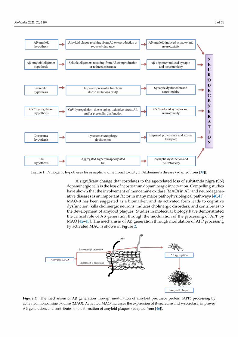
There are several AD profiles, which include deficits in episodic memory, language,semantic knowledge, visuospatial abilities, executive functions in terms of planning andorganisation as well as apraxia [32]. Apart from neuronal loss, amyloid plaques andneurofibrillary tangles are inter-related to the presence of reactive astrocytes and activatedmicroglial cells [33–35]. Aβ is the most widely studied component of AD pathogenesis,where it can induce neuronal toxicity and activate microglia leading to the indirect damageof neurons [36]. Proteolytic cleavage from the type I cell-surface protein amyloid precursorprotein (APP) was known to yield several forms of Aβ [37,38]. The pathogenic hypothesesfor synaptic and neuronal toxicity in Alzheimer’s disease is shown in Figure 1.
Molecules 2021, 26, 1107 3 of 61
Figure 1. Pathogenic hypotheses for synaptic and neuronal toxicity in Alzheimer’s disease (adapted from [39]).
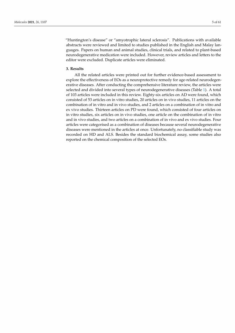
A significant change that correlates to the age-related loss of substantia nigra (SN)dopaminergic cells is the loss of neostriatum dopaminergic innervation. Compelling studieshave shown that the involvement of monoamine oxidase (MAO) in AD and neurodegener-ative diseases is an important factor in many major pathophysiological pathways [40,41].MAO-B has been suggested as a biomarker, and its activated form leads to cognitivedysfunction, kills cholinergic neurons, induces cholinergic disorders, and contributes tothe development of amyloid plaques. Studies in molecular biology have demonstratedthe critical role of Aβ generation through the modulation of the processing of APP byMAO [42–45]. The mechanism of Aβ generation through modulation of APP processingby activated MAO is shown in Figure 2.
Figure 2. The mechanism of Aβ generation through modulation of amyloid precursor protein (APP) processing byactivated monoamine oxidase (MAO). Activated MAO increases the expression of β-secretase and γ-secretase, improvesAβ generation, and contributes to the formation of amyloid plaques (adapted from [46]).

Molecules 2021, 26, 1107 4 of 61
PD is the second most prevalent condition after AD, and also develops slowly overtime [47]. PD can be identified and clinically characterised via motor impairment, whichincludes bradykinesia, rigidity, resting tremor and postural instability [48]. PD cases can bedivided into sporadic (sPD) and familial (fPD), the latter of which represents approximately20–25% of all PD cases. A common hallmark of sPD and fPD is the presence of intracellularinclusions, termed Lewy bodies [49,50].
α-Synuclein (α-Syn) has been identified as a major component of Lewy bodies insporadic and familial cases, and is believed to be the central player in PD aetiology [51].It is worthwhile to note that research conducted on PD has mainly focused on proteinaggregation, neurotoxicity, increased oxidative stress, and mitochondrial dysfunction, aswell as defects in the protein degradation machinery [52].
Apart from the role caused by α-Syn, the presence of neurotoxins, in particular6-hydroxydopamine (6-OHDA) and 1-methyl-4-phenylpyridinium (MPP+), are widelyaccepted to induce neurotoxicity in PD patients. Both neurotoxins are thought to inducedopaminergic toxicity by intra- and extracellular oxidation, hydrogen peroxide formation,and direct inhibition of the mitochondrial respiratory chain [53].
The next common neurodegenerative disease is HD; caused by the recurrent devel-opment of cytosine–adenine–guanine (CAG) in the huntingtin (HTT) gene and involves anetwork of complex pathogenic mechanisms. HD is a profoundly penetrating, autosomaldominant, progressive neurodegenerative movement and neurobehavioural disorder asso-ciated with a variety of motor signs, psychological symptoms and cognitive dysfunctionthat progress with dementia. Knowledge of HD’s causal mutation allows the detection ofan ever-expanding number of HD phenotypes and phenocopies. The mean starting age isaround 40 years, with a recorded range of 2 to 79+ years [54–57].
Progressive physical impairment of HD could be contributed to by various movementaspects such as hyperkinetic movements (dystonia, myoclonus, tics) and other motor mani-festations (bradykinesia, incoordination, oculomotor function changes, gait impairment)along with chorea as the most distinct involuntary motion. As the disease progresses overtime, dystonia becomes more prevalent and replaces chorea.
On the other hand, ALS is categorised as a heterogeneous neurodegenerative condi-tion clinically, genetically, and pathologically [58–60]. The degeneration of cortical motorneurons and anterior horn cells of the spinal cord is characterised by ALS, also knownas Charcot’s or Lou Gehrig’s disease. This contributes, usually within 3–5 years of diag-nosis, to muscle atrophy, loss of muscle function, and death resulting from respiratoryfailure. The diverse clinical variability in ALS is believed to be due to differences in uppermotor neuron (UMN) and lower motor neuron (LMN) involvement, extra-motor symp-toms, onset age, survival, and progression rates. Heterogeneity of the disease preventsbiomarker production which hinders the accurate evaluation of candidate drugs in clinicaltrials [59–61]. Various studies have shown that oxidative stress plays a major role in thisdisease’s pathogenesis, identified as an unusual family type that often exhibits superoxidedismutase 1 (SOD1) gene mutations [62,63].
EOs are now commonly used and their advantages in all facets of life are investigated.The attractiveness of EO potential and possible mechanism of actions are ventured ona continual basis around the globe. In this review, we focus on and analyse the effec-tiveness of EOs as neuroprotective remedies among the four selected age-related neu-rodegenerative disorders mentioned above. We strongly believe that this review will bebeneficial to many researchers and academicians that have great interest in EOs and theirextensive applications.
2. Materials and MethodsSearch Strategy
Original articles were searched in three databases (PubMed, Web of Science andGoogle Scholar) from the year 2010 to 2020, using the medical subject heading (MeSH)terms “essential oils”, crossed with the term “Alzheimer’s disease”, “Parkinson’s disease”,
Molecules 2021, 26, 1107 5 of 61
“Huntington’s disease” or “amyotrophic lateral sclerosis”. Publications with availableabstracts were reviewed and limited to studies published in the English and Malay lan-guages. Papers on human and animal studies, clinical trials, and related to plant-basedneurodegenerative medication were included. However, review articles and letters to theeditor were excluded. Duplicate articles were eliminated.
3. Results
All the related articles were printed out for further evidence-based assessment toexplore the effectiveness of EOs as a neuroprotective remedy for age-related neurodegen-erative diseases. After conducting the comprehensive literature review, the articles wereselected and divided into several types of neurodegenerative diseases (Table 1). A totalof 103 articles were included in this review. Eighty-six articles on AD were found, whichconsisted of 53 articles on in vitro studies, 20 articles on in vivo studies, 11 articles on thecombination of in vitro and in vivo studies, and 2 articles on a combination of in vitro andex vivo studies. Thirteen articles on PD were found, which consisted of four articles onin vitro studies, six articles on in vivo studies, one article on the combination of in vitroand in vivo studies, and two articles on a combination of in vivo and ex vivo studies. Fourarticles were categorised as a combination of diseases because several neurodegenerativediseases were mentioned in the articles at once. Unfortunately, no classifiable study wasrecorded on HD and ALS. Besides the standard biochemical assay, some studies alsoreported on the chemical composition of the selected EOs.

Molecules 2021, 26, 1107 6 of 61
Table 1. Effects of essential oils (EOs) on neurodegenerative diseases.
No. Disease Study Source of Essential Oils Ref Method Findings Conclusions and/or Recommendations
1.
ALZ
HEI
MER
’SD
ISEA
SE
Invi
tro
Ajuga chamaecistus subsp scoparia (Boiss.) Rech.f. [64]
• Chemical profiling [Gaschromatography (GC), Gaschromatography–mass spectrometry(GC–MS), High-performance liquidchromatography with diode-arraydetection (HPLC–DAD)]
• Cholinesterase inhibition assay(AChE, BChE—Ellman assay)
• Major constituents in the EO—Spathulenol (18.0%),thymol (15.1%), octen-3-ol (14.3%),and linalool oxide (11.2%).
• Inhibit the activity of AChE(1.96 mg gallic acid equivalents/g),BChE (2.2 mg gallic acid equivalents/g).
• This study should be extended toindividual constituents of the EOand extracts of A. chamaecistus.
2. Allium tuncelianum (Amaryllidaceae) [65]
• Chemical profiling [Gaschromatography–flame ionisationdetector (GC–FID), GC–MS)]
• Anti-lipid peroxidation activity• Cholinesterase inhibition assay
(AChE, BChE—Ellman assay)
• Major components—Diallyl disulfide (49.8%), diallyltrisulfide (27.9%) and allyl methyl trisulfide (6.9%).
• EO inhibition against BChE (28.65 ± 2.58%) andAChE (7.59 ± 2.90%) at 100 µg/mL.
• A. tuncelianum could be a novelpotency source of nativeantioxidant andanticholinesterase components.
3. Aloysia citrodora Palau [66]
• Chemical profiling (GC–MS)• Radioligand binding profile• MTT cell viability assay• Treatment of cell cultures with Aβ• CytoTox 96 nonradioactive assay
• The major chemical components—limonene, geranial,neral, 1, 8-cineole, curcumene, spathulenol andcaryophyllene oxide.
• A. citrodora leaf EO inhibited [3H] nicotine binding towell-washed rat forebrain membranes, and increasediron-chelation in vitro.
• A. citrodora EO displays effective antioxidant,radical-scavenging activities and significantprotective properties vs both hydrogen peroxide- andβ-amyloid-induced neurotoxicity.
• A. citrodora EO possess nicotiniccholinergic, oxidative andsignificant neuroprotectiveactivities. These activities are ofrelevance to potential AD therapy,as well as other.
4.
ALZ
HEI
MER
’SD
ISEA
SE
Invi
tro
Artemisia macrocephala [26]• Chemical profiling (GC–MS)• Cholinesterase inhibition assay
(AChE, BChE—Ellman assay)
• The major components were α-humulene (46.3%),β-caryophyllene (9.3%), α-copaene (8.2%),β-myrcene (4.3%), Z(E)-α-farnesene (3.7%), andcalarene (3.5%).
• Diverse volatile organic constituents (VOCs) of A.maderaspatana have significant AChE inhibitoryactivity (IC50 value = 31.33 ± 1.03 mg/mL).
• The results of this study confirmthe beneficial use.
5. Artemisia maderaspatana [67]• Chemical profiling (GC–FID, GC–MS)• Cholinesterase inhibition assay
(AChE—Ellman assay)
• The major components were α-humulene (46.3%),β-caryophyllene (9.3%), α-copaene (8.2%),β-myrcene (4.3%), Z(E)-a-farnesene (3.7%), andcalarene (3.5%).
• The experimental results showed that diverse VOCsof A. maderaspatana have significant AChE inhibitoryactivity (IC50 value of 31.33 ± 1.03 mg/mL).
• The possibility of novel AChEinhibitors might exist in VOCs ofthis plant.

Molecules 2021, 26, 1107 7 of 61
Table 1. Cont.
No. Disease Study Source of Essential Oils Ref Method Findings Conclusions and/or Recommendations
6. Artemisia absinthium [27]
• Niosomal vesicles employingphosphatidylcholine, span 60,cholesterol anddistearoylphosphatidylethanolaminewith covalently linked polyethyleneglycol of molecular weight 2000(DSPE-PEG2000) by the thin-filmmethod. A. absinthium was loaded intothe niosomes.
• Inhibitory effect on aggregation of Aβpeptides using Thioflavin Tfluorescence and atomicforce microscopy.
• Niosomes containing A. absinthium have a size of174 ± 2.56 nm, the encapsulation efficiency of 66.73%,zeta potential of −26.5 ± 1/42 mV and polydispersityindex (PDI) of 0.373 ± 0/02.
• The nano-niosomes containingEO from art A. absinthium has thecapability to precludeamyloid development.
7.
ALZ
HEI
MER
’SD
ISEA
SE
Invi
tro
Boswellia dalzielii [68]
• Chemical profiling (GC–FID, GC–MS)• Cholinesterase inhibition assay
(AChE—Ellman assay)• Anti-inflammatory activity
(5-lipoxygenase (LOX) inhibition assay)
• Fifty compounds were identified, including 3-carene(27.72%) and α-pinene (15.18%). 2,5-Dihydroxyacetophenone and β-D-xylopyranose were identifiedin the methanol extract.
• IC50 = 35.10 mg/L for antixanthine oxidase and 28.01mg/L for anti-5-lipoxygenase.
• For AChE inhibition, the best IC50 (76.20 and 67.10mg/L) were observed, respectively, with an ethylacetate extract and the EO. At 50 mg/L, thedichloromethane extract inhibited OVCAR-3 celllines by 65.10%, while cyclohexane extract inhibitedIGROV-1 cell lines by 92.60%.
• The ethyl acetate and methanolextracts could be considered aspotential alternatives for use indietary supplements. They havea natural antioxidant,antihyperuricemic andanti-inflammatory activities.
8. Chamaecyparis obtusaCryptomeria japonica [69]
• Chemical profiling [(GC–MS, Nuclearmagnetic resonance (NMR)]
• Cholinesterase inhibition assay (AChE,BChE)
• Anti-AChE activity of C. japonica with 64.8% ofinhibition at 100 µg/mL.
• The active principles of anti-AChE were investigatedby activity-guided fractionation, and kaur-16-ene,nezukol and ferruginol were successfully identified,the IC50 values were 640, 300 and 95 µM, respectively.
• Kaur-16-ene and nezukol inhibited AChE in themixed type mode, while ferruginol inhibited it in thecompetitive mode. In addition, nezukol andferruginol showed anti-BChE activity, the IC50 valueswere 155 and 22 µM, respectively.
• Ethyl acetate extract of C. obtusa showed anti-AChEactivity of 37.7% inhibition at 100 µg/mL. The activeprinciple was determined to be (−)-hinokinin byactivity-guided fractionation and the IC50 value was176 µM.
• The leaf of C. japonica andheartwood of C. obtusa may besuitable agents for AD therapywhen administered through thenasal system as an aromasupplement.
9. Cinnamomum tamala (Buch.-Ham.) [18]
• Chemical profiling [(High-performanceliquid chromatography–photodiodearray detection (HPLC-PDA)]
• Cholinesterase inhibition assay (AChE,BChE—Ellman assay and thin-layerchromatography (TLC)bio-autography)
• HPLC analysis confirmed the presence of linalool inC. tamala EO.
• The cinnamon oil obtained from leaves of C. tamalapossess maximum inhibition against AChE (IC50:94.54 ± 0.774 µg/mL) and BChE (IC50: 135.56 ±0.912 µg/mL). The result also explains that C. tamalais more sensitive to AChE enzyme than the BChE.
• C. tamala may be explored as ananti-cholinesterase agent furtherfor the better and safermanagement of ADs.

Molecules 2021, 26, 1107 8 of 61
Table 1. Cont.
No. Disease Study Source of Essential Oils Ref Method Findings Conclusions and/or Recommendations
10.
ALZ
HEI
MER
’SD
ISEA
SE
Invi
tro
Cinnamomum zeylanicum [19]
• Chemical profiling (GC–FID, GC–MS)• Monoamine oxidase (MAO)-A and
MAO-B inhibitory activity(fluorometric assay)
• Cholinesterase inhibition assay (AChE,BChE—Ellman assay)
• Inhibition of self- and Cu2+-inducedAβ1–42 peptide aggregation(Thioflavin T-fluorometric assay)
• Tyrosinase inhibitory activity (L-dopa)
• Twenty-two components were identified representing99.79% of the oil. (E)-Cinnamaldehyde (CAL)(81.39%) and (E)-cinnamyl acetate (CAS) (4.20%) weredetermined as the major compounds.
• The oil and the major compounds, CAL and CAS,showed over 78.0% inhibitory activityon cholinesterases.
• In MAO A and MAO-B inhibition assay, both oil(96.44% and 95.96%, respectively) and CAL (96.32%and 96.29%, respectively) showed remarkable activityas high as rasagiline(97.42% and 97.38%, respectively).
• The oil also showed 57.78% and 84.53% inhibitoryactivity in self- and Cu2+ induced aggregation ofAβ1–42, respectively. In tyrosinase inhibition assay,CAL (83.75%) and CAS (45.58%) showed higheractivity than that of the oil (18.08%).
• Total oil exhibited remarkableanti-Alzheimer and skinwhitening activity (over 80%).
11. Citrus Sinensis [L.] Osbeck [70]
• Chemical profiling (GC–FID)• Cholinesterase inhibition assay
(AChE, BChE)• MAO inhibition assay• Lipid peroxidation assay
• Forty-four compounds were identified in peels andseed EOs, respectively.
• The EOs inhibited AChE, BChE and MAO in adose-dependent manner. Both EOs inhibited AChEactivity. However, peel EO (IC50 = 2.64 µg/mL) hadsignificantly (p < 0.05) higher inhibition than that ofthe seeds (IC50 = 3.54 µg/ mL).
• Similarly, the peel EO (IC50 of 2.61 µg/mL) hadhigher BChE inhibitory ability than seed EO(IC50 = 3.52 µg/ mL).
• MAO activity was inhibited compared to thereference test when the EO were added to the sampletest. However, the EO from the seeds(IC50 = 6.93 µg/mL) had a significantly (p < 0.05)higher inhibitory ability compared to peel EO(IC50 = 8.53 µg/mL).
• In addition, EOs significantly (p < 0.05) inhibitedmalondialdehyde (MDA) produced in the brainhomogenates to 168.21 and 141.47% for peel and seedEOs, respectively.
• EOs from sweet orange peels andseeds inhibited cholinergic andmonoaminergic enzymes andexhibited antioxidant.

Molecules 2021, 26, 1107 9 of 61
Table 1. Cont.
No. Disease Study Source of Essential Oils Ref Method Findings Conclusions and/or Recommendations
12.
ALZ
HEI
MER
’SD
ISEA
SE
Invi
tro
Citrus aurantifolia Swingle,C. aurantium L.
C. bergamia Risso and Poit.[71]
• Chemical profiling (GC–FID, GC–MS)• Cholinesterase inhibition assay (AChE,
BChE—spectrophotometric method)
• The analysed EOs contain mainly limonene,α-pinene, β-pinene, γ -terpinene, and linalyl acetate.
• C. aurantifolia inhibited more selectively AChE. Thebest activity was exerted by C. aurantifolia and C.aurantium that inhibited AChE of EOs with IC50values of 139.3 and 147.5 µg/mL, respectively.
• A lower bio-activity was observed against BChE withIC50 values ranging from 235.5 to 266.6 µg/mL for C.aurantifolia and C. aurantium, respectively.
• Obtained data suggest a potentialuse of citrus oils as a valuablenew flavor with functionalproperties for food ornutraceutical products withparticular relevance tosupplements for the elderly.
13.
Cistus creticusCistus salvifoliusCistus libanotis
Cistus monspeliensisCistus villo-sus
[72]• Chemical profiling (GC–FID, GC–MS)• Cholinesterase inhibition assay
(AChE, BChE—Ellman assay)
• C. monspeliensis exhibited the most promising activityin β-carotene bleaching test (IC50 of 54.7 µg/mL).
• C. salvifolius showed the highest activity againstAChE (IC50 of 58.1 µg/mL) while C. libanotis, C.creticus and C. salvifolius demonstrated a goodinhibitory activity against BChE with IC50 values of23.7, 29.1 and 34.2 µg/mL, respectively.
• Not possible to clearlydemonstrate the relationshipbetween phytochemicals andbioactivity further studies mustbe conducted.
14. Clinopodium serpyllifolium (M.Bieb.) Kuntze [73]• Chemical profiling (GC–MS)• Cholinesterase inhibition assay
(AChE, BChE—NA-FB)
• The best AChE inhibitory activity was exhibited bythe decoction extracts (IC50 = 2.17–2.36 mg/mL),followed by the MeOH extracts(IC50 = 3.03–3.12 mg/mL).
• The best BChE was exhibited by EO(IC50 = 2.86–3.10 mg/mL), followed by the decoction(IC50 = 3.31–3.87 mg/mL), and the MeOH extracts(IC50 = 4.43–4.66 mg/mL).
• C. serpyllifolium could be avaluable natural source ofantioxidants andcholinesterase inhibitors.
15. Daucus aristidis Coss. [74]• Chemical profiling (GC–MS)• Cholinesterase inhibition assay
(AChE, BChE—Ellman assay)
• The main components of D. aristidis oils from Ghoufiwere α-pinene (49–74.1%) and Bousaada wereβ-pinene (19.2–11.9%).
• The EO of D. aristidis from Boussaada regiondisplayed modest inhibition against AChE and BChE(61.75% and 56.79%, respectively).
• The aerial parts, stems, leaves and umbels of D.aristidis EO from Ghoufi displayed low to moderateinhibition against AChE (51.0, 34.69, 13.44, and33.07%, respectively) and BChE (41.46, 22.32, 23.70,and 30.00%, respectively).
• EOs are complex mixtures ofcomponents that are usuallymore active than their isolatedcomponents. Their final activitiesare due to the combined effects ofa number of minor components.

Molecules 2021, 26, 1107 10 of 61
Table 1. Cont.
No. Disease Study Source of Essential Oils Ref Method Findings Conclusions and/or Recommendations
16.
ALZ
HEI
MER
’SD
ISEA
SE
Invi
tro
Hedychium gardnerianum,Sheppard ex Ker-Gawl [75]• Chemical profiling (GC–MS)• Cholinesterase inhibition assay
(AChE—Ellman assay)
• All the oils inhibited AChE, with IC50 values ofapproximately 1 mg/mL.
• Three oils presented mixed inhibition, whilst one wasalmost truly competitive.This activity can beattributed to presence of sesquiterpenes, whichconstituted more than 60% of the composition ofthe oils.
• Oils may contribute to theincrease in acetylcholine incholinergic neurons and to thefight againstdeleterious oxidation.
17. Hymenocrater bituminous [76]• Chemical profiling (GC–MS)• Cholinesterase inhibition assay
(AChE, BChE)
• GC–MS analysis of EO showed the presence ofα-pinene (18.2%), β-pinene (11.3%), trans-phytol(11.0%), and spathulenol (8.5%) as themajor components
• EO showed high AChE (3.8 mg GEs/g oil) and BChE(4.7 mg GEs/g oil) inhibitory activities.
• The results indicated that H.bituminous has promisingpotential for possible uses in foodand pharmaceutical industriesdue to its valuablephytoconstituents andbiological activities.
18. Illicium verum Hook.f [77]
• Chemical profiling (GC–MS)• Cholinesterase inhibition assay
(AChE, BChE—Ellman assay)• Bioautography method for detection of
AChE and BchE inhibition
• Present study confirmed that anethole contributed tothe anticholinesterase activity of I. verum, with morespecificity towards AChE.
• IC50 for AChE and BChE inhibitory activity ofanethole was 39.89 ± 0.32 µg/mL and75.35 ± 1.47 µg/mL, whereas for the oil,36.00 ± 0.44 µg/mL and70.65 ± 0.96 µg/mL, respectively
• I. verum fruit has potentialinhibitory activity of AChE andBChE. This fruit and its oil couldpotentially lead to thedevelopment ofanticholinesterase agents for themanagement of AD.
19.
ALZ
HEI
MER
’SD
ISEA
SE
Invi
tro
Lavandula luisieri [78]
• Chemical profiling (GC, GC–MS)• Inhibitory activity on Beta-site
APP-cleaving enzyme 1 (BACE-1).• Endogenous BACE-1 in cultured cells
• High contents of oxygen-containing monoterpenes,mainly necrodane derivatives, which are absent fromany other oil.
• EO from L. luisieri, was signalised as inhibiting theBACE-1 enzyme.
• This oil was tested on the endogenous BACE-1 incultured cells, being responsible for a reduction inAβ production, with no significant toxicity.
• The main inhibitory activity was assigned to themonoterpenic ketone2,3,4,4-tetramethyl-5-methylene-cyclopent-2-enone,one of the distinctive components of L. luisieri EO.
• This inhibitor could be used tostudy functional mechanisms ofBACE-1 or other asparticproteases in cells or animalmodels. It could contribute to theresearch of disorders related toderegulation of these proteins.
20. Lavandula angustifolia [24]• Thioflavin T measurement• Atomic force microscope
(AFM) Imaging
• The thioflavin T method showed that EO enhancesthe Aβ aggregation.
• The results of the AFM method also confirmedthese effects.
• Study suggests that combinationof EOs (rose oil and lavender oil),vitamin C and Trolox withL-dopa can reduce oxidativetoxicity, and may play a key rolein ROS/reactive nitrogen species(RNS) disarming.
Molecules 2021, 26, 1107 11 of 61
Table 1. Cont.
No. Disease Study Source of Essential Oils Ref Method Findings Conclusions and/or Recommendations
21. Lavandula pubescens [23]• Chemical profiling (GC–MS)• Cholinesterase inhibition assay
(AChE, BChE)
• EO composition revealed 25 constituents, of whichcarvacrol (65.27%) was the most abundant.
• EO exhibited strong antiAChE (IC50 0.9 µL/mL) andantiBChE (IC50 6.82 µL/mL) effects.
• LP EO makes a valuable naturalsource of bioactive moleculesshowing substantial potential asneuroprotective agents.
22. Lavandula viridis L’Her [79]
• Chemical profiling (GC–FID, GasChromatography–Ion Trap MassSpectrometry (GC–ITMS)
• Cholinesterase inhibition assay(AChE, BChE—Ellman assay)
• Qualitative determination offalse-positive effects in AChE andBChEinhibition assay—TLC
• Camphor was the main component identified in theEO (31.59 ± 1.32%), and in extracts from the first(1.61 ± 0.34%) and second supercritical fluidextraction (SFE) separators (22.48 ± 1.49%) at 12 MPa.
• The first separator SFE extract at 18 MPa wasdominated by myrtenol (5.38 ± 2.04%) and camphor(4.81 ± 1.93%), second separator SFE extract wasdominated by verbenone (13.97 ± 5.27%).
• Anti-cholinesterase activities EO was the mosteffective AChE inhibitor(IC50 = 411.33 ± 72.73 µg/mL) than first separatorSFE extracts.
• First separator SFE extracts were the most effectiveBChE inhibitors (IC50 = 215.56 ± 13.60 andIC50 = 204.76 ± 22.86 µg/mL at extraction pressuresof 12 and 18 MPa). The second separator extracts didnot achieve 50% inhibition at the highest finalconcentration (2.5 mg/mL).
• Phytochemicals from the aerialparts of L. viridis could bedeveloped as natural antioxidantand anti-cholinesterase drugs,with particular applications in thesymptomatic treatment of AD.
23.
ALZ
HEI
MER
’SD
ISEA
SE
Invi
tro
Mentha spicata L. [80]
• Chemical profiling (GC–MS)• Cholinesterase inhibition assay: AChE,
BChE—New micro-well plate AChEactivity (NA-FB) method
• Chemical analysis of EO revealed 31 compounds withoxygenated monoterpenes (90%) as the mostabundant components followed by sesquiterpene andmonoterpene hydrocarbons (6% and 3%,respectively). M spicata can be characterised as acarvone chemotype (65%).
• The EO of M. spicata showed high levels of inhibitoryactivity against AChE and BChE inhibitedcholinesterase enzymes in a concentration-dependentmanner, with AChE (IC50 = 23.1 µL/mL) and BuChE(IC50 = 35.0 µL/mL) inhibitory activities
• The current study supports theutilisation of M. spicata EO as atraditional medicine and opensperceptions to find more potentsubstances in the EO for themanagement of obesity, AD, anddermatophytosis and forcombating drug-resistantbacterial infections.
24. Neomitranthes obscura (DC.) N. Silveira [81]• Chemical profiling (GC–MS)• Cholinesterase inhibition assay
(AChE—Ellman assay)
• 17 compounds were identified, corresponding to86.6% of the relative composition of this oil, being3.9% of the oil constituted by monoterpenes and82.7% constituted by sesquiterpenes.
• The major compounds found were cis-nerolidol,trans-nerolidol and β-bisabolene, corresponding to19.3, 17.1, and 11.7% of relative composition,respectively.
• EO presented AChE inhibition with an IC50 of75.93 ± 0.85 µg/mL.
• EO from species of “Restinga deJurubatiba” National Park couldbe used as anticholinesteraseinhibitors. Neomitranthesobscura remains unexploredregarding itsbiological properties.
Molecules 2021, 26, 1107 12 of 61
Table 1. Cont.
No. Disease Study Source of Essential Oils Ref Method Findings Conclusions and/or Recommendations
25.
ALZ
HEI
MER
’SD
ISEA
SE
Invi
tro
Origanum rotundifolium Boiss. [82]• Chemical profiling (GC–FID, GC–MS)• Cholinesterase inhibition assay
(AChE, BChE—Ellman assay)
• The major component was identified as carvacrol(56.8%) along with p-cymene (13.1%), (Z)-β-ocimene(5.4%), β-caryophyllene (3.9%), borneol (3.4%) andthymol (3.2%)
• Anticholinesterase activity against AChE with 38.66%inhibition, and BChE with 50.66% inhibition at25 µg/mL concentration.
• This is the first report oncholinesterase inhibitory activityof the EO of the plant.
26.Panax ginseng
Panax japonicusP. notoginseng
P. quinquefolius[83]
• Chemical profiling (GC–MS)• Cholinesterase inhibition assay (AChE,
BChE)• β-Secretase Inhibition
• Spathulenol (8.82%), bicyclogermacrene (6.23%),β-elemene (3.94%), and α-humulene (3.69%) wereidentified as high content by GC–MS.
• EO (250 µg/mL) showed 41.4% inhibition againstβ-secretase, 77.4% inhibition against AChE, and94.1% inhibition against BChE.
• Furthermore, β-elemene and α-humulene showedhigh activity among 3 compounds with 50%inhibitory concentration (IC50) values of 77.2 and137.3 µM for AChE, and 298.2 and >2000 µM forBChE, respectively.
• An EO extract of P. japonicus showed the most potentactivity with 51.3% inhibition at 500 µg/mL againstβ-secretase.
• P. ginseng showed the most potent inhibitory activityagainst AChE and BChE with 70.4% and 84.4%inhibition at 50 µg/mL, respectively.
• P. notoginseng extract showed the most potent activitywith 57.3% inhibition at 500 µg/mL againstAβ aggregation.
• An EO extract of P. ginseng couldbe an effective agent to improveAD symptoms.
27. Pinus sp. [84]• Chemical profiling (GC, GC–MS)• Cholinesterase inhibition assay
(AChE, BChE—Ellman assay)
• Terpinolene, β-phellandrene, linalyl acetate,trans-caryophyllene, and terpinen-4-ol wereidentified
• P. heldreichii subsp. leucodermis exhibited the mostpromising activity, with IC50 values of 51.1 and80.6 µg/mL against AChE and BChE, respectively.
• An activity against AChE was also observed with P.nigra subsp. nigra EO, with an IC50 value of94.4 µg/mL.
• Trans-caryophyllene and terpinen-4-ol inhibitedBChE with IC50 values of 78.6 and 107.6 µg/mL,respectively. β-Phellandrene was selective againstAChE (IC50 value of 120.2 µg/mL).
• Results demonstrated that PinusEOs and some constituents haveproperties for the treatmentof AD.
Molecules 2021, 26, 1107 13 of 61
Table 1. Cont.
No. Disease Study Source of Essential Oils Ref Method Findings Conclusions and/or Recommendations
28.
ALZ
HEI
MER
’SD
ISEA
SE
Invi
tro
Piper divaricatum [85]
• Chemical profiling (GC–MS)• Cholinesterase inhibition assay
(AChE—TLC)• Molecular docking
• Methyl eugenol was the compound with the highestconcentration ranging from 48.01% to 61.85%.
• The molecular docking revealed that β-elemene,eugenol, eugenyl acetate, and methyl eugenol arecapable of interacting with different residuesbelonging to the active site of AChE, such as His447.
• The results of per-residue freeenergy decompositiondemonstrated that the molecules,during the simulation, performedinteractions with residues of theactive site that are important forthe enzymatic activity inhibition.
29. Polygonum hydropiper L. [22]• Chemical profiling (GC–FID, GC–MS)• Cholinesterase inhibition assay
(AChE, BChE—Ellman assay)
• 141 and 122 compounds were identified in Ph.LO(leaf oils) and Ph.FO (flower oils), respectively.
• Caryophyllene oxide (41.42%) was the majorcomponent in Ph.FO while decahydronaphthalene(38.29%) was prominent in Ph.LO.
• In AChE inhibition, Ph.LO and Ph.FO exhibited 87.00and 79.66% inhibitions at 1000 µg/mL with IC50 of120 and 220 µg/mL, respectively.
• The IC50 value for galanthamine was 15 µg/mL. InBChE inhibitory assay, Ph.LO and Ph.FO caused82.66 (IC50 130 µg/mL) and 77.50% (IC50 225 µg/mL)inhibitions, respectively,at 1000 µg/mL concentration.
• Leaves and flowers of P.hydropiper exhibiteddose-dependentanticholinesterase andantioxidant activities.
30. Prangos gaubae [86]• Chemical profiling (GC–MS)• Cholinesterase inhibition assay
(AChE, BChE)
• EO analysis showed the presence of germacrene D(26.7%), caryophyllene oxide (14.3%),(E)-caryophyllene (13.8%), and spathulenol (11.3%) asthe major volatile components.
• EO showed AChE (2.97 mg GEs/g oil) and BChE(3.30 mg GEs/g oil) inhibitory activities.
• Results indicated that P. gaubaehas promising potential forpossible uses in food, cosmetic,and pharmaceutical industries.
31.
ALZ
HEI
MER
’SD
ISEA
SE
Invi
tro
Rumex hastatus D. Don [87]• Chemical profiling (GC–FID, GC–MS)• Cholinesterase inhibition assay
(AChE, BChE—Ellman assay)
• The GC–MS analysis of EO showed 123 components.• Anticholinesterase assays demonstrated a marked
potential against AChE and BChE with IC50 values of32.54 and 97.38 µg/mL, respectively, which werecomparable with the positive control, i.e.,galantamine (AChE, IC50 = 4.73 µg/mL and BChE,IC50 = 11.09 µg/mL).
• R. hastatus is an effective sourceof EO components havinganticholinesterase andantioxidant potential.
32. Salvia officinalis [88]
• Chemical profiling (GC–FID, GC–MS)• Cholinesterase inhibition assay
(AChE—Ellman assay)• Anti-inflammatory activity (5-LOX
inhibition assay)
• Eighteen constituents were identified correspondingto 96.94% of present compounds.
• The main components were camphor (33.61%),1,8-cineole (22.22%) and α-thujone (21.43%).
• EO inhibited the tested enzymes AChE(IC50 = 8.71 ± 2.09 mg/L), 5-LOX(IC50 = 36.15 ± 1.27 mg/L) and XOD(IP (%) = 36.89 ± 1.83 at a final concentration of50 mg/L in the well.
• S. officinalis EO can be consideredas a source of bioactivephytochemicals andphytotherapeutics for humansdue to itsanti-Alzheimer activities.
33. Salvia chionantha [89]• Chemical profiling (GC–FID, GC–MS)• Cholinesterase inhibition assay
(AChE—Ellman assay)
• Germacrene D (25.03%), β-caryophyllene (8.71%),spathulenol (5.86%) and α-humulene (4.82%) wereidentified as the major compounds.
• At 0.5 mg/mL concentration, the EO showedmoderate AChE (56.7 ± 1.9%) and BChE(41.7 ± 2.9%) inhibitory activity.
• The EO may be useful as amoderate anticholinesteraseagent, particularly against AChE.

Molecules 2021, 26, 1107 14 of 61
Table 1. Cont.
No. Disease Study Source of Essential Oils Ref Method Findings Conclusions and/or Recommendations
34. Salvia chrysophylla Staph [90]• Chemical profiling (GC–FID, GC–MS)• Cholinesterase inhibition assay (AChE,
BChE—Ellman assay)
• The major components of the EO were α-terpinylacetate (36.31%), β-caryophyllene (15.29%), linalool(8.12%) and β-elemene (4.26%).
• Anticholinesterase activity of the EO showed weakantioxidant activity. At 1 mg/mL concentration, theEO exhibited mild AChE (52.5 ± 2.0%) and moderateBChE (76.5 ± 2.7%) inhibitory activity.
• Even if the EO demonstrated lessAChE and BChE inhibitoryactivity than galantamine, it maybe useful as a moderate BChEinhibitory agent. Some furtherstudies should be done.
35.
ALZ
HEI
MER
’SD
ISEA
SE
Invi
tro
Salvia urmiensis [20]• Chemical profiling (GC–FID, GC–MS)• Cholinesterase inhibition assay (AChE,
BChE—Ellman assay)
• EO of leaves was rich in ester compounds such asethyl linoleate (19%), methyl hexadecanoate (17%),and methyl linoleate (7.5%).
• The major compound of EO of flowers was6,10,14-trimethyl-2-pentadecanone (55.7%).
• Exhibited moderate anticholinesterase activity(IC50 = 44–892 µg/mL).
• EO-F showed the highest AChE and BChE inhibitorypotential (IC50 = 44 and 86 µg/mL, respectively)while EO-L showed inhibitory with IC50 of 211 and422 µg/mL, respectively
• The results indicated that S.urmiensis could be considered avaluable source for functionalfoods and pharmaceuticals.
36. Salvia nemorosa L. [91]• Chemical profiling (GC–FID, GC–MS)• Cholinesterase inhibition assay
(AChE—Ellman assay)
• Sixteen components were identified in the oilextracted from flowers (91.3% of the total oil) and 8components were identified in the oil extracted fromleaves (94.0% of the total oil).
• Obtained oils from leaves and flowers contain similarmajor compounds which are spathulenol (57.8 and23.0%) and caryophyllene oxide (28.2 and 45.0%,respectively).
• EO-L and EO-F are also dominated by oxygenatedsesquiterpenes (86 and 68%, respectively).
• EO from S. nemorosa L.were generally less potent asan anti-AChE (IC 50 of EO-F: 488.9 ± 12.4 µg/mLand EO-L: 434.1 ± 11.6 µg/mL).
• S. nemorosa maybe useful fornovel applications in functionalfood and pharmaceuticalindustries
37. Salvia officinalis L. [21]
• Chemical profiling (Fast GC–MS,Enantioselective GC–MS)
• Cholinesterase inhibition assay(AChE—Ellman assay)
• LOX inhibition assay
• The main components were α-thujone (22.8–41.7%),camphor (10.7–19.8%), 1,8-cineole (4.7–15.6%) andβ-thujone (6.1–15.6%).
• Enantioselective gas chromatography identified(−)-α-thujone and (+)-camphor as the mainenantiomers in all the analysed EOs.
• EO from S. officinalis showed AChE inhibition withthe following IC50 values expressed in µg/mL:SoEO-3 (326.7 ± 24.8) ≤ SoEO-1 (338.1 ± 13.8) ≤SoEO-4 (450.0 ± 25.6) < SoEO-2 (867.4 ± 82.3).
• All four SoEO degrees of LOX inhibition (%) were asfollows: SoEO-4 (52.8 ± 1.2) > SoEO-2 (45.5 ± 1.5) ≥SoEO-1 (44.3 ± 1.3) ≥ SoEO-3 (42.0 ± 0.9).
• The characterisation carried outincreases our awareness of thepossible uses of S. officinalis EO asnatural additives in food,cosmetics and pharmaceuticals
Molecules 2021, 26, 1107 15 of 61
Table 1. Cont.
No. Disease Study Source of Essential Oils Ref Method Findings Conclusions and/or Recommendations
38.
ALZ
HEI
MER
’SD
ISEA
SE
Invi
tro
Salvia syriaca L. [92]• Chemical profiling (GC–MS)• Cholinesterase inhibition assay
(AChE, BChE—Ellman assay)
• GC–MS analysis showed that spathulenol (87.4%),isospathulenol (7.6%), and bornyl acetate (2.7%) arethe major compounds in EO.
• HPLC analysis indicated that rutin, quercetin,apigenin, rosmarinic acid, and ferulic acid are themost abundant phenolic components.
• The enzyme inhibitory potential of the tested EO andextracts in comparison with galantamine were weak(EO, AChE: 1.9 ± 0.1 mg/mL,BChE: 3.06 ± 0.1 mg/mL).
• S. syriaca could be considered as avaluable source of bioactivenatural compounds for functionalfoods, medical, andpharmaceutical applications.
39. Satureja thymbra L. [93]• Chemical profiling (GC–FID, GC–MS)• Cholinesterase inhibition assay
(AChE, BChE—Ellman assay)
• Twenty-five compounds were identified by GC andGC–MS.
• The identified main compounds of the EO, carvacrol(34.6%), γ-terpinene (22.9%), p-cymene (13.0%) andthymol (12.8%) were also tested in the same manner.
• EO showed AChE (IC50: 150 ± 1.50 µg/mL) andBChE (IC50: 166 ± 2.00 µg/mL) inhibitory activities.
• The extract (IC50: 13.1 ± 0.23 µg/mL), the oil(IC50: 26.7 ± 0.56 µg/mL) and γ-terpinene(IC50: 11.9 ± 0.21 µg/mL) exhibited a good lipidperoxidation inhibitory activity.
• Consumption of S. thymbra mayprotect people against oxidativestress and amnesia without anyside effect.
40.Stachys inflata,
S. lavandulifolia,S. byzantina
[94]• Chemical profiling (GC–MS)• Cholinesterase inhibition assay
(AChE, BChE—Ellman assay)
• The major volatile components of S. inflata wereidentified as germacrene D (21.6%) and β-pinene(15.6%).
• S. lavandulifolia contained mainly germacrene D(22.5%) and α-pinene (15.5%) whereas S. byzantinashowed hexahydrofarnesyl acetone (25.7%) andvaleranone (17.1%) as major volatile constituents.
• S. inflata EO had the highest activity with 7.68 and5.20 mg GALAEs/g EO, S. byzantina EO were at thesame level approximately at 6.62 and 5.06 mgGALAEs/g EO and S. lavandulifolia EO showedremarkable activity at 4.96 and 4.88 mg GALAEs/gEO against BChE and AChE, respectively.
• EOs of hedgenettle (betony)plants could be employed in thepreparation of formulations to beused in cosmetics, food, andpharmaceutical products duetotheir valuable antioxidant,neuroprotective, hypoglycemic,anti-obesity, and skin-care effects.
Molecules 2021, 26, 1107 16 of 61
Table 1. Cont.
No. Disease Study Source of Essential Oils Ref Method Findings Conclusions and/or Recommendations
41.
ALZ
HEI
MER
’SD
ISEA
SE
Invi
tro Sideritis galatica Bornm. [95]
• Chemical profiling (GC–FID, GC–MS)• Cholinesterase inhibition assay
(AChE, BChE—Ellman assay)
• 23 components, representing 98.1% of S. galatica EO(SGEO) were identified. Monoterpene hydrocarbons(74.1%), especially α- (23.0%) and β-pinene (32.2%),were the main constituents in SGEO.
• The main sesquiterpene hydrocarbons wereβ-caryophyllene (16.9%), germacrene-D (1.2%) andcaryophyllene oxide (1.2%), respectively.
• The cholinesterase inhibitory activity of SGEO wasvery low when compared to galantamine. AChE(IC50: 0.618 mg/mL) and BChE (IC50: 0.632 mg/mL)inhibition ability of SGEO appear to be close.
• This investigation suggests thatSGEO can be considered as asource of natural agents for thedevelopment of new naturalproducts such as food additivesand drugs.
42.
S. ballsianaS. cyanescensS. divaricataS. hydrangea
S. kronenburgiiS. macrochlamys
S. nydeggeriS. pachystachys
S. pseudeuphraticaS. rusellii
[96]• Chemical profiling (GC–FID, GC– MS)• Cholinesterase inhibition assay
(AChE, BChE—Ellman assay)
• S. pseudeuphratica, S. hydrangea and S. divaricata EOsdemonstrated the most potent AChE inhibitory effect[50% inhibition concentration(IC50) = 26.00 ± 2.00 µg/mL, 40.0 ± 4.00,64.68 ± 4.16, respectively].
• The EO of S. pseudeuphratica demonstrated thehighest inhibitory activity against AChE and BChEamong the tested Salvia EOs.
• Evidences from the studyaugment the importance of EOsobtained from Salvia sp. Salvia sp.could be used to treatAlzheimer’s disease.
43. Thymus mastichina L. [97]
• Chemical profiling (fast GC–MS,enantioselective GC–MS)
• Cholinesterase inhibition assay(AChE—Ellman assay)
• 5- LOX inhibition assay
• 1,8-Cineole and linalool were the main components,followed by α-pinene, β-pinene and α-terpineol.(–)-Linalool, (+)-α-terpineol and (+)-α-pinene werethe most abundant enantiomers.
• All four T. mastichina EO (α-pinene, β-pinene,limonene and 1,8-cineole) inhibited bothlipoxygenase and AChE activities.1,8-cineole was thebest AChE inhibitor with an IC50 of35.2 ± 1.5 µg/mL.
• Bornyl acetate and limonene showed the highestlipoxygenase inhibition and 1,8-cineole was the bestAChE inhibitor.
• These results support thepotential applications of TmEO asnatural ingredients innutracosmeceutical products.
44.
ALZ
HEI
MER
’SD
ISEA
SE
Invi
tro
Thymus haussknechtii Velen. [98]
• Chemical profiling (GC–FID, GC–MS)• Cholinesterase inhibition assay (AChE,
BChE—Ellman assay)
• The major component of the EO was thymol (52.2%).• T. haussknechtii showed anticholinesterase activity
against AChE with 57.33% and BChE with 40.11%inhibition at 25 µg/mL concentration.
• The EO obtained from T. haussknechtii was rich inthymol (52.2%) and thymol exhibited strong acetyl-(83.0%) and BChE (98.0%) inhibitory activities.
• These results indicate that T.haussknechtii could be a goodsource for natural antioxidantswhich were very important inprevention of many disease andprotection of health.
Molecules 2021, 26, 1107 17 of 61
Table 1. Cont.
No. Disease Study Source of Essential Oils Ref Method Findings Conclusions and/or Recommendations
45. Thymus lotocephalus [99]
• Chemical profiling (GC–FID,GC–IT-MS)
• Cholinesterase inhibition assay(AChE, BChE—Ellman assay)
• Linalool (10.43 ± 1.63%) was the main component inEO, whereas camphor (7.91 ± 0.84%) and cis-linalooloxide (7.25 ± 1.45%) were the major compounds inthe extracts; second separator obtained at pressuresof 12 and 18 MPa, respectively.
• Caryophyllene oxide was the primary constituentidentified in the extracts; first separator (4.34 ± 0.51and 4.41 ± 1.25% obtained at 12 and 18 MPa,respectively).
• The inhibitory effect produced by the EO andextracts-1st separator were clearly more pronouncedthan that of the extracts; second separator, which didnot reach 50% inhibition of either enzymes at thehighest concentration tested (2.5 mg/mL).
• The EO was significantly (p < 0.05) more activeagainst BChE than AChE. Extraction pressureinfluenced the bioactivity of the SFE extract (firstseparator) isolated at the lowest pressure, which wasthe most active in inhibiting AChE(IC50 = 1.54 ± 0.04 mg/mL). BChE activity at thesimilar pressure indicates IC50 of0.14 ± 0.02 mg/mL).
• BChE activity was more affected by EO thanreference drug galanthamine, which was more activeagainst AChE.
• This research suggests T.lotocephalus as a source ofbioactive compounds withdifferent targets and pathways.
46.
ALZ
HEI
MER
’SD
ISEA
SE
Invi
tro
Zingiber cassumunar [100]• Chemical profiling (GC–MS)• Cholinesterase inhibition assay (AChE,
BChE—Ellman assay)
• IC50 values of native ZCEO show that it can becharacterised as a moderate BChE inhibitor with IC50of 0.355 ± 0.137 mg/mL and a weak AChE inhibitorwith IC50 of 5.573 ± 0.176 mg/mL.
• Native ZCEO was formulated with microemulsion(ME) technique, and the suitable ZCEO ME wascomposed of Triton X-114 in combination withpropylene glycol.
• The anticholinesterase activity of ME was higher thannative ZCEO. It exhibited twenty times and twentyfive times higher inhibitory activity against AChE(IC50 = 0.252 ± 0.096 mg/mL) and BChE(IC50 = 0.014 ± 0.002 mg/mL), respectively.
• ZCEO loaded ME is an attractiveformulation for furthercharacterisation and an in vitrostudy in an animal modelwith AD.

Molecules 2021, 26, 1107 18 of 61
Table 1. Cont.
No. Disease Study Source of Essential Oils Ref Method Findings Conclusions and/or Recommendations
47. Zosima absinthifolia Link [14]
• Chemical profiling (GC–FID, GC–MS)• Cholinesterase inhibition assay
(AChE, BChE—Ellman assay)• Anti-lipid peroxidation activity
(TBA assay) Molecular docking
• The GC–FID and GC–MS analysis revealed that themain components of the aerial parts, roots, flowersand fruits extracts were octanol (8.8%), octyloctanoate (7.6%), octyl acetate (7.3%);trans-pinocarvyl acetate (26.7%), β-pinene (8.9%);octyl acetate (19.9%), trans-p-menth-2-en-1-ol (4.6%);octyl acetate (81.6%), and (Z)-4-octenyl acetate (5.1%).
• The dichloromethane fraction of fruit had the bestinhibition against BChE enzyme (82.27 ± 1.97%)which was higher than AChE inhibition(61.09 ± 4.46%) of umbelliferone.
• The highest antioxidant potential in the TBA assaywas seen in the fruit CH2Cl2 fraction and flower EO(IC50 = 48.98 and 97.11 g/mL, respectively).
• The fruit CH2Cl2 fraction and flower EO indicatedconsiderable inhibition against BChE (82.27 ± 1.97and 78.65 ± 2.66%, respectively).
• The CH2Cl2 fractions of root and fruit also showedconsiderable inhibition against AChE (29.15 ± 2.45and 31.46 ± 2.78%).
• Study shows that the flowers andfruit of Z. absinthifolia can be anew potential resource of naturalantioxidant andanticholinesterase compounds.
• Could be an herbal alternative tosynthetic drugs in theprophylaxis of AD.
48.
ALZ
HEI
MER
’SD
ISEA
SE
Invi
tro
Calamintha nepeta, Foeniculum vulgare, Menthaspicata and Thymus mastichina [101]
• Chemical profiling• Cholinesterase inhibition assay
(AChE, BChE—Ellman assay)
• The main components of EOs were oxygenatedmonoterpenes, and aqueous extracts were rich inphenol and flavonoid compounds.
• EOs (0.5 mg/mL) showed high ability to inhibitcholinesterase activities, with IC50 (concentration thatinhibit 50% of enzyme activity) ranging from 0.08 to0.24 and 0.09 to 0.23 mg/mL, for AChE and BChE,respectively.
• EOs and aqueous extracts of C.nepeta, T. mastichina, M. spicataand F. vulgare from Alentejoregion showed high antioxidantpotential that may have animportant role in the oxidativestress protection.
49.Cymbopogon citratus (Gramineae), Citrus hystrix
(Rutaceae) and Zingiber cassumunar(Zingiberaceae)
[102]• Cholinesterase inhibition assay (AChE,
BChE—Ellman assay)• C. citratus oil exhibited the highest activity with IC50
values of 0.34 ± 0.07 µl/mL and 2.14 ± 0.18 µl/mLagainst BChE and AChE activity, respectively.
• Citratus oil loadedmicroemusions are attractivesystems for further in vivostudies in animal modelswith AD.
50.
Alpinia galanga Linn., Centella asiatica Urban.,Cinnamomum bejolghota (Buch. Ham.) Sweet, Citrus
aurantifolia Swing, Citrus hystrix DC., Citrusmaxima (Burm.) Merr., Citrus reticulata Blanco cv.
Shogun, Citrus reticulata var. Fremont, Cymbopogoncitratus Stapf., Eupatorium odoratum Linn,. Melissaofficinalis Linn., Ocimum basilicum Linn., Ocimumcanum Sims., Ocimum gratissimum Linn., Ocimum
sanctum Linn., Piper sarmentosum Roxb., Polygonumodoratum Lour., Polyscias fruticosa, Harms.Zingiber
cassumunar Roxb,.Zingiber officinale Rosco
[25]• Chemical profiling (GC–MS)• Cholinesterase inhibition assay
(AChE, BChE—Ellman assay)
• GC–MS analysis revealed that the major constituentsof C. aurantifolia leaf oil are monoterpenoidsincluding limonene, l-camphor, citronellol, o-cymeneand 1,8-cineole.
• EOs obtained from Melissa officinalis leaf and Citrusaurantifolia leaf showed high AChE and BChEco-inhibitory activities.
• C. aurantifolia leaf oil revealed in this study had anIC50 value on AChE and BChE of 139 ± 35 and42 ± 5 µg/mL, respectively.
• The excellent inhibitory activityof the EO of C. aurantifolia onboth enzymes suggests that thisoil could be a promisingsubstance for prevention andtreatment of AD.
• C. aurantifolia is an attractivenatural source to inhibitanticholinesterase and thereforewarrants further study.
• The effect of combination ofnatural EOs would also be aninteresting issue forfurther studies.

Molecules 2021, 26, 1107 19 of 61
Table 1. Cont.
No. Disease Study Source of Essential Oils Ref Method Findings Conclusions and/or Recommendations
51.
ALZ
HEI
MER
’SD
ISEA
SE
Invi
tro
Lavandula angustifoliaCoriandrum sativum [103]
• Assessment of cell viability bywater-soluble tetrazolium salt (WST)-8
• Assessment of nuclear morphology• Caspase-3 activity assay• Assessment of intracellular reactive
oxygen species (ROS) production
• Lavender and coriander EOs (10 µg/mL) as well aslinalool at the same concentration were able toimprove viability and to reduce nuclearmorphological abnormalities in cells treated withAβ1–42 oligomers for 24 h.
• Lavender, coriander EOs and linalool were alsoshowed to counteract the increase in intracellularROS production and the activation of thepro-apoptotic enzyme caspase-3 induced byAβ1–42 oligomers.
• This study suggests that EOs andtheir main constituent linaloolcould be natural agents oftherapeutic interest.
52. Artemisia annua L. (Asteraceae)Glycyrrhiza glabra L. (Fabaceae) [104]
• Cholinesterase inhibition assay(AChE—Ellman assay)
• 1,8-cineole, carvacrol, myrtenal and verbenoneapparently inhibited AChE; the highest inhibitoryactivity was observed for myrtenal (IC50 = 0.17 mM).
• This is the first report to show thepotential of myrtenal present inthe EO of A. annua L. or G. glabraL. as an AChE inhibitor.
53.
ALZ
HEI
MER
’SD
ISEA
SE
Invi
tro
Angelica sinensis (Oliv) Diels.Ligusticum Chuanxiong Hort [105]
• Aβ25–35 and Aβ1–42 fibrilspreparation
• 3-(4,5-dimethylthiazol-2-yl)-2,5-diphenyltetrazolium bromide (MTT)assay (SH-SY5Y and PC12 cells)
• Lactate dehydrogenase (LDH) releasemeasurement
• Flow cytometric analysis• Intracellular ROS measurement
• Z-ligustilide (Z-LIG) is an essential oil originallyisolated from umbelliferous plants. Z-LIG at1–30 mM provided an effective neuroprotection, asevidenced by the increase in cell viability, as well asthe decrease in LDH release and intracellularaccumulation of reactive oxygen species.
• Z-LIG markedly blocked Aβ fibril-inducedcondensed nuclei and sub-G1 accumulationsuggestive of apoptosis.
• Furthermore, Z-LIG substantially reversed theactivation of phosphorylated p38 and the inhibitionof phosphorylated Akt caused by Aβ25–35.
• Z-LIG effectively protects against fibrillar aggregatesof Aβ25–35 and Aβ1–42-induced toxicity inSH-SY5Y and differentiated PC12 cells, possiblythrough the concurrent activation of PI3-K/Aktpathway and inhibition of p38 pathway.
• The results taken togetherindicate that Z-LIG protectsagainst Aβ fibrils-inducedneurotoxicity possibly throughthe inhibition of p38 andactivation of PI3-K/Akt signalingpathways concurrently. Z-LIGmight be a potential candidate forfurther preclinical study aimed atthe prevention and treatmentof AD.
54.
Invi
vo
Achillea biebersteinii (Asteraceae) [106]
• Six groups of adult female Wistar rats(3 months old) were used. These are,control group, Sco-alone-treated group,diazepam alone-treated group,tramadol alone-treated group, Sco + A.biebersteinii EO 1% administrated groupand Sco + A. biebersteinii EO 3%administrated groups.
• Memory performances were assessedby Y-maze task and radial-arm mazetask. Anxiety and depressive-likebehaviour were evaluated by elevatedplus-maze and forced swimming tasks,respectively.
• Chemical profiling (GC–FID, GC–MS)
• The scopolamine-only administered group showedimpaired spatial memory as evidenced by a decreasein the spontaneous alternation percentage in theY-maze test, and increase in the number of workingand reference memory errors in the radialarm-maze test.
• In addition, scopolamine-only administered groupdisplayed increased anxiety and depressive-likebehaviour as evidenced by decrease in theexploratory activity, the percentage of the time spentand the number of entries in the open arm withinelevated plus-maze test and decrease in swimmingtime and increase in immobility time within forcedswimming test.
• A. biebersteinii EO significantlyimproved memory formation,and reduced anxiety anddepression-like behaviour inscopolamine-induced rats ascompared to scopolamine-aloneinduced rats. The EO of this plantcould be a good candidate forcomplementary therapy againstneurological diseases such as AD.
Molecules 2021, 26, 1107 20 of 61
Table 1. Cont.
No. Disease Study Source of Essential Oils Ref Method Findings Conclusions and/or Recommendations
55.
ALZ
HEI
MER
’SD
ISEA
SE
Invi
vo
Boswellia serrata(Burseraceae) [107]
• Mice were administered the 42-aminoacid form of amyloid β-peptide(Aβ1–42) to induce AD and thentreated with olibanum EO (OEO) at 150,300, and 600 mg/kg, for two weeks.Olibanum, the resin of the B. serrata
• Following treatment, the AD mice wereassessed by step-down test (SDT), darkavoidance test (DAT), and Morris watermaze test (MWM). Blood and braintissues were collected forbiochemical assessments.
• Chemical profiling (GC–MS)
• The main constituents of OEO were limonene,α-pinene, and 4-terpineol.
• Treatment with OEO prolonged t latency in SDT andDAT, but decreased error times.
• Escape latency decreased and crossing times rose inthe MWM following OEO treatment (p < 0.5).Treatment with OEO also enhanced the acetylcholinelevels and decreased the AChE levels in serum andbrain tissue (p < 0.5).
• Additionally, OEO reduced amyloid plaques in thehippocampus and protected hippocampal neuronsfrom damage. Furthermore, OEO decreased c-fosexpression in hippocampus tissues from AD mice(p < 0.5).
• OEO has a significantameliorative effect AD-induceddeterioration in learning andmemory in AD mice. Themechanisms of these effects arerelated to the increasein acetylcholine.
56. Chamaecyparis obtuse [108]
• To model AD, 4 µg of aggregated Aβwas injected into the hippocampus. Totest the effects of EO of C. obtuse(EOCO), behavioural performance inthe MWM was tested 4 days afterinjection. After behavioural testing,brain sections were prepared for2,3,5-triphenyltetrazolium chloride(TTC) staining and terminaldeoxynucleotidyl transferase dUTPnick end labeling (TUNEL) assay.
• Chemical profiling (GC–MS)• AChE activity assays
• Inhaled EOCO protected spatial learning andmemory from the impairments induced by Aβ1–40injection. In addition, the behavioural deficitsaccompanying Aβ1–40-induced AD were attenuatedby inhalation of EOCO.
• Furthermore, AChE activity and neuronal apoptosiswere significantly inhibited in rats treated withAβ1–40 and EOCO compared to rats treated onlywith Aβ1–40. AChE activity was significantlyelevated (0.46 ± 0.05 U/mg protein, p < 0.001) andAChE content was decreased markedly(18.54 ± 1.17 umole/mg protein, p < 0.001) in theAβ1–40 group.
• However, after treatment with donepezil or EOCO,AChE activity was significantly decreased and AChEcontent was significantly increased compared withthe Aβ1–40 group. In addition, the EOCO group hadless of a reduction in AChE activity than thedonepezil group. The EOCO was composed of45 main compounds with significant differences inthe contributions of major monoterpenes (67.97%)and sesquiterpenes (25.97%).
• EOCO suppressed bothAD-related neuronal cellapoptosis and AD-relateddysfunction of the memorysystem. Thus, the results of thisstudy support EOCO as acandidate drug for the treatmentof AD.
57.
ALZ
HEI
MER
’SD
ISEA
SE
Invi
vo
Citrus limon [109]
• APP/PS1 double transgenic AD mice• The effects of lemon EO (LEO) on
learning and memory were examinedusing the MWM test, novel objectrecognition test, and correlativeindicators, including aneurotransmitter (AChE), a nervegrowth factor (brain-derivedneurotrophic factor, BDNF), apostsynaptic marker (PSD95), andpresynaptic markers (synapsin-1, andsynaptophysin), in APP/PS1 mice.
• Histopathology was performed toestimate the effects of LEO on AD mice.
• A significantly lowered brain AChE depression inAPP/PS1 and wild-type C57BL/6L (WT) mice.PSD95/ Synaptophysin, the index of synaptic density,was noticeably improved in histopathologic changes.
• Hence, it can be summarised that memory-enhancingactivity might be associated with a reduction in theAChE levels and is elevated by BDNF, PSD95, andsynaptophysin through enhancing synaptic plasticity.
• Offer in vivo evidence that LEOprovides protection againstAD-related synaptic loss andmemory impairment. The findingalso supports the importance ofLEO as a “memory enhancer” inboth the WT and APP/PS1 brainto mediate AChE, synapticplasticity and cognitive function.

Molecules 2021, 26, 1107 21 of 61
Table 1. Cont.
No. Disease Study Source of Essential Oils Ref Method Findings Conclusions and/or Recommendations
58.
ALZ
HEI
MER
’SD
ISEA
SE
Invi
vo
Cymbopogon giganteusIllicium pachyphyllum
Carum carvi[110]
• Twenty-five rats were divided into fivegroups (/group): first group—control;second group— Aβ1–42 (1 mM)received donepezil treatment(5 mg/kg); third group— Aβ1–42(1 mM); fourth and fifthgroups—Aβ1–42 (1 mM) that received(−)-cis-carveol treatment groups(1% and 3%)
• Determination of the hippocampalAChE, SOD, CAT, Glutathioneperoxidase (GPx) activity, Proteincarbonyl and MDA level
• The total hippocampal content ofreduced glutathione (GSH)
• The results of this study demonstrated that(−)-cis-carveol improved Aβ1–42-induced memorydeficits examined by using Y-maze and radial armmaze in vivo tests.
• Additionally, the biochemical analyses of thehippocampus homogenates showed that(−)-cis-carveol reduced hippocampal oxidative stresscaused by Aβ1–42.
• The present work suggested that(−)-cis-carveol providesneuroprotection against Aβ1-42and can be regarded as analternative therapeutic agent fordementia-related neurologicalconditions, including AD.
59. Ferulago angulata [111]
• 24 Male Wistar rats were divided intofour groups: (1) Control group received0.9% saline with 1% Tween 80treatment; (2) Sco-alone received 0.9%saline with 1% Tween 80 treatment, asnegative control; (3) Sco-treatedreceived F. angulata EO 1%; and (4)Sco-treated received F. angulata EO 3%.
• Chemical profiling (GC–FID)• Sco-induced memory impairments
(Y-maze and radial arm-maze tasks).• Decreased activities of superoxide
dismutase, glutathione peroxidase andcatalase; increase in AChE and decreasein total content of reduced glutathione
• Production of protein carbonyl and malondialdehydesignificantly increased in the rat hippocampalhomogenates of scopolamine-treated animals ascompared with control, as a consequence of impairedantioxidant enzymes activities.
• Additionally, in scopolamine-treated rats exposure toF. angulata EO significantly improved memoryformation and decreased oxidative stress, suggestingmemory-enhancing and antioxidant effects.
• Our results suggest that multipleexposures to F. angulata EOamelioratescopolamine-inducedspatial memory impairment byattenuation of the oxidative stressin the rat hippocampus.
60.
ALZ
HEI
MER
’SD
ISEA
SE
Invi
vo Lavandula angustifolia ssp. angustifolia Mill.(Lamiaceae)
Lavandula hybrida Rev. (Lamiaceae)[112]
• 50 male Wistar rats were divided into 5groups: (1) Control received salinetreatment (0.9% NaCl); (2) Sco-treatedreceived silexan, as + control; (3) Scoalone-treated; (4) Sco-treated receivedLavandula angustifolia EO (LO1 + Sco);and (5) Sco-treated received Lavandulahybrida EO (LO2 + Sco).
• Chemical profiling (GC–MS)• Behavioural recovery (maze task,
forced swimming test) followingchronic exposure to the extracted EOusing sco-induced dementia rat model,are investigated.
• The main components in both analysed samples, LO1and LO2, were linalool (28.0% and 21.5%,respectively) and linalyl acetate (17%, 22.5%,respectively) followed by terpinen-4-ol (3.3% and16.7%), lavandulyl acetate (8.3% and 8.4%).Interesting is that the presence of camphor andborneol was in trace amounts.
• Chronic exposures to lavender EOs (daily, for 7continuous days) significantly reduced anxiety-likebehaviour and inhibited depression in elevatedplus-maze and forced swimming tests, suggestinganxiolytic and antidepressant activity.
• Additionally, spatial memory performance in Y-mazeand radial arm-maze tasks was improved, suggestingpositive effects on memory formation.
• Multiple exposures to lavenderEOs could effectively reversespatial memory deficits in the ratbrain and might provide anopportunity for managementneurological abnormalities indementia conditions.
Molecules 2021, 26, 1107 22 of 61
Table 1. Cont.
No. Disease Study Source of Essential Oils Ref Method Findings Conclusions and/or Recommendations
61.
ALZ
HEI
MER
’SD
ISEA
SE
Invi
vo
Ocimum basilicum [113]
• Forty male Swiss albino mice dividedinto four groups (n = 10); the control,chronic unpredictable mild stress(CUMS), CUMS + Fluoxetine, CUMS +OB were used.
• Behavioural tests, serum corticosteronelevel, hippocampus protein level of theglucocorticoid receptors (GRs) andbrain derived neurotropic factor(BDNF) were determined afterexposure to CUMS. Hippocampus washistopathologically examined.
• O. basilicum diminished the depression manifestationas well as impaired short term memory observed inthe mice after exposure to the CUMS as evidenced bythe forced swimming and elevated plus maze test.
• O. basilicum also up-regulated the serumcorticosterone level, hippocampal protein level of theglucocorticoid receptor and the brain-derivedneurotropic factor and reduced theneurodegenerative and atrophic changes induced inthe hippocampus after exposure to CUMS.
• EO of O. basilicum alleviated thememory impairment andhippocampal neurodegenerativechanges induced by exposure tothe chronic unpredictable stressindicating that it is the time totest its effectiveness on patientssuffering fromAlzheimer’s disease.
62. Ocimum sanctum L.Ocimum basilicum L. [114]
• 80 male Wistar rats (3 were divided into8 groups: (1) Control received salinetreatment (0.9% NaCl); (2) Diazepamalone-treated (DZP, 1.5 mg/kg); (3)Tramadol alone-treated (TRM, 10mg/kg); (4) Aβ1–42 alone-treated; (5)Aβ1-42-treated received O. sanctum oil1%; (6) Aβ1–42-treated received O.sanctum oil 3%; (7) Aβ1–42-treatedreceived O. basilicum oil 1%; and (8)Aβ1–42-treated received O. basilicumoil 3%. Control, DZP, TRM and Aβ1-42alone-treated groups were caged in thesame conditions but in the absence ofthe tested oils.
• Chemical profiling (GC–MS)• The anxiolytic- and antidepressant-like
effects of inhaled basil EOs werestudied by means of in vivo (elevatedplus-maze and forced swimming tests)approaches.
• The Aβ1–42-treated rats exhibited the following:decrease in the exploratory activity (number ofcrossing), the percentage of the time spent and thenumber of entries in the open arm within elevatedplus-maze test and increase in the swimming timeand decrease in the immobility time within forcedswimming test.
• The main compounds found in both samples werelinalool (31% Ob, 19% Os), camphor, β-elemene,α-bergamotene and bornyl-acetate, estragole (15.57and 7.59%, respectively), eugenol (2.64 and 1.39%,respectively) and 1,8-cineole (3.29 and 3.90%,respectively).
• The exposure to basil EOs significantly improved thebehaviour of the animals, suggesting anxiolytic- andantidepressant-like effects.
• Multiple exposures to basil EOscan be useful to counteractanxiety and depression in ADconditions. Future perspectivesof this study include a rather newcultivar of basil (O. basilicum var.purpurascens).
63.
ALZ
HEI
MER
’SD
ISEA
SE
Invi
vo
Pimpinella peregrina [115]
• Y-maze and radial arm-maze tests wereused for assessing memory processes.Additionally, the anxiety anddepressive responses were studied bymeans of the elevated plus-maze andforced swimming tests
• Chemical profiling (GC–MS).
• The scopolamine alone-treated rats exhibited thefollowing: decrease in the spontaneous alternationpercentage in Y-maze test, increase in the number ofworking and reference memory errors in radialarm-maze test, along with decrease in the exploratoryactivity, the percentage of the time spent and thenumber of entries in the open arm within elevatedplus-maze test and decrease in swimming time andincrease in immobility time within forcedswimming test.
• Inhalation of the P. peregrina EO significantlyimproved memory formation and exhibitedanxiolytic- and antidepressant-like effects inscopolamine-treated rats.
• Results suggest that the P.peregrina EO inhalationameliorates scopolamine-inducedmemory impairment, anxiety,and depression. studies on the P.peregrina EO may open a newtherapeutic window for theprevention of neurologicalabnormalities closely relatedto AD.
Molecules 2021, 26, 1107 23 of 61
Table 1. Cont.
No. Disease Study Source of Essential Oils Ref Method Findings Conclusions and/or Recommendations
64. Pinus halepensis [116]
• Rats were behaviourally tested (radialarm maze and Y-maze activities used).Rats were divided into five groups: firstgroup—vehicle, secondgroup—Aβ1–42, the third and fourthgroup—P. halepensis EO (PNO)treatment groups (1% and 3%), andfifth group—donepezil group (aspositive control, 5 mg/kg injected inAβ1–42-treated rats). Additionally,biochemical estimations of the brainhomogenates for AChE and oxidativestress biomarkers were carried out.
• Chemical profiling (GC–FID, GC–MS)• Determination of hippocampal AChE,
SOD, CAT, GPX activity• Determination of hippocampal protein
carbonyl and MDA level
• The EO reversed the Aβ1–42-induced decreasing ofthe spontaneous alternation in the Y-maze test andthe Aβ1–42-induced increasing of the working andreference memory errors in the radial arm maze test.
• The Aβ1–42-induced modification of the balanceoxidant-antioxidant and AChE action in thehippocampus of the rat has been ameliorated usingthe EO.
• Most of the volatile components belong to thesesquiterpene group (54.14%), and monoterpenesrepresent the rest of them, and only 2.50% arediterpenes (mainly cembrene).
• Aβ1–42 administration resulted in a substantialincrease in the hippocampus AChE action (p < 0.0001)in comparison with the vehicle group. PNO (1% and3%) treated groups significantly decreased theelevated AChE action (p < 0.0001 for PNO1% andp < 0.0001 for PNO3%) as compared to Aβ1–42 rats.
• In comparison with the vehicle group, injection of theAβ1–42 resulted in significant decrease in SOD(p < 0.0001), CAT (p < 0.0001), and GPX (p < 0.0001).GSH levels were decreased after Aβ1–42 treatment(p < 0.0001), while elevated protein carbonyl(p < 0.0001) and MDA (p < 0.0001) levels were noticedas compared to vehicle groups.
• P. halepensis EO may be regardedas a therapeutic tool forattenuation of Aβ toxicity andneuronal dysfunction.
65.
ALZ
HEI
MER
’SD
ISEA
SE
Invi
vo
Kushui rose (Rosa setate × Rosa rugosa) [117]
• Paralysis assay, Fluorescence stainingof Aβ deposits assay, Exogenousrerotonin sensitivity assay, RNAinterference (RNAi) assay, SubcellularDAF-16 or skinhead-1 (SKN-1) nuclearlocalisation assay, The gst-4::gfpexpression assay, Western blotting
• Rose EO (REO) significantly inhibited AD-likesymptoms of worm paralysis and hypersensitivity toexogenous 5-HT in a dose-dependent manner.
• Its main components of β-citronellol and geraniolacted less effectively than the oil itself.
• REO significantly suppressed Aβ deposits andreduced the Aβ oligomers to alleviate the toxicityinduced by Aβ overexpression. Additionally, theinhibitory effects of REO on worm paralysisphenotype were abrogated only after skn-1 RNAi butnot daf-16 and hsf-1 RNAi.
• REO markedly activated the expression of gst-4 gene,which further supported SKN-1 signaling pathwaywas involved in the therapeutic effect of REO on ADC. elegans..
• Our results provided directevidence on REO for treating ADon an organism level and relativetheoretical foundation forreshaping medicinal products ofREO in the future.
66.
ALZ
HEI
MER
’SD
ISEA
SE
Invi
vo Rosmarinus officinalis [118]
• Mice were administered Rosemary EO(EORO) by inhalation. Then,scopolamine was used to prepareAlzheimer’s type dementia model mice.To evaluate cognitive function, theY-maze test was used for assessment ofshort-term memory.
• Chemical profiling (GC–MS)
• EORO produced a significant improvement in therate of spontaneous alternation behaviour.
• Furthermore, 1,8-cineole, α-pinene, and β-pinene,the main components of EORO, were detected in thebrain in a concentration-dependent manner followinginhalation of EORO.
• Components such as 1,8-cineole and others are likelyinvolved in the effects on the brain..
• The effect of improving cognitivefunction by inhaledadministration of EORO, whichhas been used empirically, wasclarified for the first time.
Molecules 2021, 26, 1107 24 of 61
Table 1. Cont.
No. Disease Study Source of Essential Oils Ref Method Findings Conclusions and/or Recommendations
67. Salvia miltiorrhiza Bge [119]
• Chemical profiling (GC–MS)• The AD mice model was induced by
D-gal plus AlCl3, and then the mice inthe experimental group were treatedwith EO from aerial parts ofS. miltiorrhiza.
• The protective effects of EO on thememory impairment of mice weredetermined by the MWM test.
• Biochemical determinations:superoxide dismutase (SOD), catalase(CAT), (MDA), AChE kits were used).
• Terpenoids was the main components of EO fromaerial parts of S. miltiorrhiza, accounting for about50.18% of the total EO, and β-caryophyllene (8.58%),6,10,14-trimethyl-2-pentadecanone (7.97%),dihydro-neoprene (7.96%), germacrene D (6.37%)caryop, hyllene (4.22%) were the main andcharacteristic compositions of the EO.
• The EO, given orally, prevented cognitiveimpairment in AD mice induced by D-gal plus AlCl3.Compared to the model group, SOD activity, CATactivity and Ach content were found to be increasedin test group mice, while AchE activity and MDAcontent were decreased.
• All the above suggest that EOfrom aerial parts of S. miltiorrhizaimproves AD-like symptoms inmice induced by D-gal and AlCl3,and has the potential to develop anew drug for the treatmentof AD.
68.
ALZ
HEI
MER
’SD
ISEA
SE
Invi
vo
Schisandra chinensis [120]
• Male KM mice weighing 20–25 g.• Behaviour tests include open-field
behaviour, Y-maze and MWM tests.• Biochemical analysis include Western
blot, Enzyme-linked immunosorbentassay (ELISA), immunofluorescencestaining and histology assay.
• Cell viability assay and cytotoxicity(MTT assay) of S. chinensis EO (SEO)are also evaluated along withmeasurement of nitric oxide level.
• SEO improved the cognitive ability of mice withAβ1-42 or LPS-induced AD and suppressed theproduction of tumor necrosis factor-α (TNF-α),interleukin-6 (IL-6), and interleukin-1β (IL-1β) inthe hippocampus.
• Furthermore, SEO inhibited p38 activation, but hadlittle effect on other signaling proteins in the MAPKfamily, such as extracellular signal-regulated kinase1/2 (ERK1/2) and c-Jun N-terminal kinase 1/2 (JNK).The SEO and BV-2 microglia co-culture wasperformed to further confirm the anti-inflammatoryactivity of SEO.
• The data showed that SEO decreased nitric oxide(NO) levels in LPS-stimulated BV-2 microglia andsignificantly blocked LPS-induced MAPKs activation.
• Study suggests that SEOproduces anti-AD effects on ADmice by modulatingneuroinflammation through theNF-κB/MAPKsignaling pathway.
69. SuHeXiang Wan (SHXW) [121]
• We evaluated the effects of a modifiedSHXW (called KSOP1009) intake on theAD-like phenotypes of Drosophila ADmodels, which express human Aβ1–42in their developing eyes or neurons.
• When the flies were kept on the media containing 5µg/mL of KSOP1009 extract, Aβ1–42-induced eyedegeneration, apoptosis, and the locomotivedysfunctions were strongly suppressed. However,Aβ1–42 fibril deposits in the Aβ1-42 overexpressingmodel were not affected by treatment with KSOP1009extract. Conversely, KSOP1009 extract intakesignificantly suppressed the constitutive active formof hemipterous, a JNK activator, while it induced eyedegeneration and JNK activation.
• Study suggests KSOP1009 confersa therapeutic potential to AD-likepathology of Aβ1-42overexpressingDrosophila model.

Molecules 2021, 26, 1107 25 of 61
Table 1. Cont.
No. Disease Study Source of Essential Oils Ref Method Findings Conclusions and/or Recommendations
70.
ALZ
HEI
MER
’SD
ISEA
SE
Invi
vo
Tetraclinis articulata [122]
• Chemical profiling (GC–FID, GC–MS)• T. articulata EO was administered by
inhalation to male Wistar rats once daily for 15min period at doses of 1% and 3% for 21 daysafter the intracerebroventricularadministration of Aβ1–42 right-unilaterally toinduce memory deficits. Spatial memory ofrats was tested using Y-maze and radial armmaze tests.
• In vivo brain antioxidant and AChE inhibitoryeffect.
• The GC–MS and GC–FID data showed that the EOhas a high percent of monoterpene hydro-carbons.
• EO reversed the Aβ1–42-induced decreasing of thespontaneous alternation in the Y-maze test and theAβ1–42-induced increasing of the working andreference memory errors in the radial arm maze test.Furthermore, the Aβ1–42-decreased the AChEactivity and the oxidant-antioxidant status in the rathippocampus was retrieved by the treatment withthe EO.
• AChE activity in the hippocampal tissues of Aβ1–42group was higher as compared to control group (p <0.01), while treatment by EO decreased AChE activityin the hippocampus as compared to Aβ1–42 group (p< 0.0001 for TLO1% and p < 0.001 for TLO3%). Thus,TLO reduced the cholinergic deficits produced afterAβ1–42 administration resulting in enhancednootropic effect in the Y-maze and radial armmaze tests.
• The study demonstrates that theEO could be a potentpharmacological agent againstdementia by modulatingcholinergic activity andpromoting antioxidant action inthe rat hippocampus.
71. Zataria multiflora Boiss. [123]
• Forty male adult rats were categorised intofour groups and treated as follows: 1.Negative Control (NC): no treatment; 2. Shamcontrol (sham): distilled water byIntracerebroventricular (ICV) injection; 3. TheAD control (AD): Aβ1–42 by ICV injection;and 4. The Z. multiflora EO (ZMEO) group:Aβ1–42 by ICV injection and ZMEO at 100µL/kg/d orally for 20 days.
• Chemical profiling (GC–MS)• AChE activity assay.
• After Congo red staining of the hippocampus, arelative decrease in amyloid deposits was observed inthe ZMEO group. Moreover, rats showed betteroutcomes in MWM test, reduced hippocampal AChEactivity, and higher BDNF content as compared withthe AD group (p < 0.05). However, no significantchanges in antioxidant status was observed (p > 0.05).
• Thymol and carvacrol were the major constituentseach comprising more than 30% of the EO, followedby p-cymene with 9.5%.
• The AChE enzyme activity significantly increased inthe AD group as compared with the NC or shamgroups (p < 0.001). Rats in the ZMEO group showed asignificantly decreased activity of the enzyme inhippocampal tissue as compared with the AD group(p < 0.001). Interestingly, no significant difference wasobserved between the ZMEO, and NC or shamgroups (p > 0.05).
• ZMEO improves spatial learningand memory of rats with AD asassessed by MWM test. Theseeffects are associated withdecreased concentrations ofhippocampal tau protein andTNF-α.
72.
ALZ
HEI
MER
’SD
ISEA
SE
Invi
vo
Z. multiflora Boiss. [124]
• Thirty-five adult male Sprague Dawley ratswere randomly divided into 5 groups control(intact rats); sham (normal saline); AD control;vehicle control (rats with AD that orallyreceived tween 80, 5% (ZMEO vehicle) for 20days) and experimental (rats with AD thatorally received ZMEO 100 µl/kg/day for 20days). AD was induced by bidirectionalmicroinjection of Aβ1–42.
• Tau protein and TNF-α concentrations weremeasured by ELISA methods.
• Spatial cognitive and noncognitive behaviourwere determined by the MWM test.
• ZMEO significantly improved latency time, timespent in the target quarter and cognitive behaviour ofrats with AD compared to control and sham groups(p < 0.05). Hippocampal tau protein and TNF-αconcentrations were significantly higher in both ADcontrol and vehicle groups compared to control andsham groups, respectively (p < 0.01 and p < 0.001),administration of ZMEO reduced these parameters ascompared to AD control and vehicle groups,respectively (p < 0.01 and p < 0.001).
• ZMEO improves spatial learningand memory of rats with AD asassessed by MWM test. Theseeffects are associated withdecreased concentrations ofhippocampal tau protein andTNF-α.

Molecules 2021, 26, 1107 26 of 61
Table 1. Cont.
No. Disease Study Source of Essential Oils Ref Method Findings Conclusions and/or Recommendations
73. Origanum vulgareThymus vulgaris [125]
• Forty male albino rats divided into fourgroups as follows: control, AlCl3 induced AD,carvacrol oil treated and carvacrolnanoemulsion treated groups.
• Brain nor-epinephrine, serotonin anddopamine were analysed by HPLC.
• Levels of brain thiobarbituric acid-reactivesubstances (TBARS), SOD, GSH,cholinesterase, and advanced oxidationprotein product (AOPP) were evaluated.Urinary 8-hydroxyguanosine (8-OHdG) levelwas evaluated by HPLC.
• Brain cyclooxygenase 1 and 2 (COX 1 and 2)were analysed by immunohistochemistry.
• AD induced by AlCl3 in rats was depicted by thesignificant increase in the neurotransmitters levelswhich is accompanied with high degree of oxidativestress that was revealed in the elevated level ofurinary 8-OHdG along with significant elevation inAOPP, TBARS, and cholinesterase levels and asignificant decrease in SOD and GSH; these resultsare confirmed by immunohistochemistry analysis ofCOX 1 and 2.
• Treatment with carvacrol oil and carvacrolnanoemulsion were capable of mitigating effectsmediated by AlCl3 administration in treated rats.While the treatment with both approaches succeededto retract the negative impact of AlCl3; but the effectof carvacrol nanoemulsion was more notable than theEO. Carvacrol oil and carvacrol nanoemulsion wereeminent to overturn AlCl3 induced brain AD whichcould be imputed to antioxidant andanti-inflammatory capabilities of carvacrol to alteroxidative stress effect. In extension; carvacrolnanoemulsion was evident to give a more effectiveand efficient way in carvacrol delivery to passthrough blood–brain barriers and amelioratebrain changes.
• Carvacrol nanoemulsion as apossible treatment for ADsymptoms and its biochemicalchanges inside the brain is a newsuccessful approach that combinebetween both.
74.
ALZ
HEI
MER
’SD
ISEA
SE
Invi
tro
and
Invi
vo
Acori graminei [126]
• Using PC12 cells and primary cultures ofcortical neurons treated with Aβ1–40 orAβ1–42 peptide. AβPP/PS1 mice at the ageof 3 months and age-matched wild-type micewere intragastrically administered β-asarone(7 mg/kg/day, 21 mg/kg/day) or a vehicledaily for 4 months.
• β-asarone can protect PC12 cells and cortical neuronsand inhibit neuronal apoptosis by activating theCaMKII-α/p-CREB/Bcl-2 pathway.
• CaMKII-α overexpression enhanced theβ-asarone-induced p-CREB-Bcl-2 expression andanti-apoptotic effects. Interestingly, suppression ofCaMKII-α by siRNA or a specific inhibitor cansignificantly reduce the β-asarone-induced p-CREBand Bcl-2 expression and Aβ1–40 induced neuronalapoptosis in PC12 cells. β-asarone improvedcognitive function of the AβPP/PS1 mice andreduced neuronal apoptosis in the cortex of theAβPP/PS1 mice. A significant increase inCaMKII/CREB/Bcl-2 expression was observed in thecortex of the AβPP/PS1 mice treated with β-asarone.
• β-asarone can inhibit neuronalapoptosis via theCaMKII/CREB/Bcl-2 signalingpathway in in vitro models and inAβPP/PS1 mice. Therefore,β-asarone can be used as apotential therapeutic agent in thelong-term treatment of AD.
Molecules 2021, 26, 1107 27 of 61
Table 1. Cont.
No. Disease Study Source of Essential Oils Ref Method Findings Conclusions and/or Recommendations
75.
ALZ
HEI
MER
’SD
ISEA
SE
Invi
tro
and
Invi
vo
Foeniculi vulgare aetheroleum [127]
• Chemical profiling (GC–FID, GC–MS)• Inhibition of 15-LOX• Elevated plus-maze and forced
swimming tests
• trans-Anethole (58.1%), a phenylpropanoid, wasfound as the main component. Camphor (21.3%), aterpene ketone, was the second major compounddetected in fennel EO, followed by carvone (7.8%),trans-sabinene hydrate (3.1%), β-pinene (2.6%), andothers were found to be the minor components in theEO of fennel seeds.
• The Aβ1–42-treated rats exhibited the following:decrease in the exploratory activity, the percentage ofthe time spent and the number of entries in the openarm within elevated plus-maze test and decrease inswimming time and increase in immobility timewithin forced swimming test. Inhalation of the fennelEO significantly exhibited anxiolytic- andantidepressant-like effects. Our results suggest thatthe fennel EO inhalation amelioratesAβ1–42-induced anxiety and depression inlaboratory rats.
• The results of the present studyindicate that the fennel EO mayhave potential clinicalapplications in the managementof anxiety and depression relatedto AD conditions.
76. Lavandula angustifolia Mill. [128]
• Cognitive deficits were induced in C57BL/6Jmice treated with sco (1 mg/kg, i.p.) and wereassessed by MWM and step-through passiveavoidance tests.
• Biochemical assays (AChE), SOD, GPxand MDA.
• In vitro, the cytotoxicity were induced by 4 hexposure to H2O2 in PC12 and evaluated bycell viability (MTT), lactate dehydrogenase(LDH) level, nitric oxide (NO) release, ROS)production and mitochondrial membranepotential (MMP).
• LO (100 mg/kg) could improve the cognitiveperformance of scopolamine induced mice inbehavioural tests. Meanwhile, it significantlydecreased the AChE activity, MDA level, andincreased SOD and GPx activities of the model.Moreover, LO (12 µg/mL) protected PC12 cells fromH2O2-induced cytotoxicity by reducing LDH, NOrelease, intracellular ROS accumulation and MMPloss. AChE activity in the hippocampus of the modelgroup was increased significantly compared with thatof control mice (p < 0.05). The LO (100 mg/kg) anddonepezil decreased the AChE activity inscopolamine treated mice (p < 0.05). Similarly, withthe treatment of LO (100 mg/kg) and donepezil,significant attenuation of MDA level increase wasobserved in scopolamine treated mice (p < 0.05).
• It was suggested that LO couldshow neuroprotective effect inAD model in vitro. LO may be apotential drug for the treatmentof cognitive.
Molecules 2021, 26, 1107 28 of 61
Table 1. Cont.
No. Disease Study Source of Essential Oils Ref Method Findings Conclusions and/or Recommendations
77.
ALZ
HEI
MER
’SD
ISEA
SE
Invi
tro
and
Invi
vo
Lavandula angustifolia Mill. [129]
• A total of 90 male C57BL/6J mice (12 weeks)were randomly divided into 6 groups, control(given vehicle), AD group (given vehicle),lavender EO (LO) + AD groups (50, 100mg/kg/d), and linalool + AD groups (50, 100mg/kg/d).
• Behavioural tests were performed usingopen-field test (OFT), MWM and passiveavoidance task (PAT).
• Chemical profiling (GC–MS)• AChE activity in the hippocampus and cortex
were measured according to the protocol ofassay kit
• LO and linalool significantly protected the decreasedactivity of SOD, GPX, and protected the increasedactivity of AChE and content of MDA. Besides, theyprotected the suppressed nuclear factor erythroid2-related factor 2 (Nrf2) and heme oxygenase-1(HO-1) expression significantly. Moreover, thedecreased expression of synapse plasticity-relatedproteins, calcium-calmodulin-dependent proteinkinase II (CaMKII), p-CaMKII, BDNF, and TrkB in thehippocampus were increased with drug treatment.AChE activity in the hippocampus and cortex of theD-gal and AlCl3 group was increased significantlycompared with that of the control group. The LO (100mg/kg) and LI (100 mg/kg) protected the activity ofAChE in the cortex of model mice markedly. Besides,linalool (100 mg/kg) also significantly reversed theD-gal and AlCl3-induced AChE activity increase inthe hippocampus.
• LO and its active componentlinalool have protected theoxidative stress, activity ofcholinergic function andexpression of proteins ofNrf2/HO-1 pathway, andsynaptic plasticity. It suggeststhat LO, especially linalool, couldbe a potential agent forimproving cognitive impairmentin AD.
78. Lavandula luisieri [130]
• β-Secretase and cathepsin D inhibition assays,cellular Aβ production inhibition assays,viability assays, sandwich ELISA, Detection ofsAPPβ by Western blot
• BACE-1 is an aspartic protease involved in theconversion of amyloid precursor protein (APP) to Aβin vivo, which is one of the key steps in thedevelopment and progression of AD. In a previousscreening procedure for inhibitors of BACE-1 activity,the oil of L. luisieri was identified as the most potentamong several EOs. The overall results showed thatcompound 1 displayed a dose-dependent inhibitionof BACE-1 in cellular and mouse models of AD and istherefore capable of passing through cellularmembranes and the blood–brain barrier.
• This inhibitor could be used tostudy functional mechanisms ofBACE-1 or other asparticproteases in cells or animalmodels. It could contribute to theresearch of disorders related toderegulation of these proteins.
79.
ALZ
HEI
MER
’SD
ISEA
SE
Invi
tro
and
Invi
vo
Mentha longifolia [131]
• Chemical profiling (GC–MS)• Cholinesterase inhibition assay (AChE)• In vivo anti-inflammatory test—Carrageenan
induced pawoedema (Wistar rats)
• The EO highest yield was recorded in the springseason. Pulegone (26.92%), 1.8 cineole (21.3%), andL-menthone (10.66%) were determined as its majorcompounds in the winter season. In the spring oil,the main components were pulegone (38.2%) andoleic and palmitic acids (23.79% and 15.26%,respectively). A remarkable AChE inhibitory activityof the ethyl acetate fraction (Ml EtOAcF) (IC50 = 12.3µg/mL) and EO were also observed suggesting theirneuroprotective property against AD.
• It was found that its activity levelwas season-dependent. Furtherstudies are still recommended toaccurately identify the bioactivemolecules in this extract.

Molecules 2021, 26, 1107 29 of 61
Table 1. Cont.
No. Disease Study Source of Essential Oils Ref Method Findings Conclusions and/or Recommendations
80. Salviae aetheroleum [8]
• Chemical profiling (GC–MS)• We used in vitro and in vivo (the
determination of SOD activity and AChEinhibitory activity) studies to fully assessits potential.
• For the in vivo testing, the EO wasadministered by inhalation to rats withinduced AD and brain tissue sampleswere analysed.
• The GC–MS analysis indicated the presence of 45compounds and the principal components of the EO,in addition to thujone, were 1,8-cineole and camphor.
• The results of in vitro tests indicated that S.aetheroleum is a powerful inhibitor against 15-LOX(IC50 0.064 µL/mL) and cholinesterase (IC50 0.478µL/mL) and a good scavenger of free radicals (IC5010.5 µL/mL). For the in vivo testing, daily exposuresfor one week to sage EO increased antioxidantenzymes activity, suggesting that the mainmechanism to prevent neurodegeneration is relatedto antioxidant properties.
• The EO extracted from dry leavesof S. officinalis L. has a significantantioxidant activity. It exhibitedan anticholinesterase effect. Itmight be used for aromatherapyin patients with Alzheimer’sdementia in order to improve theoxidative status.
81. SHXW [132]
• SHXW EO was extracted from nine herbs.• The mouse AD model was induced by a single
injection of Aβ1–42 into the hippocampus.The animals were divided into four groups,the negative control group injected withAβ1–42, the Aβ group injected with Aβ1–42,the SHXW group inhaled SHXW EO andreceived Aβ1–42 injection, and the positivecontrol group administered withdocosahexaenoic acid (DHA,10 mg/kg) andwith subsequent Aβ1–42 injection.
• Mice were analysed by behavioural tests andimmunological examination inthe hippocampus.
• In vitro investigation was performed toexamine whether SHXW EO inhibits Aβ1–42induced neurotoxicity in a humanneuroblastoma cell line, SH-SY5Y cells.
• Pre-inhalation of SHXW EO improved the Aβ1–42induced memory impairment and suppressedAβ1–42 induced JNK, p38 and Tau phosphorylationin the hippocampus. SHXW EO suppressedAβ-induced apoptosis and ROS production via anup-regulation of HO-1 and Nrf2 expression inSH-SY5Y cells.
• SHXW EO may have potential asa therapeutic inhalation drug forthe prevention and treatmentof AD.
82.
ALZ
HEI
MER
’SD
ISEA
SE
Invi
tro
and
Invi
vo
Zelkova serrata [133]• Chemical profiling (GC–MS)• Oxidative stress resistance assays in C. elegans,
C. elegans paralysis assays
• The EO of Z. serrata heartwood exhibited greatradical scavenging activities and high total phenoliccontent. In vivo assays showed significant inhibitionof oxidative damage in wild-type C. elegans underjuglone induced oxidative stress and heat shock.Based on results from both in vitro and in vivo assays,the major compound in EO of heartwood, (−)-(1S,4S)-7-hydroxycalamenene (1S, 4S-7HC), maycontribute significantly to the observed antioxidantactivity. Further evidence showed that 1S, 4S-7HCsignificantly delayed the paralysis phenotype inamyloid beta-expressing transgenic C. elegans.
• These findings suggest that 1S,4S-7HC from the EO of Z. serrataheartwood has potential as asource for antioxidant orAD treatment.
Molecules 2021, 26, 1107 30 of 61
Table 1. Cont.
No. Disease Study Source of Essential Oils Ref Method Findings Conclusions and/or Recommendations
83.
ALZ
HEI
MER
’SD
ISEA
SE
Invi
tro
and
Invi
vo
Zingiber officinale [134]
• Phytochemical study was carried out usingsemi-preparative HPLC and GC–MS systems.
• Ginger methanolic extract (GME), the isolatedpure compounds and ginger EO (GEO) weretested for their inhibiting activity in vitroagainst AChE using Ellman’s assay.
• The methanolic extract and the EO werestudied in vivo using an AD model induced inrats using oral AlCl3.
• GME and GEO showed moderate AChE inhibitoryactivity in vitro. On the other hand, the treatmentwith GME and GEO showed improvement in thelearning and memory in Alzheimer’s model inducedin rats also they showed significant inhibitory activityagainst AChE as compared to AD group (positivecontrol group). Moreover, they showed animprovement of the morphological structure of thebrain tissue with disappearance of most amyloidplaques. The preliminary screening of differentconcentrations (0.25, 0.50, 1 mg/mL) of GME andGEO showed inhibitory activity in a dose-dependentmanner. However, at high concentration of 1 mg/mL,GME and GEO gave 41.4 ± 0.9% and 35.0 ± 1.9%inhibition of AChE, respectively
• Study revealed the ability ofGME and GEO to improve thesymptoms of AD induced in rats.The inhibitory activity of bothcould be attributed to thepresence of bioactive.
84. Pistacia khinjukAllium sativum [135]
• P. khinjuk EO(PKEO) and A. sativum EO(ASEO) were prepared and analysed in termsof extraction yield, phenolic content, andcholinergic markers in vitro.
• Moreover, both were administered orally toadult male Wistar rats at concentrations of 1, 2,and 3%. The inhibitory potential of PKEO andASEO was compared with Donepezil (0.75mg/kg) against the high activities of AChEand BChE enzymes.
• PKEO reached an inhibition rate of 83.6% and 81.4%against AChE and BChE, respectively. ASEO hadlower anti-cholinesterase activity (65.4% and 31.5%for the inhibition of AChE and BChE). PKEO wasfound to have more phenolic content than ASEO. Asignificantly positive correlation was observedbetween the total phenolics and anti-cholinesterasepotential. In rats, both EO decreased the enzymeactivity in a concentration-dependent manner. Ascompared with Donepezil, the significant differencein the AChE and BChE inhibition occurred as ratswere treated with PKEO 3% (p < 0.05).
• It could be concluded that PKEOand ASEO are potent inhibitors ofAChE and BChE in rats that holdpromise to be used for thetreatment of AD. For futurestudies, it will be of interest toinvestigate their effect onoxidative stress in AD as one ofthe major cause of neurotoxicity.
85.
ALZ
HEI
MER
’SD
ISEA
SE
Invi
tro
and
Exvi
vo
Salvia sp [136]
• The AChE inhibitory activity of differentextracts from S. trichoclada, S. verticillata, and S.fruticosa was determined by the Ellman andisolated guinea pig ileum methods.
• The AChE inhibitory activity of the majormolecule rosmarinic acid was determined byin silico docking and isolated guinea pigileum methods.
• The methanol extract of S. trichoclada showed thehighest inhibition on AChE. The same extract androsmarinic acid showed significant contractionresponses on isolated guinea pig ileum. All theextracts and rosmarinic acid showed high radicalscavenging capacities. Docking results of rosmarinicacid showed high affinity to the selectedtarget, AChE.
• In this study in vitro and ex vivostudies and in silico dockingresearch of rosmarinic acid wereused simultaneously for the firsttime. Rosmarinic acid showedpromising results in all themethods tested.
• Our results suggest thatrosmarinic acid may become anovel therapeutic candidate forthe treatment of AD.
Molecules 2021, 26, 1107 31 of 61
Table 1. Cont.
No. Disease Study Source of Essential Oils Ref Method Findings Conclusions and/or Recommendations
86. Syzygium aromaticumXylopia aethiopica [137]
• Chemical profiling (GC–MS)• Cholinesterase inhibition assay (AChE,
BChE—Ellman assay)• TBARS—ex vivo
• 37 and 30 compounds were identified in the EOs fromclove bud and Ethiopian pepper, respectively.
• High amounts of eugenol (85.61%) andβ-caryophyllene (5.80%) were found in clove budoils. Other minor components found in clove bud oilsinclude α-caryophyllene (1.52%) andlimonene (1.00%).
• The major components found in Ethiopian pepper,however, were eugenol (35.02%), terpinen-4-ol(7.21%), β-caryophyllene (5.98%), germacrene D(5.49%), (−) spathulenol (4.57%), and limonene(3.04%). The other minor components of theEthiopian pepper oil include α-terpineol (2.71),α-caryophyllene (2.67%), β-phellandrene (2.67%),β-pinene (2.60%), eucalyptol (2.58%), β-linalool(2.28%) and α-pinene (1.75%).
• The EOs also inhibited AChE and BChE activities in aconcentration-dependent manner (7.7–23.1 µL/L).Both oils were observed to have a stronger inhibitoryeffect on AChE than BChE. Furthermore, eugenolwas identified as the major component in both EOs.
• These results reveal the clove budand Ethiopian pepper oils aspotential sources of activemetabolites with cholinesteraseand antioxidant properties,although the Ethiopian pepperEO showed better potentials.
87.
PAR
KIN
SON
’SD
ISEA
SE
Invi
tro
Cinnamomum verumCinnamomum cassia [138]
• The cytotoxicity and cell apoptosis has beeninduced by 6-OHDA in PC12 cells. Theprotective effect was determined bymeasuring cell viability, the amount of reactiveoxygen species (ROS), and apoptosis.
• Cell viability and apoptosis were assessedusing resazurin assay, flow cytometry ofpropidium iodide (PI) stained cells, andWestern blot analysis
• 6-OHDA resulted in the death and apoptosis of cellswhile, pretreatment with the extract and EOs of C.verum and C. cassia at 20 µg/mL andcinnamaldehyde at 5 and 10 µM for 24 h couldsignificantly increase the viability (p < 0.001), anddecrease ROS content (p < 0.05). Pretreatment withthe extracts increased survivin and decreased cyt-cwhereas pretreatment with the EOs decreased cyt-c,increased survivin, and reduced P-p44/42/p44/42levels to a level near that of the related control. Theextract and EOs of C. verum and C. cassia can beeffective against 6-OHDA cytotoxicity.
• It is suggested that, thesynergistic efects ofcinnamaldehyde and othercomponents of extract and EOspromote cinnamon’s medicinalproperties. Our study suggeststhat cinnamon components maybe considered as neuroprotectiveagents in PD treatment.
88. Eryngium sp. [139]• Chemical profiling (GC–MS)• MAO inhibition assay
• EFEO, EEEO, ENEO, EHEO, and EPEO GC–MSanalysis showed (E)-caryophyllene (4.9–10.8%),germacrene D (0.6–35.1%), bicyclogermacrene(10.4–17.2), spathulenol (0.4–36.0%), and globulol(1.4–18.6%) as main constituents.
• None of the EO inhibited MAO-A activity (4 and 40µg/mL). However, EHEO inhibited MAO-B activitywith an IC50 value of 5.65 mg/mL (1–200 mg/mL).Pentadecane (10 mM), its major constituent (53.5%),did not display significant MAOB inhibition.
• The study demonstrates thepromising application ofEryngium species as a source ofpotential central nervous systembioactive secondary metabolites,specially related toneurodegenerative disorders.

Molecules 2021, 26, 1107 32 of 61
Table 1. Cont.
No. Disease Study Source of Essential Oils Ref Method Findings Conclusions and/or Recommendations
89.
PAR
KIN
SON
’SD
ISEA
SE
Invi
tro
Myrtus communisFerula gummosa
Eucalyptus globulusSatureja hortensis
Rosmarinus officinalisMentha spicataMentha piperita
Cuminum cyminumArtemisia dracunculus
Citrus sinensisCitrus limonumThymus vulgaris
Lavandula officinalisMentha pulegium
Foeniculum vulgare
[140]
• The fibrillation process was monitored bythioflavin T fluorescence intensity.
• According to an increase in thepermeabilisation of vesicles in the presence ofthe toxic aggregated species includingoligomers and protofibrils, release of calceinfrom liposomes and the subsequent increase inthe fluorescence signal due to dilutionwere measured.
• The MTT assay for cell viability evaluations.
• M. communis particularly increased α-Syn fibrillationin a concentration-dependent manner. By using aunilamellar vesicle, it was shown that the aggregatedspecies with tendency to perturb membrane wereincreased in the presence of M. communis. In thisregard, the cytotoxicity of α-Syn on SH-SH5Y cellswas also increased significantly. Inappropriately, theeffects of fibrillation inhibitors, baicalein andcuminaldehyde, were modulated in the presence ofM. communis. However, major components of M.communis did not induce fibrillation and also theeffect of M. communis was limited on otherfibrinogenic proteins.
• Assuming that EOs have theability to pass through the bloodbrain barrier along with thepopular attention onaromatherapy for the incurableND, these findings suggest animplementation of fibrillationtests for EOs.
90. Cuminum cyminum [141]
• Site-directed mutagenesis of human α-SN andlabeling with MBBr, Protein labeling withFluorescein isothiocyanate (FITC)
• α-SN fibrillation process was monitored byThioflavin T fluorescenceintensity
• Fractionation of C. cyminum (silica gelchromatography) and investigation theinfluences of the fractions on the α-SN fibrilformation
• Chemical profiling (GC–MS)• Discriminating apoptosis/necrosis using
double stainingflow cytometric assay
• Analysis of different fractions from the total extractidentified cuminaldehyde as the active compoundinvolved in the anti-fibrillation activity.
• In comparison with baicalein, a well-known inhibitorof α-SN fibrillation, cuminaldehyde showed thesame activity in some aspects and a different activityon other parameters influencing α-SN fibrillation.
• The presence of spermidine, an α-SN fibrillationinducer, dominantly enforced the inhibitory effects ofcuminaldehyde even more intensively than baicalein.
• Cuminaldehyde prevents α-SN fibrillation even inthe presence of seeds, having no disaggregatingimpact on the preformed fibrils.
• Cytotoxicity assays on PC12 cells showed thatcuminaldehyde is a nontoxic compound, treatmentwith cuminaldehyde throughout α-SN fibrillationshowed no toxic effects on the cells.
• Cuminaldehyde can modulateα-SN fibrillation. have potentialtherapeutic applications.
91.
PAR
KIN
SON
’SD
ISEA
SE
Invi
vo
Acorus tatarinowii Schott (Shi Chang Pu) [142]
• To prove the hypothesis, we investigated themRNA levels of glucose regulated protein 78(GRP78) and C/EBP homologous bindingprotein (CHOP) in 6-hydroxydopamine(6-OHDA) induced parkinsonian rats afterβ-asarone treatment. Furthermore, theinositol-requiring enzyme 1/X-Box BindingProtein 1 (IRE1/XBP1) ER stress pathway wasalso studied.
• β-asarone could improve the behaviour ofparkinsonian rats; increase the HVA, Dopacl, and5-HIAA levels; and reduce α-synuclein levels. Here,we assumed that the protective role of β-asarone onparkinsonian rats was mediated via the ER stresspathway. β-asarone inhibited the mRNA levels ofGRP78 and CHOP, accompanied with the declinedexpressions of phosphorylated IER1 (p-IRE1) andXBP1. We deduced that β-asarone might have aprotective effect on the 6-OHDA inducedparkinsonian rats via IRE1/XBP1 Pathway
• Collectively, all data indicatedthat β-asarone might be apotential candidate of medicinefor clinical therapy of PD.
Molecules 2021, 26, 1107 33 of 61
Table 1. Cont.
No. Disease Study Source of Essential Oils Ref Method Findings Conclusions and/or Recommendations
92. Acorus tatarinowii Schott (Shi Chang Pu) [143]
• To support this hypothesis, we investigatedthe expressions of glucose regulated protein 78(GRP78), PERK phosphorylation (p-PERK),C/EBP homologous binding protein (CHOP),Bcl-2 and Beclin-1 in 6-OHDA-inducedparkinsonian rats after β-asarone treatment.
• The results showed that the β-asarone group andPERK inhibitor group had lower levels of GRP78,p-PERK, CHOP and Beclin-1 while having higherlevels of Bcl-2.
• β-asarone might regulate the ERstress-autophagy via inhibition ofthe PERK/CHOP/Bcl-2/Beclin-1pathway in 6-OHDA-inducedparkinsonian rats.
93.
PAR
KIN
SON
’SD
ISEA
SE
Invi
vo
Eplingiella fruticosa [144]
• Chemical profiling (GC–FID, GC–MS).• We evaluated the effects of EO (EPL) and EPL
complexed with β-cyclodextrin EPL-βCD (5mg/kg, p.o. for 40 days) on male micesubmitted to the progressive reserpine PDmodel. Behavioural evaluations, lipidperoxidation quantification andimmunohistochemistry for tyrosinehydroxylase were conducted.
• Phytochemical analysis showed the mainconstituents comprised β-caryophyllene,bicyclogermacrene and 1,8-cineole.
• EPL delayed the onset of catalepsy and decreasedmembrane lipid peroxides levels in the striatum.EPL-βCD also delayed the onset of catalepsy,reduced the frequency of oral dyskinesia, restoredmemory deficit, produced anxiolytic activity andprotected against dopaminergic depletion in thestriatum and substantia nigra pars compacta (SNpc).
• These findings showed that EPLhas a potential neuroprotectiveeffect in a progressive PD animalmodel. Furthermore, EPL-βCDenhanced this protective effects,suggesting a novel therapeuticapproach to ameliorate thesymptoms of PD.
94. Foeniculum vulgare [145]
• To create a model of Parkinson’s drugreserpine subcutaneously at a rate of 3 mg wasused. Rats were surgically ovariectomized.Ten groups, each consisting of 8 rats, wereused. The authors studied a total of 48ovariectomized rats. EO of fennel (50, 100, 200mg/kg) were used for treatment for 5 days.After the 5 days of behavioural and motortests were performed, the amount of oestrogenwas measured.
• In this study, rats were ovariectomized and fennel EOdoses (50, 100, 200 mg/kg) significantly increasedamounts of oestrogen. The motor disorders of thetreated groups were reduced. Furthermore, thenon-ovariectomized groups were treated with EO offennel. The amount of oestrogen increased and motoractivity as well as behaviour had improved in thecontrol groups.
• The present study affirms that EOof fennel improve Parkinson’s inanimal models. Further studiesare needed to demonstrate thepossible effects of EO of fennel onindividuals with Parkinson’s andpostmenopausal women.
95.
PAR
KIN
SON
’SD
ISEA
SE
Invi
vo
Pulicaria undulata [146]
• Chemical profiling (GC–FID, GC–MS)• The neuroprotective effects of three dose levels
(50, 100, and 200 mg/kg) of P. undulata EO(PUEO) in rotenone-induced model in maleWistar rats were investigated.
• Carvotanacetone was the major component (80.14%).• The middle and high doses of PUEO attenuated
rotenone-induced behavioural deficits besides,hindering the decrease in striatal dopamine and ATPlevels, with partial retardation in rotenone-inducedbody weight loss. Biochemical assessmentsillustrated that PUEO mitigated rotenone-inducedincrement in striatal interleukin-1β (IL-1β), tumornecrosis factor-α (TNF-α), and inducible nitric oxidesynthase (iNOS). The reduction in malondialdehydeand increase in glutathione striatal contents depictedits antioxidant potential. Molecular docking study ofcarvotanacetone might justify the observednormalisation of the elevated iNOS level inducedafter exposure to rotenone.
• This is the first study indicatingthe ability of PUEO to protect ratsagainst rotenone-induced PD viaanti-inflammatory andantioxidant activities with theability to reduce α-synucleingene expression.
Molecules 2021, 26, 1107 34 of 61
Table 1. Cont.
No. Disease Study Source of Essential Oils Ref Method Findings Conclusions and/or Recommendations
96. Zingiber officinaleSyzygium aromaticum [28]
• Dopamine (DA), 3,4-dihydroxyphenylaceticacid (DOPAC), and homovanillic acid (HVA)Levels, SOD activity, CAT activity, GPxactivity, GSH and Ascorbic determination.
• Daily oral administration of eugenol/zingerone andinjection of L-dopa intraperitoneally for 4 weeksfollowing a single 6-OHDA injection did not improveabnormal behaviours induced by L-dopa treatment.6-OHDA reduced the DA level in the striatum;surprisingly, zingerone and eugenol enhanced thereduction in striatal DA and its metabolites.Zingerone decreased catalase activity, and increasedglutathione peroxidase activity and the oxidisedL-ascorbate level in the striatum.Previously reportedfindings showed that pre-treatment with zingerone oreugenol prevents 6-OHDA-induced DA depressionby preventing lipid peroxidation. However, thepresent study shows that post-treatment with thesesubstances enhanced the DA decrease. Thesesubstances had adverse effects dependent on the timeof administration relative to model PD onset.
• These results suggest that weshould be wary of ingesting thesespice elements after the onset ofPD symptoms.
97.
PAR
KIN
SON
’SD
ISEA
SE
Invi
tro
and
Invi
vo
SHXW [147]
• Cell toxicity, apoptosis, and ROS levels wereanalysed in the human neuroblastoma cell lineSH-SY5Y.
• After that, changes in animal behaviour andtyrosine hydroxylase (TH) protein levels in thesubstantia nigra (SN) of1-methyl-4-phenyl-1,2,3,6-tetrahydropyridine(MPTP)-injected mice were examined.
• Three different doses of KSOP1009 (30, 100,and 300 mg/kg, n = 8 for each group) wereadministered daily for 7 d before MPTPinjection and 14 d after MPTP injection,totaling 21 d.
• MPP+, the active metabolite of MPTP, decreased theviability of SH-SY5Y cells, whereas KSOP1009alleviated MPP+-induced cytotoxicity. KSOP1009 (10and 50 mg/mL) reduced MPP+-induced ROSgeneration compared with the control group.Treatment with 1 mM MPP+ increased the percentageof depolarised/live cells, whereas KSOP1009 intakeat a dose of 10 mg/mL decreased the percentage ofthese cells.
• The mean latency to fall in the rotarod test wasreduced in mice treated with MPTP compared withthe control group. However, mice receiving threedifferent doses of KSOP1009 performed better thanMPTP-treated animals. MPTP-treated mice weremore hesitant and took longer to traverse the balancebeam than the control animals. In contrast,KSOP1009-treated mice performed significantlybetter than MPTP-treated mice. Furthermore, theKSOP1009-treated groups had a significantly highernumber of TH-positive neurons in the lesioned SNand significantly higher expression of TH in thestriatum than the MPTP-treated group. MPTPtreatment strongly induced Jun-N-terminal kinase(JNK) activation, whereas KSOP1009 suppressedMPTP-induced JNK activation. In addition,KSOP1009 intake reversed the decrease in thephosphorylation levels of cAMP-responseelement-binding protein in the brain of MPTP-treatedmice. KSOP1009 also restored the decrease indopaminergic neurons and dopamine levels in thebrain of MPTP-treated mice.
• KSOP1009 protected mice againstMPTP-induced toxicity bydecreasing ROS formation andrestoring mitochondrial function.

Molecules 2021, 26, 1107 35 of 61
Table 1. Cont.
No. Disease Study Source of Essential Oils Ref Method Findings Conclusions and/or Recommendations
98.
PAR
KIN
SON
’SD
ISEA
SE
Invi
voan
dex
vivo
Rosa damascena Mill.Lavandula angustifolia Mill, [148]
• Male non-inbred albino mice (25–40 g) weredivided into six groups. The groupsundergoing combination therapy werepre-treated first for one hour with i.p.injections in doses of 400 mg/kg of ascorbicacid, Trolox, rose oil or lavender oils andreceived L-dopa and benserazide.
• The antioxidants protective effects againstL-dopa oxidative by the levels of the threebiomarkers of oxidative stress (OS)—MDAreactive substances, protein carbonyl content(PCC) and NO radicals in brain homogenateand blood of experimental mice.
• Statistically significant increase relative to controlswere observed in the combination rose oil + L-dopa(mean 3.06 ± 0.28 nmol/mL vs mean1.51 ± 0.07 nmol/mL, p < 0.05, t-test).
• Similar results were seen in combinations of lavenderoil + L-dopa, statistically significantly higher thancontrols (mean 3.74 ± 0.21 nmol/mL vs mean1.51 ± 0.07 nmol/mL, p < 0.05, t-test)
• The levels of MDA in the combinations of rose oil +L-dopa were slightly higher vs controls (mean2.11 ± 0.1 µmol/mL vs mean 1.88 µmol/mL ± 0.02,p < 0.1, t-test), and was statistically lower than thesamples of mice treated with L-dopa (mean2.11 ± 0.11 µmol/mL vs mean 2.87 ± 0.07 µmol/mL,p < 0.00, t-test).
• The combination lavender oil + L-dopa showed astatistically significant increase in comparison withthe controls (mean 2.05 ± 0.15 µmol/mL vs mean1.88 ± 0.02 µmol/mL, p < 0.00, t-test), and comparedto samples treated only with L-dopa observed astatistically significant decrease (mean2.05 ± 0.15 µmol/mL compared to an average of2.87 ± 0.07 µmol mL, p < 0.3, t-test).
• Study suggests that combinationof EOs (rose oil and lavender oil),vitamin C and Trolox withL-dopa can reduce oxidativetoxicity, and may play a key rolein ROS/RNS disarming.

Molecules 2021, 26, 1107 36 of 61
Table 1. Cont.
No. Disease Study Source of Essential Oils Ref Method Findings Conclusions and/or Recommendations
99.
PAR
KIN
SON
’SD
ISEA
SE
Invi
voan
dex
vivo
Rosa Damascena Mill. [149]
• Male non-inbred albino mice (25–40 g) andwere divided into four groups. The controlgroup of mice was inoculated two i.p.injections with solvent, only. The secondinjection was administered 45 min afterthe first.
• To study the L-dopa effect we used the acutemodel. The mice from all tested groups(except controls) received either two i.p.injections of L-dopa (100 mg/kg) followed bybenserazide (10 mg/kg). The second injectionwas administered 45 min after the first. Thegroups undergoing combination therapy werepre-treated first for 1 h with i.p. injections withascorbic acid (400 mg/kg), rose oil(400 mg/kg) and after that received L-dopaand benserazide. All mice were sacrificed bylight anesthesia (Nembutal 50 mg/kg i.p.)after 30 min.
• Levels of lipid peroxidation measured asMDA, Protein carbonyl content (PCC), andadvanced glycation end products (AGEs) inblood plasma of experimental model ofhealthy mice
• Statistically significant increased MDA levels, PCCand AGEs were found in the blood L-dopa treatedmice compared to the controls, while the sameparameters were significantly decreased in the grouppre-treated with antioxidants compared to the samecontrols.
• The MDA level in the combination L-dopa + ascorbicacid was statistically significantly higher than incontrols (mean 2.08 ± 0.04 µmol/mL vs mean1.77 ± 0.03 µmol/mL, p < 0.00, t-test) and statisticallysignificantly lower than in mice treated with L-dopaalone (mean 2.08 ± 0.04 µmol/mL vs mean3.75 ± 0.12 µmol/mL, p < 0.00, t-test).
• MDA level in combination L-dopa + Rose oil isstatistically significantly higher than controls (mean2.16 ± 0.05 µmol/mL vs mean 1.77 ± 0.03 µmol/mL,p < 0.00, t-test), and statistically lower than samplestreated with L-dopa alone (mean 2.16 ± 0.05µmol/mL vs mean 23.75 ± 0.12 µmol/mL, p < 0.00,t-test).
• Statistically significant increase in PCC is observed ingroup treated with L-dopa + Ascorbic acid versuscontrols (mean 3.18 ± 0.09 nmol/mg vs mean1.55 ± 0.04 nmol /mg, p < 0.00, t-test), but comparedwith group treated with L-dopa alone, the PCC levelwas statistically significant lower (mean3.18 ± 0.09 nmol/mg vs. 5.23 ± 0.01 nmol/mg,p < 0.00, t-test).
• The L-dopa + rose oil combination compared tocontrols was statistically significantly higher (mean3.07 ± 0.02 nmol/mg vs. mean 1.55 ± 0.04 nmol/mg,p < 0.00, t-test), and statistically significantly lower insamples treated with L-dopa alone (mean3.07 ± 0.02 nmol/mg vs. mean 5.23 ± 0.35 nmol/mg,p < 0.00, t-test).
• Ascorbic acid + L-dopa (686 ± 21.6 µg/mL, vs432 ± 19.05 µg/mL, p <.00, t-test), and rose oil +L-dopa (489 ± 13.1 µg/mL, vs 432 ± 19.05 µg/mL,p < 0.000, t-test). Both groups pretreated withantioxidants showed a statistically significantdecrease compared to group treated with L-dopaalone (p < 0.00).
• The studied antioxidants canprotect organisms from inducedL-dopa oxidative toxicity andmay play a key role in endproducts protection.
Molecules 2021, 26, 1107 37 of 61
Table 1. Cont.
No. Disease Study Source of Essential Oils Ref Method Findings Conclusions and/or Recommendations
100.
OT
HER
NEU
RO
DEG
ENER
AT
IVE
DIS
EASE
Invi
tro
Lavandula pedunculata subsp lusitanica(Chaytor) Franco [150]
• Chemical profiling (GC–MS, HPLC-DAD)• TBARS assay• Cholinesterase inhibition assay (AChE,
BChE—Ellman assay)
• GC–MS analysis revealed that oxygen-containingmonoterpenes was the principal group of compoundsidentified in the EO. Camphor (40.6%) and fenchone(38.0%) were found as the major constituents.HPLC–DAD analysis allowed the identification ofhydroxycinnamic acids (3-O-caffeoylquinic,4-O-caffeoylquinic, 5-O-caffeoylquinic and rosmarinicacids) and flavones (luteolin and apigenin) in the polarextracts, with rosmarinic acid being the maincompound in most of them.
• The bioactive compounds from L. pedunculata polarextracts were the most efficient free-radical scavengers,Fe2+ chelators and inhibitors of malondialdehydeproduction, while the EO was the most activeagainst AChE.
• Subspecies of L. pedunculata is apotential source of activemetabolites with a positive effecton human health. This researchsuggests L. pedunculata subsp.lusitanica as a source of naturalcompounds able to preventneurodegenerative diseases.
101. Pelargonium graveolens [151]
• Chemical profiling (GC–MS)• Preparation of primary cultures of microglial
cells, nitrite quantification, Western blotanalysis
• GC–MS analysis revealed presence of citronellol (26%),citronellyl formate (16%), linalool (10%), geraniol (8%),isomenthone (6%), and menthone (4%).
• Geranium oil inhibited NO production, as well as theexpression of the proinflammatory enzymescyclooxygenase-2 (COX-2) and induced nitric oxidesynthase (iNOS) in primary cultures of activatedmicroglial cells. When tested at natural relativeconcentrations in the oil, none of the major constituentsof the oil could inhibit NO production. At higherconcentrations, citronellol exhibited aninhibitory activity.
• The results suggest a possiblesynergistic interaction betweenthese components. Thus,geranium oil might be beneficial inthe prevention/treatment ofneurodegenerative diseases whereneuroinflammation is part ofthe pathophysiology.
102.
OT
HER
NEU
RO
DEG
ENER
AT
IVE
DIS
EASE
Invi
tro
Myrcianthes myrsinoides (Kunth) GrifoMyrcia mollis (Kunth) DC. (Myrtaceae) [152]
• Chemical profiling (GC–FID, GC–MS),enantioselective gas chromatography, and gaschromatography–olfactometry (GC–O)
• Anticholinesterase activity (AChE, BChE)
• A total of 58 compounds for Myrcianthes myrsinoidesEO and 22 compounds for Myrcia mollis EO wereidentified and quantified by GC–MS with apolar andpolar columns (including undetermined components).Major compounds (> 5.0%) were limonene (5.3–5.2%),1,8-cineole (10.4–11.6%), (Z)-caryophyllene(16.6–16.8%), trans-calamenene (15.9–14.6%), andspathulenol (6.2–6.5%). The enantiomeric excess ofeight chiral constituents was determined, beingenantiomerically pure (+)-limonene and(+)-germacrene D. Eight components were identified asdeterminant in the aromatic profile: α-pinene,β-pinene, (+)-limonene, γ-terpinene, terpinolene,linalool, β-elemene and spathulenol. For M. mollis, themajor compounds (> 5.0%) were α-pinene(29.2%–27.7%), β-pinene (31.3–30.0%), myrcene(5.0–5.2%), 1,8-cineole (8.5–8.7%), and linalool(7.7–8.2%). The enantiomeric excess of five chiralconstituents was determined, with (S)-α-pinene and(+)-germacrene D enantiomerically pure.
• Finally, the M. myrsinoides EO has an inhibitory activityfor cholinesterase enzymes (IC50 of 78.6 µg/mL and18.4 µg/mL vs. AChE and BChE, respectively.
• M. myrsinoides and M. mollis havedifferent chemical compositions.The similarity in the aroma extractdilution analysis evaluation couldjustify the similarity. This activityis of interest to treat AD.

Molecules 2021, 26, 1107 38 of 61
Table 1. Cont.
No. Disease Study Source of Essential Oils Ref Method Findings Conclusions and/or Recommendations
103.
OT
HER
NEU
RO
DEG
ENER
AT
IVE
DIS
EASE
Invi
vo
Anthriscus nemorosa [153]
• Anthriscus nemorosa EO was administered byinhalation in the doses of 1% and 3% for 21continuous days and scopolamine (0.7 mg/kg)was injected intraperitoneally 30 min before thebehavioural testing. Y-maze and radialarm-maze tests were used for assessing memoryprocesses. Additionally, the anxiety anddepressive responses were studied by elevatedplus-maze and forced swimming tests.
• The scopolamine alone-treated rats exhibited thefollowing: decrease the percentage of the spontaneousalternation in Y-maze test, increase the number ofworking and reference memory errors in radialarm-maze test, decrease in the exploratory activity, thepercentage of the time spent and the number of entriesin the open arm within elevated plus-maze test anddecrease in swimming time and increase in immobilitytime within forced swimming test. However, dualscopolamine and A. nemorosa EO-treated rats showedsignificant improvement of memory formation andexhibited anxiolytic- and antidepressant-like effects inscopolamine-treated rats.
• These results suggest that A.nemorosa EO inhalation canprevent scopolamine-inducedmemory impairment, anxietyand depression.
Molecules 2021, 26, 1107 39 of 61
4. Discussion
EOs contain the essence of different scents and the properties from their originatingplants. These volatile oils display various biological activities [154]. They are mainly usedin the beverage, fruit, cosmetic, and fragrance industries [155]. EOs derived from steamdistillation process are mainly used in pharmacological activities and food products, whilethe extracts from lipophilic solvents are utilised in the fragrance industry [156]. SeveralEOs have been well-known for their usage in fragrances and flavours for hundreds of years.EO usage in the fragrance industry is mainly due to by their attractive odour. The extensivebenefits offered by EOs signify the continuous demand that is seen to be increasing steadily.
4.1. The Source of EOs
As mentioned before, EOs can be extracted and obtained from various parts of plants.Clove’s EO derived from the Syzygium aromaticum tree’s aromatic flower buds with ori-gin from Maluku, Indonesia contains the powerful scent used in spiced foods [154,155].Eucalyptus globulus oil is mint-like, with properties such as a decongestant, pain relievers,antimicrobial agent, immunostimulant, flu and cold/cough treatment, as well as for mentalclarity in aromatherapy [157,158]. One of the most influential EO is from Lavandula angusti-folia, which is also known as English lavender. Lavender oil possesses strong antioxidant,anti-inflammatory, antibacterial, and antimicrobial properties, and can be used to treatvarious skin diseases (e.g., eczema, ringworm, acne), improve digestive system, minimisesore muscle swelling, and additionally, alleviate pain [157,158]. Citrus limon EOs are usedas antimicrobial and antifungal agents, pain relievers, aids in weight loss, and to reduceextreme nausea as well as for usage as soaps, hair shampoo, furniture polishes, and fresh-eners [157,158]. Oregano (Origanum vulgare) EOs are often used for skin care, menstrualproblems, stomach problems, and to control flu and cold infections [157,158]. Rosemary’sEO originates from the Rosmarinus officinalis evergreen shrub with characteristics of acrisp woody, herbal and balsamic odour, similar to camphor. The usage of rosemary oilranges from various treatments of skin care, dandruff, and scalp health, as well as to coldprevention and boosting the immune system [157,158]. EO from Mentha piperita is calledpeppermint oil, which is mainly used in the prevention of flu and colds, reduces headachesymptoms, and also in relieving muscle and joint pains [157,158].
Apart from the general usage of EOs, their extensive benefits have also been noticedand reported in relevance to age-related neurodegenerative disorders. Based on availablestudies, EOs have been proposed as an effective preventive and treatment approach foranti-ageing and neurodegenerative disorders. Therefore, we attempted to describe andhighlight the various EOs, and the effectiveness of their components with respect to thefour common neurodegenerative diseases (AD, PD, HD, and ALS), as mentioned above.The different parameters that are commonly used for the evaluation of each disease areexplained, accordingly.
Based on Table 1, a total of sixty-nine types of EOs from different genera of plantswere evaluated for their effectiveness against neurodegenerative diseases among studiesconducted between 2010 and 2020. In reference to all the compiled literature, we observedthat the IC50 results were presented in several formats, in particular to the units of measure-ment. Among the units that were reported included mg gallic acid equivalents/g [64,76],percentage [65,74], mg/mL or mg/L [26,67,68,73,75], µm [69], and µg/mL [18,70–72]. Thisdiversity, however, was considered troublesome because direct comparison among studieswith different measurement units is not possible without conversion.
4.2. Major Component of EOs
The chemical structures of several major components commonly found in EOs thathave been reported to have anti-neurodegenerative properties are presented in Figure 3.Based on our review, 1,8-cineole has been identified as one of the major components foundfrom various types of EOs. The compound 1,8-cineole is a saturated monoterpene that
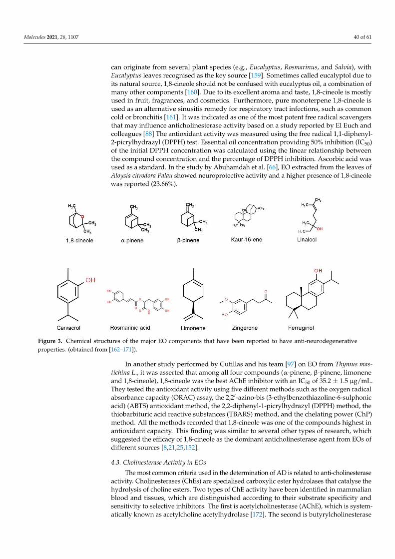
Molecules 2021, 26, 1107 40 of 61
can originate from several plant species (e.g., Eucalyptus, Rosmarinus, and Salvia), withEucalyptus leaves recognised as the key source [159]. Sometimes called eucalyptol due toits natural source, 1,8-cineole should not be confused with eucalyptus oil, a combination ofmany other components [160]. Due to its excellent aroma and taste, 1,8-cineole is mostlyused in fruit, fragrances, and cosmetics. Furthermore, pure monoterpene 1,8-cineole isused as an alternative sinusitis remedy for respiratory tract infections, such as commoncold or bronchitis [161]. It was indicated as one of the most potent free radical scavengersthat may influence anticholinesterase activity based on a study reported by El Euch andcolleagues [88] The antioxidant activity was measured using the free radical 1,1-diphenyl-2-picrylhydrazyl (DPPH) test. Essential oil concentration providing 50% inhibition (IC50)of the initial DPPH concentration was calculated using the linear relationship betweenthe compound concentration and the percentage of DPPH inhibition. Ascorbic acid wasused as a standard. In the study by Abuhamdah et al. [66], EO extracted from the leaves ofAloysia citrodora Palau showed neuroprotective activity and a higher presence of 1,8-cineolewas reported (23.66%).
Figure 3. Chemical structures of the major EO components that have been reported to have anti-neurodegenerativeproperties. (obtained from [162–171]).
In another study performed by Cutillas and his team [97] on EO from Thymus mas-tichina L., it was asserted that among all four compounds (α-pinene, β-pinene, limoneneand 1,8-cineole), 1,8-cineole was the best AChE inhibitor with an IC50 of 35.2 ± 1.5 µg/mL.They tested the antioxidant activity using five different methods such as the oxygen radicalabsorbance capacity (ORAC) assay, the 2,2′-azino-bis (3-ethylbenzothiazoline-6-sulphonicacid) (ABTS) antioxidant method, the 2,2-diphenyl-1-picrylhydrazyl (DPPH) method, thethiobarbituric acid reactive substances (TBARS) method, and the chelating power (ChP)method. All the methods recorded that 1,8-cineole was one of the compounds highest inantioxidant capacity. This finding was similar to several other types of research, whichsuggested the efficacy of 1,8-cineole as the dominant anticholinesterase agent from EOs ofdifferent sources [8,21,25,152].
4.3. Cholinesterase Activity in EOs
The most common criteria used in the determination of AD is related to anti-cholinesteraseactivity. Cholinesterases (ChEs) are specialised carboxylic ester hydrolases that catalyse thehydrolysis of choline esters. Two types of ChE activity have been identified in mammalianblood and tissues, which are distinguished according to their substrate specificity andsensitivity to selective inhibitors. The first is acetylcholinesterase (AChE), which is system-atically known as acetylcholine acetylhydrolase [172]. The second is butyrylcholinesterase
Molecules 2021, 26, 1107 41 of 61
(BChE), which is systematically referred to as acetylcholine acyl hydrolase [173–176]. Thepreferred substrate for AChE is acetylcholine (ACh), while butyrylcholine (BCh) and propi-onylcholine (PCh) are ideal for BChE [175–177]. AChE activity is known to be inhibited byseveral compounds, with toxins and drugs as the major inhibitors [178]. AChE activity isused in verifying treatment effects, especially in AD [177].
Both AChE and BChE possess active sites at the bottom of 20 Å-deep gorges with50% identical amino acid sequence, whereas the gorge entrance locates the peripheralsite [179]. The active site for both enzymes comprises a catalytic triad, acyl-bindingpocket, and choline binding site [180]. A total of 14 aromatic amino acids are found inthe active site of AChE, whereas six of these are substituted by aliphatic amino acids forBChE [181]. Binding and hydrolysis processes of bulky ligands are restricted in AChE dueto presence of phenylalanine residues in the acyl binding pocket. In contrast, these residuesare substituted with two flexible amino acids that are selective for BChE and allow thebinding of bulkier ligands [182]. The different mechanisms involved in relevance to theactive gorge site specific for each enzymes have been investigated via molecular modelling,structure-based virtual screening, or even crystallographic studies [181–184].
Generally, traditional Ellman assay is used with some modifications, applied for thedetermination of anti-cholinesterase activities [185,186]. This technique is a simple, accu-rate, and rapid method of measuring ChE activity, that is based on the reaction betweenthiocholine with the sulfhydryl group of a chromogen such as 5,5′-dithiobis-(2-nitrobenzoicacid) (DTNB or Ellman’s reagent). The shift of electrons to sulphur atoms yields a yel-low substance called 5-thio-2-nitrobenzoic acid (TNB), which is measured by monitoringabsorbance at 410 nm [187–190]. DTNB is a water-soluble compound and is useful forits fast reaction with thiocholine and minor side effects at neutral pH [185,187–191]. Thistechnique, however, is also subject to certain limitations; it is restricted for testing antidotsagainst organophosphorus AChE inhibitors or for measuring AChE activity in samplesof such treated individuals [190]. In addition to the Ellman assay, another method thatcan also be used for measuring ChE activities is the electrometric method of Michel [192].This technique is applied based on pH changes that arise from H+ synthesis via cholinesterhydrolysis [175,176,193].
4.4. Extracellular Plaque Deposits
Extracellular plaque deposits of the Aβ-peptide and flame-shaped neurofibrillarytangles of the microtubule-binding protein tau are the two hallmark pathologies requiredfor AD patients. Familial early-onset forms of AD are associated with mutations either inthe precursor protein for Aβ (APP) or in presenilin-1 (PS1) or presenilin-2 (PS2). Peptidegeneration pathways synthesise γ-secretase with either PS1 or PS2 as the catalytic subunit.APP is sequentially cleaved, where β-secretase first cleaves APP to release a large, secretedderivative, sAPPβ, followed by γ-secretase that cleaves a fragment of 99 amino acids(CTFβ) to generate Aβ. The process of γ-secretase cleaving can be inaccurate, leading toC-terminal heterogeneity of the resulting peptide population that generates numerous Aβ
species, with Aβ1—40 of the highest abundance followed by Aβ42. The slightly longerforms of Aβ, particularly Aβ1–42, are the principal species deposited in the brain thatare more hydrophobic and fibrillogenic [193]. In view of their vital role in Aβ synthesis,both β- and γ-secretase are considered as key components in anti-AD pharmaceuticalsdevelopments [193,194]. Normal pathology tests refer to the density in the affected brainregions of neuritic amyloid plaques and neurofibrillary tangles of tau protein. AD diagnosisinvolves the presence of large neuritic plaque portions, consisting of highly insoluble Aβ inthe brain parenchyma. There are also deposits of tau protein, although they occur amongless common neurodegenerative disorders, especially in the absence of neuritic plaques.There are some distinctive morphological features of the neurofibrillary tangles in thevarious diseases, and may exhibit a distinct composition of tau isoforms that vary fromAD [195].

Molecules 2021, 26, 1107 42 of 61
It is not only humans which have amyloid beta; non-human primates (NHPs) have thesame Aβ sequences as humans, an almost identical APP sequence, and they overlap withrelated human biochemical pathways in many aspects, however surprisingly with ageing,they develop relatively few AD-like neuropathologies. Aged canines also develop severeamyloid deposition; canines tend to demonstrate extensive amyloid deposition from aboutten years of age, unlike in aged NHPs, where it could take several decades [196]. Amyloiddeposition in canines is also interrelated with age-related cognitive dysfunction [197],although little neuronal loss is detected. Due to a poor understanding of AD and thehuman brain complexity, it has been deduced that there is no natural animal model ofthe disease [198]. For the past 25 years, pharmacological and genetic AD-models, as wellas various animal species (primates, dogs, rodents, etc.), have been used in AD researchactivities [199,200]. The resurgence of interest in rats as the appropriate animal model of ADled to the usage of various types of rat models. As of current practice, transgenic mice havebeen extensively used in studies on AD. In any selected models, all need the introductionof some combined familial AD mutation into APP or PS1, or even in both [200].
4.5. Current EOs on AD
Salvia is the largest genus of plants in the family Lamiaceae, with the number of speciesestimated to range from 700 to nearly 1000. Fifteen species of Salvia (namely, officinalis L.,chionantha, chrysophylla Staph, urmiensis, nemorosa L., syriaca, ballsiana, cyanescens, divaricate,hydrangea, kronenburgii, macrochlamys, nydeggeri, pachystachys, pseudeuphratica, and rusellii)were studied for cholinesterase inhibition assay, and happen to be the most widely studiedsource of EOs. Most of the researchers found that Salvia spp. were weak AChE andBChE inhibitors, except for EO from S. pseudeuphratica, which demonstrated the highestinhibitory activity against AChE, at IC50 = 26.00 ± 2.00 µg/mL compared to S. cyanescensand S. pachystachys. In contrast, BChE activity did not show 50% inhibition even at thehighest concentration, where IC50 was reported to be above 80 µg/mL for S. pseudeuphraticaand S. hydrangea [96]. Salvia officinalis was studied twice, in 2017 and 2019. Based on GC–MS and GC–FID analysis, the main components of S. officinalis were α-thujone, camphor,1,8-cineole, and β-thujone. Salvia has also been used in ex vivo-based research, using theisolated guinea pig ileum method where the major molecule, rosmarinic acid, showedsignificant contraction responses on an isolated guinea pig ileum. Docking results ofrosmarinic acid also showed a high affinity to the selected target, AChE [136]. The authorsuggested the potential of rosmarinic acid to become a novel therapeutic candidate for thetreatment of AD.
Other than Salvia spp., EOs from Lavandula spp. have also been studied for the treat-ment of AD. L. luisieri has been found to comprise high contents of oxygen-containingmonoterpenes, mainly necrodane derivatives, which are absent from any other oil. This oilwas tested on the endogenous beta-site APP-cleaving enzyme 1 (BACE-1) in cultured cells,being responsible for a reduction in Aβ production, with no significant toxicity. Althoughthe study was conducted in vitro, the low molecular weight and high hydrophobicity ofterpenoids are properties that provide a good chance for them to cross cellular membranesand the blood–brain barrier, an essential attribute for BACE-1 inhibition in vivo [78]. How-ever, EO from L. angustifolia did not give a required finding because it enhanced the Aβ
aggregation based on the thioflavin T method; this effect was further confirmed by atomicforce microscope (AFM) imaging. EO of L. angustifolia was also showed to counteract theincrease in intracellular reactive oxygen species production and the activation of the pro-apoptotic enzyme caspase-3 induced by Aβ1–42 oligomers [23,24]. Meanwhile, EO fromL. pubescens exhibited strong anti-AChE and anti-BChE effects at IC50 of 0.9 µL/mL and6.82 µL/mL, respectively. Carvacrol (CAR, 2-methyl-5-isopropylphenol) was also found tobe higher in L. pubescens. Carvacrol was found to be abundant among EOs of the Lamiaceaefamily, and is known for various benefits including antibacterial, antifungal, antioxidant,antinociceptive, anti-inflammatory, anti-apoptosis, and anti-cancer activities [201]. Severalstudies on EOs have reported that carvacrol exerts some actions on the neuronal system,
Molecules 2021, 26, 1107 43 of 61
including AChE inhibition [104,202], anxiolytic [203], and antidepressant [204] properties.In addition, carvacrol has the ability to modulate central neurotransmitter pathways, suchas dopaminergic, serotonergic and γ-aminobutyric acid (GABA)-ergic systems [201].
Only two types of cell lines, SH-SY5Y and PC-12, were reported to have been usedin AD research. The in vitro toxic effects of amyloid peptides are usually examined usingthe human neuroblastoma-derived SH-SY5Y cell line, because differentiated neuron-likeSH-SY5Y cells are extra-sensitive to amyloid peptides compared to non-differentiated cells,because the latter lack long neurites [205]. Z-ligustilide (Z-LIG) EOs effectively protectagainst fibrillar aggregates of Aβ25–35- and Aβ1–42-induced toxicity in SH-SY5Y anddifferentiated PC12 cells, possibly through the concurrent activation of the PI3-K/Akt path-way and inhibition of the p38 pathway [105]. Aβ25–35 represents a neurotoxic fragmentof Aβ1–40 or Aβ1–42, and retains the toxicity of the full-length peptide [206]. Aβ25–35 isoften selected as a model for full-length peptides because it retains both its physical andbiological properties [207]. In general, declining levels of PI3K subunits as well as bluntedAkt kinase phosphorylation have been observed in the AD brain, which is characterised byAβ and tau pathologies [208].
There was also a study on the potential therapeutic effect of hybrid EO from Kushuiroses. Kushui rose (R. setate× R. rugosa) refers to a natural hybrid of cog rose and traditionalChinese rose that has been cultivated for more than 200 years [209]. In this study, transgenicworm strains purchased from the Caenorhabditis Genetics Center (CGC) were used insteadof rat or mice models. They found that rose EO (REO) significantly inhibited AD-likesymptoms of worm paralysis and hypersensitivity to exogenous serotonin (5-HT) in a dose-dependent manner. Although the GC–MS analysis revealed the presence of 40 components,the major components, β-citronellol and geraniol, were found to act less effectively thanthe oil itself. Intriguingly, REO significantly suppressed Aβ deposits and reduced the Aβ
oligomers to alleviate the toxicity induced by Aβ overexpression [209].Su He Xiang Wan (SHXW) has also been studied for its neurodegenerative remedy
potential. SHXW is a distinct EO, and is a patent medicine comprising borneol, styraxresin, musk, aquilaria, frankincense, piper, benzoin, saussurea, cyperus, sandalwood, clove,terminallia, aristolachia fruit, rhino horn, and cinnabar. This ancient prescription wasrecorded in the He Ji Ju Fang of the Song Dynasty [210]. For this plant, the researchersevaluated the effects of a modified SHXW (KSOP1009 formulation) intake on the AD-likephenotypes of Drosophila AD models, which express human Aβ1–42 in their developingeyes or neurons. They found that Aβ1–42-induced eye degeneration, apoptosis, andlocomotive dysfunctions were strongly suppressed. However, Aβ1–42 fibril depositsin the Aβ1–42 overexpressing model were not affected by treatment with KSOP1009extract. Conversely, KSOP1009 extract intake significantly suppressed the constitutiveactive form of hemipterous, a c-Jun N-terminal kinase (JNK) activator, while it induced eyedegeneration and JNK activation. In Drosophila, flies with mutations that augment JNKsignalling accumulate less oxidative damage and live dramatically longer than wild-typeflies [211,212].
Cinnamomum zeylanicum consisting of (E)-cinnamaldehyde (CAL) (81.39%) and (E)-cinnamyl acetate (CAS) (4.20%) as the main compounds showed over 78.0% inhibitoryactivity in cholinesterase. In MAO-A and MAO-B inhibition assays, EOs from C. zeylanicum(96.44%, 95.96%) and CAL (96.32%, 96.29%) demonstrated comparable activity to rasagiline(97.42%, 97.38%, respectively). Research by Murata and co-workers [69] found that kaur-16-ene, nezukol, and ferruginol isolated from plants had anti-AChE (IC50) activity at 640,300 and 95 µm, respectively. Even though ferruginol activity has already been highlightedbefore, by Gulacti et al. [213], this study documented the activities of kaur-16-ene andnezukol for the first time.
Meanwhile, Citrus limon has been found to significantly lower AChE brain depressionin APP/PS1 and wild-type C57BL/6L (WT) mice. PSD95/synaptophysin, the synapticdensity index, was substantially improved in histopathological shifts [109]. Based on theprevious analysis by other researchers, nobiletin 3′,4′,5,6,7,8-hexamethoxyflavone was
Molecules 2021, 26, 1107 44 of 61
found to be the major component of polymethoxylated flavones in citrus peels, such as C.depressa, C. reticulata, C. sinensis, and C. limon [214,215]. Thus, nobiletin may potentiallybe the compound that substantially alters the development of these diseases. Other thanthat, Acori graminei, which was found to be rich in β-asarone, enhanced cognitive functionof AβPP/PS1 mice and decreased neuronal apoptosis in the AβPP/PS1 mouse cortex. Inaddition, a substantial increase in the expression of CaMKII/CREB/Bcl-2 was observed inthe cortex of AβPP/PS1 mice treated with β-asarone.
In a study conducted by Ayuob et al. [113], Ocimum basilicum up-regulated the serumcorticosterone level, the hippocampal protein glucocorticoid receptor, and the brain-derivedneurotropic factor (BDNF); however, it down-regulated the neurodegenerative and at-rophic changes induced in the hippocampus, which decreased after exposure to chronicunpredictable mild stress (CUMS). According to the data collected by Avetisyan and co-workers [216], the major components of O. basil includes methyl chavicol and linalool.Interestingly, many linalool-producing plants are commonly used in folk medicine and aro-matherapy to alleviate symptoms and treat multiple acute and chronic diseases [217,218].Linalool is frequently used in the manufacture of fragrances for shampoos, soaps, de-tergents, and in pharmaceutical formulations [219,220]. Research conducted by Sabogal-Guáqueta et al. [221] found that oral administration of monoterpene linalool to elderlymice (21–24 months old) with a triple transgenic form of AD (3x Tg-AD) at 25 mg/kg forthree months at an interval of 48 h resulted in enhanced learning and spatial memory andincreased risk assessment activity in the elevated plus maze. Hippocampi and amygdalaefrom 3x Tg-AD linalool-treated mice also showed a large reduction in extracellular β-amyloidosis, tauopathy, astrogliosis, and microgliosis, as well as pro-inflammatory markerlevels of p38 MAPK, NOS2, COX2 and IL-1β. Thus, linalool is suitable as an AD preventioncandidate for pre-clinical studies. Based on the articles that we have selected, linaloolis a major volatile component of EOs in a number of aromatic plant species, such as L.angustifolia Mill., M. officinalis L., R. officinalis L., and C. citrate DC. The presence of linaloolcan also help to reduce the deposits of Aβ, based on a study conducted by Gradinariuet al. [114] where Aβ1–42-treated rats exhibited the following: a decrease in exploratoryactivity (crossing number); smaller percentage of time spent and fewer entries in the openarm in the elevated plus-maze test; increase in swim time; and decrease in the immobilitytime within the forced swimming test.
4.6. Current EOs on PD
In terms of PD, the current therapy in practice is applied as a combination of gold-standard dopaminergic reposition with 3-(3,4-dihydroxyphenyl)-L-alanine (L-dopa), alongwith other agents such as MAO-B, catechol O-methyltransferase (COMT) inhibitors, dopamin-ergic agonists, and cholinergic blockers [222]. However, the available treatments are subjectto consequences of motor and non-motor side effects, which leads to poor efficacy in ad-vanced stages of PD [144]. These arising phenomena are the main reasons that suggest andemphasise the necessity for the synthesis of anti-PD drugs that could delay the progressionof neurodegeneration [144]. As mentioned in the results, PD studies included in this reviewcomprise in vitro and in vivo, as well as combinations of in vitro with in vivo or ex vivoresearch. Cinnamomum sp., Eryngium sp., Myrtus sp., Acorus sp., Eplingiella sp., Foeniculumsp., Pulicaria sp., Rosa sp., Zingiber sp., and Lavandula sp. were among the identified EOsbased on the respective included PD studies.
Four of the included studies on PD were based on in vitro approaches, with variousEOs. The first study was focused on the evaluation of protective effects of EOs extractedfrom Cinnamomum sp. (C. verum and C. cassia) and cinnamaldehyde, in comparison tohydroalcoholic extracts using 6-OHDA-induced PC12 cytotoxicity as the representativemodel of PD [138]. Cinnamomum sp., or more commonly known as cinnamon, belongs tothe Lauraceae family that is composed of almost 250 species and has been acknowledgedfor extensive health benefits [223,224]. Among the various species, C. verum and C. cassiawere the two main species that have been widely applied for their medicinal and culinary

Molecules 2021, 26, 1107 45 of 61
applications, especially in Iran [225–227]. It is important to note that cinnamaldehyderepresents one of the key components of both species, and EOs were reported to exhibitstrong antioxidant properties [223]. The findings of this study indicated that 6-OHDAled to cell death, cell apoptosis, and suppression of the p44/42 pathway. On the whole,the study concluded that synergistic effects of cinnamaldehyde and EOs as well as otherextract components could promote cinnamon’s roles as neuroprotective agents, specificallyfor PD treatment [138].
Eryngium sp. belong to the Apiaceae family, and are recognised for their EOs’ po-tentials in MAO inhibition [139]. MAO is available in two forms (A, B) where MAO-Ainhibition is linked to antidepressant effects, while MAO-B is correlated with PD treat-ment [228,229]. An in vitro study conducted by Klein-Júnior et al. demonstrated theassessment of Eryngium sp. (E. floribundum: EP, E. horridum: EH, E. pandanifolium: EP, E.eriophorum: EE and E. nudicaule: EN) EOs for their MAO inhibitory effect. Intriguingly, thefindings of this study indicated that MAO-A activity was not inhibited by any EOs, whileEPEO and EHEO resulted in MAO-B inhibition. The literature search has also highlightedthat PD patients usually represent elevated levels of MAO-B which arise due to gliosis,and hence contribute towards the collapse of the dopaminergic system [229]. Thus, thisstudy puts forward the claim that Eryngium sp. could have potential applications as CNSbioactive secondary metabolites, particularly for neurodegenerative disease, in relevanceto characteristics exhibited by EHEO [139].
Another important aspect that is usually associated with studies on PD is on α-Synfibrillation. It is presumed that protein structural modifications resulting in amyloid fibrilformation progresses towards neurodegenerative disorders [140]. However, the exact fac-tors of α-Syn actions of misfolding and aggregation in the brain are still scarce. In addition,it should also be noted that preventive measures against α-Syn fibrillation are yet to beavailable; hence, the acceleration of the fibrillation process via certain factors should beavoided. Among some of the common factors are metal ions, small molecules, nanopar-ticles, and, particularly, toxins that could intensify the aggregation process [140,230–233].The next two in vitro studies were performed by the same research team, where theyhighlighted the effects of 15 various Iranian EOs against α-Syn fibrillation [140,141].
Among all the 15 oils tested, it was shown that M. communis demonstrated potentialbenefits because it elevated the fibrillation in a concentration-dependent manner. However,it is necessary to understand that the major components of this oil are not responsiblefor the observed changes, suggesting complexity of both extract and synergistic effectsof the available compounds, regardless of their amount [140]. In the second study, theinvestigation on C. cyminum EO signified the presence of cuminaldehyde as the majoractive compound that plays its role in the inhibition of α-Syn fibrillation. In addition,cytotoxicity assays on PC12 cells indicated the absence of toxic effects with cuminaldehydetreatment throughout α-Syn fibrillation [141].
Apart from in vitro studies, PD studies are also extensively performed under in vivoconditions. EOs of Acorus sp. cover two of the in vivo studies in this review. Both of thesestudies were conducted by the same research team, and focused on the regulation effect ofβ-asarone that was isolated from A. tatarinowii Schott on 6-OHDA-induced parkinsonianrats via two distinct endoplasmic reticulum (ER) stress pathways [142,143]. ER is knownfor its role in protein folding, where the build-up of protein unfolding/misfolding couldinitiate a phenomenon called ER stress that further activates the cellular process of unfoldedprotein response (UPR) [234]. ER stress has been noticed in a number of PD experimentalmodels, and is also provoked by an increase in wild-type α-Syn [235–237]. Three mainpathways that are categorised as UPR are inositol requiring enzyme 1 (IRE1), protein kinaseRNA (PKR)-like ER kinase (PERK), and activating transcription factor 6 (ATF6) [238].
In general, GRP78 functions in the regulation of ER stress, where it binds the threeproteins of the UPR pathway and maintains them as inactive when the cells are notexposed to stress conditions. However, under conditions of accumulated protein unfoldingor misfolding, GRP78 will bind to the proteins and release them [239–241]. In terms of

Molecules 2021, 26, 1107 46 of 61
autophagy, the latest research has claimed that it could be induced due to ER stress [242,243].Beclin-1 is known for its role in forming autophagosomes and is an essential part of theinitial autophagy process. The pro-autophagic role of Beclin-1 could be inhibited via itsreaction with Bcl-2, however this interaction is also subject to disruption caused by Bcl-2phosphorylation that leads to Beclin-1 release and accelerates autophagy [244]. As per thesecond pathway study, it was proven that β-asarone leads to Beclin-1 downregulation,which highlights that Bcl-2 could possibly be the main linkage between autophagy andER stress. The findings of both studies lead us to conclude that diminishing ER stressvia β-asarone regulation is proven to be useful in the impairment of PD pathologicalprogression [142,143].
Another study that also applies the use of 6-OHDA-induced PD mouse models focusedon the investigation of the effects of zingerone and eugenol on dopamine concentration, be-havioural changes, and antioxidant activities upon 6-OHDA administration and treatmentof L-dopa [28]. Zingerone is extracted from the ginger root, while eugenol originates fromcloves and was reported to be protective against 6-OHDA-induced depletion of striataldopamine via increases in SOD activity and elevation of reduced glutathione (GSH) and L-Ascorbate (Asc) concentration, respectively [245,246]. Although these groups of researcherspreviously reported positive findings where pre-treatment with zingerone or eugenol inhib-ited 6-OHDA-induced dopamine depression by preventing lipid peroxidation, the currentstudy, which involved post-treatment with similar compounds, resulted in contradictoryfindings, where dopamine decrease was more pronounced [28,246]. Despite the availabilityof other findings that propose the benefits of consuming these compounds, Kabuto andYamanushi [28] suggested that intake of these specific substances upon the onset of PDsymptoms should be more carefully monitored to prevent further injury aggravation.
In addition to studies among 6-OHDA-induced Parkinson’s rat models, the possi-bility to achieve positive effects of EOs when complexed with β-cyclodextrin (βCD) wasevaluated by Filho and colleagues using reserpine-induced progressive models for PD inmice [144]. Cyclodextrins are cyclic oligosaccharides that could form host–guest complexeswith hydrophobic molecules and were also reported to protect EOs from heat, evaporation,moisture, oxidation and light effects along with facilitating easy solubility [247–249]. Com-plexation effects of cyclodextrins with EOs were shown to be more prominent in exertingpositive effects, especially in the treatment of chronic diseases, as published by severalstudies [250,251]. In this particular study, the same approach was applied using leaf EOextracted from Eplingiella fruticosa (EPL), where one of the key components is 1,8-cineole.Eplingiella sp. belongs to the Lamiaceae family, and was reported for its benefits as anti-inflammatory and antioxidant effects [252,253]. This research demonstrated and proved thehypothesis whereby both treated groups of EPL and EPL-βCD deferred reserpine effectson catalepsy time. However, this effect was noticed to be more remarkable with EPL-βCDtreated mice groups.
Another study also applied induction with reserpine, with a different PD model ofovariectomized and non-ovariectomized rats [145]. Reserpine is known as an irreversibleinhibitor of the vesicular monoamine transporter 2 (VMAT-2). The approach of reserpineinjection to rats as a mode of PD model was proposed in response to its action on thedepletion of monoamine and locomotor activities [254]. Ovariectomized rats are subjectedto oestrogen deficits, similar to surgically menopaused women, where cognitive damage ishighly possible [255]. Lower oestrogen levels are correlated with many side effects, suchas mental disorders, memory defects, emotional issues, and other cognitive failures [256].These incidences have led to much attraction towards phytoestrogens for its protectivenature against certain diseases. Fennel plant (Foeniculum vulgare) is classified as in theApiaceae family, and is known for its phytoestrogen compounds; it showed promisingresults in the treatment of cognitive disorders, such as dementia and AD [145,257–259].The evaluation of this study indicated that protective oestrogen effects against neurodegen-erative disorders were significantly decreased among reserpine-induced ovariectomizedrats. Injection of reserpine resulted in a more remarkable observation on limb movement

Molecules 2021, 26, 1107 47 of 61
disorder among ovariectomized rats. Fennel treatment at various doses for both groupsgave better results on the motor activity, which stressed the importance of oestrogensand phytoestrogens as a protective measure of dopaminergic neurons and improved PDsymptoms [145].
Rotenone administration to rats, as induction of a PD model, is an alternative approachwhere it induces nigrostriatal dopaminergic neuron degeneration that is associated withα-Syn Lewy bodies [260]. Rotenone is an insecticide with high lipophilic nature, and isknown to inhibit mitochondrial complex-1 along with causing oxidative stress [261,262].This rotenone-induced model was reported in the study by Issa and colleagues, based onthe neuroprotective effects of Pulicaria undulata EO in male Wistar rats [146]. P. undulatabelongs to the Asteraceae family, which is commonly distributed in Asia, Europe, andNorth Africa [263]. From this study, it was shown that EO of P. undulata could exert itsneuroprotective effects via anti-inflammatory and antioxidant properties. The mechanismsinvolved in neuroinflammation suppression include downregulation of induced nitricoxide synthase (iNOS) expression, followed by lower gene expression of α-Syn [146].
Compared to individual studies, there are also several approaches that examine com-bined effects that could incorporate in vitro, in vivo, and also ex vivo applications. Onesuch attractive research is on the combined in vitro/in vivo evaluation of SHXW EO with1-methyl-4-phenyl-1,2,3,6-tetrahydropyridine (MPTP)-induced PD mice and SH-SY5Y celllines [147]. In this study, SHXW was a Chinese herbal formulation that consisted of 15crude herbs called KSOP1009, composed of eight medicinal plants of different families(Hamamelidaceae, Myristicaceae, Umbelliferae, Santalaceae, Piperaceae, Myrtaceae, Ty-phaceae, and Lamiaceae). MPTP is known to cause fast degeneration of dopaminergicneurons, and as such, it was believed that the use of this specific model could assist inexplaining certain aspects of PD disease mechanisms [147,264]. Positive findings of thestudy demonstrated that ingestion of KSOP1009 was successful in the protection of MPTPtoxicity, where this could be correlated with dopamine reduction that also decreases ROSand restores mitochondrial roles [147].
In terms of in vivo and ex vivo combinatorial approaches, two articles were high-lighted in this review, with several authors originating from the same team [148,149]. Bothstudies explored the approach of L-dopa induction of oxidative toxicity. L-dopa has beenrecognised as the most effective symptomatic treatment of PD for more than 30 years;however, toxicity issues that were raised via in vitro studies seem to be an unresolvedchallenge [148]. It was also mentioned that prolonged L-dopa treatment is often associatedwith side effects that often result in a delay in its administration [148]. Past studies haveclaimed that L-dopa therapy in combination with antioxidants could lessen the possibleside effects. As such, efforts were taken to evaluate the effects; this research team investi-gated the combined effects of EOs from Lavandula angustifolia, Rosa damascena, vitamin Cand Trolox in the initial study [148], followed by another study in 2019 on the combinedeffects of pre-treatment with Rosa damascena and vitamin C [149].
For the first study, the obtained results indicated that both EOs from herbal plantsshowed noticeable radical scavenging and antioxidant properties against L-dopa toxicity.Similarly, the second study also put forward equivalent claims with R. damascena charac-teristics where it was in parallel to vitamin C, and exhibited a significant role of rose oilin its interference against the acute oxidative toxicity of L-dopa [148,149]. Based on allthe collective studies, it could be observed that PD treatments remain centralised amongseveral parameters that include mainly α-Syn fibrillation, MAO-B, β-asarone regulationof ER stress pathways, toxicity-induced models with 6-OHDA, MPTP, L-dopa, reserpine,and rotenone with common animal models of rats and mice. Although the regulationmechanisms involved in each of the parameters may differ, the main focus remains towardseffective and improved treatments for PD patients.
Molecules 2021, 26, 1107 48 of 61
4.7. Current EOs on Other Neurodegenerative Diseases
Our review findings also revealed that several studies did not specify the studieddisease, and instead examined general neurodegenerative disorders. The in vitro studyconducted by Costa et al. [150] using Lavandula pedunculata subsp. lusitanica (Chaytor)Franco EO suggested the ability of L. pedunculata as a suitable choice to prevent neurode-generative disease. A study performed by Elmann et al. [151] with Pelargonium graveolensEO showed that some constituents may depend on synergistic interactions to function.Geranium oil from Pelargonium graveolens inhibited nitric oxide (NO) production, as wellas the expression of the proinflammatory enzymes cyclooxygenase-2 (COX-2) and inducednitric oxide synthase (iNOS) in primary cultures of activated microglial cells. The findingshowed that none of the major constituents could inhibit NO production when examinedat natural relative oil concentrations, with excellent inhibitory activity of citronellol athigher concentrations. The findings indicated that the presence of synergistic interac-tions between these components are of considerable importance. Thus, geranium oil canbe useful in neurodegenerative disease prevention where neuroinflammation is part ofpathophysiology [151].
EOs could also be applied as inhalation-based treatment, which could signify a naturalway to heal one’s mind, body, and soul [265]. In Bagci et al.’s study [153], it was investigatedwhether inhalation of the Anthriscus nemorosa EO leads to behavioural changes that indi-cated significant memory improvement and exhibited both anxiolytic- and antidepressant-like effects in dual-treated rats. The results suggested that A. nemorosa EO inhalation canprevent scopolamine-induced memory impairment, anxiety, and depression [153].
In the in vivo research conducted by Satou et al. [118], mice were administered Rosmar-inus officinalis EO (EORO) by inhalation and it was concluded that the rate of spontaneousalternation activity was significantly improved. The key components (1,8-cineole, α-pineneand β-pinene) were detected in the brain in a concentration-dependent manner uponEORO inhalation, which indicated its possible exerted effects. However, in several otherinterpretations, it was proposed that 1,8-cineole might require synergistic interaction effects.This was highlighted by Costa et al. [150], where α-pinene and 1,8-cineole from Lavandulapedunculata subsp. Lusitanica were shown to be effective cholinesterase inhibitors evenat low concentrations and are likely to contribute to this behaviour. The synergistic andantagonistic interactions between certain terpenes that could result in combined effectsshould also be taken into consideration. Although this compound was also mentioned ina study conducted by Hritcu et al. [112], the study focused more on the positive effect oflinalool in the improvement of spatial memory deficit in a scopolamine-induced dementiarat model instead of the 1,8-cineole effect on neurodegeneration. In addition, a study byKaufmann and colleagues [104] signified the potential of myrtenal as an effective AChEinhibitor, compared to 1,8-cineole with EOs of Artemisia annua L. (Asteraceae) or Glycyrrhizaglabra L. (Fabaceae).
5. Conclusions
In conclusion, our review highlighted that EOs provide many other benefits apartfrom its common usage as fragrances and flavours, especially for its significant role inneurodegenerative disease therapy. Their roles in reducing disease severity are exertedby different mechanisms that vary respective of their origin. Although human studieswere not obtained as part of our search, we strongly believe that the presence of essentialcomponents such as 1,8-cineole, carvacrol, or β-asarone could play significant roles inefforts towards the prevention and treatment of neurodegenerative disorders. Therefore, itis vital that the search for novel species of EOs are continued, to explore oils which couldbe applied towards the application of EO-based therapies or treatment strategies intendedfor age-related neurodegenerative disorders.
Molecules 2021, 26, 1107 49 of 61
Author Contributions: Conceptualization, all authors; literature review: all authors; tables: allauthors, writing and review all authors; editing: all authors; revisions and final editing: all authors.All authors have read and agreed to the published version of the manuscript.
Funding: This research received no external funding.
Acknowledgments: The authors thank the Director General of Health Malaysia and the Directorof Institute for Medical Research (IMR), Malaysia, for giving permission to publish this article. Wealso thank the staff of the Nutrition, Metabolism and Cardiovascular Centre, Institute for MedicalResearch, NIH, for their continuous support.
Conflicts of Interest: The authors declare no conflict of interest.
Abbreviations
Aβ Amyloid-betaACh AcetylcholineAChE AcetylcholinesteraseAD Alzheimer’s diseaseAFM Atomic force microscopeALS Amyotrophic lateral sclerosisAOPP Advanced oxidation protein productAPP Amyloid precursor proteinAsc L-Ascorbateα-Syn α-SynucleinATF6 Activating transcription factor 6BACE-1 Beta-site APP-cleaving enzyme 1BCh ButyrylcholineBChE ButyrylcholinesteraseβCD β-cyclodextrinBDNF Brain-derived neurotropic factorCA1 Cornu ammonisCAG Cytosine–adenine–guanineCAL (E)-cinnamaldehydeCAS (E)-cinnamyl acetateCAT CatalaseCGC Caenorhabditis Genetics CenterChE CholinesterasesCNS Central nervous system (human)COMT Catechol O-methyltransferaseCOX-2 Cyclooxygenase-2CUMS Chronic unpredictable mild stressDA DopamineDAT Dark avoidance testDOPACDSPE-PEG2000
3,4-dihydroxyphenylacetic acidDistearoylphosphatidylethanolamine with covalently linked polyethylene glycol of molecular weight 2000
DTNBELISA
5,5′-dithiobis-(2-nitrobenzoic acid)Enzyme-linked immunosorbent assay
ER Endoplasmic reticulumEO Essential oilfPDFITC
Familial PDFluorescein isothiocyanate
GABA γ-aminobutyric acidGC–FID Gas chromatography–flame ionisation detectorGC–MSGC–ITMS
Gas chromatography–mass spectrometryGas Chromatography/Ion Trap Mass Spectrometry
GC–O Gas chromatography–olfactometryGPx Gluthathione peroxidaseGRP78 Glucose regulated protein 78GSH Reduced glutathione

Molecules 2021, 26, 1107 50 of 61
HDHPLC–DADHPLC–PDA
Huntington’s diseaseHigh-performance liquid chromatography–diode-array detectionHigh-performance liquid chromatography–photodiode array detection
HTT Huntingtin geneHVA Homovanillic acidiNOS Induced nitric oxide synthaseIRE1 Inositol requiring enzyme 1IRE1/XBP1 IRE1/X-Box Binding Protein 1JNK c-Jun N-terminal kinaseLDH Lactate dehydrogenaseL-dopa LevodopaLMNLOX
Lower motor neuronLipoxygenase
MAO Monoamine oxidaseMDA MalondialdehydeME MicroemulsionmHTT Mutant HTTMMP Mitochondrial membrane potentialMPP+ 1-methyl-4-phenylpyridiniumMPTP 1-methyl-4-phenyl-1,2,3,6-tetrahydropyridineMTT 3-(4,5-dimethylthiazol-2-yl)-2,5-diphenyltetrazolium bromideMWMNA–FB
Morris water maze testNew micro-well plate AChE activity
NHPsNMR
Non-human primatesNuclear magnetic resonance
NOOS
Nitric oxideOxidative stress
PCC Protein carbonyl contentPCh PropionylcholinePD Parkinson’s diseasePDI Polydispersity indexPERK Protein kinase RNA (PKR)-like ER kinasep-PERK PERK phosphorylationPI3K Phosphatidylinositol-4,5-bisphosphate 3-kinasePKR Protein kinase RNAPS PresenilinRNAi RNA interferenceRNS Reactive nitrogen speciesROS Reactive oxygen speciesSDT Step-down testSHXWSFESKN-1SN
Su He Xiang WanSupercritical fluid extractionSkinhead-1Substantia nigra
SNpc Substantia nigra pars compactaSOD Superoxide dismutasesPD Sporadic PDTBARSTLC
Thiobarbituric acid-reactive substancesThin-layer chromatography
TNB 5-thio-2-nitrobenzoic acidTNFTTCTUNEL
Tumor necrosis factor2,3,5-triphenyltetrazolium chlorideTerminal deoxynucleotidyl transferase dUTP nick end labeling
UMN Upper motor neuron

Molecules 2021, 26, 1107 51 of 61
UPR) Unfolded protein responseVMAT-2 Vesicular monoamine transporter 2VOCsWST
Volatile organic constituentsWater-soluble tetrazolium salt
Z-LIG Z-ligustilide5-HT Serotonin6-OHDA 6-hydroxydopamine8-OHdG Urinary 8-hydroxyguanosine
References1. Osbourn, A.E.; Lanzotti, V. Plant-Derived Natural Products: Synthesis, Function, and Application; Springer Science & Business Media:
New York, NY, USA, 2009; p. 612.2. Esposito, E.R.; Bystrek, M.V.; Klein, J.S. An Elective Course in Aromatherapy Science. Am. J. Pharm. Edu. 2014, 78, 79. [CrossRef]3. Robins, J.L.W. The Science and Art of Aromatherapy. J. Holist. Nurs. 1999, 17, 5–17. [CrossRef]4. Smith, C.A.; Collins, C.T.; Crowther, C.A. Aromatherapy for pain management in labour. Cochrane Database Syst. Rev. 2011.
[CrossRef]5. Garcia-Salas, P.; Morales-Soto, A.; Segura-Carretero, A.; Fernández-Gutiérrez, A. Phenolic-compound-extraction systems for fruit
and vegetable samples. Molecules 2010, 15, 8813–8826. [CrossRef]6. Singer, A.C.; Crowley, D.E.; Thompson, I.P. Secondary plant metabolites in phytoremediation and biotransformation. Trends
Biotechnol. 2003, 21, 123–130. [CrossRef]7. Hsieh, M.T.; Peng, W.H.; Wu, C.R.; Wang, W.H. The ameliorating effects of the cognitive-enhancing Chinese herbs on scopolamine-
induced amnesia in rats. Phytother. Res. 2000, 14, 375–377. [CrossRef]8. Cioanca, O.; Mircea, C.; Hritcu, L.; Trifan, A.; Marius, M.; Aprotosoaie, A.C.; Robu, S.; Gille, E.; Hancianu, M. In vitro—In vivo
correlation of the antioxidant capacity of Salviae aetheroleum essential oil. Farmacia 2015, 63, 34–39.9. Jové, M.; Portero-Otín, M.; Naudí, A.; Ferrer, I.; Pamplona, R. Metabolomics of Human Brain Aging and Age-Related Neurode-
generative Diseases. J. Neuropath. Exp. Neur. 2014, 73, 640–657. [CrossRef]10. Yan, M.H.; Wang, X.; Zhu, X. Mitochondrial defects and oxidative stress in Alzheimer disease and Parkinson disease. Free Radic.
Biol. Med. 2013, 62, 90–101. [CrossRef]11. Sarrafchi, A.; Bahmani, M.; Shirzad, H.; Rafieian-Kopaei, M. Oxidative stress and Parkinson’s disease: New hopes in treatment
with herbal antioxidants. Curr. Pharm. Des. 2016, 22, 238–246. [CrossRef]12. González-Burgos, E.; Gómez-Serranillos, M.P. Terpene compounds in nature: A review of their potential antioxidant activity.
Curr. Med. Chem. 2012, 19, 5319–5341. [CrossRef] [PubMed]13. Essa, M.; Braidy, N.; Bridge, W.; Subash, S.; Manivasagam, T.; Vijayan, R.; Al-Adawi, S.; Guillemin, G. Review of natural products
on Parkinson’s disease pathology. J. Aging Res. Clin. Pract 2014, 3, 1–8.14. Karakaya, S.; Koca, M.; Yılmaz, S.V.; Yıldırım, K.; Pınar, N.M.; Demirci, B.; Brestic, M.; Sytar, O. Molecular Docking Studies of
Coumarins Isolated from Extracts and Essential Oils of Zosima absinthifolia Link as Potential Inhibitors for Alzheimer’s Disease.Molecules 2019, 24, 722. [CrossRef] [PubMed]
15. Forrester, L.T.; Maayan, N.; Orrell, M.; Spector, A.E.; Buchan, L.D.; Soares-Weiser, K. Aromatherapy for dementia. CochraneDatabase Syst. Rev. 2014, CD003150, 1–56. [CrossRef] [PubMed]
16. Xu, P.; Wang, K.; Lu, C.; Dong, L.; Gao, L.; Yan, M.; Aibai, S.; Yang, Y.; Liu, X. Protective effects of linalool against amyloidbeta-induced cognitive deficits and damages in mice. Life Sci. 2017, 174, 21–27. [CrossRef]
17. Bansal, A.; Kirschner, M.; Zu, L.; Cai, D.; Zhang, L. Coconut oil decreases expression of amyloid precursor protein (APP) andsecretion of amyloid peptides through inhibition of ADP-ribosylation factor 1 (ARF1). Brain Res. 2019, 1704, 78–84. [CrossRef]
18. Dalai, M.; Bhadra, S.; Chaudhary, S.; Bandyopadhyay, A.; Mukherjee, P. Anti-cholinesterase potential of Cinnamomum tamala(Buch.-Ham.) T.Nees & Eberm. leaves. Indian J. Tradit. Know. 2014, 13, 691–697.
19. Sihoglu Tepe, A.; Ozaslan, M. Anti-Alzheimer, anti-diabetic, skin-whitening, and antioxidant activities of the essential oil ofCinnamomum zeylanicum. Ind. Crop. Prod. 2020, 145, 112069. [CrossRef]
20. Bahadori, M.B.; Salehi, P.; Sonboli, A. Comparative study of the essential oil composition of Salvia urmiensis and its enzymeinhibitory activities linked to diabetes mellitus and Alzheimer’s disease. Int. J. Food Prop. 2017, 20, 2974–2981. [CrossRef]
21. Cutillas, A.B.; Carrasco, A.; Martinez-Gutierrez, R.; Tomas, V.; Tudela, J. Salvia officinalis L. Essential Oils from Spain: Deter-mination of Composition, Antioxidant Capacity, Antienzymatic, and Antimicrobial Bioactivities. Chem. Biodivers. 2017; 14.[CrossRef]
22. Ayaz, M.; Junaid, M.; Ullah, F.; Sadiq, A.; Khan, M.A.; Ahmad, W.; Shah, M.R.; Imran, M.; Ahmad, S. Comparative chemicalprofiling, cholinesterase inhibitions and anti-radicals properties of essential oils from Polygonum hydropiper L: A Preliminary anti-Alzheimer’s study. Lipids Health Dis. 2015, 14, 141. [CrossRef]
23. Ali-Shtayeh, M.S.; Abu-Zaitoun, S.Y.; Dudai, N.; Jamous, R.M. Downy Lavender Oil: A Promising Source of Antimicrobial,Antiobesity, and Anti-Alzheimer’s Disease Agents. Evid-Based Complement. Altern. Med. 2020, 2020, 5679408. [CrossRef] [PubMed]
Molecules 2021, 26, 1107 52 of 61
24. Soheili, M.; Khalaji, F.; Mirhashemi, M.; Salami, M. The Effect of Essential Oil of Lavandula Angustifolia on Amyloid BetaPolymerization: An In Vitro Study. Iran. J. Chem. Chem. Eng. 2018, 37, 201–207.
25. Chaiyana, W.; Okonogi, S. Inhibition of cholinesterase by essential oil from food plant. Phytomedicine 2012, 19, 836–839. [CrossRef][PubMed]
26. Shoaib, M.; Shah, I.; Ali, N.; Ali, W. In vitro acetylcholinesterase and butyrylcholinesterase inhibitory potentials of essential oil ofArtemisia macrocephala. Bangladesh J. Pharm. 2015, 10, 87–91. [CrossRef]
27. Ansari, M.; Eslami, H. Preparation and study of the inhibitory effect of nano-niosomes containing essential oil from artemisiaabsinthium on amyloid fibril formation. Nanomed. J. 2020, 7, 243–250. [CrossRef]
28. Kabuto, H.; Yamanushi, T.T. Effects of zingerone [4-(4-hydroxy-3-methoxyphenyl)-2-butanone] and eugenol [2-methoxy-4-(2-propenyl) phenol] on the pathological progress in the 6-hydroxydopamine-induced parkinson’s disease mouse model. Neurochem.Res. 2011, 36, 2244. [CrossRef]
29. Alzheimer’s Association. 2019 Alzheimer’s disease facts and figures. Alzheimer’s Dement. 2019, 15, 321–387. [CrossRef]30. Reitz, C.; Mayeux, R. Alzheimer disease: Epidemiology, diagnostic criteria, risk factors and biomarkers. Biochem. Pharm. 2014, 88,
640–651. [CrossRef]31. Ferretti, C.; Sarti, F.M.; Nitrini, R.; Ferreira, F.F.; Brucki, S.M.D. An assessment of direct and indirect costs of dementia in Brazil.
PLoS ONE 2018, 13, e0193209. [CrossRef]32. Weintraub, S.; Wicklund, A.H.; Salmon, D.P. The neuropsychological profile of Alzheimer disease. Cold Spring Harb. Perspect.
Med. 2012, 2, a006171. [CrossRef]33. Aliev, G.; Palacios, H.H.; Walrafen, B.; Lipsitt, A.E.; Obrenovich, M.E.; Morales, L. Brain mitochondria as a primary target in the
development of treatment strategies for Alzheimer disease. Int. J. Biochem. Cell Biol. 2009, 41, 1989–2004. [CrossRef] [PubMed]34. Serrano-Pozo, A.; Mielke, M.L.; Gómez-Isla, T.; Betensky, R.A.; Growdon, J.H.; Frosch, M.P.; Hyman, B.T. Reactive glia not only
associates with plaques but also parallels tangles in Alzheimer’s disease. Am. J. Pathol. 2011, 179, 1373–1384. [CrossRef] [PubMed]35. Villa, V.; Thellung, S.; Bajetto, A.; Gatta, E.; Robello, M.; Novelli, F.; Tasso, B.; Tonelli, M.; Florio, T. Novel celecoxib analogues
inhibit glial production of prostaglandin E2, nitric oxide, and oxygen radicals reverting the neuroinflammatory responses inducedby misfolded prion protein fragment 90–231 or lipopolysaccharide. Pharm. Res. 2016, 113, 500–514. [CrossRef] [PubMed]
36. Masters, C.L.; Selkoe, D.J. Biochemistry of amyloid β-protein and amyloid deposits in Alzheimer disease. Cold Spring Harb.Perspect. Med. 2012, 2, a006262. [CrossRef] [PubMed]
37. Nhan, H.S.; Chiang, K.; Koo, E.H. The multifaceted nature of amyloid precursor protein and its proteolytic fragments: Friendsand foes. Acta. Neuropathol. 2015, 129, 1–19. [CrossRef] [PubMed]
38. Campora, M.; Francesconi, V.; Schenone, S.; Tasso, B.; Tonelli, M. Journey on Naphthoquinone and Anthraquinone Derivatives:New Insights in Alzheimer’s Disease. Pharmaceuticals 2021, 14, 33. [CrossRef]
39. Kocahan, S.; Dogan, Z. Mechanisms of Alzheimer’s Disease Pathogenesis and Prevention: The Brain, Neural Pathology, N-methyl-D-aspartate Receptors, Tau Protein and Other Risk Factors. Clin. Psychopharmacol. Neurosci. 2017, 15, 1–8. [CrossRef][PubMed]
40. Zheng, H.; Fridkin, M.; Youdim, M.B. From antioxidant chelators to site-activated multi-target chelators targeting hypoxiainducing factor, beta-amyloid, acetylcholinesterase and monoamine oxidase A/B. Mini Rev. Med. Chem. 2012, 12, 364–370.[CrossRef]
41. Huang, L.; Lu, C.; Sun, Y.; Mao, F.; Luo, Z.; Su, T.; Jiang, H.; Shan, W.; Li, X. Multitarget-directed benzylideneindanone derivatives:Anti-β-amyloid (Aβ) aggregation, antioxidant, metal chelation, and monoamine oxidase B (MAO-B) inhibition properties againstAlzheimer’s disease. J. Med. Chem. 2012, 55, 8483–8492. [CrossRef]
42. Youdim, M.B.H.; Amit, T.; Bar-Am, O.; Weinreb, O.; Yogev-Falach, M. Implications of co-morbidity for etiology and treatment ofneurodegenerative diseases with multifunctional neuroprotective-neurorescue drugs; ladostigil. Neurotox. Res. 2006, 10, 181–192.[CrossRef]
43. Bar-Am, O.; Amit, T.; Weinreb, O.; Youdim, M.B.; Mandel, S. Propargylamine containing compounds as modulators of proteolyticcleavage of amyloid-beta protein precursor: Involvement of MAPK and PKC activation. J. Alzheimers Dis. 2010, 21, 361–371.[CrossRef] [PubMed]
44. Yogev-Falach, M.; Bar-Am, O.; Amit, T.; Weinreb, O.; Youdim, M.B. A multifunctional, neuroprotective drug, ladostigil (TV3326),regulates holo-APP translation and processing. FASEB J. 2006, 20, 2177–2179. [CrossRef] [PubMed]
45. Weinreb, O.; Amit, T.; Bar-Am, O.; Sagi, Y.; Mandel, S.; Youdim, M.B.H. Involvement of multiple survival signal transductionpathways in the neuroprotective, neurorescue and APP processing activity of rasagiline and its propargyl moiety. In Parkinson’sDisease and Related Disorders, 2nd ed.; Springer: Vienna, Austria, 2006; Volume 70, pp. 457–465.
46. Cai, Z. Monoamine oxidase inhibitors: Promising therapeutic agents for Alzheimer’s disease (Review). Mol. Med. Rep. 2014, 9,1533–1541. [CrossRef]
47. Manouchehrabadi, M.; Farhadi, M.; Azizi, Z.; Torkaman-Boutorabi, A. Carvacrol Protects Against 6-Hydroxydopamine-InducedNeurotoxicity in In Vivo and In Vitro Models of Parkinson’s Disease. Neurotox Res. 2020, 37, 156–170. [CrossRef] [PubMed]
48. Fahn, S. Description of Parkinson’s disease as a clinical syndrome. Ann. N. Y. Acad. Sci. 2003, 991, 1–14. [CrossRef]49. Goedert, M. Alpha-synuclein and neurodegenerative diseases. Nat. Rev. Neurosci. 2001, 2, 492–501. [CrossRef]50. Jellinger, K.A. The pathology of Parkinson’s disease. Adv. Neurol. 2001, 86, 55–72. [CrossRef]

Molecules 2021, 26, 1107 53 of 61
51. Spillantini, M.G.; Crowther, R.A.; Jakes, R.; Hasegawa, M.; Goedert, M. α-Synuclein in filamentous inclusions of Lewy bodiesfrom Parkinson’s disease and dementia with Lewy bodies. Proc. Natl. Acad. Sci. USA 1998, 95, 6469–6473. [CrossRef]
52. Schulz, J.B. Update on the pathogenesis of Parkinson’s disease. J. Neurol. 2008, 255, 3–7. [CrossRef]53. Blum, D.; Torch, S.; Lambeng, N.; Nissou, M.; Benabid, A.L.; Sadoul, R.; Verna, J.M. Molecular pathways involved in the
neurotoxicity of 6-OHDA, dopamine and MPTP: Contribution to the apoptotic theory in Parkinson’s disease. Prog. Neurobiol.2001, 65, 135–172. [CrossRef]
54. Wexler, N.S. Venezuelan kindreds reveal that genetic and environmental factors modulate Huntington’s disease age of onset.Proc. Natl. Acad. Sci. USA 2004, 101, 3498–3503. [PubMed]
55. Quarrell, O.W.; Nance, M.A.; Nopoulos, P.; Paulsen, J.S.; Smith, J.A.; Squitieri, F. Managing juvenile Huntington’s disease.Neurodegener. Dis. Manag. 2013, 3, 267–276. [CrossRef]
56. Lee, J.M.; Ramos, E.M.; Lee, J.H.; Gillis, T.; Mysore, J.S.; Hayden, M.R.; Warby, S.C.; Morrison, P.; Nance, M.; Ross, C.A.; et al.CAG repeat expansion in Huntington disease determines age at onset in a fully dominant fashion. Neurology 2012, 78, 690–695.[CrossRef] [PubMed]
57. Koutsis, G.; Karadima, G.; Kladi, A.; Panas, M. Late-onset Huntington’s disease: Diagnostic and prognostic considerations.Parkinsonism Relat. D. 2014, 20, 726–730. [CrossRef] [PubMed]
58. Miller, R.G.; Mitchell, J.D.; Lyon, M.; Moore, D.H. Riluzole for amyotrophic lateral sclerosis (ALS)/motor neuron disease (MND).Cochrane Database Syst. Rev. 2007, 7, 1–26. [CrossRef]
59. Katyal, N.; Govindarajan, R. Shortcomings in the current amyotrophic lateral sclerosis trials and potential solutions for improve-ment. Front. Neurol. 2017, 8, 521. [CrossRef]
60. Al-Chalabi, A.; Jones, A.; Troakes, C.; King, A.; Al-Sarraj, S.; van den Berg, L.H. The genetics and neuropathology of amyotrophiclateral sclerosis. Acta. Neuropathol. 2012, 124, 339–352. [CrossRef]
61. Menke, R.A.; Körner, S.; Filippini, N.; Douaud, G.; Knight, S.; Talbot, K.; Turner, M.R. Widespread grey matter pathologydominates the longitudinal cerebral MRI and clinical landscape of amyotrophic lateral sclerosis. Brain 2014, 137, 2546–2555.[CrossRef]
62. Hadzhieva, M.; Kirches, E.; Wilisch-Neumann, A.; Pachow, D.; Wallesch, M.; Schoenfeld, P.; Paege, I.; Vielhaber, S.; Petri, S.;Keilhoff, G. Dysregulation of iron protein expression in the G93A model of amyotrophic lateral sclerosis. Neuroscience 2013, 230,94–101. [CrossRef]
63. De Assis Lima, I.V.; Bastos, L.F.S.; Limborço-Filho, M.; Fiebich, B.L.; de Oliveira, A.C.P. Role of prostaglandins in neuroinflamma-tory and neurodegenerative diseases. Mediat. Inflamm. 2012, 2012, 1–13. [CrossRef]
64. Movahhedin, N.; Zengin, G.; Bahadori, M.B.; Sarikurkcu, C.; Bahadori, S.; Dinparast, L. Ajuga chamaecistus subsp. scoparia (Boiss.)Rech.f.: A new source of phytochemicals for antidiabetic, skin-care, and neuroprotective uses. Ind. Crop. Prod. 2016, 94, 89–96.[CrossRef]
65. Karakaya, S.; Eksi, G.; Koca, M.; Demirci, B.; Kaymak, H.C.; Kaplan, M.E.; Aksakal, O. Chemical and morphological characteriza-tion of Allium tuncelianum (Amaryllidaceae) and its antioxidant and anticholinesterase potentials. Jard. Bot. Madr. 2019, 76, 85.[CrossRef]
66. Abuhamdah, S.; Abuhamdah, R.; Howes, M.J.; Al-Olimat, S.; Ennaceur, A.; Chazot, P.L. Pharmacological and neuroprotectiveprofile of an essential oil derived from leaves of Aloysia citrodora Palau. J. Pharm. Pharm. 2015, 67, 1306–1315. [CrossRef]
67. Jyotshna; Srivastava, N.; Singh, B.; Chanda, D.; Shanker, K. Chemical composition and acetylcholinesterase inhibitory activity ofArtemisia maderaspatana essential oil. Pharm. Biol. 2015, 53, 1677–1683. [CrossRef] [PubMed]
68. Kohoude, M.J.; Gbaguidi, F.; Agbani, P.; Ayedoun, M.A.; Cazaux, S.; Bouajila, J. Chemical composition and biological activities ofextracts and essential oil of Boswellia dalzielii leaves. Pharm. Biol. 2017, 55, 33–42. [CrossRef]
69. Murata, K.; Tanaka, K.; Akiyama, R.; Noro, I.; Nishio, A.; Nakagawa, S.; Matsumura, S.; Matsuda, H. Anti-cholinesterase Activityof Crude Drugs Selected from the Ingredients of Incense Sticks and Heartwood of Chamaecyparis obtusa. Nat. Prod. Commun. 2018,13. [CrossRef]
70. Ademosun, A.O.; Oboh, G.; Olupona, A.J.; Oyeleye, S.I.; Adewuni, T.M.; Nwanna, E.E. Comparative Study of ChemicalComposition, In Vitro Inhibition of Cholinergic and Monoaminergic Enzymes, and Antioxidant Potentials of Essential Oil fromPeels and Seeds of Sweet Orange (Citrus Sinensis [L.] Osbeck) Fruits. J. Food Biochem. 2016, 40, 53–60. [CrossRef]
71. Tundis, R.; Loizzo, M.R.; Bonesi, M.; Menichini, F.; Mastellone, V.; Colica, C.; Menichini, F. Comparative Study on the AntioxidantCapacity and Cholinesterase Inhibitory Activity of Citrus aurantifolia Swingle, C. aurantium L., and C. bergamia Risso and Poit. PeelEssential Oils. J. Food Sci. 2012, 77, H40–H46. [CrossRef]
72. Loizzo, M.; Ben Jemia, M.; Senatore, F.; Bruno, M.; Menichini, F.; Tundis, R. Chemistry and functional properties in prevention ofneurodegenerative disorders of five Cistus species essential oils. Food Chem. Toxicol 2013, 59. [CrossRef]
73. Ali-Shtayeh, M.S.; Jamous, R.M.; Abu-Zaitoun, S.Y.; Akkawi, R.J.; Kalbouneh, S.R.; Bernstein, N.; Dudai, N. Chemical profile andbioactive properties of the essential oil isolated from Clinopodium serpyllifolium (M.Bieb.) Kuntze growing in Palestine. Ind. Crop.Prod. 2018, 124, 617–625. [CrossRef]
74. Lamamra, M.; Laouer, H.; Amira, S.; Erdogan Orhan, I.; Senol, F.; Demirci, B.; Akkal, S. Chemical Composition and CholinesteraseInhibitory Activity of Different Parts of Daucus aristidis Coss. Essential Oils from Two Locations in Algeria. Rec. Nat. Prod. 2017,11, 147–156.
Molecules 2021, 26, 1107 54 of 61
75. Arruda, M.; Viana, H.; Rainha, N.; Neng, N.R.; Rosa, J.S.; Nogueira, J.M.F.; Barreto, M.d.C. Anti-acetylcholinesterase andantioxidant activity of essential oils from Hedychium gardnerianum Sheppard ex Ker-Gawl. Molecules 2012, 17, 3082–3092.[CrossRef] [PubMed]
76. Bahadori, S.; Bahadori, M.B.; Zengin, G.; Maggi, F.; Dinparast, L.; Aktumsek, A. Chemical composition profile of the essential oilfrom Hymenocrater bituminous and its health functionality. Int. J. Food Prop. 2017, 20, S972–S980. [CrossRef]
77. Bhadra, S.; Mukherjee, P.K.; Kumar, N.S.; Bandyopadhyay, A. Anticholinesterase activity of standardized extract of Illicium verumHook. f. fruits. Fitoterapia 2011, 82, 342–346. [CrossRef] [PubMed]
78. Videira, R.; Castanheira, P.; Grãos, M.; Salgueiro, L.; Faro, C.; Cavaleiro, C. A necrodane monoterpenoid from Lavandula luisieriessential oil as a cell-permeable inhibitor of BACE-1, the β-secretase in Alzheimer’s disease. Flavour Frag J. 2013, 28, 380–388.[CrossRef]
79. Costa, P.; Grosso, C.; Gonçalves, S.; Andrade, P.B.; Valentão, P.; Gabriela Bernardo-Gil, M.; Romano, A. Supercritical fluidextraction and hydrodistillation for the recovery of bioactive compounds from Lavandula viridis L’Hér. Food Chem. 2012, 135,112–121. [CrossRef]
80. Ali-Shtayeh, M.S.; Jamous, R.M.; Abu-Zaitoun, S.Y.; Khasati, A.I.; Kalbouneh, S.R. Biological Properties and Bioactive Componentsof Mentha spicata L. Essential Oil: Focus on Potential Benefits in the Treatment of Obesity, Alzheimer’s Disease, Dermatophytosis,and Drug-Resistant Infections. Evid-Based Complement. Altern Med. 2019, 2019, 3834265. [CrossRef]
81. Amaral, R.; Fernandes, C.; Botas, G.; Cruz, R.; Santos, M.; Rocha, L. Anticholinesterase Activity of Essential oil from Leaves ofNeomitranthes obscura (DC.) N. Silveira. Lat. Am. J. Pharm. 2014, 33, 860–863.
82. Özbek, H.; Güvenalp, Z.; Ozek, T.; Sevindik, H.; Yuca, H.; Yerdelen, K.; Demirezer, L.Ö. Chemical Composition, Antioxidant andAnticholinesterase Activities of the Essential oil of Origanum rotundifolium Boiss. from Turkey. Rec. Nat. Prod. 2017, 11, 485–490.[CrossRef]
83. Kawamoto, H.; Takeshita, F.; Murata, K. Inhibitory Effects of Essential Oil Extracts From Panax Plants Against β-Secretase,Cholinesterase, and Amyloid Aggregation. Nat. Prod. Commun. 2019, 14. [CrossRef]
84. Bonesi, M.; Menichini, F.; Tundis, R.; Loizzo, M.; Conforti, F.; Passalacqua, N.; Statti, G.; Menichini, F. Acetylcholinesterase andbutyrylcholinesterase inhibitory activity of Pinus species essential oils and their constituents. J. Enzym Inhib. Med. Ch. 2010, 25,622–628. [CrossRef] [PubMed]
85. De Oliveira, M.S.; da Cruz, J.N.; Gomes Silva, S.; da Costa, W.A.; de Sousa, S.H.B.; Bezerra, F.W.F.; Teixeira, E.; da Silva,N.J.N.; de Aguiar Andrade, E.H.; de Jesus Chaves Neto, A.M.; et al. Phytochemical profile, antioxidant activity, inhibitionof acetylcholinesterase and interaction mechanism of the major components of the Piper divaricatum essential oil obtained bysupercritical CO2. J. Supercrit Fluid 2019, 145, 74–84. [CrossRef]
86. Bahadori, M.B.; Zengin, G.; Bahadori, S.; Maggi, F.; Dinparast, L. Chemical Composition of Essential Oil, Antioxidant, Antidiabetic,Anti-obesity, and Neuroprotective Properties of Prangos gaubae. Nat. Prod. Commun. 2017, 12. [CrossRef]
87. Ahmad, S.; Ullah, F.; Sadiq, A.; Ayaz, M.; Imran, M.; Ali, I.; Zeb, A.; Ullah, F.; Shah, M.R. Chemical composition, antioxidant andanticholinesterase potentials of essential oil of Rumex hastatus D. Don collected from the North West of Pakistan. BMC Complement.Altern. Med. 2016, 16, 29. [CrossRef]
88. El Euch, S.K.; Hassine, D.B.; Cazaux, S.; Bouzouita, N.; Bouajila, J. Salvia officinalis essential oil: Chemical analysis and evaluationof anti-enzymatic and antioxidant bioactivities. S. Afr. J. Bot. 2019, 120, 253–260. [CrossRef]
89. Tel, G.; Oztürk, M.; Duru, M.E.; Harmandar, M.; Topçu, G. Chemical composition of the essential oil and hexane extract of Salviachionantha and their antioxidant and anticholinesterase activities. Food Chem. Toxicol 2010, 48, 3189–3193. [CrossRef]
90. Duru, M.; Tel Çayan, G.; Öztürk, M.; Harmandar, M. Chemical Composition, Antioxidant and Anticholinesterase Activities of theEssential Oil of Salvia chrysophylla Staph. Rec. Nat. Prod. 2012, 6, 175.
91. Bahadori, M.B.; Asghari, B.; Dinparast, L.; Zengin, G.; Sarikurkcu, C.; Abbas-Mohammadi, M.; Bahadori, S. Salvia nemorosa L.: Anovel source of bioactive agents with functional connections. LWT 2017, 75, 42–50. [CrossRef]
92. Bahadori, M.B.; Dinparast, L.; Zengin, G.; Sarikurkcu, C.; Bahadori, S.; Asghari, B.; Movahhedin, N. Functional components,antidiabetic, anti-Alzheimer’s disease, and antioxidant activities of Salvia syriaca L. Int. J. Food Prop. 2017, 20, 1761–1772.[CrossRef]
93. Öztürk, M. Anticholinesterase and antioxidant activities of Savoury (Satureja thymbra L.) with identified major terpenes of theessential oil. Food Chem. 2012, 134, 48–54. [CrossRef]
94. Bahadori, M.B.; Maggi, F.; Zengin, G.; Asghari, B.; Eskandani, M. Essential oils of hedgenettles (Stachys inflata, S. lavandulifolia,and S. byzantina) have antioxidant, anti-Alzheimer, antidiabetic, and anti-obesity potential: A comparative study. Ind. Crop. Prod.2020, 145, 112089. [CrossRef]
95. Zengin, G.; Aktumsek, A.; Ceylan, R. Antioxidant Potential and Inhibition of Key Enzymes Linked to Alzheimer’s Diseases andDiabetes Mellitus by Monoterpene-Rich Essential Oil from Sideritis galatica Bornm. Endemic to Turkey. Rec. Nat. Prod. 2016, 75,42–50.
96. Temel, H.E.; Demirci, B.; Demirci, F.; Celep, F.; Kahraman, A.; Dogan, M.; Hüsnü Can Baser, K. Chemical characterization andanticholinesterase effects of essential oils derived from Salvia species. J. Essent Oil Res. 2016, 28, 322–331. [CrossRef]
97. Cutillas, A.B.; Carrasco, A.; Martinez-Gutierrez, R.; Tomas, V.; Tudela, J. Thymus mastichina L. essential oils from Murcia (Spain):Composition and antioxidant, antienzymatic and antimicrobial bioactivities. PLoS ONE 2018, 13, e0190790. [CrossRef] [PubMed]

Molecules 2021, 26, 1107 55 of 61
98. Sevindik, H.; Ozek, T.; Yerdelen, K.; Önal, M.; Özbek, H.; Güvenalp, Z.; Demirezer, L.Ö. Chemical Composition, AntioxidantCapacity, Acetyl- and Butyrylcholinesterase Inhibitory Activities of the Essential Oil of Thymus haussknechtii Velen. Rec. Nat. Prod.2016, 10, 503–507.
99. Costa, P.; Gonçalves, S.; Grosso, C.; Andrade, P.B.; Valentão, P.; Bernardo-Gil, M.G.; Romano, A. Chemical profiling and biologicalscreening of Thymus lotocephalus extracts obtained by supercritical fluid extraction and hydrodistillation. Ind. Crop. Prod. 2012, 36,246–256. [CrossRef]
100. Okonogi, S.; Chaiyana, W. Enhancement of anti-cholinesterase activity of Zingiber cassumunar essential oil using a microemulsiontechnique. Drug Discov. 2012, 6, 249–255. [CrossRef]
101. Arantes, S.; Piçarra, A.; Candeias, F.; Teixeira, D.; Caldeira, A.T.; Martins, M.R. Antioxidant activity and cholinesterase inhibitionstudies of four flavouring herbs from Alentejo. Nat. Prod. Res. 2017, 31, 2183–2187. [CrossRef]
102. Chaiyana, W.; Saeio, K.; Hennink, W.E.; Okonogi, S. Characterization of potent anticholinesterase plant oil based microemulsion.Int. J. Pharm. 2010, 401, 32–40. [CrossRef]
103. Caputo, L.; Piccialli, I.; Ciccone, R.; de Caprariis, P.; Massa, A.; de Feo, V.; Pannaccione, A. Lavender and coriander essential oilsand their main component linalool exert a protective effect against amyloid-β neurotoxicity. Phytother. Res. 2020. [CrossRef]
104. Kaufmann, D.; Dogra, A.; Wink, M. Myrtenal inhibits acetylcholinesterase, a known Alzheimer target. J. Pharm. Pharm. 2011, 63,1368–1371. [CrossRef] [PubMed]
105. Xu, W.; Yang, L.; Li, J. Protection against β-amyloid-induced neurotoxicity by naturally occurring Z-ligustilide through theconcurrent regulation of p38 and PI3-K/Akt pathways. Neurochem. Int. 2016, 100, 44–51. [CrossRef] [PubMed]
106. Akbaba, E.; Hassan, S.; Sur, T.; Bagci, E. Memory Enhancing, Anxiolytic and Antidepressant Effects of Achillea biebersteinii(Asteraceae) Essential Oil on Scopolamine-Induced Rats. J. Essent Oil-Bear Plants 2018, 21, 825–839. [CrossRef]
107. Zhang, Z.; Chen, W.; Luan, J.; Chen, D.; Liu, L.; Feng, X. Ameliorative effects of olibanum essential oil on learning and memory inAβ1-42-induced Alzheimer’s disease mouse model. Trop. J. Pharm. Res. 2020, 19, 1643–1651.
108. Bae, D.; Seol, H.; Yoon, H.G.; Na, J.R.; Oh, K.; Choi, C.Y.; Lee, D.W.; Jun, W.; Youl Lee, K.; Lee, J.; et al. Inhaled essential oil fromChamaecyparis obtuse ameliorates the impairments of cognitive function induced by injection of β-amyloid in rats. Pharm. Biol.2012, 50, 900–910. [CrossRef]
109. Liu, B.; Kou, J.; Li, F.; Huo, D.; Xu, J.; Zhou, X.; Meng, D.; Ghulam, M.; Artyom, B.; Gao, X.; et al. Lemon essential oil amelioratesage-associated cognitive dysfunction via modulating hippocampal synaptic density and inhibiting acetylcholinesterase. Aging2020, 12, 8622–8639. [CrossRef]
110. Hritcu, L.; Boiangiu, R.S.; de Morais, M.C.; de Sousa, D.P. (-)cis-Carveol, a Natural Compound, Improves β-Amyloid-Peptide1-42-Induced Memory Impairment and Oxidative Stress in the Rat Hippocampus. Biomed. Res. Int. 2020, 2020, 1–9. [CrossRef]
111. Hritcu, L.; Bagci, E.; Aydin, E.; Mihasan, M. Antiamnesic and Antioxidants Effects of Ferulago angulata Essential Oil AgainstScopolamine-Induced Memory Impairment in Laboratory Rats. Neurochem. Res. 2015, 40, 1799–1809. [CrossRef]
112. Hritcu, L.; Cioanca, O.; Hancianu, M. Effects of lavender oil inhalation on improving scopolamine-induced spatial memoryimpairment in laboratory rats. Phytomedicine 2012, 19, 529–534. [CrossRef]
113. Ayuob, N.N.; El Wahab, M.G.A.; Ali, S.S.; Abdel-Tawab, H.S. Ocimum basilicum improve chronic stress-induced neurodegenerativechanges in mice hippocampus. Metab. Brain Dis. 2018, 33, 795–804. [CrossRef]
114. Gradinariu, V.; Cioanca, O.; Hritcu, L.; Trifan, A.; Gille, E.; Hancianu, M. Comparative efficacy of Ocimum sanctum L. and Ocimumbasilicum L. essential oils against amyloid beta (1–42)-induced anxiety and depression in laboratory rats. PhytoChem. Rev. 2014, 14,1–9. [CrossRef]
115. Aydin, E.; Hritcu, L.; Dogan, G.; Hayta, S.; Bagci, E. The Effects of Inhaled Pimpinella peregrina Essential Oil on Scopolamine-Induced Memory Impairment, Anxiety, and Depression in Laboratory Rats. Mol. Neurobiol. 2016, 53, 6557–6567. [CrossRef][PubMed]
116. Postu, P.A.; Sadiki, F.Z.; El Idrissi, M.; Cioanca, O.; Trifan, A.; Hancianu, M.; Hritcu, L. Pinus halepensis essential oil attenuates thetoxic Alzheimer’s amyloid beta (1-42)-induced memory impairment and oxidative stress in the rat hippocampus. Biomed. Pharm.2019, 112, 1–8. [CrossRef] [PubMed]
117. Zhu, S.; Li, H.; Dong, J.; Yang, W.; Liu, T.; Wang, Y.; Wang, X.; Wang, M.; Zhi, D. Rose Essential Oil Delayed Alzheimer’sDisease-Like Symptoms by SKN-1 Pathway in C. elegans. J. Agric. Food Chem. 2017, 65, 8855–8865. [CrossRef]
118. Satou, T.; Hanashima, Y.; Mizutani, I.; Koike, K. The effect of inhalation of essential oil from Rosmarinus officinalis on scopolamine-induced Alzheimer’s type dementia model mice. Flavour Frag J. 2018, 33, 230–234. [CrossRef]
119. Chen, Y.; Hu, J.-h.; Zhang, Y.; Han, C.; Li, J. Anti-Alzheimer’s disease effect of essential oil from aerial parts of Salvia miltiorrhizaBge. Int. J. Clin. Exp. Med. 2018, 11, 641–652.
120. Xu, M.; Zhang, X.; Ren, F.; Yan, T.; Wu, B.; Bi, K.; Bi, W.; Jia, Y. Essential oil of Schisandra chinensis ameliorates cognitive decline inmice by alleviating inflammation. Food Funct. 2019, 10, 5827–5842. [CrossRef]
121. Hong, Y.K.; Park, S.H.; Lee, S.; Hwang, S.; Lee, M.J.; Kim, D.; Lee, J.H.; Han, S.Y.; Kim, S.T.; Kim, Y.K.; et al. Neuroprotective effectof SuHeXiang Wan in Drosophila models of Alzheimer’s disease. J. Ethnopharmacol. 2011, 134, 1028–1032. [CrossRef]
122. Sadiki, F.Z.; Idrissi, M.E.; Cioanca, O.; Trifan, A.; Hancianu, M.; Hritcu, L.; Postu, P.A. Tetraclinis articulata essential oil mitigatescognitive deficits and brain oxidative stress in an Alzheimer’s disease amyloidosis model. Phytomedicine 2019, 56, 57–63.[CrossRef]

Molecules 2021, 26, 1107 56 of 61
123. Eskandari-Roozbahani, N.; Shomali, T.; Taherianfard, M. Neuroprotective Effect of Zataria Multiflora Essential Oil on Rats WithAlzheimer Disease: A Mechanistic Study. Basic Clin. Neurosci. 2019, 10, 85–97. [CrossRef]
124. Ahmadi, M.; Taherianfard, M.; Shomali, T. Zataria multiflora could improve hippocampal tau protein and TNF(α) levels andcognitive behavior defects in a rat model of Alzheimer’s disease. Avicenna J. Phytomed. 2019, 9, 465–473. [PubMed]
125. Medhat, D.; El-Mezayen, H.A.; El-Naggar, M.E.; Farrag, A.R.; Abdelgawad, M.E.; Hussein, J.; Kamal, M.H. Evaluation of urinary8-hydroxy-2-deoxyguanosine level in experimental Alzheimer’s disease: Impact of carvacrol nanoparticles. Mol. Biol. Rep. 2019,46, 4517–4527. [CrossRef] [PubMed]
126. Wei, G.; Chen, Y.B.; Chen, D.F.; Lai, X.P.; Liu, D.H.; Deng, R.D.; Zhou, J.H.; Zhang, S.X.; Li, Y.W.; Lii, H.; et al. β-Asarone inhibitsneuronal apoptosis via the CaMKII/CREB/Bcl-2 signaling pathway in an in vitro model and AβPP/PS1 mice. J. Alzheimers Dis.2013, 33, 863–880. [CrossRef] [PubMed]
127. Cioanca, O.; Hancianu, M.; Mircea, C.; Trifan, A.; Hritcu, L. Essential oils from Apiaceae as valuable resources in neurologicaldisorders: Foeniculi vulgare aetheroleum. Ind. Crop. Prod. 2016, 88. [CrossRef]
128. Xu, P.; Wang, K.; Lu, C.; Dong, L.; Gao, L.; Yan, M.; Aibai, S.; Liu, X. Protective effect of lavender oil on scopolamine inducedcognitive deficits in mice and H2O2 induced cytotoxicity in PC12 cells. J. Ethnopharmacol. 2016, 193, 408–415. [CrossRef]
129. Xu, P.; Wang, K.; Lu, C.; Dong, L.; Gao, L.; Yan, M.; Aibai, S.; Yang, Y.; Liu, X. The Protective Effect of Lavender Essential Oiland Its Main Component Linalool against the Cognitive Deficits Induced by D-Galactose and Aluminum Trichloride in Mice.Evid-Based Complement. Altern. Med. 2017, 2017, 7426538. [CrossRef]
130. Videira, R.; Castanheira, P.; Grãos, M.; Resende, R.; Salgueiro, L.; Faro, C.; Cavaleiro, C. Dose-dependent inhibition of BACE-1 bythe monoterpenoid 2,3,4,4-tetramethyl-5-methylenecyclopent-2-enone in cellular and mouse models of Alzheimer’s disease. J.Nat. Prod. 2014, 77, 1275–1279. [CrossRef]
131. Zouari-Bouassida, K.; Trigui, M.; Makni, S.; Jlaiel, L.; Tounsi, S. Seasonal Variation in Essential Oils Composition and theBiological and Pharmaceutical Protective Effects of Mentha longifolia Leaves Grown in Tunisia. Biomed. Res. Int. 2018, 2018,7856517. [CrossRef]
132. Jeon, S.; Hur, J.; Jeong, H.J.; Koo, B.S.; Pak, S.C. SuHeXiang Wan essential oil alleviates amyloid beta induced memory impairmentthrough inhibition of tau protein phosphorylation in mice. Am. J. Chin. Med. 2011, 39, 917–932. [CrossRef]
133. Yen, P.L.; Cheng, S.S.; Wei, C.C.; Lina, H.Y.; Liao, V.H.; Chang, S.T. Antioxidant Activities and Reduced Amyloid-β Toxicity of7-Hydroxycalamenene Isolated from the Essential Oil of Zelkova serrata Heartwood. Nat. Prod. Commun. 2016, 11, 1357–1362.[CrossRef]
134. Fathy, M.M.; Eid, H.; Hussein, M.A.; Ahmed, H.H.; Hussein, A. The role of Zingiber officinale in the treatment of Alzheimer’sdisease: In-vitro and in-vivo evidences. Res. J. Pharm. Biol. Chem. Sci. 2015, 6, 735–749.
135. Ghajarbeygi, P.; Hajhoseini, A.; Hosseini, M.S.; Sharifan, A. An In Vitro and In Vivo Cholinesterase Inhibitory Activity of Pistaciakhinjuk and Allium sativum Essential Oils. J. Pharm. 2019, 22, 231–238. [CrossRef]
136. Demirezer, L.; Gürbüz, P.; Kelicen Ugur, E.P.; Bodur, M.; Özenver, N.; Uz, A.; Güvenalp, Z. Molecular docking and ex vivo andin vitro anticholinesterase activity studies of Salvia sp. and highlighted rosmarinic acid. Turk. J. Med. Sci. 2015, 45, 1141–1148.[CrossRef] [PubMed]
137. Adefegha, A.; Oboh, G.; Odubanjo, O.; Ogunsuyi, O. Comparative study on the antioxidative activities, anticholinesteraseproperties and essential oil composition of Clove (Syzygium aromaticum) bud and Ethiopian pepper (Xylopia aethiopica). La RivistaItaliana Delle Sostanze Grasse 2016, 92, 257–268.
138. Ramazani, E.; YazdFazeli, M.; Emami, S.A.; Mohtashami, L.; Javadi, B.; Asili, J.; Tayarani-Najaran, Z. Protective effects ofCinnamomum verum, Cinnamomum cassia and cinnamaldehyde against 6-OHDA-induced apoptosis in PC12 cells. Mol. Biol. Rep.2020, 7, 2437–2445. [CrossRef] [PubMed]
139. Klein-Júnior, L.C.; dos Santos Passos, C.; Tasso de Souza, T.J.; Gobbi de Bitencourt, F.; Salton, J.; de Loreto Bordignon, S.A.;Henriques, A.T. The monoamine oxidase inhibitory activity of essential oils obtained from Eryngium species and their chemicalcomposition. Pharm. Biol. 2016, 54, 1071–1076. [CrossRef] [PubMed]
140. Morshedi, D.; Nasouti, M. Essential Oils May Lead α-Synuclein towards Toxic Fibrils Formation. Parkinsons Dis. 2016, 2016,6219249. [CrossRef]
141. Morshedi, D.; Aliakbari, F.; Tayaranian-Marvian, A.; Fassihi, A.; Pan-Montojo, F.; Pérez-Sánchez, H. Cuminaldehyde as the majorcomponent of Cuminum cyminum, a natural aldehyde with inhibitory effect on alpha-synuclein fibrillation and cytotoxicity. J.Food Sci. 2015, 80, H2336–H2345. [CrossRef]
142. Ning, B.; Deng, M.; Zhang, Q.; Wang, N.; Fang, Y. β-Asarone inhibits IRE1/XBP1 endoplasmic reticulum stress pathway in6-OHDA-induced parkinsonian rats. Neurochem. Res. 2016, 41, 2097–2101. [CrossRef]
143. Ning, B.; Zhang, Q.; Wang, N.; Deng, M.; Fang, Y. β-Asarone regulates ER stress and autophagy via inhibition of thePERK/CHOP/Bcl-2/Beclin-1 Pathway in 6-OHDA-induced parkinsonian rats. Neurochem. Res. 2019, 44, 1159–1166. [CrossRef]
144. Beserra-Filho, J.I.; de Macêdo, A.M.; Leão, A.H.; Bispo, J.M.M.; Santos, J.R.; de Oliveira-Melo, A.J.; Menezes, P.D.P.; Duarte,M.C.; de Souza Araújo, A.A.; Silva, R.H. Eplingiella fruticosa leaf essential oil complexed with β-cyclodextrin produces a superiorneuroprotective and behavioral profile in a mice model of Parkinson’s disease. Food Chem. Toxicol. 2019, 124, 17–29. [CrossRef][PubMed]

Molecules 2021, 26, 1107 57 of 61
145. Nemati, M.; Hemmati, A.A.; Najafzadeh, H.; Mansouri, M.T.; Khodayar, M.J. Evaluation of the Effects of Foeniculum vulgareEssence on Behavioral-Motor Disorders of Parkinson’s Disease induced by Reserpine in Ovariectomized and Non OvariectomizedRats. Jundishapur J. Nat. Pharm. Prod. 2018, 13, e67391. [CrossRef]
146. Issa, M.Y.; Ezzat, M.I.; Sayed, R.H.; Elbaz, E.M.; Omar, F.A.; Mohsen, E. Neuroprotective effects of Pulicaria undulata essential oilin rotenone model of parkinson’s disease in rats: Insights into its anti-inflammatory and anti-oxidant effects. S. Afr. J. Bot. 2020,132, 289–298. [CrossRef]
147. Liu, Q.; Sang Heon, K.; Yung-Wei, S.; Sok Cheon, P.; Wonwoong, L.; Jongki, H.; Jaehwan, J.; Kyoung Sang, C.; Songhee, J.;Byung-Soo, K. Neuroprotective effects of Suhexiang Wan on the in vitro and in vivo models of Parkinson’s disease. J. Tradit. Chin.Med. 2019, 39, 800–808.
148. Nikolova, G.; Karamalakova, Y.; Kovacheva, N.; Stanev, S.; Zheleva, A.; Gadjeva, V. Protective effect of two essential oils isolatedfrom Rosa damascena Mill. and Lavandula angustifolia Mill, and two classic antioxidants against L-dopa oxidative toxicity inducedin healthy mice. Regul. Toxicol. Pharm. 2016, 81, 1–7. [CrossRef]
149. Nikolova, G.; Karamalakova, Y.; Gadjeva, V. Reducing oxidative toxicity of L-dopa in combination with two different antioxidants:An essential oil isolated from Rosa Damascena Mill., and vitamin C. Toxicol. Rep. 2019, 6, 267–271. [CrossRef]
150. Costa, P.; Gonçalves, S.; Valentão, P.; Andrade, P.B.; Almeida, C.; Nogueira, J.M.; Romano, A. Metabolic profile and biologicalactivities of Lavandula pedunculata subsp. lusitanica (Chaytor) Franco: Studies on the essential oil and polar extracts. Food Chem.2013, 141, 2501–2506. [CrossRef]
151. Elmann, A.; Mordechay, S.; Rindner, M.; Ravid, U. Anti-neuroinflammatory effects of geranium oil in microglial cells. J. Funct.Foods 2010, 2, 17–22. [CrossRef]
152. Montalván, M.; Peñafiel, M.A.; Ramírez, J.; Cumbicus, N.; Bec, N.; Larroque, C.; Bicchi, C.; Gilardoni, G. Chemical Composition,Enantiomeric Distribution, and Sensory Evaluation of the Essential Oils Distilled from the Ecuadorian Species Myrcianthesmyrsinoides (Kunth) Grifo and Myrcia mollis (Kunth) DC. (Myrtaceae). Plants 2019, 8, 511. [CrossRef]
153. Bagci, E.; Aydin, E.; Ungureanu, E.; Hritcu, L. Anthriscus nemorosa essential oil inhalation prevents memory impairment, anxietyand depression in scopolamine-treated rats. Biomed. Pharm. 2016, 84, 1313–1320. [CrossRef]
154. Bassam, A.-S. Antibacterial activities of some plant extracts utilized in popular medicine in Palestine. Turk. J. Biol. 2004, 28,99–102.
155. Rios, J.-L.; Recio, M. Medicinal plants and antimicrobial activity. J. Ethnopharmacol. 2005, 100, 80–84. [CrossRef] [PubMed]156. Donelian, A.; Carlson, L.H.C.; Lopes, T.J.; Machado, R.A.F. Comparison of extraction of patchouli (Pogostemon cablin) essential oil
with supercritical CO2 and by steam distillation. J. Supercrit. Fluids 2009, 48, 15–20. [CrossRef]157. Irshad, M.; Subhani, M.A.; Ali, S.; Hussain, A. Biological Importance of Essential Oils. In Essential Oils-Oils of Nature; El-Shemy,
H.A., Ed.; IntechOpen: London, UK, 2020. [CrossRef]158. Natural Living Ideas. The 10 Most Popular Essential Oils & 174 Magical Ways To Use Them. Available online: https://www.
naturallivingideas.com/most-popular-essential-oils/ (accessed on 12 December 2020).159. Sadlon, A.E.; Lamson, D.W. Immune-modifying and antimicrobial effects of Eucalyptus oil and simple inhalation devices. Altern.
Med. Rev. 2010, 15, 33–47.160. ESCOP. Alchemillae Herba, The Scientific Foundation for Herbal Medicinal Products. Available online: www.escop.com (accessed
on 12 December 2020).161. Juergens, U.R. Anti-inflammatory properties of the monoterpene 1.8-cineole: Current evidence for co-medication in inflammatory
airway diseases. Drug Res. 2014, 64, 638–646. [CrossRef]162. CSID:2656. 1,8-cineole. Available online: http://www.chemspider.com/Chemical-Structure.2656.html (accessed on 15 January 2021).163. CSID:6402. α-pinene. Available online: http://www.chemspider.com/Chemical-Structure.6402.html (accessed on 15 January 2021).164. CSID:14198. β-pinene. Available online: http://www.chemspider.com/Chemical-Structure.14198.html (accessed on 15
January 2021).165. CSID:28952. Zingerone. Available online: http://www.chemspider.com/Chemical-Structure.28952.html (accessed on 15
January 2021).166. CSID:388386. Limonene. Available online: http://www.chemspider.com/Chemical-Structure.388386.html (accessed on 15
January 2021).167. CSID:390582. Ferruginol. Available online: http://www.chemspider.com/Chemical-Structure.390582.html (accessed on 15
January 2021).168. CSID:454172. Kaur-16-ene. Available online: http://www.chemspider.com/Chemical-Structure.454172.html (accessed on 15
January 2021).169. CSID:4474888. Rosmarinic acid. Available online: http://www.chemspider.com/Chemical-Structure.4474888.html (accessed on
15 January 2021).170. CSID:13849981. Linalool. Available online: http://www.chemspider.com/Chemical-Structure.13849981.html (accessed on 15
January 2021).171. CSID:21105867. Carvacrol. Available online: http://www.chemspider.com/Chemical-Structure.21105867.html (accessed on 15
January 2021).
Molecules 2021, 26, 1107 58 of 61
172. Dhanasekaran, S.; Perumal, P.; Palayan, M. In-vitro Screening for acetylcholinesterase enzyme inhibition potential and antioxidantactivity of extracts of Ipomoea aquatica Forsk: Therapeutic lead for Alzheimer’s disease. J. Appl. Pharm. Sci. 2015, 5, 012–016.[CrossRef]
173. Silver, A. The Biology of Cholinesterases, North-Holland Publishing Company: Amsterdam, The Netherlands, 1974; 1–596.174. Wilson, B.; Philip, W. Cholinesterase inhibition. In Encyclopedia of Toxicology; Elsevier: New York, NY, USA, 2005; pp. 588–599.175. Wilson, B.W. CHAPTER 48-Cholinesterases. In Handbook of Pesticide Toxicology, 2nd ed.; Krieger, R.I., Krieger, W.C., Eds.; Academic
Press: San Diego, CA, USA, 2001; pp. 967–985. [CrossRef]176. Chatonnet, A.; Lockridge, O. Comparison of butyrylcholinesterase and acetylcholinesterase. Biochem. J. 1989, 260, 625–634.
[CrossRef]177. Schwarz, M.; Glick, D.; Loewenstein, Y.; Soreq, H. Engineering of human cholinesterases explains and predicts diverse conse-
quences of administration of various drugs and poisons. Pharm. Ther. 1995, 67, 283–322. [CrossRef]178. Tecles, F.; Ceron, J. Determination of whole blood cholinesterase in different animal species using specific substrates. Res. Vet. Sci.
2001, 70, 233–238. [CrossRef]179. Sramek, J.J.; Cutler, N.R. RBC cholinesterase inhibition: A useful surrogate marker for cholinesterase inhibitor activity in
Alzheimer disease therapy? Alzheimer Dis. Assoc. Disord 2000, 14, 216–227. [CrossRef]180. Nicolet, Y.; Lockridge, O.; Masson, P.; Fontecilla-Camps, J.C.; Nachon, F. Crystal structure of human butyrylcholinesterase and of
its complexes with substrate and products. J. Biol. Chem. 2003, 278, 41141–41147. [CrossRef] [PubMed]181. Saxena, A.; Redman, A.M.; Jiang, X.; Lockridge, O.; Doctor, B.P. Differences in active site gorge dimensions of cholinesterases
revealed by binding of inhibitors to human butyrylcholinesterase. Biochemistry 1997, 36, 14642–14651. [CrossRef] [PubMed]182. Xu, Y.; Colletier, J.-P.; Weik, M.; Jiang, H.; Moult, J.; Silman, I.; Sussman, J.L. Flexibility of aromatic residues in the active-site gorge
of acetylcholinesterase: X-ray versus molecular dynamics. Biophys. J. 2008, 95, 2500–2511. [CrossRef] [PubMed]183. Dighe, S.N.; Deora, G.S.; de la Mora, E.; Nachon, F.; Chan, S.; Parat, M.-O.; Brazzolotto, X.; Ross, B.P. Discovery and Structure–
Activity Relationships of a Highly Selective Butyrylcholinesterase Inhibitor by Structure-Based Virtual Screening. J. Med. Chem.2016, 59, 7683–7689. [CrossRef] [PubMed]
184. Zhou, Y.; Lu, X.; Du, C.; Liu, Y.; Wang, Y.; Hong, K.H.; Chen, Y.; Sun, H. Novel BuChE-IDO1 inhibitors from sertaconazole: Virtualscreening, chemical optimization and molecular modeling studies. Bioorg. Med. Chem. Lett. 2021, 34, 127756. [CrossRef] [PubMed]
185. Miles, J.A.; Kapure, J.S.; Deora, G.S.; Courageux, C.; Igert, A.; Dias, J.; McGeary, R.P.; Brazzolotto, X.; Ross, B.P. Rapid discoveryof a selective butyrylcholinesterase inhibitor using structure-based virtual screening. Bioorg. Med. Chem. Lett. 2020, 30, 127609.[CrossRef]
186. Ellman, G.L.; Courtney, K.D.; Andres, V., Jr.; Featherstone, R.M. A new and rapid colorimetric determination of acetyl-cholinesterase activity. Biochem. Pharm. 1961, 7, 88–95. [CrossRef]
187. Pohanka, M.; Jun, D.; Kuca, K. Improvement of acetylcholinesterase-based assay for organophosphates in way of identificationby reactivators. Talanta 2008, 77, 451–454. [CrossRef]
188. Morizono, M.; Akinaga, Y. Studies on Tissue Cholinesterase in Domestic Animals II. Detection of Tissue Cholinesterase Isoenzymes.Mem. Fac. Agr. Kagoshima. Univ. 1981, 17, 219–234.
189. Frasco, M.; Fournier, D.; Carvalho, F.; Guilhermino, L. Do metals inhibit acetylcholinesterase (AChE)? Implementation of assayconditions for the use of AChE activity as a biomarker of metal toxicity. Biomarkers 2005, 10, 360–375. [CrossRef]
190. Šinko, G.; Calic, M.; Bosak, A.; Kovarik, Z. Limitation of the Ellman method: Cholinesterase activity measurement in the presenceof oximes. Anal. Biochem. 2007, 370, 223–227. [CrossRef] [PubMed]
191. Marrs, T.C. Organophosphates: History, chemistry, pharmacology. In Organophosphates and Health; Imperial College Press:London, UK, 2001; pp. 1–36.
192. Michel, H.O. An electrometric method for the determination of red blood cell and plasma cholinesterase activity. J. Lab. Clin. Med.1949, 34, 1564–1568.
193. Selkoe, D.J. Alzheimer’s disease: Genes, proteins, and therapy. Physiol. Rev. 2001, 81, 742–760. [CrossRef] [PubMed]194. Rochette, M.; Murphy, M. Gamma-secretase: Substrates and inhibitors. Mol. Neurobiol. 2002, 26, 81–95. [CrossRef]195. Lee, V.M.; Goedert, M.; Trojanowski, J.Q. Neurodegenerative tauopathies. Annu Rev. Neurosci. 2001, 24, 1121–1159. [CrossRef]196. Satou, T.; Cummings, B.J.; Head, E.; Nielson, K.A.; Hahn, F.F.; Milgram, N.W.; Velazquez, P.; Cribbs, D.H.; Tenner, A.J.; Cotman,
C.W. The progression of β-amyloid deposition in the frontal cortex of the aged canine. Brain Res. 1997, 774, 35–43. [CrossRef]197. Cummings, B.J.; Head, E.; Afagh, A.J.; Milgram, N.W.; Cotman, C.W. β-amyloid accumulation correlates with cognitive
dysfunction in the aged canine. Neurobiol. Learn. Mem. 1996, 66, 11–23. [CrossRef]198. De Felice, F.G.; Munoz, D.P. Opportunities and challenges in developing relevant animal models for Alzheimer’s disease. Ageing
Res. Rev. 2016, 26, 112–114. [CrossRef]199. Casadesus, G. Handbook of Animal Models in Alzheimer’s Disease, 1st ed.; IOS Press: Amsterdam, The Netherlands, 2011; Volume 1,
p. 340.200. Lecanu, L.; Papadopoulos, V. Modeling Alzheimer’s disease with non-transgenic rat models. Alzheimer’s Res. 2013, 5, 17.
[CrossRef]201. Baser, K.H. Biological and pharmacological activities of carvacrol and carvacrol bearing essential oils. Curr Pharm. Des. 2008, 14,
3106–3119. [CrossRef]
Molecules 2021, 26, 1107 59 of 61
202. Jukic, M.; Politeo, O.; Maksimovic, M.; Milos, M.; Milos, M. In vitro acetylcholinesterase inhibitory properties of thymol, carvacroland their derivatives thymoquinone and thymohydroquinone. Phytother. Res. 2007, 21, 259–261. [CrossRef] [PubMed]
203. Melo, F.H.; Venâncio, E.T.; de Sousa, D.P.; de França Fonteles, M.M.; de Vasconcelos, S.M.; Viana, G.S.; de Sousa, F.C. Anxiolytic-like effect of Carvacrol (5-isopropyl-2-methylphenol) in mice: Involvement with GABAergic transmission. Fundam. Clin. Pharm.2010, 24, 437–443. [CrossRef] [PubMed]
204. Melo, F.H.; Moura, B.A.; de Sousa, D.P.; de Vasconcelos, S.M.; Macedo, D.S.; Fonteles, M.M.; Viana, G.S.; de Sousa, F.C.Antidepressant-like effect of carvacrol (5-Isopropyl-2-methylphenol) in mice: Involvement of dopaminergic system. Fundam. Clin.Pharm. 2011, 25, 362–367. [CrossRef] [PubMed]
205. Krishtal, J.; Bragina, O.; Metsla, K.; Palumaa, P.; Tõugu, V. In situ fibrillizing amyloid-beta 1-42 induces neurite degeneration andapoptosis of differentiated SH-SY5Y cells. PLoS ONE 2017, 12, e0186636. [CrossRef]
206. Pike, C.J.; Walencewicz-Wasserman, A.J.; Kosmoski, J.; Cribbs, D.H.; Glabe, C.G.; Cotman, C.W. Structure-activity analysesof β-amyloid peptides: Contributions of the β25–35 region to aggregation and neurotoxicity. J. Neurochem. 1995, 64, 253–265.[CrossRef]
207. Kaminsky, Y.G.; Marlatt, M.W.; Smith, M.A.; Kosenko, E.A. Subcellular and metabolic examination of amyloid-β peptides inAlzheimer disease pathogenesis: Evidence for Aβ25–35. Exp. Neurol. 2010, 221, 26–37. [CrossRef]
208. Gabbouj, S.; Ryhänen, S.; Marttinen, M.; Wittrahm, R.; Takalo, M.; Kemppainen, S.; Martiskainen, H.; Tanila, H.; Haapasalo, A.;Hiltunen, M.; et al. Altered Insulin Signaling in Alzheimer’s Disease Brain—Special Emphasis on PI3K-Akt Pathway. Front.Neurosci. 2019, 13. [CrossRef]
209. Wu, Y.; Han, X.; Yuan, W.; Wang, X.; Meng, D.; Hu, J.; Lv, Z. Salt intervention for the diversities of essential oil composition,aroma and antioxidant activities of Kushui rose (R. setate× R. rugosa). Ind. Crop. Prod. 2020, 150, 112417. [CrossRef]
210. Hsu, H.; Hsu, C. Commonly used Chinese herb formulas with illustrations, 2nd ed.; Oriental Healing Arts Institute: Long Beach, CA,USA, 1980; Volume 1, p. 671.
211. Wang, M.C.; Bohmann, D.; Jasper, H. JNK signaling confers tolerance to oxidative stress and extends lifespan in Drosophila. Dev.Cell 2003, 5, 811–816. [CrossRef]
212. Wang, M.C.; Bohmann, D.; Jasper, H. JNK extends life span and limits growth by antagonizing cellular and organism-wideresponses to insulin signaling. Cell 2005, 121, 115–125. [CrossRef]
213. Gulactı, T.; Ufuk, K.; Mehmet, O.; Mehmet, B.; Seda Damla, H.; Fatemeh, B.; Burcu, C.; Tuncay, D. Investigation of Anti-cholinesterase Activity of a Series of Salvia Extracts and the Constituents of Salvia staminea. Nat. Prod. J. 2013, 3, 3–9. [CrossRef]
214. Chen, J.; Montanari, A.M.; Widmer, W.W. Two New Polymethoxylated Flavones, a Class of Compounds with Potential AnticancerActivity, Isolated from Cold Pressed Dancy Tangerine Peel Oil Solids. J. Agric. Food Chem. 1997, 45, 364–368. [CrossRef]
215. Nogata, Y.; Sakamoto, K.; Shiratsuchi, H.; Ishii, T.; Yano, M.; Ohta, H. Flavonoid Composition of Fruit Tissues of Citrus Species.Biosci. Biotechnol. Biochem. 2006, 70, 178–192. [CrossRef] [PubMed]
216. Avetisyan, A.; Markosian, A.; Petrosyan, M.; Sahakyan, N.; Babayan, A.; Aloyan, S.; Trchounian, A. Chemical composition andsome biological activities of the essential oils from basil Ocimum different cultivars. BMC Complement. Altern. Med. 2017, 17, 60.[CrossRef]
217. Batista, P.A.; Werner, M.F.; Oliveira, E.C.; Burgos, L.; Pereira, P.; Brum, L.F.; Story, G.M.; Santos, A.R. The antinociceptive effect of(-)-linalool in models of chronic inflammatory and neuropathic hypersensitivity in mice. J. Pain 2010, 11, 1222–1229. [CrossRef]
218. Elisabetsky, E.; Marschner, J.; Souza, D.O. Effects of Linalool on glutamatergic system in the rat cerebral cortex. Neurochem. Res.1995, 20, 461–465. [CrossRef]
219. Letizia, C.S.; Cocchiara, J.; Lalko, J.; Api, A.M. Fragrance material review on linalool. Food Chem. Toxicol. 2003, 41, 943–964.[CrossRef]
220. Mitic-Culafic, D.; Zegura, B.; Nikolic, B.; Vukovic-Gacic, B.; Knezevic-Vukcevic, J.; Filipic, M. Protective effect of linalool, myrceneand eucalyptol against t-butyl hydroperoxide induced genotoxicity in bacteria and cultured human cells. Food Chem. Toxicol.2009, 47, 260–266. [CrossRef]
221. Sabogal-Guáqueta, A.M.; Osorio, E.; Cardona-Gómez, G.P. Linalool reverses neuropathological and behavioral impairments inold triple transgenic Alzheimer’s mice. Neuropharmacology 2016, 102, 111–120. [CrossRef]
222. Jankovic, J.; Aguilar, L.G. Current approaches to the treatment of Parkinson’s disease. Neuropsych. Dis. Treat. 2008, 4, 743–757.[CrossRef]
223. Rao, P.V.; Gan, S.H. Cinnamon: A multifaceted medicinal plant. Evid-Based Complement. Altern. Med. 2014, 2014, 1–2. [CrossRef][PubMed]
224. Brahmachari, S.; Jana, A.; Pahan, K. Sodium benzoate, a metabolite of cinnamon and a food additive, reduces microglial andastroglial inflammatory responses. J. Immunol. 2009, 183, 5917–5927. [CrossRef] [PubMed]
225. Mollazadeh, H.; Hosseinzadeh, H. Cinnamon effects on metabolic syndrome: A review based on its mechanisms. Iran. J. BasicMed. Sci. 2016, 19, 1258. [PubMed]
226. Liu, X.C.; Cheng, J.; Zhao, N.N.; Liu, Z.L. Insecticidal activity of essential oil of Cinnamomum cassia and its main constituent,trans-cinnamaldehyde, against the booklice, Liposcelis bostrychophila. Trop. J. Pharm. Res. 2014, 13, 1697–1702. [CrossRef]
227. Nabavi, S.F.; di Lorenzo, A.; Izadi, M.; Sobarzo-Sánchez, E.; Daglia, M.; Nabavi, S.M. Antibacterial effects of cinnamon: Fromfarm to food, cosmetic and pharmaceutical industries. Nutrients 2015, 7, 7729–7748. [CrossRef] [PubMed]
Molecules 2021, 26, 1107 60 of 61
228. Youdim, M.B.; Bakhle, Y. Monoamine oxidase: Isoforms and inhibitors in Parkinson’s disease and depressive illness. Br. J. Pharm.2006, 147, S287–S296. [CrossRef]
229. Youdim, M.B.; Edmondson, D.; Tipton, K.F. The therapeutic potential of monoamine oxidase inhibitors. Nat. Rev. Neurosci. 2006,7, 295–309. [CrossRef] [PubMed]
230. McCormack, A.L.; Mak, S.K.; Shenasa, M.; Langston, W.J.; Forno, L.S.; di Monte, D.A. Pathologic modifications of α-synucleinin 1-methyl-4-phenyl-1, 2, 3, 6-tetrahydropyridine (MPTP)-treated squirrel monkeys. J. Neuropath Exp. Neur. 2008, 67, 793–802.[CrossRef]
231. Ahmad, A.; Burns, C.S.; Fink, A.L.; Uversky, V.N. Peculiarities of copper binding to α-synuclein. J. Biomol. Struct. Dyn. 2012, 29,825–842. [CrossRef]
232. Mohammad-Beigi, H.; Shojaosadati, S.A.; Marvian, A.T.; Pedersen, J.N.; Klausen, L.H.; Christiansen, G.; Pedersen, J.S.; Dong, M.;Morshedi, D.; Otzen, D.E. Strong interactions with polyethylenimine-coated human serum albumin nanoparticles (PEI-HSA NPs)alter α-synuclein conformation and aggregation kinetics. Nanoscale 2015, 7, 19627–19640. [CrossRef]
233. Silva, B.A.; Breydo, L.; Fink, A.L.; Uversky, V.N. Agrochemicals, α-synuclein, and Parkinson’s disease. Mol. Neurobiol. 2013, 47,598–612. [CrossRef] [PubMed]
234. Schröder, M.; Kaufman, R.J. ER stress and the unfolded protein response. Mutat Res./Fund Mol. M. 2005, 569, 29–63.235. Ryu, E.J.; Harding, H.P.; Angelastro, J.M.; Vitolo, O.V.; Ron, D.; Greene, L.A. Endoplasmic reticulum stress and the unfolded
protein response in cellular models of Parkinson’s disease. J. Neurosci. 2002, 22, 10690–10698. [CrossRef] [PubMed]236. Hoozemans, J.; van Haastert, E.; Eikelenboom, P.; de Vos, R.; Rozemuller, J.; Scheper, W. Activation of the unfolded protein
response in Parkinson’s disease. Biochem. Biophys. Res. Commun. 2007, 354, 707–711. [CrossRef]237. Jiang, P.; Gan, M.; Ebrahim, A.S.; Lin, W.-L.; Melrose, H.L.; Yen, S.-H.C. ER stress response plays an important role in aggregation
of α-synuclein. Mol. Neurodegener. 2010, 5, 1–15. [CrossRef]238. Gardner, B.M.; Pincus, D.; Gotthardt, K.; Gallagher, C.M.; Walter, P. Endoplasmic reticulum stress sensing in the unfolded protein
response. Cold Spring Harb Perspect. Biol. 2013, 5, a013169. [CrossRef]239. Hotamisligil, G.S. Endoplasmic reticulum stress and atherosclerosis. Nat. Med. 2010, 16, 396–399. [CrossRef]240. Hotamisligil, G.S. Endoplasmic reticulum stress and the inflammatory basis of metabolic disease. Cell 2010, 140, 900–917.
[CrossRef]241. Todd, D.J.; Lee, A.-H.; Glimcher, L.H. The endoplasmic reticulum stress response in immunity and autoimmunity. Nat. Rev.
Immunol. 2008, 8, 663–674. [CrossRef]242. Senft, D.; Ze’ev, A.R. UPR, autophagy, and mitochondria crosstalk underlies the ER stress response. Trends Biochem. Sci. 2015, 40,
141–148. [CrossRef]243. Yorimitsu, T.; Nair, U.; Yang, Z.; Klionsky, D.J. Endoplasmic reticulum stress triggers autophagy. J. Biol. Chem. 2006, 281,
30299–30304. [CrossRef] [PubMed]244. Wei, Y.; Pattingre, S.; Sinha, S.; Bassik, M.; Levine, B. JNK1-mediated phosphorylation of Bcl-2 regulates starvation-induced
autophagy. Mol. Cell 2008, 30, 678–688. [CrossRef] [PubMed]245. Kabuto, H.; Nishizawa, M.; Tada, M.; Higashio, C.; Shishibori, T.; Kohno, M. Zingerone [4-(4-hydroxy-3-methoxyphenyl)-2-
butanone] prevents 6-hydroxydopamine-induced dopamine depression in mouse striatum and increases superoxide scavengingactivity in serum. Neurochem. Res. 2005, 30, 325–332. [CrossRef] [PubMed]
246. Kabuto, H.; Tada, M.; Kohno, M. Eugenol [2-methoxy-4-(2-propenyl) phenol] prevents 6-hydroxydopamine-induced dopaminedepression and lipid peroxidation inductivity in mouse striatum. Biol. Pharm. Bull. 2007, 30, 423–427. [CrossRef] [PubMed]
247. Siqueira-Lima, P.S.; Brito, R.G.; Araújo-Filho, H.G.; Santos, P.L.; Lucchesi, A.; Araújo, A.A.; Menezes, P.P.; Scotti, L.; Scotti, M.T.;Menezes, I.R. Anti-hyperalgesic effect of Lippia grata leaf essential oil complexed with β-cyclodextrin in a chronic musculoskeletalpain animal model: Complemented with a molecular docking and antioxidant screening. Biomed. Pharm. 2017, 91, 739–747.[CrossRef] [PubMed]
248. Oliveira, M.A.; Guimarães, A.G.; Araújo, A.A.; Quintans-Júnior, L.J.; Quintans, J.S. New drugs or alternative therapy to blurringthe symptoms of fibromyalgia—a patent review. Expert Opin. Pat. 2017, 27, 1147–1157. [CrossRef]
249. Araújo-Filho, H.G.; Pereira, E.W.; Rezende, M.M.; Menezes, P.P.; Araújo, A.A.; Barreto, R.S.; Martins, A.O.; Albuquerque, T.R.;Silva, B.A.; Alcantara, I.S. D-limonene exhibits superior antihyperalgesic effects in a β-cyclodextrin-complexed form in chronicmusculoskeletal pain reducing Fos protein expression on spinal cord in mice. Neuroscience 2017, 358, 158–169. [CrossRef]
250. Santos, P.L.; Brito, R.G.; Quintans, J.S.S.; Araujo, A.A.S.; Menezes, I.R.A.; Brogden, N.K.; Quintans-Junior, L.J. Cyclodextrins ascomplexation agents to improve the anti-inflammatory drugs profile: A systematic review and meta-analysis. In Curr. Pharm.Des.; 2017; Volume 23, pp. 2096–2107.
251. Brito, R.G.; Araujo, A.A.; Quintans, J.S.; Sluka, K.A.; Quintans-Junior, L.J. Enhanced analgesic activity by cyclodextrins–asystematic review and meta-analysis. Expert Opin Drug Del. 2015, 12, 1677–1688. [CrossRef]
252. Andrade, A.M.; Oliveira, J.P.; Santos, A.L.; Franco, C.R.; Antoniolli, Â.R.; Estevam, C.S.; Thomazzi, S.M. Preliminary study on theanti-inflammatory and antioxidant activities of the leave extract of Hyptis fruticosa Salzm. ex Benth., Lamiaceae. Rev. Bras. Farm.2010, 20, 962–968. [CrossRef]
253. De Lima, A.C.B.; Paixão, M.S.; Melo, M.; de Santana, M.T.; Damascena, N.P.; Dias, A.S.; Porto, Y.C.B.S.; Fernandes, X.A.; Santos,C.C.S.; Lima, C.A.; et al. Orofacial antinociceptive effect and antioxidant properties of the hydroethanol extract of Hyptis fruticosasalmz ex Benth. J. Ethnopharmacol. 2013, 146, 192–197. [CrossRef]

Molecules 2021, 26, 1107 61 of 61
254. Tadaiesky, M.T.; Andreatini, R.; Vital, M.A. Different effects of 7-nitroindazole in reserpine-induced hypolocomotion in twostrains of mice. Eur. J. Pharm. 2006, 535, 199–207. [CrossRef] [PubMed]
255. Ben, J.; Soares, F.M.; Scherer, E.B.; Cechetti, F.; Netto, C.A.; Wyse, A.T. Running exercise effects on spatial and avoidance tasks inovariectomized rats. Neurobiol. Learn. Mem. 2010, 94, 312–317. [CrossRef] [PubMed]
256. Scott, E.L.; Zhang, Q.G.; Han, D.; Desai, B.N.; Brann, D.W. Long-term estrogen deprivation leads to elevation of Dickkopf-1 anddysregulation of Wnt/β-Catenin signaling in hippocampal CA1 neurons. Steroids 2013, 78, 624–632. [CrossRef] [PubMed]
257. Sarkaki, A.; Badavi, M.; Hoseiny, N.; Gharibnaseri, M.K.; Rahim, F. Postmenopausal effects of intrastriatal estrogen on catalepsyand pallidal electroencephalogram in an animal model of Parkinson’s disease. Neuroscience 2008, 154, 940–945. [CrossRef][PubMed]
258. Namavar Jahromi, B.; Tartifizadeh, A.; Khabnadideh, S. Comparison of fennel and mefenamic acid for the treatment of primarydysmenorrhea. Int. J. Gynaecol. Obs. 2003, 80, 153–157. [CrossRef]
259. Modaress Nejad, V.; Asadipour, M. Comparison of the effectiveness of fennel and mefenamic acid on pain intensity in dysmenor-rhoea. East. Mediterr. Health J. 2006, 12, 423–427.
260. Betarbet, R.; Sherer, T.B.; MacKenzie, G.; Garcia-Osuna, M.; Panov, A.V.; Greenamyre, J.T. Chronic systemic pesticide exposurereproduces features of Parkinson’s disease. Nat. Neurosci. 2000, 3, 1301–1306. [CrossRef]
261. Talpade, D.J.; Greene, J.G.; Higgins, D.S., Jr.; Greenamyre, J.T. In vivo labeling of mitochondrial complex I (NADH:ubiquinoneoxidoreductase) in rat brain using [(3)H]dihydrorotenone. J. Neurochem. 2000, 75, 2611–2621. [CrossRef]
262. Sherer, T.B.; Richardson, J.R.; Testa, C.M.; Seo, B.B.; Panov, A.V.; Yagi, T.; Matsuno-Yagi, A.; Miller, G.W.; Greenamyre, J.T.Mechanism of toxicity of pesticides acting at complex I: Relevance to environmental etiologies of Parkinson’s disease. J.Neurochem. 2007, 100, 1469–1479. [CrossRef]
263. Liu, L.-L.; Yang, J.-L.; Shi, Y.-P. Phytochemicals and biological activities of Pulicaria species. Chem. Biodivers. 2010, 7, 327–349.[CrossRef]
264. Lee, K.S.; Lee, J.K.; Kim, H.G.; Kim, H.R. Differential Effects of 1-methyl-4-phenyl-1,2,3,6-tetrahydropyridine on Motor Behaviorand Dopamine Levels at Brain Regions in Three Different Mouse Strains. Korean J. Physiol. Pharm. 2013, 17, 89–97. [CrossRef][PubMed]
265. Worwood, V.A. Aromatherapy for the Healthy Child.: More Than 300 Natural, Nontoxic, and Fragrant Essential Oil Blends, 3rd ed.; NewWorld Library: Novato, CA, USA, 2000; p. 384.