airway management - si.mahidol.ac.th
TRANSCRIPT

AIRWAY MANAGEMENT
Angkana Lurngnateetape, MD.
Department of Anesthesiology
Siriraj Hospital
Perhaps the most important
responsibility of the anesthesiologist
is “management of the patient’s
airway”
Miller RD’s Anesthesia 2000
Barash PG, Cullen BF, Stoelting RK’s
Clinical Anesthesia 2001

What should we know
about “airway management”?
- Maintenance and ventilation
- Intubation and extubation
- Difficult airway management
● Airway anatomy and function
● Evaluation of airway
● Clinical management of the airway

Airway anatomy
The term “airway” refers to the
upper airway, consisting of
● Nasal and oral cavities
● Pharynx
● Larynx
● Trachea
● Principle bronchi

Anatomy of upper airway


Larynx in laryngoscopic view

Nerves
V1V2
V3
IX

Vagus nerve• Superior laryngeal n
– External br(Motor)
• cricothyroid m
– Internal br(Sensory)
• area above cord
• Recurrent laryngeal n
- Motor br• intrinsic m
– Sensory br• area below cord
SL
RL

Evaluation of the airway
● History
● Physical examination
● Special investigation

Evaluation of the airway
● Previous history of difficult airway
● Airway-related untoward events
● Airway-related symptoms/diseases
“History”

Evaluation of the airway
● Ease of open airway and maintenance
● Ease of tracheal intubation
● Teeth
● Neck movement
● Intubation hazards
● Signs of airway distress
Physical examination

Evaluation of the airway
● Short muscular neck
● Receding mandible
● Protruding maxillary incisors
● Long high-arched palate
● Inability to visualize uvula
● Limited temporomandibular joint mobility
● Limited cervical spine mobility
● Interincisor distance < 2 FB or 3 cm
Anatomic characteristics associated with difficult airway management

Evaluation of the airway
● Mallampati’s classification
● Atlanto-occipital joint extension
● Hyoid-mental distance
● Thyromental distance
● Horizontal length of mandible
● Sternomental distance
Assessment of airway associated with difficult airway management
> Class III
< 35O
< 3 cm or 2 FB
< 6 cm or 3 FB
< 9 cm
< 12 cm

Mallampati’s classificaton
Soft palate
Fauces
Uvula
Soft palate Hard palateSoft palate
Fauces
Uvula
Pillar

Signs of upper airway
obstruction/airway distress
● Hoarse voice
● Decreased air in and out
● Stridor
● Retraction of suprasternal / supraclavicular / intercostal space
● Tracheal tug
● Restlessness
● Cyanosis

How to open the airway?
Non equipment :- head tilt / chin lift / jaw thrust
With equipment :- oral/nasopharyngeal airway
- endotracheal intubation
- laryngeal mask airway (LMA)
- combitube
- cricothyrotomy
- tracheostomy

Basic Airway Management
(Manual / Non equipment)
1. Head tilt
2. Chin lift
3. Jaw thrust

Face Mask
22 mm orifice
Transparent/ black rubber
HookMinimize dead space

One-handed face mask technique

Two-handed face mask technique


Indications for tracheal intubation
● Airway protection
● Maintenance of patent airway
● Pulmonary toilet
● Application of positive pressure ventilation
● Maintenance of adequate oxygenation
● Route for emergency drug during cardiac
arrest
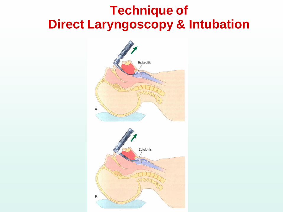
Technique of Direct Laryngoscopy & Intubation

How is
the best
laryngoscopic
view
achieved?

“Sniffing Position”


01/11/56 Bencharatana Yokubol 28
Cormack - Lehanegrading system
Laryngoscopic view ( LV classification )
Grade I Grade IIIGrade II Grade IV

Oral endotracheal tube size guideline
AgeInt diameter
(mm)
Length
(cm)
Full term 3.5 12
Child 4 + Age/4 12+ Age/2
Adult
Female
Male
7.0 – 7.5
7.5 – 8.0
20-23
21-24

Preparation for Rigid Laryngoscopy
● Suction
● Airway
● Laryngoscope
● Endotracheal tube (ET or ETT)
● Stylet
● Anesthetic machine / Breathing system / Self-
inflating bag
● Monitoring : Pulse oximeter, Capnograph, ECG
● Local anesthetics infiltration / spray

Stylet

Signs of Tracheal Intubation• Respiratory gas moisture disappearing on
inhalation and reappearing on exhalation
• Chest rise & fall
• No gastric distention
• ICS filling out during inspiration
• Reservoir bag having the appropriate
compliance

Signs of Tracheal Intubation
● Breath sounds over chest wall
● No breath sounds over stomach
● Hearing air exit from ET when chest
is compressed
● Large spontaneous exhaled
tidal volumes

Signs of Tracheal Intubation“More reliable signs”
● CO2 excretion waveform
● Rapid expansion of a tracheal indicator bulb

Signs of Tracheal Intubation“Most reliable signs”
• ET visualized between vocal cords
• Fiberoptic visualization of cartilaginous rings of the trachea and tracheal carina

Techniques for routine intubation
● (Preoxygenation)
● Administration of induction agent
● Adequate mask ventilation
● Administration of neuromuscular
(NM) blocking agent
● Continue mask ventilation
● Intubation
● Confirm ET in trachea

Techniques for “rapid-sequence” (crash) induction and intubation
● Preoxygenation 5 min (or 8 deep breaths)
● Administration of induction and NM
blocking agents
● Cricoid pressure (Sellick’s maneuver)
● “No” mask ventilation
● Intubation
● Check ET in trachea
● Release cricoid pressure


Cricoid Pressure (Sellick’s maneuver)

Complications:
• During laryngoscopy & intubation
• While tube in place
• Following extubation

During laryngoscopy & intubation● Malpositions
- Esophageal intubation
- Bronchial intubation
● Trauma
- Dental damage
- Lip, tongue, pharyngeal, laryngeal, tracheobronchial injuries
- Dislocated mandible
- Retropharyngeal dissection
- Cervical spine injury
Complications:
● Aspiration

● Physiologic reflexes
– HT, arrthymia
– Intracranial HT
– Intraocular HT
– Bronchospasm
● Tube malfunction
– Cuff perforation
During laryngoscopy & intubation
Complication:

Oropharyngeal airway Nasopharyngeal airway

● Malpositioning
– Unintentional extubation
– Endobronchial intubation
– ET cuff malposition
● Airway trauma
– Mucosal inflammation
– Excruciation of nose
● Tube malfunction
– Ignition
– Obstruction / Kinking
● Aspiration
While tube in place
Complications:

● Airway trauma
– Edema, Stenosis
– Hoarseness / Sorethroat
– Laryngeal trauma / malfunction
● Physiologic reflexes
● Laryngospasm
● Aspiration
Following Extubation
Complications:

● Techniques for Difficult Ventilation
● Techniques for Difficult Intubation
Techniques for Difficult Airway Management

Techniques for Difficult Ventilation
● Oral/Nasal Airway Insertion
● Two-person mask ventilation
● Laryngeal mask airway (LMA)
● Esophageal-tracheal combitube
● Surgical airway access

Two person Mask Ventilation
Three-hand jaw-thrust/mask seal
Secondperson
First person
Two-hand jaw-thrust/mask seal
First person
Secondperson

Laryngeal Mask Airway


The Esophageal-tracheal combitube

Surgical airway management:Percutaneous cricothyrotomy

Jet Ventilation

Techniques for Difficult Intubation
● Stylet
● Intubating stylet-tube changer
● Alternative laryngoscope (e.g. McCoy,
Bullard, Intubating LMA,etc)
● Awake intubation
● Blind intubation (oral or nasal)
● Fiberoptic intubation
● Illuminating stylet / Light wand
● Retrograde intubation
● Surgical airway access
Techniques for Difficult Intubation

Stylet

Bullard laryngoscope


Illumination Stylet
Light wand

Retrograde
intubation


Practice Guidelines for Management of the Difficult Airway
An update report by the American
Society of Anesthesiologist
Anesthesiology
Feb 2013


1. Assess the likelihood and clinical impact of basic management problems:

2. Actively pursue opportunities to deliver supplemental oxygen throughout the process of difficult airway mangement

3. Consider the relative merits and feasibility of basic management choices:
•Awake intubation v.s. intubation after induction of general anesthesia
•Non-invasive technique v.s. invasive technique for the initial approach to intubation
•Video-assisted laryngoscope as an initial approach to intubation
•Preservation v.s. ablation of spontaneous ventilation

4. Develop primary and alternative strategies:

4. Develop primary and alternative strategies:

Initial Intubation Attempts UNSUCCESSFUL
(Call for Help)

Supragloltic airway (SGA)
• Laryngeal mask airway (LMA)
• Intubation LMA (ILMA)
• Laryngeal tube

• LMA proseal
• LMA (Fastrach)
• LMA CTrach
• C MAC video laryngoscope
• Airtraq laryngoscope
• Glidescope intubation
• Lightwand intubation
• Fiberoptic intubation
ADVANCED AIRWAY AND INTUBATION TECHNIQUES

Thank you
and Good Luck