alginate oligosaccharides inhibit fungal cell...
TRANSCRIPT

Alginate Oligosaccharides Inhibit Fungal Cell Growthand Potentiate the Activity of Antifungals againstCandida and Aspergillus sppAnne Tøndervik1, Havard Sletta1*, Geir Klinkenberg1, Charlotte Emanuel2, Lydia C. Powell2,
Manon F. Pritchard2, Saira Khan2, Kieron M. Craine2, Edvar Onsøyen3, Phil D. Rye3, Chris Wright4,
David W. Thomas2, Katja E. Hill2
1 Department of Bioprocess Technology, SINTEF Materials and Chemistry, N-7465 Trondheim, Norway, 2 Advanced Therapies Group, Tissue Engineering and Reparative
Dentistry, Cardiff University School of Dentistry, Cardiff, CF14 4XY, United Kingdom, 3 AlgiPharma AS, Industriveien 33, N-1337 Sandvika, Norway, 4 Centre for NanoHealth,
Systems and Process Engineering Centre, College of Engineering, Swansea University, Swansea, SA2 8PP, United Kingdom
Abstract
The oligosaccharide OligoG, an alginate derived from seaweed, has been shown to have anti-bacterial and anti-biofilmproperties and potentiates the activity of selected antibiotics against multi-drug resistant bacteria. The ability of OligoG toperturb fungal growth and potentiate conventional antifungal agents was evaluated using a range of pathogenic fungalstrains. Candida (n = 11) and Aspergillus (n = 3) spp. were tested using germ tube assays, LIVE/DEAD staining, scanningelectron microscopy (SEM), atomic force microscopy (AFM) and high-throughput minimum inhibition concentration assays(MICs). In general, the strains tested showed a significant dose-dependent reduction in cell growth at $6% OligoG asmeasured by optical density (OD600; P,0.05). OligoG (.0.5%) also showed a significant inhibitory effect on hyphal growthin germ tube assays, although strain-dependent variations in efficacy were observed (P,0.05). SEM and AFM both showedthat OligoG ($2%) markedly disrupted fungal biofilm formation, both alone, and in combination with fluconazole. Cellsurface roughness was also significantly increased by the combination treatment (P,0.001). High-throughput robotic MICscreening demonstrated the potentiating effects of OligoG (2, 6, 10%) with nystatin, amphotericin B, fluconazole,miconazole, voriconazole or terbinafine with the test strains. Potentiating effects were observed for the Aspergillus strainswith all six antifungal agents, with an up to 16-fold (nystatin) reduction in MIC. Similarly, all the Candida spp. showedpotentiation with nystatin (up to 16-fold) and fluconazole (up to 8-fold). These findings demonstrate the antifungalproperties of OligoG and suggest a potential role in the management of fungal infections and possible reduction ofantifungal toxicity.
Citation: Tøndervik A, Sletta H, Klinkenberg G, Emanuel C, Powell LC, et al. (2014) Alginate Oligosaccharides Inhibit Fungal Cell Growth and Potentiate theActivity of Antifungals against Candida and Aspergillus spp. PLoS ONE 9(11): e112518. doi:10.1371/journal.pone.0112518
Editor: Alix T. Coste, Institute of Microbiology, Switzerland
Received May 12, 2014; Accepted October 3, 2014; Published November 19, 2014
Copyright: � 2014 Tøndervik et al. This is an open-access article distributed under the terms of the Creative Commons Attribution License, which permitsunrestricted use, distribution, and reproduction in any medium, provided the original author and source are credited.
Data Availability: The authors confirm that all data underlying the findings are fully available without restriction. All relevant data are within the paper and itsSupporting Information files.
Funding: This work was supported by AlgiPharma AS. The funding organization was directly involved in the study design, decision to publish and preparation ofthe manuscript.
Competing Interests: EO and PDR are employees and shareholders at AlgiPharma AS. This does not alter the authors’ adherence to PLOS ONE policies onsharing data and materials.
* Email: [email protected]
Introduction
The increase in invasive fungal infections over the last decade is
a growing cause for concern as they are associated with high
mortality rates [1]. The likely reasons for this increase include
greater use of immunosuppressive therapy, selective pressure
through broad-spectrum antibiotic use, medical and prosthetic
device-related infections [2], as well as the spread of HIV/AIDS
and an increasingly ageing population [1,3]. Candida albicans and
Aspergillus fumigatus are the most common species associated
with fungal invasive diseases, but the importance of other Candidaspecies and filamentous fungi like Scedosporium and Fusarium [4]
is becoming apparent. In addition to C. albicans, C. glabrata, C.parapsilosis, C. tropicalis and C. krusei, together account for 92%
of the detected cases of candidemia [5]. In aspergillosis, after A.fumigatus, A. flavus is the most common causative species [6].
Amphotericin B has for many years been the first line of
treatment for fungal infections despite its nephrotoxic side effects.
Amphotericin B and polyene resistance is occasionally reported,
but until now, has had limited clinical importance [4]. However,
azole antifungals such as fluconazole, itraconazole and voricona-
zole, although effective antifungal agents, have shown develop-
ment of resistance [7]; it being well-recognised among Aspergillusspecies in particular. Echinocandins, a newer class of antifungals
which are generally well tolerated, are considered to be a potential
first line treatment for many patients due to their activity against
azole-resistant strains [8,9]. In general, the clinical value of many
common antifungal therapeutics is limited by toxicity, and
considerable efforts are being made to reduce this toxicity in
PLOS ONE | www.plosone.org 1 November 2014 | Volume 9 | Issue 11 | e112518
some of the conventional antifungal drugs. For example, less toxic
lipid formulations of amphotericin B have now been produced and
approved for clinical use [10].
Another strategy for enhancing efficacy and reducing drug
toxicity is the use of combined therapies. Several in vitro studies
performed using combinations of antifungals have shown syner-
gistic effects, demonstrating the potential of this strategy
[11,12,13,14]. An alternative approach has been to potentiate
the effect of antifungals through the use of different non-antibiotic
compounds, such as peptides [15,16], ion-chelators [17], essential
oils [18,19] or secondary metabolites of plant and microbial origin
[20,21,22]. Combining antifungals with antibiotics such as colistin
(an antibiotic in current use with significant toxicity issues) has also
recently proved successful [23]. To date, however, none of these
products have progressed as far as clinical use.
The novel alginate oligomer OligoG is an oligosaccharide
enriched from sodium alginate polysaccharides and composed
predominantly of a-L-guluronic acid (.96%), possessing only a
small percentage of the b-D-mannuronic acid isomer (,4%).
OligoG has recently been shown to perturb multi-drug resistant
bacteria by influencing biofilm formation and reducing resistance
to antibiotic treatment [24]. In the present study, we tested this
alginate oligomer in combination with conventional antifungal
treatment (selected polyenes, azoles and allylamines) against a
panel of Aspergillus and Candida strains, to determine if this
potentiation was evident against fungal pathogens.
Materials and Methods
Fungal strains, alginate oligomers and antibioticsThe strains used in this study represent both culture collection
strains and clinical isolates and are shown in Table 1. The alginate
oligomer, OligoG, used in the study was prepared, purified and
characterized as described previously [24] and the antifungals
employed were pharmaceutical grade (Sigma-Aldrich).
Preparation of freezer-stock cultures and growthcharacterization of the test strains
Candida strains were grown at 34uC for 48 h on Yeast Mold-
agar (YM-agar, Difco). One to three fungal colonies from the
plates were grown in 6 mL YM-broth at 34uC for 14 h before
freezing in 6% glycerol at 280uC. Aspergillus strains were grown
at 30uC for 96 h on YM-agar. Spores and aerial mycelia were then
cut from the agar, suspended in 1 mL YM-broth and dispersed
with glass beads (1 mm) in a mini bead-beater (2 min). Glycerol
was added to a final concentration of 10% and the suspension was
frozen at 280uC. Each batch of frozen stock culture was then
characterized in separate growth experiments (results not shown)
using Mueller-Hinton broth (MH, Lab114, LabM) and RPMI
with 0.2% glucose (RPMI-1640, Sigma-Aldrich) to determine the
minimum amount of inoculum giving satisfactory growth after
48 h under conditions relevant for the determination of minimum
inhibitory concentration assays (MICs) as described below. This
inoculum procedure was used to reduce inter-experimental
variation in the bioassays. For characterization of growth in the
two different media, strains were grown with static incubation in
384-well microplates (30 mL per well, Nunc 242757) and optical
density (OD600) measured between 24–96 h using a Beckman
Coulter Paradigm microplate reader. Prior to OD measurements,
the microplates were shaken at 1800 rpm (2.5 mm amplitude) for
2 mins.
Susceptibility testing by robotic minimum inhibitoryconcentration (MIC) assay
The ability of alginate oligomers to potentiate the activity of
selected antifungals against the Candida and Aspergillus test
strains was studied by susceptibility testing using high-throughput
robotic screening (HTS) as described previously [24]. Alginate
oligomers were dissolved in medium to 1.25 times the desired
assay concentrations (2, 6 and 10%). Selected antifungals from
different classes included: nystatin, amphotericin B (polyenes),
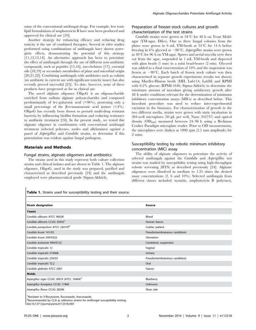
Table 1. Strains used for susceptibility testing and their source.
Strain designation Source
Yeasts
Candida albicans ATCC 90028 Blood
Candida albicans CCUG 393431 Human faeces
Candida parapsilosis ATCC 22019T2 Coeliac patient
Candida krusei 141/03 Pseudomembraneous candidosis
Candida krusei 249/03(2) Ulceration
Candida lusitaniae 994/01(2) Candidosis suspension
Candida tropicalis 12 Vaginal
Candida tropicalis 519468 Urinary
Candida tropicalis 250/03 Pseudomembraneous candidosis
Candida tropicalis T2.2 Oral
Candida glabrata ATCC 2001 Faeces
Molds
Aspergillus niger CCUG 18919 (ATCC 16404)2 Blueberry
Aspergillus fumigatus CCUG 17460 Unknown
Aspergillus flavus CCUG 28296 Shoe sole
1Resistant to 5-flucytosine, fluconazole, itraconazole.2Recommended by CLSI as reference strains for antifungal susceptibility testing.doi:10.1371/journal.pone.0112518.t001
Alginate Oligosaccharides Potentiate Antifungal Activity
PLOS ONE | www.plosone.org 2 November 2014 | Volume 9 | Issue 11 | e112518
fluconazole, miconazole and voriconazole (azoles) and the topical
antifungal, terbinafine (allylamine). The Candida strains were
tested against nystatin, fluconazole and terbinafine, (one from each
class) whilst the Aspergillus strains were tested against all six
antifungals. Two-fold serial dilutions of antifungals were made in
medium with different concentrations of OligoG, and the solutions
were placed in four parallel wells in 384-well micro plates (30 mL
per well). Serial dilutions were performed with a Tecan Genesis
RSP 200 liquid handling workstation equipped with an 8-channel
pipetting tool, using sterile disposable 200 mL barrier tips. Into
each well in the 384-well assay plates 7.5 mL, of the medium
inoculated with frozen stock culture of the relevant strains
(described above) was added. The microtitre plates were placed
in plastic bags and incubated in a Thermo Cytomat 2 450 S
robotic incubator without shaking at 34uC. Optical density
(OD600) was measured at specific time points between 24–96 h.
The microplates were shaken at 1800 rpm (2.5 mm amplitude) for
2 mins prior to taking the absorbance readings. The readings
made at 48 h were used for all strains except for the fluconazole
test with C. tropicalis 519468 and T2.2. For these strains OD600
readings after incubation for 36 h were used, since the readings
after 48 h were inconclusive.
Synergy for the antifungal drug/OligoG MIC combinations
tested was determined from the Fractional Inhibitory Concentra-
tion Index (FICI; [25]) where FICI#0.5 is indicative of synergy.
The FICI was calculated from the MIC of the drug in
combination, divided by the MIC of the drug acting alone.
Germ Tube AssayOvernight cultures of C. albicans CCUG 39343, ATCC 90028,
C. tropicalis 519468 and C. glabrata ATCC 2001 were prepared
in Sabouraud-dextrose broth (SAB, Oxoid) and incubated at
37uC. C. glabrata as a non-hyphae producer was the negative
control. One mL of culture was then washed twice with phosphate
buffered saline (PBS) and the resulting pellet resuspended in
500 mL PBS, to obtain approximately 56106 cells/mL. Serial
dilutions (100 mL) were spiral plated onto Sabouraud-dextrose
agar (SAA; Oxoid) to calculate colony forming units per mL
(CFU/mL). Donor horse serum (500 mL; TCS Biosciences Ltd)
supplemented with 0, 0.2, 0.5, 2, 6 or 10% OligoG was inoculated
with 50 mL of the washed candidal suspension, and incubated for
2 h at 37uC. Following incubation, 100 mL of the candidal/serum
suspension was serially diluted and spiral plated onto SAA to
calculate post incubation CFU/mL. The remaining suspension
was washed with 0.9% NaCl (x3) to remove OligoG, and
resuspended in 200 mL PBS. The percentage number of cells
with hyphal growth was calculated using a Neubauer haemocy-
tometer under phase contrast microscopy. Light microscopy
images of the different hyphal growth were also taken.
LIVE-DEAD Staining of Candida biofilmsAn overnight culture of C. tropicalis 519468 was grown in
RPMI (Invitrogen). Biofilms were grown in 8-well chamber slides
(BD Falcon), using 35 mL of the overnight culture per well (107
cells), followed by the addition of either 350 mL of RPMI or
350 mL of 2% OligoG solubilized in RPMI. These were grown for
24 h at 37uC, with gentle rocking. The supernatant was then
removed and the biofilm stained with the LIVE/DEAD BacLight
Bacterial Viability Kit (Invitrogen, Paisley, UK) containing SYTO
9 dye and propidium iodide, prior to being imaged under a
fluorescent microscope (Olympus Provis AX70).
Scanning Electron Microscopy ImagingC. tropicalis 519468 was grown overnight in RPMI, and the
culture was diluted in the same medium to attain 0.46107/mL
cells. Biofilms were then formed on thermanox slides (Agar
Scientific) in the bottom of 12-well Cellstar plates (Greiner Bio-
One, Stonehouse, UK) using 1 mL of culture incubated with
gentle rocking at 37uC for 4 h. Each well was then washed (x3)
with pre-warmed RPMI. Then, 1 mL of either 2, 1, 0.5 mg/mL
fluconazole and/or 2% OligoG solubilized in RPMI was added to
each well and further incubated with gentle rocking at 37uC for
24 h. The supernatant was then removed and each well immersed
in 2.5% glutaraldehyde for 1.5 h and then washed thoroughly with
distilled water. Biofilms were then frozen (220uC) after addition of
1 mL distilled water to each well. Once frozen, the well plates
were then freeze-dried for 24 h. The thermanox slides were then
removed from the well plates and imaged using an Hitachi S4800
scanning electron microscope.
Atomic Force Microscopy ImagingCandida cultures for atomic force microscopy (AFM) imaging
were prepared following a similar protocol to that of Murillo et al.
[26]. C. tropicalis 519468 was grown as a shaken overnight culture
in SAB. The overnight culture (10 mL) was centrifuged at
2,1006g for 10 mins and resuspended in fresh SAB at 37uC to
attain 107/mL cells (OD520). Biofilms were formed in polystyrene
petri dishes (60 mm615 mm) using 6 mL of 107 cells in SAB, and
incubated with gentle rocking at 37uC for 30 mins. The biofilms
were then thoroughly washed (x3) in pre-warmed media and 6 mL
of fresh media and/or 2% OligoG and fluconazole (1 mg/L)
added followed by incubation with gentle rocking at 37uC for a
further 90 mins. The use of this shorter biofilm growing time was
necessary to optimize AFM imaging. The biofilms were rinsed
twice with de-ionised water and dried at room temperature for 1 h
before imaging with a Dimension 3100 AFM instrument (Bruker)
using a scan speed of 0.4 Hz. Mean surface roughness (Ra)
measurements were used to investigate the impact of the cell
treatments on the morphology of the fungal cell wall by measuring
a 1 mm2 area at the centre of each fungal cell observed within
50 mm2 tapping mode images.
Statistical AnalysisStatistical analysis was performed using GraphPad Prism3
(GraphPad software Inc, California, USA). A paired T-test with
95% confidence intervals was used to test the significance in the
differences in growth at various OligoG-concentrations and in the
mean number of cells producing hyphae following incubation with
OligoG. Where the rules of a paired T-test were not obeyed, a
Mann-Whitney test was performed. The non-parametric Kruskal-
Wallis one-way analysis of variance was used for analysis of the
surface roughness data. P,0.05 was considered significant.
Results
Growth characteristics of Candida and Aspergillus sppInitial studies were performed to characterize the growth of
Candida and Aspergillus strains in 384-well plates in RPMI and
MH. According to standardized protocols these media are
recommended for antibiotic susceptibility testing in yeast and
bacteria respectively [27,28]. For all strains tested, the cell mass
obtained after incubation for 24–48 h (which is the recommended
duration of incubation before MIC determination) in RPMI was
lower than that observed in MH, as shown for C. tropicalis 519468
and A. flavus (Figure S1). The primary objective of the present
study was to explore the effects of OligoG on fungal growth, either
Alginate Oligosaccharides Potentiate Antifungal Activity
PLOS ONE | www.plosone.org 3 November 2014 | Volume 9 | Issue 11 | e112518
alone or in combination with antifungal agents. Even though MH
is used mostly for bacterial susceptibility testing, we chose to use
this broth for our studies since all our test strains exhibited better
growth characteristics under the relevant cultivation conditions in
MH. However, parallel experiments were performed in RPMI
with selected strains to ensure that the effects observed were not
exclusive to MH (Figure S2). Aspergillus strains reached a higher
cell density (1.3–1.7 OD600) than the Candida strains (0.5–0.8
OD600) in MH.
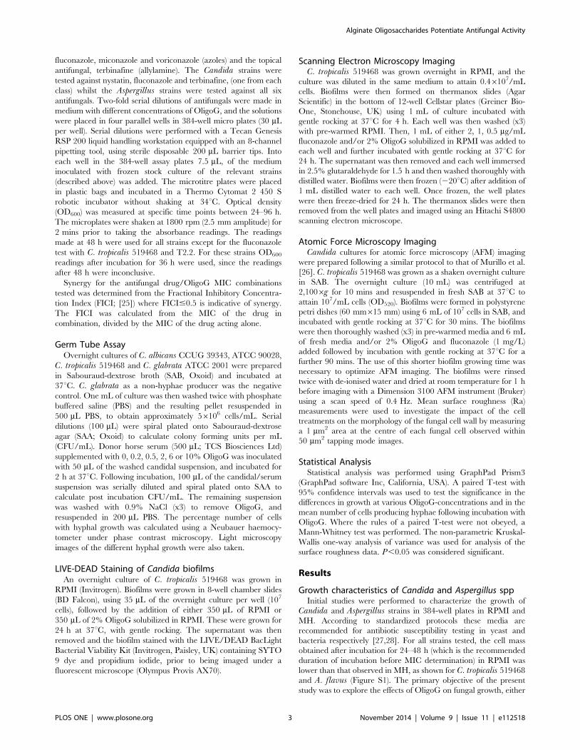
Addition of OligoG to MH reduced the growth of all the fungal
strains tested in a concentration dependent manner (Figure 1).
Hence, the effect was most pronounced with 10% OligoG leading
to a 15–50% reduction in cell density after 48 h incubation.
Growth of selected strains of Candida (n = 5) and Aspergillus(n = 3) in RPMI also showed a reduction in OD600 of 8–35% with
10% OligoG (Figure S2), showing that this effect was not media-
dependent. Analysis of the growth kinetics showed that the specific
growth rate (m) in MH was only slightly reduced by the addition of
OligoG. Typically for A. flavus and C. tropicalis 519468 growth
rates of m = 0.22/t (without OligoG) and m = 0.19–0.2/t (with 10%
OligoG) were observed.
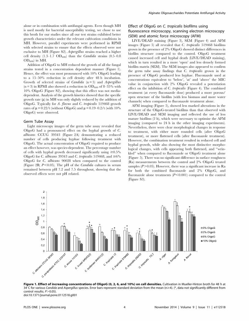
Germ Tube AssayLight microscopy images of the germ tube assay revealed that
OligoG had a pronounced effect on the hyphal growth of C.albicans CCUG 39343 (Figure 2A) demonstrating a reduced
number of cells producing hyphae following treatment with
OligoG. The actual concentration of OligoG required to produce
an effect however, was species-dependent. The percentage number
of cells with hyphal growth decreased significantly using $0.5%
OligoG for C. albicans 39343 and C. tropicalis 519468, and $6%
OligoG for C. albicans 90028 when compared to the control
(Figure 2B; P,0.05). The pH of the Candida cultures in serum
remained between pH 7.2 and 7.5 throughout, showing that the
observed effects were not pH related.
Effect of OligoG on C. tropicalis biofilms usingfluorescence microscopy, scanning electron microscopy(SEM) and atomic force microscopy (AFM)
LIVE/DEAD staining (Figure 3), SEM (Figure 4) and AFM
images (Figure 5) all revealed that C. tropicalis 519468 biofilms
grown in the presence of 2% OligoG showed distinct differences in
biofilm structure compared to the control. OligoG treatment
caused increased cell and hyphal death (LIVE/DEAD staining),
which in turn resulted in a more ‘open’ and less densely formed
biofilm matrix (SEM). The SEM images also appeared to confirm
the germ tube assay findings that C. tropicalis grown in the
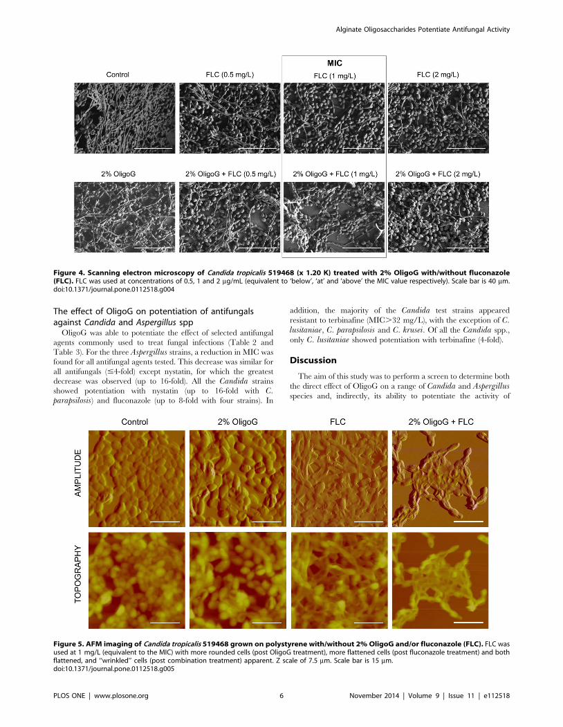
presence of OligoG produced less hyphae. Fluconazole used at
concentrations equivalent to ‘below’, ‘at’ and ‘above’ the MIC
value in conjunction with 2% OligoG revealed a potentiation
effect on the inhibition of C. tropicalis (Figure 4). The combined
treatment (at every fluconazole dose) produced a more porous/
open structure of the biofilm (with less biomass and more water
channels) when compared to fluconazole treatment alone.
AFM imaging (Figure 5), showed less marked alterations in the
structure of the OligoG-treated biofilms than that observed with
LIVE/DEAD and SEM imaging and reflected the use of less
mature biofilms (2 h), which were necessary to optimise the AFM
imaging (compared to 24 h in the other imaging experiments).
Nevertheless, there were clear morphological changes in response
to treatment, with either more rounded cells (after OligoG
treatment), or more flattened cells (after fluconazole treatment).
However, the combination treatment resulted in reduced cell and
hyphal growth, while also showing the most distinctive morpho-
logical changes, with cells appearing both flattened, and ‘‘wrin-
kled’’ when compared to fluconazole or OligoG treatment alone
(Figure 5). There was no significant difference in surface roughness
(Ra) measurements between the control and 2% OligoG treated
samples (P.0.05). However, there was a significant increase in Ra
for both the combined fluconazole and 2% OligoG, and
fluconazole alone treatments (P,0.001) compared to the control
(Figure S3).
Figure 1. Effect of increasing concentrations of OligoG (0, 2, 6, and 10%) on cell densities. Cultivation in Mueller-Hinton broth for 48 h at34uC for various Candida and Aspergillus species. Error bars represent standard deviation from the mean (n$4). (*, data not significantly different fromcontrol results; P.0.05).doi:10.1371/journal.pone.0112518.g001
Alginate Oligosaccharides Potentiate Antifungal Activity
PLOS ONE | www.plosone.org 4 November 2014 | Volume 9 | Issue 11 | e112518
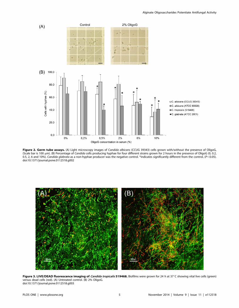
Figure 2. Germ tube assays. (A) Light microscopy images of Candida albicans (CCUG 39343) cells grown with/without the presence of OligoG,(Scale bar is 100 mm). (B) Percentage of Candida cells producing hyphae for four different strains grown for 2 hours in the presence of OligoG (0, 0.2,0.5, 2, 6 and 10%). Candida glabrata as a non-hyphae producer was the negative control. *indicates significantly different from the control, (P,0.05).doi:10.1371/journal.pone.0112518.g002
Figure 3. LIVE/DEAD fluorescence imaging of Candida tropicalis 519468. Biofilms were grown for 24 h at 37uC showing vital live cells (green)versus dead cells (red). (A) Untreated control. (B) 2% OligoG.doi:10.1371/journal.pone.0112518.g003
Alginate Oligosaccharides Potentiate Antifungal Activity
PLOS ONE | www.plosone.org 5 November 2014 | Volume 9 | Issue 11 | e112518
The effect of OligoG on potentiation of antifungalsagainst Candida and Aspergillus spp
OligoG was able to potentiate the effect of selected antifungal
agents commonly used to treat fungal infections (Table 2 and
Table 3). For the three Aspergillus strains, a reduction in MIC was
found for all antifungal agents tested. This decrease was similar for
all antifungals (#4-fold) except nystatin, for which the greatest
decrease was observed (up to 16-fold). All the Candida strains
showed potentiation with nystatin (up to 16-fold with C.parapsilosis) and fluconazole (up to 8-fold with four strains). In
addition, the majority of the Candida test strains appeared
resistant to terbinafine (MIC.32 mg/L), with the exception of C.lusitaniae, C. parapsilosis and C. krusei. Of all the Candida spp.,
only C. lusitaniae showed potentiation with terbinafine (4-fold).
Discussion
The aim of this study was to perform a screen to determine both
the direct effect of OligoG on a range of Candida and Aspergillusspecies and, indirectly, its ability to potentiate the activity of
Figure 4. Scanning electron microscopy of Candida tropicalis 519468 (x 1.20 K) treated with 2% OligoG with/without fluconazole(FLC). FLC was used at concentrations of 0.5, 1 and 2 mg/mL (equivalent to ‘below’, ‘at’ and ‘above’ the MIC value respectively). Scale bar is 40 mm.doi:10.1371/journal.pone.0112518.g004
Figure 5. AFM imaging of Candida tropicalis 519468 grown on polystyrene with/without 2% OligoG and/or fluconazole (FLC). FLC wasused at 1 mg/L (equivalent to the MIC) with more rounded cells (post OligoG treatment), more flattened cells (post fluconazole treatment) and bothflattened, and ‘‘wrinkled’’ cells (post combination treatment) apparent. Z scale of 7.5 mm. Scale bar is 15 mm.doi:10.1371/journal.pone.0112518.g005
Alginate Oligosaccharides Potentiate Antifungal Activity
PLOS ONE | www.plosone.org 6 November 2014 | Volume 9 | Issue 11 | e112518
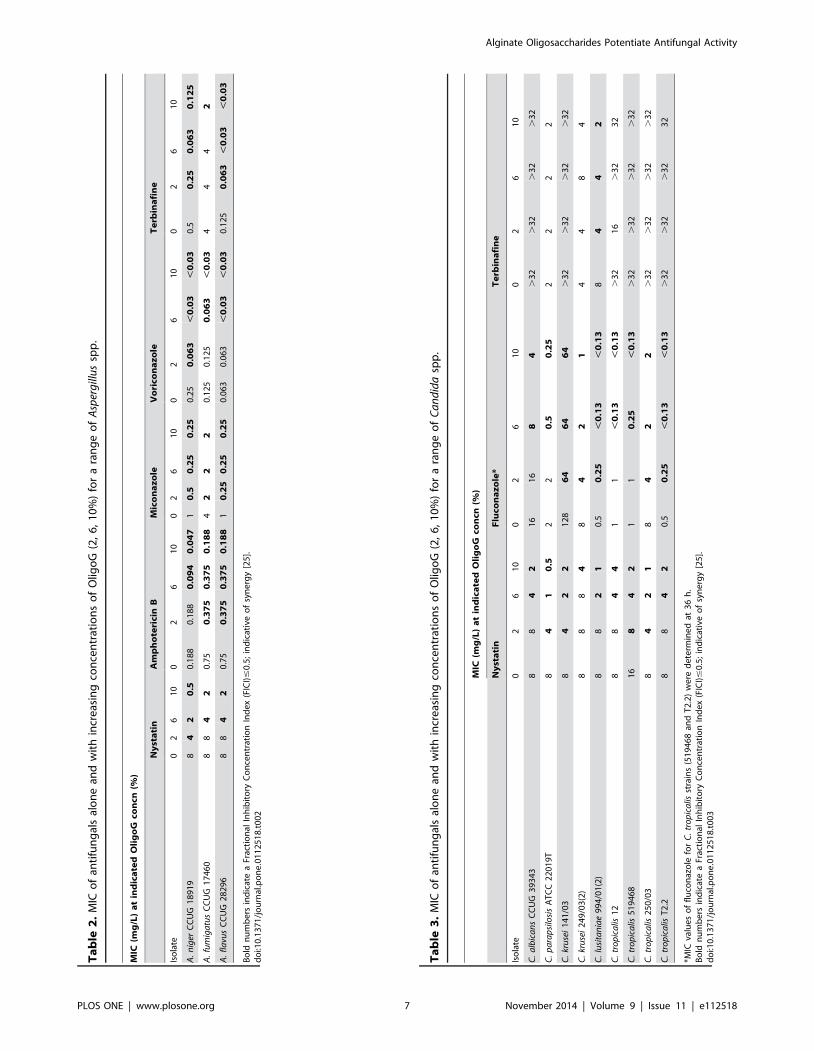
Ta
ble
2.
MIC
of
anti
fun
gal
sal
on
ean
dw
ith
incr
eas
ing
con
cen
trat
ion
so
fO
ligo
G(2
,6
,1
0%
)fo
ra
ran
ge
of
Asp
erg
illu
ssp
p.
MIC
(mg
/L)
at
ind
ica
ted
Oli
go
Gco
ncn
(%)
Ny
sta
tin
Am
ph
ote
rici
nB
Mic
on
az
ole
Vo
rico
na
zo
leT
erb
ina
fin
e
Iso
late
02
61
00
26
10
02
61
00
26
10
02
61
0
A.
nig
erC
CU
G1
89
19
84
20
.50
.18
80
.18
80
.09
40
.04
71
0.5
0.2
50
.25
0.2
50
.06
3,
0.0
3,
0.0
30
.50
.25
0.0
63
0.1
25
A.
fum
iga
tus
CC
UG
17
46
08
84
20
.75
0.3
75
0.3
75
0.1
88
42
22
0.1
25
0.1
25
0.0
63
,0
.03
44
42
A.
fla
vus
CC
UG
28
29
68
84
20
.75
0.3
75
0.3
75
0.1
88
10
.25
0.2
50
.25
0.0
63
0.0
63
,0
.03
,0
.03
0.1
25
0.0
63
,0
.03
,0
.03
Bo
ldn
um
be
rsin
dic
ate
aFr
acti
on
alIn
hib
ito
ryC
on
cen
trat
ion
Ind
ex
(FIC
I)#
0.5
;in
dic
ativ
eo
fsy
ne
rgy
[25
].d
oi:1
0.1
37
1/j
ou
rnal
.po
ne
.01
12
51
8.t
00
2
Ta
ble
3.
MIC
of
anti
fun
gal
sal
on
ean
dw
ith
incr
eas
ing
con
cen
trat
ion
so
fO
ligo
G(2
,6
,1
0%
)fo
ra
ran
ge
of
Ca
nd
ida
spp
.
MIC
(mg
/L)
at
ind
ica
ted
Oli
go
Gco
ncn
(%)
Ny
sta
tin
Flu
con
az
ole
*T
erb
ina
fin
e
Iso
late
02
61
00
26
10
02
61
0
C.
alb
ica
ns
CC
UG
39
34
38
84
21
61
68
4.
32
.3
2.
32
.3
2
C.
pa
rap
silo
sis
AT
CC
22
01
9T
84
10
.52
20
.50
.25
22
22
C.
kru
sei
14
1/0
38
42
21
28
64
64
64
.3
2.
32
.3
2.
32
C.
kru
sei
24
9/0
3(2
)8
88
48
42
14
48
4
C.
lusi
tan
iae
99
4/0
1(2
)8
82
10
.50
.25
,0
.13
,0
.13
84
42
C.
tro
pic
alis
12
88
44
11
,0
.13
,0
.13
.3
21
6.
32
32
C.
tro
pic
alis
51
94
68
16
84
21
10
.25
,0
.13
.3
2.
32
.3
2.
32
C.
tro
pic
alis
25
0/0
38
42
18
42
2.
32
.3
2.
32
.3
2
C.
tro
pic
alis
T2
.28
84
20
.50
.25
,0
.13
,0
.13
.3
2.
32
.3
23
2
*MIC
valu
es
of
flu
con
azo
lefo
rC
.tr
op
ica
lisst
rain
s(5
19
46
8an
dT
2.2
)w
ere
de
term
ine
dat
36
h.
Bo
ldn
um
be
rsin
dic
ate
aFr
acti
on
alIn
hib
ito
ryC
on
cen
trat
ion
Ind
ex
(FIC
I)#
0.5
;in
dic
ativ
eo
fsy
ne
rgy
[25
].d
oi:1
0.1
37
1/j
ou
rnal
.po
ne
.01
12
51
8.t
00
3
Alginate Oligosaccharides Potentiate Antifungal Activity
PLOS ONE | www.plosone.org 7 November 2014 | Volume 9 | Issue 11 | e112518
conventional antifungal therapies. The potentiation effects of
OligoG were investigated with antifungal agents from different
chemical classes; with polyenes, azoles and allylamines being
employed in these assays. The antifungals which were studied
included those agents most commonly prescribed for human and
veterinary use. From a mechanistic and novel therapy perspective,
it was of interest to determine whether any potentiation effects of
antifungals seen with OligoG were similar for the entire selected
range of Candida and Aspergillus spp., whilst recognising that all
antifungals tested were not currently deemed clinically relevant for
the test strains that were utilized.
This in vitro study clearly demonstrates that the novel alginate
oligomer, OligoG, was able to modulate both fungal growth and
fungal biofilm formation. Moreover, OligoG was also shown to
potentiate the activity of a range of antifungal agents, giving up to
a 16-fold reduction in MIC values. Given the toxicity of many
antifungals, these findings represent a potential clinical benefit in
the management of fungal infections. This is supported by similar
observations for OligoG in potentiating the activity of antibiotics
against multi-drug resistant gram-negative bacteria [24].
Resistance to antifungal agents is commonplace. The majority
of the Candida strains tested in this study were resistant to
terbinafine. A large variation in susceptibility towards terbinafine
among different clinical Candida isolates has previously been
described [29] and resistance has been linked to overexpression of
the efflux pump genes CDR1 and CDR2 from the ABC
transporter gene family [30]. The polyenes such as amphotericin
B bind to ergosterol, the major sterol in the fungal cell membrane,
and form pores, thereby causing membrane damage. In contrast,
azoles target sterol biosynthesis by inhibiting the sterol 14 a-
demethylase, the product of the ERG11 gene. Resistance to
fluconazole has been associated with point mutations and
increased ERG11 expression or over-expression of efflux-pump
genes CDR1, CDR2 and MDR1 [31]. Importantly, resistance of
Candida spp. to classic triazole antifungal agents is increasing. The
MIC testing of the Candida strains demonstrated potentiation (up
to 8-fold) of antifungal azoles.
Several efflux pumps of A. fumigatus have also been identified.
However, their involvement in drug resistance is less well
established and little is known about how their expression is
regulated [31]. Recently, a deletion mutant for the transcription
factor SrbA in A. fumigatus was found to confer susceptibility to
fluconazole, which is not normally active against this pathogen
[32]. Unsurprisingly therefore, there was little effect of fluconazole
on the Aspergillus strains tested; two being resistant (MIC.128)
while the A. flavus strain showed only a low level of potentiation
(2-fold) with OligoG.
Although there is some controversy surrounding the pathoge-
nicity of Aspergillus spp. and the decision to treat infections caused
by this genera in some indications (i.e. lung infection in cystic
fibrosis patients) these arguments are strongly influenced by the
immune status of the host, clinical signs of infection and patient
microbiology [33]. Overuse of antifungal agents, antifungal
resistance and drug costs are all problems for these patients. The
ability of OligoG to reduce the MIC and thereby potentiate the
effect of antifungal agents in vivo, may provide wider treatment
options in the management of resistant fungal lung infections,
which are an increasing problem with triazoles.
In addition to the potentiation activity of OligoG, it clearly
showed antifungal activity when used alone. The modification of
biofilm structure, together with growth inhibition, and hyphae
effects were evident in all of the imaging techniques (conventional
light and fluorescence microscopy, SEM and AFM) employed in
this study. These methods demonstrated that the altered growth
characteristics were associated with distinct morphological chang-
es, which subsequently resulted in biofilm disruption, increased cell
death, and reduced hyphal formation. In previous studies,
inhibition of bacterial growth by OligoG was associated with
marked decreases in cell motility as seen in bacterial swarming
assays [24,34]. These earlier observations support the current
findings that OligoG has a direct effect on the invasive, hyphal
growth phase of Candida spp.
AFM is increasingly being used to study the effect of
antimicrobial agents on the cell surface [24,34]. Recently using
AFM, we showed strong binding of OligoG to the cell surface of
the pathogenic bacteria Pseudomonas aeruginosa, which remained
bound even after hydrodynamic shear [35]. Although similar
antimicrobial effects were observed with fungal pathogens, there
was no apparent similarity in the binding of OligoG to the fungal
cell wall. Nevertheless, morphological changes were clearly
evident. While these studies are not directly comparable (organ-
isms were in different growth states, planktonic versus biofilm), it
would suggest that different mechanisms of action are involved in
the antimicrobial effects observed in bacterial and fungal
pathogens. This is unsurprising given the contrasting differences
in cell wall structure and charge. The fungal cell wall is principally
composed of three main components: b-glucans (microfibrillar
polymers of glucose; ,60%), chitin (,2%) and mannoproteins
(,39%) [36]. As a means of combating potential cell lysis, the
composition of the cell wall can be altered using a ‘compensatory
mechanism’ which is activated in response to changes in their
immediate environment such as cell wall perturbing agents or
mutations. This allows the cell wall to be remodeled [37] and can
result in an increased level of chitin [38]. The response to such
cues can effectively direct changes in fungal growth, cell wall mass,
ultrastructure, elasticity and adhesion, enzyme production and
pathogenicity [39]. While it is not known whether OligoG induces
this kind of remodeling effect, it is a charged molecule and so could
influence, or be influenced by, molecular changes in the cell wall.
The observation that OligoG inhibits hyphae formation is
particularly relevant in light of recent co-infection studies with
Staphylococcus aureus and C. albicans [40]. Both organisms are
commonly isolated bloodstream pathogens, often found as co-
infecting agents. They are also both capable of forming biofilms,
and show increasing evidence of antimicrobial resistance, thereby
representing a significant and growing problem in the manage-
ment of polymicrobial infections. Specific binding of S. aureus to
the hyphae of co-cultured C. albicans (via Als3p binding) has been
shown to enable tissue infiltration and subsequent deep tissue
infection by S. aureus [40]. Undoubtedly, antimicrobials capable
of targeting both bacteria and yeasts, such as OligoG, have a
distinct advantage for such polymicrobial infections.
Although a precise molecular mechanism of action has yet to be
established, it is thought that the synergistic and antimicrobial
activity of OligoG in disrupting the biofilm matrix/architecture
could potentially permit: a) better access for antimicrobial agents
and/or host innate defenses to biofilm-embedded organisms and,
b) previously dormant (drug-tolerant) cells to move into a more
drug-susceptible phase of growth. The current in vitro observa-
tions would appear to support some of these hypotheses, but will
need to be tested with appropriate in vivo models.
Clearly further studies are needed to determine the precise
molecular mechanisms of OligoG in microbial infections.
Notwithstanding, the antifungal effects and potentiating activity
observed in this study represent considerable promise for the
clinical utility of OligoG in the treatment and management of
fungal infection and reduction of antifungal toxicity in clinical
practice. Phase I and IIa studies have demonstrated that OligoG is
Alginate Oligosaccharides Potentiate Antifungal Activity
PLOS ONE | www.plosone.org 8 November 2014 | Volume 9 | Issue 11 | e112518
safe for human use, in vivo testing having shown no intolerance to
inhaled doses of up to 540 mg/day (https://ClinicalTrials.gov,
NCT00970346 and NCT01465529; EudraCT, 2009-009330-33).
OligoG is currently being developed for topical wound applica-
tion, and studies are ongoing for the use of OligoG as an inhaled
therapy for cystic fibrosis.
Supporting Information
Figure S1 Growth of (A) C. tropicalis 519468 and (B) A. flavusat 34uC in Mueller-Hinton broth (open squares) and RPMI (solid
squares).
(TIF)
Figure S2 Effect of increasing concentrations of OligoG (0%,
2%, 6%, and 10%) on cell densities after cultivation in RPMI
broth for 48 h at 34uC for various Candida and Aspergillusspecies. Error bars represent standard deviation from the mean
(n$4). (*, data not significantly different from control results; P.
0.05).
(TIF)
Figure S3 Mean surface roughness expressed as (Ra) 6 standard
error. *indicates significantly different from the control. FLC,
fluconazole.
(TIF)
Acknowledgments
For Candida strains we thank Prof. David W. Williams (Cardiff University)
and Dr. Mariana Henriques (Minho University, Portugal).
Author Contributions
Conceived and designed the experiments: AT HS GK PDR EO DWT
KEH. Performed the experiments: AT HS GK MFP SK CE LCP MFP
KC. Analyzed the data: AT HS GK CW KC CE DWT KEH.
Contributed reagents/materials/analysis tools: AT HS GK PDR EO.
Contributed to the writing of the manuscript: AT HS DWT KEH PDR
EO LCP MFP CE CW.
References
1. Lass-Florl C (2009) The changing face of epidemiology of invasive fungal disease
in Europe. Mycoses 52: 197–205.
2. Ramage G, Martinez JP, Lopez-Ribot JL (2006) Candida biofilms on implanted
biomaterials: a clinically significant problem. FEMS Yeast Res 6: 979–986.
3. Silva S, Negri M, Henriques M, Oliveira R, Williams DW, et al. (2012) Candidaglabrata, Candida parapsilosis and Candida tropicalis: biology, epidemiology,
pathogenicity and antifungal resistance. FEMS Microbiol Rev 36: 288–305.
4. Alcazar-Fuoli L, Mellado E (2014) Current status of antifungal resistance and its
impact on clinical practice. Br J Haematol 166: 471–484.
5. Guinea J (2014) Global trends in the distribution of Candida species causing
candidemia. Clin Microbiol Infect 20: 5–10.
6. Walsh TJ, Gamaletsou MN (2013) Treatment of fungal disease in the setting of
neutropenia. Hematology-American Society of Hematology Education Pro-
gram: 423–427.
7. Aperis G, Myriounis N, Spanakis EK, Mylonakis E (2006) Developments in the
treatment of candidiasis: more choices and new challenges. Expert Opin Investig
Drugs 15: 1319–1336.
8. Emri T, Majoros L, Toth V, Pocsi I (2013) Echinocandins: production and
applications. Appl Microbiol Biotechnol 97: 3267–3284.
9. Simon J, Sun HY, Leong HN, Barez MYC, Huang PY, et al. (2013)
Echinocandins in invasive candidiasis. Mycoses 56: 601–609.
10. Bes DF, Sberna N, Rosanova MT (2012) Advantages and drawbacks of
amphotericin formulations in children: literature review. Arch Argent Pediatr
110: 46–51.
11. Kalkanci A, Dizbay M, Sari N, Yalcin B, Fidan I, et al. (2010) Fluconazole,
caspofungin, voriconazole in combination with amphotericin B. Cent Eur J Med
5: 194–197.
12. Yalcin B, Kalkanci A, Gurelik F, Fidan I, Kustimur S, et al. (2010) In vitrosynergistic effect of moxifloxacin and amphotericin B combination against
Candida strains. Mikrobiyol Bul 44: 65–70.
13. Alves IA, Bandeira LA, Mario DAN, Denardi LB, Neves LV, et al. (2012) Effects
of antifungal agents alone and in combination against Candida glabrata strains
susceptible or resistant to fluconazole. Mycopathologia 174: 215–221.
14. Veiga-Santos P, Barrias ES, Santos JFC, Moreira TLD, de Carvalho TMU, et
al. (2012) Effects of amiodarone and posaconazole on the growth and
ultrastructure of Trypanosoma cruzi. Int J Antimicrob Agents 40: 61–71.
15. Harris MR, Coote PJ (2010) Combination of caspofungin or anidulafungin with
antimicrobial peptides results in potent synergistic killing of Candida albicansand Candida glabrata in vitro. Int J Antimicrob Agents 35: 347–356.
16. Kamysz W, Nadolski P, Kedzia A, Cirioni O, Barchiesi F, et al. (2006) In vitro
activity of synthetic antimicrobial peptides against Candida. Pol J Microbiol 55:
303–307.
17. Zarember KA, Cruz AR, Huang CY, Gallin JI (2009) Antifungal activities of
natural and synthetic iron chelators alone and in combination with azole and
polyene antibiotics against Aspergillus fumigatus. Antimicrob Agents Che-
mother 53: 2654–2656.
18. Giordani R, Regli P, Kaloustian J, Mikail C, Abou L, et al. (2004) Antifungal
effect of various essential oils against Candida albicans. Potentiation of antifungal
action of amphotericin B by essential oil from Thymus vulgaris. Phytother Res
18: 990–995.
19. Giordani R, Regli P, Kaloustian J, Portugal H (2006) Potentiation of antifungal
activity of amphotericin B by essential oil from Cinnamomum cassia. Phytother
Res 20: 58–61.
20. Ali I, Sharma P, Suri KA, Satti NK, Dutt P, et al. (2011) In vitro antifungal
activities of amphotericin B in combination with acteoside, a phenylethanoid
glycoside from Colebrookea oppositifolia. J Med Microbiol 60: 1326–1336.
21. Veras HNH, dos Santos IJM, dos Santos ACB, Fernandes CN, Matias EFF, et
al. (2011) Comparative evaluation of antibiotic and antibiotic modifying activity
of quercetin and isoquercetin in vitro. Curr Top Nutraceutical Res 9: 25–29.
22. Fukuda T, Hasegawa Y, Sakabe Y, Tomoda H, Omura S (2008) Citrinamides,
new potentiators of antifungal miconazole activity, produced by Penicillium sp
FKI-1938. J Antibiot 61: 550–555.
23. Zeidler U, Bougnoux ME, Lupan A, Helynck O, Doyen A, et al. (2013) Synergy
of the antibiotic colistin with echinocandin antifungals in Candida species.
J Antimicrob Chemother 68: 1285–1296.
24. Khan S, Tøndervik A, Sletta H, Klinkenberg G, Emanuel C, et al. (2012)
Overcoming drug resistance with alginate oligosaccharides able to potentiate the
action of selected antibiotics. Antimicrob Agents Chemother 56: 5134–5141.
25. Odds FC (2003) Synergy, antagonism, and what the chequerboard puts between
them. J Antimicrob Chemother 52: 1–1.
26. Murillo LA, Newport G, Lan CY, Habelitz S, Dungan J, et al. (2005) Genome-
wide transcription profiling of the early phase of biofilm formation by Candidaalbicans. Eukaryot Cell 4: 1562–1573.
27. Arendrup MC, Cuenca-Estrella M, Lass-Florl C, Hope W, Eucast A (2012)
EUCAST technical note on the EUCAST definitive document EDef 7.2:
method for the determination of broth dilution minimum inhibitory concentra-
tions of antifungal agents for yeasts EDef 7.2 (EUCAST-AFST). Clinical
Microbiology and Infection 18: E246–E247.
28. Institute CaLS (2005) Performance standards for antimicrobial susceptibility
testing; 15th informational supplement, M100-S15. Wayne PA.
29. Ryder NS, Wagner S, Leitner I (1998) In vitro activities of terbinafine against
cutaneous isolates of Candida albicans and other pathogenic yeasts. Antimicrob
Agents Chemother 42: 1057–1061.
30. Odds FC (2009) In Candida albicans, resistance to flucytosine and terbinafine is
linked to MAT locus homozygosity and multilocus sequence typing clade 1.
FEMS Yeast Res 9: 1091–1101.
31. Morschhauser J (2010) Regulation of multidrug resistance in pathogenic fungi.
Fungal Genet Biol 47: 94–106.
32. Willger SD, Puttikamonkul S, Kim KH, Burritt JB, Grahl N, et al. (2008) A
sterol-regulatory element binding protein is required for cell polarity, hypoxia
adaptation, azole drug resistance, and virulence in Aspergillus fumigatus. Plos
Pathog 4.
33. De Pauw B, Walsh TJ, Donnelly JP, Stevens DA, Edwards JE, et al. (2008)
Revised definitions of invasive fungal disease from the European Organization
for Research and Treatment of Cancer/Invasive Fungal Infections Cooperative
Group and the National Institute of Allergy and Infectious Diseases Mycoses
Study Group (EORTC/MSG) Consensus Group. Clin Infect Dis 46: 1813–
1821.
34. Powell LC, Sowedan A, Khan S, Wright CJ, Hawkins K, et al. (2013) The effect
of alginate oligosaccharides on the mechanical properties of Gram-negative
biofilms. Biofouling 29: 413–421.
35. Powell LC, Pritchard MF, Emanuel C, Onsoyen E, Rye PD, et al. (2014) A
nanoscale characterization of the interaction of a novel alginate oligomer with
the cell surface and motility of Pseudomonas aeruginosa. Am J Respir Cell Mol
Biol 50: 483–492.
36. Aguilar-Uscanga B, Francois JM (2003) A study of the yeast cell wall
composition and structure in response to growth conditions and mode of
cultivation. Lett Appl Microbiol 37: 268–274.
Alginate Oligosaccharides Potentiate Antifungal Activity
PLOS ONE | www.plosone.org 9 November 2014 | Volume 9 | Issue 11 | e112518
37. Klis FM, Mol P, Hellingwerf K, Brul S (2002) Dynamics of cell wall structure in
Saccharomyces cerevisiae. FEMS Microbiol Rev 26: 239–256.38. Lagorce A, Le Berre-Anton V, Aguilar-Uscanga B, Martin-Yken H, Dagkessa-
manskaia A, et al. (2002) Involvement of GFA1, which encodes glutamine-
fructose-6-phosphate amidotransferase, in the activation of the chitin synthesispathway in response to cell-wall defects in Saccharomyces cerevisiae. Eur -
J Biochem 269: 1697–1707.
39. Ene IV, Adya AK, Wehmeier S, Brand AC, MacCallum DM, et al. (2012) Host
carbon sources modulate cell wall architecture, drug resistance and virulence in
a fungal pathogen. Cell Microbiol 14: 1319–1335.
40. Peters BM, Ovchinnikova ES, Krom BP, Schlecht LM, Zhou H, et al. (2012)
Staphylococcus aureus adherence to Candida albicans hyphae is mediated by the
hyphal adhesin Als3p. Microbiology (SGM) 158: 2975–2986.
Alginate Oligosaccharides Potentiate Antifungal Activity
PLOS ONE | www.plosone.org 10 November 2014 | Volume 9 | Issue 11 | e112518