algorithms (uab/bu): interactive segmentation feedback model
TRANSCRIPT

Algorithms (UAB/BU): Interactive segmentation feedback model

Slicer visualization and fiducials enable effective expert input
Figure: Progressive interactivesegmentation of bone fracture.
Figure: Percentage of voxels in which there was user input compared to the totalnumber of voxels making up boundary of ground-truth segmentation.
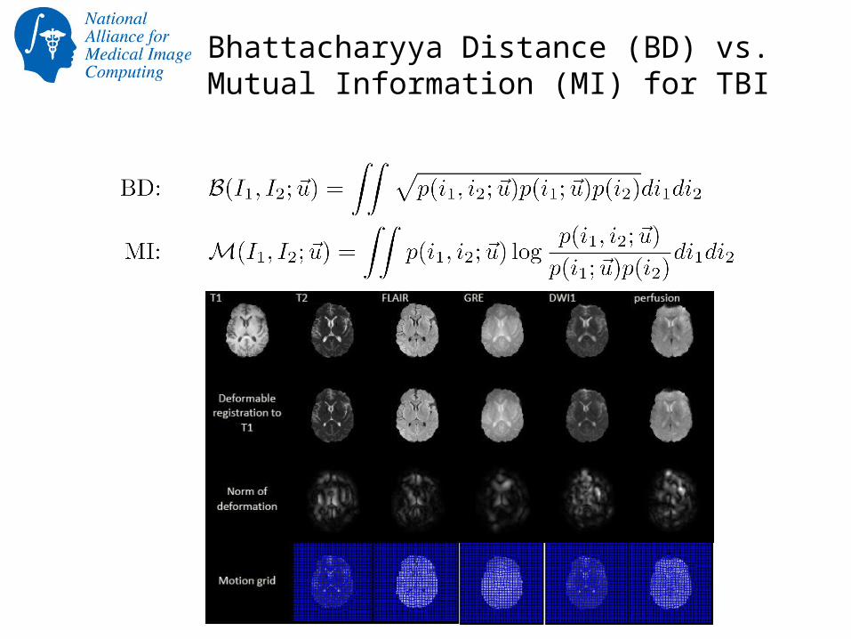
Bhattacharyya Distance (BD) vs.Mutual Information (MI) for TBI

Stochastic point set registration for Adaptive Radiotherapy

Left atrium segmentation
• Salient features + shape based evolution– Combine shape priors (Zernike moments) with RSSegmenter in
Slicer
– Working on Slicer module: Seed region and shape priors
– Multi-atlas Kalman filter based approach
L. Zhu Y. Gao, A. Yezzi, R. MacLeod, J. Cates, A. Tannenbaum. Automatic Segmentation of the Left Atrium from MRI Images Using Salient Feature and Contour Evolution. EMBC, 2012.L. Zhu, Y. Gao, A. Yezzi, A. Tannenbaum, Automatic Segmentation of the Left Atrium from MR Images Via Variational Region Growing with a Moments-based Shape Prior. TMI (submitted).

Scar segmentation
• Segment myocardium wall– start from endo-cardium– distance modulated contour evolution– local/global evolution method– Sobolev active contours
• Scar segmentation in wall– Mixture of Gaussians models intensity– Parameters estimated via EM
• Slicer module• LA wall seg • Scar seg in C++
Y. Gao, L. Zhu, A. Yezzi, S. Bouix , A. Tannenbaum. Scar Segmentation in DE-MRI, cDEMRs challenge. ISBI 2012.
Wall segmenter Slicer3 module

Longitudinal registration
• Post-to-pre ablation MR registration• Accurate around left atrium
• Registration couples MR images and LA masks
• Delivered as Slicer module
Slicer module UI Pre-endocardium overlay on registered post-MRI

Longitudinal shape analysis
• Two groups of LA shapes• AFib cured/recurred after ablation
• Each subject has two time points
• Shape differences along time?• Shape interpolation using optimal mass transport
• Shape analysis at continuous time points
• Statistical differences in the distribution of the contrast agent (related with fibrosis) between cured/recurred groups?