aloe vera - iarc monographs on the evaluation of...
TRANSCRIPT

37
1. Exposure Data
The first record of human use of Aloe vera is in Sumerian hieroglyphics engraved on clay tablets during the Mesopotamia civilization circa 2200 BC, in which it is described as a laxative. Use of aloe in ancient times is also documented in Egypt, Greece, and China. Aloe vera was culti-vated on the islands of Barbados and Curacao in the Caribbean by Spain and the Netherlands, and was sold in various parts of Europe during the 17th century (Park & Jo, 2006). Commercial cultivation of Aloe vera in the USA began in the 1920s in Florida (Grindlay & Reynolds, 1986). Although Aloe vera originated in the warm, dry climates of Africa, the plant is readily adaptable and grows worldwide (Steenkamp & Stewart, 2007).
Use of Aloe vera gel extracts in health foods and beverages, and moisturizing cosmetics, began during the 1970s, starting in the USA and parts of Europe (Park & Jo, 2006). Historically, Aloe vera was used topically to heal wounds and for various skin conditions, and orally as a laxa-tive (Steenkamp & Stewart, 2007). The dried latex of other Aloe species, such as Aloe ferox Miller (Cape aloe or bitter aloe) has also been used as a laxative (EMA, 2006). Today, Aloe vera is also used as a folk or traditional remedy for a variety of conditions and is found in some dietary supplements and food products. Aloe vera gel can be found in hundreds of skin products, including lotions and sunblocks (NCCAM, 2012).
A glossary of commonly used terms for Aloe vera products is provided in Table 1.1.
1.1 Identification of the agent
1.1.1 Botanical data
(a) Nomenclature
For details on botanical nomenclature, see Newton (2004).
Chem. Abstr. Serv. Reg. No.: 8001-97-6Chem. Abstr. Name: Aloe barbadensisBotanical name: Aloe vera (L.) Burm. f. (synonym, Aloe barbadensis, Aloe humilis Blanco, Aloe indica Royle, nomen nudum, Aloe perfoliata var. vera L., Aloe vulgaris Lam.) (GRIN, 2013).Family: XanthorrhoeaceaeGenus: AloePlant part: LeafCommon names: Aloe vera; Aloe vera Linné; True aloe; Aloe barbadensis; Barbados aloe; Curaçao aloe; Mediterranean aloe; Ghritakumari; Lu Hui; Luhui, etc.
From WHO (1999), Eur Ph (2008), O’Neil et al. (2006), SciFinder (2013), IASC (2013a), Boudreau et al. (2013a).
ALOE VERA

IARC MONOGRAPHS – 108
38
(b) Description
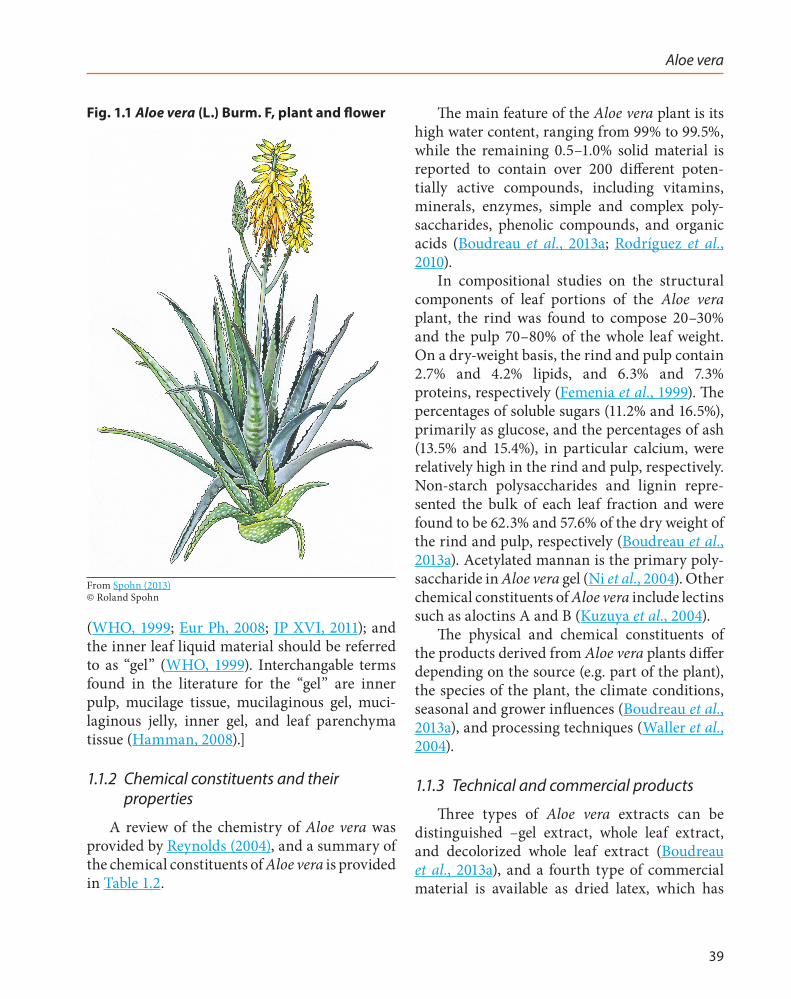
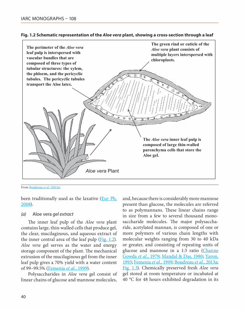
Aloes are perennial succulents or xero-phytes; they can adapt to habitats with low or erratic water availability, are characterized by the capacity to store large volumes of water in their tissue, and are able to use crassulacean acid metabolism, an adaptation to the photosynthetic pathway that involves the formation of malic acid (Boudreau et al., 2013a). Aloe plants, such as Aloe vera (Fig. 1.1), all have green fleshy leaves covered by a thick cuticle or rind, under which is a thin vascular layer covering an inner clear pulp (Boudreau et al., 2013a; Fig. 1.2) The leaves are 30–50 cm in length and 10 cm in width at the base, pea-green in colour (when young spotted with white), and with bright yellow tubular flowers 25–35 cm in length arranged in a slender loose spike (WHO, 1999).
The vascular bundles, located within the leaf pulp, transport (i) water and minerals from the roots to the leaves; (ii) synthesized materials to the roots; and (iii) latex along the margins of the leaf for storage (Ni et al., 2004; Fig. 1.2). The number of vascular bundles varies depending on the size of the leaves and the age of the plant (Ni et al., 2004).
Aloe vera plants contain two major liquid materials (Fig. 1.2): first, a bitter yellow latex located under the strongly cutinized epidermis of the leaves in the vascular layer and containing a high concentration of anthraquinone compounds, which has been used throughout the centuries as a cathartic and for medicinal purges; and, second, a clear mucilaginous gel produced by the thin-walled tubular cells in the inner central zone (parenchyma) that has been used since ancient times to treat burns and other wounds, where it is thought to increase the rate of healing and reduce the risk of infection (Joseph & Raj, 2010). A third liquid may also be obtained by macerating the whole leaf.
[Both the scientific and the lay literature (e.g. on internet sites) are extremely inconsistent when referring to products obtained from Aloe vera. The problem starts with the fact that the three types of liquids that are obtained from Aloe vera leaves are interchangeably referred to as “Aloe juice,” which has caused confusion in the literature. For disambiguation reasons, the term “Aloe juice” should be restricted – if used at all – to the latex material of the pericycle, which is in accordance with the pharmacopoeial definitions
Table 1.1 Definition of terms commonly used in the Aloe industry
Term Definition
Leaf The part of the Aloe vera plant used in commerce, where processing is begun without stripping off the rind.Whole leaf Historically used to describe products derived from the entire leaf that were filtered/purified. However, use of
this terminology without adequate additional descriptors is not recommended. This terminology is now seen on products or in reference to raw material where the entire leaf is used as a starting ingredient to create Aloe vera juice.
Decolorized whole leaf
A process, usually involving filtration with activated charcoal, that clarifies the liquid aloe mass.
Inner leaf Plant part used to describe the clear, central parenchymatous tissues of the aloe leaf.Aloe latex Brown, yellow-brown, or occasionally red exudate found between the rind and inner leaf. Also called “sap,” it
contains several constituents, but most notably anthraquinones.Anthraquinone An organic compound primarily found in the aloe latex, whose structure serves as the basic building block
for several naturally occurring plant pigments. The substance is commonly used for laxative purposes.Gel Liquid product typically derived from the inner leaf.Juice Liquid product derived from Aloe vera leaf [the Working Group noted that the term “juice” is used arbitrarily
and may either apply to products from the latex or from the gel].Adapted from IASC (2009)
Aloe vera
39
(WHO, 1999; Eur Ph, 2008; JP XVI, 2011); and the inner leaf liquid material should be referred to as “gel” (WHO, 1999). Interchangable terms found in the literature for the “gel” are inner pulp, mucilage tissue, mucilaginous gel, muci-laginous jelly, inner gel, and leaf parenchyma tissue (Hamman, 2008).]
1.1.2 Chemical constituents and their properties
A review of the chemistry of Aloe vera was provided by Reynolds (2004), and a summary of the chemical constituents of Aloe vera is provided in Table 1.2.
The main feature of the Aloe vera plant is its high water content, ranging from 99% to 99.5%, while the remaining 0.5–1.0% solid material is reported to contain over 200 different poten-tially active compounds, including vitamins, minerals, enzymes, simple and complex poly-saccharides, phenolic compounds, and organic acids (Boudreau et al., 2013a; Rodríguez et al., 2010).
In compositional studies on the structural components of leaf portions of the Aloe vera plant, the rind was found to compose 20–30% and the pulp 70–80% of the whole leaf weight. On a dry-weight basis, the rind and pulp contain 2.7% and 4.2% lipids, and 6.3% and 7.3% proteins, respectively (Femenia et al., 1999). The percentages of soluble sugars (11.2% and 16.5%), primarily as glucose, and the percentages of ash (13.5% and 15.4%), in particular calcium, were relatively high in the rind and pulp, respectively. Non-starch polysaccharides and lignin repre-sented the bulk of each leaf fraction and were found to be 62.3% and 57.6% of the dry weight of the rind and pulp, respectively (Boudreau et al., 2013a). Acetylated mannan is the primary poly-saccharide in Aloe vera gel (Ni et al., 2004). Other chemical constituents of Aloe vera include lectins such as aloctins A and B (Kuzuya et al., 2004).
The physical and chemical constituents of the products derived from Aloe vera plants differ depending on the source (e.g. part of the plant), the species of the plant, the climate conditions, seasonal and grower influences (Boudreau et al., 2013a), and processing techniques (Waller et al., 2004).
1.1.3 Technical and commercial products
Three types of Aloe vera extracts can be distinguished –gel extract, whole leaf extract, and decolorized whole leaf extract (Boudreau et al., 2013a), and a fourth type of commercial material is available as dried latex, which has
Fig. 1.1 Aloe vera (L.) Burm. F, plant and flower
From Spohn (2013)© Roland Spohn
IARC MONOGRAPHS – 108
40
been traditionally used as the laxative (Eur Ph, 2008).
(a) Aloe vera gel extract
The inner leaf pulp of the Aloe vera plant contains large, thin-walled cells that produce gel, the clear, mucilaginous, and aqueous extract of the inner central area of the leaf pulp (Fig. 1.2). Aloe vera gel serves as the water and energy storage component of the plant. The mechanical extrusion of the mucilaginous gel from the inner leaf pulp gives a 70% yield with a water content of 99–99.5% (Femenia et al., 1999).
Polysaccharides in Aloe vera gel consist of linear chains of glucose and mannose molecules,
and, because there is considerably more mannose present than glucose, the molecules are referred to as polymannans. These linear chains range in size from a few to several thousand mono-saccharide molecules. The major polysaccha-ride, acetylated mannan, is composed of one or more polymers of various chain lengths with molecular weights ranging from 30 to 40 kDa or greater, and consisting of repeating units of glucose and mannose in a 1:3 ratio (Channe Gowda et al., 1979; Mandal & Das, 1980; Yaron, 1993; Femenia et al., 1999; Boudreau et al., 2013a; Fig. 1.3). Chemically preserved fresh Aloe vera gel stored at room temperature or incubated at 40 °C for 48 hours exhibited degradation in its
Fig. 1.2 Schematic representation of the Aloe vera plant, showing a cross-section through a leaf
The green rind or cuticle of the Aloe vera plant consists of multiple layers interspersed with chloroplasts.
The perimeter of the Aloe veraleaf pulp is interspersed with vascular bundles that are composed of three types of tubular structures: the xylem, the phloem, and the pericyclictubules. The pericyclic tubules transport the Aloe latex.
The Aloe vera inner leaf pulp is composed of large thin-walled parenchyma cells that store the Aloe gel.
Aloe vera Plant
From Boudreau et al. (2013a)

Aloe vera
41
rheological properties, a decrease in the content and composition of polysaccharides, and a substantial increase in the mannose:glucose ratio, from 2.9 in the fresh gel to 13.4 in the incu-bated gel (Yaron, 1993).
(b) Aloe vera whole leaf extract
The Aloe vera whole leaf extract (sometimes referred to as whole leaf Aloe vera juice, Aloe juice or nondecolorized whole leaf extract), is the aqueous extract of the whole leaf with lignified fibres removed. The whole leaf extract contains both the gel from the inner parenchyma leaf pulp and the latex. The restricted distribution of the bitter latex within the margins of the leaves of the Aloe vera plant suggests that this thin layer is the primary site of secondary metabolites biosynthesis: compounds that do not function directly in plant growth and development and serve as a plant defence strategy (Boudreau et al., 2013a). A wide variety of secondary compounds have been isolated from the Aloe vera latex (Reynolds, 2004). The isolated compounds are
largely phenolic in nature, and many are anth-raquinone C-glycosides, anthrones, and free anthraquinones (Park et al., 1998). The levels of anthraquinone C-glycosides in Aloe vera latex are quite variable; however, they may constitute up to 30% of the dry weight of the latex (Groom & Reynolds, 1987). Aloe vera latex contains four major C-glycosyl constituents: aloin A, aloin B, aloesin, and aloeresin A (Fig. 1.3; Saccù et al., 2001). Aloin A, a C-glycosyl anthrone, also referred to as barbaloin, is the major component of aloe latex. Aloin A and its epimer, aloin B, also referred to as isobarbaloin, have a 9-anthrone skeleton and a β-D-glucopyranosyl substit-uent. Aloesin, also known as aloeresin B, is a 5-methyl chromone with an 8-β-D-glucopyra-nosyl substituent, and aloeresin A is a 5-methyl chromone with an 8-β-D-glucopyranosyl-2-O-trans-p-coumarol substituent. Several other C-glycosyl-chromones and anthrones have been isolated from Aloe vera, including aloe-emodin, the anthraquinone of barbaloin and isobarbaloin (Boudreau et al., 2013a).
Table 1.2 Summary of chemical constituents of Aloe vera products
Class Compounds
Anthraquinones/anthrones
Aloe-emodin, aloetic acid, anthranol, aloin A and B (or collectively known as barbaloin), isobarbaloin, emodin, ester of cinnamic acid
Carbohydrates Pure mannan, acetylated mannan, acetylated glucomannan, glucogalactomannan, galactan, galactogalacturan, arabinogalactan, galactoglucoarabinomannan, pectic substance, xylan, cellulose
Chromones 8-C-Glucosyl-(2′-O-cinnamoyl)-7-O-methylaloediol A, 8-C-glucosyl-(S)-aloesol, 8-C-glucosyl-7-O-methyl-(S)-aloesol, 8-C-glucosyl-7-O-methylaloediol, 8-C-glucosyl-noreugenin, isoaloeresin D, isorabaichromone, neoaloesin A
Enzymes Alkaline phosphatase, amylase, carboxypeptidase, catalase, cyclooxidase, cyclooxygenase, lipase, oxidase, phosphoenolpyruvate carboxylase, superoxide dismutase
Minerals Calcium, chlorine, chromium, copper, iron, magnesium, manganese, potassium, phosphorous, sodium, zinc
Lipids and miscellaneous organic compounds
Arachidonic acid, γ-linolenic acid, steroids (campestrol, cholesterol, β-sitosterol), triglycerides, triterpenoid, gibberillin, lignins, potassium sorbate, salicylic acid, uric acid
Amino acids Alanine, arginine, aspartic acid, glutamic acid, glycine, histidine, hydroxyproline, isoleucine, leucine, lysine, methionine, phenylalanine, proline, threonine, tyrosine, valine
Proteins Lectins, lectin-like substanceSaccharides Mannose, glucose, L-rhamnose, aldopentoseVitamins B1, B2, B6, C, β-carotene, choline, folic acid, α-tocopherolAdapted from Hamman (2008)

IARC MONOGRAPHS – 108
42
Fig.
1.3
Che
mic
als
pres
ent i
n ge
l and
late
x pr
epar
ed fr
om A
loe
vera
HO
OO
OH
OH
OH
O
HO
O
Aloe
vera
who
lele
af
O
HO
OO
OO
OA
cO
H
OA
cOH
OH
O
HO
OO
O
OA
cO
H
OH
OA
cOH
Ace
tyla
ted
man
nan
(Ace
man
nan)
Aloe
ver
a ge
l
Aloe
ver
a la
tex
Alo
in A
(B
arba
loin
)
1
2 3
410
5
67
89
OH
OH
OH
O
H
OH
OH
OH
O
HO
OH
OH
OH
O
OH
OH
OH
O
HO
H
Alo
in B
(Is
obar
balo
in)
Alo
esin
(A
loer
esin
B)
OH
OH
OH
O
HO
O
HO
O O
O
Alo
enin
HO
OO
OH
O
OH
O
HO
O
OH
OA
loer
esin
A
AcO
AcO
Ac,
ace
tyl g
roup
From
Bou
drea
u et
al.
(201
3a)

Aloe vera
43
The occurrence in Aloe vera latex of endoge-nous free anthraquinones and anthrones results from oxidative processes acting on the glycosides rather than from metabolic synthesis (Boudreau et al., 2013a). In addition, the latex from Aloe vera contains several aromatic compounds, such as aldehydes and ketones (Saccù et al., 2001). The sugar moiety in aloins is D-glucose, and studies indicate that carbon atom 1 of the D-glucose moiety is linked directly to carbon atom 10 of the anthracene ring in a β-configuration (Fig. 1.3). The carbon–carbon bond is quite resistant to acid and alkaline conditions; however, the intestinal microflora of humans and animals have been shown to cleave the β-C-glucosyl bond, although considerable variation in response among animal species occurs. Cleavage of the β-C-glucosyl bond results in the formation of aloe-emodin, the cathartic principle of the latex, and other free anthraquinones and anthrones (Boudreau et al., 2013a; see Section 4.1.1b). In commercial prod-ucts containing whole leaf extract, a rapid dete-rioration of aloin was detected during storage, especially at higher temperatures (Pellizzoni et al., 2011).
(c) Aloe vera decolorized whole leaf extract
Activated carbon treatment of the Aloe vera whole leaf extract is used to remove bitterness and colour caused by the anthraquinone compo-nents of the latex. This results in a product termed “decolorized whole leaf extract” that has quite different properties from the whole leaf extract. Aloe vera decolorized whole leaf extract is also referred to as “whole leaf Aloe vera gel” (Boudreau et al., 2013a). Dentali (2013) noted that an industry standard for aloin content of decolorized Aloe vera whole leaf extract is < 10 ppm. Sehgal et al. (2013) reported results of toxicological assessment of a commercial decol-orized whole leaf extract that contained approx-imately Aloin A at 0.9 ppm, Aloin B at 1.3 ppm, and aloe-emodin at 0.2 ppm. A decolorized Aloe vera whole leaf extract assessed for safety by Shao
et al. (2013) was reported to contain combined Aloin A and Aloin B at < 0.1 ppm.
Although Aloe vera gel and the decolor-ized whole leaf extract are similar in that each contain little or no latex anthraquinones, carbon adsorption changes the physical and chemical properties of the whole leaf extract. Aloe vera decolorized whole leaf extract differs from the gel in that it exhibits a degradation in rheological properties and a loss of approximately 19–23% of the complex polysaccharide content (Pelley et al., 1998).
(d) Dried Aloe vera latex (pharmaceutical material)
The dried Aloe vera latex is the solidified liquid originating in the cells of the pericycle and adjacent leaf parenchyma, and flowing sponta-neously from the cut leaf, allowed to dry with or without the aid of heat (WHO, 1999). The material is used for medicinal purposes and its composition is specified in several official phar-macopoeias (see Section 1.6).
1.2 Analysis
For Aloe vera sold for medicinal purposes, analyses are defined in pharmacopoeial mono-graphs (see Section 1.6). Most of the published analytical methods (Table 1.3) deal with the determination of the anthraquinone compounds in the latex, and fewer and mostly qualitative methods are available for authentication.
To carry out an exhaustive quality control of commercial Aloe vera gel products (e.g. for food or cosmetic uses), the following analyses should be carried out: (i) investigation of authenticity; (ii) test for identification of additives (to control the labelling or regulatory limits); and (iii) deter-mination of the aloin content (Lachenmeier et al., 2005; Rodríguez et al., 2010). The investigation of authenticity aims at confirming the amount of Aloe vera in the preparation; adulteration

IARC MONOGRAPHS – 108
44
Tabl
e 1.
3 Se
lect
ed m
etho
ds o
f ana
lysi
s of
Alo
e ve
ra c
onst
itue
nts
in v
ario
us m
atri
ces
Sam
ple
mat
rix
Ana
lyte
/pur
pose
of a
naly
sis
Sam
ple
prep
arat
ion
Ass
ay m
etho
dD
etec
tion
lim
itR
efer
ence
Uri
neA
loin
/det
ectio
n of
laxa
tive
abus
eG
lucu
roni
dase
, SPE
HPT
LC10
–20
mg/
LPe
rkin
s & L
ives
ey (1
993)
Uri
neA
loe-
emod
in/d
etec
tion
of la
xativ
e ab
use
Glu
curo
nida
se, E
xtra
ctio
n w
ith
chlo
rofo
rm/is
opro
pano
l 9+1
HPL
C/U
V0.
015
mg/
LSt
olk
& H
oogt
ande
rs
(199
9)Se
rum
Alo
in/p
harm
acok
inet
ic st
udy
Extr
actio
n w
ith e
thyl
ace
tate
TLC
0.03
3 m
g/L
Ishi
i et a
l. (1
987)
Plas
ma
Alo
e-em
odin
/pha
rmac
okin
etic
stud
yD
ichl
orom
etha
ne e
xtra
ctio
nH
PLC
/FD
4.5
µg/L
Zaffa
roni
et a
l. (2
003)
Alo
e le
ave
exud
ates
Alo
in/t
axon
omy
Met
hano
lic so
lutio
nH
PLC
/UV
NA
Gro
om &
Rey
nold
s (1
987)
Alo
e ver
a ge
l13
phe
nolic
com
poun
ds/q
ualit
y co
ntro
l and
stan
dard
izat
ion
Liqu
id–l
iqui
d ex
trac
tion
HPL
C/U
VN
AK
im &
Par
k (2
006)
Alo
e ver
a pr
oduc
tsA
cety
late
d po
lysa
ccha
ride
s, gl
ucos
e,
mal
ic a
cid,
lact
ic a
cid,
and
ace
tic a
cid/
qual
ity c
ontr
ol
Non
e (d
isso
lve
in D
2O)
NM
R<
0.05
µg/
LJia
o et
al.
(201
0)
Alo
e ex
trac
ts
and
com
mer
cial
fo
rmul
atio
ns
Alo
e-em
odin
/iden
tity
confi
rmat
ion
Prep
arat
ive
TLC
HPL
C/U
V a
nd
FDU
V: 3
µg/
L FD
: 0.8
µg/
LM
andr
ioli
et a
l. (2
011)
Alo
e ver
a pl
ants
Met
abol
ite p
rofil
ing/
met
abol
omic
sEx
trac
tion
with
met
hano
l. D
eriv
atiz
atio
n w
ith M
STFA
for
GC
-IT-
MS
anal
ysis
GC
-IT-
MS
and
UPL
C-Q
-TO
F-M
S
NA
Lee
et a
l. (2
012)
Alo
e ver
a le
aves
Alo
in d
eriv
ativ
es/r
egul
ator
y co
ntro
lU
ltras
ound
-ass
iste
d ex
trac
tion
in
met
hano
lH
PLC
-DA
D,
HPL
C-M
S,
UPL
C
3–10
mg/
LA
zaro
ual e
t al.
(201
2)
Phar
mac
eutic
al
form
ulat
ions
Alo
e ver
a po
lysa
ccha
ride
s/co
ntro
l for
ad
ulte
ratio
n or
deg
rada
tion
Non
e (d
isso
lve
in D
2O)
NM
R2
g/L
Dav
is &
Gou
x (2
009)
Cos
met
ics
Man
nose
/det
erm
inat
ion
of q
uant
ity
of A
loe i
n pr
oduc
tEx
trac
tion
with
die
thyl
eth
er a
nd
hydr
olys
is w
ith su
lfuri
c ac
idH
PTLC
3% o
f Alo
e ver
a in
cos
met
ic
prod
uct
Gei
sser
& K
ratz
(201
0)
Alo
e spe
cies
Alo
in d
eriv
ativ
es/a
uthe
ntic
ity c
ontr
olM
etha
nolic
ext
ract
ion
HPL
C/U
V<
0.05
mg/
LO
kam
ura
et a
l. (1
996)
Alo
e exu
date
Vola
tiles
/flav
our c
hara
cter
izat
ion
for
beve
rage
indu
stry
Etha
nol 4
0% fo
r HPL
C, H
S sa
mpl
ing
for G
CH
PLC
, HS-
GC
/M
SN
ASa
ccù
et a
l. (2
001)

Aloe vera
45
Sam
ple
mat
rix
Ana
lyte
/pur
pose
of a
naly
sis
Sam
ple
prep
arat
ion
Ass
ay m
etho
dD
etec
tion
lim
itR
efer
ence
Alo
e ver
a be
vera
ges
Profi
ling
for i
dent
ity, a
dulte
ratio
n,
dilu
tion
Non
eH
PTLC
, HS-
SPM
E-G
C/M
SN
ALa
chen
mei
er et
al.
(200
5)
Alo
e spe
cies
13 P
heno
lic c
ompo
unds
/sea
sona
l va
riat
ion
Extr
actio
n w
ith e
than
olH
PLC
/UV
NA
Park
et a
l. (1
998)
Com
mer
cial
alo
e pr
oduc
tsH
igh
mol
ecul
ar-w
eigh
t po
lysa
ccha
ride
sD
ilutio
n w
ith w
ater
and
0.2
M
NaC
lSE
CN
ATu
rner
et a
l. (2
004)
Com
mer
cial
alo
e pr
oduc
tsA
loe-
emod
in, a
loin
AEx
trac
tion
with
eth
yl a
ceta
te/
met
hano
l 9+1
HPL
C/M
SA
loin
-A,
1 µg
/L; A
loe
emod
in,
2.5
µg/L
Elso
hly
et a
l. (2
007)
Com
mer
cial
alo
e pr
oduc
tsA
loin
A, a
loin
BEx
trac
tion
with
eth
anol
/wat
er
(90+
10)
HPL
C0.
06 m
g/L
Ram
írez
Dur
ón et
al.
(200
8)D
AD
, dio
de a
rray
det
ecto
r; FD
, fluo
resc
ence
det
ectio
n; G
C, g
as c
hrom
atog
raph
y; H
PLC
, hig
h-pe
rfor
man
ce li
quid
chr
omat
ogra
phy;
HPT
LC, h
igh-
perf
orm
ance
thin
-laye
r ch
rom
atog
raph
y; H
S, h
eads
pace
; IT;
ion
trap
; MS,
mas
s spe
ctro
met
ry; M
STFA
, N-m
ethy
l-N-(
trim
ethy
lsily
l)tri
fluor
oace
tam
ide;
NA
, not
app
licab
le; N
MR
, nuc
lear
mag
netic
re
sona
nce
spec
tros
copy
; Q-T
OF,
qua
drup
ole-
time
of fl
ight
; SEC
, siz
e-ex
clus
ion
chro
mat
ogra
phy;
SPE
, sol
id-p
hase
ext
ract
ion;
SPM
E, so
lid-p
hase
mic
roex
trac
tion;
TLC
, thi
n-la
yer
chro
mat
ogra
phy;
UPL
C, u
ltra-
perf
orm
ance
liqu
id c
hrom
atog
raph
y; U
V, u
ltrav
iole
t
Tabl
e 1.
3 (
cont
inue
d)

IARC MONOGRAPHS – 108
46
has been a major concern as a consequence of the high cost of the raw materials. Common adulterants have included maltodextrin in Aloe vera gel, powder or water in the liquid prepa-rations (Pelley et al., 1998). Many authors have reviewed the considerable available amount of literature for analysis and authenticity control of Aloe vera. Besides various chromatographic approaches, nuclear magnetic resonance spec-troscopy appears to be the method of choice for this purpose (Table 1.3). Common addi-tives found in Aloe vera gel preparations, which can be detected by chromatographic methods, include preservatives such as benzoic acid and sorbic acid, or antioxidants such as ascorbic acid (Lachenmeier et al., 2005). Several methods to control the gel material for contamination with aloin are available (see review by Rodríguez et al. (2010) and Table 1.3).
1.3 Use
1.3.1 Indications
(a) Medicinal use
The Aloe vera plant has been used in folk medicine for more than 2000 years, and it remains an important component of traditional medicine in many contemporary cultures, such as China, India, the Caribbean, and Japan (Grindlay & Reynolds, 1986). Aloe vera first gained popularity in the USA in the 1930s with reports of successful use of freshly cut leaves in treating X-ray burns (Ulbricht et al., 2007). Both classes of Aloe vera leaf products, gel and latex, are reported to possess a wide range of pharma-ceutical activities.
WHO lists the short-term treatment of occa-sional constipation as a use for Aloe vera latex that is supported by clinical data (WHO, 1999). The well established cathartic properties of anth-raquinone glycosides provide strong evidence in support of the laxative properties of Aloe vera (Ulbricht et al., 2007). The European Medicines
Agency also found that the therapeutic indica-tion as an “herbal product for short-term use in cases of occasional constipation” is a well estab-lished use of Aloe vera latex (EMA, 2006).
For the gel, WHO identified no uses supported by clinical data. Traditional uses include the external treatment of minor wounds and inflam-matory skin disorders. The gel may be used in the treatment of minor skin irritations, including burns, bruises, and abrasions (WHO, 1999).
In recent times, the oral consumption of Aloe vera has been promoted as prophylaxis and therapy for a variety of unrelated systemic conditions. The scientific literature yields little to substantiate claims of usefulness for systemic conditions by the ingestion of Aloe vera (Boudreau et al., 2013a).
Aloe vera may be used in veterinary medicine as laxative or in topical applications, e.g. in udder disinfectants (Leon, 2003).
(b) Food use
Aloe vera extracts may be used in beverages as bitter flavouring agent (O’Neil et al., 2006). Food products include health and soft drinks, yoghurts, jams, instant tea granules, candies, alcoholic beverages, and ice cream (Ahlawat & Khatkar, 2011). Aloe vera may also be used in food supplements (Steenkamp & Stewart, 2007). The Dietary Supplements Label Database lists 43 products that contain Aloe vera as an active ingredient in amounts of 0.33 to 750 mg per capsule (NLM, 2012). Aloe vera whole leaf extract (which combines both the gel and latex) and Aloe vera decolorized whole leaf extract (from which most of the latex components have been removed) are popular as dietary supplements for various systemic ailments. The anthraquinone components of these products appear to vary significantly in their content of aloe-emodin and aloin A, the major anthraquinone constituent of Aloe vera latex. (Elsohly et al., 2007) evaluated 53 liquid and 30 semisolid and solid aloe-based commercial products. The liquid samples all

Aloe vera
47
contained either aloe-emodin or aloin A at ≤ 10 ppm, with many having no detectable levels of either of the two compounds. Unlike liquid prod-ucts, many solid and semisolid products (11 out of 30) contained one or both of the compounds, aloe-emodin and aloin A, at ≥ 10 ppm.
(c) Cosmetic use
The gel may be used as emollient and mois-turizer in cosmetics and personal care prod-ucts (O’Neil et al., 2006). The gel is used in the cosmetics industry as a hydrating ingredient in liquids, creams, sun lotions, shaving creams, lip balms, healing ointments, and face packs (WHO, 1999). Other products containing Aloe vera include after-shave gel, mouthwash, hair tonic, shampoo, and skin-moistening gel (Newton, 2004).
Aloe vera may be used in cosmetics for marketing reasons (i.e. to impart a touch of “nature” to the product) rather than for actual effects, and the content may be normally kept at a low level (Committee of Experts on Cosmetic Products, 2008).
A study on skin hydration found that a single application of a cosmetic formulation containing > 0.25% of a commercial freeze-dried Aloe vera gel 200:1 concentrate improved the water content of the stratum corneum (Dal’Belo et al., 2006). However, the concentrations of Aloe vera raw materials in cosmetics vary widely from 0.1% or less up to 20% (Cosmetic Ingredient Review Expert Panel, 2007).
Anthraquinone-rich Aloe vera extracts may function as absorbers of ultraviolet radiation in suncreens, because anthraquinones absorb ultraviolet radiation (Committee of Experts on Cosmetic Products, 2008). Regulatory author-ities in Germany have proposed that cosmetic products for which claims are made regarding Aloe vera should contain at least 5 g of Aloe vera per 100 g of product (Kratz, 2009).
1.3.2 Dosage
For medicinal use as a laxative, the correct individual dose is the smallest amount required to produce a soft-formed stool. For adults and children aged more than 10 years, the dose is 40–110 mg of the dried latex, corresponding to 10–30 mg of hydroxyanthraquinones per day, or 100 mg as a single dose in the evening (WHO, 1999). The European Medicines Agency suggests a maximum daily dose of hydroxyanthracene glycosides of 30 mg, and that the correct indi-vidual dose is the smallest required to produce a comfortable soft-formed motion (EMA, 2006). As for other laxatives, there is potential for abuse of Aloe vera latex (Perkins & Livesey, 1993; Stolk & Hoogtanders, 1999). It is difficult to estimate rates of laxative abuse, and more so for cases of abuse attributable to Aloe vera alone.
For medicinal use of Aloe vera gel, 25 to 100 mL per day of a 4.5:1 gel concentrate was suggested as typical oral dose range in adults (Morgan et al., 2005). The International Aloe Science Council recommended a total daily consumption of Aloe vera of 2–8 fluid ounces (59–237 mL) of single-strength leaf gel (IASC, 2013b). For topical use, pure Aloe vera gel is often used liberally on the skin. Hydrophilic cream of 0.5% (by weight) of a 50% ethanol extract of Aloe vera, three times per day for five consecutive days per week has been used for treatment of genital herpes and psoriasis vulgaris (Ulbricht et al., 2007).
1.4 Production, sales, and consumption
1.4.1 Production
(a) Production process
Aloe vera grows best in dry chalky soil or in a sandy loam (Grindlay & Reynolds, 1986). While the plant needs warm semi-tropical conditions, overexposure to sun results in stunted plants with low gel yield. Therefore, Aloe vera is commonly

IARC MONOGRAPHS – 108
48
interplanted with other crops, such as fruit trees. The quality of Aloe vera plant products varies considerably due to differences in growing, harvesting, processing, and storage techniques (Boudreau et al., 2013a), and may also depend on the regulatory regime under which the product is sold (see Section 1.6).
Mexico, followed by the rest of Latin America, China, Thailand, and the USA were described as main producing countries (Rodríguez et al., 2010). Aloe vera has become an important plant crop in Arizona and in the Rio Grande valley of southern Texas (Boudreau et al., 2013a).
The production processes for Aloe vera prod-ucts include various steps such as crushing, grinding or pressing, filtration, decoloriza-tion, stabilization, heat processing, and may be followed by addition of preservatives and stabilizers. A complete overview of production was provided by Ahlawat & Khatkar (2011). The technology for processing of Aloe vera gel was reviewed by Ramachandra & Rao (2008).
Harvesting of the leaves of the Aloe vera plant is generally performed by hand, with the leaves cut from the base of the plant (Grindlay & Reynolds, 1986). Individual leaves are wrapped, crated, and transported to processing plants. Ideally, the leaves are processed within a few hours after harvesting, as temperature, light, air, and humidity can affect the stability of the plant components (Paez et al., 2000). At the processing step, the leaves may be cleaned with water and a mild chlorine solution (Grindlay & Reynolds, 1986).
Aloe vera gel from the fillet of the inner leaf pulp is obtained either by manual removal of the outer layers of the leaf with a knife or by machine. Either method can be flawed and has the poten-tial to contaminate the gel with latex (Grindlay & Reynolds, 1986). This process yields crude Aloe vera gel. High quality gel appears opaque, slightly off-white in colour, and is viscous (Vogler & Ernst, 1999).
Aloe vera whole leaf extract is obtained by grinding the whole fresh leaves, without removal of the rind. Extraneous material and lignified fibres are then removed by homogenizing and filtering the crude gel or whole leaf extracts (Yaron, 1993). Since various amounts of latex and rind may be present in the whole leaf extracts, the extracts may appear yellow to yellow-green in colour.
Activated carbon adsorption to produce Aloe vera decolorized whole leaf extract is the first processing step where an extract is intention-ally subjected to chemical alteration. Aloe vera decolorized whole leaf has lower rheological values than the gel and has a lower content of complex carbohydrates than either gel or whole leaf extracts (Pelley et al., 1998).
The processed extracts are difficult to keep stable, a problem that may cause differences in product potency; therefore, the gel or whole leaf extracts can undergo a stabilization process before being bottled. This process may involve pasteurization, ultraviolet stabilization, chemical oxidation with hydrogen peroxide, addition of chemical preservatives and additives, or concen-tration, and/or drying (Boudreau et al., 2013a).
(b) Production volume
In the cosmetic industry, Aloe vera ingredi-ents hold a prominent position at the top of the list showing the relative frequency of use of plant ingredients within formulations filed with the United States Food and Drug Administration (FDA) (Committee of Experts on Cosmetic Products, 2008).
1.4.2 Sales
According to the 2012 Nutrition Business Journal Annual Report, Aloe vera was 20th among best-selling dietary supplements in the USA. There has been a general upward trend in sales from US$ 31 million in 2000 to US$

Aloe vera
49
72 million in 2011 (Fig. 1.4; Nutrition Business Journal, 2010, 2012).
In 2006, the industry size for Aloe species raw material was estimated to be about US$ 125 million worldwide, while the industry for finished products containing Aloe vera was around US$ 110 billion (Ahlawat & Khatkar, 2011).
Global sales of Aloe species products in 2012 totalled US$ 351 million, according to IMS Health MIDAS data. Most products were reported as derived from Aloe vera (90%). Substantial sales as a dietary supplement were reported in Brazil (US$ 74 million), Indonesia (US$ 50 million), India (US$ 34 million), USA (US$ 29 million), the Russian Federation (US$ 19 million), Japan (US$ 15 million), and Mexico (US$ 12 million) (IMS Health, 2012).
1.4.3 Consumption
Consumers of products specified in Section 1.3 are exposed to Aloe vera. While the occa-sional short-term use of the latex as a laxative may allow exposure to be estimated for use in that context, it is unclear whether or not the gel products or liquid preparations are used over the short or long-term.
According to a representative survey conducted by the National Health and Nutrition Examination Survey from 1999 to 2010 (NHANES, 2010), the consumption of dietary supplements containing Aloe vera in the USA (prevalence of use in the past 30 days among adults in the USA) was 0.3% in 1999–2006 and 0.1% in 2007–2010 [figures calculated by the Working Group from publicly available data; due to the small use, the coefficient of variation is > 30%, so that the data for Aloe vera are less reliable than for other herbs]. In the context of complementary and alternative medicine, use
Fig. 1.4 Sales of dietary supplements containing Aloe vera in the USA
0
10
20
30
40
50
60
70
80
2000 2001 2002 2003 2004 2005 2006 2007 2008 2009 2010 2011
Aloe
ver
a sa
les (
mill
ion
US$
)
Year
Compiled by the Working Group from data in Nutrition Business Journal (2010, 2012).

IARC MONOGRAPHS – 108
50
Tabl
e 1.
4 Re
gula
tion
s fo
r diff
eren
t Alo
e ve
ra p
rodu
cts
Reg
ulat
ion
WH
O M
onog
raph
on
Sele
cted
Med
icin
al
Plan
ts (1
999)
aJa
pane
se P
harm
acop
oeia
Si
xtee
nth
Edit
ion
(201
1)b
Euro
pean
Ph
arm
acop
oeia
7.0
(2
008)
c
The
Inte
rnat
iona
l Alo
e Sc
ienc
e C
ounc
il (2
013)
d
Regu
late
d A
loe
prod
uct
Dri
ed ju
ice
Gel
Dri
ed ju
ice
Con
cent
rate
d an
d dr
ied
juic
e Ra
w m
ater
ials
for u
se in
pro
duct
s for
or
al c
onsu
mpt
ion
Con
tent
Min
. 28%
of
hydr
oxya
nthr
acen
e de
riva
tives
, exp
ress
ed a
s al
oin
Min
. 4%
alo
in (d
ried
m
ater
ial)
Min
. 28%
of
hydr
oxya
nthr
acen
e de
riva
tives
, exp
ress
ed
as a
loin
(dri
ed d
rug)
Max
. 10
ppm
(alo
in A
+ B
)
Iden
tity
test
sM
acro
scop
ic a
nd
mic
rosc
opic
exa
min
atio
ns,
solv
ent s
olub
ility
; TLC
Col
our r
eact
ions
with
so
dium
tetr
abor
ate
and
nitr
ic a
cid;
TLC
TLC
; fluo
resc
ence
w
ith d
isod
ium
te
trab
orat
e; c
olou
r re
actio
n w
ith
brom
ine
wat
er
Min
. 5%
ace
tyla
ted
man
nan
cont
ent b
y dr
y w
eigh
t O
rgan
olep
tic st
anda
rds:
Alo
e so
lids i
n si
ngle
stre
ngth
juic
e (1
%
in le
af ju
ice
and
0.5%
for i
nner
leaf
ju
ice)
M
alic
aci
d an
d gl
ucos
e m
ust b
e pr
esen
t at
a m
inim
um
Who
le le
af m
arke
r (is
ocitr
ate)
. Max
. 5%
for i
nner
leaf
by
dry
wei
ght.
Moi
stur
eM
ax. 1
2% fo
r Cur
acao
or
Barb
ados
Alo
eC
onta
ins 9
8.5%
w
ater
Tota
l ash
Max
. 2%
Max
. 2%
Max
. 2%
< 40
%Lo
ss o
n dr
ying
Max
. 12%
Max
. 12%
Fore
ign
subs
tanc
es/
cont
amin
ants
Lim
its fo
r cer
tain
m
icro
orga
nism
s, ab
senc
e of
adu
ltera
nts s
uch
as b
lack
ca
tech
u, p
iece
s of i
ron,
and
st
ones
; lim
its fo
r cer
tain
pe
stic
ides
, hea
vy m
etal
s, an
d ra
dioa
ctiv
e re
sidue
s
Lim
its fo
r ce
rtai
n m
icro
orga
nism
s, pe
stic
ides
, he
avy
met
als,
radi
oact
ive
resid
ues
Two
diffe
rent
pur
ity te
sts
are
spec
ified
Mic
robi
olog
ical
s (pa
thog
ens,
lact
ic
acid
, mou
ld, y
east
), he
avy
met
als,
mal
tode
xtri
n
Extr
act c
onte
ntM
in. 5
0% (w
ater
-sol
uble
ex
trac
t), m
ax. 1
0% (a
lcoh
ol-
inso
lubl
e ex
trac
t)
Min
. 40%
(wat
er-s
olub
le
extr
act)
a W
HO
(199
9)b
Eur P
h (2
008)
c JP
XV
I (20
11)
d IA
SC (2
013b
)m
ax.,
max
imum
; min
., m
inim
um; T
LC, t
hin-
laye
r chr
omat
ogra
phy

Aloe vera
51
of Aloe vera has been reported in 8.5–13.8% of people in predominantly Hispanic populations in the southern USA; according to surveys, it is also used frequently by 10.8%, 10.3%, and 7.6% of adults in Australia, Italy, and Jamaica, respec-tively (Ngo et al., 2010).
1.5 Occupational exposure
No specific studies on occupational exposure were identified. It can be assumed that workers in the production of Aloe vera may be exposed, as well as workers in pharmaceutical, cosmetic, and food industries that use Aloe vera as an ingredient.
1.6 Regulations and guidelines
Products made with various components of Aloe vera (aloin, aloe-emodin, and barbaloin) were at one time regulated by the FDA as oral over-the-counter (OTC) laxatives (NCCAM, 2012). In 2002, the FDA promulgated a regula-tion stating that the stimulant laxative ingre-dient Aloe vera in over the counter (OTC) drug products is not “generally recognized as safe and effective” or is misbranded (FDA, 2002). Because the companies that manufactured such products did not provide the necessary safety data, the FDA required that all OTC Aloe vera laxative products be removed from the USA market or reformulated (NCCAM, 2012). [The Working Group noted that currently no medicinal OTC Aloe vera products are available in the USA, unlike Europe where some medicinal Aloe vera products are still available.]
According to FDA regulations, Aloe vera may be safely used as a flavouring in foods as defined in 21CFR172.510. The Environmental Protection Agency (EPA) classified Aloe vera gel as a List 3 substance (inerts of unknown toxicity), and also listed Aloe vera gel as an inert ingredient of pesti-cide products (SciFinder, 2013).
A published tabulation of acceptable levels of natural flavourings by the Flavor and Extract Manufacturers’ Association indicates that an acceptable level of Aloe vera extract is 5–2000 ppm. No distinction is given for the part of the plant or type of plant extract used to produce the extract used as a flavouring additive (Duke & Beckstrom-Sternberg, 1994).
For cosmetic uses, many of the manufac-turers of Aloe vera gel take care to supply an ingredient containing anthraquinones at no more than 50 ppm (Committee of Experts on Cosmetic Products, 2008). This maximum level was also demanded in a safety assessment of the cosmetic industry (Cosmetic Ingredient Review Expert Panel, 2007).
Aloe vera is specified in several official phar-macopoeias, and an industry quality standard of the International Aloe Science Council is also available (Table 1.4). An American Herbal Pharmacopoeia on “Aloe vera leaf, Aloe vera leaf juice, Aloe vera inner leaf juice” was provided (AHP, 2012).
2. Cancer in Humans
No data were available to the Working Group.
3. Cancer in Experimental Animals
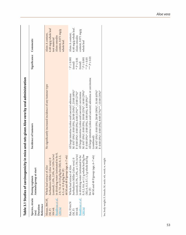
3.1 Studies of carcinogenicityWhole leaf extract of Aloe barbadensis Miller
[Aloe vera] was tested for carcinogenicity by oral administration (drinking-water) in one study in mice and one study in rats.
3.1.1 Mouse
In a 2-year study of carcinogenicity, groups of 48 male and 48 female B6C3F1 mice (age, 6–7 weeks) were given drinking-water containing 0 (controls), 1.0%, 2.0%, or 3.0% (wt/wt) whole leaf extract of Aloe barbadensis Miller [Aloe vera]

IARC MONOGRAPHS – 108
52
for 104 weeks. The average content of aloin A and aloe-emodin of the whole leaf test material was 6.40 and 0.071 mg/g respectively. The doses of whole leaf extract were equivalent to average daily doses of approximately 0, 2.9, 7.0, or 11.8 g/kg body weight (bw) in males; and 0, 2.2, 6.3, or 11.8 g/kg bw in females (Boudreau et al., 2013a). Survival of exposed groups was similar to that of controls. There was no significantly increased incidence of any tumour type in male or female mice. Whole leaf extract increased the incidence of goblet cell hyperplasia in the intestine of male and female mice (Table 3.1).
3.1.2 Rat
In a 2-year study of carcinogenicity, groups of 48 male and 48 female F344/N rats were given drinking-water containing whole leaf extract of Aloe barbadensis Miller [Aloe vera] at 0 (controls), 0.5%, 1.0%, or 1.5% (wt/wt) for 104 weeks. The average content of aloin A and aloe-emodin of the whole leaf test material was 6.40 and 0.071 mg/g, respectively. The doses of whole leaf extract were equivalent to average daily doses of approximately 0, 0.2, 0.6, or 1.1 g/kg bw in males and 0, 0.3, 0.7, or 1.3 g/kg bw in females (Boudreau et al., 2013a). Survival of exposed groups was similar to that of controls. Whole leaf extract caused increased incidences of adenoma and carcinoma of the large intestine (colon and caecum) in males and females. Other treat-ment-related lesions included hyperplasia and/or inflammation in the mesenteric lymph node, forestomach, small intestine, and large intestine in males and females (Table 3.1). [The Working Group noted that large intestine tumours are rare spontaneous neoplasms in F344/N rats.]
3.2 Photo-co-carcinogenicity studies
Mouse
There has been one study reported in which Aloe barbadensis Miller [Aloe vera] test articles were studied by dermal application in mice.
Groups of 36 male and 36 female Crl:SKH-1 (hr-/hr-) hairless mice (age, 8 weeks) received topical applications of control cream or creams containing: 3% or 6% (w/w) gel; 3% or 6% (w/w) whole leaf extract; 3% or 6% (w/w) decolorized whole leaf extract; or 7.46 or 74.6 μg/g of aloe-emodin to the dorsal skin region, for 5 days per week, for up to 40 weeks. After application of the cream in the morning, mice were exposed to filtered solar simulated light (SSL) at 0 (0.00 mJ.CIE/cm2 per day) or 0.6 (13.70 mJ.CIE/cm2 per day) minimal erythema doses of light (NTP, 2010). The minimal erythema dose is defined as the minimal amount of radiation that causes slight erythema within 24 hours after irradiation (Table 3.2). The mice were killed after a recovery/observation period of 12 weeks.
At 52 weeks, there was no significant increase in the incidence of skin neoplasms in any group receiving any of the four creams containing Aloe preparations without exposure to SSL. [The Working Group noted that the duration of the experiment, 1 year, was too short to consider this arm of the experiment as a full carcinogenicity study.]
There was no treatment-related increase in the incidence of skin neoplasms in any groups receiving any of the four creams containing Aloe preparations followed by SSL when compared with the groups receiving control cream followed by SSL. Almost all mice in groups exposed to SSL presented with skin neoplasms due to SSL exposure. [As a result, the primary experimental end-point was multiplicity of skin tumours.]
There was a significant enhancing effect of Aloe gel cream or of aloe-emodin cream on the photocarcinogenic activity of SSL in female mice, and there was a significant enhancing effect of the cream containing whole leaf extract, or cream containing decolorized whole leaf extract, on the photocarcinogenic activity of SSL in male and female mice, based on an increase in the multi-plicity of squamous cell papilloma, carcinoma or carcinoma in situ (combined) (NTP, 2010).
Aloe vera
53
Tabl
e 3.
1 St
udie
s of
car
cino
geni
city
in m
ice
and
rats
giv
en A
loe
vera
by
oral
adm
inis
trat
ion
Spec
ies,
stra
in
(sex
) D
urat
ion
Ref
eren
ce
Dos
ing
regi
men
A
nim
als/
grou
p at
star
tIn
cide
nce
of tu
mou
rsSi
gnifi
canc
eC
omm
ents
Mou
se, B
6C3F
1 (M
, F)
104
wk
Boud
reau
et a
l. (2
013a
)
Who
le le
af e
xtra
ct o
f Alo
e ba
rbad
ensi
s Mill
er [A
loe v
era]
: 0
(con
trol
), 1.
0%, 2
.0%
, or 3
.0%
(w/w
) in
dri
nkin
g-w
ater
(est
imat
ed to
be
0,
2.9,
7.0,
or 1
1.8
g/kg
bw
(M);
0, 2
.2,
6.3,
or 1
1.8
g/kg
bw
(F)
48 M
and
48
F/gr
oup
(age
, 6–7
wk)
No
signi
fican
tly in
crea
sed
inci
denc
e of
any
tum
our t
ype
Alo
in A
con
tent
, 6.
40 m
g/g
who
le le
af
test
mat
eria
l A
loin
-em
odin
co
nten
t, 0.
071
mg/
g w
hole
leaf
.
Rat,
F344
/N
(M, F
) 10
4 w
k Bo
udre
au et
al.
(201
3a)
Who
le le
af e
xtra
ct o
f Alo
e ba
rbad
ensi
s Mill
er [A
loe v
era]
: 0
(con
trol
), 0.
5%, 1
.0%
, or 1
.5%
(w/w
) in
dri
nkin
g-w
ater
(est
imat
ed to
be
0, 0
.2, 0
.6, o
r 1.1
g w
hole
leaf
/kg
bw
(M);
0, 0
.3, 0
.7, 1
.3 g
who
le le
af/k
g bw
(F)
48 M
and
48
F/gr
oup
(age
, 6–7
wk)
All
larg
e in
test
ine
(col
on a
nd c
aecu
m) a
deno
ma:
M
: 0/4
7 (0
%)# ,
0/48
(0%
), 26
/48
(54%
)*, 2
3/48
(48%
)*
F: 0
/48
(0%
)# , 0/
48 (0
%),
6/48
(13%
)**,
13/4
8 (2
7%)*
A
ll la
rge
inte
stin
e (c
olon
and
cae
cum
) car
cino
ma:
M
: 0/4
7 (0
%)# ,
0/48
(0%
), 10
/48
(21%
)*, 1
4/48
(29%
)*
F: 0
/48
(0%
)##, 0
/48
(0%
), 3/
48 (6
%),
4/48
(8%
)**
All
larg
e in
test
ine
(col
on a
nd c
aecu
m) a
deno
ma
or c
arci
nom
a (c
ombi
ned)
: M
: 0/4
7 (0
%)# ,
0/48
(0%
), 28
/48
(58%
)*, 3
1/48
(65%
)*
F: 0
/48
(0%
)# , 0/
48 (0
%),
8/48
(17%
)***
, 15/
48 (3
1%)*
# P ≤
0.0
01
(tren
d)
## P
≤ 0
.01
(tren
d)
* P ≤
0.0
01
** P
< 0
.05
*** P
≤ 0
.01
Alo
in A
con
tent
, 6.
40 m
g/g
who
le le
af.
Alo
in-e
mod
in
cont
ent,
0.07
1 m
g/g
who
le le
af
bw, b
ody
wei
ght;
F, fe
mal
e; M
, mal
e; w
k, w
eek;
w, w
eigh
t

IARC MONOGRAPHS – 108
54
Tabl
e 3.
2 Co
-car
cino
geni
city
stu
dies
in S
KH
-1 m
ice
give
n A
loe
vera
or a
loe-
emod
in b
y sk
in a
pplic
atio
n fo
llow
ed b
y ex
posu
re
to s
imul
ated
sol
ar li
ght
Spec
ies,
stra
in
(sex
) D
urat
ion
Ref
eren
ce
Dos
ing
regi
men
A
nim
als/
grou
p at
star
tO
vera
ll ag
e-ad
just
ed tu
mou
r mul
tipl
icit
ySi
gnifi
canc
eC
omm
ents
Mou
se,
Crl:
SKH
-1 (h
r- /hr
- ) ha
irle
ss
(M, F
) 52
wk
NTP
(201
0)
Alo
e ver
a ge
l cre
am a
t 0%
, 3%
, or 6
% (w
/w) +
SSL
(1
3.70
mJ•
CIE
/cm
2 per
day
). C
ream
app
lied
in th
e m
orni
ng; S
SL in
the
after
noon
. 5
d pe
r wk
for 4
0 w
k, fo
llow
ed b
y 12
-wk
reco
very
/ob
serv
atio
n pe
riod
36
M a
nd 3
6 F/
grou
p (a
ge 8
wk)
Squa
mou
s cel
l pap
illom
a, sq
uam
ous c
ell
carc
inom
a in
situ
, and
/or s
quam
ous c
ell
carc
inom
a of
the
skin
: M
: 5.8
(4.7
–7.2
), 6.
8 (5
.6–8
.4),
7.1 (5
.8–8
.7)
F: 6
.4 (5
.3–7
.6)*
, 9.2
(7.8
–10.
8)**
, 8.1
(6.9
–9.6
)***
* P <
0.0
5 (tr
end)
**
P =
0.0
06
*** P
< 0
.05
No
signi
fican
t inc
reas
e in
the
inci
denc
e of
sk
in n
eopl
asm
s in
any
grou
p re
ceiv
ing
crea
ms c
onta
inin
g A
loe v
era
prep
arat
ions
w
ithou
t exp
osur
e to
SS
LM
ouse
, C
rl:SK
H-1
(hr- /
hr- )
hair
less
(M
, F)
52 w
k N
TP (2
010)
Who
le le
af A
loe v
era
crea
m a
t 0%
, 3%
, or 6
% (w
/w)
+ SS
L (1
3.70
mJ•
CIE
/cm
2 per
day
). C
ream
app
lied
in th
e m
orni
ng; S
SL in
the
after
noon
. 5
d pe
r wk
for 4
0 w
k, fo
llow
ed b
y 12
-wk
reco
very
/ob
serv
atio
n pe
riod
36
M a
nd 3
6 F/
grou
p (a
ge 8
wk)
Squa
mou
s cel
l pap
illom
a, sq
uam
ous c
ell
carc
inom
a in
situ
, and
/or s
quam
ous c
ell
carc
inom
a of
the
skin
: M
: 5.8
* (4.
7–7.
2), 6
.4 (5
.2–7
.9),
8.4*
* (6.
8–10
.3)
F: 6
.4* (
5.3–
7.6)
, 8.7
** (7
.4–1
0.3)
, 7.7
(6.5
–9.1)
* P <
0.0
5 (tr
end)
**
P <
0.0
5
Mou
se,
Crl:
SKH
-1 (h
r- /hr
- ) ha
irle
ss
(M, F
) 52
wk
NTP
(201
0)
Dec
olor
ized
who
le le
af A
loe v
era
crea
m a
t 0%
, 3%
, or 6
% (w
/w) +
SSL
(13.
70 m
J•C
IE/c
m2 p
er
day)
. Cre
am a
pplie
d in
the
mor
ning
; SSL
in th
e aft
erno
on.
5 d
per w
k fo
r 40
wk,
follo
wed
by
12-w
k re
cove
ry/
obse
rvat
ion
peri
od
36 M
and
36
F/gr
oup
(age
8 w
k)
Squa
mou
s cel
l pap
illom
a, sq
uam
ous c
ell
carc
inom
a in
situ
, and
/or s
quam
ous c
ell
carc
inom
a of
the
skin
: M
: 5.8
(4.6
–7.3
), 8.
0* (6
.5–9
.9),
6.4
(5.2
−8.
0)
F: 6
.4**
(5.2
–7.7
), 10
.0**
* (8.
4–12
.0),
9.3*
***
(7.8
–11.
1)
* P <
0.0
5 **
P =
0.0
07
(tren
d)
*** P
= 0
.002
**
** P
= 0
.007
Mou
se,
Crl:
SKH
-1 (h
r- /hr
- ) ha
irle
ss
(M, F
) 52
wk
NTP
(201
0)
Alo
e-em
odin
cre
am a
t 0, 7
.46,
or 7
4.6
µg/g
+ S
SL
(13.
70 m
J•C
IE/c
m2 p
er d
ay).
Cre
am a
pplie
d in
the
mor
ning
; SSL
in th
e aft
erno
on.
5 d
per w
k fo
r 40
wk,
follo
wed
by
12 w
k re
cove
ry/
obse
rvat
ion
peri
od
36 M
and
36
F/gr
oup
(age
, 8 w
k)
Squa
mou
s cel
l pap
illom
a, sq
uam
ous c
ell
carc
inom
a in
situ
, and
/or s
quam
ous c
ell
carc
inom
a of
the
skin
: M
: 5.8
(4.7
–7.2
), 6.
3 (5
.1–7
.8),
7.1 (5
.8–8
.7)
F: 6
.4* (
5.3–
7.7) 7
.9 (6
.6–9
.4),
8.9*
* (7.
5–10
.6)
* P <
0.0
5 (tr
end)
**
P <
0.0
5
bw, b
ody
wei
ght;
CIE
, Com
mis
sion
Inte
rnat
iona
le d
e l’E
clai
rage
[Int
erna
tiona
l Com
mis
sion
on Il
lum
inat
ion]
; d, d
ay; F
, fem
ale;
M, m
ale;
SSL
, sim
ulat
ed so
lar l
ight
; wk,
wee
k

Aloe vera
55
4. Mechanistic and Other Relevant Data
In reviewing studies relevant to the possible carcinogenicity of Aloe vera, the Working Group noted that attributing appropriate weight to indi-vidual studies was complicated by the consid-eration that, despite the terminology used, the material tested may not have been identical across various studies and/or may not have been identical to the material that was studied in experimental animals, as described in Section 3 of this Monograph.
4.1 Absorption, distribution, metabolism, and excretion
4.1.1 Humans
There were no reports of studies to determine the absorption, distribution, metabolism, or excretion of topically applied Aloe vera gel, whole leaf extract or decolorized whole leaf extract in experimental animals or humans.
Aloe vera whole leaf extract is composed of gel and latex. Aloe vera gel contains non-starch polysaccharides of high molecular weight (the major one being acemannan) that are composed of sugar moieties linked by β-1,4-glycosyl bonds (Fig. 1.3 in Section 1). Aloe vera latex contains the anthrone C-glycosides aloin A (barbaloin) and aloin B (isobarbaloin) that are linked by β-glycosyl bonds to D-glucopyranose. Other C-glycosides found in Aloe vera latex include aloesin (aloeresin B) and aloeresin A in which the glycosyl linkage is to the benzo ring of benzopyran-4-one (Boudreau & Beland, 2006). Aloenin, an O-β-glucoside, is also a component of Aloe vera latex (Hirata et al., 1981; Matsuda et al., 2008).
(a) Components of Aloe vera gel: metabolism ex vivo
Incubation of acemannan (aloemannan; molecular weight > 400 kDa) labelled with fluo-resceinyl isothiocyanate (FITC) with a suspen-sion of fresh human faeces for 5 days gave two metabolites, with molecular weights of 10 and 30 kDa, in 1% yield, meaning that aloemannan is catabolized by human intestinal bacteria (Yagi et al., 1999).
(b) Components of Aloe vera latex
Orally ingested anthrone C-glycosides (i.e. aloin A and aloin B) pass intact through the upper portion of the gastrointestinal tract and upon reaching the lower gastrointestinal tract are cleaved to aloe-emodin-9-anthrone by human Eubacterium sp. BAR given to germ-free rats (Che et al., 1991; Hattori et al., 1993; Akao et al., 1996). The free aglycone is then absorbed, undergoes oxidation, and is excreted in the urine as rhein, as was shown in three volunteers receiving Aloe vera or barbaloin (Vyth & Kamp, 1979; Fig. 4.1).
4.1.2 Experimental systems
(a) Components of Aloe vera gel
In beagle dogs, the oral administration of radiolabelled acemannan at a dose of 20 mg/kg body weight (bw) per day for 3 months resulted in peak blood concentrations at 4–6 hours and a half-life of > 48 hours (Fogleman et al., 1992).
Male ddY mice were given FITC-labelled acemannan (aloemannan; molecular weight, 500 kDa) at a dose of 120 mg/kg bw by gavage, and urinary and faecal excretion was monitored for 48 hours. Of the administered dose, 95% was excreted in the faeces, with > 90% occurring within 24 hours. Only 0.3% of the material was found in the urine. In both urine and faeces, FITC-labelled acemannan was converted to substances of low molecular weight (< 9 kDa).

IARC MONOGRAPHS – 108
56
Fig. 4.1 Metabolites of aloin A and aloin B
1
2
3
4105
6
7
8 9
OHOH
OH
O
H
OHOH
OH
O
HO
OHOH
OH
O
OHOH
OH
O
HO H
Aloin A (Barbaloin) Aloin B (Isobarbaloin)
OH
OH OHO
Hydrolysis of the β-glycosidicbond by intestinal bacteria
Aloe-emodin-9-anthrone
OH
OH OHO
O
Aloe-emodin anthraquinone
[O]
[O]
COOH
OH OHO
O
Rhein
Aloin A and B are constituents of Aloe vera whole leaf latex. Compiled by the Working Group.

Aloe vera
57
FITC-labelled acemannan was also adminis-tered to mice by intravenous injection at a dose of 120 mg/kg bw. Of the administered dose, 73% was excreted in the urine, with > 60% occurring within 24 hours; 13% of the material was found in the faeces. In both urine and faeces, FITC-labelled acemannan was converted to substances of low molecular weight (10–70 kDa in urine; 5 kDa in faeces) (Yagi et al., 1999).
(b) Components of Aloe vera latex
In male Wistar rats, oral administration of aloin A (barbaloin; 100 mg/kg bw) resulted in maximum serum concentrations of aloin A [340 ng/mL; ~0.8 μM, based upon the molec-ular weight of aloin A of 404 Da] 1.5 hours after administration, followed by a decrease in concen-tration, with aloin A still detectable 6 hours after dosing (Ishii et al., 1987).
The ability to cleave anthrone C-glycosides varies among species; free anthrones are detected in faecal contents from humans and rats, but not mice or guinea-pigs (Dreessen & Lemli, 1988; Hattori et al., 1988).
In male and female Brown-Norway rats, oral administration of [14C]aloe-emodin (4.5 mg/kg bw) resulted in maximum blood concentrations [~350 ng/mL; ~1.3 μM, based upon the molecular weight of aloe-emodin of 270 Da] 2 hours after dosing, and a terminal half-life of elimination of approximately 50 hours. Seven metabolites were detected in the plasma. These were char-acterized as aloe-emodin, rhein, an unidentified aglycone, and conjugates of these aglycones. Approximately 20% of the radiolabel was elim-inated in the urine, primarily as aloe-emodin, rhein, and their conjugates. More than 75% of the radiolabel was excreted in the faeces and nearly all of this was aloe-emodin. At early time-points (< 48 hours), a great majority of the radiolabel was associated with the gastrointestinal tract. At later time-points (i.e. 96 hours), the highest levels of radioactivity were found in the kidney and liver.
This material was characterized as aloe-emodin, rhein, and their conjugates (Lang, 1993).
Intracaecal administration of [14C]rhein to male Wistar rats resulted in 37% of the radioac-tivity being excreted in the urine and 53% in the faeces. The highest tissue concentrations were found in the kidney (De Witte & Lemli, 1988).
The oral administration of [14C]-labelled aloenin to rats resulted in the faecal and urinary excretion of the aglycone 4-methoxy-6-(2,4-di-hydroxy-6-methylphenyl)-2-pyrone (Hirata et al., 1981).
4.1.3 Alterations in enzymes involved in metabolism
Incubation of the human colon carcinoma cell line LS180 with Aloe vera juice resulted in a significant increase in the expression of CYP1A2, CYP3A4, and multidrug resistance 1 genes (Brandin et al., 2007).
Commercial preparations of Aloe vera were tested for their ability to inhibit the activities of CYP3A4 and CYP2D6 in vitro. Inhibition was observed with half maximal inhibitory concen-trations IC50 in the range 8–43 mg/mL, concen-trations that were probably sufficiently high as to preclude any significant inhibition in vivo (Djuv & Nilsen, 2012). Rhein was shown to inhibit CYP1A2, CYP2C9, CYP2D6, CYP2E1, and CYP3A activities in rat liver microsomes, with Ki in the range of 10–74 μM (Tang et al., 2009).
4.2 Genetic and related effects
4.2.1 Humans
No data were available to the Working Group.

IARC MONOGRAPHS – 108
58
4.2.2 Experimental systems
(a) DNA damage
An Aloe vera whole leaf extract induced single-strand breaks in pUC 9.1 plasmid DNA, and this was associated with decreased transfor-mation efficiency of the plasmid (Table 4.1; Paes-Leme et al., 2005).
Aloe-emodin and/or rhein induced DNA damage in NPC-039 and NPC-076 human naso-pharyngeal carcinoma cells and SCC-4 human tongue cancer cells, as measured by comet assays (Table 4.2; Lin et al., 2007, 2010; Chen et al., 2010).
(b) End-points associated with DNA damage
In addition to inducing DNA damage (as indicated by comet assays), aloe-emodin signif-icantly inhibited expression of genes associated with DNA damage and repair: ataxia telangiec-tasia mutated (ATM), ataxia telangiectasia and Rad3-related (ATR), 14–3-3σ, breast cancer 1, early onset (BRCA1), and DNA-dependent serine/threonine protein kinase (DNA-PK) in SCC-4 human tongue squamous cancer cells (Chen et al., 2010). Aloe-emodin also induced the formation of reactive oxygen species (ROS) in SCC-4 cells, which was accompanied by S-phase cell-cycle arrest, apoptosis, and several molecular markers associated with apoptosis (Chiu et al., 2009). Rhein induced the formation of ROS in NPC-039 human nasopharyngeal carcinoma cells, SCC-4 human tongue squamous cancer cells, and A-549 human lung cancer cells, which was accompa-nied by apoptosis and several molecular markers associated with apoptosis (Lin et al., 2007; Hsia et al. 2009; Lai et al., 2009).
Aloe-emodin induced DNA damage in mouse lymphoma L5178 cells, as measured by comet assay (Table 4.2; Müller et al., 1996).
(c) Gene mutation
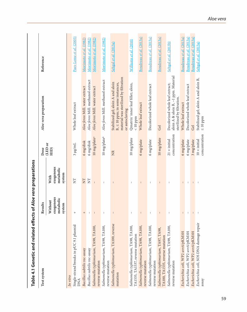
Extracts in water, ethanol or methanol of Aloe ferox Mill., stabilized Aloe vera gel, Aloe vera whole leaf extract, Aloe vera decolorized whole
leaf extract, Aloe vera gel, acemannan, and aloin were tested in Salmonella typhimurium reverse mutation assays, Bacillus subtilis rec-assays, and/or SOS DNA damage-repair assays. With the exception of the Bacillus subtilis rec-assay with water extracts of Aloe ferox Mill., all gave negative results (Table 4.1; Table 4.2; Brown & Dietrich, 1979; Morimoto et al., 1982; Boudreau et al., 2013a; Sehgal et al., 2013a, b).
Aloe-emodin was mutagenic in reversion assays with various strains of Salmonella typh-imurium, at the Tk+/– locus in mouse lymphoma L5178Y cells, and the Gpt locus in AS52 Chinese hamster cells (Table 4.2; Brown et al., 1977; Brown & Dietrich, 1979; Westendorf et al., 1990; Heidemann et al., 1996; Müller et al., 1996; Müller & Stopper, 1999; Nesslany et al., 2009).
Mutation analysis of eight adenomas and four carcinomas from the large intestine of F344 rats given drinking-water containing an Aloe vera whole leaf extract (Boudreau et al., 2013a, b) indicated four point mutations in exons 1 and 2 of the Kras gene and four point mutations in exon 2 of the Ctnnb1 gene (Table 4.1; Pandiri et al., 2011).
(d) Other genotoxicity end-points
Aloe vera inner leaf fillet Qmatrix® did not induce chromosomal aberration in Chinese hamster lung cells in vitro; micronuclei were not formed in bone-marrow cells of mice treated orally in vivo (Williams et al., 2010; Table 4.1).
Aloe-emodin induced unscheduled DNA synthesis in primary hepatocytes from male Wistar rats, micronucleus formation in mouse lymphoma L5178Y cells and TK6 human lymph-oblastoid cells, and chromosomal aberrations in Chinese hamster ovary cells. Aloe-emodin also inhibited topoisomerase II, gave positive results in comet assays in mouse lymphoma L5178Y cells, SCC-4 human tongue cancer cells, and NPC-039 human nasopharyngeal carcinoma cells, and transformed C3H/M2 mouse cells
Aloe vera
59
Tabl
e 4.
1 G
enet
ic a
nd re
late
d eff
ects
of A
loe
vera
pre
para
tion
s
Test
syst
emR
esul
tsD
ose
(LED
or
HID
)
Alo
e ver
a pr
epar
atio
nR
efer
ence
Wit
hout
ex
ogen
ous
met
abol
ic
syst
em
Wit
h ex
ogen
ous
met
abol
ic
syst
em
In v
itro
Sing
le-s
tran
d br
eaks
in p
UC
9.1
pla
smid
D
NA
+N
T3
μg/m
LW
hole
leaf
ext
ract
Paes
-Lem
e et
al.
(200
5)
Baci
llus s
ubtil
is re
c-as
say
+N
T6
mg/
disk
Alo
e fer
ox M
ill. w
ater
ext
ract
Mor
imot
o et
al.
(198
2)Ba
cillu
s sub
tilis
rec-
assa
y–
NT
6 m
g/di
skA
loe f
erox
Mill
. met
hano
l ext
ract
Mor
imot
o et
al.
(198
2)Sa
lmon
ella
typh
imur
ium
, TA
98, T
A10
0,
reve
rse
mut
atio
n–
–10
mg/
plat
eaA
loe f
erox
Mill
. wat
er e
xtra
ctM
orim
oto
et a
l. (1
982)
Salm
onel
la ty
phim
uriu
m, T
A98
, TA
100,
re
vers
e m
utat
ion
––
10 m
g/pl
ateb
Alo
e fer
ox M
ill. m
etha
nol e
xtra
ctM
orim
oto
et a
l. (1
982)
Salm
onel
la ty
phim
uriu
m, T
A10
0, re
vers
e m
utat
ion
––
NR
Stab
ilize
d ge
l; al
oin
A a
nd a
loin
B,
≤ 1
0 pp
m; i
n so
me
inst
ance
s, m
ater
ial w
as st
erili
zed
by fi
ltrat
ion
or a
utoc
lavi
ng
Sehg
al et
al.
(201
3a)
Salm
onel
la ty
phim
uriu
m, T
A98
, TA
100,
TA
1535
, TA
1537
, rev
erse
mut
atio
n–
–10
mg/
plat
eQ
mat
rix® i
nner
leaf
fille
t; al
oin,
<
10 p
pmW
illia
ms e
t al.
(201
0)
Salm
onel
la ty
phim
uriu
m, T
A98
, TA
100,
re
vers
e m
utat
ion
––
6 m
g/pl
ate
Who
le le
af e
xtra
ctBo
udre
au et
al.
(201
3a)
Salm
onel
la ty
phim
uriu
m, T
A98
, TA
100,
re
vers
e m
utat
ion
––
6 m
g/pl
ate
Dec
olor
ized
who
le le
af e
xtra
ctBo
udre
au et
al.
(201
3a)
Salm
onel
la ty
phim
uriu
m, T
A97
, TA
98,
TA10
0, T
A15
35, r
ever
se m
utat
ion
––
10 m
g/pl
ate
Gel
Boud
reau
et a
l. (2
013a
)
Salm
onel
la ty
phim
uriu
m, T
A98
, TA
100,
re
vers
e m
utat
ion
––
21 ×
initi
al
conc
entr
atio
nD
ecol
oriz
ed w
hole
leaf
ext
ract
; al
oin
A &
alo
in B
, ~1
ppm
. Mat
eria
l st
erili
zed
by fi
ltrat
ion.
Sehg
al et
al.
(201
3b)
Esch
eric
hia
coli,
WP2
uvr
A/p
KM
101
––
6 m
g/pl
ate
Who
le le
af e
xtra
ctBo
udre
au et
al.
(201
3a)
Esch
eric
hia
coli,
WP2
uvr
A/p
KM
101
––
6 m
g/pl
ate
Dec
olor
ized
who
le le
af e
xtra
ctBo
udre
au et
al.
(201
3a)
Esch
eric
hia
coli,
WP2
uvr
A/p
KM
101
––
3 m
g/pl
ate
Gel
Boud
reau
et a
l. (2
013a
)Es
cher
ichi
a co
li, S
OS
DN
A d
amag
e re
pair
as
say
––
10 ×
initi
al
conc
entr
atio
nSt
abili
zed
gel;
aloi
n A
and
alo
in B
, ≤
10 p
pmSe
hgal
et a
l. (2
013a
)

IARC MONOGRAPHS – 108
60
Test
syst
emR
esul
tsD
ose
(LED
or
HID
)
Alo
e ver
a pr
epar
atio
nR
efer
ence
Wit
hout
ex
ogen
ous
met
abol
ic
syst
em
Wit
h ex
ogen
ous
met
abol
ic
syst
em
Esch
eric
hia
coli,
SO
S D
NA
dam
age
repa
ir
assa
y–
–21
× in
itial
co
ncen
trat
ion
Dec
olor
ized
who
le le
af e
xtra
ct;
aloi
n A
and
alo
in B
, ~1
ppm
. M
ater
ial s
teri
lized
by
filtr
atio
n.
Sehg
al et
al.
(201
3b)
Chr
omos
omal
abe
rrat
ions
, Chi
nese
ham
ster
lu
ng c
ells
––
10 m
g/pl
ate
Qm
atri
x® inn
er le
af fi
llet;
aloi
n,<
10
ppm
Will
iam
s et a
l. (2
010)
In v
ivo
Mic
ronu
cleu
s for
mat
ion,
mal
e IC
R m
ice,
bo
ne-m
arro
w c
ells
–N
T50
00 m
g/kg
bw
, po
Qm
atri
x® inn
er le
af fi
llet;
aloi
n,<
10
ppm
Will
iam
s et a
l. (2
010)
Gen
e m
utat
ions
in e
xon
1 an
d 2
of K
ras g
ene
in la
rge
inte
stin
e ad
enom
as a
nd c
arci
nom
as,
F344
rats
+N
T1%
Who
le le
af p
repa
ratio
nPa
ndir
i et a
l. (2
011)
Gen
e m
utat
ions
in e
xon
2 of
Ctn
nb1
gene
in
larg
e in
test
ine
aden
omas
and
car
cino
mas
, F3
44 ra
ts
+N
T1%
Who
le le
af p
repa
ratio
nPa
ndir
i et a
l. (2
011)
+, p
ositi
ve; –
, neg
ativ
e; L
ED, l
owes
t effe
ctiv
e do
se; H
ID, h
ighe
st in
effec
tive
dose
; NR
, not
repo
rted
; NT,
not
test
ed; p
o, p
er o
ral
a To
xic
in T
A98
, with
out e
xoge
nous
met
abol
ic sy
stem
, and
in T
A10
0, w
ith o
r with
out e
xoge
nous
met
abol
ic sy
stem
b To
xic
in T
A98
, with
out e
xoge
nous
met
abol
ic sy
stem
Tabl
e 4.
1 (
cont
inue
d)

Aloe vera
61
Tabl
e 4.
2 G
enet
ic a
nd re
late
d eff
ects
of c
onst
itue
nts
of A
loe
vera
Con
stitu
ent
Test
syst
emR
esul
tsD
ose
(LED
or
HID
)
Ref
eren
ce
Wit
hout
ex
ogen
ous
met
abol
ic
syst
em
Wit
h ex
ogen
ous
met
abol
ic
syst
em
Ace
man
nan
Salm
onel
la ty
phim
uriu
m re
vers
e m
utat
ion
in v
itro
––
800
μL/p
late
Fogl
eman
et a
l. (1
992)
Alo
inSa
lmon
ella
typh
imur
ium
, TA
1535
, TA
100,
TA
1537
, TA
1538
, TA
98, T
A10
0FR
50, r
ever
se
mut
atio
n in
vitr
o–
–25
0 μg
/pla
teBr
own
& D
ietr
ich
(197
9)
Alo
e-em
odin
in v
itro
Salm
onel
la ty
phim
uriu
m, T
A15
37, r
ever
se m
utat
ion
+–
100
μg/p
late
Brow
n et
al.
(197
7),
Brow
n &
Die
tric
h (1
979)
Salm
onel
la ty
phim
uriu
m, T
A15
35, T
A10
0, T
A15
38, T
A98
, TA
100F
R50
, rev
erse
mut
atio
n–
–25
0 μg
/pla
teBr
own
et a
l. (1
977)
, Br
own
& D
ietr
ich
(197
9)Sa
lmon
ella
typh
imur
ium
, TA
1537
, TA
102,
TA
1538
, TA
98, T
A19
78, r
ever
se m
utat
ion
+a+b
10 μ
g/pl
ate
Wes
tend
orf e
t al.
(199
0)Sa
lmon
ella
typh
imur
ium
, TA
98, T
A10
0, T
A15
35, T
A15
37, T
A15
38, r
ever
se m
utat
ion
+c+d
10 μ
g/pl
ate
Hei
dem
ann
et a
l. (1
996)
Salm
onel
la ty
phim
uriu
m, T
A15
37, T
A98
, TA
100,
reve
rse
mut
atio
n+
+0.
62 μ
g/pl
ate
Nes
slany
et a
l. (2
009)
Uns
ched
uled
DN
A sy
nthe
sis,
mal
e W
ista
r rat
pri
mar
y he
pato
cyte
s+
NT
25 μ
g/m
LW
este
ndor
f et a
l. (1
990)
Gen
e m
utat
ion,
Chi
nese
ham
ster
lung
V79
cel
ls, 8
-aza
guan
ine
resi
stan
ce
(+)
NT
10 μ
g/m
LW
este
ndor
f et a
l. (1
990)
Gen
e m
utat
ion,
Chi
nese
ham
ster
lung
V79
cel
ls, H
prt l
ocus
, 6-t
hiog
uani
ne re
sist
ance
–
–35
0 μg
/mL
Hei
dem
ann
et a
l. (1
996)
Gen
e m
utat
ion,
Chi
nese
ham
ster
lung
V79
cel
ls, H
prt l
ocus
, 6-t
hiog
uani
ne re
sist
ance
–
–35
0 μg
/mL
Brus
ick
& M
engs
(199
7)G
ene
mut
atio
n, m
ouse
lym
phom
a L5
178Y
cel
ls, T
k+/− l
ocus
, tri
fluor
othy
mid
ine
resi
stan
ce
+N
T37
μM
Mül
ler e
t al.
(199
6)G
ene
mut
atio
n, m
ouse
lym
phom
a L5
178Y
cel
ls, T
k +/
− loc
us, t
riflu
orot
hym
idin
e re
sist
ance
+e
NT
100
μMM
ülle
r & S
topp
er (1
999)
Gen
e m
utat
ion,
AS5
2 C
hine
se h
amst
er c
ells
, Gpt
locu
s +
NT
28 μ
MM
ülle
r et a
l. (1
996)
Mic
ronu
cleu
s for
mat
ion,
mou
se ly
mph
oma
L517
8Y c
ells
+N
T37
μM
Mül
ler e
t al.
(199
6)M
icro
nucl
eus f
orm
atio
n, T
K6
hum
an ly
mph
obla
stoi
d ce
lls
++
3.12
μg/
mL
Nes
slany
et a
l. (2
009)
Chr
omos
omal
abe
rrat
ions
, Chi
nese
ham
ster
ova
ry c
ells
++
18.7
5 μg
/mL
Hei
dem
ann
et a
l. (1
996)
Topo
isom
eras
e II
inhi
bitio
n, d
ecat
enat
ion
of k
DN
Af
+N
T1
mM
Mül
ler e
t al.
(199
6)To
pois
omer
ase
II in
hibi
tion,
dec
aten
atio
n of
kD
NA
f +
NT
741
μMg
Mül
ler &
Sto
pper
(199
9)D
NA
dam
age,
com
et a
ssay
, mou
se ly
mph
oma
L517
8Y c
ells
+N
T55
μM
Mül
ler e
t al.
(199
6)D
NA
frag
men
tatio
n, c
omet
ass
ay, S
CC
-4 h
uman
tong
ue c
ance
r cel
ls +
NT
100
μMC
hen
et a
l. (2
010)

IARC MONOGRAPHS – 108
62
Con
stitu
ent
Test
syst
emR
esul
tsD
ose
(LED
or
HID
)
Ref
eren
ce
Wit
hout
ex
ogen
ous
met
abol
ic
syst
em
Wit
h ex
ogen
ous
met
abol
ic
syst
em
DN
A fr
agm
enta
tion,
com
et a
ssay
, NPC
-039
& N
PC-0
76 h
uman
nas
opha
ryng
eal c
arci
nom
a ce
lls
+N
T60
μM
Lin
et a
l. (2
010)
Cel
l tra
nsfo
rmat
ion,
C3H
/M2
mou
se fi
brob
last
cel
ls +
NT
3 μg
/mL
Wes
tend
orf e
t al.
(199
0)A
loe-
emod
in in
viv
oC
hrom
osom
al a
berr
atio
ns, W
ista
r rat
s, bo
ne-m
arro
w c
ells
–N
T20
0 m
g/kg
bw
, po
Hei
dem
ann
et a
l. (1
996)
Mic
ronu
cleu
s for
mat
ion,
pol
ychr
omat
ic e
ryth
rocy
tes,
NM
RI m
ice,
bon
e-m
arro
w c
ells
–N
T15
00 m
g/kg
bw
, po
Hei
dem
ann
et a
l. (1
996)
Spot
test
in F
1 off
spri
ng o
f NM
RI f
emal
e m
ice,
X D
BA m
ale
mic
e –
NT
2000
mg/
kg
bw, p
ohH
eide
man
n et
al.
(199
6)
Uns
ched
uled
DN
A sy
nthe
sis,
hepa
tocy
tes o
f Wis
tar r
ats t
reat
ed in
viv
o–
NT
1000
mg/
kg
bw, p
oH
eide
man
n et
al.
(199
6)
DN
A d
amag
e, c
omet
ass
ay, m
ale
OF1
mic
e, k
idne
y an
d co
lon,
trea
ted
in v
ivo
+N
T50
0 m
g/kg
bw
, po
Nes
slany
et a
l. (2
009)
Rhei
nSa
lmon
ella
typh
imur
ium
, TA
1537
, TA
102,
TA
1538
, TA
98, T
A19
78, r
ever
se m
utat
ion
–(+
)iN
RW
este
ndor
f et a
l. (1
990)
Uns
ched
uled
DN
A sy
nthe
sis,
mal
e W
ista
r rat
pri
mar
y he
pato
cyte
s–
NT
NR
Wes
tend
orf e
t al.
(199
0)G
ene
mut
atio
n, C
hine
se h
amst
er lu
ng V
79 c
ells
, 8-a
zagu
anin
e re
sist
ance
–N
TN
RW
este
ndor
f et a
l. (1
990)
DN
A fr
agm
enta
tion,
com
et a
ssay
, NPC
-039
hum
an n
asop
hary
ngea
l car
cino
ma
cells
+
NT
180
μMLi
n et
al.
(200
7)D
NA
frag
men
tatio
n, c
omet
ass
ay, S
CC
-4 h
uman
can
cer t
ongu
e ce
lls
+N
T50
μM
Che
n et
al.
(201
0)C
ell t
rans
form
atio
n, C
3H/M
2 m
ouse
cel
ls–
NT
NR
Wes
tend
orf e
t al.
(199
0)C
ell t
rans
form
atio
n, C
3H/M
2 m
ouse
fibr
obla
st c
ells
–N
T10
μg/
mL
Wöl
fle e
t al.
(199
0)+,
pos
itive
; (+)
, wea
kly
posit
ive;
–, n
egat
ive;
bw
, bod
y w
eigh
t; H
ID, h
ighe
st in
effec
tive
dose
; LED
, low
est e
ffect
ive
dose
; NR
, not
repo
rted
; NT,
not
test
ed; p
o, o
ral
a N
ot p
ositi
ve in
TA
102
b N
ot p
ositi
ve in
TA
102
or T
A19
78c
Posit
ive
only
in T
A98
, TA
1537
, and
TA
1538
d Po
sitiv
e on
ly in
TA
1538
e M
utat
ions
ass
ocia
ted
with
loss
of h
eter
ozyg
osity
.f
kDN
A, k
inet
opla
st D
NA
, a c
aten
ated
net
wor
k of
mito
chon
dria
l DN
A ri
ngs i
sola
ted
from
Cri
thid
ia fa
scic
ulat
e.g
IC50
= 7
41 ±
272
μM
, rep
rese
nts t
he c
once
ntra
tion
at w
hich
the
cata
lytic
act
ivity
of t
he to
pois
omer
ase
II w
as re
duce
d to
50%
.h
The
mot
her m
ice
wer
e tr
eate
d on
day
9 o
f the
pre
gnan
cy.
i In
TA
1537
onl
y.
Tabl
e 4.
2 (
cont
inue
d)

Aloe vera
63
(Table 4.2; Heidemann et al., 1996; Müller et al., 1996; Müller & Stopper, 1999; Lin et al., 2010).
Rhein gave positive results in comet assays in SCC-4 human tongue cancer cells and NPC-039 human nasopharyngeal carcinoma cells (Table 4.2; Lin et al., 2007; Chen et al., 2010).
4.3 Toxic effects and other mechanistic data
[The Working Group noted that the following studies would have been strengthened by the inclusion of positive controls, and Aloe vera whole leaf was not included for comparison.]
Short-term studies were conducted in which technical-grade acemannan (acemannan, 78%) was fed to male and female Sprague-Dawley rats at doses up to 2000 mg/kg bw per day for 6 months, and to male and female beagle dogs at doses up to 1500 mg/kg bw per day for 90 days. There were no significant gross or microscopic lesions in either species that could be associated with the dietary administration of acemannan (Fogleman et al., 1992).
Short-term studies were conducted in which Aloe vera gel (designated “process A aloe”) or charcoal-treated Aloe vera gel (designated “process B aloe”) were fed to male F344 rats at dietary concentrations of 1% or 10% for up to 7 months [corresponding to doses of Aloe vera gel of ~0.33 and 3.3 g per kg bw per day]. Gross and microscopic histopathological analyses indi-cated that there were no significant differences between the control rats and rats fed either of the Aloe vera preparations (Herlihy et al., 1998).
Male F344 rats were given drinking-water containing Aloe vera gel at a concentration of 1% [~0.2 g/kg bw per day], or charcoal-treated Aloe vera gel at 1% [~0.2 g/kg bw per day], or charcoal-treated Aloe vera whole leaf at 0.02% [amount consumed could not be determined] for 30 months. None of the treatments caused any obvious histopathological changes (Ikeno et al., 2002).
Male and female Sprague-Dawley rats were treated by gavage with an Aloe vera preparation designated Qmatrix® at a dose of 0, 500, 1000, or 2000 mg/kg bw per day for 90 days. This material was prepared from Aloe vera inner leaf fillets and was further treated to reduce the aloin content to < 10 ppm. At necroscopy, there were no gross or histopathological alterations that could be ascribed to treatment with Aloe vera (Williams et al., 2010).
Short-term studies were conducted in which charcoal-treated Aloe vera gel (designated “Aloe juice”) was fed to male and female B6C3F1 mice at a dose of 350–540 mg per day for 13 weeks. At necropsy, no significant differences were observed between mice fed Aloe juice and the control mice. Likewise, histopathological exami-nation of the livers revealed no treatment-related lesions (Sehgal et al., 2013a).
Male and female Wistar Hannover rats fed whole leaf powder from Aloe arborescens Miller var. natalensis Berger at a dietary concentra-tion of 0%, 0.16%, 0.8%, or 4% [corresponding to doses of whole leaf powder of 0, 99, 486, and 2447 mg per kg bw per day] for 1 or 2 years showed severe sinus dilatation and yellowish pigmentation of the ileocaecal lymph nodes, as well as yellow-brown pigmentation of the renal tubules. Rats from the 2-year study also showed pigmentation, epithelial thickening, and atypical hyperplasia of the large intestine (Matsuda et al., 2008; Yokohira et al., 2009).
Male and female F344Du rats given drink-ing-water containing Aloe vera decolorized whole leaf juice at 0%, 0.5%, 1%, or 2% [corre-sponding to doses of decolorized whole leaf juice of 0, 565, 1201, and 2382 mg per kg bw per day] for 3 months. No alterations were detected upon histopathological examination of the caecum and colon (Shao et al., 2013).
Male and female B6C3F1 mice were given an Aloe vera decolorized whole leaf extract to which had been added a high-molecular-weight Aloe vera polysaccharide designated Aloesorb,

IARC MONOGRAPHS – 108
64
by gavage, twice in 24 hours. The material was administered at 1% of the mouse body weight [the absolute amount of the decolorized whole leaf extract administered could not be deter-mined] and the mice were monitored for up to 14 days after treatment. No adverse effects were detected. The same Aloe preparation was also mixed in the diet at a concentration of 100 g/kg and fed to male and female F344 rats for 3 months [total amount administered, 10 g of decolorized whole leaf extract per g bw]. Histopathological examination of the caecum, colon, and rectum indicated no significant alterations (Sehgal et al., 2013b).
Male and female F344/N rats exposed to drinking-water containing Aloe vera whole leaf extract at 1%, 2%, or 3% [corresponding to whole leaf extract at approximately 1.2, 2.4, and 3.6 g per kg bw per day] for 13 weeks, or Aloe vera whole leaf extract at 0.5%, 1.0%, or 1.5% [corresponding to whole leaf extract at 0.25, 0.65, and 1.2 g per kg bw per day] for 2 years, had dose-related increases in the incidence and severity of goblet cell and lymph node hyperplasia in the large intestine. Goblet cell hyperplasia also occurred in the large intestine of B6C3F1 mice given drinking-water containing Aloe vera whole leaf extract at 1%, 2%, or 3% [corresponding to whole leaf extract at 2.55, 6.65, and 11.8 g per kg bw per day] for 13 weeks, but with lesser severity than that observed in rats (Boudreau et al., 2013a, b).
Aloe vera Liliaceae was extracted with 95% ethanol, the solvent was evaporated, and the residue was administered in the drinking-water at a dose of 100 mg/kg bw to male Swiss albino mice for 3 months. Of the treated mice, 30% (6/20) died compared with 10% (2/20) of the control mice, a difference that was not statisti-cally significant. Mice treated with the Aloe vera preparation had an increased incidence (P < 0.01) of sperm abnormalities, including megacephaly, flat head, swollen acrosome, and rotated head (Shah et al., 1989). [The identity of the material being tested was not certain.]
4.4 Other mechanistic data
Gastrointestinal transit times were meas-ured in B6C3F1 mice and F344/N rats given drinking-water containing Aloe vera whole leaf extract (aloin A, 14.1–15.9 mg per g of extract), Aloe vera decolorized whole leaf extract (aloin A, 0.06–0.2 mg per g of extract), or Aloe vera gel (aloin A, 1.1–1.4 mg per g of gel) for 14 days. Without treatment, B6C3F1 mice have shorter transit times than F344/N rats. Aloe vera whole leaf extract decreased transit times in the rats, but not in mice. A decrease in gastrointestinal transit times was also observed in a 13-week study in F344/N rats, but not B6C3F1 mice, receiving Aloe vera whole leaf extract (Boudreau et al., 2013a, b).
Molecular pathways shown to be important in human colorectal carcinogenesis, including mitogen-activated protein kinases MAPK, WNT, and transforming growth factor TGF-β signal-ling pathways, were also altered in adenomas and carcinomas of the large intestine in F344 rats given drinking-water containing Aloe vera whole leaf preparations at 1% and 1.5% (Pandiri et al., 2011).
Aloe vera whole leaf preparations were incu-bated with pure and mixed human gut-bacteria cultures. The Aloe vera preparations possessed bacteriogenic activity and altered the production of acetic acid, butyric acid, and propionic acid (Pogribna et al., 2008).
4.5 Susceptibility
No data were available to the Working Group.
4.6 Mechanistic considerations
Upon oral ingestion, Aloe vera components pass through the upper portion of the gastroin-testinal tract; upon reaching the lower gastroin-testinal tract, the anthrone C-glycosides aloin A and aloin B are converted by the intestinal microflora to aloe-emodin-9-anthrone, which

Aloe vera
65
undergoes sequential oxidation to aloe-emodin and rhein (Fig. 4.1). Likewise, intestinal microflora metabolize acemannan to smaller compounds by cleavage of the β-1→4 linkages.
The oral administration of Aloe vera whole leaf preparation induces hyperplasia of the large intestine in mice and rats, and adenomas and carcinomas of the large intestine in rats. Rats dosed orally with acemannan for up to 6 months did not display any treatment-related patholog-ical changes. Likewise, rats given Aloe vera gel orally, and mice and rats given charcoal-treated Aloe vera gel did not show any treatment-related non-neoplastic or neoplastic lesions.
Aloe vera preparations, acemannan, and aloin A do not display genotoxic activity in bacterial assays for mutagenesis and/or other assays for genotoxicity. In contrast, aloe-emodin is mutagenic in Salmonella typhimurium reversion assays, induces unscheduled DNA synthesis, gene mutations, micronucleus forma-tion, and chromosomal aberrations, inhibits topoisomerase II, and gives positive results in comet assays. These data suggest that the neoplastic response observed with Aloe vera is a consequence of the conversion of the anthrone C-glycosides to aloe-emodin, which by itself or in combination with other Aloe vera components is responsible for the development of adenomas and carcinomas in the large intestine.
In the 2-year bioassays with Aloe vera whole leaf preparations, mice were exposed to nearly 10 times more test material (on a per kg bw basis) than rats. In spite of this higher exposure, the mice did not develop adenoma or carcinoma of the large intestine, which may be due to the fact that the intestinal bacteria in mice are less efficient than the intestinal bacteria of rats in converting aloin A and aloin B to aloe-emodin anthrone and subequently aloe-emodin. In addition, mice have shorter gastrointestinal tracts and faster gastro-intestinal transit times than rats, which could contribute to the lack of a tumour response in mice.
Although the carcinogenicity of Aloe vera appears to be dependent upon the presence of the anthraquinone fraction, in particular aloe-emodin, the mechanism by which this fraction causes intestinal tumours is presently unknown. The administration of Aloe vera whole leaf prepa-rations to rats induced non-neoplastic lesions (i.e. goblet cell hyperplasia) that are not observed with other Aloe vera preparations (e.g. Aloe vera gel, decolorized gel, or decolorized whole leaf). These non-neoplastic changes may result in the formation of ROS that could promote neoplastic progression. Aloe-emodin has been shown to generate ROS after incubation with human cells in vitro, probably as a result of its anthraquinone structure. Aloe-emodin also contains a benzylic hydroxy moiety that has the potential to undergo esterification (e.g. sulfation) to a reactive elec-trophile that could bind to DNA. This pathway does not appear to have been investigated. The incubation of Aloe vera whole leaf preparations with human intestinal microflora results in an increased production of short-chain fatty acids. The impact of this increased production is pres-ently not clear. Although the mechanism by which Aloe vera whole leaf preparations induce intestinal neoplasms in rats is not fully under-stood, it is clear that the molecular pathways observed in the intestinal neoplasms induced in rats by Aloe vera whole leaf preparations are also observed in human colorectal cancers.
5. Summary of Data Reported
5.1 Exposure data
Aloe vera, also known as Aloe barbadensis, is a perennial succulent plant with green fleshy leaves. The leaves contain two types of liquids: a yellow bitter latex under the skin, and a viscous gel in the inner section. Commercial products are made from processed leaves. Four major types of processed products were identified: whole leaf

IARC MONOGRAPHS – 108
66
extract; decolorized whole leaf extract; inner-leaf gel; and dried bitter latex. Decolorization removes pigments and anthraquinones from the whole leaf extract. The dried latex has medicinal uses as a laxative. The other forms are used in foods, dietary supplements, beverages, and cosmetic products. Exposure data, where they exist, do not identify the nature of products containing Aloe vera used by consumers.
5.2 Human carcinogenicity data
No data were available to the Working Group.
5.3 Animal carcinogenicity data
Whole leaf extract of Aloe vera was tested for carcinogenicity after oral administration in one 2-year study in mice, and one 2-year study in rats.
In male and female rats, drinking-water containing whole leaf extract of Aloe vera caused significantly increased incidences of adenoma of the large intestine (colon and caecum) and carci-noma of the large intestine (colon and caecum), tumours rarely developed spontaneously in rats.
In the 2-year study in mice, there was no significantly increased incidence of any type of tumours in males or females given drink-ing-water containing whole leaf extract of Aloe vera.
In a study of photo-co-carcinogenesis with simulated sunlight, four articles were studied by skin application in hairless mice: three test arti-cles containing Aloe vera that included gel, whole leaf extract, and decolorized whole leaf extract; and an aloe-emodin preparation. Almost all mice exposed to simulated sunlight developed skin neoplasms. No increase in the incidence of skin neoplasms was observed in the groups receiving any of the four test articles applied as a cream followed by simulated sunlight when compared with the group receiving control cream followed by simulated sunlight. There was a significant
enhancing effect of Aloe vera gel cream or aloe-emodin cream on the photocarcinogenic activity of simulated sunlight in female mice based on an increase in the multiplicity of squamous cell papilloma, carcinoma or carcinoma in situ (combined). There was a significant enhancing effect of the whole leaf extract cream or decol-orized whole leaf extract cream on the photocar-cinogenic activity of simulated sunlight in both male and female mice, based on an increase in the multiplicity of squamous cell papilloma, carcinoma or carcinoma in situ (combined).
5.4 Mechanistic and other relevant data
The C-glycosides aloin A and aloin B, which are components of Aloe vera latex, are converted to aloe-emodin-9-anthrone by bacteria present in the gastrointestinal tract of rats and humans. Aloe-emodin-9-anthrone undergoes sequen-tial oxidation to aloe-emodin and rhein. Preparations of Aloe vera, acemannan, and aloin A, do not display genotoxic activity in assays for mutagenesis in bacteria and/or other assays for genotoxicity. In contrast, aloe-emodin has genotoxic activity. These data suggest that the neoplastic response observed with Aloe vera is a consequence of the conversion of the anthrone C-glycosides to aloe-emodin, which by itself or in combination with other components of Aloe vera is responsible for the adenomas and carci-nomas in the large intestine of rats.
6. Evaluation
6.1 Cancer in humans
There is inadequate evidence in humans for the carcinogenicity of Aloe vera.

Aloe vera
67
6.2 Cancer in experimental animals
There is sufficient evidence in experimental animals for the carcinogenicity of whole leaf extract of Aloe vera.
6.3 Overall evaluation
Whole leaf extract of Aloe vera is possibly carcinogenic to humans (Group 2B).
References
Ahlawat KS & Khatkar BS (2011). Processing, food appli-cations and safety of aloe vera products: a review. J Food Sci Technol, 48(5):525–33. doi:10.1007/s13197-011-0229-z PMID:23572784
AHP; American Herbal Pharmacopoeia (2012). Aloe Vera Leaf, Aloe Vera Leaf Juice, Aloe Vera Inner Leaf Juice. Scotts Valley (CA): American Herbal Pharmacopoeia.
Akao T, Che Q-M, Kobashi K, Hattori M, Namba T (1996). A purgative action of barbaloin is induced by Eubacterium sp. strain BAR, a human intestinal anaerobe, capable of transforming barbaloin to aloe-emodin anthrone. Biol Pharm Bull, 19(1):136–8. doi:10.1248/bpb.19.136 PMID:8820926
Azaroual L, Liazid A, Barbero GF, Brigui J, Palma M, Barroso CG (2012). Improved chromatographic methods for determination of bioactive compounds from Aloe vera leaves. ISRN Chromatography, 2012:1–7. doi:10.5402/2012/609095
Boudreau MD & Beland FA (2006). An evaluation of the biological and toxicological properties of Aloe barbadensis (miller), Aloe vera. J Environ Sci Health C Environ Carcinog Ecotoxicol Rev, 24(1):103–54. doi:10.1080/10590500600614303 PMID:16690538
Boudreau MD, Beland FA, Nichols JA, Pogribna M (2013a). Toxicology and carcinogenesis studies of a noncolorized whole leaf extract of Aloe barbadensis Miller (Aloe vera) in F344/N rats and B6C3F1 mice (drinking water study). Natl Toxicol Program Tech Rep Ser, 577(577):1–266. PMID:24042237
Boudreau MD, Mellick PW, Olson GR, Felton RP, Thorn BT, Beland FA (2013b). Clear evidence of carcinogenic activity by a whole-leaf extract of Aloe barbadensis miller (aloe vera) in F344/N rats. Toxicol Sci, 131(1):26–39. doi:10.1093/toxsci/kfs275 PMID:22968693
Brandin H, Viitanen E, Myrberg O, Arvidsson A-K (2007). Effects of herbal medicinal products and food supplements on induction of CYP1A2, CYP3A4 and MDR1 in the human colon carcinoma cell line LS180.
Phytother Res, 21(3):239–44. doi:10.1002/ptr.2057 PMID:17163579
Brown JP & Dietrich PS (1979). Mutagenicity of anthraqui-none and benzanthrone derivatives in the Salmonella/microsome test: activation of anthraquinone glyco-sides by enzymic extracts of rat cecal bacteria. Mutat Res, 66(1):9–24. doi:10.1016/0165-1218(79)90003-X PMID:370585
Brown JP, Dietrich PS, Brown RJ (1977). Frameshift mutagenicity of certain naturally occurring phenolic compounds in the ‘Salmonella/microsome’ test: acti-vation of anthraquinone and flavonol glycosides by gut bacterial enzymes [proceedings]. Biochem Soc Trans, 5(5):1489–92. PMID:336434
Brusick D & Mengs U (1997). Assessment of the genotoxic risk from laxative senna products. Environ Mol Mutagen, 29(1):1–9. doi:10.1002/(SICI)1098-2280(1997)29:1<1::AID-EM1>3.0.CO;2-J PMID:9020301
Channe Gowda D, Neelisiddaiah B, Anjaneyalu YV (1979). Structural studies of polysaccharides from Aloe vera. Carbohydr Res, 72:201–5. doi:10.1016/S0008-6215(00)83936-1
Che Q-M, Akao T, Hattori M, Kobashi K, Namba T (1991). Isolation of a human intestinal bacterium capable of transforming barbaloin to aloe-emodin anthrone. Planta Med, 57(1):15–9. doi:10.1055/s-2006-960007 PMID:2062951
Chen Y-Y, Chiang S-Y, Lin J-G, Yang JS, Ma YS, Liao CL et al. (2010). Emodin, aloe-emodin and rhein induced DNA damage and inhibited DNA repair gene expres-sion in SCC-4 human tongue cancer cells. Anticancer Res, 30(3):945–51. PMID:20393018
Chiu T-H, Lai W-W, Hsia T-C, Yang JS, Lai TY, Wu PP et al. (2009). Aloe-emodin induces cell death through S-phase arrest and caspase-dependent path-ways in human tongue squamous cancer SCC-4 cells. Anticancer Res, 29(11):4503–11. PMID:20032398
Committee of Experts on Cosmetic Products (2008). Aloe extracts with anthraquinones. Active ingredients used in cosmetics: safety survey. Strasbourg, France: Council of Europe Publishing; pp. 9–27.
Cosmetic Ingredient Review Expert Panel (2007). Final report on the safety assessment of AloeAndongensis Extract, Aloe Andongensis Leaf Juice,aloe Arborescens Leaf Extract, Aloe Arborescens Leaf Juice, Aloe Arborescens Leaf Protoplasts, Aloe Barbadensis Flower Extract, Aloe Barbadensis Leaf, Aloe Barbadensis Leaf Extract, Aloe Barbadensis Leaf Juice,aloe Barbadensis Leaf Polysaccharides, Aloe Barbadensis Leaf Water, Aloe Ferox Leaf Extract, Aloe Ferox Leaf Juice, and Aloe Ferox Leaf Juice Extract. Int J Toxicol, 26(1):Suppl 2: 1–50. doi:10.1080/10915810701351186 PMID:17613130
Dal’Belo SE, Gaspar LR, Maia Campos PM (2006). Moisturizing effect of cosmetic formulations containing Aloe vera extract in different concentrations assessed

IARC MONOGRAPHS – 108
68
by skin bioengineering techniques. Skin Res Technol, 12(4):241–6. doi:10.1111/j.0909-752X.2006.00155.x PMID:17026654
Davis B & Goux WJ (2009). Single-laboratory validation of an NMR method for the determination of aloe vera polysaccharide in pharmaceutical formulations. J AOAC Int, 92(6):1607–16. PMID:20166576
De Witte P & Lemli J (1988). Excretion and distribution of [14C]rhein and [14C]rhein anthrone in rat. J Pharm Pharmacol, 40(9):652–5. doi:10.1111/j.2042-7158.1988.tb05330.x PMID:2907037
Dentali S (2013). “Nondecolorized” essential qualifier for NTP aloe vera study material. Toxicol Sci, 133(2):342 doi:10.1093/toxsci/kft072 PMID:23492813
Djuv A & Nilsen OG (2012). Aloe vera juice: IC₅₀ and dual mechanistic inhibition of CYP3A4 and CYP2D6. Phytother Res, 26(3):445–51. PMID:21842479
Dreessen M & Lemli J (1988). Studies in the field of drugs containing anthraquinone derivatives. XXXVI. The metabolism of cascarosides by intestinal bacteria. Pharm Acta Helv, 63(9-10):287–9. PMID:3237734
Duke JA & Beckstrom-Sternberg SM (1994). “Acceptable” Levels of Flavoring Ingredients? Developmental Food Science, 34:741–757.
Elsohly MA, Gul W, Avula B, Khan IA (2007). Determination of the anthraquinones aloe-emodin and aloin-A by liquid chromatography with mass spectrometric and diode array detection. J AOAC Int, 90(1):28–42. PMID:17373434
EMA (2006). Community herbal monograph on Aloe barbadensis MILLER and on Aloe (various species, mainly Aloe ferox MILLER and its hybrids). London, UK: European Medicines Agency.
Eur Ph; European Pharmacopoeia (2008). European pharmacopoeia. 7.0. Strasbourg, France: European Directorate for the Quality of Medicines & HealthCare.
FDA; Food and Drug Administration (2002). Status of certain additional over-the-counter drug cate-gory II and III active ingredients. Fed Regist, 67(90):31125–7. Availabe from: http://www.fda.gov/downloads/Drugs/DevelopmentApprovalProcess/D e v e l o p m e n t R e s o u r c e s / O v e r - t h e -CounterOTCDrugs/StatusofOTCRulemakings/ucm094018.pdf. PMID:12001972
Femenia A, Sanchez E, Simal S, Rosselló C (1999). Compositional features of polysacchardies from Aloe vera (Aloe barbadensis Miller) plant tissues. Carbohydr Polym, 39(2):109–17. doi:10.1016/S0144-8617(98)00163-5
Fogleman RW, Shellenberger TE, Balmer MF, Carpenter RH, McAnalley BH (1992). Subchronic oral admin-istration of acemannan in the rat and dog. Vet Hum Toxicol, 34(2):144–7. PMID:1509675
Geisser J & Kratz E (2010). Determination of Aloe vera gel in cosmetics. CBS Camag Bibliography Service, 104:13–15.
GRIN; Germplasm Resources Information Network (2013). National Germplasm Resources Laboratory, Beltsville (Md): USDA. Available from: http://www.ars-grin.gov/cgi-bin/npgs/html/genform.pl, accessed 5 June 2013
Grindlay D & Reynolds T (1986). The Aloe vera phenom-enon: a review of the properties and modern uses of the leaf parenchyma gel. J Ethnopharmacol, 16(2-3):117–51. doi:10.1016/0378-8741(86)90085-1 PMID:3528673
Groom QJ & Reynolds T (1987). Barbaloin in aloe species. Planta Med, 53(4):345–8. doi:10.1055/s-2006-962735 PMID:17269040
Hamman JH (2008). Composition and applications of Aloe vera leaf gel. Molecules, 13(8):1599–616. doi:10.3390/molecules13081599 PMID:18794775
Hattori M, Akao T, Kobashi K, Namba T (1993). Cleavages of the O- and C-glucosyl bonds of anthrone and 10,10′-bianthrone derivatives by human intes-tinal bacteria. Pharmacology, 47:Suppl 1: 125–33. doi:10.1159/000139851 PMID:8234419
Hattori M, Kanda T, Shu Y-Z, Akao T, Kobashi K, Namba T (1988). Metabolism of barbaloin by intestinal bacteria. Chem Pharm Bull (Tokyo), 36(11):4462–6. doi:10.1248/cpb.36.4462 PMID:3246014
Heidemann A, Völkner W, Mengs U (1996). Genotoxicity of aloeemodin in vitro and in vivo. Mutat Res, 367(3):123–33. doi:10.1016/0165-1218(95)00084-4 PMID:8600368
Herlihy JT, Bertrand HA, Kim JD, Ikeno Y, Yu BP (1998). Effects of Aloe vera ingestion in the rat I. Growth, food and fluid intake and serum chemistry. Phytother Res, 12(3):183–8. doi:10.1002/(SICI)1099-1573(199805)12:3<183::AID-PTR219>3.0.CO;2-Q
Hirata T, Sakano S, Suga T (1981). Biotransformation of aloenin, a bitter glucoside constituent of Aloe arbo-rescens, by rats. Experientia, 37(12):1252–3. doi:10.1007/BF01948341 PMID:7035211
Hsia T-C, Yang J-S, Chen G-W, Chiu TH, Lu HF, Yang MD et al. (2009). The roles of endoplasmic reticulum stress and Ca2+ on rhein-induced apoptosis in A-549 human lung cancer cells. Anticancer Res, 29(1):309–18. PMID:19331167
IASC (2009). IASC Labeling Guidance. Silver Spring (MD): International Aloe Science Council. Available from: http://www.iasc.org/pdfs/10_0405_IASC_Labeling_Guidance_Definitions.pdf.
IASC (2013a). The nomenclature of Aloe vera. Silver Spring (MD): International Aloe Science Council. Available from: http://www.iasc.org/Aloe_Taxonomy.pdf, accessed 3 June 2014.
IASC (2013b). Aloe vera quality standard. Silver Spring (MD), USA: International Aloe Science Council. Available from: http://iasc.org/pdfs/AloeVeraQualityStandard.pdf, accessed 3 June 2014.
Ikeno Y, Hubbard GB, Lee S, Yu BP, Herlihy JT (2002). The influence of long-term Aloe vera ingestion on

Aloe vera
69
age-related disease in male Fischer 344 rats. Phytother Res, 16(8):712–8. doi:10.1002/ptr.1022 PMID:12458471
IMS Health (2012). Multinational Integrated Data Analysis (MIDAS). Worldwide Sales of Selected Products. Plymouth Meeting, Pennsylvania: IMS Health
Ishii Y, Tanizawa H, Takino Y (1987). Determination of barbaloin in rat serum. Chem Pharm Bull (Tokyo), 35(11):4642–4. doi:10.1248/cpb.35.4642 PMID:3442898
Jiao P, Jia Q, Randel G, Diehl B, Weaver S, Milligan G (2010). Quantitative 1H-NMR spectrometry method for quality control of Aloe vera products. J AOAC Int, 93(3):842–8. PMID:20629385
Joseph B & Raj SJ (2010). Pharmacognostic and phyto-chemical properties of Aloe vera Linn−an overview. International Journal of Pharmaceutical Sciences Review and Research, 4:106–110.
JP XVI, The Japanese Pharmacopoeia (2011). The Japanese Pharmacopoeia. 16th ed. English Version, Tokyo, Japan: Ministry of Health, Labour and Welfare.
Kim KH, Park JH (2006). Quality control and standard-ization of Aloe products. In: Park YI, Lee SK, editors. New Perspectives on Aloe. New York (NY), USA: Springer Science and Business Media; pp. 169–189
Kratz E (2009). Aloe vera. Evidence of active content. Cossma, 10:12–13.
Kuzuya H, Shimpo K, Beppu H (2004). Aloe lectins and their activities. In: Reynolds T, editor. Aloes: The genus Aloe. 38th ed. Boca Raton (FL), USA: CRC Press; pp. 88–110
Lachenmeier K, Kuepper U, Musshoff F et al. (2005). Quality control of Aloe vera beverages. Electronic Journal of Environmental, Agricultural and Food Chemistry, 4:1033–1042.
Lai W-W, Yang J-S, Lai K-C, Kuo CL, Hsu CK, Wang CK et al. (2009). Rhein induced apoptosis through the endoplasmic reticulum stress, caspase- and mito-chondria-dependent pathways in SCC-4 human tongue squamous cancer cells. In Vivo, 23(2):309–16. PMID:19414420
Lang W (1993). Pharmacokinetic-metabolic studies with 14C-aloe emodin after oral administration to male and female rats. Pharmacology, 47:Suppl 1: 110–9. doi:10.1159/000139849 PMID:8234417
Lee S, Do SG, Kim SY, Kim J, Jin Y, Lee CH (2012). Mass spectrometry-based metabolite profiling and antioxidant activity of Aloe vera ( Aloe barbadensis Miller) in different growth stages. J Agric Food Chem, 60(45):11222–8. doi:10.1021/jf3026309 PMID:23050594
Leon L (2003). The medicinal plant Aloe. Ganzheitliche Tiermedizin, 17:138–143.
Lin M-L, Chen S-S, Lu Y-C, Liang RY, Ho YT, Yang CY et al. (2007). Rhein induces apoptosis through induction of endoplasmic reticulum stress and Ca2+-dependent mitochondrial death pathway in human nasopharyn-geal carcinoma cells. Anticancer Res, 27:5A: 3313–22. PMID:17970076
Lin M-L, Lu Y-C, Chung J-G, Li YC, Wang SG, N G SH et al. (2010). Aloe-emodin induces apoptosis of human naso-pharyngeal carcinoma cells via caspase-8-mediated activation of the mitochondrial death pathway. Cancer Lett, 291(1):46–58. doi:10.1016/j.canlet.2009.09.016 PMID:19942342
Mandal G & Das A (1980). Structure of the D-galactan isolated from Aloe barbadensis Miller. Carbohydr Res, 86(2):247–57. doi:10.1016/S0008-6215(00)85902-9
Mandrioli R, Mercolini L, Ferranti A, Fanali S, Raggi MA (2011). Determination of aloe emodin in Aloe vera extracts and commercial formulations by HPLC with tandem UV absorption and fluorescence detection. Food Chem, 126(1):387–93. doi:10.1016/j.foodchem.2010.10.112
Matsuda Y, Yokohira M, Suzuki S, Hosokawa K, Yamakawa K, Zeng Y et al. (2008). One-year chronic toxicity study of Aloe arborescens Miller var. natalensis Berger in Wistar Hannover rats. A pilot study. Food Chem Toxicol, 46(2):733–9. doi:10.1016/j.fct.2007.09.107 PMID:17981383
Morgan M, Bone K, Mills S et al. (2005). Aloe. Safety monograph. In: Mills S, Bone K, editors. The essential guide to herbal safety. St. Louis (MO), USA: Elsevier Churchill Livingstone; pp. 233–240
Morimoto I, Watanabe F, Osawa T, Okitsu T, Kada T (1982). Mutagenicity screening of crude drugs with Bacillus subtilis rec-assay and Salmonella/microsome rever-sion assay. Mutat Res, 97(2):81–102. doi:10.1016/0165-1161(82)90007-3 PMID:6804865
Müller SO & Stopper H (1999). Characterization of the genotoxicity of anthraquinones in mammalian cells. Biochim Biophys Acta, 1428(2-3):406–14. doi:10.1016/S0304-4165(99)00064-1 PMID:10434060
Müller SO, Eckert I, Lutz WK, Stopper H (1996). Genotoxicity of the laxative drug components emodin, aloe-emodin and danthron in mammalian cells: topoi-somerase II mediated? Mutat Res, 371(3-4):165–73. doi:10.1016/S0165-1218(96)90105-6 PMID:9008718
NCCAM (2012). Aloe vera. Bethesda (MD): National Center for Complementary and Alternative Medicine. Available from: http://nccam.nih.gov/health/aloevera, accessed 5 June 2014.
Nesslany F, Simar-Meintières S, Ficheux H, Marzin D (2009). Aloe-emodin-induced DNA fragmentation in the mouse in vivo comet assay. Mutat Res, 678(1):13–9. doi:10.1016/j.mrgentox.2009.06.004 PMID:19559101
Newton LE (2004). Aloes in habitat. In: Reynolds T, editor. Aloes: the genus Aloe. Boca Raton (FL), USA: CRC Press; pp. 3–14
Ngo MQ, Nguyen NN, Shah SA (2010). Oral aloe vera for treatment of diabetes mellitus and dyslipidemia. Am J Health Syst Pharm, 67(21):1804–11, 1806, 1808 passim. doi:10.2146/ajhp100182 PMID:20966143
NHANES (2010). National Health and Nutrition Examination Survey (1999–2010). Atlanta (GA):

IARC MONOGRAPHS – 108
70
Centers for Disease Control and Prevention. Available from: http://www.cdc.gov/nchs/nhanes.htm; accessed 5 June 2014.
Ni Y, Turner D, Yates KM, Tizard I (2004). Isolation and characterization of structural components of Aloe vera L. leaf pulp. Int Immunopharmacol, 4(14):1745–55. doi:10.1016/j.intimp.2004.07.006 PMID:15531291
NLM (2012). Products that contain active ingredient - Aloe vera. Dietary supplements labels database. United States National Library of Medicine. Available from: http://www.dsld.nlm.nih.gov/dsld/; accessed 5 June 2014.
NTP; National Toxicology Program (2010). Photocarcinogenesis study of aloe vera [CAS NO. 481–72–1(Aloe-emodin)] in SKH-1 mice (simulated solar light and topical application study). Natl Toxicol Program Tech Rep Ser, 553(553):7–33, 35–97, 99–103 passim. PMID:21031007
Nutrition Business Journal (2010). NBJ’s Supplement Business Report. An analysis of markets, trends, competition and strategy in the U.S. dietary supple-ment industry. New York (NY), USA: Penton Media, Inc.
Nutrition Business Journal (2012). NBJ’s Supplement Business Report. An analysis of markets, trends, competition and strategy in the U.S. dietary supple-ment industry. . New York (NY), USA: Penton Media, Inc. Available from: http://newhope360.com/2012-supplement-business-report; accessed 5 June 2014.
O’Neil MJ, Heckelman PE, Koch CB et al. (2006). The Merck Index - An Encyclopedia of Chemicals, Drugs, and Biologicals. 14th Ed. Version 14.6. Whitehouse Station (NJ), USA: Merck & Co., Inc.
Okamura N, Asai M, Hine N, Yagi A (1996). High-performance liquid chromatographic determination of phenolic compounds in Aloe species. J Chromatogr A, 746(2):225–31. doi:10.1016/0021-9673(96)00342-1
Paes-Leme AA, Motta ES, De Mattos JCP, Dantas FJ, Bezerra RJ, Caldeira-de-Araujo A (2005). Assessment of Aloe vera (L.) genotoxic potential on Escherichia coli and plasmid DNA. J Ethnopharmacol, 102(2):197–201. doi:10.1016/j.jep.2005.06.013 PMID:16054315
Paez A, Michael Gebre G, Gonzalez ME, Tschaplinski TJ (2000). Growth, soluble carbohydrates, and aloin concentration of Aloe vera plants exposed to three irra-diance levels. Environ Exp Bot, 44(2):133–9. doi:10.1016/S0098-8472(00)00062-9 PMID:10996366
Pandiri AR, Sills RC, Hoenerhoff MJ, Peddada SD, Ton TV, Hong HH et al. (2011). Aloe vera non-decolorized whole leaf extract-induced large intestinal tumors in F344 rats share similar molecular pathways with human sporadic colorectal tumors. Toxicol Pathol, 39(7):1065–74. doi:10.1177/0192623311422081 PMID:21937742
Park MK, Park JH, Kim NY, Shin YG, Choi YS, Lee JG et al. (1998). Analysis of 13 phenolic compounds in Aloe species by high performance liquid chromatography.
Phytochem Anal, 9(4):186–91. doi:10.1002/(SICI)1099-1565(199807/08)9:4<186::AID-PCA406>3.0.CO;2-#
Park YI, Jo TH (2006). Perspectives of industrial appli-cation of Aloe vera. In: Park YI, Lee SK, editors. New Perspectives on Aloe. New York (NY), USA: Springer Science+Business Media; pp. 191–200
Pelley RP, Martini WJ, Liu DQ et al. (1998). Multiparameter analysis of commercial “Aloe vera” materials and comparison to Aloe barbadensis Miller extracts. Subtropical Plant Science, 50:1–14.
Pellizzoni M, Molinari GP, Lucini L (2011). Stability of the main Aloe fractions and Aloe-based commercial prod-ucts under different storage conditions Agrochimica, 5:288–296.
Perkins SL & Livesey JF (1993). A rapid high-performance thin-layer chromatographic urine screen for laxative abuse. Clin Biochem, 26(3):179–81. doi:10.1016/0009-9120(93)90023-Y PMID:8330386
Pogribna M, Freeman JP, Paine D, Boudreau MD (2008). Effect of Aloe vera whole leaf extract on short chain fatty acids production by Bacteroides fragilis, Bifidobacterium infantis and Eubacterium limosum. Lett Appl Microbiol, 46(5):575–80. doi:10.1111/j.1472-765X.2008.02346.x PMID:18363656
Ramachandra CT & Rao PS (2008). Processing of Aloe vera leaf gel: a review. Am J Agric Biol Sci, 3(2):502–10. doi:10.3844/ajabssp.2008.502.510
Ramírez Durón R, Ceniceros Almaguer L, Cavazos Rocha NC, Silva Flores PG, De Torres NW (2008). Comparison of high-performance liquid chromatographic and thin-layer chromatographic methods for determination of aloin in herbal products containing Aloe vera. J AOAC Int, 91(6):1265–70. PMID:19202785
Reynolds T (2004). Aloe chemistry. In: Reynolds T, editor. Aloes: The genus Aloe. 38th Ed. Boca Raton (FL), USA: CRC Press; pp. 39–74
Rodríguez ER, Martín JD, Romero CD (2010). Aloe vera as a functional ingredient in foods. Crit Rev Food Sci Nutr, 50(4):305–26. doi:10.1080/10408390802544454 PMID:20301017
Saccù D, Bogoni P, Procida G (2001). Aloe exudate: characterization by reversed phase HPLC and head-space GC-MS. J Agric Food Chem, 49(10):4526–30. doi:10.1021/jf010179c PMID:11599983
SciFinder (2013). CAS Registry Number 8001–97–6. Columbus (OH). USA: Chemical Abstracts Service, American Chemical Society. Accessed: 15 January 2013
Sehgal I, Winters WD, Scott M, David A, Gillis G, Stoufflet T et al. (2013b). Toxicologic assessment of a commer-cial decolorized whole leaf aloe vera juice, lily of the desert filtered whole leaf juice with aloesorb. J Toxicol, Epub802453 doi:10.1155/2013/802453 PMID:23554812
Sehgal I, Winters WD, Scott M, Kousoulas K (2013). An in vitro and in vivo toxicologic evaluation of a stabi-lized aloe vera gel supplement drink in mice. Food

Aloe vera
71
Chem Toxicol, 55:363–70. doi:10.1016/j.fct.2013.01.012 PMID:23376510
Sehgal I, Winters WD, Scott M, Kousoulas K (2013a). An in vitro and in vivo toxicologic evaluation of a stabi-lized aloe vera gel supplement drink in mice. Food Chem Toxicol, 55:363–70. doi:10.1016/j.fct.2013.01.012 PMID:23376510
Shah AH, Qureshi S, Tariq M, Ageel M (1989). Toxicity studies on six plants used in the traditional Arab system of medicine. Phytother Res, 3(1):25–9. doi:10.1002/ptr.2650030107
Shao A, Broadmeadow A, Goddard G, Bejar E, Frankos V (2013). Safety of purified decolorized (low anth-raquinone) whole leaf Aloe vera (L) Burm. f. juice in a 3-month drinking water toxicity study in F344 rats. Food Chem Toxicol, 57:21–31. doi:10.1016/j.fct.2013.03.002 PMID:23500775
Spohn R (2013). Water colour of Aloe vera. Available from: http://www.spohns.de/heilpf lanzenillus/aloevera.html; accessed 28 May 2014.
Steenkamp V & Stewart MJ (2007). Medicinal appli-cations and toxicological activities of Aloe prod-ucts. Pharmaceutical Biology, 45(5):411–20. doi:10.1080/13880200701215307
Stolk LM & Hoogtanders K (1999). Detection of laxa-tive abuse by urine analysis with HPLC and diode array detection. Pharm World Sci, 21(1):40–3. doi:10.1023/A:1008647424809 PMID:10214668
Tang JC, Yang H, Song XY, Song XH, Yan SL, Shao JQ et al. (2009). Inhibition of cytochrome P450 enzymes by rhein in rat liver microsomes. Phytother Res, 23(2):159–64. doi:10.1002/ptr.2572 PMID:18814214
Turner CE, Williamson DA, Stroud PA, Talley DJ (2004). Evaluation and comparison of commercially available Aloe vera L. products using size exclusion chroma-tography with refractive index and multi-angle laser light scattering detection. Int Immunopharmacol, 4(14):1727–37. doi:10.1016/j.intimp.2004.07.004 PMID:15531289
Ulbricht C, Armstrong J, Basch E, Basch S, Bent S, Dacey C et al. (2007). An evidence-based systematic review of Aloe vera by the natural standard research collaboration. J Herb Pharmacother, 7(3-4):279–323. doi:10.1080/15228940802153339 PMID:18928148
Vogler BK & Ernst E (1999). Aloe vera: a systematic review of its clinical effectiveness. Br J Gen Pract, 49(447):823–8. PMID:10885091
Vyth A & Kamp PE (1979). Detection of anthraquinone laxatives in the urine. Pharm Weekbl, 1(1):456–9. doi:10.1007/BF02293250
Waller TA, Pelley RP, Strickland FM (2004). Industrial processing and quality control of Aloe barbadensis (Aloe vera) gel. In: Reynolds T, editor. Aloes: The genus Aloe. 38th Ed. Boca Raton (FL), USA: CRC Press; pp. 139–205
Westendorf J, Marquardt H, Poginsky B, Dominiak M, Schmidt J, Marquardt H (1990). Genotoxicity of naturally occurring hydroxyanthraquinones. Mutat Res, 240(1):1–12. doi:10.1016/0165-1218(90)90002-J PMID:2294411
WHO (1999). Aloe and Aloe vera gel. WHO Monographs on selected medicinal plants. Vol. 1. Geneva, Switzerland: World Health Organization; pp. 33–49 (available from http://apps.who.int/medicinedocs/en/d/Js2200e/5.html)
Williams LD, Burdock GA, Shin E, Kim S, Jo TH, Jones KN et al. (2010). Safety studies conducted on a propri-etary high-purity aloe vera inner leaf fillet prepara-tion, Qmatrix. Regul Toxicol Pharmacol, 57(1):90–8. doi:10.1016/j.yrtph.2010.01.002 PMID:20096744
Wölfle D, Schmutte C, Westendorf J, Marquardt H (1990). Hydroxyanthraquinones as tumor promoters: enhancement of malignant transformation of C3H mouse fibroblasts and growth stimulation of primary rat hepatocytes. Cancer Res, 50(20):6540–4. PMID:2208114
Yagi A, Nakamori J, Yamada T, Iwase H, Tanaka T, Kaneo Y et al. (1999). In vivo metabolism of aloemannan. Planta Med, 65(5):417–20. doi:10.1055/s-1999-14018 PMID:10418327
Yaron A (1993). Characterization of Aloe vera gel before and after autodegradation, and stabilization of the natural fresh gel. Phytother Res, 7(7):11 doi:10.1002/ptr.2650070706
Yokohira M, Matsuda Y, Suzuki S, Hosokawa K, Yamakawa K, Hashimoto N et al. (2009). Equivocal colonic carcinogenicity of Aloe arborescens Miller var. natalensis berger at high-dose level in a Wistar Hannover rat 2-y study. J Food Sci, 74(2):T24–30. doi:10.1111/j.1750-3841.2009.01070.x PMID:19323775
Zaffaroni M, Mucignat C, Pecere T, Zagotto G, Frapolli R, D’Incalci M et al. (2003). High-performance liquid chromatographic assay for the determination of Aloe Emodin in mouse plasma. J Chromatogr B Analyt Technol Biomed Life Sci, 796(1):113–9. doi:10.1016/j.jchromb.2003.08.012 PMID:14552822
