alphalisa assay development guide - perkinelmer · assay development guide for laboratory use only...
TRANSCRIPT
M-6772001/2/3/4-04
PERKINELMER, INC.
AlphaLISA®
Assay Development Guide
For Laboratory Use Only Research Chemicals for Research Purposes Only
- 2 -
TABLE OF CONTENTS
I. BEFORE STARTING....................................................................................4
II. INTENDED USE ...........................................................................................6
III. PRINCIPLE OF THE ASSAY .......................................................................7
IV. ASSAY DEVELOPMENT .............................................................................7 A. Selection of antibody pairs .............................................................................................. 8 B. Biotinylation of the antibody ............................................................................................ 9 C. AlphaLISA acceptor bead conjugation with the antibody................................................ 9 D. Determination of the best antibody pair ........................................................................ 13 E. Assay buffer optimization .............................................................................................. 18 F. Optimization of the assay volume, incubation time and order of addition..................... 19 G. Determination of the level of analyte in unknown samples by interpolation from the
calibration curve ............................................................................................................ 19 V. REFERENCES ...........................................................................................23
Appendix I: Summary of AlphaLISA Assay Development Steps..................24
Appendix II: Serum Sample Preparation........................................................25
- 3 -
Precautions Alpha streptavidin-coated Donor beads (referred in this guide as streptavidin
Donor beads) are light-sensitive. All assays using the streptavidin Donor beads should be performed under subdued laboratory lighting of less than 100 lux. Alternatively, green filters (e.g., Roscolux filters #389 from Rosco Laboratories, Inc., or equivalent) can be applied to light fixtures. Any incubation of streptavidin Donor beads should be performed in the dark.
Due to the small volumes used in the assay, it is recommended to cover
microplates with TopSeal-ATM adhesive sealing film to reduce evaporation during incubation periods (PerkinElmer, Inc., Cat. No. 6005185). The microplates can be read with the TopSeal-A film in place, except if condensation is present.
AlphaLISA Acceptor beads in the stock solution may slightly sediment over
several days. This is normal. It is advised to vortex the beads prior to use. Streptavidin Donor beads and AlphaLISA Acceptor beads should be stored in
the dark at 4oC.
AlphaLISA is intended for research purposes only and is not for use in diagnostic procedures.
- 4 -
I. BEFORE STARTING
Receiving the AlphaLISA Acceptor Beads
Upon receiving the AlphaLISA Acceptor beads, ensure that they are on blue ice and that the ice packs are not completely thawed.
Reagents Provided
The following sizes are available:
1 mg (catalog number 6772001) 5 mg (catalog number 6772002) 50 mg (catalog number 6772003) Bulk format (catalog number 6772004)
Table I: Reagents provided
COMPONENT 1 mg 6772001
5 mg 6772002
50 mg 6772003
AlphaLISA Acceptor beads stored in 0.1M Tris-HCl pH 8.0,
0.05% Proclin-300
50 µL (20 mg/mL)
250 µL (20 mg/mL)
5 x 500 µL (20 mg/mL)
Notes before use
For maximum recovery of contents, briefly spin the vials prior to removing the caps and resuspend the beads by vortexing.
AlphaLISA Acceptor beads should be stored at +2 to +8°C.
- 5 -
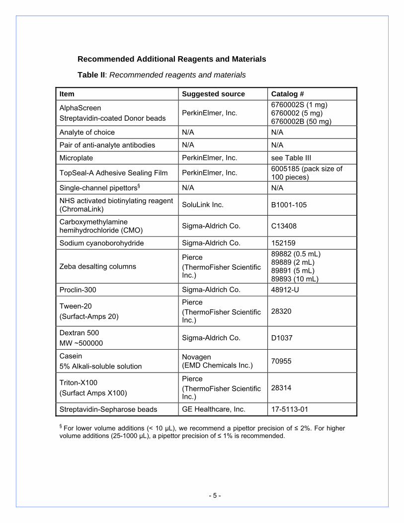
Recommended Additional Reagents and Materials
Table II: Recommended reagents and materials
Item Suggested source Catalog #
AlphaScreen Streptavidin-coated Donor beads
PerkinElmer, Inc. 6760002S (1 mg) 6760002 (5 mg) 6760002B (50 mg)
Analyte of choice N/A N/A
Pair of anti-analyte antibodies N/A N/A
Microplate PerkinElmer, Inc. see Table III
TopSeal-A Adhesive Sealing Film PerkinElmer, Inc. 6005185 (pack size of 100 pieces)
Single-channel pipettors§ N/A N/A
NHS activated biotinylating reagent (ChromaLink) SoluLink Inc. B1001-105
Carboxymethylamine hemihydrochloride (CMO) Sigma-Aldrich Co. C13408
Sodium cyanoborohydride Sigma-Aldrich Co. 152159
Zeba desalting columns Pierce (ThermoFisher Scientific Inc.)
89882 (0.5 mL) 89889 (2 mL) 89891 (5 mL) 89893 (10 mL)
Proclin-300 Sigma-Aldrich Co. 48912-U
Tween-20 (Surfact-Amps 20)
Pierce (ThermoFisher Scientific Inc.)
28320
Dextran 500 MW ~500000
Sigma-Aldrich Co. D1037
Casein 5% Alkali-soluble solution
Novagen (EMD Chemicals Inc.) 70955
Triton-X100 (Surfact Amps X100)
Pierce (ThermoFisher Scientific Inc.)
28314
Streptavidin-Sepharose beads GE Healthcare, Inc. 17-5113-01
§ For lower volume additions (< 10 μL), we recommend a pipettor precision of ≤ 2%. For higher volume additions (25-1000 μL), a pipettor precision of ≤ 1% is recommended.
- 6 -
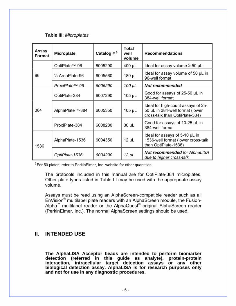
Table III: Microplates
Assay Format Microplate Catalog # §
Total well volume
Recommendations
OptiPlate™-96 6005290 400 µL Ideal for assay volume ≥ 50 µL
½ AreaPlate-96 6005560 180 µL Ideal for assay volume of 50 µL in 96-well format 96
ProxiPlate™-96 6006290 100 µL Not recommended
OptiPlate-384 6007290 105 µL Good for assays of 25-50 µL in 384-well format
AlphaPlate™-384 6005350 105 µL Ideal for high-count assays of 25-50 µL in 384-well format (lower cross-talk than OptiPlate-384)
384
ProxiPlate-384 6008280 30 µL Good for assays of 10-25 µL in 384-well format
AlphaPlate-1536 6004350 12 µL Ideal for assays of 5-10 µL in 1536-well format (lower cross-talk than OptiPlate-1536) 1536
OptiPlate-1536 6004290 12 µL Not recommended for AlphaLISA due to higher cross-talk
§ For 50 plates; refer to PerkinElmer, Inc. website for other quantities
The protocols included in this manual are for OptiPlate-384 microplates. Other plate types listed in Table III may be used with the appropriate assay volume.
Assays must be read using an AlphaScreen-compatible reader such as all EnVision® multilabel plate readers with an AlphaScreen module, the Fusion-Alpha™ multilabel reader or the AlphaQuest® original AlphaScreen reader (PerkinElmer, Inc.). The normal AlphaScreen settings should be used.
II. INTENDED USE
The AlphaLISA Acceptor beads are intended to perform biomarker detection (referred in this guide as analyte), protein-protein interaction, intracellular target detection assays or any other biological detection assay. AlphaLISA is for research purposes only and not for use in any diagnostic procedures.

- 7 -
III. PRINCIPLE OF THE ASSAY
The AlphaLISA technology allows the detection of molecules of interest in serum, plasma, cell culture supernatants or cell lysates in a very sensitive, quantitative, reproducible, and user-friendly assay. Almost, any sandwich assay can be developed to detect an analyte of interest. As shown in Figure 1, an anti-analyte antibody which is biotinylated binds the streptavidin Donor bead while another anti-analyte antibody is conjugated to AlphaLISA Acceptor beads. In the presence of the analyte, the beads come into close proximity. The excitation of the Donor beads provokes the release of singlet oxygen molecules that triggers a cascade of energy transfer in the Acceptor beads, resulting in light emission. For more details about the AlphaLISA technology, please refer to the guide entitled ‘A Practical Guide to Working with AlphaScreen’.
Figure 1: Illustration of the detection of an analyte (insulin in this example) using AlphaLISA Acceptor beads
IV. ASSAY DEVELOPMENT
A few simple steps are needed to develop an AlphaLISA analyte assay. This generic protocol is the starting point for the development of all AlphaLISA assays. Some assays may require modified protocols based on the specific nature of the analyte under investigation.

- 8 -
A. Selection of antibody pairs
To develop an AlphaLISA analyte detection assay, an antibody pair that recognizes the targeted analyte is required. It is always preferable to find an antibody pair that has already been tested in a sandwich assay. Most of the identified pairs should perform well in AlphaLISA. If no identified pairs are available, different antibodies need to be tested to find a pair that will perform optimally in AlphaLISA. All antibodies must be selected according to the following guidelines:
Each antibody should recognize a different epitope on the analyte.
The two antibodies must be specific to the analyte.
Monoclonal or purified polyclonal antibodies will perform best; avoid the use of anti-sera.
The antibodies must not be in any amine-based buffer, including Tris, glycine, bicine, tricine, etc. If buffer exchange is necessary, the buffer should be replaced with a neutral to slightly alkaline buffer, such as PBS or carbonate buffer pH 8.
Antibody solutions must not contain any protein or peptide-based stabilizers (such as BSA or gelatin).
Optimal results will be obtained with antibody concentrations above 0.5 mg/mL.
One of the two antibodies will have to be biotinylated and the other one should be conjugated to AlphaLISA Acceptor beads. It is recommended to test the two possible combinations as the sensitivity and the level of counts may differ dramatically depending on the set-up. The two combinations are as follows:
1. biotinylated antibody A + antibody B-AlphaLISA Acceptor beads
2. biotinylated antibody B + antibody A-AlphaLISA Acceptor beads
Therefore, it is recommended to biotinylate both antibodies of the pair as well as conjugating both to AlphaLISA acceptor beads to eventually select the optimal assay configuration.
(Note: Alternatively, the antibodies can be captured in an indirect assay set-up using anti-species IgG AlphaLISA Acceptor beads and/or biotinylated anti-species IgG. See AlphaLISA products on p. 24.)

- 9 -
B. Biotinylation of the antibody
The following protocol is used for biotinylation of antibodies:
a. A preliminary check of the antibody to be biotinylated is mandatory. The User must check for the following: The biotinylation will perform best when the antibody
concentration is at least 0.5 mg/mL. The antibodies must not be in any amine-based buffer, including
Tris, glycine, bicine, tricine, etc. If buffer exchange is necessary, the buffer should be replaced with a neutral to slightly alkaline buffer, such as PBS or carbonate buffer pH 8. Optimal performance will be obtained in sodium azide-free conditions.
Antibody solutions must not contain any protein or peptide-based stabilizers (such as BSA or gelatin).
The antibody will be labeled at slightly alkaline pH values (7.0-8.0) in aqueous buffer. Ensure that the antibody is soluble in these conditions.
b. Prepare antibody solution. If the antibody is provided in a powder form, resuspend at 5 mg/mL in PBS. If already in solution at neutral to slightly alkaline pH (pH ≥ 7.0), use as provided.
c. On the day of the assay, prepare a fresh solution of N-hydroxysuccinimido-ChromaLink-biotin (NHS-ChromaLink-biotin) at 2 mg/mL in PBS. Alternatively, other NHS reagents such as NHS-biotin, NHS-LC-biotin or NHS-LC-LC-biotin can also be used.
d. Add NHS-ChromaLink-biotin to the antibody solution. A 30:1 molar ratio of biotin to antibody is recommended. This represents using 7.6 µL of a 2 mg/mL NHS-ChromaLink-biotin solution for 100 µg of antibody. Adjust the volume to 200 µL with phosphate buffer pH 7.4.
e. Incubate for 2 hours at 21-23ºC. f. When using NHS-ChromaLink-biotin, a purification step using Zeba
columns is required to remove free biotin. To evaluate biotinylation efficiency, refer to SoluLink’s website (http://solulink.com).
C. AlphaLISA acceptor bead conjugation with the antibody
C1. Preliminary Notes
A preliminary check of the material to be conjugated is mandatory. The User must check for the followings:
The conjugation will perform best when the antibody concentration is at least 1 mg/mL (when conjugating 1-2 mg of

- 10 -
beads) or 0.53 mg/mL (when conjugating 2.5 mg of beads or higher amounts). Lower concentrations of antibody yield lower coupling efficiency.
The antibodies must not be in any amine-based buffer, including Tris, glycine, bicine, tricine, etc. If buffer exchange is necessary, the buffer should be replaced with a neutral to slightly alkaline buffer, such as PBS or carbonate buffer pH 8. (Note: Although both buffers can be utilized, for clarity purposes, phosphate buffer will be used in the protocol below).
Ideally, antibody solutions should not contain any protein or peptide-based stabilizers (such as BSA or gelatin). However in the presence of such substances, the conjugation process can still be performed, but may result in lower coupling efficiency in some cases.
Glycerol will significantly impact coupling efficiency (10% glycerol final in the conjugation mix will cause approx. 50% signal reduction). Dialysis of the antibody is recommended prior to coupling.
The ratio of antibody to mg of beads is an important parameter for a successful assay development. Typical coupling weight ratios (amount of beads to amount of antibody) vary from 10:1 to 50:1. When preparing low amounts of beads (1-2 mg), a 10:1 ratio is recommended (i.e. 1 mg of Acceptor beads to 0.1 mg of antibody), while a ratio of 50:1 is used with bead amounts equal to or higher than 2.5 mg to minimize the antibody consumption (i.e. 5 mg of Acceptor beads to 0.1 mg of antibody).
C2. Protocol for conjugating 1 mg AlphaLISA Acceptor beads (10:1 coupling ratio)
This procedure is appropriate for a solution of antibody ≥ 1 mg/mL; if the concentration is below 1 mg/mL, the antibody solution must be concentrated using an iCON Concentrator (Pierce Cat# 89886), Microcon or Centricon (Ultracell YM-30, Millipore, cat# 4208 or 42409). Washing
Wash AlphaLISA Acceptor beads (50 μL at 20 mg/mL) once with 50 μL PBS: centrifuge at 16,000 × g or maximum speed for 15 min and then discard the supernatant.
Conjugation In an eppendorf tube, add the following reagents: 1 mg of AlphaLISA Acceptor bead pellet (prepared as described
above)
- 11 -
0.1 mg of antibody the appropriate volume of 0.13 M phosphate buffer pH 8.0 to
obtain a final reaction volume of 200 µL
1.25 μL of 10% Tween-20 10 µL of a 25 mg/mL solution of NaBH3CN in water (prepare fresh
as required). Incubate for 18-24 hours at 37ºC. Longer incubation times up to 48 hours might be used, which could result in higher conjugation yields.
Blocking Prepare a fresh 65 mg/mL solution of carboxy-methoxylamine
(CMO) in a 0.8 M NaOH solution. Add 10 µL of CMO solution to the reaction (to block unreacted
sites). Incubate for 1 hour at 37ºC.
Purification Centrifuge for 15 minutes at 16,000 × g (or maximum speed). Remove the supernatant with a micropipette and resuspend the
bead pellet in 200 μL of 0.1 M Tris-HCl pH 8.0. Centrifuge for 15 minutes at 16,000 × g (or maximum speed),
then remove the supernatant. Repeat the washing steps (resuspend the pellet and centrifuge)
another time. After the last centrifugation, resuspend the beads at 5 mg/mL in
storage buffer (200 μL of PBS + 0.05% Proclin-300 as a preservative).
Vortex, briefly spin down and sonicate the bead solution (20 short pulses of 1 second using a probe sonicator).
Storage Store the conjugated Acceptor bead solution at 4ºC.
Important note: always vortex conjugated AlphaLISA Acceptor beads before use, as beads tend to settle with time.
C3. Protocol for conjugating 5 mg AlphaLISA Acceptor beads (50:1 coupling ratio)
This procedure is appropriate for a solution of antibody ≥ 0.53 mg/mL; if the concentration is below 0.53 mg/mL the antibody solution must be concentrated using an iCON Concentrator (Pierce Cat# 89886), Microcon or Centricon (Ultracell YM-30, Millipore, cat# 4208 or 42409).
- 12 -
Washing Wash AlphaLISA Acceptor beads (250 μL at 20 mg/mL) once with
250 μL PBS: centrifuge at 16,000 × g or maximum speed for 15 min and then discard the supernatant.
Conjugation In an eppendorf tube, add the following reagents: 0.1 mg of antibody to 5 mg of bead pellet (prepared as described
above) the appropriate volume of 0.13 M phosphate buffer pH 8.0 to
obtain a final reaction volume of 200 µL 1.25 µL of 10 % Tween-20 10 µL of a 25 mg/mL solution of NaBH3CN in water (prepare fresh
as required). Incubate for 18-24 hours at 37ºC.
Blocking Prepare a fresh 65 mg/mL solution of carboxy-methoxylamine
(CMO) in a 0.8 M NaOH solution. Add 10 µL of CMO solution to the reaction (to block unreacted
sites). Incubate for 1 hour at 37ºC.
Purification Centrifuge for 15 minutes at 16,000 × g (or maximum speed). Remove the supernatant with a micropipette and resuspend the
bead pellet in 1 mL of 0.1 M Tris-HCl pH 8.0 (200 μg per mg of beads).
Centrifuge for 15 minutes at 16,000 × g (or maximum speed), then remove the supernatant.
Repeat the washing steps (resuspend the pellet and centrifuge) another time.
After the last centrifugation, resuspend the beads at 5 mg/mL in storage buffer (1 mL of PBS + 0.05% Proclin-300 as a preservative).
Vortex, briefly spin down and sonicate the bead solution (20 short pulses of 1 second using a probe sonicator).
Storage Store the conjugated Acceptor bead solution at 4ºC.
- 13 -
Important note: always vortex conjugated AlphaLISA Acceptor beads before use, as beads tend to settle with time.
D. Determination of the best antibody pair
One of the first development steps is the selection of the antibody combination. As previously mentioned, both combinations of antibody set-up must be tested:
1. biotinylated antibody A + antibody B-AlphaLISA Acceptor beads
2. biotinylated antibody B + antibody A-AlphaLISA Acceptor beads
This section describes the method to be used to perform this selection.
D1. Selection of optimal antibody pair(s) using a matrix assay
A matrix assay should be performed using fixed concentrations of antibodies and 2-3 different concentrations of analyte (within the usual working range) and a negative control (no analyte).
It is important to note that this method is a guideline; it will need to be optimized for every analyte studied.
These preliminary conditions could be used for this experiment:
Typical assay buffer: 25 mM HEPES pH 7.4 0.5% Triton X-100 0.1% Casein 1 mg/mL Dextran 500 Adjust pH to 7.4
This buffer is available from PerkinElmer, Inc. as a 10X solution: AlphaLISA ImmunoAssay Buffer (10X) (Cat No. AL000). Dilute 10-fold with water prior to use.
In an OptiPlate-384 microplate add: 5 µL of the analyte diluted in assay buffer (use various
concentrations that are in the working range for the target detection)
10 µL of fixed amount of the biotinylated antibody (Use a final concentration between 0.3 nM and 3 nM of biotin-antibody. Often the optimal biotinylated antibody
- 14 -
concentration is 1 nM.) 10 µL of antibody-coupled AlphaLISA Acceptor beads at
50 µg/mL (10 µg/mL final concentration in each well) Incubate at 23ºC for 1 h then add: 25 µL of streptavidin Donor beads at 80 µg/mL prepared
under subdued light conditions (40 µg/mL final concentration in each well)
Incubate at 23ºC in the dark for 30 min and read on an AlphaScreen reader.
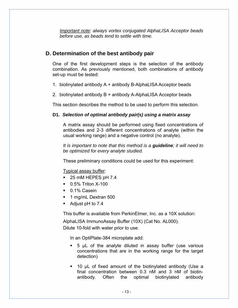
Select the pair(s) of antibodies providing the highest signal-to-background (S/B) ratio and the best sensitivity as defined as the highest S/B ratio for the condition with lowest concentration of analyte. Once selected, determination of optimal biotinylated antibody concentration can be performed.
2.5 nM analyte
Ab1Ab2
Ab3Ab4
0
10
20
Ab1
Ab2Biotinylatedantibodies
255075
100125150175200
Ab3
Ab4
Antibodies conjugated on AlphaLISA Acceptor Beads
S/B
rat
io
2.5 pM analyte
Ab1Ab2
Ab3Ab4
0
5
10
15
20
25
30
35
Ab1
Ab2
Ab3
Ab4Biotinylatedantibodies
Antibodies conjugated on AlphaLISA Acceptor Beads
S/B
rat
io
Figure 2: Determination of the best antibody pair. S/B ratio obtained for each antibody pair (each permutation possible), with 2.5 nM and 2.5 pM of analyte (S/B ratio is calculated as AlphaLISA signal obtained for 2.5 nM or 2.5 pM analyte ÷ AlphaLISA signal obtained without analyte (background)). In this example, Antibody-Ab3-conjugated AlphaLISA Acceptor Beads and Biotinylated-Antibody-Ab4 were selected.
- 15 -
D2. Determination of optimal biotinylated antibody concentration
An antibody titration curve should be performed using a fixed concentration of analyte (within the usual working range).
The following preliminary conditions may be used for this titration.
It is important to note that the method is a guideline; it will need to be optimized for every analyte studied.
In an OptiPlate-384 microplate add: 5 µL of the analyte diluted in assay buffer (use a fixed
concentration that is in the working range for the target detection)
10 µL of increasing amounts of the biotinylated antibody (as a starting point, use concentrations between 0.1 nM to 100 nM final concentration of biotin-antibody)
10 µL of antibody-coupled AlphaLISA Acceptor beads at 50 µg/mL (10 µg/mL final concentration in the well)
Incubate at 23ºC for 1 h then add: 25 µL of streptavidin-Donor bead solution at 80 µg/mL
prepared under subdued light conditions (40 µg/mL final concentration in each well)
Incubate at 23ºC in the dark for 30 min and read on an AlphaScreen reader.
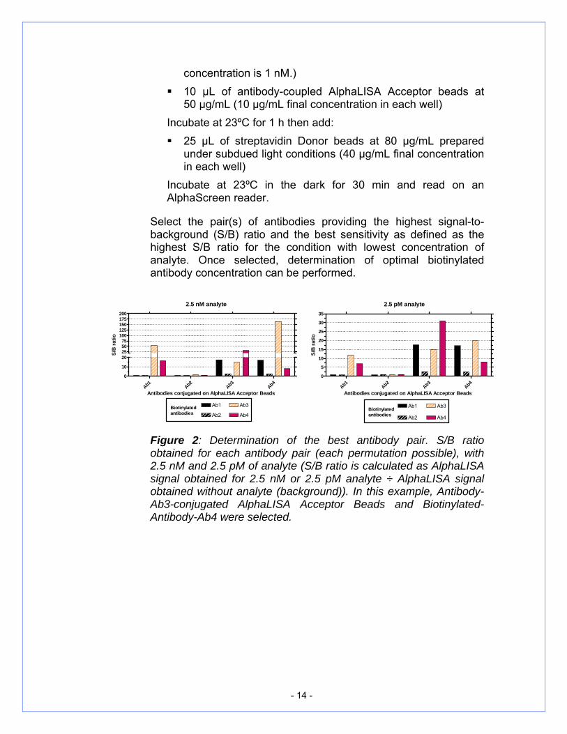
A bell-shaped curve should be obtained (Figure 3). The highest signal obtained indicates the hook point (highest signal before saturation of the bead binding capacity) of the biotinylated anti-analyte antibody. A sub-hooking concentration should be used for the next optimization steps. A lack of specific signal likely indicates that the selected antibodies are not able to capture the analyte simultaneously in this specific configuration.
For the selection of the best combination of antibody pair, the usual criterion is the highest S/B signal. However, in order to confirm the choice of the antibody pair, a calibration curve of the analyte should be performed. This will allow the measurement of the analyte detection limit and the dynamic range of the specific pair of antibody in the AlphaLISA assay.
- 16 -
Figure 3: Titration curve for an insulin detection assay, prepared in assay buffer: in this particular example, the optimal antibody concentration was determined to be 1 nM.
D3. Calibration curve of analyte
The most important parameters to determine the best combination of antibodies are the detection limit, the assay window (S/B) and the assay dynamic range.
The following conditions may be used for the analyte calibration curve.
In an OptiPlate-384 microplate add: 5 µL of the analyte diluted in assay buffer (use dilutions that
cover the working range for the target detection plus few points above and below the range of target detection as well as negative controls (background) consisting of 3 independent conditions without analyte in triplicate for a total of 9 points)
20 µL of the antibody mix: biotinylated antibody (as a starting point, use the concentration determined by titration, Section D2)
AND antibody-conjugated AlphaLISA Acceptor beads at 25 µg/mL (10 µg/mL final concentration in each well)
Incubate at 23ºC for 1 h then add: 25 µL of streptavidin-Donor bead solution at 80 µg/mL
prepared under subdued light conditions (40 µg/mL final concentration in the well)
Incubate at 23ºC in the dark for 30 min and read on an
-12 -11 -10 -9 -8 -70
50000
100000
150000
200000
250000
300000
350000Hook point
[biotin-antibody] (M)
Alp
haLI
SA S
igna
l (co
unts
)

- 17 -
AlphaScreen reader.
The lower detection limit (LDL) is usually calculated as follows: Average the 9 background values and calculate the standard
deviation (SD). Multiply the standard deviation (SD) by 3 Add the 3XSD value calculated to the average background
signal. On the graph, extrapolate the value obtained (AlphaLISA
signal counts) to determine the corresponding analyte concentration. This concentration is the lowest the assay can detect and corresponds to the lower detection limit (LDL).
The assay dynamic range corresponds to the concentration window in the standard curve running from the lower detection limit to the maximum concentration up to (but excluding) the hook point.
Data can be analyzed using linear or nonlinear regression analysis. However, wider dynamic ranges are usually interpreted using nonlinear regression Eq.1. Four-Parameter Logistic Model also described as Sigmoidal Dose-Response (variable slope) where the four parameters to be estimated are Top, Bottom, EC50 and Slope. For more details refer to the NIH Chemical Genomics Center manual (http://ncgc.nih.gov/guidance/manual_toc.html).
Slope
ECionconcentrat
TopBottomTopsponse)(1
)(Re
50
+
−+= Eq. (1)
Top’ refers to the top asymptote, ‘Bottom’ refers to the bottom asymptote, and ‘EC50’ refers to the concentration at which the response is halfway between Top and Bottom.
- 18 -
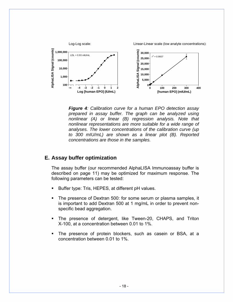
Log-Log scale: Linear-Linear scale (low analyte concentrations):
100
1,000
10,000
100,000
1,000,000
-4 -3 -2 -1 0 1 2-∞
LDL = 0.93 mIU/mL
Log [human EPO] (IU/mL)
Alp
haLI
SA S
igna
l (co
unts
)
0 100 200 300 4000
5,000
10,000
15,000
20,000
25,000
30,000r2 = 0.9937
[human EPO] (mIU/mL)
Alp
haLI
SA S
igna
l (co
unts
)
Figure 4: Calibration curve for a human EPO detection assay prepared in assay buffer. The graph can be analyzed using nonlinear (A) or linear (B) regression analysis. Note that nonlinear representations are more suitable for a wide range of analyses. The lower concentrations of the calibration curve (up to 300 mIU/mL) are shown as a linear plot (B). Reported concentrations are those in the samples.
E. Assay buffer optimization
The assay buffer (our recommended AlphaLISA Immunoassay buffer is described on page 11) may be optimized for maximum response. The following parameters can be tested:
Buffer type: Tris, HEPES, at different pH values.
The presence of Dextran 500: for some serum or plasma samples, it is important to add Dextran 500 at 1 mg/mL in order to prevent non-specific bead aggregation.
The presence of detergent, like Tween-20, CHAPS, and Triton X-100, at a concentration between 0.01 to 1%.
The presence of protein blockers, such as casein or BSA, at a concentration between 0.01 to 1%.
- 19 -
F. Optimization of the assay volume, incubation time and order of addition
The assay may be further optimized by changing the following parameters.
F1. Assay volume
Most analyte assays will perform best in 50-75 µL final volume in 96-well plates, 25-50 µL in 384-well plates and 5-10 µL in 1536-well plates. However, the volume can be adjusted to fulfill specific user requirements. If working with serum or plasma samples, it is strongly recommended that the sample volume represents no more than 10% of the total volume in the well in order to reduce interference. Less interference will be observed using the lowest sample volume possible. It is always possible to pre-dilute the sample in the matrix solution if the concentration of the analyte in the sample permits.
F2. Incubation time
A time course could be performed following each addition step. A time course could also be performed after the addition of Donor beads. Usually the signal will reach a plateau after a few hours.
F3. Order of addition
For the majority of analyte assays, the order of addition described in Section D3 performs best. However, the addition steps can be changed in order to optimize the assay sensitivity. It could be beneficial to add the biotin-antibody and the Acceptor beads separately. In this case, an incubation time may be necessary between each addition step.
From our experience, pre-mixing the biotinylated antibody with the Donor beads lowers the sensitivity of the assay and should in most cases be avoided.
G. Determination of the level of analyte in unknown samples by interpolation from the calibration curve
In order to determine the concentration of an analyte in a serum or plasma sample, a calibration curve with known concentrations of the analyte should be performed first. The calibrators (solutions containing a known concentration of analyte) should be prepared in a “matrix
- 20 -
solution”. We recommend using a serum or plasma (similar to the samples) first depleted of the analyte of interest as matrix solution then spiked with known concentrations of analyte.
G1. Analyte depletion of serum
It is possible to prepare analyte-depleted sera by treatment with streptavidin-Sepharose beads and the biotinylated antibody.
Evaluate the amount of biotinylated-antibody and streptavidin-Sepharose beads required for the depletion. Those conditions will need to be adjusted and we suggest using as starting condition a molar ratio of analyte/antibody of 1/100 and a molar ratio of antibody/streptavidin of 1/20.
Wash the Sepharose beads with PBS buffer prior to use.
Mix biotinylated antibody with Sepharose beads for 2 h at 4°C.
Centrifuge at 1000 × g for 5 min, remove the supernatant and add serum to pelleted beads and incubate overnight at 4°C.
Remove beads from serum by centrifuging at 16,000 × g (or maximum speed) for 5 min. Transfer supernatant to a new tube and centrifuge again at 16,000 × g (or maximum speed) for 5 min.
Collect the supernatant and store depleted serum in aliquots at -20°C.
Alternatively, a charcoal treatment may be used to remove the analyte of interest. Several protocols are described in the literature (see references page 20).
An artificial matrix solution could also be prepared. Such a matrix should behave the same way in the AlphaLISA assay as the real samples (same level of signal interference). In such instance, high concentrations of BSA or any other signal quencher could be used for this purpose. Finally, the concentrations of analyte used to perform the calibration curve must cover the range expected to be found in the samples.
- 21 -
G2. Calibration curve
A general protocol to generate a calibration curve in the matrix solution and determine the analyte concentration of unknown samples is presented below. As mentioned earlier, it might be necessary to optimize the assay conditions to meet the specific assay requirements.
For calibration curve: Add 1-5 µL of calibrators diluted in matrix solution
For unknown samples: Add 1-5 µL of unknown samples
Then to calibrator or unknown sample, Add 20-24 µL of antibody-conjugated AlphaLISA Acceptor
beads + biotinylated antibody solution Incubate for 30 min at 23ºC. Add 25 µL of streptavidin-Donor bead solution
Incubate for 60 min at 23ºC, in the dark and read plate on an AlphaScreen reader.
It is advised to prepare three to five replicates of each calibrator concentration.
The calibrator serial dilutions are prepared in the matrix solution, while the solutions of antibody-conjugated AlphaLISA Acceptor beads and of biotinylated antibody are prepared in the assay buffer (see page 11).
It is also possible to predilute the calibrators in the matrix solution, aliquot the solutions and store the vials at -80°C for future use.
On the calibration curve (Figure 5), the increasing concentrations of analyte are on the x-axis and the AlphaLISA signal on the y-axis.
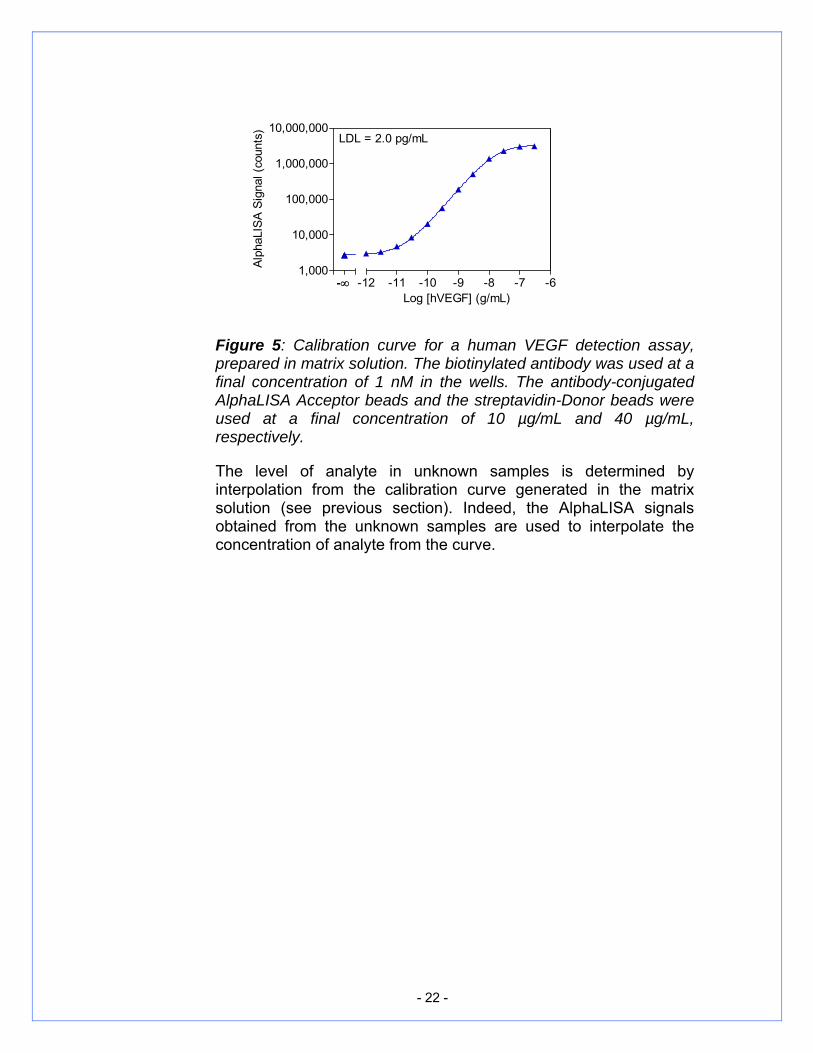
- 22 -
Figure 5: Calibration curve for a human VEGF detection assay, prepared in matrix solution. The biotinylated antibody was used at a final concentration of 1 nM in the wells. The antibody-conjugated AlphaLISA Acceptor beads and the streptavidin-Donor beads were used at a final concentration of 10 µg/mL and 40 µg/mL, respectively.
The level of analyte in unknown samples is determined by interpolation from the calibration curve generated in the matrix solution (see previous section). Indeed, the AlphaLISA signals obtained from the unknown samples are used to interpolate the concentration of analyte from the curve.
1,000
10,000
100,000
1,000,000
10,000,000
-12 -11 -10 -9 -8 -7 -6-∞
LDL = 2.0 pg/mL
Log [hVEGF] (g/mL)
Alph
aLIS
A Si
gnal
(cou
nts)
- 23 -
V. REFERENCES
A PRACTICAL GUIDE TO WORKING WITH ALPHASCREEN® (PerkinElmer, Inc.).
APPLICATION NOTE (PerkinElmer, Inc.): AlphaScreen® Insulin Detection Assay Using AlphaLISA® Acceptor beads.
CHARCOAL-TREATED SERUM/PLASMA PREPARATION: Myrick J.E. et al., (1989), Clin. Chem. 35, pp 37-42 Glick R.D. et al., (2000), J. Pediat. Surg. 35, pp 465-472 Raffo A.J. et al., (1995), Cancer Res. 55, pp 4438-4445
LUMINESCENT OXYGEN CHANNELING ASSAY (LOCI™): sensitive, broadly applicable homogeneous immunoassay method: Ullman E.F. et al., (1996), Clin. Chem. 42, pp 1518-1526.
A LUMINESCENT OXYGEN CHANNELING IMMUNOASSAY FOR THE DETERMINATION OF INSULIN IN HUMAN PLASMA: Poulsen F. and Jensen K.B. (2007), J. Biomol Screen. 12(2):240-247
DEVELOPMENT OF HOMOGENEOUS 384-WELL HIGH-THROUGHPUT SCREENING ASSAYS FOR Aβ1-40 and Aβ1-42 USING ALPHASCREENTM TECHNOLOGY: Szekeres P., Leong K., Day T., Kingston A., and Karran E. (2008), J. Biomol. Screen. 13(2):101-111
- 24 -
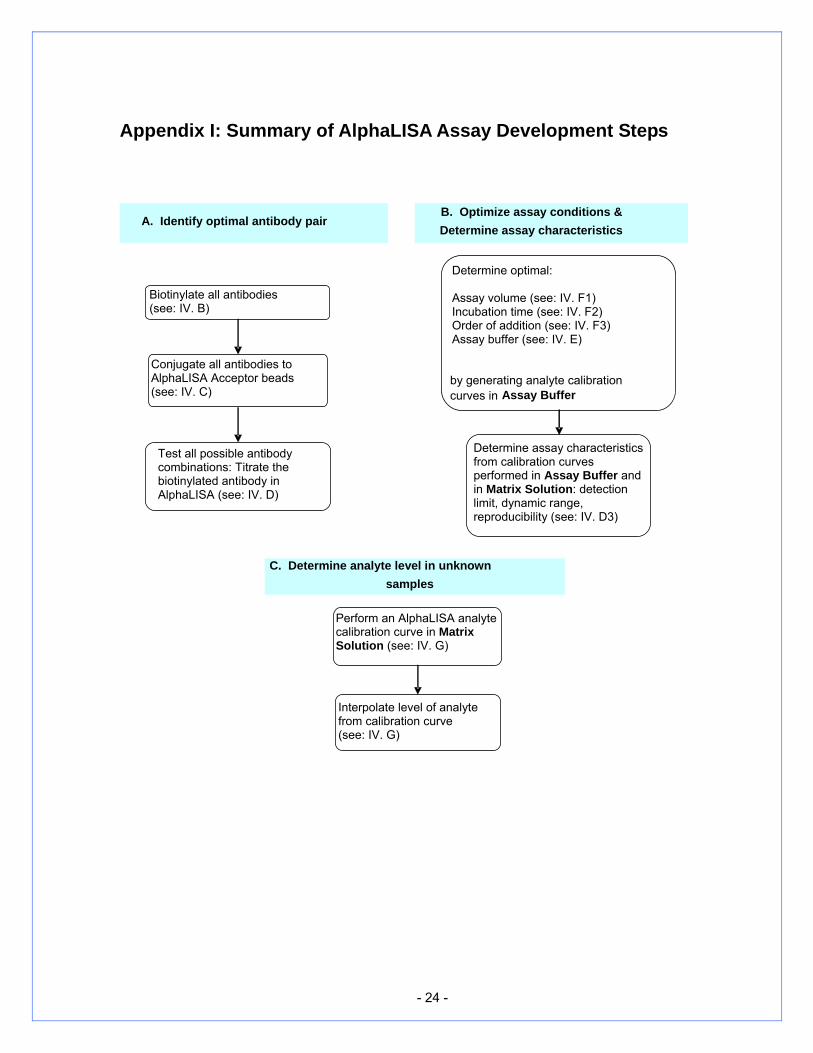
Appendix I: Summary of AlphaLISA Assay Development Steps
B. Optimize assay conditions & Determine assay characteristics
A. Identify optimal antibody pair
C. Determine analyte level in unknown samples
Biotinylate all antibodies (see: IV. B)
Conjugate all antibodies to AlphaLISA Acceptor beads (see: IV. C)
Test all possible antibody combinations: Titrate the biotinylated antibody in AlphaLISA (see: IV. D)
Determine optimal: Assay volume (see: IV. F1) Incubation time (see: IV. F2) Order of addition (see: IV. F3) Assay buffer (see: IV. E)
by generating analyte calibration curves in Assay Buffer
Perform an AlphaLISA analyte calibration curve in Matrix Solution (see: IV. G)
Determine assay characteristics from calibration curves performed in Assay Buffer and in Matrix Solution: detection limit, dynamic range, reproducibility (see: IV. D3)
Interpolate level of analyte from calibration curve (see: IV. G)
- 25 -
Appendix II: Serum Sample Preparation
a. To prepare serum samples, whole blood is directly drawn into a Vacutainer® serum tube that contains no anticoagulant. Let blood clot at room temperature for 30 minutes.
b. Promptly centrifuge the clotted blood at 2,000 – 3,000 × g for 15 minutes at 4 +/- 2oC.
c. Transfer and store serum samples in separate tubes. Date and identify each sample.
d. Use freshly prepared serum or store samples in aliquots at ≤ -20oC for later use. Avoid freeze/thaw cycles.

- 26 -
MANUFACTURED BY: PerkinElmer BioSignal, Inc. 1744, William Street Montreal, Quebec Canada H3J 1R4 To place an order Please contact: PerkinElmer, Inc. 940 Winter St. Waltham, MA 02451 (800) 762-4000 or (+1) 203-925-4600
To find your local PerkinElmer office, please visit: www.perkinelmer.com/lasoffices For technical information and support In US and rest of the world, please contact: [email protected] In Europe, Middle-East and Africa, please contact: [email protected]

- 27 -
OTHER ALPHALISA PRODUCTS Stand-Alone Reagents*
AlphaLISA Protein A Acceptor beads (Cat No. AL101) AlphaLISA Protein G Acceptor beads (Cat No. AL102) AlphaLISA anti-human IgG Acceptor beads (Cat No. AL103) AlphaLISA anti-rabbit IgG Acceptor beads (Cat No. AL104) AlphaLISA anti-mouse IgG Acceptor beads (Cat No. AL105) AlphaLISA anti-rat IgG Acceptor beads (Cat No. AL106) AlphaLISA anti-goat IgG Acceptor beads (Cat No. AL107) AlphaLISA Nickel Chelate Acceptor beads (Cat No. AL108) AlphaLISA Glutathione Acceptor beads (Cat No. AL109) AlphaLISA anti-GST Acceptor beads (Cat No. AL110) AlphaLISA anti-c-myc Acceptor beads (Cat No. AL111) AlphaLISA anti-FLAG Acceptor beads (Cat No. AL112) AlphaLISA anti-DIG Acceptor beads (Cat No. AL113) AlphaScreen Nickel Chelate Donor beads (Cat No. AS101) AlphaScreen Glutathione Donor beads (Cat No. 6765300) AlphaLISA ImmunoAssay Buffer (10X) (Cat No. AL000) AlphaLISA Universal Assay Buffer (5X) (Cat No. AL001)
* Visit our Web site for the different formats available: www.perkinelmer.com/nowashelisa AlphaLISA ImmunoAssay Kits For a complete list of kits, visit our Web site: www.perkinelmer.com/nowashelisa Note: AlphaLISA is for research purposes only and not for use in diagnostic procedures.

M-6772001/2/3/4-04
PerkinElmer, Inc. 940 Winter St.
Waltham, MA 02451 USA (800) 762-4000 or (+1) 203-925-4602
www.perkinelmer.com For a complete listing of our global offices, visit www.perkinelmer.com/lasoffices