amino-acid sequenceof equine renal metallothionein-ib · 3416 biochemistry: kojimaetal. ginineis...
TRANSCRIPT
Proc. Natl. Acad. Sci. USAVol. 73, No. 10, pp. 3413-3417, October 1976Biochemistry
Amino-acid sequence of equine renal metallothionein-IB(protein primary structure/cadmium- and zinc-binding protein/metal-chelating sequence/cysteine distribution in sequence/mercaptide binding site)
YUTAKA KoJIMA*, CHRISTINE BERGER*, BERT L. VALLEEt, AND JEREMIAS H. R. KAGI***Biochemisches Institut der Universitit Zurich, Zuirichbergstrasse 4, CH-8028 Zurich, Switzerland; tBiophysics Research Laboratory, Department of BiologicalChemistry, Harvard Medical School, and the Division of Medical Biology, Peter Bent Brigham Hospital, Boston, Massachusetts 02115
Contributed by Bert L. Vallee, July 12, 1976
ABSTRACT The amino-acid sequence of a metallothioneinis reported. Metallothionein is a widely distributed, extremelycysteine-rich, low-molecular-weight protein containing largeamounts of cadmium and/or zinc. Metallothionein-IB is oneof the two principal variants occurring in equine kidney cortex.The single-chain protein contains 61 amino acids and hasthe composition Cys2oSer8Lys7ArglAla7GlysVal3Asp2Asnl-GluiGln2Pro2ThriMeti(Cd+Znh. Its amino-terminal residueis N-acetylmethionine. The sequence shows distinct clusteringof the twenty cysteinyl residues into seven groups separated bystretches of at least three other residues. Within these groupsthe cysteines occur seven times in alternating Cys-X-Cys se-quences and three times each in Cys-Cys and Cys-X-X-Cys se-quences, where X is an amino acid other than cysteine. Anotherunique feature is the close association of serine and of the basicamino acids with cysteine, as manifested by the occurrence ofseven Ser-Cys, four Cys-Lys, one Cys-Arg, and three Lys-Cyssequences. These findings are in agreement with the previoussuggestion that metallothionein has structurally definedmetal-binding sites, most of which contain three cysteinylresidues as the principal metal-binding ligands. The chargedifference between the metal-free and the metal-containingprotein is consistent with the formation of negatively chargedtrimercaptide complexes with cadmium and/or zinc ions. Thepossible additional involvement of serine and the basic aminoacids in metal binding is discussed.
Metallothionein, a cadmium- and zinc-containing, cysteine-richprotein of low molecular weight, was recognized in equine renalcortex by Margoshes and Vallee in 1957 (1, 2). The same proteinoccurs in large quantity also in human kidney (3), in equine andhuman liver (4, 5), and accumulates in various parenchymatoustissues of laboratory animals after administration of salts ofcadmium, zinc, or certain other heavy metals, implicating a rolein metal metabolism and detoxication (6-17). Similar proteinshave recently also been identified in lower vertebrates (18) andyeast (19).The metallothioneins that are characterized best contain
between 6 and 11% metal and between 30 and 35% cysteinewhen calculated on the basis of the weight of the polypeptidechain, but are completely devoid of aromatic amino acids andhistidine. Early spectroscopic and chemical studies establishedthat the metal ions are bound to the cysteinyl side chainsthrough mercaptide bonds (20). To obtain a more completeunderstanding of the metal-protein interactions and thephysiological functions dependent on them, as well as of evo-lutionary relationships, studies were undertaken to determinethe covalent structure of several genetic variants of equine andhuman metallothionein. In this preliminary note we report thecomplete amino-acid sequence of variant 1B of equine renalmetallothionein.
MATERIALS AND METHODSSeparation of Metallothionein Variants. Metallothionein§
purified from horse kidney cortex by the procedure of Kagi etal. (4) was resolved into two main fractions by ion exchangechromatography on DEAE-cellulose DE-23, using as elutingbuffer 0.025 M ammonium acetate adjusted to pH 10.0 withammonium hydroxide. The two fractions constituting metal-lothionein variants of equal molecular size but of differingamino-acid and metal composition (unpublished results) weredesignated metallothionein-lA (MT-1A) and metallothio-nein-iB (MT-1B). The studies reported in this communicationare restricted to MT-1B, the fraction more negatively chargedat pH 10.0.Chemical Analyses. Analyses for amino acids (21) and metals
were made by established procedures. Acetic acid liberated onacid hydrolysis from MT-1B or its amino-terminal peptide wasidentified and quantified by combined gas chromatographyand mass spectroscopy. Thin-layer chromatography of theamino-terminal N-acetyl-peptide was performed accordingto Brenner and Niederwieser (22).Sequence Determination. Most studies were made on the
S-pyridylethylated derivative of MT-1B. Following removalof the metal by exposure to 6 M guanidine-HCl in 0.1 M HCIthe protein moiety, thionein-iB, was reacted with 4-vinylpy-ridine at pH 8.0 using a modification of the method of Fried-man et al. (23). The principal sets of fragments employed wereobtained either by selective cleavage of the acid-labile bondbetween Asp-2 and Pro-3 or by tryptic cleavage at the singlearginyl bond (Arg-25). For acid cleavage S-pyridylethylatedthionein-lB was incubated in 70% (vol/vol) formic acid for 96hr at 40°. The resulting fragments AP-I and AP-II were sepa-rated on a column of Sephadex G-25. For selective trypticcleavage the lysyl C-amino groups of S-pyridylethylated thi-onein-iB were blocked by succinylation (24). The fragmentsT-I and T-II obtained by tryptic digestion (25) were separatedon a column of Bio-Gel P-6.Automated Edman degradation was carried out with a
Beckman Sequencer model 890B (updated) using a modifica-tion (26) of the method of Edman and Begg (27). The phenyl-thiazolinones were converted to the phenylthiohydantoins foridentification by gas chromatography (26, 28) and by thin-layerchromatography (26, 27). Where indicated phenylthiohy-dantoin derivatives were identified and quantitated also byamino-acid analysis after reconversion to the free amino acidsby hydrolysis with constant-boiling hydriodic acid (29).The sequence of the carboxyl-terminal region was deter-
mined by digestion with bovine pancreatic carboxypeptidases
§ Metallothionein-1 refers to the major anionic fraction of equine renalmetallothionein obtained by the isolation procedure described pre-
viously (4, 20).
Abbreviations: MT-1A, equine renal metallothionein-lA; MT-1B,equine renal metallothionein-lB.* To whom correspondence should be addressed.
3413
Dow
nloa
ded
by g
uest
on
Apr
il 5,
202
0
Proc. Natl. Acad. Sci. USA 73 (1976)
A and B (Worthington) (30) using the S-carbamidomethylatedderivative of the protein, prepared by reacting thionein-lB withiodoacetamide by a modification of the procedure employedfor alkylation with iodoacetate (31). In some cases amino-acidpositions were confirmed by manual subtractive Edman deg-radation (25) and by digestion with carboxypeptidases A, B, andC (R6hm GmbH, Darmstadt, Germany) (32) of tryptic peptidesof the S-carboxymethylated derivative separated on Dowex AG1W-X2 (Bio-Rad Laboratories) with a 2,4,6-trimethylpyri-dine/pyridine/acetic acid gradient system (33).
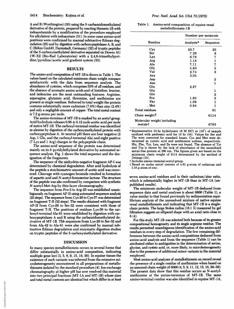
RESULTSThe amino-acid composition of MT-1B is shown in Table 1. Thevalues based on the calculated minimum chain weight comparesatisfactorily with the data from sequence analysis. Theabundance of cysteine, which comprises 33% of all residues, andthe absence of aromatic amino acids and of histidine, leucine,and isoleucine are the most outstanding features. Arginine,asparagine, glutamic acid, threonine, and methionine are
present as single residues. Referred to total weight the proteincontains substantially more cadmium (7.6%) than zinc (2.4%)and only a negligible amount of copper. The total metal contentis 7.2 g-atoms per mole.The amino-terminus of MT-lB is masked by an acetyl group.
Acid hydrolysis releases 0.96 + 0.12 mole acetic acid per moleof native MT-1B. The carboxyl-terminal residue was identifiedas alanine by digestion of the carboxymethylated protein withcarboxypeptidase A. At neutral pH there are four negative (2Asp, 1 Glu, and the carboxyl-terminal Ala) and eight positive(7 Lys and 1 Arg) charges in the polypeptide chain.The amino-acid sequence of the protein was determined
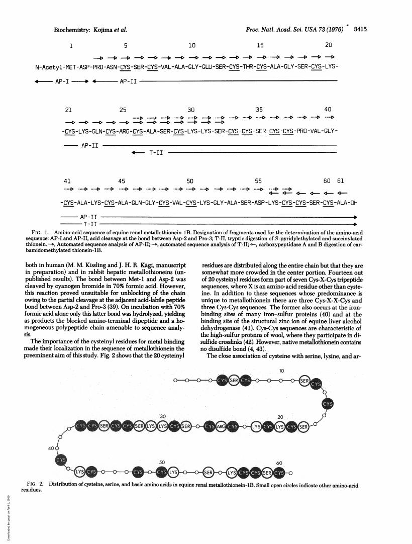
mainly on its S-pyridylethylated derivative by automated se-quence analysis. Fig. 1 shows the total sequence and the des-ignation of the fragments.The sequence of the ninhydrin-negative fragment AP-I was
determined by chemical degradation. After acid hydrolysis ofthe peptide a stoichiometric amount of acetic acid was recov-
ered. Cleavage with cyanogen bromide resulted in formationof aspartic acid and N-acetyl-homoserine lactone. The structureof the peptide was also confirmed by comparison with syntheticN-acetyl-Met-Asp by thin-layer chromatography.The sequence from Pro-3 to Arg-25 was established unam-
biguously on fragment AP-1I using automated sequence analysis(23 steps). The sequence from Cys-26 to Cys-57 was determinedon fragment T-II (32 steps). The results obtained with fragmentAP-II from Cys-26 to Ser-32 were consistent with those offragment T-II. The positions of residues Lys-56 to the car-
boxyl-terminal Ala-61 were established by digestion with car-
boxypeptidases A and B using the carbamidomethylated de-rivative of MT-1B. The sequences from Lys-20 to Ser-32 andfrom Ala-42 to Ala-61 were also confirmed by manual sub-tractive Edman degradation and enzymatic digestion studieson tryptic peptides of the S-carboxymethylated derivative.
DISCUSSION
In many species metallothionein occurs in several forms thatdiffer substantially in amino-acid composition, indicatingmultiple gene loci (3, 5, 8, 9, 15, 16, 36). In equine tissues theexistence of such variants was inferred from the extensive mi-croheterogeneity encountered in all preparations of metallo-thionein isolated by the standard procedure (4). Ion-exchangechromatography at higher pH has now resolved this materialinto two principal fractions (MT-lA and MT-lB) whose sizes
Table 1. Amino-acid composition of equine renalmetallothionein-lB
Number per molecule
Residue Analysis* Sequence
Cys 20.7 20Ser 7.29 8Lys 6.79 7Arg 1.16 1Ala 7.11 7Gly 5.60 5Val 2.74 3Asx 3.05Asp 2Asn 1Glx 2.87Glu 1Gln 2Pro 1.80 2Thr 1.09 1Met 0.94 1
Total residues 61
Chain weightt 6114
Molecular weight includingmetalst 6793
* Representative 24-hr hydrolysate (6 M HOl at 1100) of sampleoxidized with performic acid for 16 hr (34). Values for Ser andThr were corrected for standard losses. Cys and Met were de-termined as cysteic acid and methionine sulfone, respectively.His, Phe, Tyr, Leu, and Re were not found. The absence of Tyrand Trp is shown by the lack of absorbance of the unoxidizedmetal-free protein at 280 nm. The figures given are based on theminimum chain weight of 6114 determined by the method ofDelaage (35).
t Includes amino-terminal acetyl group.T Based on molar metal content of 4.61 g-atom of cadmium and2.55 g-atom of zinc.
seven amino-acid residues and in their cadmium/zinc ratio,which is substantially higher in MT-1B than in MT-1A (un-published results).The minimum molecular weight of MT-1B deduced from
sequence data and metal analyses is about 6800 (Table 1), avalue similar to that found previously by sedimentation equi-librium analysis of the unresolved mixture of native equinerenal metallothionein and indicating that MT-1B is a single-chain protein. The large Stokes radius (16.1 A) measured by gelfiltration suggests an ellipsoid shape with an axial ratio close to6 (4).
For this study MT-1B was selected both because of its greatercompositional homogeneity and its single arginine residue. Theresults permitted unambiguous identification of the amino-acidresidues in every step of degradation. The few remaining dif-ferences between the amino-acid compositions deduced fromamino-acid analysis and from the sequence (Table 1) can beattributed either to ambiguities in the determination of serine,glycine, and cysteic acid, or, more likely, to microheterogeneitydue to the presence of additional minor variants in the materialemployed.
Most amino-acid analyses of metallothionein on record revealthe presence of a single residue of methionine when based onan assumed chain weight of 6000 (4, 5, 9, 11, 12, 15, 16, 36-38).The present data show that this residue occurs as N-acetyl-methionine at- the amino-terminus of MT-1B. The sameamino-terminal residue was also identified in equine MT-1A,
3414 Biochemistry: Kojima et al.
and total metal contents are identical but which differ in at least
Dow
nloa
ded
by g
uest
on
Apr
il 5,
202
0
Proc. Natl. Acad. Sci. USA 73 (1976) 3415
1 5 10 15 20
- -0 -. -4 - _- - .---4-4- 4-4-4--4-4
N-Acetyl-MET-ASP-PRO-ASN-CYS-SER-CYS-VAL-ALA-GLY-GLU-SER-CYS-THR-CYS-ALA-GLY-SER-CYS-LYS-
44 AP-I -* 4 - AP-II
21 25 30 35 40
-4-C> -4-.C> -4-C>-04-4 -4-4 -4 --4-C --D-D*-CYS-LYS-GLN-CYS-ARG-CYS-ALA-SER-CYS-LYS-LYS-SER-CYS-CYS-SER-CYS-CYS-PRO-VAL-GLY-
AP-II
41
4 T-II
45 50 55 60 61-*- **---*..* .--*----D ....**.* .**D**--4 *.*.*... *...- ---*>*.... *.... *- **---*
-CYS-ALA-LYS-CYS-ALA-GLN-GLY-CYS-VAL-CYS-LYS-GLY-ALA-SER-ASP-LYS-CYS-CYS-SER-CYS-ALA-OH
AP-IIT-II t
FIG. 1. Amino-acid sequence of equine renal metallothionein-lB. Designation of fragments used for the determination of the amino-acidsequence: AP-I and AP-II, acid cleavage at the bond between Asp-2 and Pro-3; T-II, tryptic digestion of S-pyridylethylated and succinylatedthionein. --, Automated sequence analysis of AP-II; .., automated sequence analysis of T-II; -, carboxypeptidase A and B digestion of car-bamidomethylated thionein-lB.
both in human (M. M. Kissling and J. H. R. Kagi, manuscriptin preparation) and in rabbit hepatic metallothioneins (un-published results). The bond between Met-I and Asp-2 wascleaved by cyanogen bromide in 70% formic acid. However,this reaction proved unsuitable for unblocking of the chainowing to the partial cleavage at the adjacent acid-labile peptidebond between Asp-2 and Pro-3 (39). On incubation with 70%formic acid alone only this latter bond was hydrolyzed, yieldingas products the blocked amino-terminal dipeptide and a ho-mogeneous polypeptide chain amenable to sequence analy-sis.The importance of the cysteinyl residues for metal binding
made their localization in the sequence of metallothionein thepreeminent aim of this study. Fig. 2 shows that the 20 cysteinyl
residues are distributed along the entire chain but that they aresomewhat more crowded in the center portion. Fourteen outof 20 cysteinyl residues form part of seven Cys-X-Cys tripeptidesequences, where X is an amino-acid residue other than cyste-ine. In addition to these sequences whose predominance isunique to metallothionein there are three Cys-X-X-Cys andthree Cys-Cys sequences. The former also occurs at the iron-binding sites of many iron-sulfur proteins (40) and at thebinding site of the structural zinc ion of equine liver alcoholdehydrogenase (41). Cys-Cys sequences are characteristic ofthe high-sulfur proteins of wooL where they participate in di-sulfide crosslinks (42). However, native metallothionein containsno disulfide bond (4, 43).The close association of cysteine with serine, lysine, and ar-
10
50 60
FIG. 2. Distribution of cysteine, serine, and basic amino acids in equine renal metallothionein-lB. Small open circles indicate other amino-acidresidues.
Biochemistry: Kojima et al.
Dow
nloa
ded
by g
uest
on
Apr
il 5,
202
0
3416 Biochemistry: Kojima et al.
ginine is the second outstanding feature of the sequence. Sevenout of eight seryl residues occur in a Ser-Cys sequence. Simi-larly, seven out of eight basic residues occupy positions directlyadjacent to cysteine, i.e., as four Cys-Lys, one Cys-Arg, andthree Lys-Cys sequences. Together these residues form distinctclusters separated by short stretches of the remaining aminoacids. The center portion of the chain (residues 18-37) consistsalmost exclusively of these four residues. Virtually the samedistribution of cysteine, serine, and basic amino acids exists inequine -renal metallothionein-LA and in the human hepaticmetallothioneins, suggesting a special functional importanceof these residues (M. M. Kissling and J. H. R. Kiigi, manuscriptin preparation).
Although definite conclusions about the mode of metalbinding in metallothionein must wait until the three-dimen-sional structure is clarified by x-ray crystallography, the se-quence data in combination with the results of earlier studiespermit a number of interesting inferences. All available dataindicate that the metal ions are bound through mercaptidelinkages (3, 20, 43, 44) and that all cysteinyl residues of theprotein participate in metal coordination. The ratio of cysteinylresidues to the sum of cadmium and zinc is close to S in all formsof metallothionein examined thus far (2-5, 16, 20, 45). Fur-thermore, spectrophotometric and complexometric titrationdata have shown that binding of a cadmium or zinc ion dis-places three protons and have suggested that thionein possessesequivalent and independent binding sites, each containing threecysteinyl residues (20) and forming with the divalent metal iona negatively charged trimercaptide complex [Metal2+ (Cys-)3]-(43).The stoichiometry of cysteine to total metal content in MT-1B
is in essential agreement with this mode of metal binding. Thetwenty cysteinyl residues established by the sequence can ac-commodate six of the seven metal ions in the postulated tri-mercaptide complexes. The existence of these complexes is alsostrongly supported by the net difference of six negative chargesbetween the metal-free polypeptide chain (four positive chargesat neutral pH) and the metal-containing native equine renalmetallothionein, which by moving boundary electrophoresiswas found to carry two negative charges between pH 7 and 9(A. J. Budreau and J. L. Bethune, personal communication).
In model compounds coordination of metal ions to only threeligands has been observed infrequently (46). Hence, in additionto the cysteinyl residues, each metal-binding site might be ex-pected to include yet additional ligands donated by amino-acidside chains, the peptide backbone, or the solvent. The sequencemight suggest that serine and/or basic amino acids participatein metal binding together with cysteine. Seryl residues havebeen shown to bind to calcium in carp muscle calcium-bindingprotein (47), and lysyl residues can also form metal complexes(48). However, the formation of an inner-sphere complex ofthe metal with the three mercaptide ligands and an unpro-tonated lysyl side chain is excluded, since the resultant chargeof MT-1B would be much higher than is actually observed. Itis conceivable, though, that the protonated form of the sidechain of the basic amino acids plays a role in neutralizing thenegative charge of the trimercaptide complex through for,mation of an ion pair in the outer coordination sphere of themetal ion. Such charge neutralization by lysine or argininewould result in an entropy gain and could thus contribute in-directly to the stability of the complex (49).Of itself the primary structure alone is insufficient to identify
the cysteinyl ligands that jointly constitute individual metal-binding sites in MT-1B. Comparison with the distribution ofthe metal-binding amino-acid side chains of metalloproteins
of known three-dimensional structure may, however, suggestcertain relationships. In the majority of such proteins some ofthe metal-binding amino-acid residues are closely juxtaposedin the chain, forming chelating structures (47, 50, 11). Se-quences in which two of the metal-binding residues (L) areseparated by only one other residue (L-X-L) occur in carbonicanhydrase C (51), thermolysin (52), concanavalin A (53),erythrocyte superoxide dismutase (54), staphylococcal nuclease(55), and carp muscle calcium-binding protein (47). Ligandpairs separated by two other amino-acid residues (L-X-X-L)are reported for bovine carboxypeptidase A (56), staphylococcalnuclease (55), superoxide dismutase (54), carp muscle cal-cium-binding protein (47), iron-sulfur proteins (57-59), as wellas horse liver alcohol dehydrogenase (41). It is tempting to at:tribute a critical role in the initiation of metal binding to thesemetal-chelating oligopeptide sequences. Complex formationof the metal ion with the vicinal ligands in the chain could bethe first step in its interaction with the protein, thereby pro-viding a nucleation center for the assembling of the ultimatemultidentate metal-coordinating structure. The abundance ofCys-X-Cys sequences in MT-lB and their stoichiometric cor-respondence to the number of metal ions bound could suggestthat these sequences constitute such primary chelation sites forcadmium or zinc ions, which, through appropriate tertiary-structure folding, subsequently are completed to the fullmetal-binding site.
The authors are indebted to Mrs. Arlene C. Rusche for excellenttechnical assistance and to Miss Marietta Sutter for the isolation of theprotein. We also thank Drs. Kenneth J. Wilson and Konrad Lerch forhelpful suggestions. This investigation was supported by Grant-in-AidGM-15003 from the National Institutes of Health, of the Departmentof Health, Education and Welfare, and by Schweizerischer Nation-alfonds, Grants 3.268.69 and 3.107.73.
1. Margoshes, M. & Vallee, B. L. (1957) J. Am. Chem. Soc. 79,4813-4814.
2. Kigi, J. H. R. & Vallee, B. L. (1960) J. Biol. Chem. 235,3460-3465.
3. Pulido, P., Kigi, J. H. R. & Vallee, B. L. (1966) Biochemistry 5,1768-1777.
4. Kigi, J. H. R., Himmelhoch, S. R., Whanger, P. D., Bethune, J.L. & Vallee, B. L. (1974) J. Biol. Chem. 249,35374542.
5. Bfihler, R. H. 0. & Kigi, J. H. R. (1974) FEBS Lett. 39, 229-234.
6. Piscator, M. (1964) Nord. Hyg. Tidskr. 45,76-82.7. Wisniewska-Knypl, J. M. & Jablofiska, J. (1970) Bull. Acad. Pol.
Sci. Ser. Sci. Biol. 18, 321-327.8. Shaikh, Z. A. & Lucis, 0. J. (1971) Experientia 27, 1024-1025.9. Nordberg, G. F., Nordberg, M., Piscator, M. & Vesterberg, 0.
(1972) Biochem. J. 126, 491-498.10. Webb, M. (1972) Biochem. Pharmarcol. 21,2751-2765.11. Winge, D. R. & Rajagopalan, K. V. (1972) Arch. Biochem. Bio-
phys. 153,755-762.12. Weser, U., Donay, F. & Rupp, H. (1973) FEBS Lett. 32, 171-
174.13. Bremner, I. & Marshall, R. B. (1974) Br. J. Nutr. 32,283-291.14. Chen, R. W., Eakin, D. J. & Whanger, P. D. (1974) Nutr. Rep.
Int. 10, 195-200.15. Kimura, M., Otaki, N., Yoshiki, S., Suzuki, M., Horiuchi, N. &
Suda, T. (1974) Arch. Biochem. Biophys. 165,340-348.16. Bremner, I. & Davies, N. T. (1975) Biochem. J. 149,733-738.17. Richards, M. P. & Cousins, R. J. (1975) Bioinorg. Chem. 4,
215-224.18. Marafante, E. (1976) Experientia 32, 149-150.19. prinz, R. & Weser, U. (1975) Hoppe-Seyler's Z. Physiol. Chem.
356,767-776.20. Kagi, J. H. R. & Vallee, B. L. (1961) J. Biol. Chem. 236,2435-
2442.
Proc. Nati. Acad. Scf. USA 73 (1976)
Dow
nloa
ded
by g
uest
on
Apr
il 5,
202
0
Proc. Natl. Acad. Sci. USA 73 (1976) 3417
21. Spackman, D. H., Stein, W. H. & Moore, S. (1958) Anal. Chem.30, 1190-1206.
22. Brenner, M. & Niederwieser, A. (1967) in Methods in Enzy-mology, ed. Hirs, C. H. W. (Academic Press, New York), Vol.11, pp. 39-59.
23. Friedman, M., Krull, L. H. & Cavins, J. F. (1970) J. Biol. Chem.245,3868-3871.
24. Klotz, I. M. (1967) in Methods in Enzymology, ed. Hirs, C. H.W. (Academic Press, New York), Vol. 11, pp. 576-580.
25. Taniuchi, H. & Anfinsen, C. B. (1966) J. Biol. Chem. 241,4366-4385.
26. Hermodson, M. A., Ericsson, L. H., Titani, K., Neurath, H. &Walsh, K. A. (1972) Biochemistry 11, 4493-4502.
27. Edman, P. & Begg, G. (1967) Eur. J. Biochem. 1, 80-91.28. Pisano, J. J., Bronzert, T. J. & Brewer, H. B., Jr. (1972) Anal.
Biochem. 45, 43-59.29. Smithies, O., Gibson, D., Fanning, E. M., Goodfliesh, R. M.,
Gilman, J. G. & Ballantyne, D. L. (1971) Biochemistry 10,4912-4921.
30. Ambler, R. P. (1967) in Methods in Enzymology, ed. Hirs, C. H.W. (Academic Press, New York), Vol. 11, pp. 436-445.
31. J6rnvall, H. (1970) Eur. J. Biochem. 14,521-534.32. Tschesche, H. & Kupfer, S. (1972) Eur. J. Biochem. 26, 33-
36.33. Rudloff, V. & Braunitzer, G. (1961) Hoppe-Seyler's Z. Physiol.
Chem. 323, 129-144.34. Hirs, C. H. W. (1956) J. Biol. Chem. 219,611-621.35. Delaage, M. (1968) Biochim. Biophys. Acta 168,573-575.36. Winge, D. R., Premakumar, R. & Rajagopalan, K. V. (1975) Arch.
Biochem. Biophys. 170,242-252.37. Suda, T., Horuchi, N., Ogata, E., Ezawa, I., Otaki, N. & Kimura,
M. (1974) FEBS Lett. 42, 23-26.38. Bremner, I. & Marshall, R. B. (1974) Br. J. Nutr. 32,293-300.39. Piszkiewicz, D., Landon, M. & Smith, E. L. (1970) Biochem.
Biophys. Res. Commun. 40,1173-1178.40. Croft, L. R. (1973) Handbook of Protein Sequences (joynson-
Bruvvers Ltd, Oxford, England), pp. 65-70.41. Eklund, H., Nordstr6m, B., Zeppezauer, E., S6derlund, G.,
Ohlsson, I., Boiwe, T. & Branden, C.-I. (1974) FEBS Lett. 44,
200-204.42. Fraser, R. D. B., MacRae, T. P. & Rogers, G. E. (1972) Keratins
(Charles C Thomas, Springfield, Ill.), pp. 7 and 48-55.43. Kagi, J. H. R. (1970) Abstracts, 8th International Congress of
Biochemistry, Switzerland, Sept. 1970, pp. 130-131.44. Weser, U., Rupp, H., Donay, F., Linnemann, F., Voelter, W.,
Voetsch, W. & Jung, G. (1973) Eur. J. Biochem. 39, 127-140.45. Sokolowski, G. & Weser, U. (1975) Hoppe-Seyler's Z. Physiol.
Chem. 356, 1715-1726.46. Cotton, F. A. & Wilkinson, G. (1972) Advanced Inorganic
Chemistry (Interscience Publishers, new York), 3rd ed., p. 24.47. Kretsinger, R. H. & Nockolds, C. E. (1973) J. Biol. Chem. 248,
3313-326.48. Banaszak, L. J., Watson, H. C. & Kendrew, J. C. (1965) J. Mol.
Biol. 12, 130-137.49. Szwarc, M. (1969) Acc. Chem. Res. 2,87-96.50. Lilias, A. & Rossmann, M. G. (1974) Annu. Rev. Biochem. 43,
475-507.51. Waara, I., Lovgren, S., Liias, A., Kannan, K. K. & Bergst6n, P.-C.
(1972) Adv. Exp. Med. Biol. 28, 169-187.52. Matthews, B. W. & Weaver, L. H. (1974) Biochemistry 13,
1719-1725.53. Edelman, G. M., Cunningham, B. A., Reeke, G. N., Jr., Becker,
J. W., Waxdal, M. J. & Wang, J. L. (1972) Proc. Natl. Acad. Sci.USA 69,2580-2584.
54. Richardson, J. S., Thomas, K. A., Rubin, B. H. & Richardson, D.C. (1975) Proc. Natl. Acad. Sci. USA 72, 1349-1353.
55. Cotton, F. A., Bier, C. J., Day, V. W., Hazen, E. E., Jr., Larsen,S., Anfinsen, C. B., Schechter, A. N. & Taniuchi, H. (1971) ColdSpring Harbor Symp. Quant. Biol. 36,243-255.
56. Lipscomb, W. N., Hartsuck, J. A., Quiocho, F. A. & Reeke, G. N.,Jr. (1969) Proc. Natl. Acad. Sci. USA 64, 28-35.
57. Watenpaugh, K. D., Sieker, L. C., Herriott, J. R. & Jensen, L. H.(1971) Cold Spring Harbor Symp. Quant. Biol. 36,359-367.
58. Carter,.C. W., Jr., Freer, S. T., Xuong, N. H., Alden, R. A. &Kraut, J. (1971) Cold Spring Harbor Symp. Quant. Biol. 36,381-385.
59. Adman, E. T., Sieker, L. C. & Jensen, L. H. (1973) J. Biol. Chem.248,3987-3996.
Biochemistry: Kojima et al.
Dow
nloa
ded
by g
uest
on
Apr
il 5,
202
0