amino acids and proteins - contentextra.com · 9/23/2013 · 2 1 iochemistry of macromolecules and...
TRANSCRIPT

1
1: Biochemistry of macromolecules and metabolic pathways
All living organisms need proteins – they play a key part in metabolism and are important building blocks that enable growth and repair. Proteins are made from carbon, oxygen, hydrogen and nitrogen. They have many functions within the body, for instance, structural support, antibodies, membrane carriers, enzymes or hormones, and each protein has its own specific function. Proteins are large molecules called polymers, which are composed of small repeating monomers called amino acids.
On successful completion of this topic you will: • understand the chemical principles that apply to the structures of
biological building block molecules (LO1) • understand the structures of biological macromolecules and the
relationships to biological functions (LO2).
To achieve a Pass in this unit you need to show that you can: • explain the principal properties and classification of amino acids (1.1) • discuss the differences in protein structure at primary, secondary and
tertiary level between globular and fibrous proteins (2.1).
Key termsPolymer: A large molecule made from repeating units called monomers.
Monomer: A molecule that is a basic unit; many monomers join together to make a polymer.
Amino acid: A monomer of a protein.
Amino acids and proteins1.1

2
1: Biochemistry of macromolecules and metabolic pathways
1.1: Amino acids and proteins
1 Amino acidsBefore you startIf you find some parts of this unit challenging, remember you are working at a higher level than you may be used to. In this unit it is important that you fully understand the following themes and topics before you begin:
• structure and function of biological molecules • enzyme structure and function • aerobic respiration.
If you need to check your understanding of proteins, carbohydrates, lipids and nucleic acids, Unit 2 Module 1 of OCR AS Biology (P. Kennedy and F. Sochacki, 2008), offers a good introduction to the topic.
If you need to check your understanding of aerobic respiration and the stages of glycolysis, link reaction, the Krebs cycle and the electron transport chain, you may find Unit 1 Module 4 of OCR A2 Biology (S. Hocking, 2008) useful.
Every amino acid has one amine and one acid functional group along with a side chain specific to each amino acid, referred to as an R group (see Figure 1.1.1). There are 20 different amino acids (see Table 1.1.1) and their different properties are due to the variations in the structures of their different R groups (see Figure 1.1.2).
Amino acid Abbreviation Letter
alanine ala A
arginine arg R
asparagine asn N
aspartic acid asp D
cysteine cys C
glutamine gln Q
glutamic acid glu E
glycine gly G
histidine his H
isoleucine ile I
leucine leu L
lysine lys K
methionine met M
phenylalanine phe F
proline pro P
serine ser S
threonine thr T
tryptophan trp W
tyrosine tyr Y
valine val V
OH
OH
HR
H
CN C
Figure 1.1.1: The structure of an amino acid highlighting the
acid and amine groups.
Table 1.1.1: This table shows the 20 amino acids, their abbreviations and single letter amino acid codes.

3
1: Biochemistry of macromolecules and metabolic pathways
1.1: Amino acids and proteins
H H
H2N COOH
Glycine Alanine Serine Threonine Cysteine
Small
Hydrophobic
Aromatic
Amide Basic
Acidic
NucleophilicCH3
COOHH2N
OH
COOHH2N
OH
COOHH2N
SH
COOHH2N
Valine
Phenylalanine Tyrosine Tryptophan
COOHH2N
Leucine
COOHH2N
Isoleucine
COOHH2N
Proline
NH
Methionine
COOHH2N
S
COOH
COOHH2N COOHH2N
OH
COOHH2N
HN
Aspartic acid
COOHH2N
Glutamic acid
COOHH2N
OH
O O OH
Asparagine
COOHH2N
NH2
O
Glutamine
COOHH2N
O NH2
Histidine
COOHH2N
HN NH+
Lysine
COOHH2N
NH3+
Arginine
COOHH2N
NH
NH2+H2N
ZwitterionAn amino acid contains a basic amine group and a carboxylic acid group. An internal transfer takes place of a hydrogen ion from the –COOH group to the –NH
2
group, to leave an ion with both a negative charge and a positive charge. This ion is called a zwitterion. Although it is a neutral molecule with no overall electrical charge, it contains distinct parts that are positively and negatively charged. Figure 1.1.3 shows the internal transfer of a hydrogen ion to form a zwitterion.
When the pH of a solution is increased by adding hydroxide ions, the hydrogen ion is removed from the –NH
3+ group, therefore the ion is a negative ion (no longer a
zwitterion). If the pH of the amino acid is decreased by adding an acid, the –COO− part of the zwitterion picks up a hydrogen ion, leaving a positive ion (no longer a zwitterion). The effect of changing pH on a zwitterion is shown in Figure 1.1.4.
H
H H
C
H
C
N
O O
H H
+
Ion at low pH
H H
C
H
C
N
O O
H H
–
+
Zwitterionneutral pH
H H
C
C
N
O O
H H
–
Ion at high pH
OH–
H+
OH–
H+
The electrical charge of amino acids can be tested using electrophoresis, where positive amino acids move towards the cathode and negative ones move towards the anode. The isoelectric point is where the movement of the amino acid stops during electrophoresis as the pH changes. This point depends on the R group present and is therefore different for different amino acids.
Figure 1.1.2: The structures of all the different side chains
present on amino acids.
H H
C
C
N
O O
H H
H
H H
C
H
C
N
O O
H H
–
+
From here
To here
Zwitterion
Figure 1.1.3: The formation of a zwitterion.
Figure 1.1.4: The effect of changing pH on a zwitterion.

4
1: Biochemistry of macromolecules and metabolic pathways
1.1: Amino acids and proteins
Side chain typesThere are four different types of amino acid R group, also referred to as side chains:
• non-polar and neutral • polar and neutral • acidic and polar • basic and polar.
Non-polar side chains
Valine and phenylalanine are examples of amino acids that have hydrocarbon alkyl groups (alkane branches) or aromatic groups (benzene rings) as side chains. They make the amino acid non-polar.
Polar side chains
The amino acid will become more polar if the side chains contain functional groups such as acids, amides, alcohols, and amines. Figure 1.1.5 shows arginine, which has a polar side chain.
Neutral side chains
Normally an amino acid produces a neutral solution because the acid group and the basic amine group of the amino acid neutralise each other in the zwitterion, unless there is an extra acid or base on the side chain. If neither is present, then the amino acid is neutral. If an amide side chain is present, the amino acid does not produce a basic solution. The presence of the carbonyl group changes the property – amides are not basic. Figure 1.1.6 shows an example of an amino acid with a neutral side chain.
Basic side chains
The amino acid produces a basic solution if the side chain contains an amine functional group, because the extra amine group is not neutralised by the acid group. An example is lysine, shown in Figure 1.1.7. Lysine produces a basic solution because the side chain contains an amine functional group, which is not neutralised by the acid group.
Acidic side chains
If the functional group of the side chain contains an acid, the whole amino acid produces an acidic solution, for example, glutamic acid (see Figure 1.1.8).
OH
OH
H
O
CH2
CH2
C
H
CN C
HO
OH
OH
H
NH
CH2
CH2
CH2
NH
C
H
CN C
H2N
Figure 1.1.5: Arginine has a polar side chain.
OH
CN
HH
HH
CC
C
C OH
H
H
HH
Figure 1.1.6: Proline is slightly polar and has a neutral side chain.
Figure 1.1.7: Lysine has an extra amine group so produces a basic solution.
HO
O H
HCH2
CH2
CH2
CH2
NH2
H
CC N
Figure 1.1.8: Glutamic acid contains an acid functional group in its side chain.
5
1: Biochemistry of macromolecules and metabolic pathways
1.1: Amino acids and proteins
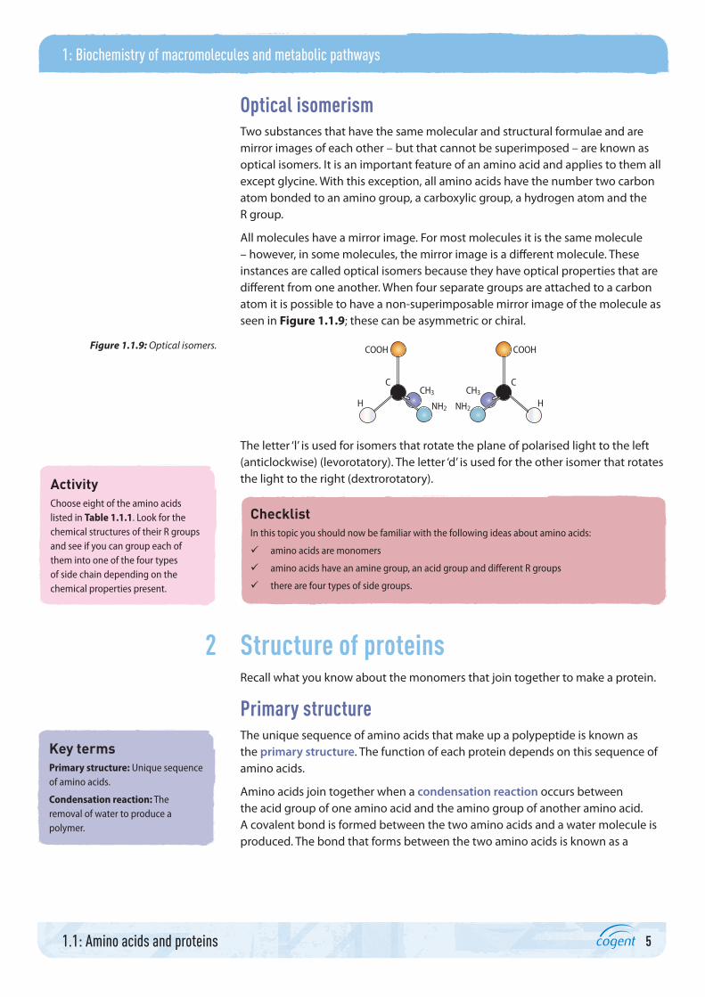
Optical isomerismTwo substances that have the same molecular and structural formulae and are mirror images of each other – but that cannot be superimposed – are known as optical isomers. It is an important feature of an amino acid and applies to them all except glycine. With this exception, all amino acids have the number two carbon atom bonded to an amino group, a carboxylic group, a hydrogen atom and the R group.
All molecules have a mirror image. For most molecules it is the same molecule – however, in some molecules, the mirror image is a different molecule. These instances are called optical isomers because they have optical properties that are different from one another. When four separate groups are attached to a carbon atom it is possible to have a non-superimposable mirror image of the molecule as seen in Figure 1.1.9; these can be asymmetric or chiral.
COOH COOH
H HCH3 CH3
C C
NH2 NH2
The letter ‘l’ is used for isomers that rotate the plane of polarised light to the left (anticlockwise) (levorotatory). The letter ‘d’ is used for the other isomer that rotates the light to the right (dextrorotatory).
ChecklistIn this topic you should now be familiar with the following ideas about amino acids:
amino acids are monomers
amino acids have an amine group, an acid group and different R groups
there are four types of side groups.
2 Structure of proteinsRecall what you know about the monomers that join together to make a protein.
Primary structure The unique sequence of amino acids that make up a polypeptide is known as the primary structure. The function of each protein depends on this sequence of amino acids.
Amino acids join together when a condensation reaction occurs between the acid group of one amino acid and the amino group of another amino acid. A covalent bond is formed between the two amino acids and a water molecule is produced. The bond that forms between the two amino acids is known as a
Figure 1.1.9: Optical isomers.
ActivityChoose eight of the amino acids listed in Table 1.1.1. Look for the chemical structures of their R groups and see if you can group each of them into one of the four types of side chain depending on the chemical properties present.
Key termsPrimary structure: Unique sequence of amino acids.
Condensation reaction: The removal of water to produce a polymer.

6
1: Biochemistry of macromolecules and metabolic pathways
1.1: Amino acids and proteins
peptide bond and a dipeptide is the molecule that is produced. When this chain of amino acids gets bigger a polypeptide is produced. Peptide bonds can be broken by adding water in a hydrolysis reaction to return the structure to the monomers. Figure 1.1.10 shows the condensation and hydrolysis of amino acids and dipeptide molecules.
In a polypeptide, there are many peptide bonds. These bonds are rigid and planar (flat) because of the electron sharing between the carbon from the carboxyl group and the nitrogen from the amide group.
OH
O
OH
OH
H
H
H
R
H
CN C
R
H
CN C
R
HHO
CN C
OH
OH
HR
H
CN C
Dipeptide molecule
Condensation reactionPeptide bond (covalent) formedWater eliminated (released)
Hydrolysis reactionPeptide bond (covalent) brokenWater used up
Amino acid Amino acidH2O
Secondary structure A protein’s secondary structure forms because the chain of amino acids folds to form either an α-helix or a β-pleated sheet. Many hydrogen bonds form between the folds and these hold the secondary structure in place. Hydrogen bonds occur where there are slightly positively-charged groups close to slightly negatively-charged groups.
Tertiary structure The tertiary structure is formed when the secondary structure coils and folds. The protein becomes a three-dimensional structure held in place by a number of different bonds and interactions. Disulfide bonds occur between two sulfur molecules. Sulfur molecules may be present in the R groups of some amino acids so, when they are close together, a disulfide bond forms. Ionic bonds form because some R groups carry a charge; when oppositely charged amino acids are close together an ionic bond forms. Hydrogen bonds occur where there are slightly positively-charged groups close to slightly negatively-charged groups. Finally, hydrophobic and hydrophilic interactions occur. Amino acids with hydrophobic R groups tend to be found in the centre of the globular protein and amino acids with hydrophilic R groups are found on the outside of the protein. Figure 1.1.11 shows the three-dimensional tertiary structure of a protein or a polypeptide.
Figure 1.1.10: Condensation and hydrolysis of amino acids
and dipeptide molecules.
Key termsPeptide bond: The bond that is formed between amino acids to form a polypeptide.
Dipeptide: Two amino acid molecules joined through a condensation reaction.
Hydrolysis reaction: The addition of water to break a polymer into monomers.
Secondary structure: The folding of the primary structure.
Tertiary structure: The folding of the secondary structure held together by bonds and interactions.
Disulfide bond: A bond occurring between two sulfur molecules that may occur in the R groups.
Ionic bond: A bond formed between two oppositely charged groups.
Hydrophobic: Repelling water.
Hydrophilic: Has an affinity to water.

7
1: Biochemistry of macromolecules and metabolic pathways
1.1: Amino acids and proteins
Disulfide (S–S) bond
α-helix
β-pleated sheet
Quaternary structure Some proteins are made from more than one polypeptide chain – this is said to be the quaternary structure. These proteins sometimes contain essential functional groups, known as prosthetic groups. An example of a protein with a quaternary structure and a prosthetic group is haemoglobin; see Figure 1.1.12 on page 8.
Portfolio activity (2.1)You can generate evidence for your portfolio by using the Internet to research images of the four structures of proteins and identify the sequences, folds, bonds and prosthetic groups. Produce a flow diagram or storyboard describing what happens to each structure in order for a complex protein to be produced. Think about the folds and bonds that are made.
• Select the relevant images (do not forget to reference them). • Place them in the correct order. • Describe what each structure consists of in terms of folds and bonds and the importance of
this structure.
3 Globular and fibrous proteinsThere are two types of proteins – fibrous and globular. Fibrous proteins, such as keratin found in human hair, consist of polypeptides bound together to form long sheets or fibres that are insoluble in water and extremely strong, making them ideal for a structural function. Globular proteins, on the other hand, are spherical in shape with intricate tertiary and quaternary structures. They are usually soluble in water and their function is biochemical. Haemoglobin is an example of a globular protein – this is an important molecule that picks up oxygen from the lungs and delivers it to respiring tissues.
Fibrous proteins – collagenCollagen is an important structural protein in the human body; it gives strength to various tissues in the body such as tendons, ligaments and skin. Collagen molecules are made from three polypeptide chains consisting of approximately 1000 amino acids, wound around each other. The hydrogen bonds that form between the chains provide the structure with immense strength.
Figure 1.1.11: Tertiary structure of a protein or polypeptide.
Key termsFibrous proteins: Large molecules consisting of polypeptide chains that make long fibres.
Globular proteins: Large molecules consisting of polypeptide chains that make a spherical shape.
8
1: Biochemistry of macromolecules and metabolic pathways
1.1: Amino acids and proteins
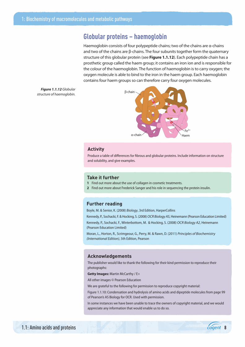
Globular proteins – haemoglobinHaemoglobin consists of four polypeptide chains; two of the chains are α-chains and two of the chains are β-chains. The four subunits together form the quaternary structure of this globular protein (see Figure 1.1.12). Each polypeptide chain has a prosthetic group called the haem group; it contains an iron ion and is responsible for the colour of the haemoglobin. The function of haemoglobin is to carry oxygen; the oxygen molecule is able to bind to the iron in the haem group. Each haemoglobin contains four haem groups so can therefore carry four oxygen molecules.
HaemFe2+
α-chain
β-chain
ActivityProduce a table of differences for fibrous and globular proteins. Include information on structure and solubility, and give examples.
Take it further1 Find out more about the use of collagen in cosmetic treatments.2 Find out more about Frederick Sanger and his role in sequencing the protein insulin.
Further readingBoyle, M. & Senior, K. (2008) Biology, 3rd Edition, HarperCollins
Kennedy, P., Sochacki, F. & Hocking, S. (2008) OCR Biology AS, Heinemann (Pearson Education Limited)
Kennedy, P., Sochacki, F., Winterbottom, M. & Hocking, S. (2008) OCR Biology A2, Heinemann (Pearson Education Limited)
Moran, L., Horton, R., Scrimgeour, G., Perry, M. & Rawn, D. (2011) Principles of Biochemistry (International Edition), 5th Edition, Pearson
AcknowledgementsThe publisher would like to thank the following for their kind permission to reproduce their photographs:
Getty Images: Martin McCarthy / E+
All other images © Pearson Education
We are grateful to the following for permission to reproduce copyright material:
Figure 1.1.10: Condensation and hydrolysis of amino acids and dipeptide molecules from page 99 of Pearson’s AS Biology for OCR. Used with permission.
In some instances we have been unable to trace the owners of copyright material, and we would appreciate any information that would enable us to do so.
Figure 1.1.12 Globular structure of haemoglobin.