& liu di jingmai k. o’connor , yuguang zhang , luis m
TRANSCRIPT

This article was downloaded by: [Institute of Vertebrate Paleontology and Paleoanthropology]On: 23 December 2013, At: 06:00Publisher: Taylor & FrancisInforma Ltd Registered in England and Wales Registered Number: 1072954 Registered office: Mortimer House,37-41 Mortimer Street, London W1T 3JH, UK
Journal of Vertebrate PaleontologyPublication details, including instructions for authors and subscription information:http://www.tandfonline.com/loi/ujvp20
A new enantiornithine from the Yixian Formation withthe first recognized avian enamel specializationJingmai K. O’Connor a b , Yuguang Zhang c , Luis M. Chiappe a , Qingjin Meng c , Li Quanguo c
& Liu Di ca Dinosaur Institute, Natural History Museum of Los Angeles County , 900 ExpositionBoulevard, Los Angeles , CA , 90007 , U.S.A.b Key Laboratory of Evolutionary Systematics of Vertebrates , Institute of VertebratePaleontology and Paleoanthroplogy , 142 Xizhimenwai Dajie, Beijing , China , 100044c Beijing Museum of Natural History , 126 Tianqiao South Street, Beijing , China , 100050Published online: 08 Jan 2013.
To cite this article: Jingmai K. O’Connor , Yuguang Zhang , Luis M. Chiappe , Qingjin Meng , Li Quanguo & Liu Di (2013) Anew enantiornithine from the Yixian Formation with the first recognized avian enamel specialization, Journal of VertebratePaleontology, 33:1, 1-12, DOI: 10.1080/02724634.2012.719176
To link to this article: http://dx.doi.org/10.1080/02724634.2012.719176
PLEASE SCROLL DOWN FOR ARTICLE
Taylor & Francis makes every effort to ensure the accuracy of all the information (the “Content”) containedin the publications on our platform. However, Taylor & Francis, our agents, and our licensors make norepresentations or warranties whatsoever as to the accuracy, completeness, or suitability for any purpose of theContent. Any opinions and views expressed in this publication are the opinions and views of the authors, andare not the views of or endorsed by Taylor & Francis. The accuracy of the Content should not be relied upon andshould be independently verified with primary sources of information. Taylor and Francis shall not be liable forany losses, actions, claims, proceedings, demands, costs, expenses, damages, and other liabilities whatsoeveror howsoever caused arising directly or indirectly in connection with, in relation to or arising out of the use ofthe Content.
This article may be used for research, teaching, and private study purposes. Any substantial or systematicreproduction, redistribution, reselling, loan, sub-licensing, systematic supply, or distribution in anyform to anyone is expressly forbidden. Terms & Conditions of access and use can be found at http://www.tandfonline.com/page/terms-and-conditions

Journal of Vertebrate Paleontology 33(1):1–12, January 2013© 2013 by the Society of Vertebrate Paleontology
FEATURED ARTICLE
A NEW ENANTIORNITHINE FROM THE YIXIAN FORMATION WITH THE FIRSTRECOGNIZED AVIAN ENAMEL SPECIALIZATION
JINGMAI K. O’CONNOR,*,1,2 YUGUANG ZHANG,3 LUIS M. CHIAPPE,1 QINGJIN MENG,3 LI QUANGUO,3 and LIU DI3
1Dinosaur Institute, Natural History Museum of Los Angeles County, 900 Exposition Boulevard, Los Angeles, CA 90007, U.S.A.;2Key Laboratory of Evolutionary Systematics of Vertebrates, Institute of Vertebrate Paleontology and Paleoanthroplogy,
142 Xizhimenwai Dajie, Beijing, China 100044, [email protected];3Beijing Museum of Natural History, 126 Tianqiao South Street, Beijing, China 100050
ABSTRACT—We report on a new enantiornithine bird, Sulcavis geeorum, gen. et sp. nov., from the Jehol Group of north-eastern China. The fossil preserves robust teeth with longitudinal grooves radiating from the occlusal tip preserved in theenamel on the lingual surface. This is the first known occurrence of specialized tooth enamel within Aves. Compared withother Mesozoic groups, stomach contents are hardly ever preserved within enantiornithine specimens; therefore, this newtooth morphology reveals new evidence regarding the diversity of trophic niches occupied by the clade.
SUPPLEMENTAL DATA— Supplemental materials are available for this article for free at www.tandfonline.com/UJVP
INTRODUCTION
New species of fossil bird are being uncovered from the JeholGroup in northeastern China at an unprecedented rate and therecognized range of morphological variation among Early Cre-taceous birds continues to grow. During the last two decades,over 40 avian species have been named (Zhou and Zhang, 2006a).More than half of these species are referable to Enantiornithes,the most specious Cretaceous clade of birds and the sister taxonto Ornithuromorpha, the lineage that includes modern birds. TheJehol Group is also important because specimens from these de-posits preserve more than osteological morphology, and revealaspects of the biology of these extinct birds, such as plumage,diet, ecology, life history, and development (Zhang and Zhou,2000; Hou et al., 2004; Zhou and Zhang, 2004; Chiappe et al.,2008; O’Connor et al., 2009).
Approximately half of all enantiornithines are known from theJehol Group, and nearly every species is represented by a partialto nearly complete articulated skeleton, often preserving integu-ment (Zhou and Zhang, 2006a; Chiappe, 2007). Unfortunately,not one published specimen preserves direct evidence of trophichabit such as stomach contents, although numerous ornithuro-morph specimens preserve ingested remains and other indicatorsof diet, with geo-gastroliths being particularly common (Zhouet al., 2004; Zhou and Zhang, 2006b). The only two enantior-nithine specimens that preserve such evidence are from outsideChina: the holotype of Eoalulavis hoyasi Sanz, Chiappe, Perez-Moreno, Buscalioni, Moratalla, Ortega, and Poyato-Ariza, 1996,preserves the exoskeletal remains of crustaceans in the thoraciccavity, interpreted as stomach contents, suggesting an aquaticfeeding habitat (Sanz et al., 1996); the holotype of Enantio-phoenix electrophyla Cau and Arduini, 2008, preserves small in-clusions of amber associated with the skeleton, which have beeninterpreted as ingested items and indicative of a diet that includedsap (Dalla Vecchia and Chiappe, 2002).
To date, the only way to infer the trophic habit of enantior-nithines from the Jehol Group is through their cranial or den-
*Corresponding author.
tal morphology. Enantiornithines exhibit a wide range of cra-nial morphologies (O’Connor and Chiappe, 2011) and the longrostrum of some of these taxa has been used to infer specifictrophic specializations (Zhang et al., 2000; Hou et al., 2004;Morschhauser et al., 2009; O’Connor et al., 2009). A diversityof dental shapes, sizes, and patterns are also apparent in theJehol enantiornithines (O’Connor and Chiappe, 2011). How-ever, although the teeth range in overall morphology, caudalcurvature, lateral compression, and size, no previous specimenshave preserved ridges, striations, denticles, or any other formof dental ornamentation. Likewise, no Jehol enantiornithinepreserves edentulous jaws, although this is present in a diver-sity of basal ornithuromorphs (e.g., Archaeorhynchus spathulaZhou and Zhang, 2006b, Hongshanornis longicresta Zhou andZhang, 2005), the confuciusornithiforms (Chiappe et al., 1999),and Zhongjianornis zhengi Zhou, Zhang, and Li, 2010 (Zhou andZhang, 2005; Zhou and Zhang, 2006b; Zhou et al., 2010), all fromthe Jehol Group.
In this paper, we describe the morphology of a new enan-tiornithine specimen (BMNH Ph-000805) from the Jehol Group,which presents a unique dental specialization—one that is sug-gestive of a durophagous diet. BMNH Ph-000805 appears to beclosely related to the recently described Shenqiornis mengi Wang,O’Connor, Zhao, Chiappe, Gao, and Cheng, 2010, with which itshares aspects of the dental morphology, and the two taxa areincluded in a phylogenetic analysis to explore this potential rela-tionship. The new specimen, although more fragmentary than theholotype of Shenqiornis mengi, also reveals distinct morphologiesin the skull and postcranial skeleton that suggest that it representsa new taxon.
Institutional Abbreviations—BMNH, Beijing Museum of Nat-ural History, Beijing, China; LACM, Natural History Museum ofLos Angeles County, Los Angeles, U.S.A.
SYSTEMATIC PALEONTOLOGY
AVES Linnaeus, 1758PYGOSTYLIA Chiappe, 2002
1
Dow
nloa
ded
by [
Inst
itute
of
Ver
tebr
ate
Pale
onto
logy
and
Pal
eoan
thro
polo
gy]
at 0
6:00
23
Dec
embe
r 20
13
2 JOURNAL OF VERTEBRATE PALEONTOLOGY, VOL. 33, NO. 1, 2013
ORNITHOTHORACES Chiappe, 1995ENANTIORNITHES Walker, 1981
SULCAVIS GEEORUM, gen. et sp. nov.(Figs. 1, 2)
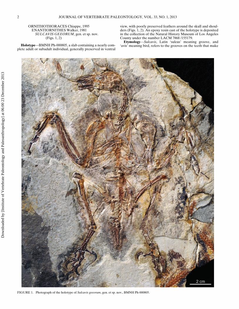
Holotype—BMNH Ph-000805, a slab containing a nearly com-plete adult or subadult individual, generally preserved in ventral
view, with poorly preserved feathers around the skull and shoul-ders (Figs. 1, 2). An epoxy resin cast of the holotype is depositedin the collection of the Natural History Museum of Los AngelesCounty under the number LACM 7868 /155179.
Etymology—Sulcavis, Latin ‘sulcus’ meaning groove, and‘avis’ meaning bird, refers to the grooves on the teeth that make
FIGURE 1. Photograph of the holotype of Sulcavis geeorum, gen. et sp. nov., BMNH Ph-000805.
Dow
nloa
ded
by [
Inst
itute
of
Ver
tebr
ate
Pale
onto
logy
and
Pal
eoan
thro
polo
gy]
at 0
6:00
23
Dec
embe
r 20
13

O’CONNOR ET AL.—ENANTIORNITHINE WITH SPECIALIZED ENAMEL 3
FIGURE 2. Camera lucida drawing of the holotype of Sulcavis geeorum, gen. et sp. nov., BMNH Ph-000805. Light gray indicates areas of poorpreservation; dark gray indicates matrix. Abbreviations: ce, cervical vertebrae; co, coracoid; fe, femur; fi, fibula; fu, furcula; h, humerus; il, ilium; mc,metacarpal; mc I, alular digit; mc II, major digit; mc III, minor digit; mt, metatarsals; py, pygostyle; r, radius; s, synsacrum; sc, scapula; ti, tibiotarsus; u,ulna; ul, ulnare.
Dow
nloa
ded
by [
Inst
itute
of
Ver
tebr
ate
Pale
onto
logy
and
Pal
eoan
thro
polo
gy]
at 0
6:00
23
Dec
embe
r 20
13
4 JOURNAL OF VERTEBRATE PALEONTOLOGY, VOL. 33, NO. 1, 2013
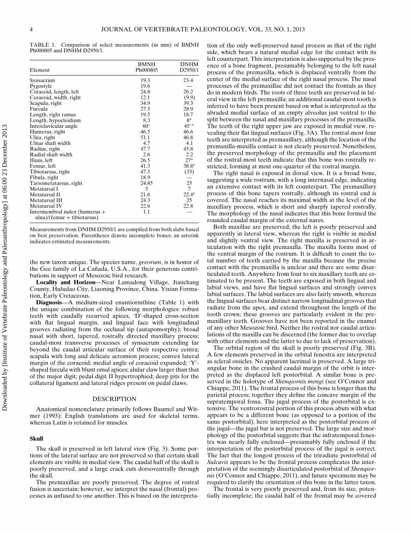
TABLE 1. Comparison of select measurements (in mm) of BMNHPh000805 and DNHM D2950/1.
BMNH DNHMElement Ph000805 D2950/1
Synsacrum 19.3 23.4Pygostyle 19.6 —Coracoid, length, left 24.8 26.2Coracoid, width, right 12.1 (9.9)Scapula, right 34.9 39.3Furcula 27.5 28.9Length, right ramus 19.5 18.7Length, hypocleidium 8.3 8∗Interclavicular angle 60◦ 45◦∗Humerus, right 46.5 46.6Ulna, right 51.1 46.8Ulnar shaft width 4.7 4.1Radius, right 47.7 45.8Radial shaft width 2.6 2.2Ilium, left 26.5 27∗Femur, left 41.3 38.8∗Tibiotarsus, right 47.3 (33)Fibula, right 18.9 —Tarsometatarsus, right 24.85 25Metatarsal I 5 7Metatarsal II 21.6 22.4∗Metatarsal III 24.3 25Metatarsal IV 22.6 22.8Intermembral index (humerus +
ulna)/(femur + tibiotarsus)1.1 —
Measurements from DNHM D2950/1 are compiled from both slabs basedon best preservation. Parentheses denote incomplete bones; an asteriskindicates estimated measurements.
the new taxon unique. The species name, geeorum, is in honor ofthe Gee family of La Canada, U.S.A., for their generous contri-butions in support of Mesozoic bird research.
Locality and Horizon—Near Lamadong Village, JianchangCounty, Huludao City, Liaoning Province, China. Yixian Forma-tion, Early Cretaceous.
Diagnosis—A medium-sized enantiornithine (Table 1) withthe unique combination of the following morphologies: robustteeth with caudally recurved apices, ‘D’-shaped cross-sectionwith flat lingual margin, and lingual face with longitudinalgrooves radiating from the occlusal tip (autapomorphy); broadnasal with short, tapered, rostrally directed maxillary process;caudal-most transverse processes of synsacrum extending farbeyond the caudal articular surface of their respective centra;scapula with long and delicate acromion process; convex lateralmargin of the coracoid; medial angle of coracoid expanded; ‘Y’-shaped furcula with blunt omal apices; alular claw larger than thatof the major digit; pedal digit II hypertrophied; deep pits for thecollateral ligament and lateral ridges present on pedal claws.
DESCRIPTION
Anatomical nomenclature primarily follows Baumel and Wit-mer (1993); English translations are used for skeletal terms,whereas Latin is retained for muscles.
Skull
The skull is preserved in left lateral view (Fig. 3). Some por-tions of the lateral surface are not preserved so that certain skullelements are visible in medial view. The caudal half of the skull ispoorly preserved, and a large crack cuts dorsoventrally throughthe skull.
The premaxillae are poorly preserved. The degree of rostralfusion is uncertain; however, we interpret the nasal (frontal) pro-cesses as unfused to one another. This is based on the interpreta-
tion of the only well-preserved nasal process as that of the rightside, which bears a natural medial edge for the contact with itsleft counterpart. This interpretation is also supported by the pres-ence of a bone fragment, presumably belonging to the left nasalprocess of the premaxilla, which is displaced ventrally from thecenter of the medial surface of the right nasal process. The nasalprocesses of the premaxillae did not contact the frontals as theydo in modern birds. The roots of three teeth are preserved in lat-eral view in the left premaxilla; an additional caudal-most tooth isinferred to have been present based on what is interpreted as theabraded medial surface of an empty alveolus just ventral to thesplit between the nasal and maxillary processes of the premaxilla.The teeth of the right upper jaw are exposed in medial view, re-vealing their flat lingual surfaces (Fig. 3A). The rostral-most fourteeth are interpreted as premaxillary, although the location of thepremaxilla-maxilla contact is not clearly preserved. Nonetheless,the preserved morphology of the premaxilla and the placementof the rostral-most teeth indicate that this bone was rostrally re-stricted, forming at most one-quarter of the rostral margin.
The right nasal is exposed in dorsal view. It is a broad bone,suggesting a wide rostrum, with a long internasal edge, indicatingan extensive contact with its left counterpart. The premaxillaryprocess of this bone tapers rostrally, although its rostral end iscovered. The nasal reaches its maximal width at the level of themaxillary process, which is short and sharply tapered rostrally.The morphology of the nasal indicates that this bone formed therounded caudal margin of the external nares.
Both maxillae are preserved; the left is poorly preserved andapparently in lateral view, whereas the right is visible in medialand slightly ventral view. The right maxilla is preserved in ar-ticulation with the right premaxilla. The maxilla forms most ofthe ventral margin of the rostrum. It is difficult to count the to-tal number of teeth carried by the maxilla because the precisecontact with the premaxilla is unclear and there are some disar-ticulated teeth. Anywhere from four to six maxillary teeth are es-timated to be present. The teeth are exposed in both lingual andlabial views, and have flat lingual surfaces and strongly convexlabial surfaces. The labial surfaces are also fairly smooth, whereasthe lingual surfaces bear distinct narrow longitudinal grooves thatradiate from the apex, and extend throughout the length of thetooth crown; these grooves are particularly evident in the pre-maxillary teeth. Grooves have not been reported in the enamelof any other Mesozoic bird. Neither the rostral nor caudal articu-lations of the maxilla can be discerned (the former due to overlapwith other elements and the latter to due to lack of preservation).
The orbital region of the skull is poorly preserved (Fig. 3B).A few elements preserved in the orbital fenestra are interpretedas scleral ossicles. No apparent lacrimal is preserved. A large tri-angular bone in the crushed caudal margin of the orbit is inter-preted as the displaced left postorbital. A similar bone is pre-served in the holotype of Shenqiornis mengi (see O’Connor andChiappe, 2011). The frontal process of this bone is longer than theparietal process; together they define the concave margin of thesupratemporal fossa. The jugal process of the postorbital is ex-tensive. The ventrorostral portion of this process abuts with whatappears to be a different bone (as opposed to a portion of thesame postorbital), here interpreted as the postorbital process ofthe jugal—the jugal bar is not preserved. The large size and mor-phology of the postorbital suggests that the infratemporal fenes-tra was nearly fully enclosed—presumably fully enclosed if theinterpretation of the postorbital process of the jugal is correct.The fact that the longest process of the triradiate postorbital ofSulcavis appears to be the frontal process complicates the inter-pretation of the seemingly disarticulated postorbital of Shenqior-nis (O’Connor and Chiappe, 2011), and future specimens may berequired to clarify the orientation of this bone in the latter taxon.
The frontal is very poorly preserved and, from its size, poten-tially incomplete; the caudal half of the frontal may be covered
Dow
nloa
ded
by [
Inst
itute
of
Ver
tebr
ate
Pale
onto
logy
and
Pal
eoan
thro
polo
gy]
at 0
6:00
23
Dec
embe
r 20
13

O’CONNOR ET AL.—ENANTIORNITHINE WITH SPECIALIZED ENAMEL 5
FIGURE 3. Detail of skull of BMNH Ph-000805. A, proximal half of rostrum; B, entire skull, right lateral view; C, interpretative drawing. Lightgray indicates areas of poor preservation; dark gray indicates matrix. Abbreviations: ang?, angular?; bo?, basioccipital; d, dentary; eo, exoccipital; fr,frontal; fm, foramen magnum; ju, postorbital process of the jugal; max, maxilla; n, nasal; oc, occipital condyle; pa, parietal; pao, paraoccipital process;pm, premaxilla; po, postorbital; scl, scleral ossicle; so, supraoccipital; sp?, splenial; sur?, surangular. (Color figure available online.)
by the parietal due to displacement of the occipital region. Theparietal is comparatively well preserved—it is large and quadran-gular, more than twice the size of the supraoccipital with whichit articulates. A ridge intersects this bone; whether this is thesagittal nuchal crest or results from an underlying bone cannotbe determined. Interpretations of the caudal portion of the skullare complicated by poor preservation and partial disarticulation(Fig. 3). The supraoccipitals are preserved, fused into a single el-ement in caudal view, with a sagittally located cerebral promi-nence that reaches the foramen magnum. The right exoccipitalis disarticulated, obscuring the size and shape of the foramenmagnum. The left exoccipital is in caudolateral view in articu-lation with the ventrolaterally tapering paraoccipital process ofthe supraoccipital. Only the right exoccipital contribution of theoccipital condyle is preserved; although incomplete, the occipi-tal condyle appears proportionately large relative to the foramen
magnum, as in other enantiornithines (e.g., Shenqiornis, Vescor-nis) (O’Connor and Chiappe, 2011). A poorly preserved boneventral to the right exoccipital may be part of the basioccipitals.
The two dentaries are not fused rostrally and no predentarybone is preserved, morphologies consistent with other enantior-nithines. The left dentary is preserved in lateral view, whereasthe right is preserved in medial view (Fig. 3B). Seven teeth arepreserved in the left dentary. A row of small foramina marksthe lateral margin of the dentary, just ventral to the dentigerousmargin. The right dentary preserves at least 7 teeth but weestimate that 9 or 10 may have been present in total. Meckel’sgroove is visible on the right dentary, restricted to the caudalhalf of the bone and tapering rostrally. The caudal half of bothdentaries is not preserved, and interpretation of the fragmentarypostdentary bones is equivocal. Several long, thin bones mayrepresent the angular or portions of the hyoid bones—one of
Dow
nloa
ded
by [
Inst
itute
of
Ver
tebr
ate
Pale
onto
logy
and
Pal
eoan
thro
polo
gy]
at 0
6:00
23
Dec
embe
r 20
13

6 JOURNAL OF VERTEBRATE PALEONTOLOGY, VOL. 33, NO. 1, 2013
FIGURE 4. Detail photograph of the pectoral girdle of BMNH Ph-000805. Abbreviations: acr, acromion process; co, coracoid; fu, furcula;hy, hypocleidium; sc, scapula; vp, ventral process. Other abbreviations,see Figure 3. (Color figure available online.)
these bones is clearly preserved beneath the occipital condyle. Atriangular bone may be a caudally displaced splenial.
Axial Skeleton
Six articulated cervical vertebrae are clearly preserved in ven-tral view (Fig. 1); the proximal-most cervical vertebrae (the at-las, axis, and possibly one additional cervical) cannot be differ-entiated, and thus the total number of cervicals is estimated tobe eight to nine. The preserved cervicals have in situ and pre-sumably fused (fully articulated, no sutures but preservation notclear enough to determine unequivocally) costal processes thatare nearly as long as the centrum—these processes are unfusedin the last two vertebrae. Small carotid processes are located me-dial to the proximal ends of the costal processes. Given the com-plete articulation of the series, we cannot determine if the ver-tebrae were truly heterocoelic. The cervical centra are keeledventrally, flanked by ventral longitudinal recesses, although theheight of this keel cannot be determined in most vertebrae. Inthe last preserved cervical (exposed in lateral view), the ventralprocess projects rostrally, extending slightly beyond the ventraledge of the cranial articular surface, and tapers caudally (Fig. 4).This last cervical clearly marks the cervicothoracic transition.
Caudal to the last cervical vertebrae, five poorly exposed artic-ulated thoracic vertebrae are visible, the first in left lateral viewand the last in ventral view. The penultimate of these vertebraeexhibits a broad excavation on the side of the centrum and thelast two exhibit centrally located parapophyses, as seen in otherenantiornithines (Chiappe and Walker, 2002). The rest of the se-ries is very poorly preserved in articulation with the synsacrum.
The synsacrum comprises seven or eight vertebrae (Fig. 5). Thetransverse processes are short and delicate along the proximalhalf of the synsacrum, and larger and more robust on the distalhalf. All but the last transverse processes are directed perpen-dicular to the sacral axis and would have braced the pelvic gir-dle. The last two transverse processes are very long, extendingfar beyond the caudal articular surface of the synsacrum, and arestrongly deflected caudally.
At least five free caudal vertebrae are preserved in ventral view(Fig. 5). The prezygapophyses are more elongate than the postzy-
FIGURE 5. Detail photograph of the pelvic girdle of BMNH Ph-000805.Abbreviations: cav, caudal vertebrae; df, dorsal fork; fe, femur; ha, hemalarch; il, ilium; pub, pubis; py, pygostyle; s, synsacrum; vlr, ventrolat-eral process. Other abbreviations, see Figure 3. (Color figure availableonline.)
gapophyses. The last two caudals bear short, laterally directedtransverse processes; those of the proximal vertebrae are not pre-served. Several isolated elements preserved beside the caudal se-ries are interpreted as hemal arches; they are somewhat longerthan the adjacent centra and distally are bluntly tapered.
The pygostyle is triangular in shape, preserved in ventral view(Fig. 5). The morphology of the pygostyle is similar to that ofother enantiornithines. Proximally, it possesses a pair of dorsalprocesses, and the lateral edges bear lateroventrally directed pro-cesses. The right lateroventral process is abraded off. As in mostother enantiornithines, the pygostyle is constricted along its distalfifth; however, this constriction is more distally located and notas abrupt as in Rapaxavis pani Morschhauser, Varricchio, Gao,
Dow
nloa
ded
by [
Inst
itute
of
Ver
tebr
ate
Pale
onto
logy
and
Pal
eoan
thro
polo
gy]
at 0
6:00
23
Dec
embe
r 20
13

O’CONNOR ET AL.—ENANTIORNITHINE WITH SPECIALIZED ENAMEL 7
Liu, Wang, Cheng, and Meng, 2009, and other enantiornithines(O’Connor et al., 2011b).
Thoracic Girdle
The coracoids are preserved in ventral view, precluding ob-servation of the dorsal surface (Fig. 4). However, a deep fossalike that of Enantiornis leali Walker, 1981, was most likely ab-sent (Chiappe and Walker, 2002). The coracoids are distallybroad and very wide, expanding rapidly from the rod-like neck,which forms the proximal one-third of the bone. They appar-ently lack a procoracoid process, and the presence of a supra-coracoidal nerve foramen and/or medial groove cannot be de-termined. The lateral margins of the coracoids are strongly con-vex over the distal third of the bone, consistent with mostenantiornithines, and most strongly resembling the coracoids ofEoalulavis hoyasi. The coracoids are preserved with their medialangles overlapping—however, the right coracoid is slightly dis-placed medially and we interpret that the coracoids would nothave overlapped in life. The distal fifth of the medial margin isslightly convex, contributing to the expanded sternal margin. Thesternal margin is straight to slightly concave and a lateral processis absent.
The coracoids are overlain by the furcula, which is ‘Y’-shapedwith an elongate hypocleidium at least half the length of the fur-cular rami (Fig. 4). Although the furcula is exposed in ventralview, breakage reveals that it was excavated dorsolaterally, as inother enantiornithines (Chiappe and Calvo, 1994; Martin, 1995;Chiappe and Walker, 2002). The omal tips are blunt and curvedslightly medially.
The scapulae are largely covered by other elements; however,the elongate acromion of the right scapula is exposed; the processis long and delicate, as in Eoalulavis hoyasi, and the proximal endof the acromion is expanded into a rounded surface that may havearticulated with the furcula (Fig. 4) (Sanz et al., 1996). A scapularcostal groove, reported for other enantiornithines (Chiappe andWalker, 2002), is absent.
No direct information regarding the morphology of the ster-num is preserved (Fig. 1). A few fragments preserved distal tothe coracoids may represent the rostral margin of the sternum, oralternatively they may be poorly preserved thoracic ribs. How-ever, the preserved position of the coracoids suggests that the ros-tral margin of the sternum would have defined an approximately100◦ angle with the apex at midpoint, similar to Rapaxavis pani,as opposed to having a rounded margin as in some other enan-tiornithines (e.g., Longipteryx chaoyangensis Zhang, Zhou, Hou,and Gu, 2000) (O’Connor et al., 2011b). Gastralia are preserved,some of them overlapping the pelvic girdle.
Thoracic Limb
The left wing is poorly preserved and all morphological infor-mation comes from the right wing (Fig. 6). The proximal marginof the humerus, exposed cranially, is typically enantiornithine—itis centrally concave, with raised ventral and dorsal ends.The proximoventral margin of the humerus projects stronglyventrally, as noted for Zhongjianornis yangi (Zhou et al.,2010; O’Connor et al., 2011a) and the basal ornithuromorphSchizooura lii Zhou, Zhou, and O’Connor, 2012. The deltopec-toral crest is more than one-third the length of the humerus,nearly the same width as the shaft, ending abruptly. The ulnais longer than the humerus and bowed proximally; the radius isstraight.
The ulnare is triangular, with only a shallow incisure. Theradiale is quadrangular and the exposed surface bears a me-dian groove. The alular metacarpal was clearly unfused to theother elements of the metacarpus (Fig. 6). The semilunate carpaland major and minor metacarpals also appear to be unfused toeach other, although the preservation makes such interpretation
equivocal. The alular metacarpal is rectangular, although in pro-file the cranial margin is slightly convex in its middle portion,possibly representing an incipient extensor process. The proxi-mal phalanx of this digit appears to be slightly bowed craniocau-dally; the second phalanx is a large claw with a recurved hornysheath. The alular digit has equal distal extent with the majormetacarpal. The proximal margin of the major metacarpal is lo-cated proximal to that of the minor metacarpal; however, thisregion of the carpometacarpus is poorly preserved. The minormetacarpal extends distally farther than the major metacarpal, asin all other enantiornithines (Martin, 1995; Chiappe and Walker,2002). The minor metacarpal is less than half the width of themajor metacarpal, and the two are not separated by an inter-metacarpal space, although this may be a taphonomic artifact.The major digit is composed of three phalanges, which decreasein length distally (Fig. 6). The first phalanx of the major digit iscranially expanded along its length, forming a blunt longitudi-nal ridge comparable to the cranial phalangeal ball (pila cranialisphalangis) that reinforces the digit in living birds (Baumel andWitmer, 1993). The caudal margin of the first phalanx constrictsjust proximal to its distal articulation; the distoventral margin ofthe constriction forms a small tubercle. The claw is smaller thanthat of the alular digit, but also strongly recurved, with the hornysheath preserved. A single phalanx is preserved for the minordigit. It is half the length of the proximal phalanx of the majordigit, with a straight cranial margin and a slightly concave caudalmargin. The distal end of this phalanx is blunt.
Pelvic Girdle
The ilia and synsacrum are unfused, and the pelvic elementsappear unfused at the level of the acetabulum (Fig. 5). What ispreserved of the pelvic girdle remains in nearly complete articu-lation. The left ilium is completely preserved, whereas only thepostacetabular wing is preserved on the right. The proximal mar-gin of the preacetabular wing is rounded in ventral view; in lat-eral view, the postacetabular wing is triangular, distally taperingbluntly. The proximal portion of the left pubis is preserved, un-fused to but in articulation with the ilium; no pectineal processis present. The pubic shaft is proximally robust but it narrowsdistally. A piece of the left ischium is preserved but reveals noanatomical information.
Pelvic Limb
The femora are preserved in near articulation with the pelvis(Fig. 5). They are straight and rather slender. The tibiotarsi areonly 10% longer than the femora. The left is poorly preserved,yielding no information; the right is preserved in cranial view(Fig. 7). The proximal cranial surface is abraded and no cnemialcrests are preserved; the short fibular crest extends less than one-third the length of the tibiotarsus. Distally, the condyles are sube-qual in size, bulbous, and separated by a narrow incisure. A talltriangular ascending process is visible on the cranial surface prox-imal to the condyles, indicating that fusion of the tibia with theproximal tarsals was not complete in this specimen at the time ofdeath. As preserved, the fibula is approximately half the lengthof the tibiotarsus.
The proximal ends of metatarsals II–IV are fused to the dis-tal tarsals; the shafts of these metatarsals appear unfused alongtheir entire length (Fig. 7). A very slight intercotylar eminence ispresent; the lateral margin of the proximal articular surface of thetarsometatarsus (lateral cotyla) projects proximally to the samelevel as the intercotylar eminence (and more strongly than themedial margin). No tubercle for the m. tibialis cranialis is pre-served on the cranial surface of metatarsals II or III; damageto the cranial surface of portions of metatarsal II and III sug-gests that if a tubercle was present, it was fairly distally located,as in Yungavolucris brevipedalis Chiappe, 1993. Proximally, the
Dow
nloa
ded
by [
Inst
itute
of
Ver
tebr
ate
Pale
onto
logy
and
Pal
eoan
thro
polo
gy]
at 0
6:00
23
Dec
embe
r 20
13
8 JOURNAL OF VERTEBRATE PALEONTOLOGY, VOL. 33, NO. 1, 2013
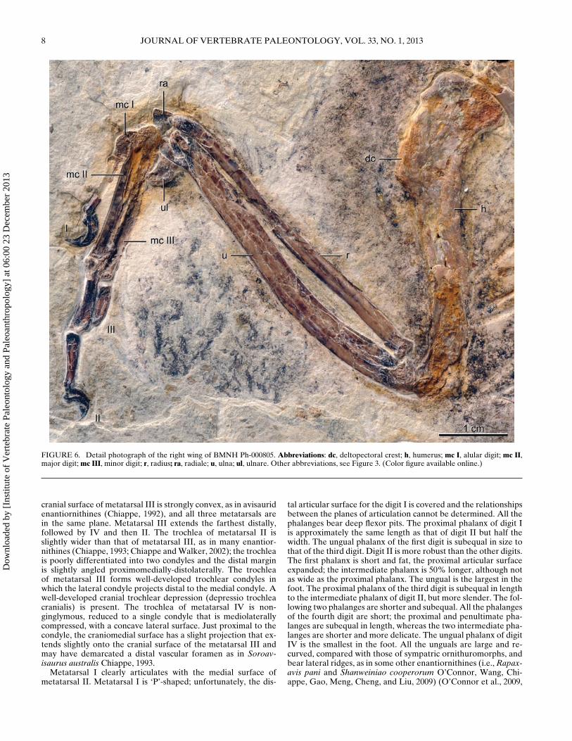
FIGURE 6. Detail photograph of the right wing of BMNH Ph-000805. Abbreviations: dc, deltopectoral crest; h, humerus; mc I, alular digit; mc II,major digit; mc III, minor digit; r, radius; ra, radiale; u, ulna; ul, ulnare. Other abbreviations, see Figure 3. (Color figure available online.)
cranial surface of metatarsal III is strongly convex, as in avisauridenantiornithines (Chiappe, 1992), and all three metatarsals arein the same plane. Metatarsal III extends the farthest distally,followed by IV and then II. The trochlea of metatarsal II isslightly wider than that of metatarsal III, as in many enantior-nithines (Chiappe, 1993; Chiappe and Walker, 2002); the trochleais poorly differentiated into two condyles and the distal marginis slightly angled proximomedially-distolaterally. The trochleaof metatarsal III forms well-developed trochlear condyles inwhich the lateral condyle projects distal to the medial condyle. Awell-developed cranial trochlear depression (depressio trochleacranialis) is present. The trochlea of metatarsal IV is non-ginglymous, reduced to a single condyle that is mediolaterallycompressed, with a concave lateral surface. Just proximal to thecondyle, the craniomedial surface has a slight projection that ex-tends slightly onto the cranial surface of the metatarsal III andmay have demarcated a distal vascular foramen as in Soroav-isaurus australis Chiappe, 1993.
Metatarsal I clearly articulates with the medial surface ofmetatarsal II. Metatarsal I is ‘P’-shaped; unfortunately, the dis-
tal articular surface for the digit I is covered and the relationshipsbetween the planes of articulation cannot be determined. All thephalanges bear deep flexor pits. The proximal phalanx of digit Iis approximately the same length as that of digit II but half thewidth. The ungual phalanx of the first digit is subequal in size tothat of the third digit. Digit II is more robust than the other digits.The first phalanx is short and fat, the proximal articular surfaceexpanded; the intermediate phalanx is 50% longer, although notas wide as the proximal phalanx. The ungual is the largest in thefoot. The proximal phalanx of the third digit is subequal in lengthto the intermediate phalanx of digit II, but more slender. The fol-lowing two phalanges are shorter and subequal. All the phalangesof the fourth digit are short; the proximal and penultimate pha-langes are subequal in length, whereas the two intermediate pha-langes are shorter and more delicate. The ungual phalanx of digitIV is the smallest in the foot. All the unguals are large and re-curved, compared with those of sympatric ornithuromorphs, andbear lateral ridges, as in some other enantiornithines (i.e., Rapax-avis pani and Shanweiniao cooperorum O’Connor, Wang, Chi-appe, Gao, Meng, Cheng, and Liu, 2009) (O’Connor et al., 2009,
Dow
nloa
ded
by [
Inst
itute
of
Ver
tebr
ate
Pale
onto
logy
and
Pal
eoan
thro
polo
gy]
at 0
6:00
23
Dec
embe
r 20
13

O’CONNOR ET AL.—ENANTIORNITHINE WITH SPECIALIZED ENAMEL 9
FIGURE 7. Detail photograph of the right foot of BMNH Ph-000805.Abbreviations: ap, ascending process of the astragalus; fi, fibula; mt,metatarsals; ti, tibia; tlc, lateral condyle of the tibiotarsus; tmc, me-dial condyle. Other abbreviations, see Figure 3. (Color figure availableonline.)
FIGURE 8. Details of the right premaxillary-maxillary teeth of BMNHPh-000805 in lingual view. A, photograph of the dentition at the rightpremaxilla-maxilla contact, in lingual view box indicates area expanded inB; B, close up of maxillary tooth; C, interpretative drawing of maxillarytooth. Arrows indicate the longitudinal grooves. (Color figure availableonline.)
2011b). Large strongly recurved horny sheaths are preserved withthe unguals.
DISCUSSION
Phylogenetic Hypothesis
BMNH Ph-000805 is considered to be an enantiornithine basedon the presence of the following features: proximally forked anddistally constricted pygostyle with ventrolateral processes; ‘Y’-shaped, dorsolaterally excavated furcula; convex lateral marginof the coracoid; proximal humerus, rising dorsally and ventrallyto centrally on the concave head; minor metacarpal projectingdistally farther than the major metacarpal; and distal tarsals fusedto the metatarsals, with metatarsals unfused along their lengths.The specimen differs from all other enantiornithines in the pres-ence of enlarged teeth with grooves on the lingual surface (Fig. 8)and a unique combination of morphological features, therefore,we erect the new taxon Sulcavis geeorum, gen. et sp. nov.
Enantiornithine systematics are notoriously poorly resolved;however, we placed the new specimen into the latest availableMesozoic bird data set (O’Connor et al., 2011a) in order to testits placement as an enantiornithine and explore a possible rela-tionship with the superficially similar Shenqiornis mengi. A totalof 64 taxa were scored for 245 characters, with Dromaeosauridaeas the outgroup; 31 characters were treated as ordered and allcharacters were weighted equally (see Supplemental Data). Anyfeature of BMNH Ph-000805 described as equivocal (i.e., postor-bital) was scored as missing data. The analysis was run using TNT(Goloboff et al., 2008). We conducted a heuristic search retainingthe single shortest tree from every 1000 trees followed by an ad-ditional round of tree bisection and reconnection (TBR) branchswapping. This analysis produced 144 shortest trees (length = 835steps; consistency index = 0.389; retention Index = 0.670). TheNelson strict consensus tree (Fig. 9) resolves Sulcavis as an enan-tiornithine more derived than the longipterygids, in a polytomywith Iberomesornis, Otogornis, and two clades of derived enan-tiornithines including the ‘avisaurids’; Shenqiornis forms the sis-ter taxon to this clade. Bremer support for this hypothesis is low(Fig. 9), but this is unfortunately characteristic of Mesozoic birdcladistic analyses and is an issue beyond the scope of this paper(O’Connor and Zhou, 2012).
Dow
nloa
ded
by [
Inst
itute
of
Ver
tebr
ate
Pale
onto
logy
and
Pal
eoan
thro
polo
gy]
at 0
6:00
23
Dec
embe
r 20
13

10 JOURNAL OF VERTEBRATE PALEONTOLOGY, VOL. 33, NO. 1, 2013
FIGURE 9. Cladogram of the strict consensus tree (length, 835 steps;consistency index = 0.389; retention index = 0.670) showing the hypothet-ical phylogenetic relationships of these Mesozoic birds. Note that Shen-qiornis is resolved as sister taxon to a polytomy formed by Sulcavis, Oto-gornis, Iberomesornis, and two clades of derived enantiornithines. Abso-lute/relative Bremer supports are indicated at major nodes.
Trophic Inferences
No previously recognized avian specimen is known to pos-sess any form of enamel ornamentation or specialization. BMNHPh-000805 preserves longitudinal grooves on the lingual surface
of its premaxillary teeth (Fig. 8); similar features, although un-known among birds, are observed in numerous organisms, bothclose (e.g., theropod teeth referred to as “Paronychodon”) anddistant relatives (e.g., globidensine mosasaurs) (Gilmore, 1912;Hwang, 2005). The presence of this feature in BMNH Ph-000805is of uncertain significance—there are several possible explana-tions for the grooves that without additional specimens are dif-ficult to differentiate (e.g., abnormal growth vs. true feature). Ifthe grooves actually had a specific function, this would imply ac-tive selection and indicate that teeth played a unique role in thislineage of enantiornithines. This is unsurprising given the diver-sity of dental morphologies and the absence of edentulous enan-tiornithines in the Early Cretaceous (O’Connor and Chiappe,2011). A number of sympatric basal ornithuromorph species areedentulous (e.g., Schizooura lii, Hongshanornis longicresta, Ar-chaeorhynchus spathula), and the recognized diversity of dentalmorphologies among basal ornithuromorphs is low. Such a dis-crepancy in dental disparity suggests trophic differences betweenenantiornithines and ornithuromorphs, a suggestion further sup-ported by the widespread presence of gastroliths among thelatter (e.g., Archaeorhynchus spathula, Yanornis martini Zhouand Zhang, 2001, Hongshanornis longicresta) (Zhou et al., 2004;Zhou and Zhang, 2005, 2006b) and their complete absence inany known specimen of enantiornithine. Teeth are energeticallycostly structures and contribute weight to the skeleton—thustheir large size and diversity in enantiornithines suggest activefunction. Mesozoic birds, however, show an overall trend towardstooth reduction, with the complete loss of teeth occurring inde-pendently at least four times (Louchart and Viriot, 2011). Thismay be linked to the development of the beak, which replacedthe importance of teeth in the manipulation of food, and the pres-ence of a well-developed gizzard (Louchart and Viriot, 2011). Agizzard is inferred to be present in at least two groups of Meso-zoic birds (Sapeornithiformes, Ornithuromorpha), as well as inclosely related groups of non-avian theropod dinosaurs (Ovirap-torosauria, Ornithomimosauria), through the preservation of gas-troliths (Zheng et al., 2011). The phylogenetic distribution of thisfeature and limited preservation in known taxa (i.e., Sapeornis)suggests that its absence in other groups may be taphonomic.However, both confuciusornithiforms and enantiornithines areknown from hundreds of specimens from the same deposits, yetnot one specimen preserves gastroliths. Recent studies also sug-gest that the appearance of a rhamphotheca on the rostrum mayhave inhibited tooth formation, and thus the evolution of thebeak and the loss of teeth are interconnected (Louchart andViriot, 2011). The sheer number of times this has occurred withinAves suggests a plesiomorphic weakness in avian odontogeneticpathways (Louchart and Viriot, 2011). These studies may suggestthat enantiornithines did not possess a well-developed gizzard orthat they were characterized by more stable odontogenesis. Thishighlights a major trophic difference between the two ornithotho-racine clades: whereas ornithurines may have evolved to rely ona horny beak for food manipulation, enantiornithines retainedteeth, and the diversity of dental shapes may reflect differencesin food items between taxa.
The large, recurved, and laterally compressed teeth ofLongipteryx are inferred to represent a carnivorous diet suchas piscivory. Pengornis houi Zhou, Clarke, and Zhang, 2008, alarge basal enantiornithine, possesses numerous small, blunt,low-crowned teeth that are interpreted for a diet of soft fooditems such as arthropods (O’Connor and Chiappe, 2011). Theteeth of BMNH Ph-000805 are similar to those in Shenqiornismengi, in which they have been interpreted as indicative of adurophagous diet, relative to other enantiornithines (O’Connorand Chiappe, 2011). Comparable enamel macrostructures havebeen reported in non-avian durophagous taxa such as theglobidensine mosasaurs, whose bulbous, low-crowned teethbear longitudinal wrinkles (Gilmore, 1912; Sanders, 2000). This
Dow
nloa
ded
by [
Inst
itute
of
Ver
tebr
ate
Pale
onto
logy
and
Pal
eoan
thro
polo
gy]
at 0
6:00
23
Dec
embe
r 20
13
O’CONNOR ET AL.—ENANTIORNITHINE WITH SPECIALIZED ENAMEL 11
suggests that this new enantiornithine taxon, Sulcavis geeorum,may have been especially well adapted for a diet of hard fooditems relative to other Jehol birds.
ACKNOWLEDGMENTS
We thank M. Walsh (Dinosaur Institute, Natural History Mu-seum of Los Angeles County) for preparing the specimen and D.Goodreau (Dinosaur Institute) for producing the cast depositedin the collection of the Natural History Museum of Los AngelesCounty. We are also grateful to S. Abramowicz (Dinosaur Insti-tute) for producing the photographs and assisting with the cre-ation of the figures. This study was supported by donations of D.and G. Gee to the Dinosaur Institute.
LITERATURE CITED
Baumel, J. J., and L. M. Witmer. 1993. Osteologia; pp. 45–132 in J. J.Baumel, A. S. King, J. E. Breazile, H. E. Evans, and J. C. Van-den Berge (eds.), Handbook of Avian Anatomy: Nomina Anatom-ica Avium, second edition. Nuttall Ornithological Club, Cambridge,U.K.
Cau, A., and P. Arduini. 2008. Enantiophoenix electrophyla gen. et sp.nov. (Aves, Enantiornithes) from the Upper Cretaceous (Cenoma-nian) of Lebanon and its phylogenetic relationships. Atti della So-cieta Italiana di Scienze Naturali e del Museo Civico di Storia Natu-rale di Milano 149:293–324.
Chiappe, L. M. 1992. Enantiornithine (Aves) tarsometatarsi and the avianaffinities of the Late Cretaceous Avisauridae. Journal of VertebratePaleontology 12:344–350.
Chiappe, L. M. 1993. Enantiornithine (Aves) tarsometatarsi from theCretaceous Lecho Formation of northwestern Argentina. AmericanMuseum Novitates 3083:1–27.
Chiappe, L. M. 1995. The phylogenetic position of the Cretaceous birds ofArgentina: Enantiornithes and Patagopteryx deferrariisi; pp. 55–63in D. S. Peters (ed.), Acta Palaeornithologica. ForschungsinstitutSenckenberg, Frankfurt, Germany.
Chiappe, L. M. 2002. Basal bird phylogeny: problems and solutions;pp. 448–472 in L. M. Chiappe and L. M. Witmer (eds.), Meso-zoic Birds: Above the Heads of Dinosaurs. University of CaliforniaPress, Berkeley, California.
Chiappe, L. M. 2007. Glorified Dinosaurs: The Origin and EarlyEvolution of Birds. John Wiley & Sons, Hoboken, New Jersey,263 pp.
Chiappe, L. M., and J. O. Calvo. 1994. Nequenornis volans, a newLate Cretaceous bird (Enantiornithes: Avisauridae) from Patag-onia, Argentina. Journal of Vertebrate Paleontology 14:230–246.
Chiappe, L. M., and C. A. Walker. 2002. Skeletal morphology and system-atics of the Cretaceous Euenantiornithes (Ornithothoraces: Enan-tiornithes); pp. 240–267 in L. M. Chiappe and L. M. Witmer (eds.),Mesozoic Birds: Above the Heads of Dinosaurs. University of Cali-fornia Press, Berkeley, California.
Chiappe, L. M., S. Ji, Q. Ji, and M. A. Norell. 1999. Anatomy and system-atics of the Confuciusornithidae (Theropoda: Aves) from the LateMesozoic of northeastern China. Bulletin of the American Museumof Natural History 242:1–89.
Chiappe, L. M., J. Marugan-Lobon, S. Ji, and Z. Zhou. 2008. Life historyof a basal bird: morphometrics of the Early Cretaceous Confuciu-sornis. Biology Letters 4:719–723.
Dalla Vecchia, F. M., and L. M. Chiappe. 2002. First avian skeleton fromthe Mesozoic of northern Gondwana. Journal of Vertebrate Paleon-tology 22:856–860.
Gilmore, C. W. 1912. A new mosasauroid reptile from the Cretaceousof Alabama. Proceedings of the United States National Museum41:479–484.
Goloboff, P. A., J. S. Farris, and K. C. Nixon. 2008. TNT, a free programfor phylogenetic analysis. Cladistics 24:774–786.
Hou, L., L. M. Chiappe, F. Zhang, and C.-M. Chuong. 2004. New EarlyCretaceous fossil from China documents a novel trophic specializa-tion for Mesozoic birds. Naturwissenschaften 91:22–25.
Hwang, S. H. 2005. Phylogenetic patterns of enamel microstructure indinosaur teeth. Journal of Morphology 266:208–240.
Linnaeus, C. 1758. Systema Naturae per Regna Tria Naturae, SecundumClasses, Ordines, Genera, Species, cum Characteribus, Differentiis,Synonymis, Locis. Volume 1: Regnum Animale. Editio decima, re-formata. Laurentii Salvii, Stockholm, 824 pp.
Louchart, A., and L. Viriot. 2011. From snout to beak: the loss of teeth inbirds. Trends in Ecology and Evolution 26:663–673.
Martin, L. D. 1995. The Enantiornithes: terrestrial birds of the Creta-ceous. Acta Palaeornithologica 181:23–36.
Morschhauser, E., D. J. Varricchio, C.-H. Gao, J.-Y. Liu, X.-R. Wang, X.-D. Cheng, and Q.-J. Meng. 2009. Anatomy of the Early Cretaceousbird Rapaxavis pani, a new species from Liaoning Province, China.Journal of Vertebrate Paleontology 29:545–554.
O’Connor, J. K., and L. M. Chiappe. 2011. A revision of enantiornithine(Aves: Ornithothoraces) skull morphology. Journal of SystematicPalaeontology 9:135–157.
O’Connor, J. K., and Z.-H. Zhou. 2012. A redescription of Chaoyan-gia beishanensis (Aves) and a comprehensive phylogeny ofMesozoic birds. Journal of Systematic Palaeontology. DOI:10.1080/14772019.2012.690455.
O’Connor, J. K., L. M. Chiappe, and A. Bell. 2011a. Pre-modern birds:avian divergences in the Mesozoic; pp. 39–114 in G. D. Dyke and G.Kaiser (eds.), Living Dinosaurs: The Evolutionary History of Birds.J. Wiley & Sons, Hoboken, New Jersey.
O’Connor, J. K., L. M. Chiappe, C.-H. Gao, and B. Zhao. 2011b.Anatomy of the Early Cretaceous enantiornithine bird Rapaxavispani. Acta Palaeontologica Polonica 56:463–475.
O’Connor, J. K., X.-R. Wang, L. M. Chiappe, C.-H. Gao, Q.-J. Meng,X.-D. Cheng, and J.-Y. Liu. 2009. Phylogenetic support for a spe-cialized clade of Cretaceous enantiornithine birds with informationfrom a new species. Journal of Vertebrate Paleontology 29:188–204.
Sanders, P. M. 2000. Prismless enamel in amniotes: terminology, funciton,and evolution; pp. 92–106 in M. F. Teaford, M. M. Smith, and M. W.J. Ferguson (eds.), Development, Function and Evolution of Teeth.Cambridge University Press, Cambridge, U.K.
Sanz, J. L., L. M. Chiappe, B. P. Perez-Moreno, A. D. Buscalioni, J. J.Moratalla, F. Ortega, and F. J. Poyato-Ariza. 1996. An Early Creta-ceous bird from Spain and its implications for the evolution of avianflight. Nature 382:442–445.
Sanz, J. L., L. M. Chiappe, B. Perez-Moreno, J. J. Moratalla, F.Hernandez-Carrasquilla, A. D. Buscalioni, F. Ortega, F. J. Poyato-Ariza, D. Rasskin-Gutman, and X. Martinez-Delclos. 1997. Anestling bird from the Lower Cretaceous of Spain: implications foravian skull and neck evolution. Science 276:1543–1546.
Walker, C. A. 1981. New subclass of birds from the Cretaceous of SouthAmerica. Nature 292:51–53.
Wang, X.-R., J. K. O’Connor, B. Zhao, L. M. Chiappe, C.-H. Gao, andX.-D. Cheng. 2010. A new species of Enantiornithes (Aves: Or-nithothoraces) based on a well-preserved specimen from the Qiao-tou Formation of Northern Hebei, China. Acta Geologica Sinica84:247–256.
Zhang, F., and Z. Zhou. 2000. A primitive enantiornithine bird and theorigin of feathers. Science 290:1955–1960.
Zhang, F., Z. Zhou, L. Hou, and G. Gu. 2000. Early diversificationof birds: evidence from a new opposite bird. Kexue Tongbao45:2650–2657.
Zheng, X.-T., L. D. Martin, Z.-H. Zhou, D. A. Burnham, F.-C.Zhang, and D. Miao. 2011. Fossil evidence of avian crops fromthe Early Cretaceous of China. Proceedings of the NationalAcademy of Sciences of the United States of America 108:15904–15907.
Zhou, S., Z.-H. Zhou, and J. K. O’Connor. 2012. A new toothless or-nithurine bird (Schizooura lii gen. et sp. nov.) from the Lower Cre-taceous of China. Vertebrata Palasiatica 50:9–24.
Zhou, Z., and F. Zhang. 2001. Two new ornithurine birds from the EarlyCretaceous of western Liaoning, China. Chinese Science Bulletin46:1258–1264.
Zhou, Z., and F. Zhang. 2004. A precocial avian embryo from the LowerCretaceous of China. Science 306:653.
Zhou, Z., and F. Zhang. 2005. Discovery of an ornithurine bird and itsimplication for Early Cretaceous avian radiation. Proceedings ofthe National Academy of Sciences of the United States of America102:18998–19002.
Zhou, Z.-H., and F.-C. Zhang. 2006a. Mesozoic birds of China—a synop-tic review. Vertebrata Palasiatica 44:74–98.
Dow
nloa
ded
by [
Inst
itute
of
Ver
tebr
ate
Pale
onto
logy
and
Pal
eoan
thro
polo
gy]
at 0
6:00
23
Dec
embe
r 20
13

12 JOURNAL OF VERTEBRATE PALEONTOLOGY, VOL. 33, NO. 1, 2013
Zhou, Z.-H., and F.-C. Zhang. 2006b. A beaked basal ornithurine bird(Aves, Ornithurae) from the Lower Cretaceous of China. ZoologicaScripta 35:363–373.
Zhou, Z., J. Clarke, and F. Zhang. 2008. Insight into diversity, bodysize and morphological evolution from the largest Early Cretaceousenantiornithine bird. Journal of Anatomy 212:565–577.
Zhou, Z.-H., F.-C. Zhang, and Z.-H. Li. 2010. A new Lower Cretaceousbird from China and tooth reduction in early avian evolution. Pro-ceedings of the Royal Society of London B 277:219–227.
Zhou, Z., J. Clarke, F. Zhang, and O. Wings. 2004. Gastroliths in Yanor-nis: an indication of the earliest radical diet-switching and gizzardplasticity in the lineage leading to living birds. Naturwissenschaften91:571–574.
Submitted February 20, 2012; revisions received July 30, 2012; acceptedJuly 30, 2012.Handling editor: Trevor Worthy.
Dow
nloa
ded
by [
Inst
itute
of
Ver
tebr
ate
Pale
onto
logy
and
Pal
eoan
thro
polo
gy]
at 0
6:00
23
Dec
embe
r 20
13