&217(176 - brabd.org
TRANSCRIPT

Renal 22(1)
1
CONTENTS
ARTICLE OF SPECIAL INTEREST Nephrotic Syndrome- Evidence Based Management 1
HU Rashid
ORIGINAL ARTICLES Peritoneal Permeability of Bangladeshi Patients and its Relation with Serum 5
Albumin and Dialysate Protein LossMA Samad, MZ Kabir, M Nasir, MF Khan, A Kumar, MN Chowdhury
Dialyzer Reuse and Its Logical Practice 9A Kashem, D Chowdhury, PK Dutta, MIH Khan, A Hussein
Doppler Ultrasonographic Measurement of Intrarenal Resistive Index 13Compared to Diuretic Radionuclide Renography : Noninvasive MethodFor Evaluating Different Grades of Obstruction In The Urinary Tract.T Begum, AB Siddique, SM Ansari
Outcome of Children of Acute Renal Failure with Seizure - In a Teaching Hospital 18S Afroz, M Hassan, F Ahmed, M Hanif
REVIEW ARTICLE Management of Rapidly Progressive Glomerulonephritis 24
MAA Chowdhury
ABSTRACT FORM CURRENT LITERATURE 28
NOTES & NEWS 32
ISSN 1015-0889Abstracted byEXCERPTA MEDICA
VOLUME 22 NUMBER 1 DHAKA, JUNE, 2003

Renal 22(1)
2
Correspondence : Dr. Abul Kashem, Consultant Nephrologist, Prince Sultan Kidney & Heart Center, Najran 1120, Saudi Arabia.
Summary :Reuse of dialyzer has been an integral part ofhemodialysis both in the developed and developingcountries since its inception. We studied the reuse ofhemodialyzer in 18 stable chronic hemodialysispatients who have been registered in the nephrologyunit of Chittagong Medical College Hospital formaintenance hemodialysis. During the study period,18 dialyzers were used for a total 108 sessions ofdialysis. After each use, dialyzer membrane clearancewas tested by measuring the fiber volume of bloodcompartment and the efficacy of the dialyzer wasdetermined by measuring the urea reduction ratio(URR). The URR was maintained between 51 ± 7% atthe first use and 50 ± 7% at the 6th use. Dialyzer volumewas maintained between 65 ml and 52.7 ml at the 1st
and at the 6th use, respectively. Total cost savings of32% was achieved with the reuse of dialyzer in ourcenter. In conclusion, we observed that dialyzer reuseis safe with maintaining its efficacy, as well as logicalwith economical benefits.
Introduction :Now a days, dialyzer reuse has become a logicalpractice in almost all countries in the world exceptJapan, most of the Middle East countries and a fewscattered countries in Europe and South America1.But controversial reports about its efficacy, safety andeconomic benefits were followed since thedevelopment of dialyzer reuse concept in 19642-4.Some previous studies suggested that there areincreased rates of infection with higher mortality andmorbidity with dialyzer reuse5. However, recent studiessuggested that reused dialyzer is safe and morebeneficial than single use dialyzer due to less risk ofhemodialysis and first use phenomenon, likeleucopenia, complement activation, and anaphylacticreaction6-8. Despite some discouraging reports, thepractice of reuse has been increasing throughout theworld due to immunological tolerance besides its majoreconomic benefits especially in the developingcountries. Recently, Agoda et al9 reported theirpreliminary evaluation of 20000 patients from theUSRDS data showing an increase in the percentageof HD-units practicing reuse from 68% to 81 % during
Dialyzer Reuse and Its Logical PracticeA KASHEM1, D CHOWDHURY1, PK DUTTA1, MIH KHAN1, A HUSSEIN2
Department of Nephrology, and Microbiology, Chittagong Medical College Hospital, Chittagong
the period of 1989- 1996.They also noticedprescription of a higher dialysis dose, greater reuseof high-flux dialyzer and higher serum albumin levelsin units practicing reuse. Although we have beenpracticing dialyzer reuse at our center for a while theefficacy of dialyzer and its exact economical advantagehave not been evaluated earlier. In this study, weevaluated the dialyzer efficacy by measuring ureareduction ratio (URR), and the cost effectiveness ofthe dialyzer reuse procedure in 18 stable chronichemodialysis patients.
Materials and Methods :Eighteen end stage renal disease (ESRD) patients(15 males and 3 females) with mean age of 43.5 ±11.1 yrs. on regular hemodialysis at our center wererandomly selected to participate in this study. Informedconsent was signed by each patient. Each patientunderwent regular hemodialysis (8 hours/week) withthe same dose of heparin, and with blood flow of 200-250 ml/min and acetate dialysate flow of 500 ml/min.We used 1.2 sq. m Hemophan dialyzer (Diacap, B-Braun, Germany) during the study period. After eachuse, the dialyzer was cleaned manually as follows.First dialyzer was cleaned with filtered water andhydrogen peroxide (6%), and then sterilized with 4%formaldehyde. The whole process of preparation of adialyzer for reuse took about thirty minutes andincluded; initial check for any fiber breaks followed bymeasurement of fiber volume of blood compartment.The fiber bundle volume of dialyzer was measured bypurging the filled blood compartment with air andmeasuring the volume of obtained fluid. The changein fiber volume was then followed after each use.Dialyzer was discarded if the membrane is rupturedor if the fiber bundle volume loss was more than 25%in one dialysis session. After all procedures thedialyzer were labeled and stored for the next use.Each dialyzer was cross-checked by the nurse forcorrect labeling before initiation of dialysis. Rinsing ofdialyzer was as usual before connecting the patients.
BANGLADESH RENAL J. 2003; VOL. 22 (1) : 9-12

Renal 22(1)
3
Blood samples were drawn from the arterial side (atthe initiation) and from the venous side (at the end),and processed for urea and creatinine estimation bya standard laboratory method. Data of each use wererecorded in a pre-structured sheet. Urea reduction ratio(URR) was calculated by the formula; (l- post HD urea/pre HD urea level) and expressed in percentage torepresent the efficacy of dialysis. During the studyperiod, if there were any adverse events such as,access problem, allergic reaction, infection or anyother medical illness were recorded.
Statistics :All data were reported as mean ± SD. Paired t testwas used to analyze the data and statisticalsignificance was considered when P value was <0.05.
Results :The demographic and the clinical characteristics ofthe dialysis patients are shown in Table 1. Out of 18patients 15 were males and 3 were females, havingdialysis duration of 6 - 40 months.
Table-IPatient demographic and clinical characteristics
Parameters Values
Age in years 43.5 ± 11.1Male/Female ratio 5:1Dialysis duration in months 6-40CRF Causes:Chronic glomerulonephritis 8(44%)Diabetic nephropathy 5(28%)
Others 5(28%).
Initial dialyzer volume was 65.0 ± 0 ml and after 6thuse it was reduced to 52.7 ± 2.9 ml which showed themean dialyzer volume reduction was 18.9%. Heparinwas used with maximum dose of 8,000 U/session.However, no episode of bleeding or prolonged post--hemodialysis hemostasis was noted. The mean ureareduction ratio (URR) was 51 ± 7% at the first useand 50 ± 7% at the 6th use of dialyzer (Fig. 1).Percentage of URR after each use of dialyzer wasshown in Fig.-2 and there was a correlation betweenthe percentage of dialyzer volume reduction and thepercentage of URR (P<0.05; Fig. 3). Baselinehemoglobin of 8.0 ± 1.2% increased to 8.3 ± 1.1% atthe end of the study. No significant episode of infection,allergic reaction or access problem were noted. Nolabeling error happened during the study and reuseprocessing was easily done by the dialysis unit staff.Cost of the dialyzer and other consumables including
cleaning and sterilization materials, and excludingreuse machine computer system and HD-personnelwas 95,400 Bangladeshi taka (approx US.$ 1645) fortotal 6 sessions of dialysis in 18 patients. Whereas,without dialyzer reuse the projected cost would havebeen 140,400 Bangladeshi taka (approx. US.$ 2430).Thus, yielding a saving of 2500 taka (32%) for only 6session dialysis i.e., yearly saving of 43,334 taka(approx. US $ 747) per patient. Dialysis expenses for6 session dialysis in a patient with and without dialyzerreuse is shown in Table-II. This calculation was doneon the basis of local market price of the dialysis itemswithout adding any percentage of profit.
0
10
20
30
40
50
60
1 2
Fig-1 : Percentage of urea reduction ratio (URR) andHemoglobulin (Hb) changes in the 1st and 6th use
URR% Hb%
1st 6th
1st 6th
Fig.-3 : Correlation between dialyzer volume decreaseand urea reduction ratio (%)
0
10
20
30
40
50
60
1 2 3 4 5 6
Pe
rcen
tage dialyzer vol
URR
Fig.-2 : Changes in URR (%) after each use of dialyzer
49
49.5
50
50.5
51
51.5
52
1 2 3 4 5
No of reuse
Pe
rcen
tage
10 Bangladesh Renal Journal

Renal 22(1)
4
Discussion :The results of this study showed that dialyzer reusedoes not affect its clearance capacity, as the URRwas maintained well all through the reuse period andthe dialyzer clearance capacity was directly relatedto the dialyzer volume reduction. In our study, we usedthe recommended cell volume of more than 80% toachieve the optimum solute clearance. Sometimes,repeated failure to reach a target number of reuse hasbeen observed because of loss of fiber bundle volume,usually resulting from clotting in the fibres10. Heparinrequirement was increased in our patients but despitethis increment, we did not notice any episodes ofbleeding and the mean hemoglobin level has improvedfrom base line. We measured the clearance of smallsolutes i.e., urea which was well maintained all throughthe study, as evidenced by other studies11,12.
The reuse process itself was not an extra burden onour staff, as HD-nurses learnt the reuse processingby a week and were able to perform the processingbetween the shifts.
Our study did not show any short-term adverse effectslike, febrile episodes or infections on patient’s morbiditywith dialyzer reuse. Infection has been reported in higherfrequency in some centers practicing reuse. This hasbeen attributed to microbial contamination of water and/or when dialyzers were reused more than 20times13,14. Although increased morbidity and mortalityhave been. described in dialyzer reuse previously, whichwere largely associated with the type of germicide,infection outbreak, type of dialysis facility (hospital
based or clinic based), and type of dialyzer (low-flux orhigh-flux)5,15. While hepatitis C infection among HD-patients is a growing concern, and the role of dialyzerreprocessing in this process has been questioned,studies have shown that reuse when performed by theconventional cleaning and sterilization procedures, isnot a cause of hepatitis C transmission.
The current trend of steady increase in dialyzer reusepractice especially in the developing countries hasbeen motivated by its economical advantage. Thisresulted to the trend for better dialysis using high-fluxdialyzers and cost containment practices at the sametime9. In our study, we saved 32% of our expenditurefor 6 session dialysis in each patient, which wassignificant enough that the overall saving would havebeen 43,334 taka per patient per year. Our HD-unitbeing small (20 patients), we could handle the reuseprocedures without need for extra nursing staff. Formost medical practices, especially in case of chronicmaintenance procedures, economic considerationsare weighed against safety and effectiveness forpatients. As no country is so rich that it can afford tosquander its health care resources unnecessarily.
We conclude that dialyzer reuse in our patients didnot affect the efficacy of the dialyzers in the clearanceof urea upto 6 times and we can reuse more withconsiderable saving of cost if the fiber bundle volumedoes not reduce more than 25%. However, a largeand multi-center study would be needed to addressthe safety and efficacy of dialyzer reuse in HD patientsin our country.
Table-IIDialysis expenses in 6 sessions with and without dialyzer reuse
Single use dialyzer Reuse dialyzer
Dialyzer Others Dialyzer Others Reprocessing cost
1st use Tk. 650 Tk. 650 Tk. 650 Tk. 650
2nd use Tk. 650 Tk. 650 Tk. 650 Tk. 150
3rduse Tk.650 Tk.650 Tk.650 Tk.150
4th use Tk. 650 Tk. 650 Tk. 650 Tk. 150
5th use Tk. 650 Tk. 650 Tk. 650 Tk. 150
6 use Tk.650 Tk.650 Tk.650 Tk.150
Total 3900/= 3900/= 650/= 3900/= Tk.750
Grand Tk. 7800 Tk. 5300
total
Bangladesh Renal Journal 11
Renal 22(1)
5
References :1. Brown C. Current opinion and controversies of dialyzer
reuse. Saudi J Kidney Dis Transplant 2001; 12 (3) : 352-63.
2. Shaldon SK Siva H, Rasen SM. The technique ofrefrigerated coil reservation hemodialysis with femoralvenous cathetenzation. Br Med J 1964; 2 : 72-74.
3. Kant KS, Pallack VE, Cathy M, et al. Multiple use ofdialyzers: safety and efficacy. Kidney Int 1981; 19 : 728-38.
4. Pollak VE, Kant KS, Parnell SL, Levin NW. R e p e a t e duse of dialyzer is safe: long term observations on morbidityand mortality in patients with end stage renal disease.Nephron 1986; 42 : 217-23.
5. Feldman HI, Kinosian M, Bilker WB et al. Effect of dialyzerreuse on survival of patients with hemodialysis. JAMA1996; 276 : 620-25.
6. Orriger EP, Mattem WD. Formaldehyde-induced hemolysisduring chronic hemodialysis. N Eng J Med 1976; 294 :1416-20.
7. Hakim RM, Lowrie EG. Effect of dialyzer reuse onleukopenia, hypoxemia and total hemolytic complementsystem. Trans Am Soc Artif Intern Organs 1980; 26 : 159-64.
8. Charoenpanich R, Pollak VE, Kant KS et al. Effect of firstand subsequent use of hemodialyzers on patient well-
being: the rise and fall of a syndrome associated withnew dialyzer use. Artif Organs 1987; 11 : 123-27.
9. Agodoa LY, Wolfe RA, Port FK. Reuse of dialyzers andclinical out comes: fact or fiction. Am J Kidney Dis 1998;32 (6) Suppl 4 : s88-s92.
10. Ouseph R, Brier MF, Ward RA. Improved dialyzer reuse afteruse of a population pharmacodynamic model to determineheparin doses. Am J Kidney Dis 2000; 35 : 89-94.
11. Leypoldt JK, Cheung AK, Deeter RB. Effect of hemodialyzerreuse: dissociation between clearance of small and largesolutes. Am J Kidney Dis 1998; 32(2) : 295--301.
12. Mitwalli AI-1, Abed J, Tarif N et al.Dialyzer reuse impacton dialyzer efficacy, patient morbidity and mortality andcost effectiveness. Saudi J Kidney Dis Transplant 2001;12 (3) : 305-11.
13. Tokars JI, Alter MJ, Favero MS et al. National surveillanceof dialysis associated diseases in the United States 1993.ASAIO J 1996; 42 : 210-29.
14. Favero MS, Alter MJ, Bland LA. Dialysis-associatedinfection and their control, Benett JV, Brachman PS (eds):Hospital infections (3rd ed), Chap Boston MA Little Brown1992; 375-403.
15. Collins AJ, Ma JZ, Constantini EG, Everson SE. Dialysisunit and patient characteristics associated with reusepractices and mortality 1989-1993. J Am Soc Nephrol1998; 9 : 2108-17.
12 Bangladesh Renal Journal

Renal 22(1)
6
Original Articles
Summary :Objective : Peritoneal membrane transportcharacteristics have a significant impact on dialysisclearance. Indian study has a different pattern ofmembrane transport characteristics, contrary to thefindings in other authors in Canada. We determinedthe peritoneal permeability pattern in Bangladeshipatients and, we also examined the relationshipbetween serum albumin level and dialysate protein lossto peritoneal transport status.
Methods : We studied 29 end stage renal failure (ESRD)patients, from June to December, 2000, in DhakaMedical college Hospital, who were on peritonealdialysis for a duration less than 3 months. Patientswith suspected peritonitis were excluded. Standard 4hour peritoneal equilibration test (PET) was done andpatients were grouped into conventional four classesaccording to permeability status. We recorded serumalbumin and estimated dialysate 24 hour protein lossin each patient to find out relationship with PET results.
Results : There were 21 men and 8 women. Mean agewas 51±10.27 (SD) years. Causes of ESRD were diabeticnephropathy in 12, Chronic Glomerulonephritis in 10,Hypertensive Nephrosclerosis in 4 and other causesin remaining 3 cases. Among the 29 patients, therewere 9 (31%) high (H), 11 (38%) high average (HA), 7(24%) low average (LA), and 2 (7%) low transporters (L)and mean serum albumin levels were 28.81,32.34,35.71 and 43.50 g/L (P<0.001) and calculated 24hours dialysate protein loss were 16.30, 8.27, 8.71 and5.6 gram (P<0.001) respectively. Serum albumin levelshowed a significant inverse correlation with the 4 hourD/P creatinine ratio (r= -579; p=<0.001). D/P creatinineratio also revealed a good correlation with 24 hourdialysate protein loss (r=0.722; p=<0.001).
Conclusion : Majority of the Bangladeshi patients wereHA and H transporters. Our study also observed thatincreased peritoneal permeability was associated with
Peritoneal Permeability of Bangladeshi Patients and its Relation withSerum Albumin and Dialysate Protein Loss
MA SAMAD1, MZ KABIR1, M NASIR2, MF KHAN1, A KUMAR2, MN CHOWDHURY2
1Department of Nephrology, National Institute of Kidney Disease and Urology, 2Dhaka Medical College Hospital, Dhaka
increased peritoneal protein loss and also withhypoalbuminemia Membrane permeability pattern ofour patients differ from those reported by Indian andCanadian authors. Further study with large number ofpatients may uncover the factors causing thedifferences.
Introduction :Continuous Ambulatory Peritoneal Dialysis (CAPD)is one of the modality of choice for treatment of End-stage Renal Disease (ESRD). In patients withperitoneal dialysis (PD), higher peritoneal membranetransport is associated with worse patientoutcome1,2,3. Similarly, low serum albumin after theinitiation of PD is associated with high peritonealmembrane transport1,3,4 and decreased patientsurvival. The patients peritoneal permeabilitycharacteristics are classified by the standard 4-hoursPeritoneal Equilibration Test (PET)5. The therapy isindividualized according to the PET result for eachpatient.
Indian study has demonstrated a different pattern ofmembrane transport characteristics contrary to thefindings in other authors in Canada 6.7. We studiedperitoneal permeability pattern in Bangladeshi patientsand we also have analyzed the relationship betweenserum albumin level and dialysate protein loss, toperitoneal transport status, since these parametershave not been analyzed for Bangladeshi peritonealdialysis population to date.
Methods :We studied 29 end stage renal failure (ESRD) patientsfrom June to December, 2000, in Dhaka MedicalCollege Hospital, who were on peritoneal dialysis forduration less than 3 months. Patients with history ofand/or having peritonitis were excluded from the study.
Correspondence : MA Samad, Department of Nephrology, National Institute of Kidney Disease and Urology, Dhaka
BANGLADESH RENAL J. 2003; VOL. 22 (1) : 5-8

Renal 22(1)
7
Age, aetiology, serum albumin, total protein and serumcholesterol levels were noted at the beginning of PETtest. Detail history was taken and physicalexamination done to exclude evidence of peritonitis.2.5% Baxter CAPD fluid was instilled and kept inperitoneal cavity overnight for duration of 10-hours,then the fluid drained away, measured and sampletaken for analysis. Thereafter a fresh 2.5% CAPD fluidinstilled again and samples were colleted from effluentat 0, 2 and end of 4 hour; Simultaneously bloodsamples were collected and analyzed. In this wayPET test were done according to standard methoddescribed by Twardowski et al.5 1987 and based onthe results the patients were divided into four groups :High (H), High-Average (HA), Low Average (LA) andLow (L). We analyzed the correlation between serumalbumin and 24 hours dialysate protein loss. We alsocompared between membrane transport pattern and -serum albumin level and membrane transport patternwith 24 hours dialysate protein loss.
Statistical analysis were done using SPSS package.Pearson’s Correlation Test, Paired T Test andStudent’s T Test were done wherever applicable.
Results :The study group consisted of 29 patients (21 malesand 8 females) : Mean age was 51± 10.27 (SD) years.
Causes of ESRD were Diabetic Nephropathy in 12,Chronic Glomerulonephritis in 10, Hypertensive
Nephrosclerosis in 4 and other causes in remaining 3
cases. Out of 29, 9 (31%) patients were classified as
High (H), 11 (38%) as High Average (HA), 7 (24%) as
Low Average (LA) and 2 (8%) as Low (L) transporter
(Table-1)and mean serum albumin levels were 28.81
± 4.85, 32.34 ± 5.33, 35.71 ± 5.84 and 43.50 ± 2.54
gm/L respectively (Table-2). The H and HA peritoneal
membrane groups had significantly diabetic patients
compared with L and LA group (P=0.01) (Table-1).
Our study showed significant inverse correlation
between peritoneal membrane permeability and serum
albumin (r=-57, p<0.001) (Fig-1). 24 hour dialysate
protein loss were 16.30, 8.27, 8.73 and 5.6 gm/d in
H, HA, LA and L group respectively. The dialysate
protein loss also revealed good correlation with
membrane permeability pattern (r=0.67, P<0.001) (Fig-
2). Serum albumin level showed a significant
correlation with the 4 hour Dialysate to plasma (D/P)
creatinine ratio (r=0.58; P<0.001). D/P creatinine ratio
also revealed a good correlation with 24 hour dialysate
protein loss (r=0.722; P<0.001)
Table - IClinical Variables of PD Patients According to Peritoneal Transport Type.
Group No. of % of Age Sex DiabeticsPatients patients SD (M/F) %
Low 2 7 54 ±14 2/0 0%
Low Average 7 24 54 ± 8 5/2 14.29%
High Average 11 38 50 ± 8 8/3 54.55%
High 9 31 48 ± 13 6/3 55.55%
Table - IIDistribution of laboratory parameters according to peritoneal transport
Group Albumin Mean (SD) Protein Loss Mean (SD)
Low 43.5 ± 2.5 gm/L 5.6 ± 0 gm/d
Low Average 35.4 ± 5.8 gm/L 8.7 ± 2 gm/d
High Average 32.3± 5.3 gm/L 8.3 ± 2 gm/d
High 28.8 ± 4.8 gm/L 16.3 ± 4 gm/d
6 Bangladesh Renal Journal

Renal 22(1)
8
Discussion :PET test can be used to tailor PD prescription. Patientswith higher transport characteristics are more likelyto have adequate Kt/V values, but may have problemswith ultrafiltration that requires shorter dwell times thanprovided by typical continuous ambulatory PD.Patients with lower transport characteristics usuallydo not have problems with ultrafiltration but may requirehigher dose prescriptions to achieve adequate Kt/Vvalues, especially as residual renal function is lost6.The systems by which PET groupings were originallydefined should identify the two subsets of patients oneither side of average e.g. H and L, who may requirethese special considerations when writing a PDprescription. In the current study majority of the patientfall in average group (62%) whereas only 38% fall inextreme group. This results contrast with thosereported by Indian authors7 where extreme grouppredominate. However our findings are consistent withthose reported by other authors8.
Protein and caloric malnutrition is highly prevalent inCAPD patients and has been shown to be associatedwith increased morbidity and mortality9,10. Multiplefactors can lead to malnutrition in dialysis patientsincluding anorexia, decreased nutritional intake, andincreased catabolism. Peritoneal protein loss is anadditional factor for PD patients. In the present studywe reviewed 24 hour dialysate protein loss and D/Pstatus according to peritoneal transport pattern.
It was previously established that albumin loss washigher in H transport group11,12. Our study revealedsimilar relationship. Multiple studies have reported thatperitoneal protein loss is greater with an increasedperitoneal permeability13,14,15. Our study agrees withthe previous studies. Our study showed goodcorrelation between protein loss and transport type.However, K.C. Prokah et al7 did not find such correlationin their patients. Further study with large sample isneeded to explain the fact making the difference.
Diabetic patients have a high permeable membrane16.Our study is consistent with the report. Diabetes wasfound in none of the low transporter whereas majorityof the high transporter were diabetic.
Conclusion :We conclude that there is likely to be an ethnicvariation in peritoneal permeability, which differs fromcountry to country. Majority of the patients in our study
Fig-2 : Correlation between transport pattern andserum albumin
Fig-3 : Correlation between transport pattern and 24hour dialysate protein loss
Bangladesh Renal Journal 7

Renal 22(1)
9
were high and high average transporters. Our studyalso observed that increased peritoneal permeabilitywas associated with increased peritoneal protein lossand also with decreased serum albumin level. Diabeticpatient revealed higher peritoneal permeability type.Membrane permeability pattern of our patients differfrom those reported by Indian and Canadian authors.Further study with large number of patients mayuncover the factors causing the differences.
References :1. Moncrief JW. Popovich RP, Broadrick LL, He ZZ, Simmons
EE, Tates RA. The Moncrief-Popovich catheter. A newperitoneal access technique for patients on peritonealdialysis. ASAIO J 1993; 39 : 62-5.
2. Moncrief JW, Popovich RP Seare W, Sorrels ,AA, MoncriefDB, Settle SM, et al. Peritoneal dialysis access technology: the Austin diagnostic Clinic experience. Perit Dial Int 1996;16 : S327-9
3. Moncrief JW, Popovich TP, Dasgupta M, Costcrton JW,simmons E, Moncrief B. Reduction in peritonitis incidencein continuous ambulatory peritoneal dialysis with a newcatheter and implantation technique. Perit Dial Int 1993; 13: 329-31.
4. de Alvaro F, Selgas R, Bajo Ma, Serrano P, Ferpandez-Rayes MJ, del Peso G, et al. Moncrief s technique forperitoneal catheter placement: experience of a CAPD unit.In: Khanna R, ed. Advances in peritoneal dialysis. Toronto:Peritoneal Dialysis Publications 1994; 10 : 199-202
5. Twardowski ZJ, Nolph KD, Khanna R, Prowant BF, LeonorPR, Herold LM, et al. Peritoneal equilibration test. Perit dialBull 1987; 7 : 138-47
6. Roger A. Rodby. Catherine A. Firanek, and Ann L. SarpolisRe-evaluation of solute transport groups using theperitoneal equilibration test. Perit Dial Int 1999; 438--440.
7. Baraskar VJ. Prakash KC. Comparison of Clinicalparameters in different peritoneal transporters in an indianpopulation. Perit Dial Int. Volume 19, Number 3, May-june,1999; 277-280.
8. Alfonso M. Cueto-Manzano, Angel Diaz-alvarenga, andricardo Correa-Rotter. Analysis of the perit ionealequilbration test in Mexico and factors influencing theperitoneal transport rate. Per dial Int, 1999; 19 : 45-50.
9. Young GA, Kopple JD Lindholm B. Nutritional assessmentof CAPD patients: An international study. Am J Kidney Dis1991; 4 : 462-71.
10. Marcus RG, Chaing E, Dimaano F, Uribarri J. Serum albumin:Associations and significance in peritoneal dialysis.Toronto: Peritoneal Dialysis Publications, 1994; 10 : 94-8.
11. Nolph KD, Khanna R, Twardowski ZJ, Moore HL. Predictorsof serum albumin concentration in CAPD (Abstract). J AmSoc Nephrol 1992; 3 : 416.
12. Diaz-Alvarenga A, Abasta-Jimenez M, Bravo B, GambaG, Correa-Rotter R. Serum albumin and body surface areaare the strongest predictors of the peritoneal transporttype. In: Khanna R, ed. Advances in peritoneal dialysis.Toronto: Peritoneal Dialysis Publications, 1994; 10 : 47-51.
13. Blumenkrantz MJ, Gahl GM, Kopple KD, et al. Protein lossesduring peritoneal dialysis. Kidney int1981; 19 : 593-602.
14. Nolph KD, Moore HL, twardowski ZJ, Khanna R, GamboaS, Keshaviah P. Chronic ambulatory peritoneal dialysiswith a high flux membrane. ASAIO J 1993; 39 : 904-9.
15. Pranay K, Harold LM, Khanna R, Twardowski ZJ, SharadG, Nolph KD. Effects of dialysis modality and membranecharacteristics on dialysate protein losses of patients onperitoneal dialysis. Perit dial Int 1997; 3 : 449-54
16. Lin JJ, Wadhwa NK, Suh H, Gabralda T, Patlak CS.Increased peritoneal solute transport in diabetic peritonealdialysis. N: Khanna R, ed. Advances in peritoneal dialysis.Toronto: Peritoneal dialysis Publications, 1995; 11 : 63-6
8 Bangladesh Renal Journal
Renal 22(1)
10
Abstract :Urinary tract obstruction may be defined clinically asrestriction to the flow of urine that gives rise toirreversible renal parenchymal damage. Distinction ofthe obstructed from the nonobstructed dilated renalcollecting system is a difficult problem. To overcomethis, 24 patients with unilateral collecting systemdilatations, either complete or partial obstruction, werestudied with diuretic radionuclide renography.Intrarenal resistive index (RI), before and afterhydration, were correlated with the results of diureticrenography with the aim, a) to assess the induced flowchanges in renal haemodynamics and b) whether suchflow changes contributes useful clinical informationregarding obstruction. Five normal volunteers werealso undergone for intrarenal RI before and afterhydration. Mean RI in normal subjects was (0.58 ± 0.01)and no significant difference was found between thetwo kidneys. Renography showed normal transit in20.8% of patients (mean RI: 0.59 ± 6.01), slow transit oftracer through excretory phase in 29.2% of patients(mean RI: 0.66 ± 0.02) and with no evidence of excretionin 50% of patients (mean RI: 0.72 ± 0.02). R e l a t i v erenal function in the affected kidney was < 42 %. Themean RI in obstructed kidneys was elevated (> 0.70)and was higher than the mean RI in normalcontralateral kidney (RI: < 0.60). Combined evaluationof diuretic renography and intrarenal RI are importanttools in the assessment of urinary tract obstruction.
Introduction :Recent advances have increased the value ofradionuclide scintigraphy by an intervention thatstresses an organ to assess its function. Stress ofthis kind may permit observation of dysfunction thatmay not appear under basal condition. Test of anyorgan’s functional reserve have a direct diagnostic,prognostic and therapeutic significance1.
Introduction of diuretic renogram is an importantmilestone in renal Nuclear Medicine. It is the onlystudy to evaluate the renal function and urodynamics
Doppler Ultrasonographic Measurement of Intrarenal Resistive IndexCompared to Diuretic Radionuclide Renography : Noninvasive Method
For Evaluating Different Grades of Obstruction In The Urinary Tract.T BEGUM1, AB SIDDIQUE1, SM ANSARI2
1Centre for Nuclear Medicine & Ultrasound, Bogra, Bangladesh, 2Chief Medical Officer, Nuclear MedicineCentre, Bogra, Bangladesh
Correspondence to : Dr. Selim M. Ansari, Centre for Nuclear Medicine & Ultrasound, Bogra, Bangladesh
in a single test2. This noninvasive test is based on ahigh endogenous rate of urine flow stimulated byadministration of diuretics and interpretation of thetest is based on the rate of the washout ofradiopharmaceuticals from the collecting system inthe upper urinary tract - instead of a measurement ofintrapelvic pressure. When a dilated collecting systemis being imaged, additional evaluation of the intrarenalResistive Index (RI) with a Doppler ultrasound mayhelp to distinguish obstructed from nonobstructeddilatation, which may be of particular benefit in patientswith conditions that usually predispose them tocollecting system dilatation3,4,5.
Due to partial obstruction by small stones or bynarrowing of ureter by chronic inflammation of theurinary tract, passage of some urine through ureter isobserved, but hydrostatic pressure proximal toobstruction is elevated6. Following ureteral obstruction,tubular concentrating ability is lost, followed by areduced ability to excrete uric acid. GFR falls as aresult of a drop in the transglomerular pressure gradientand renal blood flow decreases threrafter. Pressurewithin the nephron gradually rises and long standingobstruction leads to tubular atrophy and interstialfibrosis7, 8,9. So, obstruction must be identified andtreated rapidly because. obstruction can causeirreversible renal parenchymal damage6-9.
Diuresis augmented renography is an excellentmethod, as 20 - 30 % of the glomerular filtrate may bedelivered in the urine, causing under acute condition,urine output to be as great as 25 times normal for aperiod of at least a few minute10,11. Furosemide (0.5 -1 mg/kg) has been selected for its potent and rapidinhibition of chloride pump in the ascending limb ofloop of Henle that results in effective diuresis12-18.
The response of the glomerulus to the diuretics makesit possible to separate structural obstruction of the
BANGLADESH RENAL J. 2003; VOL. 22 (1) : 13-17

Renal 22(1)
11
collecting system from dilatation withoutobstructions1, 2,7,12- 17. The results are evaluatedeither subjectively or with the help of various derived
parameters, such as half time of the wash out after
the peak or peak to final ratio12-17. Transit time indices,
such as peak time wash out slope and excretory
indexes are extensively used in renal Nuclear
Medicine, because they provide a noninvasive
quantitative physiological measurements obtained as
a simple adjunct to imaging12 – 15. In 1982, a
multicenter study showed the utility of the tubular
transit time in evaluating obstruction. In patients with
an unobstructed dilated collecting system, rapid
washout of tracer from the pelvis and ureter has been
demonestrated 12. This is reflected by a sharp
complete change in the shape of time activity curve.
With widespread use of diuretic renography, a third
group of patients was identified characterized by
incomplete / or slow tracer washout, which is usually
considered to be consistent with partial or low grade
obstruction12.
Doppler sonography has been reported to be a useful
noninvasive technique for the diagnosis of renal
obstruction3-5, 19-22. A mean RI of more than 0.7 and
a difference of more than 0.06 to 0.08 between the
average values of the patient’s two kidneys have been
considered diagnostic of obstruction19. Recent studies
have suggest that the diagnostic accuracy of Doppler
ultrasonography in assessing renal obstruction may
improve after administration of loop diuretics3.
Materials and Methods :
Twenty-four patients with unilateral renal pelvi-calyceal
dilatation were selected for the study. Patients were
selected from the referred cases for routine ultrasound
studies for suspected renal colic. Five normal control
subjects were also included in the study group.
Patients with single kidney were excluded from the
study because increased RI may be due to a variety
of pathologic conditions unrelated to renal obstruction.
Detailed evaluation was done including clinical onset
of pain, other manifestations, and drug history. All
investigations regarding urinary system were
assessed.
Doppler sonography has been performed using a
standardized protocol of the HP Image Point HX with
a 3.5 MHz curvilinear transducer with software for RI
calculation. After an examination of the renal
morphology, three Doppler spectrum were obtained
from interlobar vessels in different regions of the kidney
under guidance of colour Doppler technique and
average RI of each kidney were measured for further
evaluation. Furthermore, the differences between the
average RI of each kidney was calculated, in both
group of cases (patients with renal colic and normal
subjects). After the basal measurement, patients were
hydrated with 10ml of fluid /kg of body weight for 30
minute, so that the urine flows during the test is 1-4
ml/min15. Then serial RI measurements were obtained
for each kidneys of each group of cases at the 30th
minute of the basal study and then every 15 min interval
for about an additional 90 min.
Diuretic renography was performed on SIEMENS
Orbiter-75 gamma camera with a low energy all
purpose collimator. 185 MBq of 99m Tc DTPA was
injected intravenously under camera for dynamic study,
pre-selected for 35 minutes. 40 mg of furosemide was
injected at 20th min of the study followed by an
additional 15 min data collection Computer generated
time-activity curves of each kidney were obtained,
which includes relative renal function, and the response
to diuretics, outflow efficiency and measurement of
transit times.
Results :
Isotope renogram shows normal progression of tracer
through all phases in both kidneys in all normal
volunteers.
RI changes before and after hydration in normal
volunteers was within normal limits (0.58±0.01). No
significant difference was found between two kidneys.
RI value after hydration shows significant decrease at
90 to 120 min of hydration (0.57 ± 0.02), shown in
Fig.1. In patients with dilated collecting system diuretic
renography shows normal transit time in 5 patients
(20.8%), slow transit in 7 patients (29.2%) and no
transit in 12 (50%) of patients, in ipsilateral kidneys
(Table. I). All the contralateral kidneys shows normal
transit of tracer through the excretory phase.
14 Bangladesh Renal Journal

Renal 22(1)
12
Relative renal function in normal cases were
(Contralateral - 50-56% : Ipsilateral - 44-50 %), in
cases of slow transit of tracer (Contralateral 57-59%
Ipsilateral 41-43%) and in obstructed kidney shows <
30 % of relative renal function in the pathologic kidney.
Patients were categorized according to gray scaleultrasound findings as
* marked hydronephrosis : severe pelvicalycealdilatation with cortical thinning.
* moderate hydronephrosis : dilatation ofpelvicalyceal system but no cortical thinning
* mild hydronephrosis : minimal amount of urineproducing slight distension.
In Doppler ultrasonography, RI were found normal inall the 5 patients with normal renogram. RI increasedwith left to right RI difference more than 0.6 in all therest patients (Table-II). Comparison of contralateralkidneys and the kidneys with obstructive feature withdifference between RI measurement before and afterhydration & furosemide administration are shown inFig. 2.
Table-IFindings of Diuretic Renogram in patients with suspected urinary obstruction.
Excretory feature of kidneys Transit time Impression No.
Normal transit of tracer t ½ < 10 minutes Non obstructed 5
through excretory phase
Slow transit of tracer through t ½ 10 - 20 minutes Indeterminate 7
excretory phase (Intermediate)
No excretion of tracer t ½ > 20 minutes Obstructed 12
through excretory phase
Table-IIDoppler Sonographic analysis before hydration and diuresis.
Non obstructed Indeterminate Obstructed
RI 0.59± 0.01 (p>0.05) 0.66+ 0.02 0.72+ 0.02 (p<0.01)
4 RI 0.07 0.16 0.22
0.5
0.52
0.54
0.56
0.58
0.6
0.62
0.64
1 2 3 4 5 6 7 8In minutes
0 30 45 60 75 90 105 120
RI
Fig- 1 : Time changes in RI before and after hydrationand furosemide administration in normal kidneys.
-0.04
-0.02
0
0.02
0.04
0.06
0.08
0.1
RI
In minutes
Normal
Non obstructed
Indeterminant
Obstructed
Discussion :In this study, we investigated the effect of diuretics insuspected unilateral renal tract obstruction and alsothe effect of diuretics and hydration on the RI in both
Bangladesh Renal Journal 15
Renal 22(1)
13
normal and pathological kidneys. In addition we triedto correlate the changes in RI with the renographicfindings for grading the different stages of obstruction.
Collecting system dilatation is the most commonsonographic sign in cases of urinary obstruction butin only 4 - 11.8 % of cases can an organic cause ofobstruction be found22. Almost 5% of patient with renalfailure suffer from urinary obstruction19. Theobstructing lesion may lie within the lumen or in thewall of urinary tract or outside the wall and overallfrequency is the same in men and women23.
Obstruction affecting the upper tract results inimmediate rise in ureteric basal pressure and increasein peristaltic activity. In the earliest stage of acuteobstruction pelvic pressure, normally ranges from 4-10 cm of H2O, may reach as high as 50-60 cm ofH2O14. Raised proximal tubular pressure causes aninitial reflex rise in renal blood flow as a result ofvasoconstriction14,19.This produces an elevated RI(>0.7) or asymmetry between the ipsilateral and contralateral RI (0.06 - 0.08 or more)19.
In our study, 99m TC DTPA renogram has been usedas the reference method to assess obstruction. Inmost studies, diagnostic accuracy of diureticrenography exceeds 85 %24-26. Neither severehydronephrosis nor any severe renal parenchymalimpairment were observed in the patients of this study.In this series, both in normal subjects and in thepatients, diuretic renography were performed within48 hours after the Doppler sonography. Our resultsare in keeping with physiologic investigations provingdecreased renal vascular resistance in response tohydration27. We considered the contralateral kidneyof the patients with suspected obstruction as normalfor ease of interpretation of the study. However, thechanges of renal haemodynamics in the contralateralkidney could not be ruled out. And as we mentionedearlier, we could not find any haemodynamic changesin the contralateral kidney in our series of patients. Inthe normal subjects, baseline RI, diuretic renographyas well as RI after hydration and furesemideadministration, all were within normal limit (Table 1 &Fig. 1).
In the group of patients under study, the baseline RIand renography were normal in all contralateralkidneys and in 5 patients in the ipsilateral kidney,supporting no obstruction. In 12 patients withobstruction in urinary tract, the average RI was 0.72
and the difference between the kidneys were 0.22 andin indeterminate cases, the average RI was 0.66 withthe difference of 0.16, which fulfill the diagnostic criteriareported in the literature as diagnostic of obstruction3
- 5, 19 - 22. In patients with primary suspicion ofindeterminate studies, the basal RI and the RIdifference were higher than those with non-obstructedkidneys, but obviously lower than the pure obstructivecases.
Therefore the unilateral elevated RI suggest thepresence of obstruction even when the hydronephrosisis mild and also suggest obstruction suspected onclinical ground even if there is no hydronephrosis. Onthe other hand, a normal RI in the setting of thehydronephrosis suggest that dilatation of the collectingsystem is not due to obstruction. One of the difficultiesof RI is that they may not detect either acute or partialobstruction.
Conclusions :Diuretic augmented renogram is an excellent methodto rule out the, obstructive uropathy. But in case oflong standing obstruction, response of the kidney todiuretics is reduced. Doppler sonography seemspromising noninvasive diagnostic tools for assessingthe presence or absence of obstruction to urinary flow.So, combination of diuretic renography and Dopplersonography in dilated pelvi-calyceal system will be agreat help to differentiate different grades and durationof obstruction.
References :1. Eugene J Fine. “Interventions in renal Scintigraphy”.
Seminars in Nuclear Medicine. 1999; 29 : 128 –145.
2. Andrew Taylor : Radionuclide renography: A personalapproach. Seminars in Nuclear Medicine. 1999 ; 29 : 102- 127.
3. J F Platt, J M Rubin, J H Ellis and M A DiPietro. DuplexDoppler US of the kidney: Differentiation of obstructivefrom non obstructive dilatation. Radiology. 1989; 171 :515 - 517.
4. J F Platt, J M Rubin, J H Ellis. Acute renal obstruction withintrarenal duplex Doppler and conventional US. Radiology.1993; 186 : 685-688.
5. M E Tublin, G D Dodd 3rd and V P Verdile. Acute renalcolic: diagnosis with duplex Doppler US. Ra d io l o g y1994; 193 : 697-701.
6. Michele Bertolotto, MD; Umberto Moro, MD;. Eugenio Gioulis,MD et al. Changes of Renal resistive index in response tohydration and diuretic administration in normal subjects
16 Bangladesh Renal Journal

Renal 22(1)
14
and in patients with small ureteral stone. J of UltrasoundMedicine. 1999; 18 : 819-825.
7. Andrew Taylor, Jack Ziffer. Urinary tract. In : Paul J early,D Bruce Sodee, ed. Principles and practice of NuclearMedicine.” 2nd edn. Mosby USA, 1995; 579.
8. Cotran, Kumar, Collins. The Kidneys. In ; Robbinspathologic basis of disease. 5th edn. W.B Saunders, India.1994; 927 - 989.
9. C. Fowler. The kidneys and Ureters. In.: Baily and Lovesshort practice of surgery. 22nd Ed. ELBS Publication. 1995;923.
10. Guyton A.C, Hall J.E. Urine formation by the kidneys :Glomerular filtration, Renal blood flow and their control. In“Textbook of Medical physiology:” 9"’ edn. W.B Saunders,India. 1996; 315 - 330.
11. William F Ganong. Renal Function and micturation. In ;Review of Medical physiology. 18th edn. Prentice Hall Int.Inc., USA. 1997; 677.
12. Dubovsky EV, Russel CD. The Kidneys. In : Wagner, Szabo,Buchanan ed. Principles of Nuclear medicine. 2nd edn.W.B.Saunders Co. USA. 1995; 973.
13. Harbert J.C, Andrich MP, Peller PJ. Genitourinary System.In : Vohn C Harbert, William C Eckelman, Ronald D Neumanned. Nuclear Medicine diagnosis and therapy. ThiemeMedical Publishers Inc. NY, USA. 1996; 721.
14. M. Donald Blaufax. Overview of Renal Nuclear Medicine.In : I P C Murray, P J Ell. Ed. Nuclear Medicine in clinicaldiagnosis and treatment. Churchill - Livingstone, USA.1994; 215.
15. N M Maisy, K E Britton and B D Collinen. Kidney and Urinarytract. In ; Clinical Nuclear medicine. 3rd edn. Chapman &Hall Medical, UK. 1998; 410 - 411.
16. David C Wong, Monica A Rossleigh, Robert H Farnsworth.F + 0 diuresis renography In infants and children. Thejournal of Nuclear medicine. 1999; 40 1805 - 1811.
17. Henry D. Royal. Genitourinary system. In ; Donald R Bernier,Paul E Christian, Tames K Langen ed. Nuclear Medicinetechnology and techniques. 4th edn. Mosby, USA. 1997 :401.
18. Alfred B Kurtz, William D Middleton. Kidney. In : Ultrasound: The requisite. Mosby, USA. 1996: 73 - 81.
19. Tublin ME, Dodd GD, Verdile VP. Acute renal colic :diagnosis with duplex Doppler US. Radiology. 1994; 193 :697.
20. Sandra L Hagen - Ansert. Textbook of diagnosticultrasonography. 4th edn.Mosby, USA. 1995; 231 - 282.
21. Octavio J Salgado, MD; Maria G Martin, MD; Bella Urdaneta,MD and et al. Serial pulsatility index measurements in renalgrafts before, during and after episodes of urinaryobstruction.”- J of ultrasound medicine. 1999; 18 : 827-830.
22. L.R.I Baker. Renal Disease. Parveen Kumar, Michael Clark.Ed. Clinical medicine. 4th edn. W.B.Saunders, UK. 1998;561.
23. Yusuke Inoue, Thoru, Ikuo Yokohama and et al. Evaluationof renal function from 99mTc - MAG3 renography withoutblood sampling. The journal of Nuclear Medicine 1999; 40: 793 - 798.
24. Senac MO, Miller JH, Stanley P. Evaluation of obstructiveuropathy in children Radionuclide Renography vs. theWhitaker test. AJR. 1984; 143 : 11.
25. Dubovsky EV, Russel CD : Advances in Radionuclideevaluation of urinary tract obstruction. Abdomen Imaging.1998; 23 : 17.
26. Taylor A, Nally JV. Clinical Application of Renalscintigraphy. AJR. 1995 ; 164 : 31. 27. Higginsd JT Jr. Roleof extracellular volume in diuretic response to salineloading. Am J Physiol. 1971; 220 : 1367.
Bangladesh Renal Journal 17

Renal 22(1)
15
Summary :This study was conducted to evaluate the aetiology,outcome and risk factors of seizure in children withAcute Renal Failure. It was conducted in Dept. ofPaediatric Nephrology Dhaka Shishu (children) Hospitalfrom January’ 97 to December 98. In this study 35children with convulsion positive ARF were comparedwith similar number of convulsion negative ARFchildren. Among the 35 convulsion positive childrenage ranged from 4 days to 144 mo. Vs 5 days to 144mo. in convulsion negative group. In convulsion positivegroup majority 12(34.28%) were with haemolytic uremicsyndrome, 10(28.57%) with septicemia, 6(17.14%) withpost diarrhoeal ARF and others. but in convulsionnegative group majority were post diarrhoeal11(31.42%), followed by AGN 6(17.14%), HUS 5(14.28%)septicemia 4(11.42%), obstructive uropathy 4(11.42%)and others. Severe hyponatremia was found in 23(65.71%) convulsion positive children Vs 5 (14.28%) inconvulsion negative children. Hypoglycemia was foundin 12 (34.28%) convulsion positive children Vs none onthe other hand. Pyogenic meningitis found in 5 (14.28%)convulsion positive children Vs none on the other hand.Thirteen (37.14%) convulsion positive children weresepticemic Vs, 5(14.28%) in convulsion negative group.Blood urea and serum creatinine levels were high inboth groups (27.22± 13mmol/I Vs 28.05 ± 16mmoI/I) &(371±219umol/I Vs 413±266moI/L) respectively.Majority of children of both groups had metabolicacidosis. Leucocytosis found in 31(88.57%) convulsionpositive children Vs 26 (74.28%) in convulsion negativechildren. Among the convulsion positive group 16(45.71%) children died, 19 (54.28%) children improvedwith normal renal function, 2(5.71%) of them withpyogenic meningitis developed hydrocephalus onfollow up and rest with normal CNS function. In
convulsion negative group 32 (91.42%) childrenimproved with normal renal function and CNS functionand only 3(8.57%) died. Outcome of neonatal age groupis worst in both groups 3 (60%) Vs 1(100%) and thosewho survived developed neurological sequelae
Outcome of Children of Acute Renal Failure with Seizure - In a Teaching HospitalS AFROZ1, M HASSAN2, F AHMED2, M HANIF2
1Assistant Professor, Department of Pediatric Nephrology, National Institute of Kidney Disease andUrology, 2Outdoor Consultant, Dhaka Shishu (children) Hospital, 3Assistant Professor, Department of
Paediatric Nephrology, Dhaka Shishu (children) Hospital, 4Departmental Head Paediatric Nephrology, DhakaShishu (children) Hospital.
Correspondence : Dr. Shireen Afroz, Assistant Professor, Department of Pediatric Nephrology, National Institute of KidneyDisease and Urology, Dhaka
(hydrocephalus). In convulsion positive group majorityof ARF children were between 1 mo-12 mo age group16 (45.71%) Vs 11 (31.42%). Out of 16 infant 8 (50%)died compared with none on the other hand. Between13 mo to 60 mo group out of 11 (31.42%) of convulsionpositive group 5 (45.45%) died whereas in convulsionnegative children out of 15 (42.85%) only 1 (6.66%) died.Children older than 5 yeas all patients in convulsionpositive group improved with normal renal andneurological function. So metabolic abnormalities,septicemia and CNS infection are the major cause ofseizure in this study. Outcome is fatal in neonatal agegroup and poor in infant
Introduction :Acute renal failure is characterized by increase in theblood concentration of creatinine and nitrogenouswaste product and by the inability of the kidney toappropriately regulate f luid and electrolytehomeostasis1.
Gastroenteritis is the number one cause of acute renalfailure in Bangladesh2. Haemorrhage, shock, pyrexia,glomerulonephritis, ureter calculus disease are othermajor causes3.
So, metabolic abnormalities are very common in renalfailure. It is well known to us that dyselectrolytemia isone of the important cause of CNS manifestation.Sodium values <120 mmol/L results weakness, values<100 mmol/L causes bulbar or pseudo bulbar palsy;and values between 90 and 105 mmol/L causes severesigns and symptoms of neurological impairment4.
The brain is totally dependent on blood glucose andvery low level of plasma glucose <20 or 30 mg/dlcauses severe CNS dysfunction and these symptomsare known as neuroglycopenia4.
Alteration in consciousness including seizure) deliriumand coma are known to occur in shigellosis5 in ourclinical practice. We have seen many of the ARFpatient presented with seizure. Several studies have
BANGLADESH RENAL J. 2003; VOL. 22 (1) : 18-23

Renal 22(1)
16
been done related to etiopathogenesis of seizure inARF associated with hemolytic uremic syndrome.However there are lots of patients of ARF of differentetiology presenting with seizure. Therefore this studywas designed to find out the risk factors and outcomeof ARF children presented with seizure. This studyalso attempted to find out the exact etiology of seizurein such children and we particularly focused on theimportance of metabolic abnormalities in ARF
Patients and Methods :A total of 35 ARF children with documented seizurewere compared with 35 ARF children without seizurein this study. This study was conducted in thedepartment of pediatric nephrology, Dhaka ShishuHospital, Sher-e-Bangla Nagar Dhaka-1207 during theperiod of January 1997 to December 1998. ARFpatients with history of seizure during the current illnessor having a documented seizure in hospital duringtreatment were selected. Patients those who hadhistory of seizure disorder like cerebral palsy, epilepsy,genetic disorders, and congenital malformations wereexcluded from this study. Thorough history was takenat first, and then clinical and neurological examinationsof each patient were done on an appropriatequestionnaire.
Biochemical and other necessary investigations weredone to find out the cause. Patients were managedconservatively and by IPD where there wereindications. Out come was assessed by observingclinical and biochemical parameters andneurodevelopmental status. Patients out come wereclassified as improved and discharged against medicaladvice and were on follow up or died in the treatmentcenter.
Data were entered on to a computer database usingSPSS programme, data analysis were performed byT test unpaired.
Results :Among the 35 convulsions positive children age rangedfrom 4 days to 144 mo. (mean age 24±37 mo;21M:14F) Vs 5 days to 144 mo. (mean age 43±41mo.;22M:13F) in convulsion negative group. Hyponatremiawas found in majority of convulsion positive childrenmean S. Na+ 117.92±18.38mmol/L Vs 127.50±l1.84mmol/L in convulsion negative group (P<0.001).Hypoglycemia was found in 12(34.28%) convulsionpositive children Vs none on the other hand (meanglucose level 4.13±1.6mmol/dl Vs 6.09±79mmol/dl)
(P<0.001). Pyogenic meningitis found in 5(14.28%)convulsion positive children Vs none on the other hand.Thirteen (37.14%) convulsion positive children weresepticemic Vs 5(14.28) in convulsion negative group.Seven (20%) convulsion positive children werehypertensive compared with 19(54.28%) in convulsionnegative children (mean systolic and diastolic BP78±37/46±37mmHg Vs 107±29/69±28 mmHg). Bloodurea and serum creatinine levels were high in bothgroups (mean blood urea 27.22±13mmol/L Vs28.05±16mmol/L and S. Creatimn 371±219mol/L Vs413±266umol/L). Significant number of children of bothgroups had metabolic acidosis (S. HC03 11±4mg/dlVs 11.1±7mmolg/dl). Leucocytosis found in31(88.57%) convulsion positive children Vs26(74.28%) convulsion negative children (mean WBCcount 21,207±16,816/cumm Vs 14700±4856/cumm).Mean duration of hospital stay in convulsion positivegroup 12±10 days Vs 19±14 days in convulsionnegative group. Among the convulsion positive group19(54.28%°) children improved with normal RFT, 2 ofthe pyogenic meningitis developed hydrocephalus andrest with normal CNS function and 16(45.71%) died.In convulsion negative group 32(91.42%) childrenimproved with normal RFT and CNS function and3(8.57%) died. Outcome of neonatal age group isworse in both groups 3(60%) Vs 1(100%) and thosewho survived developed neurological sequelae.Majority of ARF children were between 1 mo. - 12mo. age group 16 (45.71%) Vs 11(31.42%): Out of 16convulsion positive infant) 8(50%) died compared withnone on the other hand. Between 13 mo. to 60 mo.group out of 11(31.42%) of convulsion positive group5(45.45%) died whereas in convulsion negativechildren out of 15(42.85%) only 1(6.66%) died. Amongchildren older than 5 years all patients in convulsionpositive group improved with normal renal andneurological function. Diarrhoeal diseases was foundin 6(17.14%) convulsion positive children Vs11(31.42%) in convulsion negative group. Hemolyticuremic syndrome was highest in convulsion positivegroup 12(34.28%) Vs 5(14.28%) in convulsion negativegroup. Among the convulsion positive group 6(17.14%)patient were, unconscious and finally died, 11(31.42%)children were drowsy and irritable and among them3(8.57%) died. Among the rest 16(45.71%) of wellconscious children 9 (25.71%) died.
On the other hand in convulsion negative group only1(2.85%) patient was unconscious and died,5(14.28%) patient were irritable and 1(2.85%) diedand among the rest 29(82.85%) well conscious patientonly one (2.85%) died.
Bangladesh Renal Journal 19

Renal 22(1)
17
Table - lOutcome in relation to Age
Convulsion Positive ARF (n=35) Convulsion Negative ARF (n=35)Age Improved Death Improved Death
0 - 28 days 2(5.71%) 3(8.57%) - 1(2.85%)1 mo - 12 mo 8(22.85%) 8(22.85%) 11(31.42%) -13mo - 60 mo 6(17.14%) 5(14.28%) 14(40%) 1(2.85%)61 mo - 120 mo 1 (2.85%) - 6(17.14%) 1 (2.85%)121 mo-144 mo 2(5.71%) - 1(2.85%) -
Total 19 (54.28%) 16 (45.71%) 32 (91.42%) 3(8.57%)
Table –IIEtiology of ARF and outcome
Convulsion Positive Convulsion Negative
Diagnosis Improved Death Diagnosis Improved Death
HUS 7(20%) 5(14.28%) HUS 4(11.42%) 1(2.85%)n=12(34.28%) n=5(14.28%)Septicemia 6(17.14%) 4(11.42%) Septicemia 11(31,42%) -n=10(28.57%) n=4(11.42%)
ARF following 2(5.71%) 4(11.42%) ARF following 4(11.42%) -acute watery diarrhea watery diarrhean = 6(17.14%) n=11(31.42%)
ARF following 1(2.85%) - ARF following 16(17.14%) -Viral hepatitis AGNn=1(2.85%) n=6(17.14%)
ARF following severe 1(2.85%) 1(2.85%) ARF due to 4(11.42%) -perinatal asphyxia obstructive uropathyn=2(5.71%) n=4(11.42%)
ARF due to 1(2.85%) - ARF due to salmonella 1(2.85%) -Salmonella nephritis nephritisn=1(2.85%) n=1.(2.85%)
ARF due to post 1(2.85%) 1(2.85%) ARF due to post 1(2.85%) -surgical complication surgical complicationafter operation after anorectalof Wilms tumour operationn=2 (5.71%) n=1(2.85%)
Nephrotic Synd. - 1(2.85%)With septicemiaN=1(2.85%)
Bronchopneumonia 1(2.85%)With heart failureN=1(2.85%)ARF due to Nephroblastoma - 1(2.85%)n=1(2.85%)
20 Bangladesh Renal Journal

Renal 22(1)
18
Table- IIIComparison of risk factors of seizure in ARF children.
Convulsion Positive group Convulsion Negative group
Risk Factors Mean Value Mean Value P. Value
Serum Sodium (mmol/L) 117.92±18.38 127.50±11.84 <0.001
Blood Glucose (mg/dl) 4.13±1.6 6.051±0.79 <0.001
Blood Urea (mmol/L) 27.22±13 28.05±16 NS
Serum Creatinine (mol/L) 371±219 413±266 NS
Leucocyte Count (per cumm) 21207±16816 14700+4856 <0.001
Serum Bicarbonate (mmol/dl) 11±4 11±7 NS
Mean systolic & 78±37 / 46±37 107±29 / 69±28 NS
diastolic BP (mmHg)
Serum Potassium (mEq/L) 4.05±2.2 4.02±1.4 NS
* NS = Not Significant
Table-IVComparison of outcome in relation to risk factors
Risk factors Convulsion Positive (n=15) Convulsion Negative(n=35)improved Death Improved Death
Uraemia 19(54.28%) 16(45.71%) 32(91.42%) 3(8.57%)
Hyponatremia 10(28.57%) 13(37.14%) 1(2.85%) -
Hypoglycemia 6(17.14%) 6(17.14%) - -
Metabolic acidosis 24(68.57%) 11(31.42%) 15(42.85%) 3(8.57%)
Hypertension 7(20%) 2(5.71%) 17(48.57%) 2(5.71%)
Septicemia 8(22.85%) 5(14.28%) 4(11.43%) 1(2.85%)
Leucocytosis 23(65.71%) 14(40%) 25(71.43%) 1(2.85%)
Hypernatremia 3(8.57%) 1(2.85%) - -
Meningitis 3(8.57%) 2(5.71%) - -
Hyperkalemia 8(22.85%) 5(14.28%) 6(17.14%) 2(5.71%)
Hypokalemia 14(40%) 9(25.71%) 2(5.71%) -
Hypotension 9(25.71%) 5(14.28%) 2(5.71%) -
Discussion :Factors associated with seizure includes higher bodytemperature, uraemia, severe metabolic acidosis,hyponatremia, septicemia, hypertension, CNSinfection5.
Why seizures are apparently more common in ARFis not certain. In this study the high prevalence ofhyponatremia, which is thought to lower the seizurethresh hold in children6,7, may have acted in concert
with fever to precipitate seizure. The mean serum Na+
value in our patient with seizure were 117.92±18.38,considerably lower than found in previous studies8.Previous studies have found mild hyponatremia (130-134 mmol/l) in one third to one half of patients withshigellosis and seizure8 but have not identifiedhyponatremia as a risk factor for the development ofseizure9.
Other additional metabolic abnormalities i.e. elevatedserum creatinine and lower serum bicarbonate level
Bangladesh Renal Journal 21

Renal 22(1)
19
were common. Patients with seizure had higher serumpotassium concentration. This high potassiumconcentration have not been etiologically linked to thedevelopment of seizure8. Majority of septicemic childin convulsion positive group died. Although seizuresare known to increase the peripheral leucocytes countbut they are not thought to increase the proportion ofimmature leucocytes. The increase in leucocytes inpatients with seizure in this study may reflect a moresevere underlying disease, a finding consistent withtheir higher body temperature5.
The shorter duration of illness in patients with seizureis consistent with previous findings that seizures tendsto occur early in the course of illness7,8. Howevermajority of convulsion positive ARF patient referredlate to hospital and succumbed within a short period.Twenty five percent of convulsion positive children werehypotensive as diarrhoeal diseases were common inthis group and septicemia may be the underlyingcause. In convulsion negative group majority werenormotensive and 6(17.74%) were with acutegomerulonephritis. Mean blood pressure. was higherin convulsion negative group.
Hemolytic uremic syndrome is higher in convulsionpositive group, which is consistent with previousfindings that seizures are more common in HUSinduced by shigellosis and outcome is fatal in case oflate referral5.
In this study 5(14.28%) patients had meningitis. Sodirect infection of the CNS was also a major cause ofaltered consciousness or seizures in patients withARF.
Cerebral edema may occur in patients of ARF withseizure7,8,9 but in this study brain imaging were notperformed in all patients, so we are not sure aboutthat.
In our study unconscious patients were 3 times morelikely to die than conscious patient. This studysuggests that seizure in ARF occur in an age groupknown to be of increased risk of seizure from fever ormetabolic alteration. The fever could be controlled byantipyretics. Treatment of hyponatremia is alsoproblematic, as the capability to measure electrolytesis often not available at medical facilities in poorcountries and adequate treatment for this complicationis still difficult.
Hypoglycemia could presumably be prevented bycontinuous feeding, but profound anorexia is one ofthe hall mark of ARF due to uraemia. Earlyantimicrobial treatment in septicemia, shigellosis, andmeningitis has been shown to markedly reduce therate of complication like seizure in ARF patient.
So from this study we can come to a conclusion thatmetabolic abnormalities, septicemia, hypoglycemiaand CNS infections are the major cause of seizure inARF patient. Outcome is worse in neonatal age group.Septicemia still is an important cause of mortality inARF patient. HUS with CNS manifestations developlong term sequlae and increase mortality as they arereferred late with DIC.
So management of ARF if started before developingcomplication, prognosis will be good. Prompt attentionto fever reduction and metabolic alterations may helpto reduce these potentially lethal complication, butoften this is not easy to accomplish in the poorcountries where diarrhoeal diseases are more commonand responsible for majority of cases of ARF.
References :1. Pediatric Nephrology T. Martin Barratt, 4th edition Lippin
colt Williams & Wilkins. 99
2. Rashid HU, Ahmed S, Hossain RM, Khanam A. ARF inadults in a teaching hospital. Bang. Renal J. 1997; 16 : 41-
44
3. Rahman MM, Rahman MH, Rahman M, Kibria G, HossanMR, Ferdous J. Experience with 70 adult cause of ARF in
Dhaka medical College Hospital. Bang, Renal J. 1993; 12 :66-70.
4. Teitz, NW, Ed: Clinical Guide to Laboratory Tests. 2nd edPhiladelphia. W.B Saunders Co., 1990.
5. Wasif. A. Khan, Ujjwal Dhar, Mohammed A. Salam,Jeffrey K. Griffiths, William Rand, and Michacl L. Bennish.Central Nervous System Manifestations of Child hood
Shigellosis: Prevalence, Risk Factors and out come.Pediatrics 1999;103 : 1-8
6. Rutter N, O’Callaghan MJ. Hyponatremia in Children withfebrile convulsions; Arch Dis. Child. 1978;53 : 85-87
7. Kiviranta T., Airaksinem EM. Low Sodium levels in serumare associated with subsequent febrile seizures. Actapaediatr. 1995; 84 : 1372-1374.
8. Thisyakorh USA, Rienprayoon. S. shigellosis in Thai
Children : epidemiologic, clinical and laboratory features.Pediatric. Infect. Dis. 1992; 11 : 213-215.
9. Ash kenanzi S, Dinari G, Weitz R, Nitzan M. Convulsion in
shigellosis. Evaluation of possible risk factors. Am J Dis.Child. 1983; 137 : 985-987.
22 Bangladesh Renal Journal

Renal 22(1)
20
10. Avital A, Maayan C, Goitein KJ. Incidence of convulsionsand encephalopathy in childhood Shigella infections.Survey of 117 ‘hospitalized Patients. Clin. Pediatric. 1982;21 : 645-648.
11. Ashkenazi S, Dinan G. Zevulunov A; Nitzan M. Convulsionsin childhood shigellosis. Clinical and laboratory features in153 children. Am J Dis. Child. 1987; 141 : 208-210.
12. Rosenstein BJ. Shigella and salmonella enteritis in infantsand children. John Hopkins Med. J. 1964; 115 : 407-415.
13. Goren A, Freier S, Pass well JH. Lethal toxicencephalopathy due to childhood shigellosis in a developedcountry. Pediatrics. 1992; 89 : 1189-1193.
14. Waler J A, Alsaeid K. Acute encephalopathy fromshigellosis with localized brain edema. Clin. Pediatric. 1990;29 : 599-601.
15. Perles Z, Bar-Ziv J. Granol E. Brain Edema: anunderdiagnosed complication of Shigella infection. Pediatricinfect Dis J. 1995; 14 : 111-115.
Bangladesh Renal Journal 23

Renal 22(1)
21
Rapidly progressive glomerulonephritis (RPGN)remains a catastrophic illness with significantmortality and long term morbidity1 and its incidenceseems to be increasing2. Despite the severity of renalinjury in RPGN, it is one form of nephritis in whichtreatment has had profound effects on outcome. Overthe last 20 years there has been much debate as tothe ideal management of RPGN, with several wellconducted studies and a plethora of uncontrolled andanecdotal reports, based on markedly heterogeneouspopulations of patients, using a variety of interventions.
Two treatment modalities that lead to dramaticimprovement of RPGN both in renal functions andmany of the histological changes are pulse methylprednisolone and plasmapheresis . Plasmapheresisremoves free antibody, circulating immune complexesand mediators of inflammation such as fibrinogen andcomplement. But plasmapheresis is extremelyexpensive, time consuming and there are someserious complications.
RPGN is a clinical syndrome that results in a rapidloss of glomerular filtration rate (GFR) of at least 50%over a short period (a few days to 3 months). RPGNis classified pathologicaly into 3 categories. 1) AntiGBM antibody disease (including Goodpasturessyndrome) with variable incidence of app.15-20% inEurope3 and rare in developing countries with reportedincidence of 0.97% in India4. 2) Immune complexdisease caused by a miscellaneous set of conditionsincluding SLE, infective endocarditis, postinfectiousglomerulonephritis. This group is rare cause of RPGNin developed countries but common in developingcountries2,3. 3) Pauci-immune disease (>50% ofcases), usually ANCA positive. This group includesWegener’s granulomatosis, microscopic polyangiitisor systemic or renal limited vasculitis.
The diagnosis and treatment of RPGN is a medicalemergency. The only predictor of renal survival is theserum creatinine value at the time of diagnosis.Therefore, it is imperative to have a high index ofsuspicion and to establish the diagnosis quickly and
Review ArticleManagement of Rapidly Progressive Glomerulonephritis
MAA CHOWDHURY
Associate Professor of Nephrology, Mymensingh Medical College
Correspondence : Md. Ayub Ali Chowdhury, Associate Professor of Nephrology, Mymensingh Medical College
institute treatment as soon as possible. Serologicstudies including, complements, ANF, anti ds-DNA,viral markers,anti GBM antibody and ANCA withELISA sub-typing now permit early and easyidentification of renal vasculitis5. Renal biopsy for bothlight microscopy and immunofluresence is crucial inthe evaluation of a patient in RPGN both for diagnosticconfirmation and prognostic information gained basedon the degree of glomerular sclerosis and interstitialfibrosis. Also, renal biopsy helps to exclude otherdiseases that mimic crescentic GN like, thromboticmicroangiopathes, SLE, scleroderma renal crisis,acute tubular ncerosis, acute interstitial nephritis etc6.
Specific Management :A) Anti GBM - antibody associated disease.
The immediate aim is to control pulmonary hemorrhageand glomerular inflammation as swiftly as possible.Also, it may be necessary to treat underlying infectionand to support renal and pulmonary function7. Theapproach to treatment is determined by the level ofrenal function at presentation. a) If the serumcreatinine is only moderately elevated (<2 mg% dl)then the combination of glucocorticoids andcyclophosphamide alone may be used. Addition ofplasma exchange is probably not helpful. b) For nonoliguric patients with serum creatinine values between2 mg to 6 mg/dI, plasma exchange in addition toimmunosuppressive regimens are generally used. c)If serum creatinine >6 mg/dl and particulary if patientis oligoanuric, it is unlikely that, useful renal functionwill be recoverable despite triple therapy with steroids,,cytotoxic agents and plasma exchanges8.
The standard regimen for treating Goodpasturesyndrome is outlined in table I. It consists of dailywhole volume (4L for adults) plasma exchanges foralbumin combined with 60 mg. of prednisolone dailyand 100 to 150 mg of cyclophosphamide per day orallydepending on weight. In patients older than 55 yearsof age cyclophosphamide is given 100 mg/day.Plasma exchanges are performed for 14 days or untilclinical evidence of continuing disease activity has
BANGLADESH RENAL J. 2003; VOL. 22 (1) : 24-27

Renal 22(1)
22
subsided. Cyclophosphamide is discontinued after 8to 12 weeks provided that, anti GBM antibody titresare no longer detectable. The prednisolone is alsoreduced rapidly and stopped after 8-12 weeks7.
Supportive treatment for renal failure, respiratory failureanaemia and intercurrent infection should continue.Cigarette smoking must be stopped because it is animportant cause of pulmonary hemorrhage and resumptionof smoking can precipitate relapse7. Patient should bemonitored meticulously for the effects of treatment andcomplications. Assessment of disease activity is madeby monitoring anti GBM antibody titre daily for first 2 weeksand 3 times weekly thereafter are continued7.
Table-I
Regimen for Good pastures syndrome
Plasma Exchange Corticosteroids Daily 4-L exchange
for albumin for 14 days.Prednisolone : 60 mglday for 1week; thereafter 45, 30, 25, 20, 15,10 and 5 mg for 1 week each.
Cytotoxic drugs cyclophosphamide : 100 - 150 mg/day given for 8-12 weeks; 100 mg/day in patients over 55 years of age.
B) RPGN associated with immune complex deposit.
Crescentic Nephritis in infection
Exocapillary proliferation with extensive crescentformation has been found in <1% of cases of acutepost streptococcal glomerulonephritis (PSGN). In Natal,South Africa, PSGN is the commonest cause ofcrescentic glomerulonephritis. Development of RPGNin acute PSGN is an indication of renal biopsy andmay need dialysis. It has been suggested that, cresenticGN of post streptococcal origin has a better prognosisthan others but this contention was not substantiatedby the work of Bhuyan et al9 and they showed that, theextent of crescents and the proportion of glomeruliaffected mainly determines the outcome rather thanthe etiology of nephritis. With persistent oligoanurialonger than 7 days it may be worthwhile to try an I.Vmethylprednisolone bolus of 0.5 gm/day for 3 days4.
Chronic infections particularly infective endocarditis,ventriculoatrial shunt infection and deep seatedvisceral abscesses may cause crescentic GN withrapidly deteriorating renal function. The priority in thesepatients is to treat the underlinig infection withappropriate antibiotics and if necessary, by surgicalintervention. There are case reports in which renalfunction has not improved with apparent control of
infection, but in which immunotherapy (includingplasma exchange) has been followed by recovery2.Recommendations for immunosuppressive therapy inthis setting are based on ancedotal case reports. Useof such therapy should be tempered by the possiblerisk of exacerbation of the underlying infectiousprocess as well as other side effects.
Crescentic Nephritis in SLEProliferative glomerulonephritis of lupus are oftenassociated with crescent formations. They require moreintensive therapy to preserve renal function. Initialtherapy consists of I.V Methylprednisolone (10mg/Kg)for 3 days followed by oral prednisolone (0.5 to 1 mg/kg/day) in a single morning dose. Cyclophosphamidecan be given orally (1.5 to 2 mg/kg/day) for 2 to 3 monthsfollowed by azathioprine (2 mg/kg/day) for 12-14months. Alternatively intermittent IM high dosecyclophosphamide (fixed schedule) plus oral steroidmay be given10.
C) RPGN associated with paucimmunecrescenting nephritisThe outcome of patients in this group has dramaticallyimproved with oral cyclophosphamide and steroids.Even renal failure requiring dialysis is not acontraindication to treatment. Many patients are ableto come off dialysis for an extended period of time.The goals of pharmacotherapy are to induce remission,reduce morbidity and prevent complication.
The preferred treatment starts with pulse methylprednisolone therapy (Table (II) with a dose of 30 mg/kg (max. 3 g) given I. V. over 20 minutes on days 1, 3and 5. Blood pressure measurements every 5 minutesduring infusion and every hour for 6 hours are recordedbecause of risk of hypotension. 48 hours after thelast dose of methyl prednisolone, oral prednisolone isbegan in a dose of 2 mg/kg/every other day. After 2weeks prednisolone is given 1.75 mg/kg/every otherday and continued for 1 month. Patient is assessedmonthly and oral prednisolone is gradually tapered11.
Patients with ANCA positive (80% of cases) areusually given cyclophosphamide. The dose is 2 mg/kg of ideal body weight orally every day. The targetWBC count is 4,000 to 6,000 and a complete bloodcount is checked weekly for 2 months after initiationof therapy or after a dosage change. Patients areencouraged to take cyclophosmide in the morningand enough fluid intake and to empty their bladderfrequently in the first 6 to 8 hours after ingestion.Cyclophosphamide is continued for one year after thelatest remission11.
Bangladesh Renal Journal 25
Renal 22(1)
23
The combination of steroids and cyclophosphamidehas been the mainstay of treatment for bothmicroscopic polyangiitis and Wegener’sgranulomatosis. This combination can induceremission in the majority of patients (70-85%). TheCYCAZAREM (cyclophosphamide versusazathioprine for early remission phase of vasculitis)trial examined whether azathioprine was as effectiveas cyclophosphamide in maintaining remission andpreventing relapses in ANAC-associated systemicvasculitides, but with fewer side effects. Given theequal efficacy of both treatments, and the known long-term safety profile of azathioprine compared withcyclophosphamide, this trial has clearly establishedthe superiority of azathioprine over cyclophosphamidefor preventing relapse after initial induction of remissionin ANCA-associated disease3.
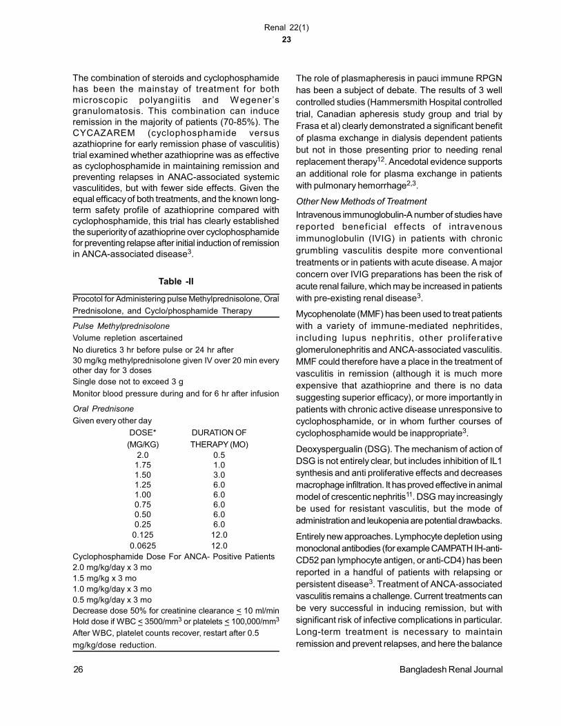
Table -II
Procotol for Administering pulse Methylprednisolone, Oral
Prednisolone, and Cyclo/phosphamide Therapy
Pulse Methylprednisolone
Volume repletion ascertained
No diuretics 3 hr before pulse or 24 hr after30 mg/kg methylprednisolone given IV over 20 min everyother day for 3 dosesSingle dose not to exceed 3 g
Monitor blood pressure during and for 6 hr after infusion
Oral Prednisone
Given every other day
DOSE* DURATION OF
(MG/KG) THERAPY (MO)2.0 0.5
1.75 1.01.50 3.01.25 6.01.00 6.00.75 6.00.50 6.00.25 6.0
0.125 12.00.0625 12.0
Cyclophosphamide Dose For ANCA- Positive Patients2.0 mg/kg/day x 3 mo1.5 mg/kg x 3 mo1.0 mg/kg/day x 3 mo0.5 mg/kg/day x 3 moDecrease dose 50% for creatinine clearance < 10 ml/minHold dose if WBC < 3500/mm3 or platelets < 100,000/mm3
After WBC, platelet counts recover, restart after 0.5
mg/kg/dose reduction.
The role of plasmapheresis in pauci immune RPGNhas been a subject of debate. The results of 3 wellcontrolled studies (Hammersmith Hospital controlledtrial, Canadian apheresis study group and trial byFrasa et al) clearly demonstrated a significant benefitof plasma exchange in dialysis dependent patientsbut not in those presenting prior to needing renalreplacement therapy12. Ancedotal evidence supportsan additional role for plasma exchange in patientswith pulmonary hemorrhage2,3.
Other New Methods of Treatment
Intravenous immunoglobulin-A number of studies havereported beneficial effects of intravenousimmunoglobulin (IVIG) in patients with chronicgrumbling vasculitis despite more conventionaltreatments or in patients with acute disease. A majorconcern over IVIG preparations has been the risk ofacute renal failure, which may be increased in patientswith pre-existing renal disease3.
Mycophenolate (MMF) has been used to treat patientswith a variety of immune-mediated nephritides,including lupus nephritis, other proliferativeglomerulonephritis and ANCA-associated vasculitis.MMF could therefore have a place in the treatment ofvasculitis in remission (although it is much moreexpensive that azathioprine and there is no datasuggesting superior efficacy), or more importantly inpatients with chronic active disease unresponsive tocyclophosphamide, or in whom further courses ofcyclophosphamide would be inappropriate3.
Deoxyspergualin (DSG). The mechanism of action ofDSG is not entirely clear, but includes inhibition of IL1synthesis and anti proliferative effects and decreasesmacrophage infiltration. It has proved effective in animalmodel of crescentic nephritis11. DSG may increasinglybe used for resistant vasculitis, but the mode ofadministration and leukopenia are potential drawbacks.
Entirely new approaches. Lymphocyte depletion usingmonoclonal antibodies (for example CAMPATH IH-anti-CD52 pan lymphocyte antigen, or anti-CD4) has beenreported in a handful of patients with relapsing orpersistent disease3. Treatment of ANCA-associatedvasculitis remains a challenge. Current treatments canbe very successful in inducing remission, but withsignificant risk of infective complications in particular.Long-term treatment is necessary to maintainremission and prevent relapses, and here the balance
26 Bangladesh Renal Journal

Renal 22(1)
24
must be struck between disease suppression andtreatment toxicity. The vasculitides are relatively rare,with an incidence of 20 new cases per millionpopulation per year3, and are heterogeneous in theirpresentation; hence the importance of well conducted,multi-centre collaborative trials to identify promisingnew therapies, and to maximize the benefit of existingtreatment regimens.
General management of Rapidly Progressiveglomerulonephritis
Hypertension almost invariably accompanies acuterenal failure caused by aggressive glomerulonephritis.Under this circumstance, hypertension is frequentlya consequence of volume expansion is addition toother intrarenal mechanisms (e.g., decreasedperfusion). Treatment of hypertension consists ofcontrol of volume expansion by fluid restriction, diuretictherapy (the dose adjusted to the level of renalinsufficiency) and anti hypertensive medications. Anyof the manifestations of uremia, if severe enough,prompt dialysis treatment is required which alsoimproves oxygenation in patients with pulmonaryedema and dyspnoea. Other indications includesevere acidosis, hyperkalemia, uremic pericarditis,and uremic encephalopathy6.
The approach to diagnosis and management ofinfection should be aggressive, with regular culturesof blood, sputum, urine, peritoneal dialysis fluid andother available material. Opportunistic infectionparticularly with nosocomial bacteria, cytomegalovirus,pneumocystis carinii and various fungui is common.The choice of antimicrobial will frequently be made onthe basis of the likely clinical diagnosis because thereis often delay or uncertainity in providing a preciseetiology13. Maintenance of adequate nutrition isimportant and early tube enteral or parenteral feedingis indicated in many patients because recovery is oftenprotracted and complicated by procatabolic events13.
Immunosuppressive and cytotoxic drug treatment inall patients must be based strictly on histopathologicalcriteria of activity. Experience in India showed use ofaggressive immunosuppressive therapy led to loss ofover 90% of patients from high chronicity index groupdue to infectious complications4. Many therapies usedto treat RPGN have the potential to induce not onlyinfections but also hemorrhages, sterility andmalignancy. Patients at risk of side effects (elderly,infected, patients with peptic ulcer) is better treated
by aggressive control of hypertension, renal failurerather than by immunosuppressive.
Long term monitoring to detect and prevent recurrenceand to preserve residual renal function is essential.Relapse may occur in patients with SLE, ANCApositive syndromes, Henoch-schonlein purpura andother conditions. Anti GBM disease does not usuallyrecur as long as the anti GBM titre has returned tobaseline. Regular follow up of patients is of utmostimportance to retain useful renal function.
References :1. Gaskin G, Pusey C D. Systemic vasculitis In : Cameron J
S. et al, eds. Oxford text book of clinical nephrology.Oxofrd university press. 1992; 612-35.
2. Levy J.B Pusey C.D. Still a role for plasma exchange inrapidly progressive glomerulonephritis ? J.N. 1997; vol10. (1) : 1-13.
3. Levy J. New aspects in the management of ANCA- positivevasculitis. Nephrol Dial transplant 2001; 16 : 1314-1317.
4. Dash S.C Rapidly progressive glomerulonephritis. In RashidH.U. editor. South asian renal disease. P 3 -8.
5. Glassock R. Management of rapidly progressiveglomerulonephritis. HOSP pract (off Ed). 2000; 15; 35 (2): 59-70.
6. Jennette J.C. Falk R.J. Diagnosis and management ofGlomerulonephritis and vasculitis presenting as AcuteRenal Failure In : Mandal A K et al eds. The medical clinicsof North America 74 (4); 893 - 908.
7. Rees A.J. Good pustures syndrome In Glassook. R.J.editor. Current therapy in Nephrology and Hypertension4th edition 1998; 166-171.
8. Wiggins R.C. Kershaw D.B Crescentic Glomerulonephritisin. Massry S G and Glassock R J eds. Text book ofNephrology 4th ed. Williams wilkins 2001; 720 - 725.
9. Bhuyan U.N. Dash S.C. et al. Immunopathology, extentand course of glomerulonephritis with crescent formation.Clin. Nephrol 18 : 280,198
10. Ponticelli C Moroni G. Lupus nephritis In . Glassock R J.editor Current therapy in Nephrology and hypertension 4th
ed. 1998; 156-160.
11. Wilkowski M J, Bolton W.K. Crescentic glomerulonephritis.In Glassock R. J. ed. Current therapy in Nephrology andhypertension 4th ed 1998; 216-220
12. Cole E, Cattram D, Magil A. et al. A prospective randomisedtrial of plasma exchange as additive therapy in idiopathiccrescentic glomerulonephritis. Am J. kidney Dis 1992; 20(3): 261-9.
13. Pusey C. D. Ress A. J. Acute renal failure due toglomerulonephritis and vasculitis. In Cameron J S et aleds. Osford text book of clinical nephrology, Oxforduniversity press. 1992; 1060-1076.
Bangladesh Renal Journal 27

Renal 22(1)
25
Introduction:Glomerulonephritis is one of leading cause of end stagerenal failure (RSRF) throughout the world. It comprises25-45% causes of ESRF in developing nations. Thisis because the treatment of primary glomerulonephritisis unsatisfactory. Although corticosteroid has longbeen used in patients with idiopathic nephroticsyndrome particularly minimal change disease (MCD),focal segmental glomerulosclerosis (FSGS) andmembranous nephropathy (MN), the response rate isvariable. The treatment of IgA nephropathy (IgAN),diffuse and focal proliferative glomerulonephritis is notsatisfactory1.
Clinical Presentation:The clinical presentation of glomerulonephritis islargely nephrotic syndrome, which comprises 80% ofcases. In 10-15% cases, the patient is presented withacute nephritic illness. The other causes ofpresentations are, recurrent hematuria and orasymptomatic proteinuria. A small number of patientspresented with chronic renal failure. The clinicalpresentation of nephrotic syndrome includes gradualonset of edema, raised or normal bloodpressure,proteinuria in excess of 3.5gm in 24 hours.Patients with minimal change nephrotic syndrome,focal segmental glomerulosclerosis and membranousnephropathy are usually presented with these typesof clinical symptoms.
Patients with acute nephritic illness usually have acuteonset with puffiness of face, oliguria and hematuria.Blood pressure is usually raised and renal failure maybe present at onset. The typical presentations areacute post-streptococcal glomerulonephritis (PSGN),some cases of IgA nephropathy and rapidly proliferativeglomerulonephritis (RPGN).
Pathogenesis:Pathogenesis of glomerulonephritis is thought to beimmunological. There is interaction of antigen and
Article of Special InterestNephrotic Syndrome- Evidence Based Management
HU RASHID
Professor & Chairman Dept. of Nephrology, BSMMU, Shahbag, Dhaka.
antibody to form the immune complexes together withcomplement and coagulation factors have beeninvolved in the pathogenesis of glomerulonephritis.Involvement of the resident glomerular cells withliberation of cytokines and growth factors are alsofound to be important in the pathogenesis ofglomerulonephritis. Cytokines regulating the responsehave been characterized, involvement of additionalmolecules and leukocyte recruitment defined andmechanism based therapy identified. These advancesare already bearing fruit in understanding mechanismof glomerular inflammation and may point to newtherapies2,3.
Diagnosis:Diagnosis of glomerulonephritis is usually made byclinical presentation, routine and microscopicexamination of urine. Renal function tests are checkedin every patient and size of the kidney is measuredthrough ultrasonography. Urinary protein excretion isdone by estimating 24 hours urinary total protein anddiagnosis is confirmated by renal biopsy. Specificdiagnosis is made by complement (C3, C4), anti-nuclear factor (ANF), anti-dsDNA, Hepatitis B&C virusand antineutrophilic cytoplasmic antibodie (ANCA).Abnormalities of lipoprotein and low serum albuminare seen in patients with nephrotic syndrome.
Histological diagnosis:In adults, histological diagnosis is usually minimalchange disease, membranous glomerulonephritis,focal segmental glomerulosclerosis (FSGS), diffuseand focal proliferative glomerulonephritis,membranoproliferative glomerulonephritis (MPGN), IgAnephropathy and heredofamililial glomerulonephritis(Alport’s syndrom). In a recent series, Rashid et al,20014 found, minimal change disease 23 (6%),membranous glomerulonephritis 88 (23%), focalsegmental glomerulosclerosis 22 (5%), diffuse andfocal proliferative glomerulonephritis 77 (22%),membranoproliferative glomerulonephritis 62 (16%),
Correspondence : HU Rashid, Professor & Chairman Dept. of Nephrology, BSMMU, Shahbag, Dhaka.
BANGLADESH RENAL J. 2003; VOL. 22 (1) : 1-4

Renal 22(1)
26
mesangial proliferative glomerulonephritis 112 (29%)among 386 cases of glomerulonephritis [abstract inWCN,]4.
Management:Management of glomerulonephritis is dependent onclinical presentation, degree of proteinuria, renal failureat presentation and above all histological diagnosis.Symptomatic treatment is necessary in almost allthe cases, restriction of fluid and salt intake is alsovery important. Edema should be appropriately treatedwith loop diuretics and hypertension by ACE inhibitorsif renal function is normal or mildly deteriorated.Proteinuria should be treated with ACE inhibitors evenin the absence of hypertension if specific treatment isnot available. Similarly, hypercholesterolaemia andhypertriglyceridemia should also be treated if there isno specific treatment for the primary disease.
Specific treatment:Specific treatment with prednisolone and prednisolonewith cytotxic drugs are available in patients withminimal change disease, focal segmentalglomerulosclerosis and indiopathic membranousglomerulonephritis. IgA nephropathy with heavyproteinuria and normal renal function can also betreated with long term steroid therapy. Patients withmembranoproliferative glomerulonephritis with hepatitisB or C can be treated with appropriate antiviral therapy.
Minimal change disease:In children: Eighty percent of patients respond to initialcorticosteroid therapy (prednisone). One widely usedregimen administers prednisone, 60mg/m2/day for 4to 6 weeks, followed by 40mg/m2/day every other dayfor another 4 to 6 weeks and then discontinuation. Incase of relapse, prednisone should be given at a doseof 60mg/m2/day till protein is nil for 3 days and then40 mg/m2 alternate day for another 4 to 6 weeks. Incase of relapse for the second time, same regimenas in 1st relapse can be repeated. In third relapsecyclophosphamide is the drug of choice, which shouldbe given at a dose of 1.5 to 2mg/kg/day for 2 to 3months. Dosage should be monitored by frequentCBCs assuming a WBC count>4000/mL.
In corticosteroid nonresponders (<5% in children),frequent relapse’s and corticosteroid-dependentpatients, prolong remission may be obtainable withoral cyclosporine 6mg/kg/day till remission. Thenreduce the dose 1 mg/kg every month to amaintenance dose of 2-3 mg/kg for about one year5.
Other drugs: Levamisole, an antihelminthic drug hasbeen used to successfully treat children with MCNS.In one study, 30 children with frequently relapsingnephrotic syndrome were treated with oral levamisole,2.5 mg/kg body weight twice a week for 10 monthsand in 16 children, corticosterioid dose could bereduced significantly without relapse while the drugwas ineffective in the remaining 14 children.
In adults: There are two widely accepted regimes forthe use of prednisone in the treatment of MCNS inadults.
A. Administer prednisone 1.5 to 2 mg/kg /day perorally for 8 to 12 weeks, then gradually taper thedose 5mg two weekly and continue for 6 months.
B. Prednisolone 2mg/kg on alternate day for 2months followed by tapering off 5-10 mg twoweekly and continue for 6 months to 1 year.
In frequently relapsing and steroid dependent nephroticpatients, cyclophosphamide is the drug of choice,which should be given at a dose of 1.5 to 2 mg/kg/dayfor 2 to 3 months. Another alternative is oralcyclosporine 5mg/kg/day, which is usually continuedfor 1 year.
Focal segmental glomerulosclerosis:Patients with focal segmental glomerulosclerosisshould be treated with prednisolone 0.5 to 2 mg/kg/day for 6 months. This dose should be continued atleast for three months before declaring that, thedisease is not FSGS. The dose of prednisolone shouldbe reduced to 0.5mg/kg/day when proteinuria is nilbut after three months. In patients not responding tosteroid therapy, additional cytotoxic drugs don’t makeany difference in the respnse rate or prevention of renalfailure. However addition of cyclosporine 6mg/kg/dayin children and 5mg/kg/day in adults showed variableresponse. Cyclosporine should be continued at leastfor one year. Treatment with tacrolimus, MMF andplasmapheresis seems to be encouraging6.
The prognosis of FSGS is variable. Those patientsresponse to prednisolone, 10 year survival is around90%, where as in non-responders 5 year survival is50-60% and 10 year survival is 25-40%. Patientspresented with nephrotic syndrome leaving untreated,5 year survival is approximately 50-90% and 10 yearsurvival is 30-50%. Patients presented with nonnephrotic proteinuria, 10 year survival is 85-90%. The
2 Bangladesh Renal Journal

Renal 22(1)
27
prognostic indicators are; initial creatinine level, degreeof proteinuria, presence of hypertension and interstitialscarring on renal biopsy.
Membranous glomerulonephritis:Asymptomatic patients with nonnephrotic range ofproteinuria should not be treated. Patients who haveedema can be controlled with diuretics orasymptomatic patients with proteinuria should befollowed, because ³ 30% will have a partial or completeremission within 3 to 4 year.
Two recommendations have been widely accepted forthe treatment of patients with symptomatic NS andfor those most at risk of progressive renal diseaseand those with an elevated serum creatinine.
Recommendation 1:
Methyl prednisolone 1 g/day slowly intrvenously for 3days followed by oral prednisone 0.5mg/kg/day for 27days followed by chlorumbucil 0.2 mg/kg/day for 30days. Repeat for another 2 cycles. Dosage should bemonitored by frequent CBCs assuming a WBC count> 4000/mL.
Recommendation 2:
Prednisone 2mg/kg alternate day for 2 month and thengradually taper the dose & continue for 6months.Cyclophasphamide is added at a dose of 1.5to 2 mg/kg/day for 3 to 6 months. Dosage should bemaintained by frequent CBC, WBC > 4000/mL.
Prognosis of membranous nephropathy is dependenton several factors. Patients with older age, male sex,heavy proteinuria (>10gm/day), presence ofhypertension, hypercholesterolemia, renal failure atonset, advanced changes on renal biopsy predict worstprognosis of membranous nephropathy. Where aspatients with younger age, female sex, non-nephroticproteinuria, persistent normal renal function for 3 years,absence of tubulointerstitial changes in renal biopsy,absence of glomerular scarring has favorableprognosis7.
IgA Nephropathy:
Treatment of IgA nephropathy is controversial.Prednisone may be given in patients with proteinuria> 3gm/day with normal renal fucntion. Howeverprednisone is not effective if renal function is impaired
and fish oil is prescribed in those circumstances. ACEin hibitor may be given for patients having proteinuria1 to 3 gm/day. Prognosis of IgA nephropathy is usuallygood unless there is heavy proteinuria of nephroticrange, presence of hypertension and impaired renalfunction at presentation. Histological evidence ofglomerular sclerosis, tubulointerstitial atrophy, fibrosisand scarring also predict bad prognosis of IgAnephropathy8.
Membranoproliferative glomerulonephritis:
Treatment of membranoproliferative glomerulonephritisis symptomatic unless there is evidence ofassociation with hepatitis virus B or C. In thosecircumstances where there is direct link of hepatitisB or C virus, appropriate therapy for the virus is usuallygiven. The symptomatic treatment includes use ofdiuretics, treatment of hypertension, fluid and saltrestriction and treatment of hyperlipidemia.
In other glomerulonephritis, specific treatment is alsonot available. Other glomerulonephritis Treatment ofFocal and diffuse proliferative glomerulonephritis andmesangial proliferative glomerulonephritis are alsosymptomatic9.
General measure:
Diet, restriction of fluid and salt is important in thegeneral management of patients with nephroticsyndrome.
Edema is treated with loop direct and the dose isincreased according to response of the drug.
Hypertension is treated with appropriateantihypertensive drugs and special emphasis is givento ACE and ARB group of drugs.
Hyper lipidaema, should also treated withantilipidaemic drugs.
Conclusion:
The prognosis of nephrotic syndrome is dependenton type of histological diagnosis degree of proteinuria,presence of hypertension and renal function atpresentation.
Male sex, older age groups, family history, type ofHLA, may also important in predicting long termprognosis of the disease.
Bangladesh Renal Journal 3
Renal 22(1)
28
References:1. Daniel C. Cattran. Management of glomerulonephritis:
Kidney Int 1999; 55 (S70) : 1-2.
2. Cameron J.S. Proteinuria and progression in humanglomerular diseases: (1990b) American Journal ofNephrology (1999b) 10 (S1):81-87.
3. Denis Glotz & Philippe Drue. Immune mechanism of glomerulardamage: In : Oxford Textbook of Clinical Nephrology; DavisonAm, Cameron JS, Grunfeld JP, Kera DNS, Ritz and WinerilsLS (Editors), Oxford University Press. 1998; 383-390.
4. Rashid et al: World Congress of Nephrology; (Abstract)Kidney Int 2001.
5. Joanne M. Bargman. Management of minimal lesionglomerulonephritis. Evidence-based recommendations:Kidney Int 1999; 55(S70): 3-16.
6. Ellen Burgess. Management of focal segmental
glomerulosclerosis. Evidence--based recommendations:
Kidney Int 1999; 55 (S70): 26-32.
7. Norman Muirhead. Management of idiopathic membranous
nephropathy. Evidence-based recommendations: Kidney
Int 1999; 55 (S70): 47-55.
8. Linda Nolin & Maryse Courteau. Management of IgA
nephropathy. Evidence--based recommendations: Kidney
Int 1999; 55 (S70):.56-62.
9. Addera Levin. Management of membranoproliferative
glomerulonephritis. Evidence-based recommendations:
Kidney Int 1999; 55 (S70): 41-46.
4 Bangladesh Renal Journal

Renal 22(1)
29
Impact of Na+ Profiled Dialysis on Control ofBlood Pressure on MHD Population
DR. NIATUZZAMAN
A prospective case-control study is conducted upon38 patients on MHD receiving about 12 hour HD/weekfor nine months (observation three months and Na+proffiling six months). Nausea and vomiting; chest pain;hypotension and hypertension, headache are recordedduring and in interdialytic period. Patients are on anti-hypertensive drugs in single or incombination withoutno gross alteration in drug groups both duringobservation and intervention (Na+profiling) period. Fiveimportant parameters are considered for statisticalanalysis-(Pre-MAP; Post-MAP, Pre-weight; Post-weight; Kt/v) before and after intervention(Nat+profiling). Objective complaints are reducedsignificantly. Nausea and vomiting (42.5 vs 31.57%)chest pain (35% vs 23.68%); Hypotension (27.5% vs18.42%); Hypertension (22.5% vs 13.15) andHeadache (60%vs 39.47%) (p-value <0.001). Thisresults are further strengthened, by reduction of anti-hypertensive drug regimen-combination-double(57.5% vs 19.44%) and triple (7.5% vs 0%) (p-value<0.001). Profiled dialysis was inter-individualised andregession analysis shows-significant linear correlationwith post-MAP; pre- and post-weight and inversecorrelation with kt/v (p-value in all cases is <0.001).Correlation with pre-MAP is not significant (p-value<0.05) suggesting other factor also regulatesinterdialytic blood pressure. Conclusion: Na+ profiledheamodialysis is helpful in MHD but with inter-individual dose regulation considering the individual’spre- and post dialytic biochemical profile. It exertspositive impact on interdialytic blood pressureregulation but other variables like-sympathetic over-activity; vasoactive hormones etc. also beenimplicated. However, this study is on short durationand small population based; so large scale study onlonger duration is needed for further evaluation.
ABV: Pre-MAP- Pre-dialysis mean arterial pressure
Post-weight- Post dialysis weight.
Abstract form Current Literature
Selected Abstract from 10th Annual Convention and International CME2nd and 3rd December 2003
Effects of ACE Inhibitor on Proteinuria and RenalFunction in Patients with Type2 Diabetes With orWithout Renal Failure
HASSAN MN, RASHID HU, ALI L
Bangabandhu Sheikh Mujib Medical University, Dhaka
Diabetic Nephropathy (DN) is the leading cause ofESRD throughout the world. Greatest importance isfocused on proteinuria and hypertension in additionto control of diabetes in declining of renal function.Few clinical trials have studied the effects of ACEInhibitors (ACEIs) on type2 diabetic patients with overtproteinuria. We have studied the effects of ACEI onproteinuria and renal function in type2 diabetic patientswith overt proteinuria with or without hypertension and/or renal failure.
This randomized, controlled, prospective study wascarried out on eighty diagnosed type2 diabetic patientswith overt proteinuria with or without hypertension and/or renal failure to see the efficacy of ACEI, lisinopril inreducing proteinuria and preserving renal function ascompared to calcium channel blocker, amlodipine.Forty one were assigned to “ACEI group” who gotlisinopril at a maximum tolerated dose. Thirty ninewere assigned to “control group” who receivedamlodipine in a dosage sufficient to control BP belowtarget level. Patients were followed three monthly fora mean period of one year for BP, s. creatinine,creatinine clearance rate (Ccr.) & 24hrs UTP.
BP was equally controlled without any significantdifference between groups. There was significantreduction in UTP from 3.24±3.0 G/24hr to 1.55±1.35G/24hr in ACEI group, whereas in control, group,reduction was from 2.52±2.1 to 1.69±1.64 G/24hr only.S.creatinine rose from 130.5±43.4 to168.1±59.4mmo1/L in ACEI group and from145.1±54.3 to 188.4±71.9 in control group. Decline inCcr was from 58.4±20.4 to 44.4±15.8 in ACEI groupand from 54.3±25.0 to 46.3±26.9 in control group.
BANGLADESH RENAL J. 2003; VOL. 22 (1) : 28-31
Renal 22(1)
30
Neither the rate of rise of s.creatinine nor the declinein Ccr. showed any statistically significant differencein between the two groups. This declining renalfunction was not affected by baseline renal functionstatus.
In the present study, although there was significantreduction of proteinuria by ACEI there was noimprovement in renal function. S.creatinine increased& Ccr decline significantly from base-line value to eachquarterly time-point throughout the one year studyperiod (all values p<0.001) in both groups.
It is concluded that ACE inhibitor causes significantreduction in proteinuria but no improvement in renalfunction as compared to calcium channel blocker inthis particular group of type2 diabetic patients withovert proteinuria with or without hypertension and/ormild renal failure.
Effect of Calcium Carbonate and Oral calcitriolon PTH in MHD patients
AMM EHTESHAMUL HOQUE
Department of Nephrology, Bangabandhu SheikhMujib Medical University, Dhaka
Secondary hyperparathyroidism is common in MHDpatients. Accumulated evidence suggests that PTHis a major uremic toxin. Oral calcitriol and calciumcarbonate therapy can effectively reduce elevated PTHlevel. A total no. of 45 MHD patients (male 24 female-21) of different duration (6 months to 60 months) werestudied in the department of nephrology and INM,BSMMU Dhaka to see to effect of oral calcitriol andcalcium carbonate on PTH. Subjects were divided into3 groups according to duration of haemodialysis.
Group-1 (6 months to 18 months, N = 12), Group-2(19 months to 36 months N = 20), Group-3 (37 months60 months, N = 13) mean age of the patients were40.24 ± 8.94 yrs. Total duration of study was 36months initial 4 months was washout period remaining32 months was study period. During this study periodall the subjects received calcium carbonate 500 mgthrice daily with meal and oral calcitriol 0.25 mg oncedaily. S PTH (midregion), S. Calcium, SAlkalinephosphatase, S. Inorganic phosphate, SAlbumin, STotal protein were measured at the end of wash outperiod and at 5th and at 9th months of study period.
Elevated S. PTH (mid region) level, hypocalcaemia,hyperphosphataemia, increased alkaline phosphatase
level was observed at beginning of study period. Atthe end of study period decreased PTH level, inorganicphosphate level, alkaline phosphatase level andincreased S. calcium level was observed in all thesubjects of 3 groups.
Median S.PTH (mid region) at beginning of study vsend of study was; In group-1 : 544. 19 pmol/L, vs 206,31 pmol/L, In group-2 476-82 pmol/L vs 196.12pmol/L, In group-3 : 440. 68pmol/L vs 128.02 pmol/L
Mean±SD S. celeium level at begining of study vsend of study was; In grou 1 1.85+0.32 mmol/L vs 2.370.10mmol/L, P<0.01, In group-2 1.94 ± 0.30 mmol/Lvs 2.43 ± 0. 10mmol/L, P<0.01, In group-3 : 1.96 ±0.27 mmol/L vs 236 ± 0.16 mmol/L P <0.05.
Mean ± SD inorganic phosphate level at beginning ofstudy vs end of study In group-1 : 2.60 ± 0.61mmol/Lvs 1.80± 0.27 mmol/L P< 0.001, In group-2 : 2.71 ±0.49 mmol/L vs 1.82 ± 0.27 mmol/L p< 0.001, In group-3 : 2.48 ± 0.67 mmol vs 1.74 ± 0.40 mmol/L P<0.06
Mean ± SD salkaline phosphatase : level at begeningof study vs end of study was In group -1 315 ± 150.64U/L vs 214.92 ± 86.68 U/L P<0.05, In group -2 : 305.34± 155.02 U/L vs 221.75 ± 96.16 U/L P < 0.05, In group-3 : 302.85 ± 94.54 U/L vs 220.69 ± 76.48 U/L p <0.05.
It is concluded that elevated S. PTH, alkalinephosphatase, inorganic phosphate andhypocalcaemia was observed in MHD patients andoral calcitriol and calcium carbonate can reduce S.PTH level, S. alkaline phosphatase level, S. inorganicphosphate level and increase S. calcium level.
Experimental Nephropathy Induced byStaphylococcus Aureus Antigens
SAMINA SHARMIN, KOYAMA AKIO, HU RASHID
Department of Internal Medicine, Institute of ClinicalMedicine, University of Tsukuba, Japan Dept. ofNephrology, BSMMU, Dhaka, Bangladesh
Purpose : Much evidence suggests that, IgAnephropathy (IgAN) is an infectious associated GNalthough exact pathogenesis is unknown. Wehypothesized that S.aureus, a common normal andpathogenic flora plays an important role in thepathogenesis of IgA nephropathy. Recently we havereported glomerulonephritis occurred during methicillin
Bangladesh Renal Journal 29
Renal 22(1)
31
resistant S. aureus infection, that have showedpolyclonal elevation of serum IgA and IgG levels withIgA, IgG and C3 deposition in the glomeruli. In thisstudy we have showed, induction of IgA -typeglomerulonephritis in Balb/c mice by using antigensderived from S.aureus.
Method : Balb/cmice (Th2 dominant type) and C57BL/6 (Thl dominant type) were immunized with antigensderived from S.aureus with Freund’s incompleteadjuvants biweekly for 4 months.
Results : Mesangial proliferative glomerulonephritiswith IgA and IgG depositions were found in all Balbfc,not in C57BL/6 mice. In Balb/c mice, S.aureusantigens were detected in glomeruli using affinity-purified human anti-S.aureus antibodies, however therewas no staining in C57BL/6 mice. In Western blotanalysis, the band which was differently recognizedby Balb/c and C57BL/6 anti-serum was 30-35 kDa.
Conclusion : S.aureus antigens, especially 30-35 kDaprotein and genetic background of the hosts may playimportant roles in the pathogenesis of IgA nephropathy.
Serum Parathormone Level and Its Correlationwith Different Biochemical Parameters inChildren with Chronic Renal Failure
M H RAHMAN, G MUINUDDIN, A BEGUM,MM HOSSAIN
Department of Paediatrics, Nephrology, BangabandhuSheikh Mujib Medical University, Dhaka
Objective : A prospective study was conducted toestimate the level of serum intact parathormone andits correlation with different biochemical parameterswith chronic renal failure in children. Setting: PaediatricNephrology Unit, Department of Paediatrics,Bangabandhu Sheikh Mujib Medical University, Dhaka,Bangladesh.
Subjects and Methods : Study was conducted in 64children (44 with chronic renal failure and 20 age andsex matched controls). Serum intact parathormone(PTH), creatinine clearance (Ccr), serum creatinine,urea, ‘serum calcium; inorganic phosphate andalkaline phosphatase were estimated for both groups.Serum intact parathormone was estimated by Inmiuliteanalyzer. Serum creatinine, urea, calcium, inorganicphosphate and alkaline phosphatase were estimatedin blood by standard technique.
Results: 44 (male 30, female 14) chronic renal failurechildren were divided into four groups according totheir Ccr, mild CRF with mean Ccr 59.17±18.53 ml/min/1.73 m2, n=6; moderate CRF with mean Ccr34.98±7.75 ml/min/1.73 M2, n=7; severe CRF withmean Ccr 17.71±5.40 ml/min/1.73 M2, n=15; End-stage renal disease with mean Ccr 6.46±1.71 ml/min/1.73 m2, n=16). Mean serum intact PTH value werein mild 48.50±4.76, moderate 67.29±7.91, severe82.42±9.67 and end-stage renal failure 130.66±58.74pg/ml, and control 16.52±9.35 pg/ml respectively .Mean serum intact PTH of CRF (93.00±46.42 pg/ml)was negatively correlated with mean Ccr (22.02±18.53ml/min/1.73 n2) (r = -0.564, P<0.001) and mean serumcalcium (7.30±1.02 mg/dl) (r = -0.50, P<0.001), andpositively correlated with mean inorganic phosphate(5.76± 1.1 mg/dl) (r = +0.345, P<0.05) and meanalkaline phosphatase (355.14±185.53 U/L) (r = +0.469,P<0.001) respectively.
Conclusion : It is concluded that progressivelyincreased PTH level may occur even early in the courseof chronic renal failure and hypocalcaemia is aninitiating factor for the development ofhyperparathyroidism.
Continuous Ambulatory Peritoneal Dialysis, ANovel Mode of Treatment in End Stage RenalFailure Patients: Yet to be Popularized InBangladesh
MA SAMAD1, HU RASHID2, H RAHMAN2, M IQBAL1
Department of Nephrology, National Institute of KidneyDisease and Urology1, BSMMU2, Dhaka, Bangladesh
Continuous Ambulatory Peritoneal dialysis (CAPD)is a well-accepted mode of renal replacement therapy(RRT) worldwide. But the method is yet to bepopularized in Bangladesh. In order to evaluate theapplicability of CAPD in Bangladesh we report theclinical outcome of the patients treated in this method.Between January 1997 to October 2003, 94 patientswere managed by CAPD in this country. 12 patientsdiscontinued or died due to unrelated cause beforeinitiation of regular exchange and were excluded.Therefore, we studied 82 patients (mean age 58±16(range 7-85] years; 50 male, 32 female.) Primaryetiology were diabetic nephropathy in 43(52.43%)glomerulonephritis in 15(18.29%), hypertension in11(13.41 %), heridofamilial disease in 2(2.43%), renal
30 Bangladesh Renal Journal
Renal 22(1)
32
cell carcinoma in one, Chronic pyelonephritis in two(2.43%), in 3 were other causes and causes in 5patients were unknown. Indications of CAPD werecardiovascular instability in 44(53.6%), long distancefrom hemodialysis center in 20(24.39%), vascularaccess problem in 7(8.53%) and others in 11(13.41%).Most of the patients used Baxter PD fluid, Y-set with2.bag system and daily 3 exchanges of 2 litre bagwere practiced. Total duration of follow up was 1256patient months; mean 15.32±17.96(range 1-77months) and, technique survival was 94%. Majorcomplications were peritonitis (one episode [ep] per14.26% patient-month) exit site infection (one ep per43 patient-month), catheter block (one ep per 34 patientmonth and were cleared in all cases with heparin washexcept two), and leakage in 2 cases and hernia in
two. Mean hemoglobin was 9.25±1.78 gm/dl,creatinine 584±153 mmol/l, urea 15.32±7.44 mmol/l,serum albumin was 30.24±5.74 gm/1 and serumpotassium was 3.12±1.4 mmol/l. At the time ofreporting, of 82 patients 20 pts. were continuing withthis model of treatment, 14 discontinued and 38 died.Of the 12, 2 patients discontinued due to leakage andhernia, 4 patients due to peritonitis and weretransferred to hemodialysis, 4 patients discontinueddue to financial reason and 3 underwent kidneytransplantation while renal function improved in 2cases. 5 of the 38 death occurred due to septicemiaresulting from peritonitis. The causes of remaining 33mortalities were unrelated to CAPD. Though most ofthe patients were elderly, diabetic, debilitated yet ourresults lead to conclusion that CAPD may be the well-accepted mode of RRT in this country.
Bangladesh Renal Journal 31

Renal 22(1)
33
Notes & News
INTERNATIONAL NEWS
International Society of Nephrology
MEMBERSHIP
The ISN provides an international forum for theexchange of knowledge, best clinical practice, andresearch. Since it was founded in 1960, the societyhas constantly made major contributions to theworldwide advancement of educational, science, andpatients care in nephrology.
The ISN’s more than 7000 members includeresearchers and clinicians from over 100 countriesinvolved in basic, transnational, and applied science.Through this global network of professionals, the ISNis focused on designing programs that aim tounderstand and diagnose kidney disease and providesuitable treatment, either to prevent the disease orslow its progression.
Join the ISN today and become a member of theworld’s leading network of nephrologists membershipincludes a 1 – year subscription of Kidney Internationaland a number of other exclusive benefits.
For complete information relating to membership orany the ISN’s programs and activities, please visitthe Membership section of the ISN Web site at http://www.isn-online.org or by contacting the ISN globalHeadquarters at Avenue des Gaulois, 7, B-1040,Brussels, Belgium. Telephone : +32-2-743 1546l Fax: +32-2-743 1550; E-mail : [email protected]
European diabetic nephropathy study group (EDNSG)will be held May 7 and 8, 2004, in Bergamo, Italy. Allabstracts on any topic relating to diabetic nephropathyresearch should be submitted by e-mail (to the addressbelow) with the abstract as an attachment in a richtext format (RTF) or as a world file. Abstracts shouldbe written with the following headings : Objective,Design, Setting, Patients, Main OutcomeMeasurements Results, and conclusion. The lengthof the abstract should notexceed on side of an A4paper. With margins of 2.5 cm, and a font size nosmaller than Times 12. Abstracts submitted by faxshould also be sent by mail (5 sets) by January 10,2004 to Dr. Roberto Trevisan, Secretary of EDNSG,U.O. Diabetologia, Ospedail Riuniti di Bergamo, Largo,
Barozzi 1, 24128 Bergamo, Italy, Fax : +39 35 266889;E-mail : [email protected] or [email protected]
The 3rd International Course on Critical CareNephrology will be held June 2-5, 2004, at the CongressCenter Ente Fiera, Vieenza, Italy. For furtherinformation, contact : Web site : http://www.vicenzanephrocourses.com or http://www.efrologiavicenza.it
The 12th International Congress on Nutritional heldMetabolism in Renal Disease will be June 19-22, 2004,in Venice/Padua, Italy. The chairpersons for thecongress are Giuseppe Bucciante and Gian-francoGuarnieri. For further information, contact : thescientific secretariat, Annamaria Bernardi at E-mail :[email protected]. Or the OrganizingSecretariat, Meet and Work. Piazza del Sole e dellapace, 5, 35031 Abano Terme (Padova), Italy. Telephone: + 39 049 8601818; Fax : + 39 049 8602389; E-mail: [email protected] ; congress web site : http://www.nutrition. Metabolism-004.it
The ISN 2004 Conference on Prevention of progressionof Renal Disease will be held June 29 – July 1, 2004,at the Hong Kong convention and exhibition Center inHong Kong. The Hong Kong Society of Nephrology isthe host organizer for this meeting in conjunction withthe ISN. This is the first ISN thematic conference heldin Asia on the prevention of progression of renaldisease. It will explore, in a multidisciplinary approach,the latest basic and clinical aspects on chronic renalfailure and the prevention of its progression. It will alsodiscuss the economic consequences of chronic renalfailure and its global health care. A global consensuswill be presented, and a way forward to help counterohonic renal diseae worldwide will evolve. Anoutstanding international cast of speakers has beenassembled to prevent the topics like health economics,diabetic nephropathy, glomerulonephritis, basicresearch, genetics, drugs and herbs, bypertensionand cardiovascular diseases, renal screeningprograms, and blueprints for prevention. For furtherinformation, contact : Conference Secretariat, c/oInternational Conference Consultants, Ltd., Units 501-3, 5/F, Far East Consortium Building, 121 Des VoeuxRoad Central Hong Kong. Telephone : (852) 2559 9973;Fax : (852) 2547 9528; E-mail : [email protected];l website : http://www.iwn2004hkconference.org.
BANGLADESH RENAL J. 2003; VOL. 22 (1) : 32