amyotrophic lateral sclerosis-linked mutations increase the … · lcs, is a component of rna...
TRANSCRIPT

Amyotrophic lateral sclerosis-linked mutations increasethe viscosity of liquid-like TDP-43 RNP granulesin neuronsPallavi P. Gopala,b, Jeffrey J. Nirschlb,c, Eva Klinmanb,c, and Erika L. F. Holzbaurb,c,1
aDepartment of Pathology and Laboratory Medicine, Perelman School of Medicine, University of Pennsylvania, Philadelphia, PA 19104; bDepartment ofPhysiology, Perelman School of Medicine, University of Pennsylvania, Philadelphia, PA 19104; and cNeuroscience Graduate Group, Perelman School ofMedicine, University of Pennsylvania, Philadelphia, PA 19104
Edited by Don W. Cleveland, University of California, San Diego, La Jolla, CA, and approved February 3, 2017 (received for review August 29, 2016)
Ribonucleoprotein (RNP) granules are enriched in specific RNAs andRNA-binding proteins (RBPs) and mediate critical cellular processes.Purified RBPs form liquid droplets in vitro through liquid–liquid phaseseparation and liquid-like non–membrane-bound structures in cells.Mutations in the human RBPs TAR-DNA binding protein 43 (TDP-43)and RNA-binding protein FUS cause amyotrophic lateral sclerosis (ALS),but the biophysical properties of these proteins have not yet beenstudied in neurons. Here, we show that TDP-43 RNP granules in axonsof rodent primary cortical neurons display liquid-like properties, includ-ing fusion with rapid relaxation to circular shape, shear stress-induceddeformation, and rapid fluorescence recovery after photobleaching.RNP granules formed from wild-type TDP-43 show distinct biophysicalproperties depending on axonal location, suggesting maturation to amore stabilized structure is dependent on subcellular context, includ-ing local density and aging. Superresolution microscopy demonstratesthat the stabilized population of TDP-43 RNP granules in the proximalaxon is less circular and shows spiculated edges, whereas more distalgranules are both more spherical and more dynamic. RNP granulesformed by ALS-linked mutant TDP-43 are more viscous and exhibitdisrupted transport dynamics. We propose these altered propertiesmay confer toxic gain of function and reflect differential propensityfor pathological transformation.
TDP-43 | ribonucleoprotein granules | liquid droplets |amyotrophic lateral sclerosis | neurons
Cellular organelles allow eukaryotic cells to organize biochemicalprocesses and concentrate specific cellular reactions in space and
time. Although the role of membrane-bound organelles in cytoplas-mic compartmentalization has long been recognized, the distinctbiophysical properties and functions of non–membrane-bound or-ganelles enriched in RNA and proteins have been recognized onlyrecently (1–5). Ribonucleoprotein (RNP) granules, such as P granulesin Caenorhabditis elegans (1), nucleoli in Xenopus laevis oocytes (2),yeast P bodies (3), and mammalian stress granules (6), show liquiddroplet properties (reviewed in refs. 5, 7, 8), including fusion withrapid relaxation to a spherical shape, dynamic internal rearrange-ments, and rapid dissolution and assembly. Proteins comprising RNPgranules share a common structure containing both RNA recogni-tion motifs (RRMs) and low-complexity sequences (LCSs), intrinsi-cally disordered regions that mediate protein–protein interactions(7, 9). In vitro characterization of human RNP granule proteins,RNA-binding protein FUS and heterogeneous nuclear ribonu-cleoprotein A1 (hnRNP A1), which are mutated in rare inheritedforms of amyotrophic lateral sclerosis (ALS) and frontotemporaldementia (FTD) (10–12), has revealed the LCS drives self-assemblyof RNP granules through a process termed liquid–liquid phaseseparation (LLPS) (6, 13–18).
Among eukaryotic cells, neurons face unique challenges in spatiotem-poral cytoplasmic organization related to their complex axonal-dendriticmorphology, separation of soma and axon terminal, and long cellularlifetimes with limited capacity for regeneration. Functionally, neuronsmaintain distinct axonal and dendritic pools of mRNAs allowing for rapid
responses to local environmental cues (19). RNA transport granules arespecialized RNP organelles composed of RNA-binding proteins (RBPs),translationally repressed mRNA, and molecular motors that play a keyrole in establishing mRNA polarity (20, 21). However, it is unclearwhether neuronal cytoplasmic RNP granules also display liquid dropletproperties, and if so, how these biophysical properties underlie physio-logical functions and relate to disease.
TAR-DNA binding protein 43 (TDP-43), a highly conserved DNA-binding protein and RBP with two RRMs and a glycine-rich C-terminalLCS, is a component of RNA transport granules and cotransports withmRNA (22–26). Mutations in TARDBP, the gene encoding TDP-43,have been linked to familial and rare sporadic cases of ALS (27, 28).Moreover, neuronal and glial cytoplasmic inclusions composed of TDP-43 serve as the neuropathological hallmark of sporadic ALS and themajority of FTD cases (29). In vitro characterization of TDP-43 has beenlimited by its tendency to fibrillize (15, 30). Although a recent study inmammalian cells demonstrates that nuclear TDP-43 may also undergoLLPS to form multiphase compartments (31), the biophysical propertiesof TDP-43 RNP granules have not been examined in neurons.
We hypothesize that TDP-43 RNP granules in the axon displayliquid droplet behaviors. If this hypothesis is true, then compared withmembrane-bound axonal cargos, RNP granule motility will show dis-tinct properties, including (i) an ability to interact with other RNAgranules to influence trajectory and composition, (ii) frequent fusionevents with rapid relaxation to spherical shape, (iii) deformability byshear stress, (iv) rapid internal redistribution, and (v) sensitivity todisruption of weak hydrophobic interactions. In addition, we predict
Significance
Mutations in TAR-DNA binding protein 43 (TDP-43), an RNA-bindingprotein (RBP) with multiple functions in RNA metabolism, causeamyotrophic lateral sclerosis (ALS), but it is uncertain howdefects inRNA biology cause disease. Purified RNA-binding protein FUS andheterogeneous nuclear ribonucleoprotein A1 (hnRNP A1) form liq-uid droplets in vitro through liquid–liquid phase separation. How-ever, the biophysical properties of ribonucleoprotein (RNP) granulescomposed of wild-type (WT) or ALS-linked TDP-43 have not beenstudied in primary neurons.We show that TDP-43WT RNP granulesexhibit distinct biophysical properties depending on their axonallocation, whereas granules formed by ALS-linked mutant TDP-43are more viscous and show disrupted axonal transport dynamics.We propose the distinct biophysical properties of these neuronalRNP granules may reflect different maturational states and differ-ential propensity for pathological transformation.
Author contributions: P.P.G., J.J.N., and E.L.F.H. designed research; P.P.G., J.J.N., and E.K.performed research; J.J.N. contributed new reagents/analytic tools; P.P.G., J.J.N., and E.L.F.H.analyzed data; P.P.G. and E.L.F.H. wrote the paper; and J.J.N. performed stimulated emissiondepletion microscopy and computational image analysis.
The authors declare no conflict of interest.
This article is a PNAS Direct Submission.1To whom correspondence should be addressed. Email: [email protected].
This article contains supporting information online at www.pnas.org/lookup/suppl/doi:10.1073/pnas.1614462114/-/DCSupplemental.
E2466–E2475 | PNAS | Published online March 6, 2017 www.pnas.org/cgi/doi/10.1073/pnas.1614462114

that ALS-linked mutations in the glycine-rich C-terminal domain ofTDP-43 would alter the viscoelastic properties of RNP granules.
Here, we show that TDP-43, as a component of neuronal RNPtransport granules, displays liquid-like properties in the axons of pri-mary neurons. Moreover, wild-type (WT) neuronal TDP-43 RNPtransport granules exhibit distinct biophysical properties in the proximaland middle (mid) axon, indicating granule properties are dependent onsubcellular context and alter over time. Further, we find that disease-linked TDP-43 mutant granules display increased viscosity com-pared with TDP-43 WT. The diverse biophysical properties observedfor neuronal RNP granules may represent different maturationalstates and/or contribute to differential propensity for pathologicaltransformation.
ResultsDistinct Populations of TDP-43 RNP Granules in the Proximal and MidAxon. We reasoned that if TDP-43 forms liquid-like RNP granules inneurons, then the neurons should demonstrate motility characteristicsand biophysical properties distinct from the characteristics and proper-ties exhibited by membrane-bound organelles. We expressed EGFP- orHalo-tagged human TDP-43 WT in rat primary cortical neurons eitheralone or in combination with RFP-LAMP1 or DsRed2-Mito. Exogenousexpression of TDP-43 WT mirrors endogenous TDP-43, with predom-inant nuclear localization and punctate axonal expression (Figs. S1 A,Left and B). In agreement with previous work that showed TDP-43cotransports with Nefl mRNA (26), most TDP-43 granules in neuronalprocesses are positive for Syto select RNA dye (Fig. S1A, Center).Furthermore, axonal TDP-43 puncta are not observed in cortical neu-rons expressing mutant TDP-43 that has F-to-L substitutions in bothRRMs, and therefore cannot bind RNA (23, 32), suggesting mRNAbinding may be a prerequisite for TDP-43 RNP granule formation inneuronal axons (Fig. S1 C and D).
Motile TDP-43 RNP granules were transported for cumulative dis-tances ranging from 10 to 130 μm and displayed instantaneous and netvelocities consistent with motor-based fast axonal transport, as previ-ously noted (25, 26). Instantaneous velocities reflect periods of oscil-latory motility and/or short runs with frequent pauses, as well as periodsof processive transport; 54% and 46% of motile granules showed netanterograde and retrograde displacement, respectively (Fig. S2). Weobserved that 21 ± 2.9% of axonal TDP-43 RNP granules underwentlong-range transport (net displacement ≥ 10 μm), whereas a significantproportion of axonal TDP-43 RNP granules were stationary (<5 μmcumulative displacement) or showed oscillatory behavior (Fig. 1 A–D).
Focusing on the motile subpopulation of axonal TDP-43 RNPgranules, we noticed that the majority of motile granules were located inthe mid axon, defined as >50 μm from the cell soma. Conversely, theproximal axon contains twofold more stationary TDP-43 granules (45 ±8.5%) than the mid axon (20 ± 4.0%; P = 0.013; Fig. 1E). This ob-servation suggests that distinct populations of TDP-43 RNP granulesmay exist in the proximal vs. mid axon.
Compared with Membrane-Bound Organelles, TDP-43 RNP Granules ShowMore Frequent Interactions with Subsequent Changes in Trajectory.Whileanalyzing TDP-43 RNP granule motility, we observed frequent interac-tions between adjacent granules, including fusion events and moretransitory interactions that often were followed by a change in trajectory(Fig. 1F and Fig. S3). Although membrane-bound organelles, such asmitochondria and endosomes/lysosomes, can also fuse, we asked whetherthe frequency of interactions with a subsequent change in trajectorywas significantly different between membrane-bound organelles andTDP-43 WT RNP granules. We examined interactions between TDP-43, LAMP1, or mitochondria, and using specific criteria (SI Materialsand Methods), we scored each interaction as resulting in “change” or“no change” in trajectory. TDP-43 interactions were followed by atrajectory change in 12 of 32 interactions, whereas only three of 32LAMP1 interactions and two of 31 mitochondrial interactions werefollowed by a trajectory change (χ2 test, P = 0.0017). Thus, TDP-43interactions result in significantly different behavior compared withmembrane-bound organelles, such as LAMP1+ vesicle or mitochondrialinteractions (Fig. 1 F and G).
0
10
20
30
40
t = 420 s
t = 435 s
t = 450 s
t = 465 s
A
time
retrograde (towards cell body)
C
B
D
LAMP1 TDP-43 (Ex. 2)
p = 0.016p = 0.0052
FN
o. o
f eve
nts
TDP-43 LAMP1 Mito
ChangeNo change
0 200 400 6000
10
20
30
Time (s)
Dis
tanc
e (μ
m) TDP-43 motile
TDP-43 non-motile
G
E
0
0.2
0.4
0.6
Frac
tion
of a
xona
l TD
P-4
3 gr
anul
esWhole axon
MotileOscillatory Stationary
Proximalaxon
Mid axon
Frac
tion
of a
xona
l TD
P-4
3 gr
anul
es p = 0.018p = 0.013
0.6
0.4
0.2
0
TDP-43 (Ex. 1)Mito
Fig. 1. Axonal TDP-43 RNP granule interactions often result in trajectorychanges. (A) TDP-43 granules are observed along the axon of a primarycortical neuron (days in vitro 7–10) expressing EGFP–TDP-43 WT. The boxedarea is enlarged in B. (B) Time-lapse images of a stationary TDP-43 granule(red arrowhead) and a motile TDP-43 granule (white arrowhead) at 15-sintervals. (Scale bars: A and B, 5 μm.) (C) Kymograph shows representativestationary (gray arrowhead), oscillatory (black arrowhead), and motile(white arrowhead) TDP-43 granules. (Scale bars: 5 μm and 100 s.) (D) Distancevs. time plot of representative motile TDP-43 granules; motile puncta show≥10 μm net displacement in 10 min. (E) Fraction of axonal TDP-43 granulesthat are stationary, oscillatory, or motile (Left), binned according to locationin the proximal or mid axon (Right) (mean ± SEM). ANOVA was performed,with a Tukey posttest (n = 17 neurons, four independent experiments).(F) Kymographs showing representative interactions between LAMP1 vesi-cles (Left), mitochondria (Center), and TDP-43 granules (Right). Intersectingtracks are highlighted in red and green below each kymograph, and inter-actions between tracks are highlighted in yellow. (Scale bars: 5 μm and 60 s.)(G) Interactions between LAMP1 vesicles (n = 32 interactions from 16 ky-mographs), mitochondria (n = 31 interactions from 16 kymographs), orTDP-43 granules (n = 32 interactions from 16 kymographs) that result in achange (gray bars) or no change (black bars) in trajectory following theinteraction (χ2 test, P = 0.0017). A Fisher exact test with Bonferroni cor-rection for multiple comparisons was performed, where P = 0.025 was usedas the significance threshold.
Gopal et al. PNAS | Published online March 6, 2017 | E2467
NEU
ROSC
IENCE
PNASPL
US

TDP-43 RNP Granules Dynamically Change Composition via Fusion andFission Events. Because TDP-43 RNP granule interactions more oftenresult in directional or motility changes compared with interactionsbetween membrane-bound organelles, we asked whether these contactevents between TDP-43 RNP granules alter granule composition, fa-cilitating exchange of proteins. To address this question, we took twocomplementary approaches. First, we live imaged neurons coexpressinglow levels of EGFP- and Halo-tagged TDP-43 WT in varying ratios.This approach resulted in stochastic differential labeling in a subset ofgranules and allowed us to identify fusion events (SI Materials andMethods). We also used photobleaching experiments to demonstratetransference of fluorescence intensity from nonbleached TDP-43 WTRNP granules to a bleached granule. Using the first approach, weobserved fusion and mixing of differentially tagged TDP-43 RNPgranules (Fig. S3 A–C and Movie S1). Interacting granules demonstratereciprocal changes in the intensity ratio of Halo/EGFP (Fig. S3C). Inaddition, we observed fission of TDP-43 RNP granules containing bothEGFP–TDP-43 and Halo–TDP-43; deformation of the granule pre-cedes splitting into two “daughter” granules, each with a distinct Halo/EGFP ratio (Fig. S4).
Using the photobleaching approach, we observed successive interac-tions between granules that result in transference of fluorescence in-tensity from neighboring nonbleached TDP-43 RNP granules (Fig. 2 andMovie S2, pink and green arrowheads) to a bleached TDP-43 RNPgranule (Fig. 2 and Movie S2, blue arrowhead). While interacting withthe first nonbleached TDP-43 granule (Fig. 2 and Movie S2, pink ar-rowhead; 5 s postbleach), the bleached TDP-43 granule gains ≈25%fluorescence intensity (ΔI, interaction 1). Similarly, the bleached TDP-43 granule gains ≈20% fluorescence intensity (ΔI, interaction 2) afterinteracting with the second nonbleached granule (Fig. 2 and Movie S2,green arrowhead; 10 s postbleach). There are corresponding reductionsin the fluorescence intensity of the donating TDP-43 RNP granules (Fig.2B). Together, these data indicate that TDP-43 RNP granules in neu-rons undergo fusion and fission events similar to other types of RNPstructures (2, 3, 6), but also undergo more transient interactions thatmediate exchange of RBPs, specifically TDP-43, between RNP granules.
Neuronal TDP-43 RNP Granules Display Liquid-Like Properties in theAxon. The dynamic nature of axonal TDP-43 RNP granules, specifi-cally fusion events, is highly suggestive of a liquid droplet state, andthese liquid-like behaviors are similar to those behaviors observed for Pgranules, nucleoli, and stress granules (1–3, 6). Three main character-istics define a phase-separated liquid-like compartment: (i) liquiddroplets should be roughly spherical due to surface tension but willdeform under shear stress, (ii) two liquid droplets will fuse into singlecircular droplet with a characteristic relaxation time, and (iii) liquiddroplets undergo rapid internal rearrangements (5, 8, 33). We askedwhether TDP-43 RNP granules in the axon display these liquid-likebiophysical properties.
Confocal microscopy demonstrates that TDP-43 RNP granules areroughly circular at rest, with an aspect ratio (AR; maximal diameter/minimal diameter) of 1.18 ± 0.09 (mean ± SEM), in agreement with aliquid droplet state (Fig. 3A). Simple Newtonian liquids suspended inanother fluid of lower viscosity (i.e., the cytoplasm) will deform in re-sponse to shear stress, unlike an elastic solid, which maintains memoryof its prior shape (5, 33, 34). The degree of deformation depends onboth the magnitude of shear stress as well as the viscoelastic propertiesof the liquid. If this assumption is true for TDP-43 granules, then wewould expect to observe transient shape changes during fast axonaltransport and relaxation back to the lowest energy (circular) form whentransport stops. In contrast, membrane-bound organelles, particularlythose organelles with a rigid internal structure, are not expected toshow significant alterations in shape.
We used near-total internal reflection fluorescence (near-TIRF)microscopy to live-image transport of TDP-43 granules, mitochondria,and LAMP1 vesicles in the axon with a higher degree of temporalresolution (seven to 10 frames per second). As expected, membrane-bound organelles, such as mitochondria and LAMP1 vesicles, did notshow significant alterations in shape during transport (Fig. 3A andFig. S5). We observed that TDP-43 granules underwent dramaticshape deformation during axonal transport at instantaneous velocities
>3 μm·s−1 (AR = 1.79 ± 0.2; P < 0.001). More modest deformation(AR = 1.58 ± 0.2) occurred at instantaneous velocities ≤3 μm·s−1.
Furthermore, a simple Newtonian liquid droplet that deforms un-der shear stress while suspended in a lower viscosity fluid will relax tothe lower energy spherical shape with a characteristic time constant:τrelax ∼ L(η/γ); this relaxation constant depends on the viscoelasticproperties of the liquid (η), droplet size (L), and surface tension be-tween the two fluids (γ) (1, 2, 33). If TDP-43 RNP granules are liquiddroplets, then we would expect a deformed TDP-43 granule to relaxback to a circular shape as its instantaneous velocity approaches zero.Indeed, we identified several examples in which we observed slowingof transport and simultaneous relaxation of the deformed TDP-43granule back to a circular AR (Fig. 3B). These data best fit a doubleexponential, with a fast relaxation phase (τ1relax = 0.19 s) that mayreflect a rapid “snap-back” following cytoskeletal release (35) and asecond slower relaxation phase (τ2relax = 3.4 s) that may reflect theviscoelastic properties of TDP-43 granules (Table S1).
Frequent fusion events with characteristic relaxation to a sphericalshape are another property of liquid droplets. The fusion of nucleoliand yeast P bodies occurs over a time scale of minutes and seconds,respectively (2, 3). Using confocal microscopy, we had noticed fusion
t = -2 s
t = -1 s
t = 2 s
t = 4 s
t = 7 s
A B
t = 11 s
t = 0 s
t = 5 s
t = 10 s
0
0.5
1
1.5
2
-4 -2 0 2 4 6 8 10 12
Nor
mal
ized
Inte
nsity
Time relative to bleach (s)
ΔI
0
0.5
1
1.5
2
-4 -2 0 2 4 6 8 10 12
Nor
mal
ized
Inte
nsity
Time relative to bleach (s)
pre-bleach post-bleach / recovery
0
0.5
1
1.5
2
-4 -2 0 2 4 6 8 10 12
Nor
mal
ized
Inte
nsity
Time relative to bleach (s)
ΔI
ΔI, interaction 1
ΔI, interaction 2
Bleach
Interaction 1
Interaction 2
Bleached TDP-43 granule
Interacting TDP-43 granule #1
Interacting TDP-43 granule #2
Fig. 2. Interactions between TDP-43 granules facilitate transfer of materialbetween granules. (A) Photobleaching experiments demonstrate transferenceof fluorescence intensity from nonbleached TDP-43 RNP granules to a bleachedgranule. A single axonal TDP-43 granule was photobleached (blue arrowhead)at time (t) = 0 s in a cortical neuron transfected with EGFP–TDP-43. Two distinctnonbleached TDP-43 granules interact with the bleached granule at t = 5 s(pink arrowhead) and at t = 10 s (green arrowhead) (also Movie S2). (Scale bar:2 μm.). (B) Normalized intensities of the bleached TDP-43 granule (blue line),nonbleached interacting granule 1 (pink), and nonbleached interacting granule2 (green) are plotted as a function of time. With each interaction, the bleachedgranule gains intensity (ΔI), and there is a corresponding loss of intensity fromeach of the “donating” nonbleached granules. The solid lines represent splinefits of the data.
E2468 | www.pnas.org/cgi/doi/10.1073/pnas.1614462114 Gopal et al.

between TDP-43 RNP granules (Fig. 1F and Fig. S3), but these fusionevents occurred rapidly, over milliseconds to seconds. To obtain bettertemporal resolution of fusion events (seven to 10 frames per second),we again used near TIRF microscopy. Fusion of two TDP-43 granulesbegins with an initial contact event and an elongated shape AR, whichrapidly relaxes into a circular AR (Fig. 3 C and D and Movie S3). Thisrelaxation curve is best fit with a single exponential, with characteristicτrelax of 2.7 s (Table S1). The relaxation constant of two fusing TDP-43granules is similar to the slower phase of relaxation (τ2relax = 3.4 s)calculated above for relaxation of deformed TDP-43 granules.
TDP-43 RNP Granules Along the Mid Axon Show Rapid FluorescenceRecovery After Photobleaching. If TDP-43 RNP granules representliquid droplets, there should be constant mixing of TDP-43 moleculeswithin RNP granules as well as exchange with the cytoplasmic pool ofTDP-43. We tested these predictions by measuring fluorescence re-covery after photobleaching (FRAP) of whole EGFP–TDP-43 granulesor approximately half of a granule (“half-bleach”) (1). We also com-pared fluorescence recovery of TDP-43 with fluorescence recovery ofmitochondria and LAMP1 vesicles (Fig. S6). FRAP of axonal TDP-43RNP granules is technically difficult due to their small size and motility(36). As a result, we performed predominantly whole-bleach experi-ments in mid axonal TDP-43 granules (n = 16), but we also were ableto half-bleach a limited number of mid axonal TDP-43 granules (n =4). We could more easily half-bleach TDP-43 granules in the proximalaxon (n = 10), where the granules were more likely to be stationary(Fig. 1E).
In the mid axon, whole-bleach analysis of TDP-43 granules demon-strates 100% recovery of fluorescence intensity on the order of secondsand suggests that TDP-43 granules exist in dynamic equilibrium with thecytoplasmic pool of unbleached TDP-43 (Fig. 4 A and D) (τ = 11.2 s;Table S2). However, we cannot exclude some contribution of reversiblephotobleaching of EGFP to the observed recovery (37).
Similarly, half-bleached TDP-43 granules in the mid axon demon-strated rapid redistribution and 100% recovery of fluorescence intensityin the bleached area (Fig. 4 B and D and Table S2). In contrast,proximal TDP-43 granules that were half-bleached only recovered 30%of prebleach fluorescence intensity (Fig. 4 C and D). The fluorescencerecovery curve for proximal axon TDP-43 granules was best fit with adouble exponential equation (τ1 = 1.43 s and τ2 = 19.8 s; Table S2) andsuggests that proximal TDP-43 granules have a small mobile fractionthat rapidly recovers fluorescence as well as a more viscous or lessdynamic stabilized region that does not readily mix with adjacent areaswithin the granule or exchange with soluble TDP-43. Recovery offluorescence intensity in half-bleached TDP-43 RNP granules occurred,at least in part, through redistribution of TDP-43 from the unbleachedarea to the bleached area. As the bleached area recovers, there is acorresponding loss of fluorescence intensity in the unbleached area(Fig. 4 E–G). These observations suggest a high degree of internalmobility within TDP-43 RNP granules and are consistent witha liquid state.
Using the proximal and mid axon half-bleach time constants andestimated bleach radius (SI Materials and Methods), we approximatedthe diffusion coefficient (DC) of EGFP–TDP-43 within proximal andmid axonal RNP granules (38) (Table S2). We assumed that TDP-43RNP granules act as equilibrium Newtonian liquids and used the DCvalues in the Stokes–Einstein relationship to calculate TDP-43 granuleviscosity (η), as previously described (1). Mid axon TDP-43 granuleshad an estimated viscosity of η ∼ 0.1 Pa·s−1, which is ∼100-fold more
Station
ary
< med
ian
veloc
ity> m
edian
veloc
ity> m
edian
veloc
ity< m
edian
veloc
ityStat
ionary
TDP-43
Sha
pe a
spec
t rat
io
LAMP1
*
n.s.
***A
B
C Time course of TDP-43 fusion
0
1
2
3
4
5
Sha
pe a
spec
t rat
io
Time (ms)
11.21.41.61.8
2
Fusion beginst = 0 ms
-500 0 500 1000 1500 2000
Time (ms)
Sha
pe a
spec
t rat
io
0 100 200 300 400 500 600
2
1.2
1.6
2.4
2.8
Prior to contact
-250 ms
0 ms
500 ms
1500 ms
1000 ms
D
Contact
Fusion begins
Fusion complete
Coalescence
Coalescence
Fig. 3. TDP-43 RNP granules show liquid-like behaviors. (A) TDP-43 granules(purple bars) deform with increasing instantaneous velocity, as assessed by AR(maximum diameter/minimum diameter). TDP-43 granules moving at speedsabove the median instantaneous velocity show statistically significant defor-mation compared with stationary TDP-43 granules (***P < 0.001, ANOVA withTukey posttest). In contrast, LAMP1 vesicles (blue bars) do not significantlychange shape regardless of instantaneous velocity. n.s., not significant. AR wasmeasured from nine representative TDP-43 granules from five neurons over fourindependent near-TIRF live-imaging experiments, including at stationary timepoints (n = 12) and at below (n = 84) and above (n = 90) the median instan-taneous velocity. LAMP1 AR measurements were obtained from nine repre-sentative vesicles from eight neurons over three independent experiments,including stationary vesicles (n = 34) and vesicles transported below (n = 87) andabove (n = 87) the median velocity. (B) Deformed TDP-43 granules undergo
characteristic relaxation to a circular shape upon slowing of transport (de-fined as t = 0 s), with fast (τ1relax = 0.19 s) and slower (τ2relax = 3.4 s) re-laxation phases. Plotted data represent mean ± SEM (n = 5 deformationevents; Table S1). (C and D) Near-TIRF microscopy was used to observe fu-sion events between TDP-43 granules and to capture the relaxation of twoTDP-43 granules into a single circular granule (also Movie S3). (Scale bar:0.5 μm.) Shape AR was plotted as a function of time, where t = 0 ms was thepoint of contact between the two fusing TDP-43 granules (n = 6 fusionevents from four neurons); negative values for time refer to time pointsbefore contact between fusing TDP-43 granules (τrelax = 2.7 s; Table S1).
Gopal et al. PNAS | Published online March 6, 2017 | E2469
NEU
ROSC
IENCE
PNASPL
US

viscous than water. Proximal axon TDP-43 granules appeared to bemore complex because the estimated viscosity of the rapidly recov-ering mobile fraction was η ∼ 0.01 Pa·s−1, whereas the less dynamicportion had an estimated viscosity of η ∼ 3.7 Pa·s−1. These datasuggest proximal TDP-43 RNP granules are biophysically distinctfrom mid axon granules and are composed of a dynamic, liquid-likephase but also more stabilized or “solid-like” areas with limitedmolecular mobility.
Superresolution Microscopy Demonstrates Enhanced RNP GranuleDensity and Irregular Granule Morphology in the Proximal Axon. Wenext asked whether TDP-43 granules in the proximal axon have dis-tinctive morphological or size characteristics that may provide insightinto the distinct bleach recovery characteristics of proximal and midaxon TDP-43 granules. Stimulated emission depletion (STED)superresolution microscopy revealed only small differences in thecross-sectional area of endogenous TDP-43 RNP granules based onaxonal location (proximal axon: 0.024 ± 0.003 μm2, mid axon: 0.025 ±0.003 μm2; Fig. 5 A and C). Exogenously expressed EGFP–TDP-43granules show a similar size distribution (Fig. 5 B and D). However,STED microscopy showed a two- to threefold greater density of TDP-43 granules in the proximal axon than in the mid axon, both for en-dogenous TDP-43 in primary cortical neurons and in neuronsexpressing EGFP–TDP-43 at low levels (Fig. 5E and Fig. S7). We also
performed nearest neighbor distance (NND) calculations on STEDimages and determined the distance from each granule to its fivenearest neighbors. We found that the average NND in the proximalaxon [1,210 ± 66 nm (mean ± SEM)] is significantly less than in themid axon (2,320 ± 350 nm; Fig. S7D).
Recent studies in vitro have shown that several factors, includingRBP concentration, molecular crowding agents, salt concentration,and presence or absence of RNA, all influence the propensity forLLPS and alter the viscous properties of RNP granules (6, 13–16, 39,40). Consistent with these findings, our STED density and NND datasuggest that TDP-43 is more concentrated in the proximal axon andthat compartment-specific differences in the local concentration ofTDP-43, and possibly other RBPs, may contribute to the biophysicaldifferences we observed in proximal and mid axonal TDP-43RNP granules.
“Aging” and repeated phase cycling of purified FUS and hnRNP A1in vitro also induce morphological and biophysical changes, suchthat some droplets lose their spherical shape and acquire anangulated or starburst appearance (6, 15, 16, 18). Morphologically,we observed that proximal TDP-43 granules possess more irregularcontours compared with mid axonal granules (Fig. 5F). Populationanalysis of granule shape and morphology from STED images revealedthat proximal TDP-43 granules have a significantly lower circularity andconvexity ratio than mid axonal TDP-43 granules (Fig. 5 G and H).
Mid axon Whole-bleach
Mid axon Half-bleach
Proximal axonHalf-bleach
Tim
e (s
)
0 255
A B
0
5
10
15
20
0 5 10 15 20 25 30 35
pre-bleach
half-bleach
4 secpost-bleach
7 secpost-bleach
Fluo
resc
ence
in
tens
ity (x
103 )
Time post half-bleach (s)
Unbleached portion
0
5
10
15
20
0 5 10 15 20 25 30 35Time post half-bleach (s)
Bleached portion
E
C
D
F
G
Fluo
resc
ence
in
tens
ity (x
103 )
pre-bleach
half-bleach
4 secpost-bleach
5 secpost-bleach
0 7010 20 30 40 50 60
Half-bleach mid axonWhole-bleach mid axonHalf-bleach proximal axon
10 secpost-bleach
10 secpost-bleach
Nor
mal
ized
inte
nsity
G
FP-T
DP
-43
Time post-bleach (s)
1.8
1.4
1
0.6
0.20
Fig. 4. FRAP identifies distinct populations of axonalTDP43 granules. TDP-43 granules in the mid axondisplay rapid recovery after whole-bleach (A) and half-bleach (B) experiments, suggesting that both re-organization within TDP-43 granules and dynamicexchange between granules and the soluble pool ofTDP-43 occur. (C) Proximal axon TDP-43 granules showincomplete recovery after half-bleach. In A–C, rectan-gular boxes highlight the bleached area. (Scale bars:0.5 μm and 5 s.) (D) Fluorescence recovery curves forproximal TDP-43 granules (n = 10) and for mid axonwhole-bleach (n = 16) and half-bleach (n = 4) experi-ments. The normalized intensity values for each con-dition represent mean ± SEM. Solid lines representbest-fit exponential curves for each condition. Datawere obtained from at least four independent exper-iments. (E, Left) Half-bleach of a representative midaxonal TDP-43 granule demonstrates rapid internalmobility and recovery of fluorescence intensity; how-ever, the size of the granule limits the ability to visu-alize this redistribution of fluorescence within thegranule. (E, Right) Therefore, a larger half-bleachedTDP-43 granule is also shown, which demonstrates lossof fluorescence intensity in the unbleached region asthe bleached region recovers. In the heat maps, reddenotes high intensity and blue represents back-ground intensity. (Scale bars: 0.5 μm.) There is loss offluorescence intensity in the unbleached area shownin E (F), whereas the bleached area recovers intensity(G). Solid lines represent best-fit curves.
E2470 | www.pnas.org/cgi/doi/10.1073/pnas.1614462114 Gopal et al.

Taken together, the motility, FRAP, and superresolution morphologydata suggest that proximal TDP-43 RNP granules are less motile, areless dynamic after photobleaching, and exhibit irregular contours;these features may reflect formation of stabilized regions withinproximal TDP-43 granules.
To address whether proximal TDP-43 granules may be older ormore “mature” than mid axonal granules, we used mEos3.2, a green-to-red photoconvertible fluorophore (41–43), tagged to TDP-43 WTto show that a photoconverted (red) pool of TDP-43 granules in theproximal axon largely “matures in place,” whereas green (non-photoconverted) granules accumulate in the mid axon at later timepoints (Fig. S8 A–D). Although we cannot completely exclude thepossibility that some of the proximal (red) granules had relocatedfrom the mid axon before photoconversion, the data suggest thatTDP-43 granules first entering the axon largely remain in the proximalaxon, whereas newer granules are transported into the mid axon. Inaddition, the fluorescence recovery characteristics of proximal TDP-43granules change with increasing time after transfection. Half-bleachedproximal TDP-43 granules in the axon display robust fluorescencerecovery at 15 h posttransfection, but show little recovery at 22–24h posttransfection (Fig. 4 C and D and Fig. S8E). These datasuggest maturation of TDP-43 granules and/or increasing TDP-43 expression levels could influence the biophysical characteris-tics of TDP-43 granules over time.
Mid Axon TDP-43 RNP Granules Are Sensitive to Disruption of WeakHydrophobic Interactions. To test our hypothesis that granule prop-erties are dependent on subcellular context, especially in morphologi-cally complex cells, such as neurons, we compared the response ofproximal and mid axon granules to chemical disruption. Recent studieshave shown that the weak hydrophobic interactions between in-trinsically disordered regions of RBPs underlie the dynamic biophysicalproperties of liquid droplet assemblies in vitro (6, 13–18) and in cells(3). To distinguish further the biophysical properties of proximal andmid axon TDP-43 granules, we treated neurons expressing EGFP–TDP-43 WT with 1,6-hexanediol, an aliphatic alcohol previously shownto disrupt weak hydrophobic interactions (44) and dissolve stressgranules and P bodies in mammalian cells (3). Based on our photo-bleaching experiments, we hypothesized that weak hydrophobic inter-actions between TDP-43 molecules underlie the dynamic liquid-likeproperties of mid axon TDP-43 granules, which would be more sus-ceptible to hexanediol, whereas TDP-43 granules in the proximal axonhave less dynamic, more stabilized regions that would be less suscep-tible to chemical disruption. Consistent with this prediction, we ob-served that 1,6-hexanediol rapidly and reversibly dissolved TDP-43granules in the mid axon but did not affect the integrity of proximalaxon TDP-43 granules (Fig. 6). We also note that mid axon TDP-43granules reform after 1,6-hexanediol washout, indicating they are moredynamic in both disassembly and reassembly. Together with data fromphotobleaching experiments, these observations suggest weak hydro-phobic interactions maintain mid axon TDP-43 granules and underlietheir dynamic liquid properties, whereas proximal axon TDP-43 RNPgranules are maintained through more stabilized interactions that arepotentially the result of time-dependent maturation.
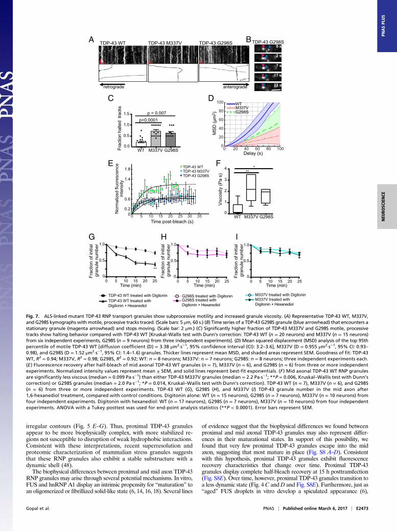
ALS-Linked Mutations Disrupt TDP-43 RNP Granule Motility and IncreaseGranule Viscosity. Characterization of FUS and hnRNP A1 liquiddroplets suggests disease-linked mutations accelerate transformation toa fibrillar state (6, 15, 16, 18). We hypothesized that ALS-linked mu-tations, M337V (27, 45) and G298S (46), in the LCS of TDP-43 wouldalter the liquid-like properties of axonal TDP-43 RNP granules anddisrupt their function. Motile TDP-43 M337V and G298S granulesdisplay similar instantaneous velocities to motile TDP-43 WT granulesbut show less efficient transport, consistent with previous character-ization of TDP-43 mutant motility (26, 47). We noted that processivetransport of motile TDP-43 mutant RNP granules is more ofteninterrupted compared with TDP-43 WT granules; TDP-43 mutantprocessive runs frequently stall or halt completely upon interactionwith another granule (Fig. 7 A–C). As a consequence, mean squareddisplacement analysis demonstrates a processive motility pattern for
TDP-43 WT, whereas both M337V and G298S mutant granules dis-play subprocessive motility (Fig. 7D).
We asked whether increased viscosity of mutant RNP granules maycontribute to the disrupted transport we observed. To test this hypoth-esis, we compared half-bleach fluorescence recovery of TDP-43 WT
proximal axon
mid axon
0.0
0.2
0.4
0.6Proximal axonMid axon
Rel
ativ
e fre
quen
cy(fr
actio
ns)
TDP
-43
gran
ule
dens
ity(n
o. o
f gra
nule
s/μm
)
Proximalaxon
Midaxon
p < 0.0001
B
C D
proximal axon
mid axon
F
0.0
0.2
0.4
0.6
0.8
1.0
0.5
0.6
0.7
0.8
0.9
1.0
Circ
ular
ity
Proximalaxon
Midaxon
Proximalaxon
Midaxon
Con
vexi
ty ra
tio
G p < 0.0001 p < 0.0001
E
A
0.00
0.02
0.04
0.06
0.08
0.10
TDP-43 granule area (μm2)0.0
00.0
40.0
80.1
20.1
6
Rel
ativ
e fre
quen
cy(fr
actio
ns)
0.0
0.2
0.4
0.6Proximal axonMid axon
0
1
2
3
H
proximal axon
mid axon
Endogenous TDP-43 eGFP-TDP-43 WT
TDP-43 granule area (μm2)
Fig. 5. Superresolution STED microscopy reveals proximal TDP-43 granulesare less round, possess more irregular contours, and are distributed at ahigher density compared with mid axonal granules. (A) Immunofluorescencefor endogenous TDP-43 shows a higher density of TDP-43 granules in theproximal axon compared with the mid axon. (Scale bars: 2.2 μm.) (B) EGFP–TDP-43 in the proximal axon shows a higher density than in the mid axon.(Scale bars: 3 μm.) Images are maximum projections of deconvolved STEDZ-stacks. (C) Size distribution of endogenous TDP-43 granules [proximalgranule area: 0.024 ± 0.003 μm2, mid axon granule area: 0.025 ± 0.003 μm2
(mean ± SEM); n = 3,085 proximal granules and n = 776 mid axonal granules;Mann–Whitney test]. (D) Size distribution of EGFP–TDP-43 granules [proximalgranule area: 0.036 ± 0.006 μm2, mid axon granule area: 0.033 ± 0.007 μm2
(mean ± SEM); n = 589 proximal granules and n = 232 mid axonal granules;Mann–Whitney test.] (E) EGFP–TDP-43 granule density was calculated foreach axon segment imaged (n = 18 proximal axon, n = 13 mid axon). TDP-43granule density in the proximal axon is significantly greater than in the midaxon (Student’s t test, P < 0.001). (F) Proximal axon TDP-43 granules oftendisplay irregular contours and sharp projections, whereas mid axonal gran-ules are usually round, with smooth borders. (Scale bar: 250 nm.) Images aresegmented masks of TDP-43 granules. Mid axonal TDP-43 granules are sig-nificantly more circular (perfect circle, circularity = 1) (G) and have a signifi-cantly higher convexity ratio than proximal TDP-43 granules (H). Convexity isdefined as the ratio of the object perimeter of the convex hull to the pe-rimeter of the object (i.e., a convexity of a perfectly circular object is 1).Wilcoxon rank sum tests with a Bonferroni correction for multiple compari-sons (P ≤ 0.001) were performed to assess for significant differences in mor-phological features between proximal (n = 589) and mid (n = 232) axonalgranules from 18 proximal axon and 13 mid axon images, respectively.
Gopal et al. PNAS | Published online March 6, 2017 | E2471
NEU
ROSC
IENCE
PNASPL
US

and mutant mid axon RNP granules. Both TDP-43 M337V and G298Sgranules show slower τ values and incomplete fluorescence recoverycompared with TDP-43WT granules (Fig. 7E and Table S2). Using theStokes–Einstein equation, TDP-43 mutant granules show an order ofmagnitude increase (∼20-fold) in viscosity compared with WT gran-ules (Fig. 7F and Table S2). Compared with mid axonal TDP-43 WTgranules, G298S and M337V TDP-43 granules are more resistant to1,6-hexanediol (Fig. 7G and Fig. S9), suggesting these mutations in theglycine-rich C-terminal domain promote a stabilized internal structure.These data suggest ALS-linked mutations in TDP-43 disrupt normaltransport granule function, but also possibly confer toxic gain offunction by enhancing viscosity of mutant TDP-43 RNP granules.
DiscussionIn this study, we examined TDP-43 RNP granule dynamics and theirbiophysical characteristics in neurons, which are morphologically com-plex and highly differentiated cells. Our data reveal properties that areboth novel and not evident in vitro or in studies of RNP granules in celllines. Recent studies have established that LLPS of purified FUS andhnRNP A1, and nuclear TDP-43, occurs in vitro and/or in mammaliancell lines (6, 15–17, 31). However, detailed characterization of TDP-43has been limited due to its tendency to aggregate (15, 30). Here, we usedlive-cell and superresolution imaging techniques to show that TDP-43–positive neuronal RNP transport granules in the axon display biophysicalproperties consistent with liquid droplets, including fusion and fissionevents with characteristic relaxation to a spherical shape within seconds,deformability by shear force, rapid internal redistribution and FRAP, andsensitivity to disruption of weak hydrophobic interactions. Furthermore,we have identified distinct subpopulations of TDP-43 RNP granules
according to axonal location (Fig. 8). Moreover, ALS-linked muta-tions in TDP-43 increase granule viscosity and disrupt transportfunction. We propose that the biophysical differences observed in TDP-43 RNP granules along the axon arise through differences in subcellularcontext and likely reflect maturational state, and, further, that ALS-linked mutations in TDP-43 may confer toxic gain-of-function effectson RNP granules, potentially increasing their propensity for path-ological transformation.
Previous studies have demonstrated the liquid droplet properties ofvarious types of cellular RNP granules and estimated their viscosity (η):C. elegans P granules, η = 1 Pa·s−1 (1); X. laevis oocyte nucleoli, η =103 Pa·s−1 (2); and FUS-positive stress granules in HeLa cells, η =0.01–0.1 Pa·s−1 (6) (Table S3). In vitro studies of purified WT FUS andAshbya gossypii Whi3 protein have estimated comparable viscosities;moreover, these systems have allowed for various manipulations thatalter the viscosity of these RBPs (18, 39). In our study, we have iden-tified two populations of neuronal TDP-43WTRNP granules, each withdistinct biophysical properties. Mid axonal TDP-43 RNP granules arehighly dynamic, motile, and susceptible to disruption by 1,6-hexanediol,suggesting they are held together by weak hydrophobic interactions.Accordingly, mid axonal TDP-43 granules show a high degree of internalmolecular mobility as well as rapid exchange with the soluble pool ofTDP-43. Mid axonal TDP-43 granules have a viscosity of 0.1 Pa·s−1,which is ∼100-fold more viscous than water and is comparable to theviscosity of FUS-positive stress granules in mammalian cells (6). Incontrast, proximal TDP-43 granules show only partial FRAP, suggestingthat they have a more limited mobile fraction and are less dynamic.Superresolution morphological analysis of axonal TDP-43 granulesreveals that proximal TDP-43 granules are less circular and exhibit
0 20 40 600.20.61.01.41.8
Digitonin
A
Mid axon
Hexanediol+ DigitoninMid axon
C
D
B
DigitoninProximal axon
0.20.61.01.41.8
0 20 40 60Position along axon (μm)
0.20.61.01.41.8
0 20 40 60
Proximal axonHexanediol+ Digitonin
0.20.61.01.41.8
0 20 40 60
Position along axon (μm)
0.20.61.01.41.8
0 20 40 60Position along axon (μm) Position along axon (μm)
Position along axon (μm)
0.20.61.01.41.8
0 20 40 60Position along axon (μm)
Position along axon (μm)
Nor
mal
ized
avg
. flu
or. i
nten
sity
0
0.5
1.0
Before drug added
Digitonin
p = 0.0025
Before drug added
Hexane-diol
DigitoninBefore drug added
Before drug added
Hexane-diol
Inte
nsity
(A
U) x
104
Inte
nsity
(A
U) x
104
Inte
nsity
(A
U) x
104
Inte
nsity
(A
U) x
104
Inte
nsity
(A
U) x
104
Inte
nsity
(A
U) x
104
Inte
nsity
(A
U) x
104
0.2
0.6
1.0
1.4
0 20 40 60Position along axon (μm)
0.2
0.6
1.01.4
0 20 40 60
Inte
nsity
(A
U) x
104
Nor
mal
ized
avg
. flu
or. i
nten
sity
Nor
mal
ized
avg
. flu
or. i
nten
sity
Nor
mal
ized
avg
. flu
or. i
nten
sity
0
0.5
1.0
0
0.5
1.0
0
0.5
1.0
Fig. 6. Disruption of weak hydrophobic interactionsdissolves TDP-43 granules in the mid axon but not inthe proximal axon. Cortical neurons expressing EGFP–TDP-43 were treated with 5 μg/mL digitonin alone(control) or 5 μg/mL digitonin with 4% 1,6-hexanedioland then imaged at 5-min intervals. (A and C) Ap-plication of digitonin does not affect proximal or midaxonal TDP-43 granules. (B and D) Digitonin with 1,6-hexanediol dissolves TDP-43 granules in the mid axonbut does not affect TDP-43 granule integrity in theproximal axon, as shown with representative imagesand intensity plot profiles (Left) and quantification offluorescence (fluor.) intensity along the axon (Right)(n = 15 neurons per condition). (D) There is a signifi-cant reduction in fluorescence intensity in the midaxon with the addition of 1,6-hexanediol (Student’s ttest, P = 0.0025). (Scale bars: 5 μm.) AU, arbitraryunits; avg., averaged.
E2472 | www.pnas.org/cgi/doi/10.1073/pnas.1614462114 Gopal et al.
irregular contours (Fig. 5 E–G). Thus, proximal TDP-43 granulesappear to be more biophysically complex, with more stabilized re-gions not susceptible to disruption of weak hydrophobic interactions.Consistent with these interpretations, recent superresolution andproteomic characterization of mammalian stress granules suggeststhat these RNP granules also exhibit a stable substructure with adynamic shell (48).
The biophysical differences between proximal and mid axon TDP-43RNP granules may arise through several potential mechanisms. In vitro,FUS and hnRNP A1 display an intrinsic propensity for “maturation” toan oligomerized or fibrillized solid-like state (6, 14, 16, 18). Several lines
of evidence suggest that the biophysical differences we found betweenproximal and mid axonal TDP-43 granules may also represent differ-ences in their maturational states. In support of this possibility, wefound that very few proximal TDP-43 granules escape into the midaxon, suggesting that most mature in place (Fig. S8 A–D). Consistentwith this hypothesis, proximal TDP-43 granules exhibit fluorescencerecovery characteristics that change over time. Proximal TDP-43granules display complete half-bleach recovery at 15 h posttransfection(Fig. S8E). Over time, however, proximal TDP-43 granules transition toa less dynamic state (Fig. 4 C andD and Fig. S8E). Furthermore, just as“aged” FUS droplets in vitro develop a spiculated appearance (6),
TDP-43 G298STDP-43 M337VTDP-43 WTA
retrograde anterograde
Nor
mal
ized
fluo
resc
ence
inte
nsity
10 20 30251550 35Time post-bleach (s)
TDP-43 WTTDP-43 M337VTDP-43 G298S
B
C
20 40 60 80 100Delay (s)
G298SM337VWT
00
20
40
60
80
100
MS
D (µ
m2 )
D
E F
TDP-43 G298S
WT M337V G298S0
1
2
3
4
Vis
cosi
ty (P
a s)
***
0 s
2 s
7 s
17 s
22 s
TDP-43 WT treated with DigitoninTDP-43 WT treated withDigitonin + Hexanediol
0.5
1.0
Time (min)0 5
015 2510 20
**
Frac
tion
of in
itial
gr
anul
e nu
mbe
r
G
0.5
Time (min)0 5
015 2510 20
0.5
1.0
Time (min)0 5
015 2510 20
0.0
0.5
1.0
1.5
WT M337V G298S
p<0.0001p = 0.007
Frac
tion
halte
d tr
acks
1.0
G298S treated with DigitoninG298S treated withDigitonin + Hexanediol
M337V treated with DigitoninM337V treated withDigitonin + Hexanediol
H I
Frac
tion
of in
itial
gr
anul
e nu
mbe
r
Frac
tion
of in
itial
gr
anul
e nu
mbe
r
1.8
1.4
1
0.6
0.20
Fig. 7. ALS-linked mutant TDP-43 RNP transport granules show subprocessive motility and increased granule viscosity. (A) Representative TDP-43 WT, M337V,and G298S kymographs with motile, processive tracks traced. (Scale bars: 5 μm, 60 s.) (B) Time series of a TDP-43 G298S granule (blue arrowhead) that encounters astationary granule (magenta arrowhead) and stops moving. (Scale bar: 2 μm.) (C) Significantly higher fraction of TDP-43 M337V and G298S motile, processivetracks show halting behavior compared with TDP-43 WT [Kruskal–Wallis test with Dunn’s correction: TDP-43 WT (n = 20 neurons) and M337V (n = 15 neurons)from six independent experiments, G298S (n = 9 neurons) from three independent experiments]. (D) Mean squared displacement (MSD) analysis of the top 95thpercentile of motile TDP-43 WT [diffusion coefficient (D) = 3.38 μm2·s−1, 95% confidence interval (CI): 3.2–3.6], M337V (D = 0.955 μm2·s−1, 95% CI: 0.93–0.98), and G298S (D = 1.52 μm2·s−1, 95% CI: 1.4–1.6) granules. Thicker lines represent mean MSD, and shaded areas represent SEM. Goodness of fit: TDP-43WT, R2 = 0.94; M337V, R2 = 0.98; G298S, R2 = 0.92; WT: n = 8 neurons; M337V: n = 7 neurons; G298S: n = 8 neurons; three independent experiments each.(E ) Fluorescence recovery after half-bleach of mid axonal TDP-43 WT granules (n = 7), M337V (n = 6), and G298S (n = 6) from three or more independentexperiments. Normalized intensity values represent mean ± SEM, and solid lines represent best-fit exponentials. (F ) Mid axonal TDP-43 WT RNP granulesare significantly less viscous (median = 0.099 Pa·s−1) than either TDP-43 M337V granules (median = 2.2 Pa·s−1; **P = 0.006, Kruskal–Wallis test with Dunn’scorrection) or G298S granules (median = 2.0 Pa·s−1; *P = 0.014, Kruskal–Wallis test with Dunn’s correction). TDP-43 WT (n = 7), M337V (n = 6), and G298S(n = 6) from three or more independent experiments. TDP-43 WT (G), G298S (H), and M337V (I) TDP-43 granule number in the mid axon after1,6-hexanediol treatment, compared with control conditions. Digitonin alone: WT (n = 15 neurons), G298S (n = 7 neurons), M337V (n = 10 neurons) fromfour independent experiments. Digitonin with hexanediol: WT (n = 17 neurons), G298S (n = 7 neurons), M337V (n = 10 neurons) from four independentexperiments. ANOVA with a Tukey posttest was used for end-point analysis statistics (**P < 0.0001). Error bars represent SEM.
Gopal et al. PNAS | Published online March 6, 2017 | E2473
NEU
ROSC
IENCE
PNASPL
US

STED superresolution microscopy revealed that in neurons, proximalaxonal TDP-43 granules display irregular contours and spiculated edgesand are less circular than mid axonal TDP-43 granules (Fig. 5 F–H).The intermolecular interactions that regulate granule maturationand formation of fibrillar and amyloid-like structures are not fullyunderstood, but several factors may contribute, including molecu-lar chaperones and high RBP concentrations within liquid-likedroplets (16, 49).
Using STED superresolution microscopy, we see clear enrichmentof endogenous TDP-43 in the proximal axon compared with the midaxon and a similar density gradient in neurons expressing EGFP–TDP-43 (Fig. 5 A–E and Fig. S7). Although additional evidence isneeded, an interesting possibility is that the relative concentrations ofTDP-43, and perhaps other RBPs, in different neuronal compart-ments allow for formation of RNP granules with distinct biophysicalproperties. Recent work has shown that RNA content influences thebiophysical properties of liquid droplets as well (39, 40). For example,Zhang et al. (39) have shown that Whi3 RNPs become more viscouswith increasing concentrations of RNA, and that addition of specificmRNA transcripts, CLN3 and BNI1, involved in nuclear division andbranching functions, respectively, result in Whi3 RNP granules withdistinct viscosities.
A third possibility is that biophysical differences between proximaland mid axon TDP-43 RNP granules may reflect differences in theirRNA content and could underlie distinct physiological functions. It hasbeen shown recently that yeast stress granules are more gel-like,whereas P bodies have more liquid-like properties (3). Our motility dataindicate that TDP-43 RNP granules in the mid axon are more likely tobe transported distances ≥10 μm, suggesting that these granules mayfunction to transport mRNAs critical for synaptic function or mainte-nance of the axon terminal (50, 51). Furthermore, weak hydrophobicinteractions that maintain mid axonal TDP-43 granules as liquiddroplets would allow for facile release of mRNA at the target site. Incontrast, proximal TDP-43 RNP granules are predominantly stationary,and we speculate that distinct mRNAs may be associated with thesegranules. These findings raise the possibility that specific mRNAs arecritical for creating biophysical diversity between cellular RNP granulesand may drive unique biological functions; it will be important to ex-amine this question in future studies.
In vitro, ALS/FTD-linked mutations in the RBPs FUS and hnRNP A1do not significantly alter the biophysical properties of newly formedliquid droplets; rather, aging liquid droplets composed of mutant RBPsexhibit enhanced maturation to a more solid-like state with decreasedmolecular mobility (15, 16, 44). Cycling mutant hnRNP A1 or FUSthrough repeated rounds of LLPS promoted conversion of liquiddroplets into fibrillar structures (15, 18). Previous studies have dem-onstrated ALS-linked mutations in TDP-43 accelerate its propensity toaggregate (30). In neurons, we observe that mid axonal granules com-posed of ALS-linked mutant TDP-43 show lower internal molecularmobility than TDP-43 WT granules (Fig. 7E); accordingly, mutantgranules are ∼20-fold more viscous that WT granules (Fig. 7F). Inaddition, we and others have observed that ALS-linked mutationsdisrupt TDP-43 RNP transport (26, 47). These data suggest the alteredviscoelastic properties of mutant TDP-43 granules may contribute todisease pathogenesis.
In this study, we show that neuronal TDP-43 RNP transport gran-ules display liquid-like properties within cortical neurons, and thusprovide insights that extend recently proposed models that suggestRNP granules are biophysically specialized structures fundamental forcellular compartmentalization. Our data demonstrate distinct bio-physical properties of TDP-43 WT RNP granules depending on axonlocation. Further, we find that granules formed from ALS-linkedmutant TDP-43 are more viscous and exhibit altered transport dy-namics. Thus, our data indicate that RNP granules must be studiedwithin the complex morphology of neurons to appreciate fully theirphysiological roles and the pathological transitions of these structuresin neurodegenerative disease.
Materials and MethodsPrimary Cortical Culture and Transfection. Primary cortical neurons were dis-sociated from embryonic day 18–20 Sprague–Dawley rat embryos at the
Neuron Culture Service Center at the University of Pennsylvania, as described(52). Details are provided in SI Materials and Methods. Cortical neurons (daysin vitro 7–10) were transfected with cDNA constructs using Lipofectamine 2000reagent (Invitrogen).
Live-Cell Microscopy. Live imaging of cortical cultures was performed in Hi-bernate E (Brainbits) supplemented with 2% (vol/vol) B27 and 2mMGlutaMAXin a temperature-controlled chamber mounted on an inverted NikonTi mi-croscope with apochromat 63× and 100× 1.49-N.A. oil-immersion objectives;images were acquired on a PerkinElmer UltraVIEW VOX spinning disk confocalsystem equipped with an Ultraview Photokinesis (PerkinElmer) unit and aC9100-50 EMCCD camera (Hamamatsu) controlled by Volocity software(PerkinElmer). Axons were identified by morphological criteria (53, 54) andwere imaged at one frame every 2–3 s for 5–10 min. Near-TIRF microscopy wasperformed as described (55). Images were captured on an ImagEM C9100-13(Hamamatsu) camera at a rate of seven to 10 frames per second × 2 min using50- to 90-ms exposure times. For hexanediol experiments, cortical neuronsexpressing EGFP–TDP-43 were treated with either 5 μg/mL digitonin alone(control) or 5 μg/mL digitonin (Sigma) with 4% (wt/vol) 1,6-hexanediol (AcrosOrganics) and then imaged at 5-min intervals for 25 min.
FRAP and photoactivation/photoconversion experiments were performedusing the Ultraview Photokinesis module. EGFP–TDP-43 was photobleachedusing the 488-nm laser at 75–90% power for 30 cycles, with a 1-ms spot pe-riod within a region of interest (0.5 × 0.5 μm). Images were acquired using the488-nm laser at one frame every second for 3–5 s before and 120–180 ssubsequent to photobleaching. The photokinesis unit was calibrated beforeeach experiment to ensure tightly localized half-bleach, as described else-where (1). Photoactivation/conversion of mEos3.2–TDP-43 or PAGFP-Synapsinwas performed using the 405-nm laser at 2–3% power for 30 cycles, with a2-ms spot period (41–43, 56).
STED Superresolution Microscopy. STED microscopy was performed at theUniversity of Pennsylvania Microscopy Core on a Leica DMI 6000 Inverted laserscanning confocal microscope equipped with 592-nm and 660-nm STED
axon
axon
Proximal TDP-43 WT granules Mid axon TDP-43 WT granules
TDP-43 RRM1 and RRM2
TDP-43 C-terminal Low complexity sequence (LCS)
TDP-43 stabilized by stronginteractions
TDP-43 condensed liquid droplet (weak interactions)
soluble liquid-droplet liquid-dropletstabilized?
soluble liquid-droplet
Mid axon TDP-43 M337V or G298Smutant granules
soluble liquid-droplet highly viscousliquid-droplet
stabilized core?
***
** ******TDP-43 missense mutationC-terminal LCS*
* *********
***
***** *
Fig. 8. Summary schematic: Mid axonal TDP-43 granules comprise a dynamicpopulation of RNP granules that are distinct from proximal TDP-43 granules.Mid axonal TDP-43 RNP granules show rapid exchange with the soluble pool ofTDP-43, display rapid internal rearrangement, and readily dissolve when weakhydrophobic interactions are disrupted, consistent with liquid-like behavior. Incontrast, proximal TDP-43 granules show incomplete FRAP and are less sensi-tive to disruption of weak hydrophobic interactions, suggesting that thesegranules have a complex structure composed of more viscous and/or stabilizedregions with limited molecular mobility. In addition, TDP-43 RNP granules aremore densely arranged in the proximal axon and are less motile than midaxonal TDP-43 granules. These distinct populations of TDP-43 WT granules inthe axon may reflect different maturational states, and possibly differentfunctional roles. Mutant TDP-43 RNP granules in the mid axon also show liquiddroplet formation but display slower recovery after photobleaching, indicat-ing these liquid droplets are more viscous than TDP-43 WT granules. Increasedviscosity observed in TDP-43 mutant granules may confer toxic gain of func-tion, possibly enhancing the propensity for aggregation.
E2474 | www.pnas.org/cgi/doi/10.1073/pnas.1614462114 Gopal et al.

depletion lasers. Detailed immunofluorescence methods are provided in SIMaterials and Methods.
Image and Data Analysis. All image processing and analysis were performedusing ImageJ/Fiji and/or custom automated analyses in MATLAB R2015a(MathWorks). For amore clear presentation of Fig. 2, Figs. S3 and S4, andMoviesS1 and S2, time series were denoised as described (57). However, all dataanalysis was performed on the raw, unprocessed data as described (58, 59). Full
details of image, data, and statistical analysis are described in SI Materialsand Methods.
ACKNOWLEDGMENTS. We thank Mariko Tokito, the Neuron Culture ServiceCenter, andMargie Price for technical assistance. We thank Ekaterina Grishchuk,Amy Ghiretti, Sandra Maday, Swathi Ayloo, Andrea Stavoe, and MichaelHowland for thoughtful discussion and comments on the manuscript. Thisresearch was supported by the NIH under Awards R37 NS060698 (to E.L.F.H.),1-K08-NS094744 (to P.P.G.), and F30NS092227 (to J.J.N.).
1. Brangwynne CP, et al. (2009) Germline P granules are liquid droplets that localize bycontrolled dissolution/condensation. Science 324(5935):1729–1732.
2. Brangwynne CP, Mitchison TJ, Hyman AA (2011) Active liquid-like behavior of nucleolidetermines their size and shape in Xenopus laevis oocytes. Proc Natl Acad Sci USA108(11):4334–4339.
3. Kroschwald S, et al. (2015) Promiscuous interactions and protein disaggregases de-termine the material state of stress-inducible RNP granules. eLife 4:e06807.
4. Zhu L, Brangwynne CP (2015) Nuclear bodies: The emerging biophysics of nucleo-plasmic phases. Curr Opin Cell Biol 34:23–30.
5. Hyman AA, Weber CA, Jülicher F (2014) Liquid-liquid phase separation in biology.Annu Rev Cell Dev Biol 30:39–58.
6. Patel A, et al. (2015) A liquid-to-solid phase transition of the ALS protein FUS acceleratedby disease mutation. Cell 162(5):1066–1077.
7. Weber SC, Brangwynne CP (2012) Getting RNA and protein in phase. Cell 149(6):1188–1191.
8. Hyman AA, Brangwynne CP (2011) Beyond stereospecificity: Liquids and mesoscaleorganization of cytoplasm. Dev Cell 21(1):14–16.
9. King OD, Gitler AD, Shorter J (2012) The tip of the iceberg: RNA-binding proteins withprion-like domains in neurodegenerative disease. Brain Res 1462:61–80.
10. Kwiatkowski TJ, Jr, et al. (2009) Mutations in the FUS/TLS gene on chromosome 16cause familial amyotrophic lateral sclerosis. Science 323(5918):1205–1208.
11. Vance C, et al. (2009) Mutations in FUS, an RNA processing protein, cause familialamyotrophic lateral sclerosis type 6. Science 323(5918):1208–1211.
12. Kim HJ, et al. (2013) Mutations in prion-like domains in hnRNPA2B1 and hnRNPA1cause multisystem proteinopathy and ALS. Nature 495(7442):467–473.
13. Han TW, et al. (2012) Cell-free formation of RNA granules: Bound RNAs identifyfeatures and components of cellular assemblies. Cell 149(4):768–779.
14. Kato M, et al. (2012) Cell-free formation of RNA granules: Low complexity sequencedomains form dynamic fibers within hydrogels. Cell 149(4):753–767.
15. Molliex A, et al. (2015) Phase separation by low complexity domains promotes stressgranule assembly and drives pathological fibrillization. Cell 163(1):123–133.
16. Lin Y, Protter DS, Rosen MK, Parker R (2015) Formation and maturation of phase-separated liquid droplets by RNA-binding proteins. Mol Cell 60(2):208–219.
17. Burke KA, Janke AM, Rhine CL, Fawzi NL (2015) Residue-by-residue view of in vitro FUSgranules that bind the C-terminal domain of RNA polymerase II. Mol Cell 60(2):231–241.
18. Murakami T, et al. (2015) ALS/FTD mutation-induced phase transition of FUS liquiddroplets and reversible hydrogels into irreversible hydrogels impairs RNP granule func-tion. Neuron 88(4):678–690.
19. Holt CE, Schuman EM (2013) The central dogma decentralized: New perspectives onRNA function and local translation in neurons. Neuron 80(3):648–657.
20. Kiebler MA, Bassell GJ (2006) Neuronal RNA granules: Movers and makers. Neuron51(6):685–690.
21. Krichevsky AM, Kosik KS (2001) Neuronal RNA granules: A link between RNA locali-zation and stimulation-dependent translation. Neuron 32(4):683–696.
22. Ou SH, Wu F, Harrich D, García-Martínez LF, Gaynor RB (1995) Cloning and charac-terization of a novel cellular protein, TDP-43, that binds to human immunodeficiencyvirus type 1 TAR DNA sequence motifs. J Virol 69(6):3584–3596.
23. Buratti E, Baralle FE (2001) Characterization and functional implications of the RNAbinding properties of nuclear factor TDP-43, a novel splicing regulator of CFTR exon 9.J Biol Chem 276(39):36337–36343.
24. Elvira G, et al. (2006) Characterization of an RNA granule from developing brain. MolCell Proteomics 5(4):635–651.
25. Fallini C, Bassell GJ, Rossoll W (2012) The ALS disease protein TDP-43 is activelytransported in motor neuron axons and regulates axon outgrowth. Hum Mol Genet21(16):3703–3718.
26. Alami NH, et al. (2014) Axonal transport of TDP-43 mRNA granules is impaired by ALS-causing mutations. Neuron 81(3):536–543.
27. Sreedharan J, et al. (2008) TDP-43 mutations in familial and sporadic amyotrophiclateral sclerosis. Science 319(5870):1668–1672.
28. Kabashi E, et al. (2008) TARDBP mutations in individuals with sporadic and familialamyotrophic lateral sclerosis. Nat Genet 40(5):572–574.
29. Neumann M, et al. (2006) Ubiquitinated TDP-43 in frontotemporal lobar degenera-tion and amyotrophic lateral sclerosis. Science 314(5796):130–133.
30. Johnson BS, et al. (2009) TDP-43 is intrinsically aggregation-prone, and amyotrophiclateral sclerosis-linked mutations accelerate aggregation and increase toxicity. J BiolChem 284(30):20329–20339.
31. Schmidt HB, Rohatgi R (2016) In vivo formation of vacuolated multi-phase compart-ments lacking membranes. Cell Reports 16(5):1228–1236.
32. Elden AC, et al. (2010) Ataxin-2 intermediate-length polyglutamine expansions areassociated with increased risk for ALS. Nature 466(7310):1069–1075.
33. Stone HA (1994) Dynamics of drop deformation and breakup in viscous fluids. AnnuRev Fluid Mech 26:65–102.
34. Kasza KE, et al. (2007) The cell as a material. Curr Opin Cell Biol 19(1):101–107.35. Zajac AL, Goldman YE, Holzbaur EL, Ostap EM (2013) Local cytoskeletal and organelle
interactions impact molecular-motor-driven early endosomal trafficking. Curr Biol23(13):1173–1180.
36. Weiss AN, Bittner MA, Holz RW, Axelrod D (2014) Protein mobility within secretorygranules. Biophys J 107(1):16–25.
37. Sinnecker D, Voigt P, Hellwig N, Schaefer M (2005) Reversible photobleaching ofenhanced green fluorescent proteins. Biochemistry 44(18):7085–7094.
38. Axelrod D, Koppel DE, Schlessinger J, Elson E, Webb WW (1976) Mobility measurementby analysis of fluorescence photobleaching recovery kinetics. Biophys J 16(9):1055–1069.
39. Zhang H, et al. (2015) RNA controls PolyQ protein phase transitions. Mol Cell 60(2):220–230.
40. Elbaum-Garfinkle S, et al. (2015) The disordered P granule protein LAF-1 drives phaseseparation into droplets with tunable viscosity and dynamics. Proc Natl Acad Sci USA112(23):7189–7194.
41. Zhang M, et al. (2012) Rational design of true monomeric and bright photoactivatablefluorescent proteins. Nat Methods 9(7):727–729.
42. Lippincott-Schwartz J, Patterson GH (2009) Photoactivatable fluorescent proteins fordiffraction-limited and super-resolution imaging. Trends Cell Biol 19(11):555–565.
43. Patterson GH, Lippincott-Schwartz J (2002) A photoactivatable GFP for selectivephotolabeling of proteins and cells. Science 297(5588):1873–1877.
44. Patel SS, Belmont BJ, Sante JM, Rexach MF (2007) Natively unfolded nucleoporinsgate protein diffusion across the nuclear pore complex. Cell 129(1):83–96.
45. Corrado L, et al. (2009) High frequency of TARDBP gene mutations in Italian patientswith amyotrophic lateral sclerosis. Hum Mutat 30(4):688–694.
46. Van Deerlin VM, et al. (2008) TARDBP mutations in amyotrophic lateral sclerosis withTDP-43 neuropathology: A genetic and histopathological analysis. Lancet Neurol 7(5):409–416.
47. Liu-Yesucevitz L, et al. (2014) ALS-linked mutations enlarge TDP-43-enriched neuronalRNA granules in the dendritic arbor. J Neurosci 34(12):4167–4174.
48. Jain S, et al. (2016) ATPase-modulated stress granules contain a diverse proteome andsubstructure. Cell 164(3):487–498.
49. Ganassi M, et al. (2016) A surveillance function of the HSPB8-BAG3-HSP70 chaperonecomplex ensures stress granule integrity and dynamism. Mol Cell 63(5):796–810.
50. Polymenidou M, et al. (2011) Long pre-mRNA depletion and RNA missplicing con-tribute to neuronal vulnerability from loss of TDP-43. Nat Neurosci 14(4):459–468.
51. Puthanveettil SV (2013) RNA transport and long-term memory storage. RNA Biol10(12):1765–1770.
52. Wilcox KS, Buchhalter J, Dichter MA (1994) Properties of inhibitory and excitatorysynapses between hippocampal neurons in very low density cultures. Synapse 18(2):128–151.
53. Craig AM, Banker G (1994) Neuronal polarity. Annu Rev Neurosci 17:267–310.54. Kaech S, Banker G (2006) Culturing hippocampal neurons. Nat Protoc 1(5):2406–2415.55. Tokunaga M, Imamoto N, Sakata-Sogawa K (2008) Highly inclined thin illumination
enables clear single-molecule imaging in cells. Nat Methods 5(2):159–161.56. Ballister ER, Ayloo S, Chenoweth DM, Lampson MA, Holzbaur EL (2015) Optogenetic
control of organelle transport using a photocaged chemical inducer of dimerization.Curr Biol 25(10):R407–R408.
57. Kervrann C, Boulanger J (2006) Optimal spatial adaptation for patch-based imagedenoising. IEEE Trans Image Process 15(10):2866–2878.
58. Twelvetrees AE, et al. (2016) The dynamic localization of cytoplasmic dynein inneurons is driven by kinesin-1. Neuron 90(5):1000–1015.
59. Tarantino N, et al. (2014) TNF and IL-1 exhibit distinct ubiquitin requirements forinducing NEMO-IKK supramolecular structures. J Cell Biol 204(2):231–245.
Gopal et al. PNAS | Published online March 6, 2017 | E2475
NEU
ROSC
IENCE
PNASPL
US