an alpha-1a adrenergic receptor agonist prevents acute
TRANSCRIPT

RESEARCH ARTICLE
An Alpha-1A Adrenergic Receptor Agonist
Prevents Acute Doxorubicin Cardiomyopathy
in Male Mice
Megan D. Montgomery1,2, Trevor Chan1,2¤, Philip M. Swigart1,2, Bat-erdene Myagmar1,2,
Rajesh Dash1,2¤, Paul C. Simpson1,2*
1 Department of Medicine, Cardiology Division, VA Medical Center, San Francisco, CA, United States of
America, 2 Department of Medicine and Cardiovascular Research Institute, University of California, San
Francisco, San Francisco, CA, United States of America
¤ Current address: Stanford University, Stanford, CA, United States of America
Abstract
Alpha-1 adrenergic receptors mediate adaptive effects in the heart and cardiac myocytes,
and a myocyte survival pathway involving the alpha-1A receptor subtype and ERK activation
exists in vitro. However, data in vivo are limited. Here we tested A61603 (N-[5-(4,5-dihydro-
1H-imidazol-2-yl)-2-hydroxy-5,6,7,8-tetrahydronaphthalen-1-yl]methanesulfonamide), a
selective imidazoline agonist for the alpha-1A. A61603 was the most potent alpha-1-agonist
in activating ERK in neonatal rat ventricular myocytes. A61603 activated ERK in adult mouse
ventricular myocytes and protected the cells from death caused by the anthracycline doxoru-
bicin. A low dose of A61603 (10 ng/kg/d) activated ERK in the mouse heart in vivo, but did not
change blood pressure. In male mice, concurrent subcutaneous A61603 infusion at 10 ng/kg/
d for 7 days after a single intraperitoneal dose of doxorubicin (25 mg/kg) increased survival,
improved cardiac function, heart rate, and cardiac output by echocardiography, and reduced
cardiac cell necrosis and apoptosis and myocardial fibrosis. All protective effects were lost in
alpha-1A-knockout mice. In female mice, doxorubicin at doses higher than in males (35–40
mg/kg) caused less cardiac toxicity than in males. We conclude that the alpha-1A-selective
agonist A61603, via the alpha-1A adrenergic receptor, prevents doxorubicin cardiomyopathy
in male mice, supporting the theory that alpha-1A adrenergic receptor agonists have potential
as novel heart failure therapies.
Introduction
Cardiac myocyte alpha-1 adrenergic receptors (α1-ARs) mediate crucial adaptive functions in
the heart, including physiological hypertrophy, contractility, survival signaling, ischemic pre-
conditioning, and protection against multiple injuries, by activating multifactorial signaling
cascades, as reviewed [1,2,3]. Among the 3 α1-AR subtypes (α1A, α1B, and α1D), rodent and
human myocytes have the α1A and α1B [4], and most data implicate the α1A in adaptive
effects [5,6,7,8,9,10,11]. However, it is unknown if an α1A-AR agonist can treat ardiomyopa-
thy in vivo.
PLOS ONE | DOI:10.1371/journal.pone.0168409 January 12, 2017 1 / 22
a1111111111
a1111111111
a1111111111
a1111111111
a1111111111
OPENACCESS
Citation: Montgomery MD, Chan T, Swigart PM,
Myagmar B-e, Dash R, Simpson PC (2017) An
Alpha-1A Adrenergic Receptor Agonist Prevents
Acute Doxorubicin Cardiomyopathy in Male Mice.
PLoS ONE 12(1): e0168409. doi:10.1371/journal.
pone.0168409
Editor: Rakesh Kukreja, Virginia Commonwealth
University Medical Center, UNITED STATES
Received: August 9, 2016
Accepted: November 29, 2016
Published: January 12, 2017
Copyright: This is an open access article, free of all
copyright, and may be freely reproduced,
distributed, transmitted, modified, built upon, or
otherwise used by anyone for any lawful purpose.
The work is made available under the Creative
Commons CC0 public domain dedication.
Data Availability Statement: All relevant data are
within the paper.
Funding: This work was supported by National
Institutes of Health (PCS, HL31113), the
Department of Veterans Affairs (PCS, BX001970),
the Western States Affiliate of the American Heart
Association (RD), the Sarnoff Foundation for
Cardiovascular Research (TC), and the PhRMA
Foundation (MDM). The funders had no role in
study design, data collection and analysis, decision
to publish, or preparation of the manuscript.

A61603 (N-[5-(4,5-dihydro-1H-imidazol-2-yl)-2-hydroxy-5,6,7,8-tetrahydronaphthalen-
1-yl]methanesulfonamide), is a potent and selective imidazoline agonist for the α1A [12]. Selec-
tivity for the α1A is important for potential clinical use, since the α1D might cause constriction
of human epicardial coronary arteries [13], and the role of the α1B remains uncertain.
The anthracycline doxorubicin (DOX, Adriamycin) is a highly effective, widely used treat-
ment for many pediatric and adult cancers, but has the severe, dose-limiting adverse effect of
cardiotoxicity [14]. Previously, we found that adenoviral expression of the α1A receptor in
myocytes from the α1-AR knockout (KO) reduced necrosis and apoptosis after DOX in vitro,
through an ERK signaling mechanism [8], and that A61603 prevented DOX-induced apopto-
sis in vivo, detected by an MRI approach [9].
Here we first verified the potency of A61603 in myocyte ERK activation and protection
from DOX in vitro. We identified a very low A61603 dose, 10 ng/kg/d, which activated cardiac
ERK in vivo but did not increase blood pressure (BP), the classic role of α1-ARs [15]. Using
this low dose, we tested the hypothesis that the beneficial effects of α1A stimulation with
A61603 would translate to treatment of anthracycline cardiomyopathy in a mouse model in
vivo.
We find that A61603 can prevent DOX cardiomyopathy in male mice, that this effect
requires the α1A receptor, and that mechanisms include less cell death and fibrosis. We also
observe that female mice are less susceptible to DOX cardiotoxicity, contrary to female
humans, who are said to be more susceptible to anthracyclines [16,17]. The data support the
idea that α1A agonists might translate to new therapy for heart failure (HF).
Materials and Methods
Animals
Female Sprague-Dawley rats with newborn litters were from Charles River; litters were used
on arrival, and mothers were euthanized. Adult C57Bl/6J male and female mice age 14–18
weeks were from Jackson Laboratory, or our laboratory where wild type (WT) and α1A-AR
knockout (KO) mice were produced [15]. All studies were reviewed and approved by the
IACUC of the San Francisco VA Medical Center. Our institution is accredited by the Ameri-
can Association for the Accreditation of Laboratory Animal Care and has an Animal Welfare
Assurance on file with the NIH Office for Laboratory Animal Research. Mice were housed in
groups in sterile micro-isolator cages with food and water available ad libitum. Chow was
Envigo Harlan Teklad, 2019 for breeders and 2016 for other mice. Light was on a 12 h sched-
ule, temperature was 68–79˚F, and humidity was 30–70%. Animals were inspected daily, and a
licensed veterinarian was available at all times. Euthanasia was by cardiectomy under deep
anesthesia with isoflurane. Criteria for euthanasia were signs of distress, including inactivity,
poor grooming, hunched posture, weight loss >20% body weight (BW), or if heart rate (HR)
was less than 200 bpm. For osmotic minipump and telemeter implantation, anesthesia was
with isoflurane 1.5–3% in oxygen, and analgesia was with local bupivacaine 0.25% and sys-
temic buprenorphine 5–100 μg/kg s.c.
Drugs
Drugs were as follows: A61603 (N-[5-(4,5-dihydro-1H-imidazol-2-yl)-2-hydroxy-5,6,7,8-tetra-
hydronaphthalen-1-yl]methanesulfonamide hydrobromide) (Tocris #1052); ABT-866 (N-[3-
(1H-imidazol-4-ylmethyl)phenyl]ethanesulfonamide) (synthesized by Synterys, Union City,
CA); cirazoline (Tocris now R&D # 0888); dobutamine (Fisher Scientific, ICN#15978010);
doxorubicin (Tocris #2252); epinephrine (Sigma #E4375); methoxamine (Sigma #M6524);
midodrine (MP-Bio #155717); norepinephrine (Sigma #N5785); oxymetazoline (Tocris
Alpha-1A Agonist Therapy
PLOS ONE | DOI:10.1371/journal.pone.0168409 January 12, 2017 2 / 22
Competing Interests: UCSF owns a patent with
PCS as inventor for use of A61603 in heart failure,
and PCS is involved in a company to pursue this.
The indicated patent owned by UCSF is "Method of
treatment using alpha-1-adrenergic agonist
compounds" (inventor Paul C. Simpson) (allowed
#8324178, 2009). This patent has not resulted in
any company, product, employment, etc. This does
not alter our adherence to PLOS ONE policies on
sharing data and materials.
#1142); phenylephrine (Sigma #P-6126); propranolol (Fluka #82066); and ST-1059 (Toronto
Research Chemicals #S686650).
Neonatal rat ventricular myocyte isolation and culture
Neonatal rat ventricular myocytes (NRVMs) were isolated with trypsin digestion and mechan-
ical dissociation, preplated to remove contaminating non-myocardial cells, and counted, with
a yield of viable myocytes (excluding trypan blue) of approximately 3.5–4 million per neonatal
heart. Cells at 500 per mm2 were plated overnight in 35 mm dishes in MEM with 5% calf
serum and bromodeoxyuridine (BrdU), then maintained at 37˚C with 1% CO2, in serum-free
MEM with Hank’s salts, supplemented with penicillin, vitamin B12, BrdU, human transferrin
10 μg/ml, bovine insulin 10 μg/ml, and BSA 1 mg/ml [18,19]. Plating efficiency was approxi-
mately 25–30% of viable myocytes plated.
Adult mouse ventricular myocyte isolation and culture
Adult mouse ventricular myocytes (AMVMs) were isolated following a detailed protocol [20].
Briefly, hearts were removed from WT and α1A-KO adult male mice under deep anesthesia
with isoflurane, after heparin 100 IU in PBS i.p. to prevent coagulation of blood in the coronary
arteries. Myocytes were obtained by perfusion with collagenase in nominally calcium-free buffer
with butanedione monoxime (BDM) 10 mM, followed by mechanical dissociation and gradual
calcium reintroduction, with a yield of 1–2 million cells per heart. Sixty thousand rod-shaped
myocytes were plated onto laminin-coated 35 mm dishes in Minimal Essential Medium (MEM)
with Hank’s salts, supplemented with calf serum 10%, BDM 10 mM, and penicillin 100 U/ml.
After 2 h for myocyte attachment, cells were maintained in serum free MEM with Hank’s salts,
supplemented with BSA 1 mg/ml and penicillin at 37˚C with 2% CO2.
ERK activation
For ERK activation (phosphorylation), NRVMs or AMVMs after 1 day in culture were pre-
treated 10 min with propranolol (PROP) 200 nM, then treated 5 min with A61603 or vehicle
at 37˚C, 2% CO2. Ventricular myocardium was collected after 7 d infusion in vivo with
A61603 or vehicle. Samples were lysed in 1.5X SDS sample buffer containing Complete Mini
protease and phosphatase inhibitor cocktails 2 and 3, snap-frozen, and stored at -80˚C. For
immunoblot, samples in SDS sample buffer were boiled for 5 min at 100˚C; equal numbers of
cells (25,000 NRVMs, 12,000 AMVMs) or equal amounts of ventricular protein (Bradford,
20 μg) per lane were separated on 12.5% Criterion Tris-HCl SDS-PAGE gels; and proteins
were transferred to nitrocellulose membranes. Membranes were blocked with non-fat milk 5%
in TBS-T, then incubated overnight at 4˚C with 1:1,000 primary antibody in BSA-Tween 5%.
Antibodies were total ERK1/2 (Cell Signaling #9102) and phospho-ERK1/2 (Cell Signaling
#4370, phospho Thr202/Tyr204-p44/42 MAPK, D12.14.4E, XP1 Rabbit mAb). Secondary
antibody in milk 5% in TBS-T (anti-rabbit IgG, HRP-linked antibody, Cell Signaling #7074)
was incubated 1 h at RT, and bands were developed using SuperSignal West Dura ECL reagent
(Thermo Scientific #34076). Images were captured with the ChemiDoc XRS system (Bio-Rad),
and analyzed with QuantityOne software version 4.6.9 (Bio-Rad).
Toxicity assay in AMVMs
For toxicity assay, AMVMs the day after isolation were pre-treated 10 min with A61603 or
vehicle, and PROP 200 nM, then DOX 20 μM or MEM vehicle. The next day cells were assayed
using the MTT CellTiter Proliferation Assay protocol (ATCC #30-1010K), and absorbance of
Alpha-1A Agonist Therapy
PLOS ONE | DOI:10.1371/journal.pone.0168409 January 12, 2017 3 / 22

the purple formazan product indicating viable mitochondrial dehydrogenase activity was mea-
sured in triplicate aliquots at 570 nm on a GloMax-Multi+ Microplate reader (Promega).
Blank wells were media plus MTT and Detergent Reagents, without cells.
Drug delivery in vivo
Alzet osmotic mini-pumps (Durect) were used for continuous, subcutaneous (s.c.) drug deliv-
ery in vivo. Pump model #1002 has a mean pumping rate 0.25 μl per hour, and a maximum
duration 14 days. Pumps were filled as stated by the package instructions; mice were anesthe-
tized with isoflurane; and pumps were implanted s.c. between the scapulae.
Blood pressure (BP) by telemetry
Telemetry BP was measured in awake, unrestrained 12 w male mice using PA-C10 BP transmit-
ters (Data Sciences International, DSI). Under anesthesia with isoflurane (induction 3% in oxy-
gen 100%, maintenance 1.5%), the pressure-sensing catheter was positioned in the aortic arch
via the left carotid artery, and the transmitter body was placed s.c. along the left flank. Mice
were recovered 7 d before an experiment. The Acquisition and Analysis programs of the Data-
quest ART software (DSI) were used to collect and analyze BP data. For BP with continuous s.c.
dosing, mice were housed throughout in the same room. BP was acquired with either 24 h con-
tinuous recording (12 h rest 6 am to 6 pm; 12 h activity 6 pm to 6 am), or twice-daily sessions
during periods of rest (2 to 3 pm) or activity (12 to 1 am). Baseline BP was established over 4 d,
after which pumps were implanted with A61603 (10 ng/kg/d or 10 μg/kg/d), NE (2.5 mg/kg/d),
or vehicle (vitamin C 100 μM in 0.9% NaCl). After pump implantation, BP measurements
began immediately for the 24 h continuous recording, or the following d for the twice-daily
recordings, for a total of 7 d. BP parameters were averaged, and compared with baseline values.
Echocardiography (echo)
Conscious, gently restrained mice underwent echo using an Acuson S2000 (Siemens) with a 5-
14-MHz multi-dimension matrix transducer [21]. Ventricular dimensions were captured
using 2-dimensional M-mode, with measurements taken at the mid-left ventricle (LV), just
distal to the mitral valve leaflet plane, and acquired from five consecutive cardiac cycles in the
long axis view. The echocardiographer was blinded to the genotype and treatments of all mice.
Fractional shortening (FS), cardiac output (CO), stroke volume (SV), and ventricular volumes
were calculated as follows [22]:
FS (%) = [(LVIDd–LVIDs)/LVIDd]x 100
CO (ml/min)= (SV×HR)/1000
SV (μl) = LVEDV−LVESV
LVEDV (μl) = [7/(2.4+LVIDd)]x LVIDd3
LVESV (μl) = [7/(2.4+LVIDs)]x LVIDs3
Where LVIDd, LV internal diameter in diastole; LVIDs, LV internal diameter in systole; HR,
heart rate (from echo); LVEDV, LV end diastolic volume; LVESV, LV end systolic volume.
Acute doxorubicin (DOX)-induced cardiomyopathy
Mice were given DOX in 250 μl saline vehicle intraperitoneal (i.p.), males 25 mg/kg or females
15–40 mg/kg. Prior to DOX, picking numbers randomized males to continuous s.c. dosing
with A61603 10 ng/kg/d or vehicle (100 μM vitamin C) by osmotic mini-pump, with a ratio in
Alpha-1A Agonist Therapy
PLOS ONE | DOI:10.1371/journal.pone.0168409 January 12, 2017 4 / 22
WT mice of 2 vehicle to 1 A61603. Pumps were filled with drug or vehicle and coded by an inde-
pendent person, and the code was not broken until the end of the experiment, so that all operators
were blinded. Mice were monitored daily, and sacrificed at 7 days, or earlier if there were signs of
distress, including inactivity, poor grooming, hunched posture, weight loss>20% BW, or if HR
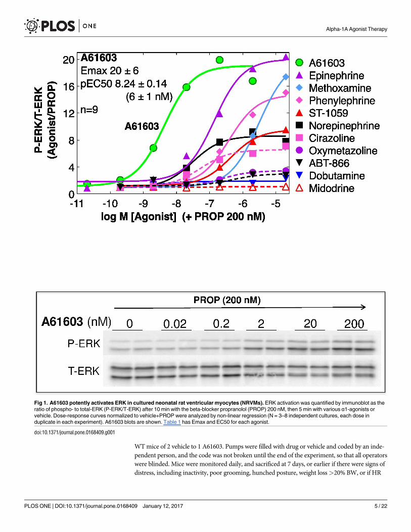
Fig 1. A61603 potently activates ERK in cultured neonatal rat ventricular myocytes (NRVMs). ERK activation was quantified by immunoblot as the
ratio of phospho- to total-ERK (P-ERK/T-ERK) after 10 min with the beta-blocker propranolol (PROP) 200 nM, then 5 min with various α1-agonists or
vehicle. Dose-response curves normalized to vehicle+PROP were analyzed by non-linear regression (N = 3–8 independent cultures, each dose in
duplicate in each experiment). A61603 blots are shown. Table 1 has Emax and EC50 for each agonist.
doi:10.1371/journal.pone.0168409.g001
Alpha-1A Agonist Therapy
PLOS ONE | DOI:10.1371/journal.pone.0168409 January 12, 2017 5 / 22

was less than 200 bpm, a sign of severe cardiomyopathy. Echocardiograms (echos) were done at
baseline in some mice, and in all mice on the day of sacrifice. At sacrifice, BW was recorded,
blood was collected, and organs and tibia were harvested.
Blood analysis for cardiomyocyte death
Blood was collected by left ventricle (LV) apical puncture with a 20-gauge needle. Whole
blood was allowed to clot, centrifuged at 13,000 rpm for 10 min, and the serum supernatant
was snap-frozen, and stored at -80˚C. Creatine kinase (CK) activity was measured in non-
hemolyzed serum samples using the CK UV-rate kit (Stanbio Labs).
Heart histology for apoptosis and fibrosis
Hearts were perfused in situ via the LV apex with KCl 60 mM to arrest in diastole, and fixed by
paraformaldehyde 4%. After paraffin embedding, 6 μm sections were cut from LV base to apex
every 100 μm, with an average of 5 sections per step collected per heart.
Sections were stained for fibrosis using the Picrosirius Red Stain kit (Polysciences, Inc.) and
for apoptosis using the APO-BrdU™ TUNEL Assay Kit (Molecular Probes/Invitrogen). Ran-
dom fields of the LV myocardium were imaged under phase microscopy at 40x magnification
using a SPOT Imaging Solutions camera and software. Images were analyzed with ImageJ.
Data analysis
Results are presented as mean ± SE. Significant differences (p<0.05) were tested using one-
way ANOVA with Newman-Keuls multiple comparison test for more than two groups, or Stu-
dent’s unpaired t-test for two groups (GraphPad Prism v4.0c). Survival data were analyzed by
Table 1. Alpha-1-AR agonist ERK potency, binding affinity, and subtype selectivity.
ERK Activation in NRVMs Alpha-1-AR Binding Affinity (pKi) Binding Selectivity
pEC50 Emax n Alpha-1A Alpha-1B Alpha-1D A/B A/D B/D
A61603 8.24 ± 0.14 20.5 ± 6.1 9 7.74 ± 0.11 (7) 5.68 ± 0.08 (5) 5.85 ± 0.01 (5) 115 78 0.7
Dobutamine 7.65 ± 0.28 2.8 ± 0.7 5 7.00 (1) na Na 0.4 0.1 0.2
Norepinephrine 7.57 ± 0.11 8.6 ± 1.3 7 5.45 ± 0.18 (17) 5.67 ± 0.13 (19) 6.83 ± 0.16 (13) 0.6 0.04 0.07
Cirazoline 7.25 ± 0.12 6.6 ± 0.1 3 6.79 ± 0.14 (5) 6.40 ± 0.17 (4) 6.94 ± 0.27 (4) 2.5 0.7 0.3
Epinephrine 7.16 ± 0.12 19.6 ± 5.9 6 5.45 ± 0.14 (13) 5.82 ± 0.15 (15) 6.44 ± 0.10 (11) 0.4 0.1 0.2
ABT-866 6.71± 0.32 2.9 ± 0.7 3 6.84 ± 0.02 (3) 6.06 ± 0.003 (3) 6.55 ± 0.003 (3) 6.1 1.9 0.3
Oxymetazoline 6.63 ± 0.09 3.5 ± 0.1 3 7.69 ± 0.09 (20) 6.62 ± 0.05 (21) 6.07 ± 0.08 (15) 12 42 3.5
Phenylephrine 6.42 ± 0.11 15.0 ± 5.2 5 5.36 ± 0.17 (14) 5.28 ± 0.14 (14) 6.24 ± 0.16 (10) 1.2 0.1 0.1
Midodrine/ST-1059 6.40 ± 0.05 9.7 ± 1.1 3 5.63 ± 0.21 (4) 5.14 ± 0.04 (4) 5.60 ± 0.18 (4) 3.1 1.1 0.3
Methoxamine 5.54 ± 0.04 20.0 ± 6.9 4 4.52 ± 0.18 (14) 3.54 ± 0.17 (15) 4.65 ± 0.15 (10) 9.4 0.7 0.1
Dose-response curves for 10 alpha-1-agonists, in duplicate dishes for each dose in each experiment (n), were generated for ERK activation (5 min agonist
treatment), in the presence of the beta-blocker propranolol 200 nM.
Potency (pEC50) and efficacy (Emax) for each experiment were from non-linear regression analysis.
Binding affinity (pKi) for each agonist at recombinant alpha-1-AR subtypes is from the literature (n = reports).
The agonist selectivity for each alpha-1-receptor subtype was calculated as A/B = (10^(pKi A—pKi B)), and similarly for A/D and B/D.
For midodrine/ST-1059, functional data are reported for the active compound ST-1059; and binding data are for the parent compound midodrine.
Values are mean ± SE.
na = not available.
Agonists are ordered according to the ERK pEC50.
doi:10.1371/journal.pone.0168409.t001
Alpha-1A Agonist Therapy
PLOS ONE | DOI:10.1371/journal.pone.0168409 January 12, 2017 6 / 22

Fig 2. A61603 activates ERK and protects WT adult mouse ventricular myocytes (AMVMs) from DOX toxicity in vitro, via the α1A.
(A) ERK activation in cultured AMVMs was quantified by immunoblot as the ratio of phospho- to total-ERK (P-ERK/T-ERK) after 10 min
with the beta-blocker PROP 200 nM, then 5 min with A61603 or vehicle. Dose-response curves normalized to vehicle+PROP were
analyzed by non-linear regression (n = 5 cultures from separate hearts, each in duplicate; values are mean ± SE). Emax is significantly
different from 1.0 by 95% confidence limits (p<0.05). Representative blots are shown. (B) WT or α1A KO cultured AMVMs were treated
Alpha-1A Agonist Therapy
PLOS ONE | DOI:10.1371/journal.pone.0168409 January 12, 2017 7 / 22

Kaplan-Meier curves. Normal distribution for continuous variables was confirmed using the
D’Agostino and Pearson omnibus test.
Results
A61603 is the most potent α1-agonist in ERK activation in myocytes
Previously, we defined an α1A-ERK survival pathway using adenoviral expression in
α1-KO cardiac myocytes [8]. Here we tested 10 α1-agonists for ERK activation in cul-
tured NRVMs. The β-AR antagonist propranolol, which does not itself activate ERK
(unpublished data), was present throughout, since some α1-agonists also activate β-ARs
(e.g. norepinephrine, epinephrine, phenylephrine). Fig 1 shows that A61603 was the most
potent in ERK activation (EC50 6 nM), and was highly efficacious (20-fold increase).
Table 1 summarizes the pEC50 and Emax values for ERK activation, and also shows the
binding affinities for each agonist at recombinant α1-subtypes, from the literature. The
calculated binding selectivity for the α1A over the α1B or α1D shows that A61603 is the
most selective of all agonists for the α1A (Table 1). These data confirmed that A61603
activated the α1A-ERK pathway with high potency and efficacy.
A61603 activates ERK and protects adult cardiac myocytes from DOX in
vitro, via the α1A
In cultured adult mouse ventricular myocytes (AMVMs), A61603 activated ERK, and pro-
tected myocytes from DOX toxicity (Fig 2). The EC50 values for these 2 effects were very simi-
lar (23 and 14 nM, Fig 2). The lower potency and efficacy of A61603 in AMVM ERK
activation in comparison with NRVMs (Fig 1) is consistent with the 10-fold lower density of
α1A-ARs in AMVMs [2]. In α1A KO myocytes, A61603 did not prevent DOX toxicity, con-
firming that A61603 was working specifically at the α1A receptor on cardiac myocytes to con-
fer a protective effect (Fig 2B). These data showed that A61603 activated the myocyte ERK
survival pathway defined previously [8].
A61603 activates cardiac ERK in vivo at subpressor dosing
We infused normal adult male WT C57Bl6J mice with A61603 (0.01 to 10 μg/kg/d) by s.c.
osmotic minipump, and quantified cardiac ERK activation by immunoblot after 7 d. A61603
increased ventricular phospho-ERK by 2.8-fold, with an EC50 9 ng/kg/d (Fig 3A). To measure
BP in awake, unrestrained mice, we inserted telemetry catheters in the aorta and infused A61603
10 ng/kg/d or 10 μg/kg/d or vehicle for 7 days, after 4 days baseline recording. From 24 h record-
ing, we identified periods of maxium res (2–3 PM) or activity (12–1 PM) for detailed analysis.
A61603 had no effect of systolic or diastolic BP in resting or active mice (Fig 3B). Previously, we
found that A61603 increases BP with an EC50 300 ng/kg given acutely intravenous [15], as
expected for an α1-AR agonist [23]. Thus, A61603 activated cardiac ERK at dosing that was far
below that required to increase BP.
overnight with DOX 20 μM and PROP 200 nM, in the presence of varying doses of A61603 or vehicle. Myocyte survival by MTT assay is
shown as mean ± SE, and was analyzed by non-linear regression and 2-way ANOVA, relative to PROP alone at 100% and DOX+PROP at
0%; n = 3–8 cultures from separate hearts, each in duplicate or triplicate. WT pEC50 and Emax are shown. Emax is significantly different
from 1.0 by 95% confidence limits (p<0.05).
doi:10.1371/journal.pone.0168409.g002
Alpha-1A Agonist Therapy
PLOS ONE | DOI:10.1371/journal.pone.0168409 January 12, 2017 8 / 22

Fig 3. A61603 activates ERK in vivo at subpressor dosing. (A) Normal male WT mice age 10–12 w were treated with A61603 (0.01–
10 μg/kg/d) or vehicle (100 μM vitamin C) continuously for 7 d via s.c. osmotic mini-pump, and cardiac ERK activation was quantified by
immunoblot as the ratio of P-ERK/T-ERK. Results are for the cytosolic fraction of ventricular homogenates, which contained approximately
75% of total- and phospho-ERK. The A61603 dose-response curve was normalized to vehicle, and analyzed by non-linear regression (N = 5
Alpha-1A Agonist Therapy
PLOS ONE | DOI:10.1371/journal.pone.0168409 January 12, 2017 9 / 22

A61603 improves survival in an acute cardiotoxic DOX HF model
Among multiple models of DOX cardiotoxicity, acute high-dose models are used to test effi-
cacy of potential therapies [24]. We did a trial in an acute, high-dose DOX model to test for
beneficial effects of A61603 in vivo.
Young male mice were given DOX in a single dose, 25 mg/kg i.p. Seventy WT mice and 21
α1A-KO mice were randomized to continuous s.c. infusion by osmotic mini-pump of A61603
or vehicle (100 μM vitamin C). Immediately after pump implantation, mice were injected once
with DOX. A61603 dosing was 10 ng/kg/d, which activates cardiac ERK but does not change
BP (Fig 3). Echo was done at baseline in some mice, and then in all mice at euthanasia, when
dictated by signs of distress or bradycardia, or at study end after 7 days, according to the proto-
col shown in Fig 4A. All operators were blinded to genotype and treatment.
In WT male mice after DOX, A61603 improved survival to day 7 to 84%, from 34% with
vehicle (p<0.0008) (Fig 4B). In α1A-KO mice, A61603 did not improve survival after DOX,
and mortality was 100% by day 6 regardless of treatment, worse than WT mice (Fig 4B). There-
fore, the beneficial survival effect of the α1A-agonist A61603 during DOX cardiotoxicity
required the α1A receptor, indicating an on-target effect.
A61603 preserves cardiac function after acute high-dose DOX
We used FS by echo in awake mice to quantify LV function after DOX. Following DOX injec-
tion, FS fell in surviving vehicle-treated WT mice, from 58% pre-DOX to 49% at euthanasia
for HF or at day 7 (n = 12, p<0.01) (Fig 5, Table 2). A61603 prevented this decline in cardiac
function in WT mice, with FS 61% post-DOX (n = 14, p<0.001 vs. DOX+Veh) (Fig 5). In α1A
KO mice, A61603 did not prevent the drop in FS (58% pre-DOX to 38% post-DOX) (Fig 5). It
was not possible to do a terminal echo on all studied mice, since many died unexpectedly dur-
ing the night.
To evaluate the A61603 mechanisms involved in preserving cardiac function measured by
FS after DOX, LV dimensions were examined. LVIDd was markedly reduced after DOX in all
mice (Table 2), consistent with cardiac atrophy, as seen in mice and humans with anthracycline
cardiomyopathy [25,26,27]. Reduced heart weight at study end supported the conclusion of car-
diac atrophy (below). WT mice treated with A61603 had trends toward a larger LVIDd, smaller
LVIDs, and greater systolic thickness of the LV posterior wall (LVPWs) and interventricular
septum (IVSs), all consistent with less atrophy and improved systolic function (Table 2). Most
of these trends were absent in α1A KO mice (Table 2).
A61603 preserves hemodynamics after acute high-dose DOX
Interestingly, HF in mice is associated with bradycardia. WT mice given vehicle with DOX had
a 55% decrease in HR vs. Pre-DOX, whereas WT mice treated with A61603 had only a 23%
decrease, a significant improvement vs. vehicle (p<0.05, Table 2, Fig 6). A61603 did not pre-
serve HR in α1A KO mice (Table 2, Fig 6). Similarly, SV, the amount of blood ejected with each
beat, was reduced by 50% vs. Pre-DOX in WT mice given vehicle with DOX, whereas the drop
in SV with DOX was only 33% with A61603 treatment (p<0.05 vs. vehicle). The improved SV
with A61603 was lost in α1A KO mice (Table 2, Fig 6). Together, the reduced HR and SV
mice per dose, mean ± SE). Emax is significantly different from 1.0 by 95% confidence limits (p<0.05). A blot is shown below. (B) Baseline BP
was recorded for 4 d, then A61603 (10 ng/kg/d and 10 μg/kg/d) or vehicle were infused continuously for 7 d by osmotic pump. Systolic BP
(upper traces) and diastolic BP (lower traces) are shown during (left) periods of rest (2–3 pm) and (right) periods of activity (12–1 am).
Numbers of mice are indicated. A hypertensive dose of NE (2.5 mg/kg/d) was a positive control.
doi:10.1371/journal.pone.0168409.g003
Alpha-1A Agonist Therapy
PLOS ONE | DOI:10.1371/journal.pone.0168409 January 12, 2017 10 / 22

Fig 4. Low-dose A61603 increases survival of WT male mice in a doxorubicin (DOX) cardiomyopathy model, but not in α1A-KO mice. (A)
Young male WT and α1A-KO mice were randomized to A61603 10 ng/kg/d (A6) or vehicle (100 μM vitamin C, Veh) via s.c. osmotic mini-pump.
Immediately after pump implantation, mice were injected with a single dose of DOX 25 mg/kg i.p. Mice were monitored daily, and euthanized for HF per
protocol or on day 7. Echo was done prior to treatment in some mice, and on the day of sacrifice in surviving mice. Operators were blinded to genotype
and treatment. (B) Kaplan-Meier 7-day survival curves, with numbers of mice indicated.
doi:10.1371/journal.pone.0168409.g004
Alpha-1A Agonist Therapy
PLOS ONE | DOI:10.1371/journal.pone.0168409 January 12, 2017 11 / 22

caused by DOX drastically reduced CO in WT mice treated with vehicle by 73%, whereas the
decrease in CO was only 41% in WT mice treated with A61603 (p<0.01 vs. vehicle). Again this
protective effect of A61603 on CO was lost in the α1A KO (Table 2, Fig 6). Preservation of
hemodynamics by A61603 was even more impressive when CO was normalized to body weight
(BW), to derive a cardiac index (88% of Pre-DOX with A61603, 25% of Pre-DOX with vehicle,
p<0.001 A61603 vs. vehicle, Table 2). Overall, these data suggested that mortality after DOX
was due to reduced cardiac function, and that A61603 improved survival by improving func-
tion, an effect that required the α1A receptor.
A61603 prevents cardiac cell death and myocardial fibrosis in the acute
DOX model
To identify potential cell mechanisms whereby A61603 preserved cardiac function and hemo-
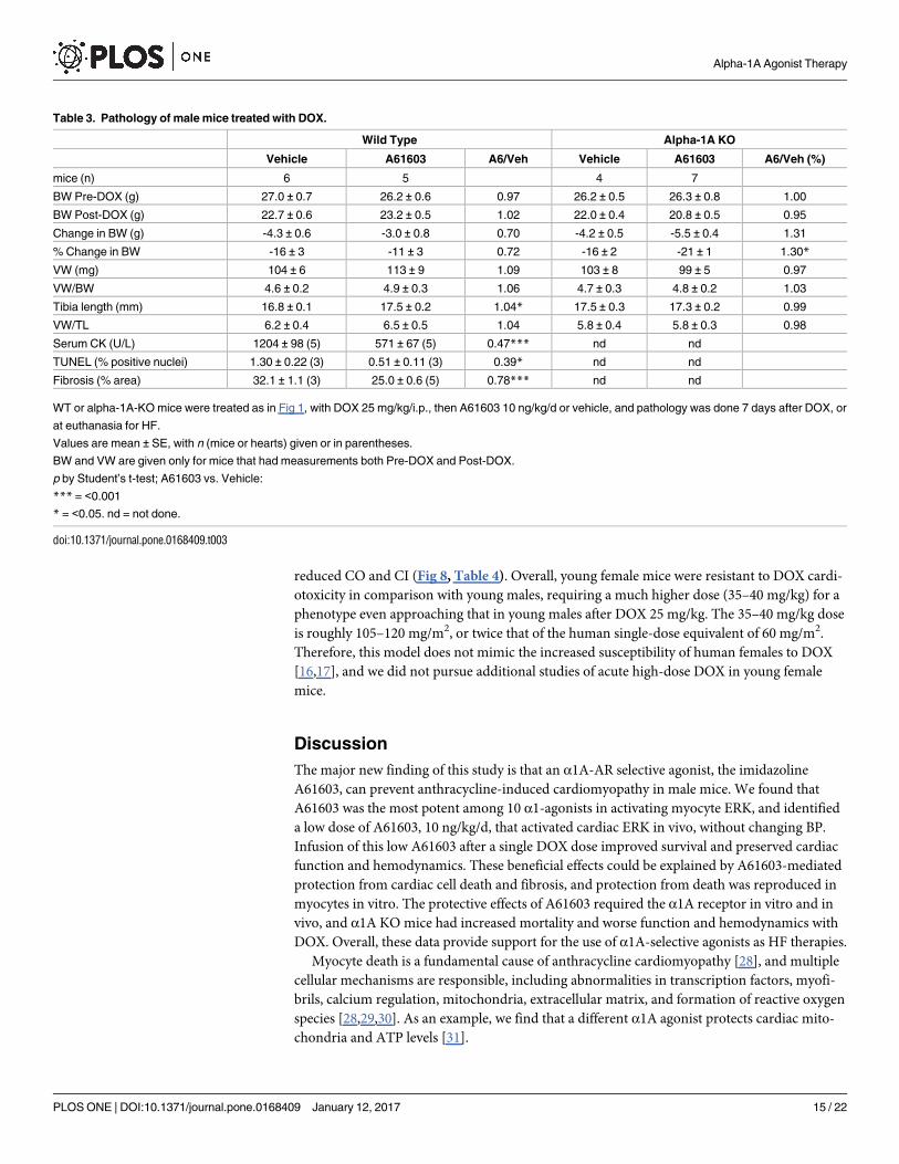
dynamics, we quantified indices of cardiac cell death and fibrosis in WT mice. DOX increased
serum creatine kinase (CK) activity, an assay for myocyte necrosis, by 4-fold at euthanasia vs.
no DOX, and A61603 prevented this increase (p<0.001 vs. vehicle) (Table 3, Fig 7). DOX
increased LV nuclear TUNEL staining, an index of cardiac apoptosis, by 7-fold vs. no DOX,
and A61603 prevented this increase (p<0.05 vs. vehicle) (Fig 7). DOX increased LV sirius red
staining, a measure of myocardial fibrosis, by 1.4-fold, and A61603 prevented this increase
(p<0.0001 vs. vehicle) (Fig 7). These data confirmed and extended our prior finding that
A61603 in the DOX model reduced fibrosis and apoptosis detected by an MRI approach [9],
and suggested that A61603 prevention of cardiac cell death and fibrosis could explain preserva-
tion of LV function and overall hemodynamics.
Fig 5. A61603 preserves cardiac function after DOX in WT male mice, but not in α1A-KO mice. Mice given DOX were treated with A61603 or
Vehicle as in Fig 4. Values are fractional shortening (FS) by echo, mean ± SE, n = mice, p by one-way ANOVA with Newman-Keuls multiple
comparisons test.
doi:10.1371/journal.pone.0168409.g005
Alpha-1A Agonist Therapy
PLOS ONE | DOI:10.1371/journal.pone.0168409 January 12, 2017 12 / 22

Effect of A61603 on body weight and heart weight in the acute DOX
model
WT mice treated with DOX lost body weight (BW) over the study, and A61603 tended to
reduce the weight loss (11% loss A61603 vs. 16% vehicle, p = 0.3), a trend that was not seen in
α1A KO mice (Table 3). Similarly, combined right and left ventricular weight (VW), VW/BW,
and VW normalized to tibia length (TL) trended higher with A61603, and this trend was absent
in α1A KO mice (Table 3). VW in mice given DOX and treated with vehicle was significantly
less than in a comparable group of 20 mice not given DOX (104 ± 6 mg, n = 6 vs. 127 ± 3,
n = 20, p<0.05). However, there was no difference when VW was normalized to BW (4.6 ± 0.2,
n = 6 DOX vs. 4.8 ± 0.1, n = 20 no DOX). Thus there was cardiac atrophy in proportion to the
loss of BW.
Female mice are less susceptible to acute DOX cardiotoxicity
To assess the clinical relevance to females of this acute DOX cardiomyopathy model, we stud-
ied a cohort of young female mice in the same model used for males (Fig 4A). We used DOX
doses (mg/kg i.p.) of 15–20, 25–30, and 35–40. In the young females, no dose reduced survival
or FS significantly; only the 35–40 mg/kg dose reduced BW, LVIDd, HR, and SV, and thereby
Table 2. Echocardiography of male mice treated with DOX.
WILD TYPE Alpha-1A KO
Pre-DOX Post-DOX Pre-DOX Post-DOX
Vehicle A61603 A6/Veh Vehicle A61603 A6/Veh
mice (n) 13 7 6 12 4 8
FS (%) 58 ± 4 49 ± 3 (12)++ 61 ± 2 (14) 1.20*** 58 ± 1 38 ± 4+++ 38 ± 4+++ 0.99
LVIDd (mm) 3.1 ± 0.1 2.5 ± 0.1+++ 2.7 ± 0.1++ 1.08 3.0 ± 0.1 2.3 ± 0.3+ 2.3 ± 0.2++ 0.98
LVIDs (mm) 1.3 ± 0.1 1.4 ± 0.1 1.3 ± 0.1 0.89 1.2 ± 0.1 1.4 ± 0.1 1.4 ± 0.2 1.01
LVPWd (mm) 0.9 ± 0.0 1.0 ± 0.1 0.9 ± 0.1 0.92 0.9 ± 0.0 1.1 ± 0.1 1.1 ± 0.1 1.02
LVPWs (mm) 1.6 ± 0.1 1.2 ± 0.1++ 1.4 ± 0.1 1.15 1.5 ± 0.0 1.1 ± 0.1++ 1.3 ± 0.1++ 1.12
IVSd (mm) 0.9 ± 0.1 1.00 ± 0.1 0.9 ± 0.1 0.95 0.9 ± 0.0 0.9 ± 0.1 1.0 ± 0.1 1.05
IVSs (mm) 1.7 ± 0.1 1.4 ± 0.1 1.6 ± 0.1 1.11 1.7 ± 0.0 1.4 ± 0.2+ 1.4 ± 0.1+ 1.06
SV (μV (±l) 34 ± 1 17 ± 2+++ 23 ± 3+++ 1.37* 31 ± 2 15 ± 1++ 14 ± 3+++ 0.74
HR (bpm) 663 ± 12 300 ± 79+++ 513 ± 77+ 1.71* 657 ± 11 252 ± 16+++ 214 ± 47+++ 0.85
CO (ml/min) 22 ± 1 6 ± 2+++ 13 ± 2+++ 2.27** 20 ± 1 4 ± 2+++ 3 ± 1+++ 0.79
CO/BW (ml/min/g) 0.8 ± 0.0 0.2 ± 0.1+++ 0.7 ± 0.1++ 3.04*** 0.8 ± 0.0 0.2 ± 0.1+++ 0.1 ± 0.0+++ 0.59
WT or alpha-1A-KO mice average age 16.5 weeks had DOX 25 mg/kg i.p., then were treated with A61603 10 ng/kg/d or Vehicle by mini-pump.
2D-guided M-mode echo was done Pre-DOX in some mice, and Post-DOX in all mice, either at euthanasia for HF or at 7 days.
Values are mean ± SE, with n (mice) given or in parentheses.
The n for Post-DOX FS is higher, since this was the only value recorded in an initial series of mice.
p by ANOVA with Newman-Keuls multiple comparisons test: A61603 vs. Vehicle
*** = <0.001
** = <0.01
* = <0.05
Post-DOX vs. Pre-DOX
+++ = <0.001
++ = <0.01
+ = <0.05.
Pre-DOX values for WT vs. alpha-1A-KO did not differ (p>0.1).
doi:10.1371/journal.pone.0168409.t002
Alpha-1A Agonist Therapy
PLOS ONE | DOI:10.1371/journal.pone.0168409 January 12, 2017 13 / 22

Fig 6. A61603 preserves hemodynamics in male mice after DOX, via the α1A. Mice given DOX were treated with A61603 or
Vehicle as in Fig 4. Shown are (A) HR, (B) SV, and (C) CO, all from echo, for each treatment group and genotype. Values are
mean ± SE, n = mice, p by one-way ANOVA with Newman-Keuls multiple comparisons test.
doi:10.1371/journal.pone.0168409.g006
Alpha-1A Agonist Therapy
PLOS ONE | DOI:10.1371/journal.pone.0168409 January 12, 2017 14 / 22
reduced CO and CI (Fig 8, Table 4). Overall, young female mice were resistant to DOX cardi-
otoxicity in comparison with young males, requiring a much higher dose (35–40 mg/kg) for a
phenotype even approaching that in young males after DOX 25 mg/kg. The 35–40 mg/kg dose
is roughly 105–120 mg/m2, or twice that of the human single-dose equivalent of 60 mg/m2.
Therefore, this model does not mimic the increased susceptibility of human females to DOX
[16,17], and we did not pursue additional studies of acute high-dose DOX in young female
mice.
Discussion
The major new finding of this study is that an α1A-AR selective agonist, the imidazoline
A61603, can prevent anthracycline-induced cardiomyopathy in male mice. We found that
A61603 was the most potent among 10 α1-agonists in activating myocyte ERK, and identified
a low dose of A61603, 10 ng/kg/d, that activated cardiac ERK in vivo, without changing BP.
Infusion of this low A61603 after a single DOX dose improved survival and preserved cardiac
function and hemodynamics. These beneficial effects could be explained by A61603-mediated
protection from cardiac cell death and fibrosis, and protection from death was reproduced in
myocytes in vitro. The protective effects of A61603 required the α1A receptor in vitro and in
vivo, and α1A KO mice had increased mortality and worse function and hemodynamics with
DOX. Overall, these data provide support for the use of α1A-selective agonists as HF therapies.
Myocyte death is a fundamental cause of anthracycline cardiomyopathy [28], and multiple
cellular mechanisms are responsible, including abnormalities in transcription factors, myofi-
brils, calcium regulation, mitochondria, extracellular matrix, and formation of reactive oxygen
species [28,29,30]. As an example, we find that a different α1A agonist protects cardiac mito-
chondria and ATP levels [31].
Table 3. Pathology of male mice treated with DOX.
Wild Type Alpha-1A KO
Vehicle A61603 A6/Veh Vehicle A61603 A6/Veh (%)
mice (n) 6 5 4 7
BW Pre-DOX (g) 27.0 ± 0.7 26.2 ± 0.6 0.97 26.2 ± 0.5 26.3 ± 0.8 1.00
BW Post-DOX (g) 22.7 ± 0.6 23.2 ± 0.5 1.02 22.0 ± 0.4 20.8 ± 0.5 0.95
Change in BW (g) -4.3 ± 0.6 -3.0 ± 0.8 0.70 -4.2 ± 0.5 -5.5 ± 0.4 1.31
% Change in BW -16 ± 3 -11 ± 3 0.72 -16 ± 2 -21 ± 1 1.30*
VW (mg) 104 ± 6 113 ± 9 1.09 103 ± 8 99 ± 5 0.97
VW/BW 4.6 ± 0.2 4.9 ± 0.3 1.06 4.7 ± 0.3 4.8 ± 0.2 1.03
Tibia length (mm) 16.8 ± 0.1 17.5 ± 0.2 1.04* 17.5 ± 0.3 17.3 ± 0.2 0.99
VW/TL 6.2 ± 0.4 6.5 ± 0.5 1.04 5.8 ± 0.4 5.8 ± 0.3 0.98
Serum CK (U/L) 1204 ± 98 (5) 571 ± 67 (5) 0.47*** nd nd
TUNEL (% positive nuclei) 1.30 ± 0.22 (3) 0.51 ± 0.11 (3) 0.39* nd nd
Fibrosis (% area) 32.1 ± 1.1 (3) 25.0 ± 0.6 (5) 0.78*** nd nd
WT or alpha-1A-KO mice were treated as in Fig 1, with DOX 25 mg/kg/i.p., then A61603 10 ng/kg/d or vehicle, and pathology was done 7 days after DOX, or
at euthanasia for HF.
Values are mean ± SE, with n (mice or hearts) given or in parentheses.
BW and VW are given only for mice that had measurements both Pre-DOX and Post-DOX.
p by Student’s t-test; A61603 vs. Vehicle:
*** = <0.001
* = <0.05. nd = not done.
doi:10.1371/journal.pone.0168409.t003
Alpha-1A Agonist Therapy
PLOS ONE | DOI:10.1371/journal.pone.0168409 January 12, 2017 15 / 22

Fig 7. A61603 prevents cardiac cell death and myocardial fibrosis after DOX in WT male mice. Male mice were treated with DOX or
saline injection, and randomized to A61803 or Vehicle, as in Fig 4. Shown are indices at end of study for (A) Cardiac myocyte necrosis
measured by serum CK activity in blood collected by LV puncture, and TUNEL staining for apoptosis in LV sections; and (B) LV myocardial
fibrosis by sirius red staining. Number (n) is hearts; p by one-way ANOVA with Newman-Keuls multiple comparisons test.
doi:10.1371/journal.pone.0168409.g007
Alpha-1A Agonist Therapy
PLOS ONE | DOI:10.1371/journal.pone.0168409 January 12, 2017 16 / 22

The signaling mechanisms involved in α1A protection from myocyte death include ERK
activation, which we found is required for adenoviral expression of the α1A to rescue α1A/B
KO cardiomyocytes from DOX-induced death [8]. We show here that A61603 potently acti-
vates this α1A-ERK survival pathway in cultured myocytes and also protects adult myocytes
from DOX-induced death via the α1A (Figs 1 and 2). We also show that A61603 activates ERK
in the normal heart In vivo with an EC50 9 ng/kg/d (Fig 3), suggesting that dosing in the car-
diomyopathy model, 10 ng/kg/d, is activating ERK in myocytes. ERK activation by A61603 in
the injury model in vivo is difficult to test directly, since, whereas the α1A receptor is expressed
in myocytes but not nonmyocytes [39], ERK is activated robustly in nonmyocytes in cardiac
injury [32], obscuring direct effects in myocytes.
ERK is a central myocyte protective kinase [33], and several potential cardioprotective path-
ways downstream of α1-ARs and/or ERK are identified, such as PKCε, p90 ribosomal S6
kinase, SOD, iNOS, anti-apoptotic Bcl proteins, adenosine, cyclooxygenase-2, GATA-4, and
others [1,2,33]. Indeed, the ability of α1-ARs in general to activate pleiotropic signaling could
explain their efficacy against multiple injuries, including oxygen radicals, ischemia-reperfu-
sion, myocardial infarction, and pressure overload (review in [1,2,3]).
The current DOX model has limitations. The DOX dosing of 25 mg/kg in males is high,
but was used in many prior studies [34,35,36,37,38], and is equivalent by allometric scaling to
about 75 mg/m2 in a mouse [39], only 25% higher than a typical single human dose of 60 mg/m2.
Fig 8. Young female mice are resistant to DOX cardiac toxicity. Female WT mice age 15 weeks were
randomized to vehicle or a single dose of DOX 15–40 mg/kg i.p. Mice were monitored daily for 7 days, and
euthanized for HF per protocol or on day 7. Echo was done prior to treatment, and on the day of sacrifice.
Operators were blinded. The highest DOX dose reduced HR (A) and SV (B), and tended to reduce survival
(C), but DOX did not change FS (D). p by one-way ANOVA with Newman-Keuls multiple comparisons test.
doi:10.1371/journal.pone.0168409.g008
Alpha-1A Agonist Therapy
PLOS ONE | DOI:10.1371/journal.pone.0168409 January 12, 2017 17 / 22

However, mortality is high in this model, most likely from the severe reductions in heart rate and
stroke volume, producing a 75% decrease in cardiac index in vehicle-treated mice, vs. only 12%
decrease with A61603 (Table 2). Humans do not typically have reduced heart rate in HF. Also
unlike humans, where females are more susceptible to anthracyclines [16,17], female mice were
resistant relative to males, as in other heart disease models [40]. We studied only young mice
(14–18 weeks), equivalent to approximately 25 human years, and effects in aged males and
females are unknown. DOX caused weight loss, which was reduced but not prevented by A61603
(Table 3), consistent with poor feeding from low cardiac index and/or systemic toxicity, such as
liver or bone marrow damage [41,42,43,44], which can also be seen in humans. Alternative
chronic dosing models might better simulate cardiac toxicity with less systemic toxicity [45].
Overall, this high dose model might be more relevant for the acute DOX toxicity observed in
about 10–30% of patients [46,47,48].
These limitations not withstanding, the data do show clearly that a small molecule α1A ago-
nist can prevent cardiac injury and improve cardiac function and hemodynamics, at a very low
dose that does not change BP. Besides this report, several lines of evidence support the
Table 4. Echocardiography and pathology of WT female mice treated with DOX.
Pre-DOX DOX dose (mg/kg i.p.)
15–20 25–30 35–40
mice (n) 12 2 5 5
FS (%) 58 ± 2 53 ± 5 52 ± 3 56 ± 5
LVIDd (mm) 3.0 ± 0.1 3.2 ± 0.1 3.1 ± 0.1 2.1 ± 0.3+++
LVIDs (mm) 1.3 ± 0.0.08 1.5 ± 0.2 1.5 ± 0.1 1.0 ± 0.2
LVPWd (mm) 0.8 ± 0.06 0.9 ± 0.1 0.9 ± 0.1 1.2 ± 0.2+
LVPWs (mm) 1.5 ± 0.05 1.5 ± 0.1 1.4 ± 0.1 1.6 ± 0.1
IVSd (mm) 0.9 ± 0.04 0.8 ± 0.1 0.8 ± 0.1 1.1 ± 0.1
IVSs (mm) 1.6 ± 0.06 1.6 ± 0.1 1.4 ± 0.1 1.6 ± 0.1
SV (microl) 31± 2 35 ± 1 31 ± 2 13 ± 4+++
HR (bpm) 613 ± 19 663 ± 12 577 ± 64 343 ± 83+++
CO (ml/min) 19 ± 1 23 ± 0 18 ± 2 5 ± 3+++
CO/BW (ml/min/g) 0.9 ± 0.1 1.1 ± 0.0 0.8 ± 0.1 0.3 ± 0.1+++
BW (g) 21.7 ± 0.3 21.1 ± 0.2 21.0 ± 1.1 17.1 ± 1.0++
Change in BW (g) -0.7 ± 0.2 -0.7 ± 1.1 -4.0 ± 1.0
% Change in BW -3 ± 1 -5 ± 6 -24 ± 6
VW (mg) 103 ± 3 105 ± 6 83 ± 6#
VW/BW 4.9 ± 2.0 5.0 ± 0.4 4.7 ± 0.1
TL (mm) 17.0 ± 0.2 17.3 ± 0.2 17.3 ± 0.2
VW/TL 6.1 ± 2.0 6.1 ± 0.4 4.8 ± 0.3
WT female mice had baseline echo, then were treated with varying doses of DOX.
Mouse dosing was as follows (mg/kg i.p.): 15–20 (1 mouse 15, 1 mouse 20), 25–30 (3 mice 25, 2 mice 30), 35–40 (4 mice 35, 1 mouse 40).
Echo was repeated at euthanasia for distress or bradycardia or at 7 days, and pathology was done.
Values are mean ± SE, with n (mice) given.
p by ANOVA with Newman-Keuls multiple comparisons test: DOX dose vs. Pre-DOX
+++ = <0.001
++ = <0.01
+ = <0.05.
high DOX vs. low or medium DOX:
# = <0.05
doi:10.1371/journal.pone.0168409.t004
Alpha-1A Agonist Therapy
PLOS ONE | DOI:10.1371/journal.pone.0168409 January 12, 2017 18 / 22
translation of α1A-agonist cardioprotection to human HF. α1-ARs levels and regulation are
similar in mouse and human heart [4], and A61603 activates protective ERK signaling in
human LV myocardium ex vivo [11]. Although α1-AR levels represent roughly 10% of the AR
population in non-diseased hearts, with β-ARs at 90%, β-AR down-regulation in HF increases
the proportion of α1-ARs to nearly 25% [2,4,49]. In addition, as β-AR function deteriorates in
HF, α1-mediated inotropy can become similar to β-AR [50]. Antagonism of α1-AR signaling
with nonselective α1-blockers can cause HF and death, as shown in the ALLHAT and V-HeFT
trials [51,52]. Altogether, these studies provide indirect human proof of concept for α1A agonist
therapy.
Potential concerns regarding α1A-agonist therapy exist, and the main arguments have been
reviewed [1]. One concern would be increased BP. However, the benefit of A61603 is seen at
10 ng/kg per day, dosing that does not change BP (Fig 3), and is far below the EC50 of 300 ng/
kg for increasing BP with IV bolus dosing [15]. Thus an ample therapeutic window exists for
cardioselective dosing.
As a note of caution, several other potential drugs are reportedly cardioprotective in anthra-
cycline cardiotoxicity models, such as thrombopoietin [25], erythropoietin [53], melatonin
[36], CB1 cannabinoid receptor antagonists [54], granulocyte colony-stimulating factor [55],
and ErbB2 [56], so that proof of concept in other forms of cardiomyopathy will be required to
advance the concept of an α1A agonist as a drug to treat HF.
Conclusions
In conclusion, the α1A-AR selective agonist A61603, at a very low subpressor dose, prevents
cardiomyopathy induced by an anthracycline in male mice. Protection in males requires the
α1A receptor and involves ERK activation and prevention of cardiomyocyte death. The model
is less useful in female mice. These findings confirm and extend previous observations of car-
dioprotection by α1A-agonist therapy, and support the developing model of α1A agonists as
novel therapies for HF.
Author Contributions
Conceptualization: MDM TC RD PCS.
Formal analysis: MDM TC PS RD PCS.
Funding acquisition: PCS.
Investigation: MDM TC PS BM RD.
Methodology: MDM TC PS BM RD PCS.
Project administration: PCS.
Resources: PCS.
Supervision: PCS.
Validation: MDM TC PS RD PCS.
Visualization: MDM PCS.
Writing – original draft: MDM PCS.
Writing – review & editing: MDM TC PS PCS.
Alpha-1A Agonist Therapy
PLOS ONE | DOI:10.1371/journal.pone.0168409 January 12, 2017 19 / 22

References1. Jensen BC, O’Connell TD, Simpson PC (2011) Alpha-1-adrenergic receptors: Targets for agonist drugs
to treat heart failure. J Mol Cell Cardiol 51: 518–528. doi: 10.1016/j.yjmcc.2010.11.014 PMID:
21118696
2. O’Connell TD, Jensen BC, Baker AJ, Simpson PC (2014) Cardiac alpha1-adrenergic receptors: novel
aspects of expression, signaling mechanisms, physiologic function, and clinical importance. Pharmacol
Rev 66: 308–333. doi: 10.1124/pr.112.007203 PMID: 24368739
3. Jensen BC, O’Connell TD, Simpson PC (2014) Alpha-1-adrenergic receptors in heart failure: the adap-
tive arm of the cardiac response to chronic catecholamine stimulation. J Cardiovasc Pharmacol 63:
291–301. doi: 10.1097/FJC.0000000000000032 PMID: 24145181
4. Jensen BC, Swigart PM, De Marco T, Hoopes C, Simpson PC (2009) {alpha}1-Adrenergic receptor sub-
types in nonfailing and failing human myocardium. Circ Heart Fail 2: 654–663. doi: 10.1161/
CIRCHEARTFAILURE.108.846212 PMID: 19919991
5. Lin F, Owens WA, Chen S, Stevens ME, Kesteven S, Arthur JF, et al. (2001) Targeted alpha(1A)-adren-
ergic receptor overexpression induces enhanced cardiac contractility but not hypertrophy. Circ Res 89:
343–350. PMID: 11509451
6. Du XJ, Fang L, Gao XM, Kiriazis H, Feng X, Hotchkin E, et al. (2004) Genetic enhancement of ventricu-
lar contractility protects against pressure-overload-induced cardiac dysfunction. J Mol Cell Cardiol 37:
979–987. doi: 10.1016/j.yjmcc.2004.07.010 PMID: 15522275
7. Du XJ, Gao XM, Kiriazis H, Moore XL, Ming Z, Su Y, et al. (2006) Transgenic alpha1A-adrenergic acti-
vation limits post-infarct ventricular remodeling and dysfunction and improves survival. Cardiovasc Res
71: 735–743. doi: 10.1016/j.cardiores.2006.06.015 PMID: 16859660
8. Huang Y, Wright CD, Merkwan CL, Baye NL, Liang Q, Simpson PC, et al. (2007) An alpha1A-adrener-
gic-extracellular signal-regulated kinase survival signaling pathway in cardiac myocytes. Circulation
115: 763–772. doi: 10.1161/CIRCULATIONAHA.106.664862 PMID: 17283256
9. Dash R, Chung J, Chan T, Yamada M, Barral J, Nishimura D, et al. (2011) A molecular MRI probe to
detect treatment of cardiac apoptosis in vivo. Magn Reson Med 66: 1152–1162. doi: 10.1002/mrm.
22876 PMID: 21360750
10. Cowley PM, Wang G, Chang AN, Makwana O, Swigart PM, Lovett DH, et al. (2015) The alpha1A-
adrenergic receptor subtype mediates increased contraction of failing right ventricular myocardium. Am
J Physiol Heart Circ Physiol 309: H888–896. doi: 10.1152/ajpheart.00042.2015 PMID: 26116709
11. Thomas RC Jr, Singh A, Cowley PM, Myagmar B-E, Montgomery MD, Swigart PM, et al. (2016) A myo-
cardial slice culture model reveals alpha-1A-adrenergic receptor signaling in the human heart. JACC
Basic Transl Res 1: 155–167.
12. Altenbach RJ, Khilevich A, Kolasa T, Rohde JJ, Bhatia PA, Patel MV, et al. (2004) Synthesis and struc-
ture-activity studies on N-[5-(1H-imidazol-4-yl)-5,6,7,8-tetrahydro-1-naphthalenyl]methanesulfonami
de, an imidazole-containing alpha(1A)-adrenoceptor agonist. J Med Chem 47: 3220–3235. doi: 10.
1021/jm030551a PMID: 15163201
13. Jensen BC, Swigart PM, Laden ME, DeMarco T, Hoopes C, Simpson PC (2009) The alpha-1D Is the
predominant alpha-1-adrenergic receptor subtype in human epicardial coronary arteries. J Am Coll Car-
diol 54: 1137–1145. doi: 10.1016/j.jacc.2009.05.056 PMID: 19761933
14. Ky B, Vejpongsa P, Yeh ET, Force T, Moslehi JJ (2013) Emerging paradigms in cardiomyopathies
associated with cancer therapies. Circ Res 113: 754–764. doi: 10.1161/CIRCRESAHA.113.300218
PMID: 23989717
15. Rokosh DG, Simpson PC (2002) Knockout of the alpha 1A/C-adrenergic receptor subtype: the alpha
1A/C is expressed in resistance arteries and is required to maintain arterial blood pressure. Proc Natl
Acad Sci U S A 99: 9474–9479. doi: 10.1073/pnas.132552699 PMID: 12093905
16. Silber JH, Jakacki RI, Larsen RL, Goldwein JW, Barber G (1993) Increased risk of cardiac dysfunction
after anthracyclines in girls. Med Pediatr Oncol 21: 477–479. PMID: 8341214
17. Cardinale D, Colombo A, Bacchiani G, Tedeschi I, Meroni CA, Veglia F, et al. (2015) Early detection of
anthracycline cardiotoxicity and improvement with heart failure therapy. Circulation 131: 1981–1988.
doi: 10.1161/CIRCULATIONAHA.114.013777 PMID: 25948538
18. Simpson P (1985) Stimulation of hypertrophy of cultured neonatal rat heart cells through an alpha 1-
adrenergic receptor and induction of beating through an alpha 1- and beta 1-adrenergic receptor inter-
action. Evidence for independent regulation of growth and beating. Circ Res 56: 884–894. PMID:
2988814
19. Deng XF, Rokosh DG, Simpson PC (2000) Autonomous and growth factor-induced hypertrophy in cul-
tured neonatal mouse cardiac myocytes. Comparison with rat. Circ Res 87: 781–788. PMID: 11055982
Alpha-1A Agonist Therapy
PLOS ONE | DOI:10.1371/journal.pone.0168409 January 12, 2017 20 / 22

20. O’Connell TD, Rodrigo MC, Simpson PC (2006) Isolation and culture of adult mouse cardiac myocytes.
Methods Mol Biol 357: Cardiovascular Proteomics: Methods and Protocols: 271–296.
21. Ishizaka S, Sievers RE, Zhu BQ, Rodrigo MC, Joho S, Foster E, et al. (2004) New technique for mea-
surement of left ventricular pressure in conscious mice. Am J Physiol Heart Circ Physiol 286: H1208–
1215. doi: 10.1152/ajpheart.00011.2003 PMID: 14563661
22. Shindler D (2015) E-chocardiography Journal: An Electronic Journal of Cardiac Ultrasound. http://
rwjms1umdnjedu/shindler/echohtml.
23. Ahlquist RP (1948) A study of the adrenotropic receptors. Am J Physiol 153: 586–600. PMID:
18882199
24. Robert J (2007) Long-term and short-term models for studying anthracycline cardiotoxicity and protec-
tors. Cardiovasc Toxicol 7: 135–139. doi: 10.1007/s12012-007-0022-4 PMID: 17652818
25. Li K, Sung RY, Huang WZ, Yang M, Pong NH, Lee SM, et al. (2006) Thrombopoietin protects against in
vitro and in vivo cardiotoxicity induced by doxorubicin. Circulation 113: 2211–2220. doi: 10.1161/
CIRCULATIONAHA.105.560250 PMID: 16651473
26. Chen YL, Chung SY, Chai HT, Chen CH, Liu CF, Huang TH, et al. (2015) Early Administration of Carve-
dilol Protected against Doxorubicin-Induced Cardiomyopathy. J Pharmacol Exp Ther 355: 516–527.
doi: 10.1124/jpet.115.225375 PMID: 26511374
27. Neilan TG, Coelho-Filho OR, Pena-Herrera D, Shah RV, Jerosch-Herold M, Francis SA, et al. (2012)
Left ventricular mass in patients with a cardiomyopathy after treatment with anthracyclines. Am J Car-
diol 110: 1679–1686. doi: 10.1016/j.amjcard.2012.07.040 PMID: 22917553
28. Sawyer DB, Peng X, Chen B, Pentassuglia L, Lim CC (2010) Mechanisms of anthracycline cardiac
injury: can we identify strategies for cardioprotection? Progress in Cardiovascular Diseases 53: 105–
113. doi: 10.1016/j.pcad.2010.06.007 PMID: 20728697
29. Gianni L, Herman EH, Lipshultz SE, Minotti G, Sarvazyan N, Sawyer DB (2008) Anthracycline cardio-
toxicity: from bench to bedside. J Clin Oncol 26: 3777–3784. doi: 10.1200/JCO.2007.14.9401 PMID:
18669466
30. Sterba M, Popelova O, Lenco J, Fucikova A, Brcakova E, Mazurova Y, et al. (2011) Proteomic insights
into chronic anthracycline cardiotoxicity. J Mol Cell Cardiol 50: 849–862. doi: 10.1016/j.yjmcc.2011.01.
018 PMID: 21284945
31. Beak JY, Huang W, Parker JS, Patterson C, Simpson PC, Ma A, et al. (2016) An oral selective alpha-
1A adrenergic receptor agonist prevents doxorubicin cardiotoxicity. JACC Basic Transl Res (in
revision).
32. Yeh CC, Li H, Malhotra D, Turcato S, Nicholas S, Tu R, et al. (2010) Distinctive ERK and p38 signaling
in remote and infarcted myocardium during post-MI remodeling in the mouse. J Cell Biochem 109:
1185–1191. doi: 10.1002/jcb.22498 PMID: 20186881
33. Bueno OF, Molkentin JD (2002) Involvement of extracellular signal-regulated kinases 1/2 in cardiac
hypertrophy and cell death. Circ Res 91: 776–781. PMID: 12411391
34. Bai P, Mabley JG, Liaudet L, Virag L, Szabo C, Pacher P (2004) Matrix metalloproteinase activation is
an early event in doxorubicin-induced cardiotoxicity. Oncology Reports 11: 505–508. PMID: 14719091
35. Li S, Wang W, Niu T, Wang H, Li B, Shao L, et al. (2014) Nrf2 deficiency exaggerates doxorubicin-
induced cardiotoxicity and cardiac dysfunction. Oxid Med Cell Longev 2014: 748524. doi: 10.1155/
2014/748524 PMID: 24895528
36. Liu X, Chen Z, Chua CC, Ma YS, Youngberg GA, Hamdy R, et al. (2002) Melatonin as an effective pro-
tector against doxorubicin-induced cardiotoxicity. Am J Physiol Heart Circ Physiol 283: H254–263. doi:
10.1152/ajpheart.01023.2001 PMID: 12063298
37. Pacher P, Liaudet L, Bai P, Mabley JG, Kaminski PM, Virag L, et al. (2003) Potent metalloporphyrin per-
oxynitrite decomposition catalyst protects against the development of doxorubicin-induced cardiac dys-
function. Circulation 107: 896–904. PMID: 12591762
38. Pacher P, Liaudet L, Bai P, Virag L, Mabley JG, Hasko G, et al. (2002) Activation of poly(ADP-ribose)
polymerase contributes to development of doxorubicin-induced heart failure. J Pharmacol Exp Ther
300: 862–867. PMID: 11861791
39. Food and Drug Administration US (2005) Guidance for Industry: Estimating the Maximum Safe Starting
Dose in Initial Clinical Trials for Therapeutics in Adult Healthy Volunteers. July: page 7.
40. Blenck CL, Harvey PA, Reckelhoff JF, Leinwand LA (2016) The importance of biological sex and estro-
gen in rodent models of cardiovascular health and disease. Circ Res 118: 1294–1312. doi: 10.1161/
CIRCRESAHA.116.307509 PMID: 27081111
41. Danesi R, Fogli S, Gennari A, Conte P, Del Tacca M (2002) Pharmacokinetic-pharmacodynamic rela-
tionships of the anthracycline anticancer drugs. Clin Pharmacokinet 41: 431–444. doi: 10.2165/
00003088-200241060-00004 PMID: 12074691
Alpha-1A Agonist Therapy
PLOS ONE | DOI:10.1371/journal.pone.0168409 January 12, 2017 21 / 22

42. Bagchi D, Bagchi M, Hassoun EA, Kelly J, Stohs SJ (1995) Adriamycin-induced hepatic and myocardial
lipid peroxidation and DNA damage, and enhanced excretion of urinary lipid metabolites in rats. Toxicol-
ogy 95: 1–9. PMID: 7825176
43. Kalender Y, Yel M, Kalender S (2005) Doxorubicin hepatotoxicity and hepatic free radical metabolism in
rats. The effects of vitamin E and catechin. Toxicology 209: 39–45. doi: 10.1016/j.tox.2004.12.003
PMID: 15725512
44. Zhao X, Zhang J, Tong N, Chen Y, Luo Y (2012) Protective effects of berberine on doxorubicin-induced
hepatotoxicity in mice. Biol Pharm Bull 35: 796–800. PMID: 22687420
45. Li DL, Wang ZV, Ding G, Tan W, Luo X, Criollo A, et al. (2016) Doxorubicin blocks cardiomyocyte autop-
hagic flux by inhibiting lysosome acidification. Circulation 133: 1668–1687. doi: 10.1161/
CIRCULATIONAHA.115.017443 PMID: 26984939
46. Cardinale D, Sandri MT, Martinoni A, Tricca A, Civelli M, Lamantia G, et al. (2000) Left ventricular dys-
function predicted by early troponin I release after high-dose chemotherapy. J Amer Coll Cardiol 36:
517–522.
47. Luminari S, Montanini A, Caballero D, Bologna S, Notter M, Dyer MJ, et al. (2010) Nonpegylated lipo-
somal doxorubicin (MyocetTM) combination (R-COMP) chemotherapy in elderly patients with diffuse
large B-cell lymphoma (DLBCL): results from the phase II EUR018 trial. Ann Oncol 21: 1492–1499. doi:
10.1093/annonc/mdp544 PMID: 20007997
48. Tirelli U, Errante D, Van Glabbeke M, Teodorovic I, Kluin-Nelemans JC, Thomas J, et al. (1998) CHOP
is the standard regimen in patients > or = 70 years of age with intermediate-grade and high-grade non-
Hodgkin’s lymphoma: results of a randomized study of the European Organization for Research and
Treatment of Cancer Lymphoma Cooperative Study Group. J Clin Oncol 16: 27–34. doi: 10.1200/jco.
1998.16.1.27 PMID: 9440719
49. Baker AJ (2014) Adrenergic signaling in heart failure: a balance of toxic and protective effects. Pflugers
Arch 466: 1139–1150. doi: 10.1007/s00424-014-1491-5 PMID: 24623099
50. Skomedal T, Borthne K, Aass H, Geiran O, Osnes JB (1997) Comparison between alpha-1 adrenocep-
tor-mediated and beta adrenoceptor- mediated inotropic components elicited by norepinephrine in fail-
ing human ventricular muscle. J Pharmacol Exp Ther 280: 721–729. PMID: 9023284
51. ALLHAT CRG (2000) Major cardiovascular events in hypertensive patients randomized to doxazosin vs
chlorthalidone: the antihypertensive and lipid-lowering treatment to prevent heart attack trial (ALLHAT).
[see comments]. Jama 283: 1967–1975. PMID: 10789664
52. Cohn JN (1993) The Vasodilator-Heart Failure Trials (V-HeFT). Mechanistic data from the VA Coopera-
tive Studies. Introduction. Circulation 87: VI1–4.
53. Li L, Takemura G, Li Y, Miyata S, Esaki M, Okada H, et al. (2006) Preventive effect of erythropoietin on
cardiac dysfunction in doxorubicin-induced cardiomyopathy. Circulation 113: 535–543. doi: 10.1161/
CIRCULATIONAHA.105.568402 PMID: 16449733
54. Mukhopadhyay P, Batkai S, Rajesh M, Czifra N, Harvey-White J, Hasko G, et al. (2007) Pharmacologi-
cal inhibition of CB1 cannabinoid receptor protects against doxorubicin-induced cardiotoxicity. J Am
Coll Cardiol 50: 528–536. doi: 10.1016/j.jacc.2007.03.057 PMID: 17678736
55. Li L, Takemura G, Li Y, Miyata S, Esaki M, Okada H, et al. (2007) Granulocyte colony-stimulating factor
improves left ventricular function of doxorubicin-induced cardiomyopathy. Lab Invest 87: 440–455. doi:
10.1038/labinvest.3700530 PMID: 17334414
56. Belmonte F, Das S, Sysa-Shah P, Sivakumaran V, Stanley B, Guo X, et al. (2015) ErbB2 overexpres-
sion upregulates antioxidant enzymes, reduces basal levels of reactive oxygen species, and protects
against doxorubicin cardiotoxicity. Am J Physiol Heart Circ Physiol 309: H1271–1280. doi: 10.1152/
ajpheart.00517.2014 PMID: 26254336
Alpha-1A Agonist Therapy
PLOS ONE | DOI:10.1371/journal.pone.0168409 January 12, 2017 22 / 22