an amphiphysin-like domain in fus2p is required for ... an amphiphysin-like domain in fus2p is...
TRANSCRIPT

INVESTIGATION
An Amphiphysin-Like Domain in Fus2p Is Requiredfor Rvs161p Interaction and Cortical LocalizationRichard A. Stein,1,2 Jean A. Smith,2 and Mark D. Rose3
Department of Molecular Biology, Princeton University, New Jersey 08544
ORCID ID: 0000-0003-1112-4765 (M.D.)
ABSTRACT Cell–cell fusion fulfils essential roles in fertilization, development and tissue repair. In the buddingyeast, Saccharomyces cerevisiae, fusion between two haploid cells of opposite mating type generates thediploid zygote. Fus2p is a pheromone-induced protein that regulates cell wall removal during mating. Fus2pshuttles from the nucleus to localize at the shmoo tip, bound to Rvs161p, an amphiphysin. However, Rvs161pindependently binds a second amphiphysin, Rvs167p, playing an essential role in endocytosis. To understandthe basis of the Fus2p–Rvs161p interaction, we analyzed Fus2p structural domains. A previously describedN-terminal domain (NTD) is necessary and sufficient to regulate nuclear/cytoplasmic trafficking of Fus2p. TheDbl homology domain (DBH) binds GTP-bound Cdc42p; binding is required for cell fusion, but not localization.We identified an approximately 200 amino acid region of Fus2p that is both necessary and sufficient forRvs161p binding. The Rvs161p binding domain (RBD) contains three predicted alpha-helices; structural mod-eling suggests that the RBD adopts an amphiphysin-like structure. The RBD contains a 13-amino-acid region,conserved with Rvs161p and other amphiphysins, which is essential for binding. Mutations in the RBD,predicted to affect membrane binding, abolish cell fusion without affecting Rvs161p binding. We proposethat Fus2p/Rvs161p form a novel heterodimeric amphiphysin required for cell fusion. Rvs161p binding isrequired but not sufficient for Fus2p localization. Mutations in the C-terminal domain (CTD) of Fus2p blocklocalization, but not Rvs161p binding, causing a significant defect in cell fusion. We conclude that the Fus2pCTD mediates an additional, Rvs161p-independent interaction at the shmoo tip.
KEYWORDS
conjugationkaryogamyBAR domain
Cell fusion is ahighly conserved eukaryotic process,with fundamental rolesin fertilization, as well as development, disease pathogenesis, and tissuerepair (Buckingham 2006; Linder 2007; Wassarman and Litscher 2008;Kim et al. 2015). Placental trophoblast formation andmuscle developmentare two examples of processes containing post fertilization cell fusion
events (Mi et al. 2000; Taylor 2002; Primakoff and Myles 2002). Under-standing cell fusion could aid attempts to develop therapies targetingmuscle degenerative disorders or other myopathies (Gibson et al. 1995).
Ultrastructural studies suggest that cell fusion events are morpho-logically similar in several distinct cell types. In cells surrounded by anextracellular matrix, fusion of the plasma membrane must be precededby removal of the intervening material. In the budding yeast Saccharo-myces cerevisiae, prior to plasma membrane fusion, cell wall thinningoccurs at the site of contact between the two haploid fusion partners,coincident with the accumulation of electron-dense vesicles at the zoneof cell fusion (ZCF) (Gammie et al. 1998). Similarly, in the Drosophilamyoblast prefusion complex, electron-dense vesicles accumulate on thecytoplasmic faces of the apposed plasma membranes of the two cells(Doberstein et al. 1997; Estrada et al. 2007; Sens et al. 2010).
In S. cerevisiae, the mating pathway begins when two haploid part-ners of opposite mating types (MATa and MATa) detect secretedmating pheromone from the other cell. Pheromone binding initiatesa G-protein-coupled response, which activates a MAP kinase sig-nal transduction pathway. This pathway results in multiple down-stream responses including G1 cell cycle arrest, transcription of genes
Copyright © 2016 Stein et al.doi: 10.1534/g3.115.023960Manuscript received October 22, 2015; accepted for publication November 26,2015; published Early Online December 16, 2015.This is an open-access article distributed under the terms of the CreativeCommons Attribution 4.0 International License (http://creativecommons.org/licenses/by/4.0/), which permits unrestricted use, distribution, and reproductionin any medium, provided the original work is properly cited.Supporting information is available online at www.g3journal.org/lookup/suppl/doi:10.1534/g3.115.023960/-/DC11Present address: Department of Biochemistry and Molecular Pharmacology,New York University School of Medicine, New York, NY 10016
2These authors contributed equally to this work.3Corresponding author: Department of Molecular Biology, 320 Lewis ThomasLaboratory, Princeton University, Princeton, NJ 08544-1014. E-mail: [email protected]
Volume 6 | February 2016 | 337
involved in mating, and polarization toward the mating partner(Dohlman and Slessareva 2006; Merlini et al. 2013). After cell-cyclearrest, the two partners extend mating projections, creating pear-shaped cells called shmoos, and contact each other to form a prezy-gote. The prezygotes degrade the walls between the two cells, undergoplasma membrane fusion, and ultimately the nuclei fuse to generate adiploid zygote (White and Rose 2001; Ydenberg and Rose 2008).
Many proteins have been identified that are required for cell fusion(Ydenberg and Rose 2008; Merlini et al. 2013). Fus2p, important for cellwall degradation, was first identified by amutation that blocks cell fusionwhen present in both partners (Elion et al. 1995). Expressed in responsetomating pheromone, Fus2p is initially retained in the nucleus (Patersonet al. 2008). Upon completion of the cell cycle, Fus2p exits the nucleus ina phosphorylation-dependent manner, and localizes at the shmoo-tipcortex (Paterson et al. 2008; Ydenberg and Rose 2009; Kim and Rose2012). Mating in fus2mutants halts at the prezygote stage, before cellwall degradation, with vesicles accumulating at the ZCF (Gammieet al. 1998). Accordingly, Fus2p is thought to regulate the fusion ofthe vesicles with the plasma membrane to release hydrolases for cellwall breakdown (Gammie et al. 1998; Paterson et al. 2008). At theshmoo-tip, Fus2p interacts with GTP-bound Cdc42p through its Dbl-homology domain; Cdc42p function and the interaction with Fus2pare required for cell wall breakdown (Barale et al. 2006; Ydenberget al. 2012). Cdc42p is a Rho-like GTPase with roles in polarization,signaling, and secretion throughout mating and mitosis (Richmanet al. 1999; Johnson 1999; Kozminski et al. 2000; Adamo et al. 2001).
Rvs161p, another protein required for cell fusion, is an amphiphysinthat was first identified as a mutant showing reduced viability uponstarvation (Crouzet et al. 1991). Rvs161p is not required for viability,but mutations in the protein cause actin delocalization (Sivadon et al.1995), osmotic sensitivity (Crouzet et al. 1991), endocytosis defects(Munn et al. 1995), and random budding in diploid cells (Durrenset al. 1995). There is another amphiphysin in yeast, Rvs167p, sharingpartial homology with Rvs161p but containing an additional internalglycine-, proline-, and alanine-rich sequence (GPA) followed by aC-terminal SH3 domain (Youn et al. 2010). Amphiphysins are membersof the BAR domain family, which includes proteins that bind cellularmembranes, and promote membrane curvature by participating inmembrane remodeling processes (Dawson et al. 2006; Youn et al.2010). BAR domains mediate critical links between the actin cyto-skeleton and the membrane, and are thus highly conserved acrossspecies (Dawson et al. 2006). The crystal structures of several BARdomains reveal that they are dimers of two monomers, each of whichencompasses three a helices separated by short unstructured coils(Peter et al. 2004; Dawson et al. 2006). Kinks in the helices, togetherwith the orientation of the two monomers, are responsible for bend-ing of the dimeric BAR protein. The central region of the banana-shaped dimers contains an overlap of three a helices from eachparticipating monomer to establish a six-helical bundle. The dimershave positive residues on their concave face as well as in the distalloops formed between a helices 2 and 3 in each monomer, which areimportant for electrostatic interactions with the negatively chargedinner face of the plasma membrane (Dawson et al. 2006).
Rvs161p has at least two distinct and dissociable cellular functions(Brizzio et al. 1998). During vegetative growth andmating, Rvs161p formsa heterodimer with Rvs167p, binding membrane lipids at an early step inendocytosis (Friesen et al. 2006; Paterson et al. 2008). In mating cells,Rvs161p also binds to Fus2p, and the complex localizes to the shmoo tipin an actin- and Myo2p-dependent manner (Brizzio et al. 1998; Sheltzerand Rose 2009). An rvs161D fus2D double mutant does not show amore severe cell fusion defect, indicating that the two genes act together
in one of the pathways responsible for cell fusion (Gammie et al. 1998).The interaction with Rvs161p is required for Fus2p stability; however, theRvs161p binding domain on Fus2p is not known. Alleles of RVS161 havebeen identified that separately affect endocytosis and cell fusion, indicatingthat these functions are at least partially independent (Brizzio et al. 1998).The alleles that block cell fusion are defective for binding Fus2p, but,because they are still active for endocytosis,must still interactwithRvs167p(Paterson et al. 2008). The formation of two exclusive complexes suggeststhat the binding sites for Fus2p and Rvs167p on Rvs161p at least partiallyoverlap (Paterson et al. 2008). Therefore, it is likely that Rvs167p andFus2p interact with Rvs161p in similar ways. Rvs161p interacts with theN-terminal domain of Rvs167p; both are predicted to form conservedthree-helix bundle BAR domains (Navarro et al. 1997; Peter et al. 2004).
Here, we report that Fus2p binds to Rvs161p via an amphiphysin-like domain. Modeling the interaction between Rvs161p and Fus2pidentified key residues involved in complex formation and function.Using function specific alleles of Rvs161p, we found that Fus2p/Rvs161p and Rvs167p/Rvs161p localize to different regions in theshmoo. Details about the Fus2p-Rvs161p interaction will fill a gap inour knowledge about a fundamental step during eukaryotic cell fusion,with relevance for several organisms.
MATERIALS AND METHODS
General yeast methods, strain and plasmid constructionYeast media, general methods and transformations were performed asdescribed previously (Adams et al. 1997), with minor modifications.Strains and plasmids used in this study are presented in SupportingInformation, Table S1 and Table S2. To generate Fus2p mutants, weintroduced stop codons or pointmutations into pMR5469 by the dut ungmutagenesis protocol (Kunkel 1985). Mutations were introduced intoRvs161p by the same approach, using plasmid pMR5912 as a template.To create Rvs161-mCherry, the mCherry fragment was amplified withprimers mCherry-start.SpeI (59-TAT CTA GTG AGC AAG GGC GAGGAG-39) and mCherry-stop.BamHI (59-TAT CTG GAT CCC TTGTAC AGC TCGACCATGCC-39), and digested with SpeI and BamHI.pMR5912 was also digested with these enzymes, and the PCR fragmentwas ligated into the XbaI–SpeI site of the linearized vector. The resultingplasmid, pMR6588, in which Rvs161p-mCherry is expressed from thenative RVS161 promoter, was confirmed by sequencing.
Deletion mutants in FUS2 were generated by site-directed PCRmutagenesis (Phusion, Thermo Fisher Scientific), using pMR5469(Paterson et al., 2008) as a template. Deletions were confirmed bysequencing, and pheromone-induced protein expression was con-firmed by TCA precipitation followed by immunodetection (Ohashiet al. 1982). Fus2p C-terminal mutations were generated using thepMR5482 template, in which FUS2 with the internal GFP tag at posi-tion 104 is expressed from its own promoter. Mutations were intro-duced by the dut ung mutagenesis protocol (Kunkel 1985).
Coimmunoprecipitation assaysOvernight cultures were grown in selective media containing galactose,and used to start 100 ml cultures, which were grown to early expo-nential phase (OD600 = 0.2), and treated with 10 mg/ml syntheticmating pheromone (Syn/Seq Facility, Department of Molecular Biol-ogy, Princeton University) for 2 hr. Cell extracts were prepared aspreviously described (Brizzio et al. 1998). Cell pellets were resuspendedin 0.75 ml breaking buffer (50 mM Tris, pH 7.4, 50 mM NaCl,and 0.5% Triton X-100) supplemented with protease inhibi-tors (Miniprotease tablets supplemented with 1 mM PMSF), andthe cells were lysed for 1 min, three times. After clearing by
338 | R. A. Stein, J. A. Smith, and M. D. Rose

centrifugation for 10 min at 13,000 rpm, lysates were incubated with30 ml anti-FLAG M2 affinity gel (Sigma-Aldrich) that had beenwashed once with water, and three times with breaking buffer. Reac-tions were brought to 1 ml with breaking buffer, supplemented with10 ml 5M NaCl to bring the final concentration of the reaction to150 mM NaCl, and incubated for 1 hr at 4� with rotation. Subse-quently, the beads were washed five times in breaking buffer supple-mented with protease inhibitors (Roche). The protein was eluted with100 ml SDS loading buffer, and samples were analyzed via SDS-PAGE and western blotting.
SDS-PAGE and western blottingSamples were prepared either by TCAprecipitation (Ohashi et al. 1982)for protein controls, or via coimmunoprecipitiation. Proteins were re-
solved on SDS-PAGE gels (8% gels to visualize the Fus2p constructs,and 10% gels to visualize the Rvs161p recombinants). Proteins werethen transferred to a nitrocellulosemembrane (100 V, 2 hr), and, afterblocking for 2 hr (3% BSA milk), the blots were incubated with pri-mary antibody [a-GFP antibodies (Roche) at 1:1000 dilution for theFus2p constructs, and a-FLAG antibodies (Sigma-Aldrich) at 1:5000dilution for the Rvs161p constructs] and subsequently with secondaryantibodies (a-mouse, 1:2500) for 1 hr each, and visualized by standardchemiluminescence.
Mating assaysLimited plate mating assays were performed as described previously(Gammie and Rose 2002). For testing Rvs161p mutants, the respectiveplasmids were transformed into MY3909, which contains an rvs161::LEU2
Figure 1 Fus2p contains an internal, amphiphysin-like Rvs161p-binding domain. (A) Rvs161p-Flag85 is functional. Rvs161p was internally flag taggedon either a plasmid or at its genomic locus. rvs161Δ (MY3909) cells were transformed with plasmids containing wild-type RVS161 (pMR3234), RVS161-Flag85 (pMR5912), or an empty vector (pRS416, Sikorski and Hieter 1989), and mated to a fus1Δ fus2Δ strain (JY429) for 3 hr at 30�. (B) Residues 537–582 are required for binding to Rvs161p. fus2Δ cells containing genomic RVS161-Flag85 (MY10904) were transformed with plasmids containing eitherdeletions or alanine mutations introduced into pMR5469. Rvs161p was pulled down using anti-FLAG agarose beads, and bound Fus2p was assessedvia western blot using anti-GFP antibodies. (C) Residues 580–582 are important during fusion. The same strains as in (B) were mated to a fus1Δ fus2Δstrain (JY429) for 3 hr at 30�. (D) Fus2p shares homology with Rvs161p between residues 570 and 582. (E) C-terminal boundary of Rvs161p-bindingdomain is between residues 580 and 640. Plasmids containing C-terminal truncations made in pMR5469 were transformed into MY10904, andbinding to Rvs161p was assessed as before. (F) The minimal binding domain for Rvs161p is between residues 415 and 626 in Fus2p. Coimmuno-precipitations were performed as before. (G) Map of all Fus2p fragments tested summarizing the results of the binding experiments.
Volume 6 February 2016 | Amphiphysin-Like Domain in Fus2p | 339

mutation. The strains were patched on yeast extract/peptone/dextrose(YEPD), mated with exponentially grown lawns of MY4907, whichhas an rvs161D fus1D double mutation, and diploids were selectedby replica plating onto selective medium. For Fus2p mutants, strainswere patched on YEP + 2% galactose plates, and mated with an expo-nential lawn of strain MY1814, which has a fus1D fus2D double muta-tion. After replica-plating the strains with the testerMATa strain, plateswere incubated for 3 hr at 30�, and diploids were selected by replicaplating to minimal medium.
For quantitativemating assays, overnight yeast cultures were dilutedto early log phase and grown for approximately 4 hr. Subsequently, 3–5 · 106 cells of each mating type were combined and concentrated on2.5-cm2 nitrocellulose filter discs (Millipore), as previously described(Grote 2008). Cells were allowed to mate for 4 hr at 30�, and thenmating mixtures were resuspended in 1 ml water or YEPD. Equalvolumes of mating mixtures were plated on selective media to selectfor diploids and calculate the mating efficiency.
Cell imagingFor imagingof pheromone-inducedcellswithfluorescent proteins, earlylog phase cellswere treatedwith10 mg/ml syntheticmatingpheromone(Syn/Seq Facility, Department of Molecular Biology, Princeton Univer-sity) for 1.5 hr in rich media. Cells containing a galactose-inducibleprotein were grown to early log phase in media containing raffinose,and then induced with pheromone (10 mg/ml), and 2% galactose for
2 hr at 30�. Pheromone-induced cells were then fixed with 2% form-aldehyde for 10 min at 30�, washed twice with 1X PBS, and imaged.Mating mixtures to be imaged were prepared as above via filter mating.They were then resuspended in 1 ml of TAF buffer and imaged.
The Applied Precision Deltavision Microscopy System (Issaquah,WA) using a Nikon TE200 inverted microscope, a Photometrics Cool-snapHQCCDcamera (Tucson,AZ), and a 100Xobjectivewere used forimaging. All images were deconvolved to remove out-of-focus fluores-cence. For publication purposes, the contrast and brightness wereenhanced using Adobe Photoshop.
Data availabilityAll strains andplasmids are available upon request. Strains andplasmidsused in this studyarepresented inSupporting Information,TableS1andTable S2.
RESULTS
Residues of Fus2p from 415 to 626 are required forbinding to Rvs161pTo examine the structural basis of the Fus2p–Rvs161p interaction, weperformed coimmunoprecipitation of the two proteins in pheromone-induced cells. For these experiments, we used tagged versions of bothproteins. Fus2p, expressed from the GAL1 promoter, was tagged withan internal GFP tag inserted in-frame after amino acid 104. The tagged
Figure 2 Conservation of the Rvs161p-binding domain among fungi. Residues 419–626 from Saccharomyces cerevisiae Fus2p were comparedagainst homologs from other fungal species. Blue highlighting indicates the amount of conservation, with darker shaded residues being moreconserved. The conservation of all amino acids is depicted using the gray bars below the residues. Black bars denote every 10 amino acids.Turquoise bars indicate the aromatic residues mutated in Figure 5; red bars indicate the lysine residues mutated in Figure 6. Predicted alphahelices in this region are mapped below the residues in green.
340 | R. A. Stein, J. A. Smith, and M. D. Rose

protein has been shown to function like wild-type Fus2p in all assaystested (Paterson et al. 2008; Ydenberg and Rose 2009). Rvs161p wastagged with an internal FLAG tag, inserted after amino acid 85, in apredicted loop region that separates the first two a-helices. The taggedprotein was functional inmating cells, as shown by limited platemating(Figure 1A). As previously observed, wild-type Fus2p and Rvs161pshow an interaction by coimmunoprecipitation (Figure 1, B, E, F,and G).
To determine the region of Fus2p necessary for the Rvs161p in-teraction, we initially generated an internal Fus2p deletion that re-moved 41 amino acids in the C-terminus of the protein (residues538–579). This region of Fus2p was predicted to contain coiled-coilstructures, similar to amphiphysins (Paterson et al. 2008). It also sharesprimary structure similarity with the C-terminus of Rvs161p, whichcontains cell fusion specific alleles (Brizzio et al. 1998). Within thisregion, we also deleted 13 residues that harbor a high degree of homol-ogy to Rvs161p (residues 570–582, Figure 1D) (Paterson et al. 2008).Both protein constructs lost the ability to bind to Rvs161p (Figure 1B).Within this 13-bp region were three residues, 580ELP582, that are iden-tical in Rvs161p. We found that deletion of just these three residues(Δ580–582) resulted in loss of binding to Fus2p; however, when eachresidue was individually mutated to alanine, binding was unaf-fected (Figure 1B). Consistent with the Rvs161p binding defect ofFus2pΔ580–582, this strain showed significant mating defects (Figure 1C).We conclude that protein sequences within the C-terminal region ofFus2p, including at least residues 579–582, are required for bindingto Rvs161p.
To further define the Rvs161p interaction region on Fus2p, wemadeadditional truncations from both the N-terminal and C-terminal ends.Among the C-terminal truncations, Rvs161p was able to interact withsmall truncations of up to 37 amino acids, including Fus2p1–670,Fus2p1–660, Fus2p1–650, and Fus2p1–640 (Figure 1E). Interaction withRvs161p was lost when we truncated the protein to residue 580 (Figure1, E and F). These experiments reveal that the C-terminal border ofthe minimal binding domain is at, or upstream of, 640.
Among the N-terminal truncations, both Fus2p105–677 andFus2p415–677 were able to bind Rvs161p. However, a deletion that begandownstream, Fus2p458–677, did not interact with Rvs161p. A slightlylarger fusion, Fus2p452–677, was too unstable to assay. The most exten-sive N-terminal truncation to still bind Rvs161p was Fus2p415–677,which places the left border of the minimal binding site at, or upstreamof, residue 415 (Figure 1F). The results of all coimmunoprecipitationsperformed on truncations of Fus2p are summarized in Figure 1G.
Comparisons of the C-terminal region of Fus2p with related fungishowed extensive conservation through residue 621 (LQKDL, Figure2). Accordingly, to determine if the region of Fus2p between aminoacids 415 and 626 might be sufficient for binding to Rvs161p, wecreated a protein construct with just these amino acids fused to GFP.We found that this construct bound Rvs161p similar to the wild typeprotein (Figure 1F). We conclude that the Fus2p415-626 is sufficient forbinding Rvs161p.
Modeling the Fus2p/Rvs161p interaction region as anamphiphysin-like domainThe three-dimensional structure of the Fus2p415–626 fragment was pre-dicted using the PHYRE2 (Protein Homology/analogY RecognitionEngine V 2.0) web server, which performs a structure-based sequencealignment to identify proteins of known structure that are homologousto a protein with a given sequence (Kelley and Sternberg 2009). ThePHYRE server uses proteins from the Structural Classification of Pro-teins (SCOP) database that is augmented with more recent depositions
in the Protein Data Bank (PDB) (Murzin et al. 1995; Berman 2000).PHYRE can reveal significant structural homology between proteinpairs that harbor limited primary sequence homology, sometimes aslittle as 15%–25% (Kelley and Sternberg 2009). For Fus2p415–626, mod-eling provided a structure homologous with the Drosophila amphiphysinBAR domain, with 99.84% confidence for the three-dimensionalsuperimposition. A different model predicted by a similar program,I-Tasser, was virtually identical in structure, differing by a root meansquare deviation (RMSD) of 1.052Ǻ (Zhang 2008; Roy et al. 2010; Yanget al. 2014). The predictions indicate that this fragment is organizedinto three a-helices that are joined by short loops, adopting an overallstructure that is reminiscent of the a-helical structures described inamphiphysins (Figure 3A). The extents of the three predicted a-helices
Figure 3 Modeling of the Fus2p-Rvs161p interaction predicts abanana-shaped heterodimer. (A) Fus2p and Rvs161p are predictedto form a heterodimer similar to the Drosophila (Dm) amphiphysinhomodimer. The structure of Fus2p residues 415–626 (shown in blue),and Rvs161p residues 28–233 (shown in gray) were individually mod-eled using the PHYRE2 program (Kelley and Sternberg 2009). The twopredicted structures were then modeled on the Dm amphiphysinhomodimer (shown in light blue) using MatchMaker in the Chimeramodeling program (Pettersen et al. 2004; Meng et al. 2006). (B) Elec-trostatic mapping of the Fus2p-Rvs161p heterodimer reveals surfaceswith high positive charges on the front and concave faces. Basic res-idues are shown in blue; acidic residues are shown in red.
Volume 6 February 2016 | Amphiphysin-Like Domain in Fus2p | 341

closely match the highest regions of conservation in the Fus2pC-terminal region (Figure 2). The same prediction algorithm was usedtomodel Rvs161p, and residues 28–233 of the protein weremodeled onthe BAR domain from human Bin1/Amphiphysin II (Casal et al. 2006)with 100% confidence (Figure 3A).
To model the interaction between Fus2p415–626 and Rvs161p, weused theDrosophila (Dm) amphiphysin dimer, for which the N-terminalresidues 1–245 have been crystallized and the structure determinedat 2.6 Å resolution (Peter et al. 2004). The PHYRE output structures forFus2p415–626 and Rvs161p were modeled on the two monomers thatform the Dm amphiphysin dimer by using the homology-modelingalgorithm MatchMaker in the Chimera modeling program (Pettersenet al. 2004; Meng et al. 2006). The predicted structure formed betweenFus2p415–626 and Rvs161p creates a banana-shaped heterodimer, strik-ingly similar to the amphiphysin dimers that were previously charac-terized in other organisms (Figure 3A). The residues in the predictedFus2p and Rvs161p chains differ from those in Dm amphiphysin by0.127 and 0.941 Ǻ RMSD, respectively.
Combining this model with the previous binding data allows us tomap the residues known to be important for binding. The Rvs161189ELP191, and the Fus2p 580ELP582 sequences that are important forbinding and mating are highlighted on the heterodimer, as are the
conserved ELP residues in the Dm amphiphysin monomers (Figure3A). In this model, the tripeptides reside at the kink in helix 3, formedby the conserved proline. The bend in the alpha helix causes the char-acteristic banana shape of the heterodimer, and is therefore unsurpris-ingly required for binding and function.
Wenext created anelectrostaticmapof thepredicteddimerusing theChimera modeling program (Pettersen et al. 2004; Meng et al. 2006),where negative residues are highlighted in red and positive residues arehighlighted in blue (Figure 3B). Interestingly, there are two broad re-gions of the heterodimer that are predicted to be strongly positivelycharged, the concave inner surface and the lateral outer surface largelycomprised of Fus2p. As positively charged residues play roles in bind-ing negatively charged phospholipids, the predicted electrostatic mapsuggests that Fus2p/Rvs161p may interact with membranes along bothits concave and lateral surfaces. This may allow Fus2p to bind to highlycurved membranes along the concave surface as well as more planarmembranes along the front surface.
Residues in Rvs161p required for binding to Fus2pPreviously, a genetic screen identified twoaminoacids inRvs161p,A175and P203, that are important for cell fusion (Brizzio et al. 1998). Theseresidues flank the region of Rvs161p that is homologous to Fus2p
Figure 4 Rvs161p contains res-idues important for both bind-ing to Fus2p and cell fusion. (A)Mutations of conserved residuesin Rvs161p cause binding defects.A strain containing a doubledeletion of FUS2 and RVS161(MY10463) was transformed withboth a plasmid containing WTGFP-tagged FUS2 (pMR7042) aswell as alanine mutations made inRVS161 from pMR5912. Nega-tive controls comprised WT GFP-tagged FUS2 and an emptyvector. Binding of Fus2p toRvs161p was assessed as beforeby coimmunoprecipitation. (B, C)Alanine-scanning mutagenesisrevealed other residues impor-tant for binding. Alanine muta-tions in RVS161 were made inpMR5912, and the mutant plas-mids were transformed intoMY10463 along with pMR7042.Binding was assessed as before.(D) Residues in Rvs161p at whichcorresponding mutations causestrongly reduced Fus2p binding.(E) Rvs161p residues importantfor Fus2p-binding are also im-portant for mating. The samestrains as in B were mated to afus1Δ fus2Δ (JY429) for 3 hr at 30�.
342 | R. A. Stein, J. A. Smith, and M. D. Rose
(Figure 1D). The A175P and P203Q mutations disrupt binding be-tween untagged Rvs161p and wild-type Fus2p (Brizzio et al. 1998).To further explore the role of this region of the protein, additionalalleles in and around the conserved region were introduced intoRvs161p-Flag85. As found previously, A175P abolished binding,whereas A175F did not (Figure 4A). Based on the structural modeling,A175 is predicted to reside in helix 3, distal to the kink in the three-helixbundle (Figure 3C). Due to the location of this residue, it is likely thatthe introduction of proline at that site causes a significant change inconformation that interferes with binding. Like P203Q, the P203Ymutation, greatly decreased binding (Figure 4A). P203 is predicted toreside at the binding interface of Rvs161p and Fus2p (Figure 4C),further explaining the decrease in Fus2p binding when mutated.
Performing an alanine scanmutagenesis in Rvs161p in the regionsurrounding the Fus2p homology, we identified several amino acidsthat are important for binding Fus2p. Mutation of the highly con-served phenylalanine to alanine (F179A) abolished binding to Fus2p(Figure 4B), which is correlated with a strong mating defect. F179resides close to A175 (Figure 4D), suggesting that this mutation mayalso cause a change in the conformation of Rvs161p rather thandirectly interfering with the interaction. Mutation of the less well-conserved neighboring residue to alanine (E180A) had no effect onbinding or mating (Figure 4, B and E). Mutation of residues NL181–182 both to alanine had no effect on binding. In contrast, theNNQ183–185AAA mutation, affecting the conserved glutamine,and the ELP189–191AAA mutation, which affects the highly con-served ELP residues, abolished binding to Fus2p (Figure 4B), andresulted in strong mating defects (Figure 4E). Mutation of the NNQtripeptide (Figure 4C) likely affects the structural integrity ofRvs161p. As for Fus2p, single mutations in ELP did not affect bind-ing (Figure 4C). No other single site mutations (affecting residues171–174 and 204–208) affected binding (Figure 4C).
Conserved aromatic residues in Fus2p are important forbinding to Rvs161pAromatic residues have been shown to play important roles in theinteractions between alpha helices. These interactions are known toensure intramolecular stability and intermolecular interactions aswell as structural stability of the helices (Anderson et al. 1993;Butterfield et al. 2002). Aromatic amino acids participate in inter-actions that contribute to the stability of the native protein fold, andaromatic–aromatic interactions provide a mechanism to generatenoncovalent interactions that ensure the stabilization of proteinstructures (Burley and Petsko 1985).
When examining the primary amino acid sequence of Fus2p415–626
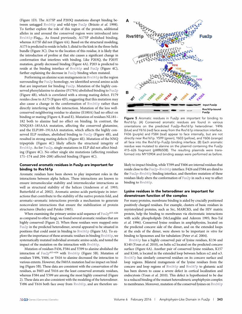
as compared to other fungi, we found several aromatic residues that arehighly conserved (Figure 2). When these residues were mapped ontoFus2p in the predicted heterodimer, several appeared to be situated inpositions that could assist in binding to Rvs161p (Figure 5A). To ex-plore the importance of these aromatic residues in binding Rvs161p, wesystematically mutated individual aromatic amino acids, and tested theimpact of the mutation on the interaction with Rvs161p.
Mutation of residues F436, F584 and Y599 to alanine abolished theinteraction of Fus2p415–626 with Rvs161p (Figure 5B). Mutation ofresidues Y496, Y606, or Y616 to alanine decreased the interaction tovarious extents. However, the F603Amutation had no impact on bind-ing (Figure 5B). These data are consistent with the conservation of theresidues, as F603 and Y616 are the least conserved aromatic residues,whereas F584 and Y599 are among the most highly conserved (Figure2). These data are also consistent with the modeling of the heterodimer.Y496 and Y616 both face away from Rvs161p, and are therefore un-
likely to impact binding, while Y599 and Y606 are internal residues thatreside close to the Fus2p–Rvs161p interface. F426 and F584 are distal tothe Fus2p–Rvs161p binding interface, and therefore mutation of theseresidues likely alters the conformation of Fus2p in such a way to affectbinding to Rvs161p.
Lysine residues in the heterodimer are important fordownstream function of the complexFor many proteins, membrane binding is aided by crucially positionedpositively charged residues. For example, clusters of basic residues inmyristoylated proteins, such as Src, MARCKS, and the HIV matrixprotein, help the binding to membranes via electrostatic interactionswith acidic phospholipids (McLaughlin and Aderem 1995; Ben-Talet al. 1996). Conserved lysine residues in amphiphysins, located onthe predicted concave side of the dimer, and on the extended loopsat the ends of the dimer, were shown to be important in vitro forbinding to liposomes and for tubulation (Peter et al. 2004).
Rvs161p has a highly conserved pair of lysine residues, K136 andK140 (Youn et al. 2010), on helix a2 located on the predicted concavesurface (Figure 6A). Another pair of conserved lysine residues, K157and K160, is located in the extended loop between helices a2 and a3.Rvs167p has similarly conserved residues on its concave surface andloop regions. Bilateral mutagenesis of the lysine residues from theconcave and loop regions of Rvs161p and Rvs167p to glutamic acidhas been shown to cause a severe defect in cortical localization andendocytosis (Youn et al. 2010). This defect is hypothesized to be dueto a reduced binding of themutant heterodimeric amphiphysin complextomembranes.Moreover, mutation of the conserved lysines on Rvs161p
Figure 5 Aromatic residues in Fus2p are important for binding toRvs161p. (A) Conserved aromatic residues are found in variousorientations on the predicted Fus2p–Rvs161p heterodimer. Y496(blue) and Y616 (red) face away from the Rvs161p interaction interface.F436 (purple) and F584 (teal) appear to face internally, but are notdirectly near Rvs161p. Y599 (green), Y603 (yellow), and Y606 (orange)all face into the Rvs161p–Fus2p binding interface. (B) Each aromaticresidue was mutated to alanine on the plasmid containing the Fus2p415–626 fragment (pMR6508). The resulting plasmids were trans-formed into MY10904 and binding assays were performed as before.
Volume 6 February 2016 | Amphiphysin-Like Domain in Fus2p | 343

results in a defect in cell fusion (Youn et al. 2010), suggesting thatmembrane association may be important for Fus2p/Rvs161p function.
The structural prediction for Fus2p415–626 helped identify con-served lysine residues that are in similar locations to the residues inamphiphysins (Figure 6A). K534 and K538 are located on the con-cave surface of the heterodimer, whereas K554 and K555 are in theloop region (Figure 6A). In wild-type Fus2p, we mutated the twolysine residues from the predicted Fus2p concave surface (“con”)and, separately, the two lysine residues from its loop region (“loop”),to glutamate residues. Con and loop mutations in either Rvs161p orFus2p caused decreased mating efficiencies compared to wild-typestrains (Figure 6C). Mutation of all four loop lysines in both Fus2pand Rvs161p had a more severe phenotype than mutation of all thecon lysines, suggesting that the loop residues are more important forfunction. When each pair of lysines on the concave surfaces weremutated in Fus2p or Rvs161p, a mating defect was still observed,although it was not as severe as in combination. The mating defectsfor fus2con or rvs161con were similar, suggesting that both surfacescontribute equally for fusion. However, when the equivalent exper-iment was performed with fus2loop and rvs161loop, mutation ofRvs161p was more deleterious than mutation of Fus2p (Figure6C), implying that the loop residues of Rvs161p are more importantfor function than those of Fus2p.
To determine if the mating defect was simply due to a defect in theFus2p–Rvs161p interaction, we performed coimmunoprecipitationsusing Rvs161pwith the con, loop, or all four lysines (4K to 4E)mutated.All strains contained wild-type Fus2p. Although each of the mutationsresulted in slightly decreased binding of Fus2p, all three constructs stillbind Fus2p (Figure 6B). Therefore, we conclude that these lysine res-idues are important formating efficiency without affecting heterodimerformation. These results support the conclusion of Youn et al. (2010)that membrane binding of Rvs161p is required for cell fusion.
The C-terminus of Fus2p is required for localizationWhile analyzing the truncations of Fus2p used to define the bindingregion for Rvs161p, we observed significantly reduced mating efficiencyfor the strain harboring a truncation of the last eight amino acids ofFus2p, fus2–670UAG (Figure 7A). Themating defect cannot be caused bya lack of heterodimer formation as this construct binds Rvs161p as wellas wild-type Fus2p (Figure 1E). Wild-type Fus2p is known to localize tothe shmoo tip in pheromone-treated cells (Paterson et al. 2008; Ydenbergand Rose 2009). We therefore assayed the cortical localization ofFus2p1–670 and found that the mutant protein was diffusely localizedthroughout the cell (Figure 7B). These data indicate that whereasRvs161p binding is necessary for Fus2p localization, it is not sufficient.
We individuallymutated eachof the eightC-terminal amino acids toalanine in otherwise wild-type Fus2p and cortical localization and mat-ing efficiency were assessed. The point mutants showed varying matingphenotypes. Mutation of V670 and K673 did not cause a distinguish-able defect. Mutation of V671, R672, E676, or L677 all caused inter-mediate phenotypes. Finally, mutation of L674 or F675 caused a severemating phenotype, comparable to the C-terminal truncation (Figure7C). The mating efficiency of each mutant was also measured by quan-titative methods and compared to the defect in localization (Table 1).The two defects are strongly correlated, and all mutations that severelyaffected localization also resulted in a mating phenotype (Table 1).However, some mutations caused mating defects that are more severethan expected based on localization (e.g., compare V671A to L677A).This observation may be understood by postulating that the mutationsmay cause additional cell fusion defects beyond localization.
Wenextwanted to determine if the C-terminus of Fus2p is sufficientfor localization. We began by assessing the localization of Fus2p415–677,which binds Rvs161p (Figure 1F), and contains the C-terminus re-quired for localization. Fus2p415–677 was localized to the cortex in70 6 2% of cells examined (Figure 7D), showing that the NTD and
Figure 6 Lysine residues on theconcave and loop regions of theheterodimer are important for cellfusion without affecting hetero-dimer formation. (A) Conservedlysine pairs highlighted on thepredicted Fus2p–Rvs161p hetero-dimer. Rvs161pK136, K140 andFus2pK534, K538 reside on the con-cave surface (“con”, highlighted ingreen), while Rvs161pK157, K160
and Fus2pK554, K555 reside on theloops (“loop”, highlighted in pur-ple). (B) Mutation of the Rvs161pcon and/or loop residues does notaffect binding to wild-type Fus2p.Plasmids containing mutation of thecon residues (pMR6543), loop resi-dues (pMR6504), or both (pMR6512)in Rvs161p-FLAG were transformedalong with wild-type Fus2p-GFP(pMR5469) into a fus2Δ rvs161Δstrain (MY10463). Fus2p bindingwas assessed via coimmunopreci-pitation as before. (C) Mutation ofthe lysine residues resulted in a
cell fusion defect. Plasmids containing RVS161 con (pMR6543) or loop (pMR6504) mutations were transformed along with plasmids containingFUS2 con (pMR6505) or loop (pMR6507) mutations into a fus2Δ rvs161Δ strain (MY10463). Mating efficiency was tested via mating to a fus1Δfus2Δ strain (JY429) for 3 or 3.5 hr at 30� to detect subtle differences in phenotypes.
344 | R. A. Stein, J. A. Smith, and M. D. Rose

DBHdomains are not required for cortical localization. To determine ifthe Rvs161p binding domain is necessary for cortical localization, wecreated a construct in which residues 626–677 of Fus2p tagged to GFPwas expressed from the GAL1 promoter. However, this construct wasnot stable in pheromone-induced cells, and did not localize. BecauseRvs161p binding is required for Fus2p stability (Brizzio et al. 1998), toassess the role of the C-terminus, we used the Rvs161p4K/4E mutant,which blocks membrane binding, but not Fus2p binding [Figure 6(Youn et al. 2010)]. The effect of the Rvs161p4K/4E mutation willtherefore indicate whether Rvs161p plays a role in the cortical localiza-tion of Fus2p, as well as stabilizing Fus2p, or is required only for Fus2pstability. We found that wild-type Fus2p was mislocalized in theRvs161p4K/4E mutant background, with 14 6 3% of cells having lo-calized Fus2p (Figure 7E). This is consistent with the observed matingdefect (Figure 6C). Given that membrane-binding of Rvs161p is alsorequired for cortical localization of Fus2p, we conclude that theC-terminus of Fus2p is necessary but not sufficient for cortical localization.
Rvs161p is localized in two pools in pheromone-induced cellsIt was shown previously that both Fus2p and Rvs161p localize to theshmoo tip and interact in pheromone-treated cells, and that this in-teraction is required for efficient cell fusion (Brizzio et al. 1998; Gammieet al. 1998; Paterson et al. 2008). However, Rvs161p also functions ina complex with Rvs167p during endocytosis, which also occurs inpheromone-induced cells (Friesen et al. 2006). Distinct patterns oflocalization to the shmoo projection have been described previously(Narayanaswamy et al. 2009). Proteins involved in exocytosis are lo-calized to the shmoo-tip, whereas proteins involved in endocytosis arelocalized more diffusely over the entire shmoo projection. Given thatthere are mutant alleles that separate the two functions of Rvs161p(Brizzio et al. 1998), we wanted to examine the effects of these alleleson the localization of Rvs161p and Fus2p.
To examine the localization of both Rvs161p and Fus2p, we usedplasmids containing Fus2p tagged with GFP at position 104 (Patersonet al. 2008), and created a plasmid with Rvs161p tagged internally withmCherry at position 85. Unless stated otherwise, the genomic copy ofRVS161 or FUS2 was deleted in these strains so that the only proteinobserved was fluorescently tagged. As previously observed, Fus2p andRvs161p were colocalized at the shmoo tip (Figure 8A, top panel).However, there is also a fainter pattern of localization of Rvs161p alongthe shmoo neck. Given that Fus2p is localized exclusively at the shmootip, we presume that localization along the neck of the shmoo is asso-ciated with endocytosis. To determine the effect of a lack of Rvs161p-Fus2p interaction on the localization of both proteins, we introducedthe F179A mutation into Rvs161p-mCherry. This allele of Rvs161p isdefective for Fus2p binding and mating (Figure 4, B and E). We foundthat Fus2p was not cortically localized in this strain, consistent with therequirement for Rvs161p interaction. The shmoo-tip localization ofRvs161pF179A was also lost; however, the shmoo neck localization
Figure 7 The C-terminus of Fus2p is required for cortical localizationand cell fusion. (A) Mutations in the C-terminal eight amino acids ofFus2p cause defects in cell fusion. fus2Δ cells (MY10904) were trans-formed with plasmids containing either WT FUS2 (pMR5482), fus2-670UAG (pMR6775), or an empty vector (pRS416), and mated to afus1Δ fus2Δ strain (JY429) for 3 hr at 30�. (B) Fus2p1-670 is defectivefor cortical localization. The same strains as in A were imaged afterincubation with pheromone for 1.5 hr. n $ 200 shmoos imaged inthree independent experiments. (C) Individual point mutations showdecreased mating efficiency. Plasmids containing individual alaninemutations for the last eight amino acids were transformed into afus2Δ strain (MY10904). Mating efficiency was assessed after matingto a fus1Δ fus2Δ strain (JY429) for 3 hr at 30�. (D) The C-terminus ofFus2p is sufficient for cortical localization. A plasmid containing a FUS2construct in which the first 414 amino acids were deleted under thecontrol of the GAL1 promoter was transformed into a fus2Δ strain
(MY9181). Cells were imaged after incubation with pheromone andgalactose for 2 hr. n $ 250 shmoos were imaged in three indepen-dent experiments. (E) Rvs161p is required for cortical localization ofFus2p. A fus2Δ rvs161Δ strain (MY10463) was transformed with a wild-type FUS2-GFP plasmid (pMR7042) as well as a plasmid containingeither wild-type RVS161 (pMR5912) or RVS1614K/4E (pMR6512).Strains were imaged after 2 hr incubation with pheromone and ga-lactose. At least 175 shmoos were imaged in three independentexperiments.
Volume 6 February 2016 | Amphiphysin-Like Domain in Fus2p | 345

remained intact (Figure 8A, middle panel), consistent with the fact thatthis allele does not affect the endocytotic function of Rvs161p (Brizzioet al. 1998). We observed the same localization for Rvs161pF179A-mCherry when a wild-type, untagged copy of Rvs161p was present.In this strain, Fus2p was able to localize, by virtue of its binding to thewild-type Rvs161p; however, Rvs161pF179A was still restricted to theshmoo neck (Figure 8A, bottom panel).We conclude that there are twolocalization pools of Rvs161p: one at the shmoo tip responsible forbinding to Fus2p and cell fusion, and the other at the shmoo neckinvolved in endocytosis.
Because it is known that binding to Rvs167p is required for thefunction of Rvs161p in endocytosis (Friesen et al. 2006), we testedthe localization of both wild-type Rvs161p and Rvs161pF179A in anrvs167Δ background. We found that wild-type Rvs161p localizationwas restricted to the shmoo tip in the rvs167Δ mutant. When thervs167Δ was combined with Rvs161pF179A, the mutant Rvs161p waslocalized diffusely throughout the strain, consistent with the loss ofboth binding partners (Figure 8B). We conclude that the localizationof Rvs161p to the shmoo neck is dependent on Rvs167p, further sup-porting that this pool of Rvs161p is involved in endocytosis.
DISCUSSION
Rvs161p and Fus2p form a heterodimeric amphiphysin-like complexAmphiphysins, defined by a shared BAR (Bin1 Amphiphysin Rvs)domain, are highly conserved throughout evolution. Amphiphysinshave been implicated in a variety of cellular processes including, butnot limited to, endocytosis, regulation of the actin cytoskeleton, tissuedifferentiation, transcriptional repression, and cell–cell fusion (Renet al. 2006). Despite the wide array of functions of BAR domains, theyshare a common mode of action in membrane remodeling. X-raycrystallography has shown that amphiphysins form an alpha-helicalbanana-shaped dimer, which is able to bind to curved membranes(Dawson et al. 2006). Themajority of amphiphysins formhomodimers;however, heterodimers are also observed, as in the case of the Rvs161p–Rvs167p complex in S. cerevisiae (Youn et al. 2010). Rvs161p also formsa heterodimer with Fus2p in mating cells, and these two complexeshave very different functions (Brizzio et al. 1998). The preference forformation of homodimers vs. heterodimers may provide insight intothe diversity of functions performed by an amphiphysin protein.
BAR-domain-containing proteins have been shown to sense curva-ture, and/or create curvature in lipid bilayers. Amphiphysin dimers canbind to the negatively charged membrane via clustering of positiveresidues along one face of the structure (Dawson et al. 2006). TheRvs161p–Rvs167p complex has been shown to oligomerize during en-
docytosis, and form helical structures at the neck of the forming endo-cytic vesicle (Engqvist-Goldstein and Drubin 2003; Youn et al. 2010).Constriction of the invaginating endocytic tubule by Rvs161p–Rvs167pis thought to help drive vesicle scission (Kishimoto et al. 2011). Duringmating, the Rvs161p–Fus2p complex localizes to the shmoo tip, whereit acts with GTP-bound Cdc42p to promote cell fusion (Brizzio et al.1998; Paterson et al. 2008; Ydenberg et al. 2012). This complex isthought to aid the fusion of glucanase-bearing vesicles with the plasmamembrane. Prior to cell fusion, Rvs161p-Fus2p may localize to theessentially planar plasma membrane at the shmoo tip, as opposed tothe highly curved endocytic tubules. However, after cell fusion, Fus2pforms an expanding ring around the cell fusion pore, possibly by bind-ing to the highly curved membrane along the edge. The dual require-ments for membrane localization before and after cell fusion mayaccount for the striking pattern of high positive charge on both thefront and concave faces of the Rvs161p-Fus2p heterodimer.
Rvs161p is required for Fus2p localization and stabilityPrevious work showed that mutations in conserved lysine residues onthe surface of the amphiphysin dimer affect the ability of the complex tobind membranes (Youn et al. 2010). Equivalent lysine mutations inRvs161p cause cell fusion defects indicating the importance for mem-brane binding in this pathway (Youn et al. 2010). Similarly, mutationsof the conserved lysines in Fus2p also block cell fusion, without affect-ing heterodimer formation (Figure 6). Thusmembrane binding of bothpartners in the complex is likely to be important for cell fusion.
The Rvs161p–Fus2p interaction is required for the stability of Fus2p(Brizzio et al. 1998; Paterson et al. 2008), which complicates assessmentof any role for Rvs161p in retaining Fus2p at the shmoo tip. To addressthis question, we studied wild-type Fus2p localization in the Rvs161p4E
mutant background. In this strain, Rvs161p has all four surface lysinesmutated to glutamic acid, which causes cell fusion defects withoutaffecting Fus2p binding. We found that wild-type Fus2p is stable butmislocalized in this strain. We conclude that membrane interaction ofRvs161p is required for the cortical localization of Fus2p (Figure 7).
We found that the C-terminus of Fus2p is also required for local-ization, indicating that the Rvs161p interaction is not sufficient forlocalization. Therefore, stable cortical localization is dependent onmul-tiple interactions, any one of which is not sufficient. Presumably, theRvs161p binding domain binds directly to the plasma membrane,whereas the C-terminus of Fus2p interacts with other cortical proteins.It has been shown previously that Fus2p is retained at the shmoo tipdependent both on Fus1p and polymerized actin (Paterson et al. 2008;Sheltzer and Rose 2009). We hypothesize that these partially redundantpathways may be acting through the C-terminus.
Although Rvs161p localized at the shmoo tip in mating cells, it wasalso localized at puncta along the shmoo neck. The shmoo tip locali-zation was Fus2p-dependent, whereas the shmoo neck localization wasRvs167p-dependent (Figure 8). Therefore, the two localization sites forRvs161p spatially segregate its two functions. Cell fusion activity wouldbe restricted to the shmoo tip, and the housekeeping function of en-docytosis would occur along the sides of the shmoo projection. Thespatial restriction of proteins required for cell fusion may help ensurethat conjugation occurs only between two partners, ensuring the pro-duction of diploid zygotes.
Potential conservation of cell fusion proteinsand mechanismsCell fusion events are ubiquitous among eukaryotes, raising the ques-tion of whether the basic mechanism and proteins required for fusionare conserved. Post fertilization, many fusion events occur during
n Table 1 Summary of mating and localization phenotypesassociated with Fus2p C-terminal mutations
FUS2 Mating (%) Localization (%)
WT . 99 . 98Δ670–677 8 6 2 8 6 2V670A 52 6 5 76 6 3V671A 19 6 1 66 6 2R672A 6 6 2 27 6 6K673A 58 6 4 44 6 6L674A , 0.1 4 6 3F675A , 0.1 , 0.1E676A 48 6 4 55 6 6L677A 31 6 34 66 6 3
346 | R. A. Stein, J. A. Smith, and M. D. Rose

mammalian development, with one of the best-characterized eventsbeing myoblast fusion during muscle formation. In Drosophila, myo-blast fusion is asymmetric, withmuscle founder cells fusing with fusioncompetent myoblasts (FCMs). The FCMs produce actin-rich, finger-like, protrusions into the founder cells, inducing inward curvature onthe founder cell membrane. The protrusions are surrounded by adhe-sion molecules required for fusion, and closely resemble podosomes.Podosomes are actin-dependent protrusions that are associated withextracellular matrix degradation, through secretion of matrix metal-loproteinases (Linder 2007). The podosome-like structure (PLS) inmyoblast fusion pushes the two membranes into closer proximity,allowing for increased surface contact of the opposing membranes(Kim et al. 2015). It is not known whether the PLS is also required todegrade extracellular matrix separating the two cells. Regardless, thePLS resembles the shmoo tip in S. cerevisiae in being an actin-dependent,actin-enriched cell fusion structure. Recent work demonstrated a sim-ilar “actin fusion focus” in the fission yeast, Schizosaccharomycespombe. Like the PLS, the fusion focus is asymmetric and Arp2/3-dependent (Dudin et al. 2015).
It is likely that curved membrane binding proteins may be requiredduring fusion to stabilize the finger-like protrusions, and/or facilitatemembrane fusion. Indeed, intracellular curvature-generating proteins,including BAR-domain-containing proteins, facilitate hemagglutinin-promoted cell–cell fusion (Richard et al. 2011). In mice, GRAF1, a
BAR-domain-containing GTPase-activating protein, is required formyoblast fusion and overexpression of GRAF1 in cultured myoblastsinduces cell fusion (Kim et al. 2015). A second BAR domain protein,Bin3, is also required for mouse muscle myogenesis and myotubeformation (Simionescu-Bankston et al. 2013). Interestingly, Bin3 isan ortholog of Rvs161p, and, like Rvs161p, contains only an N-BARdomain (Ren et al. 2006). Moreover, myotube formation requires actinpolymerization dependent on the small GTPases, Rac1, and Cdc42(Vasyutina et al. 2009). Bin3 forms a complex with active Rac1 orCdc42 (Simionescu-Bankston et al. 2013). Because Bin3 only containsa BAR domain, it is hypothesized to interact with other proteins thatbind the GTPases, similar to the Rvs161p–Fus2p complex. MouseToca-1, an F-BAR protein, also affects myoblast fusion. Toca-1 inter-acts with Cdc42, and activates Cdc42-mediated actin nucleationthrough interaction with the Arp2/3 activator N-WASP (Ho et al.2004; George et al. 2014). Thus, in both mice and yeast, BAR domainproteins are required for cell fusion.
Cdc42p is required at two stages in yeast cell fusion. First, it isrequired for cell polarization and formation of the shmoo tip (Baraleet al. 2006). Later, the Rvs161p–Fus2p complex binds GTP-boundCdc42p to promote cell fusion (Ydenberg et al. 2012). Proteins homol-ogous to Cdc42p are implicated in Drosophila, mouse, and zebrafishmyoblast fusion. In Drosophila, the GTPase Rac activates the Scarcomplex, which promotes actin polymerization via the Arp2/3
Figure 8 Rvs161p is localized intwo pools in pheromone-inducedcells. (A) Rvs161pF179A causes lossof shmoo tip localization. rvs161Δfus2Δ (MY10463, top two panels)or RVS161 fus2Δ (MY9181, bottompanel) strains were transformedwith plasmids containing wild-typeFus2p-GFP (pMR7042) along witheither wild-type Rvs161p-mCherry(pMR6588), or Rvs161pF179A-mCherry (pMR7063). Cells wereimaged after incubation with pher-omone for 1.5 hr. (B) The pool ofRvs161p localized at the shmooneck is lost in rvs167Δ cells. rvs161Δ(MY3909) or rvs161Δ rvs167Δ(MY4545) strains were trans-formed with either wild-typeRvs161p-mCherry (pMR6588),or Rvs161pF179A-mCherry (pMR7063)and imaged after 1.5 hr incuba-tion with pheromone.
Volume 6 February 2016 | Amphiphysin-Like Domain in Fus2p | 347
complex. Additionally, both the small GTPase Rac1 and Cdc42 havebeen shown to play a role in mouse myoblast fusion in vivo (Vasyutinaet al. 2009).
Invadosomes are protrusive F-actin structures, similar to podo-somes, that promote tumor cell invasion via degradation of the ECM.”Linear” invadosomes form specifically upon contact with type I col-lagen fibrils. Interestingly, whereas Rho GTPases are involved in clas-sical invadosome formation, only Cdc42p is required for the formationof linear invadosomes. Tuba is a BAR domain protein that acts as alinear invadosome-specific GEF for Cdc42p. Tuba contains an internalDBL homology domain, whichmakes it structurally the most similar toFus2p. However, Fus2p localizes GTP-bound Cdc42p rather than ac-tivating it (Juin et al. 2014).
Taken together, we perceive a common conserved pathway for all ofthese fusion events. Actin-dependent polymerization mediated by asmall GTPase (Cdc42p or Rac1) causes cells to form podosome-likeprotrusions. In fungi, the protrusion is required for the cells to makecontact; in animal cells, the protrusions may be required to deform themembranes of the fusing cells. The tip of the protrusion is also likely tobe a site where extracellularmatrix is degraded. In fungi, the ECM is cellwall, whereas in animal cells it comprises collagen and other proteinsdegraded by podosomes. Finally, many of the membrane functions ofthe protrusion are mediated by amphiphysins, which serve to regulateand localize the G-proteins that mediate cell fusion.
ACKNOWLEDGMENTSWe thank members of the Rose and Gammie laboratories for helpfulsupport and discussion. We also thank J. Youn and B. Andrews forhelpful discussions and data concerning residues in Rvs161p that arerequired for membrane binding. This work was supported by NationalInstitutes of Health Grants GM037739 (to M.D.R.). J. A. S. wassupported by National Institutes of Health Training GrantGM007388.
LITERATURE CITEDAdamo, J. E., J. J. Moskow, A. S. Gladfelter, D. Viterbo, D. J. Lew et al.,
2001 Yeast Cdc42 functions at a late step in exocytosis, specificallyduring polarized growth of the emerging bud. J. Cell Biol. 155: 581–592.
Adams, A. E., D. E. Gottschling, C. A. Kaiser, and T. Stearns, 1997 Methodsin Yeast Genetics. Cold Spring Harbor Laboratory Press, Cold SpringHarbor, NY.
Anderson, D. E., J. H. Hurley, H. Nicholson, W. A. Baase, and B. W.Matthews, 1993 Hydrophobic core repacking and aromatic-aromaticinteraction in the thermostable mutant of T4 lysozyme Ser 117/Phe.Protein Sci. 2: 1285–1290.
Barale, S., D. McCusker, R. A. Arkowitz, and D. Nice, 2006 Cdc42p GDP/GTP cycling is necessary for efficient cell fusion during yeast mating. Mol.Biol. Cell 17: 2824–2838.
Ben-Tal, N., B. Honig, R. M. Peitzsch, G. Denisov, and S. McLaughlin,1996 Binding of small basic peptides to membranes containing acidiclipids: theoretical models and experimental results. Biophys. J. 71: 561–575.
Berman, H. M., 2000 The Protein Data Bank. Nucleic Acids Res. 28: 235–242.
Brizzio, V., A. E. Gammie, and M. D. Rose, 1998 Rvs161p interacts withFus2p to promote cell fusion in Saccharomyces cerevisiae. J. Cell Biol. 141:567–584.
Buckingham, M., 2006 Myogenic progenitor cells and skeletal myogenesisin vertebrates. Curr. Opin. Genet. Dev. 16: 525–532.
Burley, S. K., and G. A. Petsko, 1985 Aromatic–aromatic interaction: amechanism of protein structure stabilization. Science 229: 23–28.
Butterfield, S. M., P. R. Patel, and M. L. Waters, 2002 Contribution ofaromatic interactions to a-helix stability. J. Am. Chem. Soc. 124: 9751–9755.
Casal, E., L. Federici, W. Zhang, J. Fernandez-Recio, E.-M. Priego et al.,2006 The crystal structure of the BAR domain from human Bin1/am-phiphysin II and its implications for molecular recognition. Biochemistry45: 12917–12928.
Crouzet, M., M. Urdaci, L. Dulau, and M. Aigle, 1991 Yeast mutant affectedfor viability upon nutrient starvation: characterization and cloning of theRVS161 gene. Yeast 7: 727–743.
Dawson, J. C., J. a. Legg, and L. M. Machesky, 2006 Bar domain proteins: arole in tubulation, scission and actin assembly in clathrin-mediated en-docytosis. Trends Cell Biol. 16: 493–498.
Doberstein, S. K., R. D. Fetter, A. Y. Mehta, and C. S. Goodman,1997 Genetic analysis of myoblast fusion: blown fuse is required forprogression beyond the prefusion complex. J. Cell Biol. 136: 1249–1261.
Dohlman, H. G., and J. E. Slessareva, 2006 Pheromone signaling pathwaysin yeast. Sci. STKE 2006: cm6.
Dudin, O., F. O. Bendezu, R. Groux, T. Laroche, A. Seitz et al., 2015 Aformin-nucleated actin aster concentrates cell wall hydrolases for cellfusion in fission yeast. J. Cell Biol. 208: 897–911.
Durrens, P., E. Revardel, M. Bonneu, and M. Aigle, 1995 Evidence for abranched pathway in the polarized cell division Saccharomyces cerevisiae.Curr. Genet. 27: 213–216.
Elion, E. A., J. Trueheart, and G. R. Fink, 1995 Fus2 localizes near the site ofcell fusion and is required for both cell fusion and nuclear alignmentduring zygote formation. J. Cell Biol. 130: 1283–1296.
Engqvist-Goldstein, A. E. Y., and D. G. Drubin, 2003 Actin assembly andendocytosis: from yeast to mammals. Annu. Rev. Cell Dev. Biol. 19: 287–332.
Estrada, B., A. D. Maeland, S. S. Gisselbrecht, J. W. Bloor, N. H. Brown et al.,2007 The MARVEL domain protein, Singles Bar, is required for pro-gression past the pre-fusion complex stage of myoblast fusion. Dev. Biol.307: 328–339.
Friesen, H., C. Humphries, Y. Ho, O. Schub, K. Colwill et al., 2006Characterization of the yeast amphiphysins Rvs161p and Rvs167p revealsroles for the Rvs heterodimer in vivo. Mol. Biol. Cell 17: 1306–1321.
Gammie, A. E., and M. D. Rose, 2002 Assays of cell and nuclear fusion.Methods Enzymol. 351: 477–498.
Gammie, A. E., V. Brizzio, and M. D. Rose, 1998 Distinct morphologicalphenotypes of cell fusion mutants. Mol. Biol. Cell 9: 1395–1410.
George, B., N. Jain, P. Fen Chong, J. Hou Tan, and T. Thanabalu,2014 Myogenesis defect due to Toca-1 knockdown can be suppressedby expression of N-WASP. Biochim. Biophys. Acta 1843: 1930–1941.
Gibson, A. J., J. Karasinski, J. Relvas, J. Moss, T. G. Sherratt et al.,1995 Dermal fibroblasts convert to a myogenic lineage in mdx mousemuscle. J. Cell Sci. 108: 207–214.
Grote, E., 2008 Cell fusion assays for yeast mating pairs. Methods Mol. Biol.475: 165–196.
Ho, H. Y. H., R. Rohatgi, A. M. Lebensohn, M. Le, J. Li et al., 2004 Toca-1mediates Cdc42-dependent actin nucleation by activating the N-WASP-WIP complex. Cell 118: 203–216.
Johnson, D. I., 1999 Cdc42: An essential Rho-type GTPase controllingeukaryotic cell polarity. Microbiol. Mol. Biol. Rev. 63: 54–105.
Juin, A., J. Di Martino, B. Leitinger, E. Henriet, A. S. Gary et al.,2014 Discoidin domain receptor 1 controls linear invadosome forma-tion via a Cdc42-Tuba pathway. J. Cell Biol. 207: 517–533.
Kelley, L. A., and M. J. E. Sternberg, 2009 Protein structure prediction onthe Web: a case study using the PHYRE server. Nat. Protoc. 4: 363–371.
Kim, J., and M. D. Rose, 2012 A mechanism for the coordination of pro-liferation and differentiation by spatial regulation of Fus2p in buddingyeast. Genes Dev. 26: 1110–1121.
Kim, J. H., P. Jin, R. Duan, and E. H. Chen, 2015 Mechanisms of myoblastfusion during muscle development. Curr. Opin. Genet. Dev. 32: 162–170.
Kishimoto, T., Y. Sun, C. Buser, J. Liu, A. Michelot et al.,2011 Determinants of endocytic membrane geometry, stability, andscission. Proc. Natl. Acad. Sci. USA 108: E979–E988.
Kozminski, K. G., A. J. Chen, A. A. Rodal, and D. G. Drubin,2000 Functions and functional domains of the GTPase Cdc42p. Mol.Biol. Cell 11: 339–354.
348 | R. A. Stein, J. A. Smith, and M. D. Rose

Kunkel, T. A., 1985 Rapid and efficient site-specific mutagenesis withoutphenotypic selection. Proc. Natl. Acad. Sci. USA 82: 488–492.
Linder, S., 2007 The matrix corroded: podosomes and invadopodia in ex-tracellular matrix degradation. Trends Cell Biol. 17: 107–117.
McLaughlin, S., and A. Aderem, 1995 The myristoyl-electrostatic switch: amodulator of reversible protein–membrane interactions. Trends Bio-chem. Sci. 20: 272–276.
Meng, E. C., E. F. Pettersen, G. S. Couch, C. C. Huang, and T. E. Ferrin,2006 Tools for integrated sequence-structure analysis with UCSF Chi-mera. BMC Bioinformatics 7: 339.
Merlini, L., O. Dudin, and S. G. Martin, 2013 Mate and fuse: how yeast cellsdo it. Open Biol. 3: 130008.
Mi, S., X. Lee, X. Li, G. M. Veldman, H. Finnerty et al., 2000 Syncytin is acaptive retroviral envelope protein involved in human placental mor-phogenesis. Nature 403: 785–789.
Munn, A. L., B. J. Stevenson, M. I. Geli, and H. Riezman, 1995 end5, end6,and end7: mutations that cause actin delocalization and block the inter-nalization step of endocytosis in Saccharomyces cerevisiae. Mol. Biol. Cell6: 1721–1742.
Murzin, A. G., S. E. Brenner, T. Hubbard, and C. Chothia, 1995 SCOP: astructural classification of proteins database for the investigation of se-quences and structures. J. Mol. Biol. 247: 536–540.
Narayanaswamy, R., E. K. Moradi, W. Niu, G. T. Hart, M. Davis et al.,2009 Systematic definition of protein constituents along the major po-larization axis reveals an adaptive reuse of the polarization machinery inpheromone-treated budding yeast. J. Proteome Res. 8: 6–19.
Navarro, P., P. Durrens, and M. Aigle, 1997 Protein–protein interactionbetween the RVS161 and RVS167 gene products of Saccharomyces cere-visiae. Biochim. Biophys. Acta 1343: 187–192.
Ohashi, A., J. Gibson, I. Gregor, and G. Schatz, 1982 Import of proteins intomitochondria. The precursor of cytochrome c1 is processed in two steps,one of them heme-dependent. J. Biol. Chem. 257: 13042–13047.
Paterson, J. M., C. A. Ydenberg, and M. D. Rose, 2008 Dynamic localizationof yeast Fus2p to an expanding ring at the cell fusion junction duringmating. J. Cell Biol. 181: 697–709.
Peter, B. J., H. M. Kent, I. G. Mills, Y. Vallis, P. J. G. Butler et al., 2004 BARdomains as sensors of membrane curvature: the amphiphysin BARstructure. Science 303: 495–499.
Pettersen, E. F., T. D. Goddard, C. C. Huang, G. S. Couch, D. M. Greenblattet al., 2004 UCSF Chimera - A visualization system for exploratoryresearch and analysis. J. Comput. Chem. 25: 1605–1612.
Primakoff, P., and D. G. Myles, 2002 Penetration, adhesion, and fusion inmammalian sperm-egg interaction. Science 296: 2183–2185.
Ren, G., P. Vajjhala, J. S. Lee, B. Winsor, and A. L. Munn, 2006 The BARdomain proteins : molding membranes in fission, fusion, and phagy 70:37–120.
Richard, J., E. Leikina, R. Langen, W. M. Henne, M. Popova et al.,2011 Intracellular curvature-generating proteins in cell-to-cell fusion.Biochem. J. 440: 185–193.
Richman, T. J., M. M. Sawyer, and D. I. Johnson, 1999 The Cdc42p GTPaseis involved in a G2/M morphogenetic checkpoint regulating the apical-isotropic switch and nuclear division in yeast. J. Biol. Chem. 274: 16861–16870.
Roy, A., A. Kucukural, and Y. Zhang, 2010 I-TASSER: a unified platformfor automated protein structure and function prediction. Nat. Protoc. 5:725–738.
Sens, K. L., S. Zhang, P. Jin, R. Duan, G. Zhang et al., 2010 An invasivepodosome-like structure promotes fusion pore formation during myo-blast fusion. J. Cell Biol. 191: 1013–1027.
Sheltzer, J. M., and M. D. Rose, 2009 The class V myosin Myo2p Is requiredfor Fus2p transport and actin polarization during the yeast mating re-sponse. Mol. Biol. Cell 20: 2909–2919.
Sikorski, R. S., and P. Hieter, 1989 A system of shuttle vectors and yeasthost strains designed for efficient manipulation of DNA in Saccharomycescerevisiae. Genetics 122: 19–27.
Simionescu-Bankston, A., G. Leoni, Y. Wang, P. P. Pham, A. Ramalingamet al., 2013 The N-BAR domain protein, Bin3, regulates Rac1- andCdc42-dependent processes in myogenesis. Dev. Biol. 382: 160–171.
Sivadon, P., F. Bauer, M. Aigle, and M. Crouzet, 1995 Actin cytoskeletonand budding pattern are altered in the yeast rvs161 mutant: the Rvs161protein shares common domains with the brain protein amphiphysin.Mol. Gen. Genet. 246: 485–495.
Taylor, M. V., 2002 Muscle differentiation: how two cells become one.Curr. Biol. 12: R224–R228.
Vasyutina, E., B. Martarelli, C. Brakebusch, H. Wende, and C. Birchmeier,2009 The small G-proteins Rac1 and Cdc42 are essential for myoblastfusion in the mouse. Proc. Natl. Acad. Sci. USA 106: 8935–8940.
Wassarman, P. M., and E. S. Litscher, 2008 Mammalian fertilization isdependent on multiple membrane fusion events. Methods Mol. Biol. 475:99–113.
White, J. M., and M. D. Rose, 2001 Yeast mating: getting close to mem-brane merger. Curr. Biol. 11: R16–R20.
Yang, J., R. Yan, A. Roy, D. Xu, J. Poisson et al., 2014 The I-TASSER Suite:protein structure and function prediction. Nat. Methods 12: 7–8.
Ydenberg, C. A., and M. D. Rose, 2008 Yeast mating: a model system forstudying cell and nuclear fusion. Methods Mol. Biol. 475: 3–20.
Ydenberg, C. A., and M. D. Rose, 2009 Antagonistic regulation of Fus2pnuclear localization by pheromone signaling and the cell cycle. J. CellBiol. 184: 409–422.
Ydenberg, C., R. A. Stein, and M. D. Rose, 2012 Cdc42p and Fus2p acttogether late in yeast cell fusion. Mol. Biol. Cell 23: 1208–1218.
Youn, J., H. Friesen, T. Kishimoto, W. M. Henne, C. F. Kurat et al.,2010 Dissecting BAR domain function in the yeast amphiphysinsRvs161 and Rvs167 during endocytosis. Mol. Biol. Cell 21: 3054–3069.
Zhang, Y., 2008 I-TASSER server for protein 3D structure prediction. BMCBioinformatics 9: 40.
Communicating editor: B. J. Andrews
Volume 6 February 2016 | Amphiphysin-Like Domain in Fus2p | 349