an approach to anemia 4-3-03 bob richard faculty.washington.edu/rrichard connect to talks for...
Post on 19-Dec-2015
214 views
TRANSCRIPT
An Approach to Anemia4-3-03
• Bob Richard
• faculty.washington.edu/rrichard
connect to Talks for handout
Anemias so far...
• Jan Abkowitz - MDS
That leaves a lot of stuff….

Anemia is operationally defined as a reduction in one or more of the major RBC measurements:
hemoglobin concentration, hematocrit, or RBC count
Keep in mind these are all concentration measures
Definition:

…most accurately measured by obtaining a RBC mass via isotopic dilution methods.

Two main approaches that are Two main approaches that are not not
mutually exclusive:mutually exclusive:1. Biologic or kinetic approach.Biologic or kinetic approach.
2. Morphology.Morphology.


Anemia?
Production? Survival/Destruction?
The key test is the …..

The reticulocyte count(kinetic approach)
• Increased reticulocytes (greater than 2-3% or 100,000/mm3 total) are seen in blood loss and hemolytic processes, although up to 25% of hemolytic anemias will present with a normal reticulocyte count due to immune destruction of red cell precursors.
• Retic counts are most helpful if extremely low (<0.1%) or greater than 3% (100,000/mm3 total).

The reticulocyte count
• To be useful the reticulocyte count must be adjusted for the patient's hematocrit. Also when the hematocrit is lower reticulocytes are released earlier from the marrow so one can adjust for this phenomenon. Thus:
• Corrected retic. = Patients retic. x (Patients Hct/45) • Reticulocyte index (RPI) = corrected retic.
count/Maturation time (Maturation time = 1 for Hct=45%, 1.5 for 35%, 2 for
25%, and 2.5 for 15%.)
• Absolute reticulocyte count = retic x RBC number.


A 43 yo man is brought to the ED for psychiatric evaluation. He was reported missing by his family 3 months ago. He is disheveled and cachectic. He does not answer questions. The psychiatry resident on call reviews the labs and consults you to bolster his plan to admit the patient to medicine for workup of anemia, r/o GI bleed.
PMH is significant for multiple traumas, struck by a car. At age thirty he had a partial gastrectomy for GI bleed.
Labs hgb 10 Hct 30 MCV 88 wbc 4.1 plts 120,000Bun 42 Cr 1.2Retic. Ct. 1.1%Hemoccult negative


Causes of Anemia (kinetic approach)
Decreased erythrocyte production
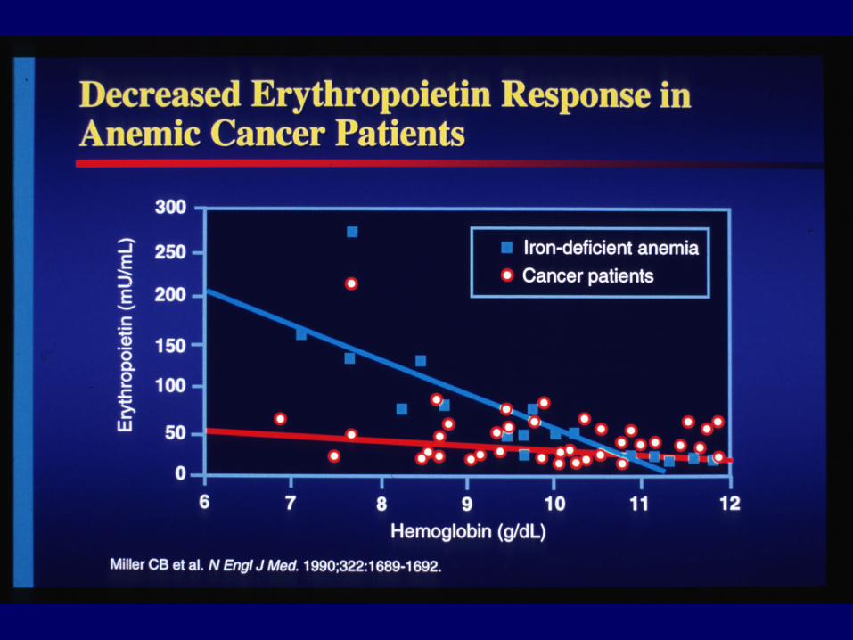
•Decreased erythropoietin production
•Inadequate marrow response to erythropoietin
Erythrocyte loss
•Hemorrhage
•Hemolysis

Anemia: Etiologies
• Production defects: – Nutritional deficiencies - Vitamin B12, folate or iron deficiency. – Inflammation/chronic disease.
– Primary marrow disorders- pure red cell aplasia,myelodysplasia. • Sequestration (hypersplenism)-usually associated with
mild pancytopenia. • Dilutional- A patient's plasma volume increases with
laying down and when they quit smoking. Possibly responsible for as much as a 3-6% drop in the hematocrit in the first two days of hospitalization.
• Blood loss. • Blood destruction.

Anemia: EtiologiesBlood Loss:
•Iatrogenic bleeding, such as from repeated venipuncture in patients undergoing a medical evaluation, blood losses associated with repeated hemodialysis procedures, or excessive blood donations •Factitious bleeding, secondary to surreptitious blood drawing by the patient.•Bleeding during or after surgical procedures may be difficult to quantitate, and is often underestimated.•Bleeding into the upper thigh and/or retroperitoneal areas can often be significant, but not clinically obvious.

Conditions Associated with Anemias Due to Reduced Erythrocyte Production
Anemias due to decreased erythropoietin production
• Renal disease
• Endocrine deficiency (pituitary, adrenal, thyroid, testis)
• Starvation
• Hemoglobinopathy (reduced oxygen affinity)
Anemias due to inadequate marrow response to erythropoietin
• Deficiency state (iron, vitamin B12, folate)
• Anemia of chronic disease (inflammation, infection, or malignancy)
• Sideroblastic anemia
• Myelodysplasia
• Pure red cell aplasia

First, measure the size of the RBCs:First, measure the size of the RBCs:• Use of volume-sensitive automated blood cell counters, such as the Coulter counter. The red cells pass through a small aperture and generate a signal directly proportional to their volume.• Other automated counters measure red blood cell volume by means of techniques that measure refracted, diffracted, or scattered light• By calculation from an independently-measured red blood cell count and hematocrit:
MCV (femtoliters) = 10 x HCT(percent) ÷ RBC (millions/µL)
Morphological Approach(big versus little)

An 18-year-old male diabetic college student who requires insulin presents to the student health service with a low-grade fever and cough of 10 days’ duration. Crackles are heard on auscultation of the lungs at the right base, but there are no signs of consolidation.Laboratory studies:
Hematocrit 41%
Hemoglobin 13.3 g/dL
Erythrocyte count 3.13million/m
Leukocyte count 11,500/mLDifferential count (automated)
Neutrophils and bands 68%
Lymphocytes 24%
Monocytes 7%
Eosinophils 1%
Basophils 0%
Mean corpuscular volume 131 fL131 fL
Mean corpuscular hemoglobin conc. 30.8 g/dL
Mean corpuscular hemoglobin 42.5 pg

Underproduction (1)(morphological approach)
MCV>115• B12, Folate• Drugs that impair
DNA synthesis (AZT, chemo., azathioprine)
• MDS
MCV 100 - 115• Ditto• endocrinopathy
(hypothyroidism)• Epo (skipped cell
divisions)• reticulocytosis

Underproduction (2)
Normocytic• Anemia of chronic
disease• Mixed deficiencies• Renal failure
Microcytic• Iron deficiency• Thal. trait• Anemia of chronic
disease (30-40%)• sideroblastic anemias

A 55 yo male artist is referred to you because his anemia has not responded to oral iron therapy. Two months ago, after repeated failure to increase the patient’s hematocrit, his physician administered a full replacement dose of parenteral iron dextran. Shortly thereafter the hematocrit level increased to normal level but has now decreased to pretreatment levels.
PE shows a pale man, liver and spleen are not enlarged, stool guaiac is negative. Repeated endoscopies fail to show a bleeding site.
Labs Hgb 6.8 g/dl, MCV 75 fl WBC 5500/ulPlts 490,000/ul, Retic ct 20,000/ul
Fe 25 ug/dl, TIBC 460 ug/dl, ferritin 15 ug/l


SELF (9 frozen pints of artists blood, frozen in sculpture)
Mark Quinn

Iron deficiency is a common form of malnutrition that affects more than 2 billion people globally.
- Project IDEA (Iron Deficiency Elimination Action)®, CDC

Prevalence (%) of iron deficiency and iron-deficiency anemia, United States, third National Health and Nutrition Examination Survey, 1988–199.
Sex and age (years) Iron deficiency Iron-deficiency anemia
Both sexes
1–2 9 3*
3–5 3 <1
6–11 2 <1
Nonpregnant females
12–15 9 2*
16–19 11* 3*
20–49 11 5*
50–69 5 2
³ 70 7* 2*
*Prevalence in non-blacks is 1 percentage point lower than prevalence in all races.

Iron Compartments in a 70 kg person
Compartment Fe content (mg) Total Body Fe (%)
Hemoglobin Fe 2000 67
Storage (ferritin, hemosiderin) 1000 27
Myoglobin Fe 130 3.5
Labile pool 80 2.2
Other tissue Fe 8 0.2
Transport Fe 3 0.08

Increased iron requirements
•Blood loss
•Gastrointestinal disorders (esophageal varices, hemorrhoids)
•Extensive and prolonged menstruation
•Pulmonary (hemoptysis, pulmonary hemosiderosis), urologic, or nasal disorders
•Chronic blood donations
•Dialysis
•Factitious removal
•Hookworm infestation
•Intravascular hemolysis with hemoglobinuria
•Paroxysmal nocturnal hemoglobinuria
•Cardiac valve prostheses
•Rapid growth in body size between 2 and 36 months of age
•Pregnancy and lactation

Inadequate iron supply
• Poor nutritional intake in children (not a common independent mechanism in adults but often a contributing factor)
• Malabsorption
• Gastric bypass surgery for ulcers or obesity
• Achlorhydria from gastritis or drug therapy
• Severe malabsorption (for example, celiac disease [nontropical sprue])
• Abnormal transferrin function
• Congenital atransferrinemia
• Autoantibodies to transferrin receptors

Oral iron failure?
• Incorrect diagnosis (eg, thalassemia)
• anemia of chronic disease?
• Patient is not taking the medication
• Not absorbed (enteric coated?)
• Rapid iron loss?

Intravenous Iron Therapy• Imferon (iron dextran BP) withdrawn from use. • InFed (iron dextran USP; Schein Pharm, Florham Park, NJ) A recent review reported 196 incidences of allergy/anaphylaxis from iron dextran between 1976 and 1996, of which 31 (15.8%) were fatal. 50 mg/mL of elemental iron in 2 mL vials.
• Ferric gluconate has been available in Europe for more than 20 years and was approved for intravenous use in the United States in 1999 (FerrlecitFerrlecit; Schein Pharm) in patients on renal dialysis. The number of allergic reactions (3.3 episodes per million doses) is lower than that from iron dextran (8.7 episodes per million doses).
Am J Kidney Dis 1999 Mar;33(3):464-70

0.5 mL test dose is given IV over at least 30 seconds, remainder given at a rate not exceeding 50 mg (one mL) per minute, and a total dose not exceeding 100 mg (two mL) per day
Intravenous Iron Therapy
60 kg woman with a hgb of 8:•Her total blood volume should be 3900 mL or 39 deciliters (65 mL/kg x 60 kg).•A normal hemoglobin concentration would be 14 g/dL. Thus, her hemoglobin deficit is 6 g/dL with a total deficit of 234 g (6 g/dL x 39 dL).•Each gram of hemoglobin contains 3.3 mg of iron. Thus, her total red cell iron deficit is 772 mg (234 g of hemoglobin x 3.3 mg Fe per gram).
For iron dextran:

When to use intravenous When to use intravenous
iron therapyiron therapy
Beneficial No benefit Investigational
Anemia of renal failure, withor without erythropoietin
therapy, Patients withongoing blood loss,
Jehovah's Witness patientswith iron deficiency, blood
loss or both
Autologous blood donationin patients with or without
iron deficiency
Blood loss, iron deficiency,and erythropoietin therapy,Anemia of chronic diseaseand erythropoietin therapy,Perisurgical anemia, with or
without erythropoietin
Absolute iron deficiency is defined as ferritin <200 µg/L with or without iron saturation <20%, or relative iron deficiency (ferritin <400 µg/L in dialysis patients
receiving erythropoietin therapy or the presence of >10% hypochromic erythrocytes, reticulocytes, or both.

• 55 yo F with moderately severe Rheumatoid Arthritis taking Prednisone 10 mg/day, Celecoxib, and monthly Etanercept is referred to you for an anemia workup
• CBC: Hct = 30%, MCV = 82, WBC = 5.4 thou/l, plt = 345 thou/ l– Smear - Normal– Retic count = 2 % (Corrected Retic = 30/40 x 2%=
1.5%)– Fe = 20 g/dL (55-155), TIBC = 200 g/dL (270-400),
Transferrin saturation = 20/200 = 10% (15-50)– Ferritin = 330 g/dL (20-160)

• A-- Marrow Failure
• B-- Iron Deficiency
• C-- Thalassemia
• D-- Inflammatory Block

Iron Deficiency Anemia vs. Inflammatory Block
– Smear:• hypochromic and microcytic (low MCV) RBCs, usually
not seen unless Hct 30%• platelet count is often elevated
Ferritin: a measure of total body iron stores, but also an acute phase reactant
• <15g/l = Fe deficiency, 150 g/l = Not Fe deficiency 15-150 g/l = ?

– Low Iron Saturation (Fe/TIBC ratio) Fe (not reliable) TIBC • Fe/TIBC (% saturation) 15%
– BM bx: absent Fe stores• Gold standard
– Therapeutic Trial of Oral Iron
Iron Deficiency Anemia vs. Inflammatory Block

Differentiation of anemia of chronic disease Differentiation of anemia of chronic disease and iron deficiency anemia by laboratory and iron deficiency anemia by laboratory measuresmeasures
Lab measure ACD Iron-def. anemia
Plasma Fe Reduced (normal) Reduced
Plasma transferrin Reduced (normal) Increased
Transferrin sat. Reduced (normal) Reduced
Plasma ferritin Increased (normal)* Reduced
Plasma TfR Normal Increased
TfR/log ferritin Low (<1) High (>4)


Papadaki HA, Kritikos HD, Valatas V, Boumpas DT, and Eliopoulos GD, Anemia of chronic disease in rheumatoid arthritis is associated with increased apoptosis of bone marrow erythroid cells: improvement following anti-tumor necrosis factor-alpha antibody therapy. Blood 2002

107 mg (triangle), 335 mg (•), and 1,102 mg (°)
Skikne BS, Flowers CH, Cook JD. Serum transferrin receptor: a quantitative measure of tissue iron deficiency. Blood. 1990;75:1870-1876

Andrews, NEJM, 1999
The transferrin cycle

Pettersson T, et al. Is serum transferrin receptor useful for detecting iron-deficiency in anemic patients with chronic inflammatory diseases? Br J Rheum. 1994; 33:740–4.
a. Low Fe – inflam.b. Nl Fe stores – inflam.c. Fe –def anemia

Diagnosis of Iron Deficiency Anemia in the Elderly by Transferrin Receptor–Ferritin Index (Arch Intern Med. 2002;162:445-449)

Barosi G. Inadequate erythropoietin response to anemia: definition and clinical relevance. Ann Hematol. 1994;68:215-223 (early review)
Utility of supraphysiologic doses of erythropoietinerythropoietin in the setting of inflammatory block.

Baer AN, et al. Blunted erythropoietin response to anemia in rheumatoid arthritis. Br J Haematol. 1987;66:559–64.
Rheumatoid arthritis Fe-deficiency

Epoetin therapy for anemia associated with cancer.
• Option to treat mild anemia and prevent severe anemia with improvement in QOL
• Epoetin 40,000u/wk = $360 to the pharmacy or about $600 to the patient.
• Darbepoetin 100mcg/wk = $330 to the pharmacy or about $550 to the patient.
• 6 cycles of chemotherapy = $12,500.
• 1/3 of all cancer patients receiving chemotherapy worldwide are being treated with epo.

Figure 1 from Seidenfeld et al., JNCI, Vol. 93:16, 2001.

Figure 2 from Seidenfeld et al., JNCI, Vol. 93:16, 2001.

Conclusions of meta-analysis
• Epoeitin reduces the odds of transfusion for cancer patients undergoing therapy.
• The number of patients needed to treat to prevent one transfusion is 4.4 (5.2 - 2.6)
• Strongest evidence for QOL improvement is in patients with <10 g/dL baseline hgb concentration (Littlewood et al., JCO;19:2865-74, 2001) but...

Relationship between changes in hemoglobin level and quality of life during chemotherapy in anemic cancer patients receiving epoetin alfa therapy.Crawford et al., Cancer, 2002.
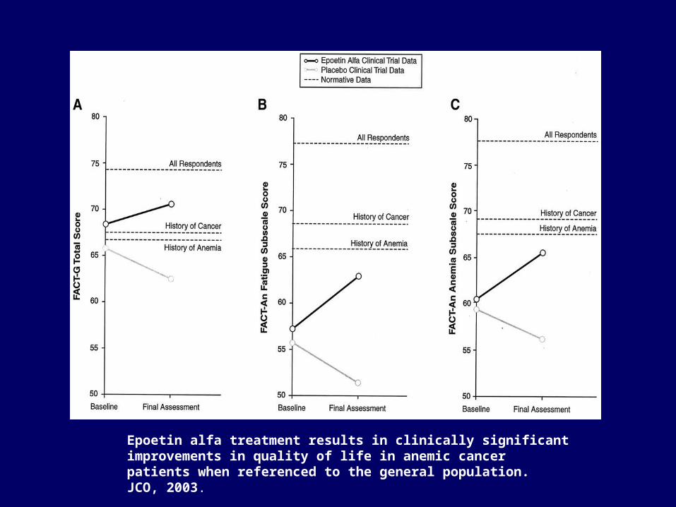
Epoetin alfa treatment results in clinically significant improvements in quality of life in anemic cancer patients when referenced to the general population.JCO, 2003.

Casadevall et al. NEJM. 346 (7): 469, Figure 1 February 14, 2002

Gershon et al. 346 (20): 1584, Figure 1 May 16, 2002
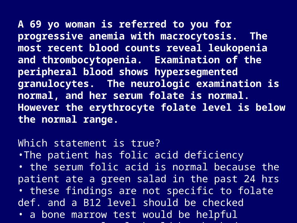
A 69 yo woman is referred to you for progressive anemia with macrocytosis. The most recent blood counts reveal leukopenia and thrombocytopenia. Examination of the peripheral blood shows hypersegmented granulocytes. The neurologic examination is normal, and her serum folate is normal. However the erythrocyte folate level is below the normal range.
Which statement is true?•The patient has folic acid deficiency• the serum folic acid is normal because the patient ate a green salad in the past 24 hrs• these findings are not specific to folate def. and a B12 level should be checked• a bone marrow test would be helpful• a serum epo level should be checked

Vitamin B12(cobalamin)
Methylmalonyl CoA Succinyl CoA
L-methylmalonylCoA mutase
Homocysteine-methioninemethyltransferase
homocysteine methionine
methyltetrahydrofolate

B12/Folate Deficiency• Etiology:
– Anemia-- Vitamin B12 and folate are needed for DNA synthesis deoxyuridate to thymidylate , including RBC precursors
– Deficiency• B12 - Dietary intake, acid-pepsin in the
stomach, pancreatic proteases, gastric secretion of intrinsic factor, an ileum with Cbl-IF receptors (fish tapeworm)
• Folate-- Poor dietary intake EtOH, malabsorption, increased demand (pregnancy, hemolytic anemias), inhibitors of DHFR

B12/Folate Deficiency (2)
• Dx:– Smear: Macrocytic (High MCV) RBCs, +/- hypersegmented
neutrophils, +/- modest neutropenia, but…– the diagnosis of B12 def. was made in patients in whom only
29 percent had anemia, and only 36 percent had a MCV greater than 100 fL (Pruthi RK, Tefferi A, Mayo Clin Proc 1994 Feb;69(2):144-50)
– B12• Low serum B12, elevated serum methylmalonic acid
levels• Anti-IF Abs, Schilling test (?), PA accounts for 75%
– Folate• Serum folate level-- can normalize with a single good
meal

B12/Folate Deficiency (3)
• Tx:– B12 deficiency: B12 1 mg/month IM, or 1-2
mg/day po
– Folate deficiency: Improved diet, folate 1 mg/day
– Monitor for a response to therapy.
– Pernicious Anemia – monitor for gi cancers.

Cobalamin deficiency and neurological problems
• Subacute combined degeneration of the dorsal and lateral spinal columns.
• Well known study of B12 deficiency in the nursing home population (Carmel R Karnaze DS, JAMA 253:1284, 1985)
• Vitamin B-12 deficiency is present in up to 15% of the elderly population as documented by elevated methylmalonic acid in combination with low or low-normal vitamin B-12 concentrations.
• Is oral B12 good enough?
• Association between nitrous oxide anesthesia and development of neurological symptoms responsive to B12 in patients with
subclinical cobalamin deficiency (methionine?).
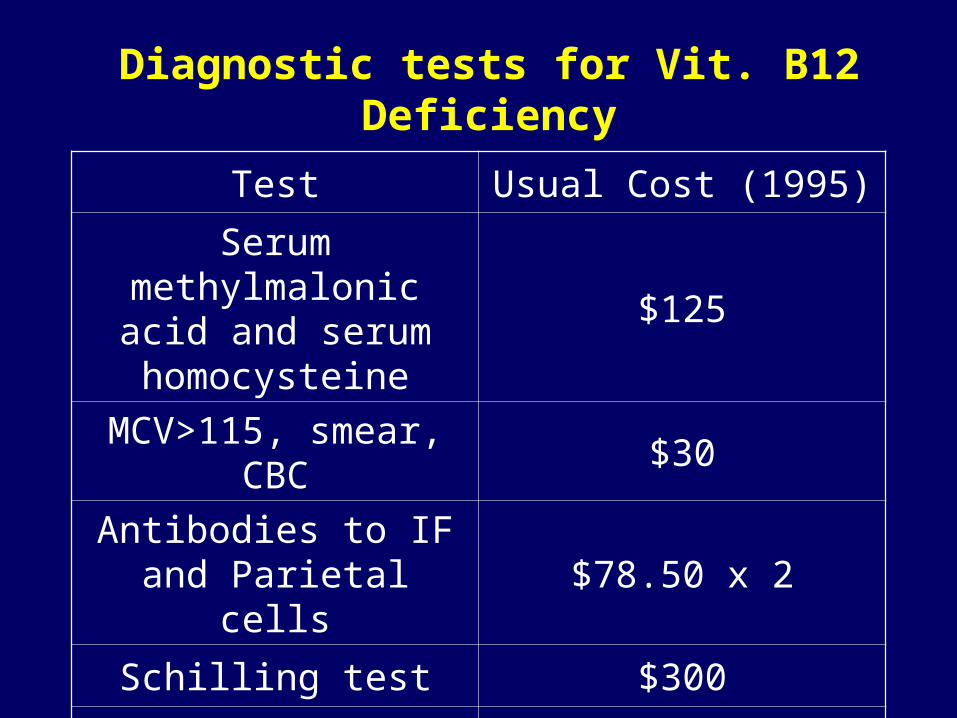
Diagnostic tests for Vit. B12 Deficiency
Test Usual Cost (1995)
Serum methylmalonic acid and serum homocysteine
$125
MCV>115, smear, CBC $30
Antibodies to IF and Parietal cells
$78.50 x 2
Schilling test $300
Spot urine for homocysteine
$18

Sideroblastic Anemias
• Heterogenous grouping of anemias defined by presence of ringed sideroblasts in the BM
• Etiologies:– Hereditary (rare), type of porphyria– Myelodysplasia– EtOH– Drugs (INH, Chloramphenicol)
• Tx:– Trial of pyridoxine for hereditary or INH induced
SA


During its approximately four month lifespan, the human red blood cell travels approximately 300 miles, making about 170,000 circuits through the heart, enduring cycles of osmotic swelling and shrinkage while traveling through the kidneys and lungs, and an equal number of deformations while passing through capillary beds.
•The normal time of RBC senescent death in adults is approximately 120 days. •Hemolysis is defined as a shortened survival of circulating red blood cells to a value of less than 100 days.•Anemia will develop in a normal person when they need to replace more than 5% of their red cell mass – corresponds to a red cell survival of 20 days.

A 32-year-old woman of Northern European descent has Crohn’s disease that has waxed and waned for 15 years. A recent flare beginning 2 weeks ago was treated with sulfasalazine and corticosteroids. Despite improvement in diarrhea and abdominal pain, she continues to feel ill and experiences easy fatigability with dyspnea and palpitations on mild exertion.
On physical examination, pallor, trace scleral icterus, and active bowel sounds are noted. Laboratory studies show: hematocrit, 22%; leukocyte count, 14,000/ul. (90% polymorphonuclear neutrophils with shift to the left); reticulocyte count, 7%; platelets noted to be “adequate on smear.”

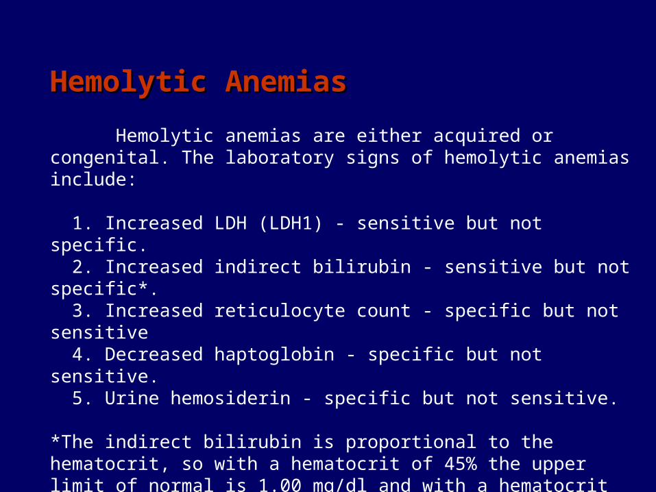
Hemolytic AnemiasHemolytic Anemias
Hemolytic anemias are either acquired or congenital. The laboratory signs of hemolytic anemias include:
1. Increased LDH (LDH1) - sensitive but not specific. 2. Increased indirect bilirubin - sensitive but not specific*. 3. Increased reticulocyte count - specific but not sensitive 4. Decreased haptoglobin - specific but not sensitive. 5. Urine hemosiderin - specific but not sensitive.
*The indirect bilirubin is proportional to the hematocrit, so with a hematocrit of 45% the upper limit of normal is 1.00 mg/dl and with a hematocrit of 22.5% the upper limit of normal for the indirect bilirubin is 0.5mg/dl. Since tests for hemolysis suffer from a lack of sensitivity and specificity, one needs a high index of suspicion for this type of anemia.

General Principles
• Anemia is a sign, not a disease.
• Anemias are a dynamic process.
• Its never normal to be anemic.
• The diagnosis of iron deficiency anemia mandates further work-up.

A primary care physician (PCP) has referred a 21-year-old married Filipino woman for evaluation of her recently documented anemia.
History of present illness: She was recently married and wants to have a family, but went to her PCP because she felt that she had less energy than her friends. She has no history of melena or bright red blood per rectum (BRBPR), and her menstrual history seemed normal. She thinks that her mother and 2 maternal aunts have anemia.
Physical examination: She is a pale but otherwise alert, healthy young woman. No scleral icterus is present and her chest and heart exam are normal. A soft spleen tip is palpable in the left upper quadrant (LUQ). No edema is present.
Labs: White blood cells (WBC) 4600, normal differential, platelets 421,000/ul, hematocrit (Hct) 27, hemoglobin (Hgb) 8.1gm/dl, red blood cells (RBC) 4.58M/ul, MCV 59, mean corpuscular hemoglobin (MCH) 17, mean corpuscular hemoglobin concentration (MCHC) 30. Retic 3.1% Absolute retics 142,000/ul, ferritin 482 ng/ml, serum iron 149, transferrin 193, % sat 77%.

The hemoglobin electrophoresis reveals HbA2 is 1%, HbF is 0.5%, and HbH is 16%.
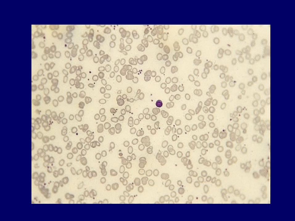

Thalassemias
• Genetic defect in hemoglobin synthesis synthesis of one of the 2 globin chains ( or )
– Imbalance of globin chain synthesis leads to depression of hemoglobin production and precipitation of excess globin (toxic)
– “Ineffective erythropoiesis”
– Ranges in severity from asymptomatic to incompatible with life (hydrops fetalis)
– Found in people of African, Asian, and Mediterranean heritage

• Dx:– Smear: microcytic/hypochromic, misshapen RBCs -thal will have an abnormal Hgb electrophoresis (HbA2,
HbF)– The more severe -thal syndromes can have HbH inclusions
in RBCs– Fe stores are usually elevated
• Tx:– Mild: None– Severe: RBC transfusions + Fe chelation, Stem cell
transplants
Thalassemias (2)