an automated drusen detection system for classifying age-related
TRANSCRIPT

AN AUTOMATED DRUSEN DETECTION SYSTEM FOR CLASSIFYING AGE-RELATEDMACULAR DEGENERATION WITH COLOR FUNDUS PHOTOGRAPHS
Yuanjie Zheng1, Brian Vanderbeek2, Ebenezer Daniel2, Dwight Stambolian2, Maureen Maguire2, David Brainard3, James Gee1
1Department of Radiology, 2Department of Ophthalmology, and 3Department of Psychology at the University of Pennsylvania, Philadelphia
ABSTRACTWe present a system of automated drusen detection from colorfundus photographs with our ultimate goal being to automati-cally assess the risk for the development of Age-related Mac-ular Degeneration (AMD). Our system incorporates learningbased drusen detection and includes fundus image analysistechniques for image denoising, illumination correction andcolor transfer. In contrast to previous work, we incorporateboth optimal color descriptors and robust multiscale local im-age descriptors in our drusen detection process. Our systemwas evaluated with color fundus photographs from two AMDclinical studies [1, 2]. By comparing our results to those ob-tained via manual drusen segmentation, we show that our sys-tem outperforms two state-of-the-art techniques.
1. INTRODUCTION
Age-related Macular Degeneration (AMD) is the most com-mon cause of blindness in developed world [1, 3]. The earlystage of AMD is an ocular condition associated with minimalvisual impairment and characterized by drusen and pigmen-tary abnormalities in the macula, and degeneration of the reti-nal pigment epithelium (RPE) [1]. The late stage of AMD ischaracterized by geographic atrophy (GA), RPE detachment,choroidal neovascularization (CNV) and disciform scar. Onlythe late stage of AMD results in moderate and severe lossesin visual function. However, the presence of drusen is a hall-mark of AMD, and magnification/multiplication of drusen in-dicates increased risk of eventual visual loss from AMD.
Grading drusen in color fundus photographs (CFPs) isan important component in the established classifications ofAMD [3, 1]. Traditional grading methods [3, 1] used to quan-titate drusen are based on the use of overlay standard circlesand subjective evaluation. The grader is asked to pick onefrom several standard circles to match each drusen in orderto decide its size, to mentally aggregate the total drusen area,and to convert the net result into a categorical number. Al-though important relationships have been demonstrated be-tween these manual grading values and AMD progression,these methods are labor-intensive, difficult to reproduce, andmay lose important information.
Automated drusen detection with computerized algo-rithms [4, 5, 6] is potentially capable of making grading of
drusen more rigorous, reproducible, quantitative and cost-effective. However, digital techniques have not as of yetgained widespread acceptance for several reasons (as shownin Fig. 1). First, it can be challenging to reliably localizedrusen against the varying background of the pigments of themacula, RPE and choroid, and to differentiate drusen fromareas of RPE hypopigmentation, exudates and scars. Second,the inherent nature of the reflectance of even a normal maculais nonuniform, and this presents an obstacle for automaticdrusen segmentation. Third, color variations between sub-jects in CFPs, caused by large natural variations in choroidalpigmentation and iris color, can mask the more subtle varia-tion between drusen and other lesions or background.
In this paper, we present an automated color-fundusdrusen detection system with our ultimate goal being toautomatically assess the risk for development of AMD. Oursystem combines a set of computerized algorithms for pat-tern recognition, computer vision and machine learning. Itis robust and accurate (as shown in Fig. 1) in the face ofchallenges described above, as validated with CFPs from twomilestone AMD clinical studies [1, 2].
2. SYSTEM OF AUTOMATED DRUSEN DETECTION
2.1. System Overview
As illustrated in Fig. 2, the integrated image analytics ofour system consist of several CFP preprocessing procedures.These include image denoising (with the Non-Local Mean-s Filter in [7]), retina mask generation (simply by imagethresholding and hole filling with certain morphological op-erations), illumination correction to correct the slow back-ground variation with our technique described in [8], andcolor transfer to render all testing images similar in color.Our color transfer is accomplished in a group-wise way byfirst creating an average intensity histogram over all testingCFPs, and then transforming the intensity of each CFP sothat the histogram of the output approximately matches thisaverage histogram. These two procedures are repeated un-til convergence. From the preprocessed CFPs, drusen arethen detected using a learning based scheme, which will bedetailed in the following section.
2013 IEEE 10th International Symposium on Biomedical Imaging:From Nano to MacroSan Francisco, CA, USA, April 7-11, 2013
978-1-4673-6454-6/13/$31.00 ©2013 IEEE 1440

Fig. 1. Drusen areas detected by our system on (left) one CAPT CFP [1] obtained by scanning color slide film and (right) oneAmish CFP [2] taken with a digital fundus camera. In each picture, the three color circles compose the grading grid used in[1]. Left: the “green”, “black” and “pink” regions indicate small detected drusen (<125µm) while the “yellow”, “cyan”, and“blue” regions indicate the large detected drusen (≥ 125µm). Right: all detected drusen are marked with the “blue” color.Automated drusen detection is challenging due to macular pigment variation, nonuniform fundus reflectance and inter-subjectcolor differences.
Fig. 2. Flowchart of our automated drusen detection system.
2.2. Learning Based Drusen Detection
Our drusen detection scheme relies on machine learningtechniques and consists of two sequential procedures of apixel-wise classification and a region-wise classification (asshown in Fig. 2). The pixel-wise classification independentlydetermines whether each pixel is a drusen or not using imagefeatures obtained from a series of robust local appearance de-scriptors. It is accomplished with a powerful Ada-LS-SVMclassifier, i.e. exploiting AdaBoost [9] for feature selectionfollowed by LS-SVM [10] for classification. The region-wiseclassification, introduced in order to remove false-positivesfrom the pixel-wise classification, applies a set of regionbased features to estimate whether spatially connected com-ponents of output of the pixel-wise classification are a drusenor not. Region-wise classification is carried out with theLS-SVM directly.
2.2.1. Features in Pixel-wise Classification
Image features used by the pixel-wise classification in oursystem are obtained from a set of optimal image color de-scriptors and a series of multiscale image local descriptors.The color descriptors characterize the local photometric prop-erties of image and include the hue histogram and color mo-ment invariants in [11]. They are computed within a localimage patch in an optimal extent specified by an optimal im-age scale, which in turn is obtained with the method in ourprevious work [12].
The multiscale image local descriptors are defined to char-acterize the local geometric structure of the image and are mo-tivated primarily by two observations. First, drusen in CFP at
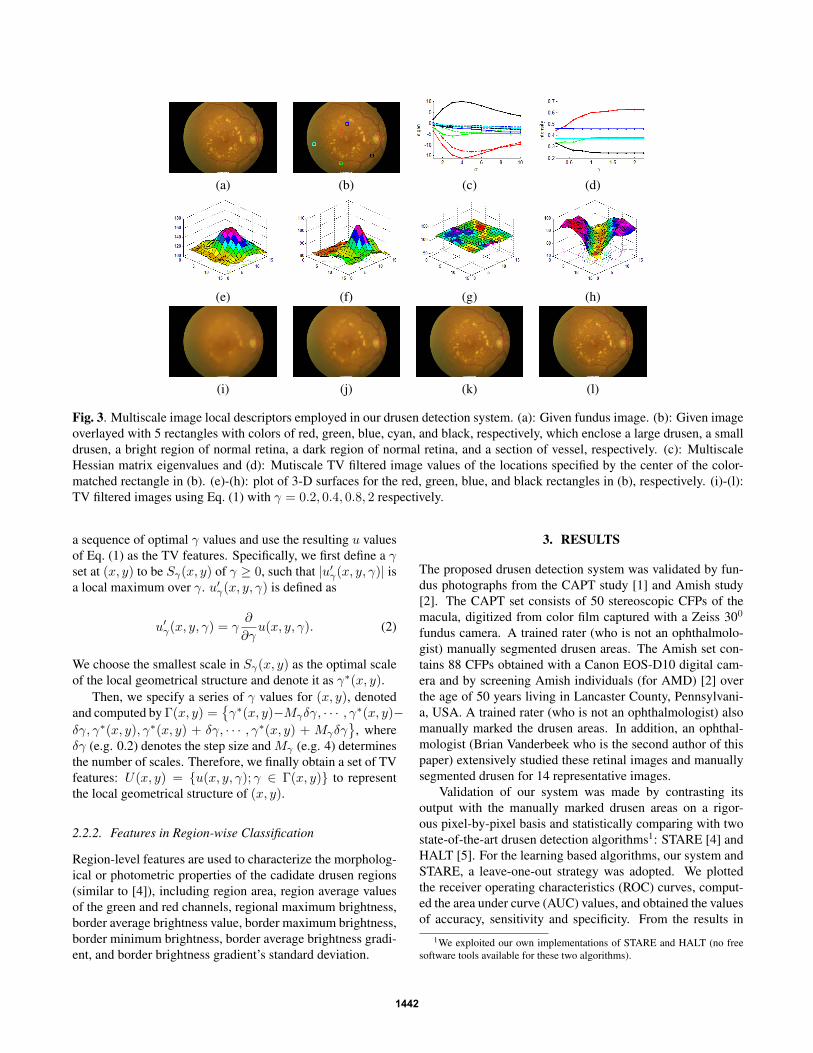
a specific scale is geometrically different from many of otherretinal components, as shown in Fig. 3 (e)-(h). Second, fea-tures characterizing these geometrical attributes demonstratevery distinct variation patterns across different image scales(as shown in Fig. 3 (c)(d)) and these patterns carry abundantinformation useful for drusen detection.
We employ two types of features to characterize thesemultiscale image properties: the Hessian features previ-ously proposed by us in [12] and a series of novel TV(total variation) features. Hessian features [12] at an arbi-trary pixel located at (x, y) can be denoted by Λ(x, y) =λ1(x, y, σ), λ2(x, y, σ);σ ∈ Σ(x, y) where λ1 and λ2 rep-resents two eigenvalues of the image Hessian matrix at scaleσ, and where Σ(x, y) is a sequence of optimal image scalesat (x, y) (as detailed in [12]).
Our TV features are obtained by solving the following TVmodel [13] based minimization with a sequence of differentvalues of the adjusting parameter γ
minu∈BV (Ω)
|u|BV (Ω) + γ|I − u|L1(Ω) (1)
where I denotes the green channel of the given CFP, Ω ⊆ R2,and where the definitions of the BV (bounded variation) spaceand L1 norm space are provided in [13]. Eq. (1) generates asmoothed version u of the given image in an anisotropically-filtering way which can remove image oscillations while p-reserving image edges. As pointed out in [13], γ in Eq. (1)indicates the scale of image analysis. The resulting value u atan arbitrary pixel is determined by the similarity of I betweenits neighborhoods in an extent specified by γ.
Similar to the Heissian features used in [12], we specify
1441
(a) (b) (c) (d)
(e) (f) (g) (h)
(i) (j) (k) (l)
Fig. 3. Multiscale image local descriptors employed in our drusen detection system. (a): Given fundus image. (b): Given imageoverlayed with 5 rectangles with colors of red, green, blue, cyan, and black, respectively, which enclose a large drusen, a smalldrusen, a bright region of normal retina, a dark region of normal retina, and a section of vessel, respectively. (c): MultiscaleHessian matrix eigenvalues and (d): Mutiscale TV filtered image values of the locations specified by the center of the color-matched rectangle in (b). (e)-(h): plot of 3-D surfaces for the red, green, blue, and black rectangles in (b), respectively. (i)-(l):TV filtered images using Eq. (1) with γ = 0.2, 0.4, 0.8, 2 respectively.
a sequence of optimal γ values and use the resulting u valuesof Eq. (1) as the TV features. Specifically, we first define a γset at (x, y) to be Sγ(x, y) of γ ≥ 0, such that |u′γ(x, y, γ)| isa local maximum over γ. u′γ(x, y, γ) is defined as
u′γ(x, y, γ) = γ∂
∂γu(x, y, γ). (2)
We choose the smallest scale in Sγ(x, y) as the optimal scaleof the local geometrical structure and denote it as γ∗(x, y).
Then, we specify a series of γ values for (x, y), denotedand computed by Γ(x, y) =
γ∗(x, y)−Mγδγ, · · · , γ∗(x, y)−
δγ, γ∗(x, y), γ∗(x, y) + δγ, · · · , γ∗(x, y) + Mγδγ
, whereδγ (e.g. 0.2) denotes the step size and Mγ (e.g. 4) determinesthe number of scales. Therefore, we finally obtain a set of TVfeatures: U(x, y) = u(x, y, γ); γ ∈ Γ(x, y) to representthe local geometrical structure of (x, y).
2.2.2. Features in Region-wise Classification
Region-level features are used to characterize the morpholog-ical or photometric properties of the cadidate drusen regions(similar to [4]), including region area, region average valuesof the green and red channels, regional maximum brightness,border average brightness value, border maximum brightness,border minimum brightness, border average brightness gradi-ent, and border brightness gradient’s standard deviation.
3. RESULTS
The proposed drusen detection system was validated by fun-dus photographs from the CAPT study [1] and Amish study[2]. The CAPT set consists of 50 stereoscopic CFPs of themacula, digitized from color film captured with a Zeiss 300
fundus camera. A trained rater (who is not an ophthalmolo-gist) manually segmented drusen areas. The Amish set con-tains 88 CFPs obtained with a Canon EOS-D10 digital cam-era and by screening Amish individuals (for AMD) [2] overthe age of 50 years living in Lancaster County, Pennsylvani-a, USA. A trained rater (who is not an ophthalmologist) alsomanually marked the drusen areas. In addition, an ophthal-mologist (Brian Vanderbeek who is the second author of thispaper) extensively studied these retinal images and manuallysegmented drusen for 14 representative images.
Validation of our system was made by contrasting itsoutput with the manually marked drusen areas on a rigor-ous pixel-by-pixel basis and statistically comparing with twostate-of-the-art drusen detection algorithms1: STARE [4] andHALT [5]. For the learning based algorithms, our system andSTARE, a leave-one-out strategy was adopted. We plottedthe receiver operating characteristics (ROC) curves, comput-ed the area under curve (AUC) values, and obtained the valuesof accuracy, sensitivity and specificity. From the results in
1We exploited our own implementations of STARE and HALT (no freesoftware tools available for these two algorithms).
1442

Data set (operator) CAPT(Rater) Amish(Ophth) Amish(Rater)Measures Accu Sens Spec Accu Sens Spec Accu Sens Spec
Our system 0.80 0.82 0.75 0.86 0.87 0.78 0.83 0.85 0.71STARE [4] 0.74 0.75 0.63 0.80 0.82 0.67 0.77 0.80 0.60HALT [5] 0.77 0.80 0.68 0.79 0.82 0.65 0.78 0.81 0.64
Table 1. Statistical results (Accu: accuracy; Sens: sensitivity; Spec: specificity) of our system, STARE [4] and HALT [5]in drusen detection on CAPT CFPs with drusen delineated by a trained rater - denoted by “CAPT(Rater)”, Amish CFPs withdrusen delineated by an ophthalmologist - denoted by “Amish(Ophth)”, and Amish CFPs with drusen delineated by a trainedrater - denoted by “Amish(Rater)”.
Fig. 4. ROC curves of our system, STARE [4] and HALT [5]in drusen detection on CAPT CFPs with drusen delineatedby a trained rater - denoted by “CAPT(Rater)”, Amish CFPswith drusen delineated by an ophthalmologist - denoted by“Amish(Ophth)”, and Amish CFPs with drusen delineated bya trained rater - denoted by “Amish(Rater)”.
Fig. 4 and in Table 1, we found our system outperforms state-of-the-art techniques. In Fig. 1, we demonstrate the drusenareas found by by our system for one CAPT CFP and oneAmish CFP. We also found that classification improvementwith the pixel-wise features selected by AdaBoost comparedwith the unselected features is significant (p < 0.005).
4. CONCLUSION AND FUTURE WORK
We present an automated drusen detection system with ourultimate goal of classifying age-related macular degeneration(AMD) from color fundus photographs (CFPs). It consist-s of several image preprocessing procedures and a learningbased drusen segmentation scheme which integrates both op-timal image color descriptors and a sequence of robust multi-scale local image descriptors. As validated using our manualdrusen delineations, our system outperforms two state-of-the-art algorithms. It can be a clinically invaluable tool for thestandardization of drusen evaluation in AMD classification.
Future work includes studies on the correlation between
our system and the manual grading scales for drusen relatedAMD characteristics.
5. REFERENCES
[1] CAPT study group, “The complications of age-related macu-lar degeneration prevention trial (CAPT): rationale, design andmethodology,” Clinical Trials, vol. 1, pp. 91–107, 2004.
[2] Dwight Stambolian and et al., “Genome-wide scan for myopiain the old order amish,” American Journal of Ophthalmology,vol. 140, no. 3, pp. 469–476, 2005.
[3] R. Klein, M. D. Davis, Y. L. Magli, P. Segal, B. E. Klein, andL. Hubbard, “The wisconsin age-related maculopathy gradingsystem,” Ophthalmology, vol. 98, no. 7, pp. 1128–1134, 1991.
[4] Lee Brandon, Automated Drusen Detection in A Retinal ImageUsing Multi-Level Analysis, Ph.D. thesis, Clemson University,2003.
[5] K. Rapantzikos, M. Zervakis, and K. Balas, “Detection andsegmentation of drusen deposits on human retina: potential inthe diagnosis of age-related macular degeneration,” MedicalImage Analysis, vol. 7, pp. 95–108, 2003.
[6] Shaoting Zhang and et al., “Towards robust and effective shapemodeling: Sparse shape composition,” Medical Image Analy-sis, vol. 16, pp. 265–277, 2012.
[7] Antoni Buades and et al., “A non-local algorithm for imagedenoising,” in IEEE CVPR, 2005, vol. 2, pp. 60–65.
[8] Yuanjie Zheng and et al., “Retrospective illumination correc-tion of retinal fundus images from gradient distribution sparsi-ty,” in ISBI, Barcelona, Spain, May 2012.
[9] Y. Freund and et al., “A decision-theoretic generalization ofon-line learning and an application to boosting,” Journal ofComputer and System Sciences, vol. 55, pp. 119–139, 1997.
[10] Johan A. K. Suykens and et al., Least squares support vectormachines, World Scientific, Singapore, 2002.
[11] Koen E.A. van de Sande and et al., “Evaluating color descrip-tors for object and scene recognition,” IEEE TPAMI, vol. 32,no. 9, pp. 1582–1596, 2010.
[12] Yuanjie Zheng and et al., “Multiscale analysis revisited: De-tection of drusen and vessel in digital retinal images,” in ISBI,Chicago, Illinois, USA, March 30 - April 2 2011.
[13] Tony F. Chan and Selim Esedoglu, “Aspects of total variationregularized l 1 function approximation,” SIAM J. Appl. Math,vol. 65, no. 5, 2005.
1443