an essential role of antibodies in the control of chikungunya virus
TRANSCRIPT

of November 21, 2018.This information is current as
Control of Chikungunya Virus InfectionAn Essential Role of Antibodies in the
Yiu-Wing Kam, Laurent Rénia and Lisa F. P. NgFok-Moon Lum, Teck-Hui Teo, Wendy W. L. Lee,
http://www.jimmunol.org/content/190/12/6295doi: 10.4049/jimmunol.1300304May 2013;
2013; 190:6295-6302; Prepublished online 13J Immunol
MaterialSupplementary
4.DC1http://www.jimmunol.org/content/suppl/2013/05/13/jimmunol.130030
Referenceshttp://www.jimmunol.org/content/190/12/6295.full#ref-list-1
, 13 of which you can access for free at: cites 44 articlesThis article
average*
4 weeks from acceptance to publicationFast Publication! •
Every submission reviewed by practicing scientistsNo Triage! •
from submission to initial decisionRapid Reviews! 30 days* •
Submit online. ?The JIWhy
Subscriptionhttp://jimmunol.org/subscription
is online at: The Journal of ImmunologyInformation about subscribing to
Permissionshttp://www.aai.org/About/Publications/JI/copyright.htmlSubmit copyright permission requests at:
Author Choice Author Choice option
The Journal of ImmunologyFreely available online through
Email Alertshttp://jimmunol.org/alertsReceive free email-alerts when new articles cite this article. Sign up at:
Print ISSN: 0022-1767 Online ISSN: 1550-6606. Immunologists, Inc. All rights reserved.Copyright © 2013 by The American Association of1451 Rockville Pike, Suite 650, Rockville, MD 20852The American Association of Immunologists, Inc.,
is published twice each month byThe Journal of Immunology
by guest on Novem
ber 21, 2018http://w
ww
.jimm
unol.org/D
ownloaded from
by guest on N
ovember 21, 2018
http://ww
w.jim
munol.org/
Dow
nloaded from
The Journal of Immunology
An Essential Role of Antibodies in the Control ofChikungunya Virus Infection
Fok-Moon Lum,*,†,1 Teck-Hui Teo,*,‡,1 Wendy W. L. Lee,*,‡ Yiu-Wing Kam,*
Laurent Renia,*,2 and Lisa F. P. Ng*,†,2
In recent years, Chikungunya virus (CHIKV) was responsible for epidemic outbreaks in intertropical regions. Although acquired
immunity has been shown to be crucial during CHIKV infection in both humans and mice, their exact role in the control of CHIKV
infection remains unclear. In this study, wild-type (WT), CD42/2, and B cell (mMT) knockout mice were infected with CHIKV. Sera
were taken at different days postinfection and measured for anti-CHIKVAb levels. Isotype and neutralizing capacity of these Abs
were assessed in vitro, and specific linear epitopes were mapped. Viremia in CHIKV-infected mMT mice persisted for more than
a year, indicating a direct role for B cells in mediating CHIKV clearance. These animals exhibited a more severe disease than WT
mice during the acute phase. Characterization of CHIKV-specific Abs revealed that anti-CHIKV Abs were elicited early and
targeted epitopes mainly at the C terminus of the virus E2 glycoprotein. Furthermore, CD42/2 mice could still control CHIKV
infection despite having lower anti-CHIKVAb levels with reduced neutralizing capacity. Lastly, pre-existing natural Abs in the
sera of normal WT mice recognized CHIKV and were able to partially inhibit CHIKV. Taken together, natural and CHIKV
infection–induced specific Abs are essential for controlling CHIKV infections. The Journal of Immunology, 2013, 190: 6295–6302.
Chikungunya virus (CHIKV) is an arthropod-borne alpha-virus transmitted by Aedes mosquitoes, namely, A. aegyptiand A. albopictus (1). Infected patients experience develop-
ment of Chikungunya fever, characterized mainly by polyarthralgia(2), febrile illness, maculopapular rashes, myalgia, headache, edemaof the extremities, and gastrointestinal complaints (3, 4). Patientsmay also experience development of neurologic complicationsand in some extreme cases, death has been reported (5, 6). Cur-rently, CHIKV is endemic in Africa, India, and many parts of Asia(7), with occasional sporadic outbreaks (8, 9). The lack of herdimmunity in countries surrounding these endemic areas presents
imminent risks for the spread of large-scale outbreaks. Hence,CHIKV remains a public threat that should not be ignored.Anti-CHIKV immunity is poorly understood, and most studies
have focused on innate immunity, particularly in elucidating theroles of type I IFN and their related antiviral pathways (10–18).Although these studies have demonstrated the importance of type IIFNs in restricting virus replication during the acute phase of in-fection, their effects are insufficient for the complete eliminationof virus in infected hosts. Moreover, CHIKV has been reported topersist in the tissues and organs in animal model studies even afterviremia has subsided, and when levels of IFN-a/b have returnedto normal (10, 14, 19). Nevertheless, CHIKV in the organs wasshown to be eliminated progressively over time (20). Furthermore,persistently high levels of viremia with no signs of joint inflam-mation were observed in infected RAG22/2 mice that have no Tand B cells (20). These observations strongly suggest that adaptiveimmunity has a crucial role in controlling and eliminating CHIKVafter the initial IFN-a/b and other innate immune responses havesubsided. It was further revealed that CD4+, but not CD8+, T cellsplay a major role in mediating the severity of joint inflammation,although both subsets have no role in the control of CHIKVreplication and dissemination (20). In addition, passive transferof anti-CHIKVAbs was able to clear CHIKV infection in mice(21, 22). These observations indicated that Abs could be the maineffectors in anti-CHIKV immunity.In this article, CHIKV infections in B cell–deficient (mMT) mice
(23) were investigated to clearly define the role of B cells in anti-CHIKV immunity. We characterized the Ab response elicited dur-ing the infection and demonstrated that the breadth of protectiveAb response is dependent on CD4+ T cells. Remarkably, we alsouncovered a role for natural Abs that are present in the sera ofuninfected mice to control early CHIKV infection.
Materials and MethodsMice
Six-week-old female wild-type (WT), mMT, and CD42/2 C57BL/6J micewere used. All mice were bred and kept under specific pathogen-free
*Singapore Immunology Network, Agency for Science, Technology and Research,Biopolis, Singapore 138648; †Department of Biochemistry, Yong Loo Lin School ofMedicine, National University of Singapore, Singapore 117597; and ‡National Uni-versity of Singapore Graduate School for Integrative Sciences and Engineering,National University of Singapore, Singapore 117456
1F.-M.L. and T.-H.T. contributed equally to this work.
2L.R. and L.F.P.N. directed this work equally.
Received for publication January 31, 2013. Accepted for publication April 9, 2013.
This work was supported by the Singapore Immunology Network, Agency for Sci-ence, Technology and Research. F.-M.L. is supported by a Yong Loo Lin School ofMedicine, National University of Singapore postgraduate scholarship. T.-H.T. is sup-ported by an Agency for Science, Technology and Research postgraduate scholarship.W.W.L.L. is supported by a National University of Singapore Graduate School forIntegrative Sciences and Engineering postgraduate scholarship.
The funders had no role in study design, data collection and analysis, decision topublish, or preparation of the manuscript.
Address correspondence and reprint requests to Prof. Lisa F.P. Ng or Prof. Laurent Renia,Singapore Immunology Network, A*STAR, Immunos, #04-06, 8A Biomedical Grove,Biopolis, 138648 Singapore (L.F.P.N.) or Singapore Immunology Network, A*STAR,Immunos, #03-15, 8A Biomedical Grove, Biopolis, 138648 Singapore (L.R.). E-mailaddresses: [email protected] (L.F.P.N.) or [email protected] (L.R.)
The online version of this article contains supplemental material.
Abbreviations used in this article: CHIKV, Chikungunya virus; dpi, days postinfec-tion; PBST, PBS containing 0.05% Tween 20; PDB, Protein Data Bank; WT, wild-type.
This article is distributed under The American Association of Immunologists, Inc.,Reuse Terms and Conditions for Author Choice articles.
Copyright� 2013 by The American Association of Immunologists, Inc. 0022-1767/13/$16.00
www.jimmunol.org/cgi/doi/10.4049/jimmunol.1300304
by guest on Novem
ber 21, 2018http://w
ww
.jimm
unol.org/D
ownloaded from
conditions in the Biological Resource Centre, Agency for Science, Tech-nology and Research, Singapore. In all experiments, age- and sex-matchedWT and deficient mice were used. All experiments and procedures wereapproved by the Institutional Animal Care and Use Committee (IACUC:120714) of the Agency for Science, Technology and Research, Singapore,in accordance with the guidelines of the Agri-Food and Veterinary Au-thority and the National Advisory Committee for Laboratory AnimalResearch of Singapore.
Virus
CHIKV SGP11 isolate, previously described (13), was used for all ex-periments. Viruses were further propagated in C6/36 cells and purified byultracentrifugation (24) before in vivo infections. Virus titer was deter-mined by standard plaque assays using Vero-E6 cells (13).
Virus infection and evaluation of disease
Mice were infected as previously described (18, 20, 24). Typically, 106
PFUs CHIKV SGP11 particles (in 50 ml PBS) were inoculated in the s.c.region of the ventral side at the right hind footpad toward the ankle.Viremia (assessed by viral RNA quantification) was monitored dailystarting from 24 h postinfection until 8 d postinfection (dpi), and subse-quently at every alternate day until 38 dpi. Viremia was assessed again at79 and 402 dpi. Joint swelling at the footpad was scored daily from 0 to 19dpi (18, 20, 24).
Viral RNA extraction and quantification
Ten microliters blood was collected from the tail vein and diluted in 120 mlPBS containing 10 ml citrate-phosphate-dextrose solution (Sigma-Aldrich).Viral RNA was extracted with the QIAamp Viral RNA Kit (Qiagen) fol-lowing manufacturer’s instructions. Viral RNA copies were quantified byquantitative RT-PCR (18). Viral RNA copies were estimated from a stan-dard curve generated using serial dilutions of CHIKV negative-sense nsP1RNA transcripts as reported previously (22).
Ab quantification and isotyping
Ab titers were assessed by a virion-based ELISA (24–26). CHIKV-coated(106 virions/well in 50 ml PBS) polystyrene 96-well MaxiSorp plates(Nunc) were blocked with PBS containing 0.05% Tween 20 (PBST) and5% w/v nonfat milk for 1.5 h at 37˚C. Sera from normal or infected groupswere heat inactivated and serially diluted in Ab diluent (0.05% PBST +2.5% w/v nonfat milk). One hundred microliters of diluted sera was addedinto each well and incubated for 1 h at 37˚C. HRP-conjugated goat anti-mouse IgG, IgG1, IgG2b, IgG2c, IgG3, and IgM Abs were used. IgG2cwas tested instead of IgG2a because only the IgG2c gene is present inC57BL/6 mice (27). Total IgG and IgM quantification assays were per-formed using sera from respective animals diluted at 1:2000 and 1:100,respectively. Pooled sera were used for IgG1, IgG2b, IgG2c, and IgG3isotyping. All HRP-conjugated Abs were from Santa Cruz, except for IgG3(Southern Biotech). ELISA assays were developed by TMB substrate
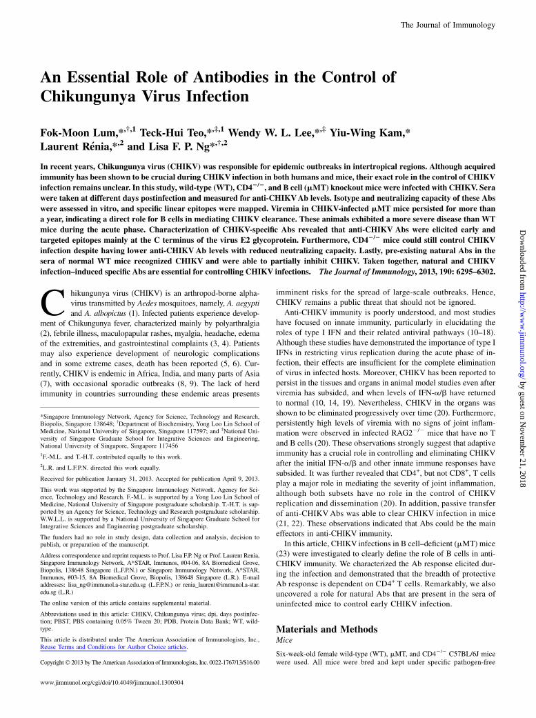
FIGURE 1. B cells mediate virus clearance and
disease pathology in CHIKV infection. (A) Viremia
and (B) joint swelling observed in both mMT (n =
6) and WT mice (n = 5). (C) Total CHIKV-specific
IgG and IgM in pooled sera of WT infected mice (n =
3–5) at 2, 4, 6, and 9 dpi in replicates of four. (D)
Total CHIKV-specific IgG and IgM Abs were mea-
sured in WT mice (n = 5) at 3, 15, and 26 dpi. (E)
Total CHIKV-specific IgG and IgM in pooled sera of
naive (n = 5) and WT infected mice (n = 5) at 402
dpi. This assay was done in quadruplicates. All IgM
and IgG titers were determined at 1:100 and 1:2000
dilutions, respectively. (F) Pooled sera from CHIKV-
infected WT mice (n = 5) were neutralizing against
CHIKV infection. Percentage infectivity was nor-
malized against virus-infected samples. All data are
presented as mean 6 SD. *p , 0.05, **p , 0.01,
***p , 0.005 by Mann–Whitney U, one-tailed test.
6296 Abs IN CHIKUNGUNYAVIRUS INFECTION
by guest on Novem
ber 21, 2018http://w
ww
.jimm
unol.org/D
ownloaded from
(Sigma-Aldrich) and terminated by Stop reagent (Sigma-Aldrich). Absor-bance was measured at 450 nm. CHIKV-specific Ab isotypes are expressedas Ab titer that is defined as the lowest dilution required for a detectablesignal above control naive pooled sera.
Detection of CHIKV-specific natural Abs
CHIKV-specific naturally occurring Abs from naive mice sera were as-sessed by Western blot, immunofluorescence (Supplemental Fig. 1), andvirion-based ELISA assays (24–26). CHIKV-coated (106 virions/well in 50ml PBS) polystyrene 96-well MaxiSorp plates (Nunc) were blocked withPBST and 5% w/v nonfat milk for 1.5 h at 37˚C. Sera from WT or mMTmice were heat-inactivated and serially diluted in Ab diluent (0.05%PBST + 2.5% w/v nonfat milk). One hundred microliters of diluted serawas added into each well and incubated for 1 h at 37˚C. HRP-conjugatedgoat anti-mouse IgG (Santa Cruz) and IgM (Santa Cruz) were used. ELISAassays were developed by TMB substrate (Sigma-Aldrich) and terminatedby Stop reagent (Sigma-Aldrich). Absorbance was measured at 450 nm,and Ab titers were determined by detectable signals above the meanbackground signal + 3 SD.
Sero-neutralization assay
Neutralizing activity of Abs was tested in a fluorescence-based cell infectionassay in HEK293T cells as described previously (24–26). In brief, CHIKVwas incubated with diluted heat-inactivated mouse sera at a multiplicity ofinfection of 10 for 2 h at 37˚C with gentle agitation (350 rpm). Virus–Abmixtures were then added to HEK293T cells seeded in a 96-well plate (104
cells/well) and incubated for another 1.5 h at 37˚C. Virus–Ab overlayswere subsequently removed and fresh DMEM medium supplemented with5% FBS was replenished. Cells were incubated for 6 h at 37˚C beforefixation with 4% paraformaldehyde followed by immunofluorescence stain-ing and quantification using the Cellomics ArrayScan V (Thermo FisherScientific, Waltham, MA). Percentage of infectivity was calculated accordingto this equation: % Infectivity = 1003 (% responder from sero-neutralizationgroup/% responder from virus infection group). All data presented arerepresentative of two independent experiments with similar results.
Epitope determination and structural localization
Peptide-based ELISA was performed using heat-inactivated pooled serafrom infected mice to screen for B cell epitopes using overlapping synthetic18-mer biotinylated-peptides (Mimotopes) (24, 25). Streptavidin-coated96-well plates (Nunc) were blocked with 1% w/v sodium caseinate (Sigma-Aldrich) in 0.1% PBST for 1 h at room temperature. Peptides were dis-solved in DMSO (15 mg/ml), diluted 1:1000 in 0.1% PBST, and coatedonto the plates (100 ml/well). Heat-inactivated pooled sera were diluted1:500 in 0.1% PBST and 100 ml diluted sera were added into each well andincubated for 1 h at 37˚C. HRP-conjugated goat anti-mouse IgG Abs(Santa Cruz) diluted 1:10,000 in Ab diluent were used to detect the boundAbs. Reactions were developed and absorbance was measured accordingly.Pools of five peptides were first used for initial screening. Positive peptidepools were determined as absorbance values greater than mean 6 6 SDvalues. Peptides from positive pools were then screened individually, andthose with absorbance values greater than mean 6 6 SD of the noninfectedcontrols were plotted. Results were expressed as percentage of Ab rec-ognition within the CHIKV structural proteome.
Computational modeling
Structural data of the E1, E2, and E3 glycoproteins were retrieved fromProtein Data Bank (PDB) (ID: 3N42) and visualized using the UCSFCHIMERA software (28). Structures of Capsid sequences were predictedusing individual I-TASSER queries (29) and visualized using UCSFCHIMERA software.
Statistical analysis
Data are presented as mean 6 SD. Differences between groups and con-trols were analyzed using Mann–Whitney U test. Statistics were performedwith GraphPad Prism 5.04.
ResultsB cells and Abs are essential for the control of CHIKV infection
CHIKV was inoculated at the joint footpad of WT and mMT miceto test for the role of B cells and Abs (23). In WT mice, CHIKVinfection developed very rapidly, with high levels of viral RNAdetected in the circulation at 2 dpi. It was observed that CHIKVinfection peaked at 2 dpi and cleared by 10 dpi (Fig. 1A). In
contrast, viremia developed more rapidly in mMT mice, reachinga peak of infection with viral load 3 logs higher than that of WTmice (Fig. 1A). After a strong reduction in CHIKV replication,infection became chronic with high levels of viral RNA copiesstill detectable at 79 dpi (Fig. 1A). Furthermore, viral RNA of2.75 3 105 copies/ml could still be detected in one of the sixinfected mMT mice that survived up to 402 dpi (data not shown).The increase in viremia and the prolonged presence of CHIKV inthe blood circulation might play a role in the exacerbated diseaseseverity observed in the joint footpad of mMT mice during thepeak of the acute phase (Fig. 1B). These observations clearlydemonstrated that B cells and Abs are necessary for the control ofviral replication.To further characterize the role of Abs induced by CHIKV
infection, we quantified CHIKV-specific Ab responses using aCHIKV virion-based ELISA (26). Anti-CHIKVAb responses werefollowed through different disease phases: the acute phase (5–10dpi when joint swelling is prominent; Fig. 1C), the convalescentto early chronic phase (11–30 dpi; Fig. 1D), and the late chronicphase (30 dpi onward; Fig. 1E). Total IgG and IgM were firsttested in pooled sera from five CHIKV-infected mice at 2, 4, 6,
FIGURE 2. Presence of natural Abs against CHIKV in the serum of
naive WT mice. Presence of naturally occurring Abs against CHIKV is
found in the sera of naive WT mice (n = 5), and these Abs can be of either
(A) IgM or (B) IgG. Presence of natural Abs against CHIKV was deter-
mined by detectable signals above the mean background signal + 3 SD
(dotted lines). (C) Pooled sera from naive WT mice (n = 5) were neu-
tralizing against CHIKV infection in an in vitro neutralization assay.
Percentage infectivity was normalized to sera isolated from mMTmice. All
data are presented as mean 6 SD. *p , 0.05, **p , 0.01, ***p , 0.005
by Mann–Whitney U or unpaired t test, one-tailed test.
The Journal of Immunology 6297
by guest on Novem
ber 21, 2018http://w
ww
.jimm
unol.org/D
ownloaded from
and 9 dpi (Fig. 1C). CHIKV-specific IgM was detected as early as2 dpi, and production peaked at 6 dpi, before declining (Fig. 1C).Contrary to IgM, CHIKV-specific IgG was detected starting at6 dpi, and their levels increased substantially and remained con-stant even after CHIKV has been cleared from the circulation (Fig.1D). Interestingly, both IgG and IgM could still be detected up to402 dpi at a level 2- to 3-fold lower than the peak (Fig. 1E). Serataken at 15 dpi showed potent neutralizing activity against CHIKVinfection in an in vitro cell-based assay, with .60% reduction ininfectivity (p , 0.005) at different dilutions (Fig. 1F).By comparing the viral kinetics, it was observed that at 1 dpi,
mMT mice already had a significantly higher viral load than theWT mice (Fig. 1A). Moreover, IgM production is normally ob-served only after 24 h even with strong polyclonal activation (30).Therefore, the difference in viral load observed in this study be-tween the WT and mMT mice is not due to the IgM induced by theinfection. One possible explanation is the presence of natural Absin the sera of naive WT animals that cross-reacted with CHIKV asseen in other viral infections (30). To test this hypothesis, weassayed low serial dilutions of sera collected from naive WT andmMT mice in the same CHIKV virion-based ELISA system for
IgM (Fig. 2A) and IgG quantification (Fig. 2B). WT mice, but notmMT mice, had very low levels of IgM detected. However, theseAbs failed to detect CHIKV by both immunofluorescence andWestern blot assays (Supplemental Fig. 1). Nevertheless, these Abswere neutralizing as sera from WT mice diluted at 1:20 were ableto reduce CHIKV infection significantly by ∼40% when comparedwith sera from naive mMT mice (p , 0.05; Fig. 2C).
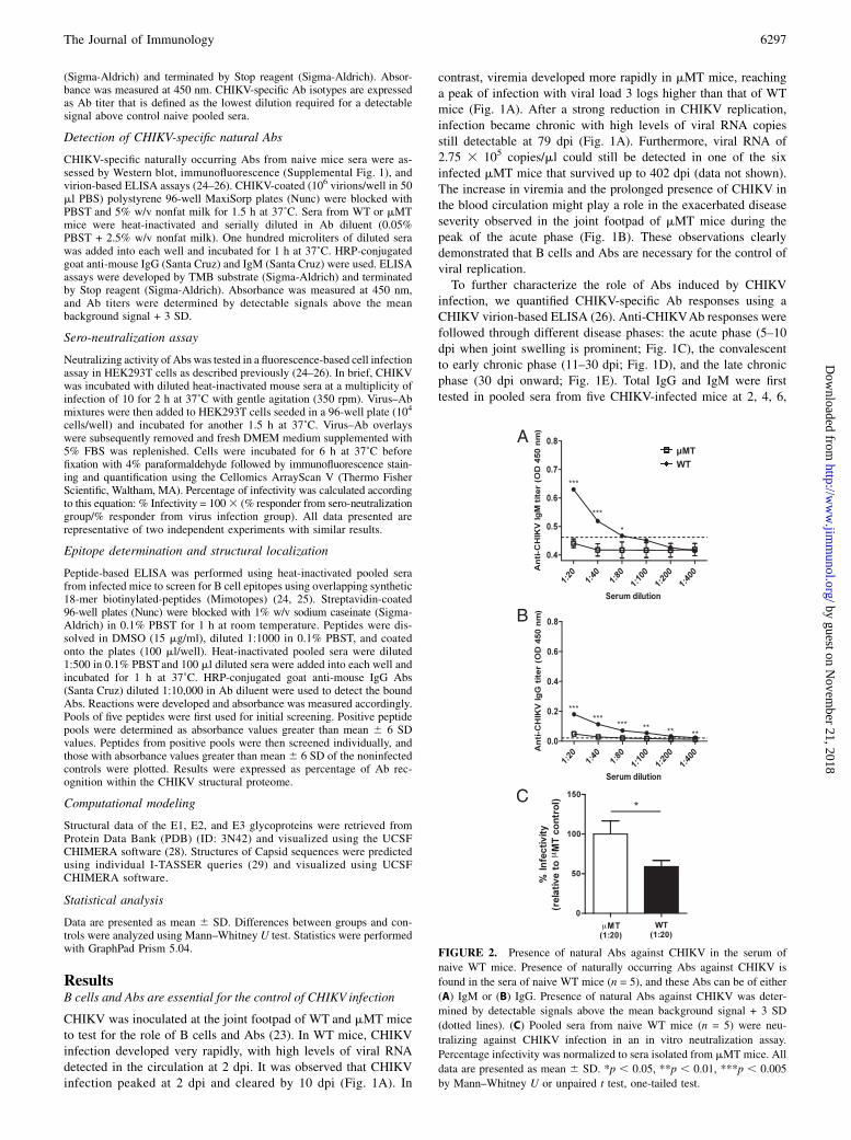
Isotype profile of anti-CHIKV Abs
Isotyping of CHIKV-specific Abs from pooled sera collectedduring the early convalescent phase of 15 dpi from WT micerevealed that the major isotype was IgG2c (Fig. 3A). This ob-servation supports earlier reports that IgG2c is the major class ofanti-CHIKV Abs present in CHIKV-infected mice (10). BecauseIgG2c production is driven mainly by IFN-g (31), a cytokine thatis induced during CHIKV infection in mice (10, 20), we went onto determine the isotype profile of IgG in IFN-g–deficient mice at15 dpi (Fig. 3B). In contrast, a modest reduction in total IgG titer,and notably only in IgG2c, was observed in these animals, sug-gesting that IFN-g is not the main driver of isotype selectionduring CHIKV infection in mice.
FIGURE 3. CD4+ T cell CHIKV-spe-
cific Abs (IgG). Isotype IgG2c is the
dominant isotype of CHIKV-specific IgG
Abs in pooled sera from (A) CD42/2
mice (n = 5) and from (B) IFN-g2/2mice
(n = 5) collected at 15 dpi. Pooled sera
fromWTmice (n = 5) were also isotyped
in parallel. Production of (C) CHIKV-
specific IgG Abs and (D) CHIKV-spe-
cific IgM Abs was significantly reduced
in CD42/2 mice (n = 5). (E) Pooled sera
from animals diluted 1:500 in serum-free
DMEMmedium were neutralizing against
CHIKV, with the neutralizing capacity of
the WT sera being significantly stronger.
Percentage infectivity was normalized
against virus-infected samples. CHIKV-
specific Ab isotypes are expressed as Ab
titer that is defined as the lowest dilution
required for a detectable signal above
control naive pooled sera. All data are
presented as mean 6 SD. *p , 0.05,
***p , 0.005 by Mann–Whitney U test.
6298 Abs IN CHIKUNGUNYAVIRUS INFECTION
by guest on Novem
ber 21, 2018http://w
ww
.jimm
unol.org/D
ownloaded from
CD4+ T cells promote a stronger production of CHIKV-specificAbs
We have previously shown that in the absence of CD4+ T cells,CHIKV viremia was still resolved in mice (20). This observationwas surprising because CD4+ T cells are important for Ab pro-duction (32, 33). To determine whether neutralizing Abs wereproduced in the absence of CD4+ T cells, we infected CD42/2
mice with CHIKV. Clearly, anti-CHIKV IgG and IgM Abs werestill being produced (Fig. 3C, 3D), albeit at a significantly lowertiter (p , 0.05) when compared with WT animals. Furthermore,IgG isotyping of the sera from CHIKV-infected CD42/2 miceshowed reduced levels of IgG2c, IgG1, and IgG2b Abs at 15 dpi(Fig. 3A). Despite the reduced titers, these Abs were still neu-tralizing (∼40% reduction; p , 0.005) against CHIKV infection,although with less efficiency than the Abs from WT animals (p ,0.005; Fig. 3E).
B cell linear epitopes by anti-CHIKV Abs
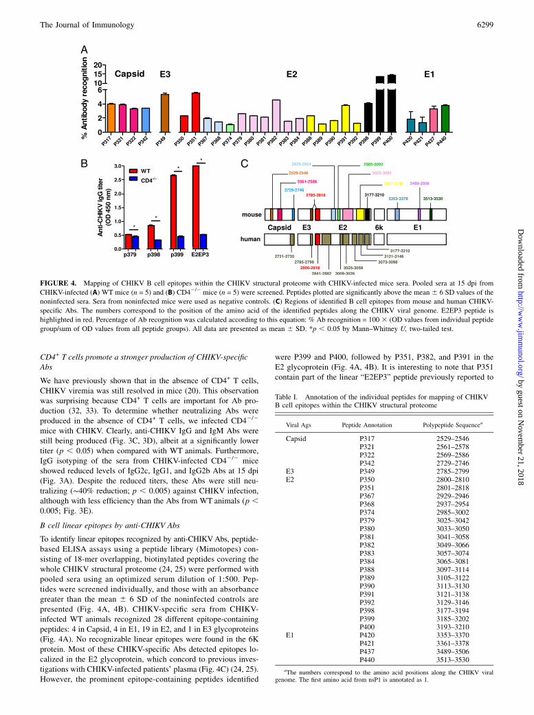
To identify linear epitopes recognized by anti-CHIKVAbs, peptide-based ELISA assays using a peptide library (Mimotopes) con-sisting of 18-mer overlapping, biotinylated peptides covering thewhole CHIKV structural proteome (24, 25) were performed withpooled sera using an optimized serum dilution of 1:500. Pep-tides were screened individually, and those with an absorbancegreater than the mean 6 6 SD of the noninfected controls arepresented (Fig. 4A, 4B). CHIKV-specific sera from CHIKV-infected WT animals recognized 28 different epitope-containingpeptides: 4 in Capsid, 4 in E1, 19 in E2, and 1 in E3 glycoproteins(Fig. 4A). No recognizable linear epitopes were found in the 6Kprotein. Most of these CHIKV-specific Abs detected epitopes lo-calized in the E2 glycoprotein, which concord to previous inves-tigations with CHIKV-infected patients’ plasma (Fig. 4C) (24, 25).However, the prominent epitope-containing peptides identified
were P399 and P400, followed by P351, P382, and P391 in theE2 glycoprotein (Fig. 4A, 4B). It is interesting to note that P351contain part of the linear “E2EP3” peptide previously reported to
FIGURE 4. Mapping of CHIKV B cell epitopes within the CHIKV structural proteome with CHIKV-infected mice sera. Pooled sera at 15 dpi from
CHIKV-infected (A) WT mice (n = 5) and (B) CD42/2 mice (n = 5) were screened. Peptides plotted are significantly above the mean 6 6 SD values of the
noninfected sera. Sera from noninfected mice were used as negative controls. (C) Regions of identified B cell epitopes from mouse and human CHIKV-
specific Abs. The numbers correspond to the position of the amino acid of the identified peptides along the CHIKV viral genome. E2EP3 peptide is
highlighted in red. Percentage of Ab recognition was calculated according to this equation: % Ab recognition = 100 3 (OD values from individual peptide
group/sum of OD values from all peptide groups). All data are presented as mean 6 SD. *p , 0.05 by Mann–Whitney U, two-tailed test.
Table I. Annotation of the individual peptides for mapping of CHIKVB cell epitopes within the CHIKV structural proteome
Viral Ags Peptide Annotation Polypeptide Sequencea
Capsid P317 2529–2546P321 2561–2578P322 2569–2586P342 2729–2746
E3 P349 2785–2799E2 P350 2800–2810
P351 2801–2818P367 2929–2946P368 2937–2954P374 2985–3002P379 3025–3042P380 3033–3050P381 3041–3058P382 3049–3066P383 3057–3074P384 3065–3081P388 3097–3114P389 3105–3122P390 3113–3130P391 3121–3138P392 3129–3146P398 3177–3194P399 3185–3202P400 3193–3210
E1 P420 3353–3370P421 3361–3378P437 3489–3506P440 3513–3530
aThe numbers correspond to the amino acid positions along the CHIKV viralgenome. The first amino acid from nsP1 is annotated as 1.
The Journal of Immunology 6299
by guest on Novem
ber 21, 2018http://w
ww
.jimm
unol.org/D
ownloaded from

be a major epitope recognized by plasma samples obtained fromboth CHIKV-infected human patients and macaques (24, 25).When E2EP3 was tested, it was also strongly recognized (Fig.4B). P399 and P400, which are located at the C terminus of the E2glycoprotein, had the strongest detection signal (.12% total IgGAbs when results were expressed as percentage of Ab recognitionfor the entire CHIKV structural proteome; percentage Ab recog-nition is defined as the percentage of total anti-CHIKV IgG specificfor a particular peptide). In contrast, sera from CHIKV-infectedCD42/2 mice recognized only four epitope-containing peptideswithin the E2 glycoprotein (P379, P398, P399, and E2EP3; Fig.4B). Annotation and the precise location of the individual epitope-containing peptides recognized by the anti-CHIKV Abs are illus-trated in Table I. Positions of the amino acid sequences recognizedby mouse Abs within the CHIKV structural proteins are illustrated(Fig. 4C). Epitopes identified from CHIKV-infected human patients’plasma (24, 25) are also illustrated for comparative purposes (Fig.4C, Table II). Position of the E2EP3 peptide (polypeptide sequence2800–2818) is highlighted in red (Fig. 4C).Linear epitope-containing sequences were next mapped onto
predicted three-dimensional structures of the Capsid proteins (Fig.5A) or available crystal structures of the E3 (Fig. 5B), E2 (Fig.5C), and E1 (Fig. 5D) glycoproteins (PDB no. 3N42).
DiscussionIn this study, we demonstrated the functional roles of B cells andAbs in a mouse model of CHIKV infection. Upon CHIKV in-fection, mice usually display a bimodal phase of joint inflamma-tion, with the shorter and less severe phase observed at 2 dpi,
Table
II.
Com
parisonofidentified
CHIK
VBcellepitopes
betweenhuman
andmouse,within
theCHIK
Vstructuralproteome
Ags
Identified
BCellEpitope(H
uman)a
AminoAcidb
Identified
BCellEpitope(M
ouse)a
AminoAcidb
Capsid
KDIVTKITPEGAEEW
2721–2735
AVPQQKPRRNRKNKKQKQ
2529–2546
PPKKKPAQKKKKPGRRERMCMKIEND
2561–2586
PEGAEEWSLAIPVMCLLA
2729–2746
E3
LLQASLTCSPHRQRR
2785–2799
LLQASLTCSPHRQRR
2785–2799
E2
STKDNFNVYKATRPYLAHC
2800–2818
STKDNFNVYKATRPYLAHC
2800–2818
TDGTLKIQVSLQIGIKTDDSHDWTKLRYMDNHMPADAERAGL
2841–2882
HHDPPVIGREKFHSRPQHGKELPCST
2929–2954
LTTTDKVINNCKVDQCHA
3009–3026
GNVKITVNGQTVRYKCNC
2985–3002
HAAVTNHKKWQYNSPLVPRNAELGDRKGKIHIPF
3025–3058
HAAVTNHKKWQYNSPLVPRNAELGDRKGKIHIPFPLANVTCRVPKARNPTVTYGKNQ
3025–3081
PTVTYGKNQVIMLLYPDHPTLLSYRN
3073–3098
RNMGEEPNYQEEWVMHKKEVVLTVPTEGLEVTWGNNEPYKYWPQLSTNGT
3097–3146
PTEGLEVTWGNNEPYKYWPQLSTNGT
3121–3146
LLSMVGMAAGMCMCARRRCITPYELTPGATVPFL
3177–3210
LLSMVGMAAGMCMCARRRCITPYELTPGATVPFL
3177–3210
6KE1
DKNLPDYSCKVFTGVYPFMWGGAYCF
3353–3378
RTPESKDVYANTQLVLQR
3489–3506
HVPYSQAPSGFKYWLKER
3513–3530
aRegionsofB
cellepitopes
foundthat
arecommonto
both
human
andmouse
areunderlined.
bThenumberscorrespondto
theam
inoacid
positionsalongtheCHIK
Vviral
genome.
Thefirstam
inoacid
from
nsP1isannotatedas
1.
FIGURE 5. Localization of identified CHIKV B cell epitopes within
CHIKV structural proteome. Schematic representation of identified B cell
epitopes in (A) Capsid, (B) E3, (C) E2, and (D) E1 proteins. Epitopes in the
E3, E2, and E1 proteins were located based on the structural data obtained
from PDB records: 3N42. Epitopes in the Capsid protein were located
based on the structures predicted via the I-TASSER server. Number cor-
responds to the position of the amino acid of the identified peptides along
the CHIKV viral genome. All representations are shown in frontal view
except for the E1 glycoprotein peptides, and peptides 2729–2746 of capsid
protein, which are shown in the back view.
6300 Abs IN CHIKUNGUNYAVIRUS INFECTION
by guest on Novem
ber 21, 2018http://w
ww
.jimm
unol.org/D
ownloaded from

whereas the longer and more severe phase was observed during thepeak of the acute phase at 5–6 dpi (10, 20). We have previouslyidentified CD4+ T cells as the major effector for maximal jointinflammation during the peak of the acute phase, but not in viralclearance (20). In WT mice, CHIKV infection induced the pro-duction of anti-CHIKV Abs that led to the rapid clearance ofCHIKV and resulted in less severe joint inflammation during thepeak of the acute phase, when compared with mMT mice. Anti-CHIKVAbs persisted for a long time. Interestingly, IgM could bedetected for more than a year postinfection. Unusual long-termpersistence for IgM was also reported in CHIKV patients (34). Wealso demonstrated that IgM Abs that are able to recognize CHIKVare present in the sera of naive WT mice. These Abs likely rec-ognize conformational epitopes (25) because reactivity to CHIKVproteins was not detected by Western blot or by immunofluores-cence. Natural Abs of IgM, IgG, and IgA isotypes were previouslyshown to be present both in human subjects (35, 36) and in mice(35). They were shown to be capable of providing early protectionand controlling dissemination of infectious pathogens via directneutralization, activation of the complement system, as well asenhancing the immune responses in secondary lymphoid organs(37). Our data clearly suggest that these natural Abs are neutral-izing against CHIKV and control viral replication in CHIKV-infected animals during the early phase of infection. Studies areunder way to determine whether such Abs exist in naive humanpopulations.We have recently demonstrated in CD42/2 mice that viremia
was cleared as efficiently as in WT mice (20). This was surprisingbecause CD4+ Th cells interact with B cells to enhance Ab pro-duction, induce class switching, and promote affinity maturation(32, 33). Therefore, in this study, we sought to characterize thespecific anti-CHIKV Ab response in both WT and CD42/2 mice.Interestingly, we observed that both CHIKV-specific total IgG andIgM were significantly reduced in infected CD42/2 mice. Despitethis significant difference in Ab levels, sera from both groups werecapable of reducing CHIKV infectivity in in vitro neutralizationassays, although sera from CD42/2 mice exhibited a reducedneutralizing capacity. This led to the conclusion that CD4+ T cellscontrol the breadth of Ab response, but nevertheless in their ab-sence, anti-CHIKV Abs against the E2 glycoprotein are still in-duced and are important in the antiviral protection during CHIKVprimary infection. In fact, we have shown that anti-CHIKV Absfrom infected CD42/2 mice, targeting just four epitope-containingregions of the E2 glycoprotein, were able to fully protect againstCHIKV infection as efficiently as the WT mice, suggesting thatthese epitope-containing regions are major targets of CD4+ T cell–independent protective Ab responses. Nevertheless, it is plausiblethat CD4+ T cells could still be required for the long-term per-sistence of anti-CHIKV Abs. In addition, the limited influence ofCD4+ T cells in the development of protective anti-CHIKV Abscould explain why CHIKV infections are not more frequent orsevere in regions where CHIKV and HIV coinfection occurs (38–43). Several CHIKVendemic regions are found in areas where HIVtransmissions are prevalent, such as India and the sub-SaharanAfrican nations (38–42). Furthermore, HIV/CHIKV coinfectionshave been previously reported in northern Tanzania (43). The res-olution of CHIKV infection in CD42/2 mice would strongly implythat HIV patients with low CD4+ T cell counts (44) could stillresolve CHIKV infection effectively.Previous studies have shown that CHIKV-specific CD4+ T cells
were induced by CHIKV infection, and the specific CD4+ T cellswere the major producers of IFN-g (20). Th1 cells have beenshown to skew the production of IgG2c Abs by B cells throughtheir production of IFN-g (31). However, this does not apply to
CHIKV infection as we have shown that CHIKV-specific IgG2cAbs were only marginally reduced in IFN-g2/2 mice at 15 dpi.This finding suggests that other alternative IFN-g–independentpathways are responsible for IgG2c production and will warrantfurther studies to decipher the mechanism.The demonstration of an essential role for B cells and Abs in
controlling CHIKV infection together with the identification ofB cell epitopes recognized by protective Abs further advocate themouse model as an important preclinical tool for diagnosis andvaccine development.
AcknowledgmentsWe thank Carla Claser (Singapore Immunology Network) for technical ex-
pertise and assistance provided in the animal experiments. We also express
gratitude to Cindy Phua (Mutant Mouse Collection, Singapore Immunology
Network) for assistance in breeding of and providing the mutant mice used
in this study.
DisclosuresThe authors have no financial conflicts of interest.
References1. Her, Z., Y. W. Kam, R. T. P. Lin, and L. F. P. Ng. 2009. Chikungunya: a bending
reality. Microbes Infect. 11: 1165–1176.2. Robinson, M. C. 1955. An epidemic of virus disease in Southern Province,
Tanganyika Territory, in 1952-53. I. Clinical features. Trans. R. Soc. Trop. Med.Hyg. 49: 28–32.
3. Borgherini, G., P. Poubeau, F. Staikowsky, M. Lory, N. Le Moullec,J. P. Becquart, C. Wengling, A. Michault, and F. Paganin. 2007. Outbreak ofchikungunya on Reunion Island: early clinical and laboratory features in 157adult patients. Clin. Infect. Dis. 44: 1401–1407.
4. Lakshmi, V., M. Neeraja, M. V. S. Subbalaxmi, M. M. Parida, P. K. Dash,S. R. Santhosh, and P. V. L. Rao. 2008. Clinical features and molecular diagnosisof Chikungunya fever from South India. Clin. Infect. Dis. 46: 1436–1442.
5. Chandak, N. H., R. S. Kashyap, D. Kabra, P. Karandikar, S. S. Saha, S. H. Morey,H. J. Purohit, G. M. Taori, and H. F. Daginawala. 2009. Neurological complica-tions of Chikungunya virus infection. Neurol. India 57: 177–180.
6. Ganesan, K., A. Diwan, S. K. Shankar, S. B. Desai, G. S. Sainani, andS. M. Katrak. 2008. Chikungunya encephalomyeloradiculitis: report of 2 caseswith neuroimaging and 1 case with autopsy findings. AJNR Am. J. Neuroradiol.29: 1636–1637.
7. Powers, A. M., and C. H. Logue. 2007. Changing patterns of chikungunya virus:re-emergence of a zoonotic arbovirus. J. Gen. Virol. 88: 2363–2377.
8. Renault, P., E. Balleydier, E. D’Ortenzio, M. Baville, and L. Filleul. 2012. Ep-idemiology of Chikungunya infection on Reunion Island, Mayotte, and neigh-boring countries. Med. Mal. Infect. 42: 93–101.
9. Chua, K. B. 2010. Epidemiology of chikungunya in Malaysia: 2006-2009. Med.J. Malaysia 65: 277–282.
10. Gardner, J., I. Anraku, T. T. Le, T. Larcher, L. Major, P. Roques, W. A. Schroder,S. Higgs, and A. Suhrbier. 2010. Chikungunya virus arthritis in adult wild-typemice. J. Virol. 84: 8021–8032.
11. Gardner, C. L., C. W. Burke, S. T. Higgs, W. B. Klimstra, and K. D. Ryman.2012. Interferon-alpha/beta deficiency greatly exacerbates arthritogenic diseasein mice infected with wild-type chikungunya virus but not with the cell culture-adapted live-attenuated 181/25 vaccine candidate. Virology 425: 103–112.
12. Werneke, S. W., C. Schilte, A. Rohatgi, K. J. Monte, A. Michault, F. Arenzana-Seisdedos, D. L. Vanlandingham, S. Higgs, A. Fontanet, M. L. Albert, andD. J. Lenschow. 2011. ISG15 is critical in the control of Chikungunya virusinfection independent of UbE1L mediated conjugation. PLoS Pathog. 7:e1002322.
13. Her, Z., B. Malleret, M. Chan, E. K. S. Ong, S. C. Wong, D. J. C. Kwek,H. Tolou, R. T. P. Lin, P. A. Tambyah, L. Renia, and L. F. Ng. 2010. Activeinfection of human blood monocytes by Chikungunya virus triggers an innateimmune response. J. Immunol. 184: 5903–5913.
14. Labadie, K., T. Larcher, C. Joubert, A. Mannioui, B. Delache, P. Brochard,L. Guigand, L. Dubreil, P. Lebon, B. Verrier, et al. 2010. Chikungunya disease innonhuman primates involves long-term viral persistence in macrophages. J. Clin.Invest. 120: 894–906.
15. Schilte, C., T. Couderc, F. Chretien, M. Sourisseau, N. Gangneux, F. Guivel-Benhassine, A. Kraxner, J. Tschopp, S. Higgs, A. Michault, et al. 2010. Type IIFN controls chikungunya virus via its action on nonhematopoietic cells. J. Exp.Med. 207: 429–442.
16. Couderc, T., F. Chretien, C. Schilte, O. Disson, M. Brigitte, F. Guivel-Benhassine, Y. Touret, G. Barau, N. Cayet, I. Schuffenecker, et al. 2008. Amouse model for Chikungunya: young age and inefficient type-I interferonsignaling are risk factors for severe disease. PLoS Pathog. 4: e29.
17. Rudd, P. A., J. Wilson, J. Gardner, T. Larcher, C. Babarit, T. T. Le, I. Anraku,Y. Kumagai, Y. M. Loo, M. Gale, Jr., et al. 2012. Interferon response factors
The Journal of Immunology 6301
by guest on Novem
ber 21, 2018http://w
ww
.jimm
unol.org/D
ownloaded from
3 and 7 protect against Chikungunya virus hemorrhagic fever and shock. J. Virol.86: 9888–9898.
18. Teng, T. S., S. S. Foo, D. Simamarta, F. M. Lum, T. H. Teo, A. Lulla, N. K. W. Yeo,E. G. L. Koh, A. Chow, Y. S. Leo, et al. 2012. Viperin restricts chikungunya virusreplication and pathology. J. Clin. Invest. 122: 4447–4460.
19. Teo, T. H., F. M. Lum, W. W. L. Lee, and L. F. P. Ng. 2012. Mouse models forChikungunya virus: deciphering immune mechanisms responsible for diseaseand pathology. Immunol. Res. 53: 136–147.
20. Teo, T. H., F. M. Lum, C. Claser, V. Lulla, A. Lulla, A. Merits, L. Renia, andL. F. P. Ng. 2013. A pathogenic role for CD4+ T cells during Chikungunya virusinfection in mice. J. Immunol. 190: 259–269.
21. Couderc, T., N. Khandoudi, M. Grandadam, C. Visse, N. Gangneux, S. Bagot,J. F. Prost, and M. Lecuit. 2009. Prophylaxis and therapy for Chikungunya virusinfection. J. Infect. Dis. 200: 516–523.
22. Lee, C. Y., Y. W. Kam, J. Fric, B. Malleret, E. G. L. Koh, C. Prakash, W. Huang,W. W. L. Lee, C. Lin, R. T. P. Lin, et al. 2011. Chikungunya virus neutralizationantigens and direct cell-to-cell transmission are revealed by human antibody-escape mutants. PLoS Pathog. 7: e1002390.
23. Kitamura, D., J. Roes, R. Kuhn, and K. Rajewsky. 1991. A B cell-deficientmouse by targeted disruption of the membrane exon of the immunoglobulinmu chain gene. Nature 350: 423–426.
24. Kam, Y. W., F. M. Lum, T. H. Teo, W. W. L. Lee, D. Simarmata, S. Harjanto,C. L. Chua, Y. F. Chan, J. K. Wee, A. Chow, et al. 2012. Early neutralizing IgGresponse to Chikungunya virus in infected patients targets a dominant linearepitope on the E2 glycoprotein. EMBO Mol. Med. 4: 330–343.
25. Kam, Y. W., W. W. L. Lee, D. Simarmata, S. Harjanto, T. S. Teng, H. Tolou,A. Chow, R. T. P. Lin, Y. S. Leo, L. Renia, and L. F. Ng. 2012. Longitudinalanalysis of the human antibody response to Chikungunya virus infection: impli-cations for serodiagnosis and vaccine development. J. Virol. 86: 13005–13015.
26. Kam, Y. W., D. Simarmata, A. Chow, Z. Her, T. S. Teng, E. K. S. Ong, L. Renia,Y. S. Leo, and L. F. P. Ng. 2012. Early appearance of neutralizing immuno-globulin G3 antibodies is associated with chikungunya virus clearance and long-term clinical protection. J. Infect. Dis. 205: 1147–1154.
27. Martin, R. M., J. L. Brady, and A. M. Lew. 1998. The need for IgG2c specificantiserum when isotyping antibodies from C57BL/6 and NOD mice. J. Immunol.Methods 212: 187–192.
28. Pettersen, E. F., T. D. Goddard, C. C. Huang, G. S. Couch, D. M. Greenblatt,E. C. Meng, and T. E. Ferrin. 2004. UCSF Chimera—a visualization system forexploratory research and analysis. J. Comput. Chem. 25: 1605–1612.
29. Roy, A., A. Kucukural, and Y. Zhang. 2010. I-TASSER: a unified platform forautomated protein structure and function prediction. Nat. Protoc. 5: 725–738.
30. Andersson, J., and F. Melchers. 1973. Induction of immunoglobulin M synthesisand secretion in bone-marrow-derived lymphocytes by locally concentratedconcanavalin A. Proc. Natl. Acad. Sci. USA 70: 416–420.
31. Barr, T. A., S. Brown, P. Mastroeni, and D. Gray. 2009. B cell intrinsic MyD88signals drive IFN-gamma production from T cells and control switching toIgG2c. J. Immunol. 183: 1005–1012.
32. Vitetta, E. S., R. Fernandez-Botran, C. D. Myers, and V. M. Sanders. 1989.Cellular interactions in the humoral immune response. Adv. Immunol. 45: 1–105.
33. Swain, S. L., K. K. McKinstry, and T. M. Strutt. 2012. Expanding roles for CD4+
T cells in immunity to viruses. Nat. Rev. Immunol. 12: 136–148.34. Malvy, D., K. Ezzedine, M. Mamani-Matsuda, B. Autran, H. Tolou,
M. C. Receveur, T. Pistone, J. Rambert, D. Moynet, and D. Mossalayi. 2009.Destructive arthritis in a patient with chikungunya virus infection with persistentspecific IgM antibodies. BMC Infect. Dis. 9: 200.
35. Ochsenbein, A. F., and R. M. Zinkernagel. 2000. Natural antibodies and com-plement link innate and acquired immunity. Immunol. Today 21: 624–630.
36. Herzenberg, L. A., and A. B. Kantor. 1993. B-cell lineages exist in the mouse.Immunol. Today 14: 79–83, discussion 88–90.
37. Ochsenbein, A. F., T. Fehr, C. Lutz, M. Suter, F. Brombacher, H. Hengartner, andR. M. Zinkernagel. 1999. Control of early viral and bacterial distribution anddisease by natural antibodies. Science 286: 2156–2159.
38. Mayer, K. 2011. The evolving Indian AIDS epidemic: hope & challenges of thefourth decade. Indian J. Med. Res. 134: 739–741.
39. De Cock, K. M., H. W. Jaffe, and J. W. Curran. 2012. The evolving epidemiologyof HIV/AIDS. AIDS 26: 1205–1213.
40. Thiboutot, M. M., S. Kannan, O. U. Kawalekar, D. J. Shedlock, A. S. Khan,G. Sarangan, P. Srikanth, D. B. Weiner, and K. Muthumani. 2010. Chikungunya:a potentially emerging epidemic? PLoS Negl. Trop. Dis. 4: e623.
41. Ravi, V. 2006. Re-emergence of chikungunya virus in India. Indian J. Med.Microbiol. 24: 83–84.
42. Sergon, K., A. A. Yahaya, J. Brown, S. A. Bedja, M. Mlindasse, N. Agata,Y. Allaranger, M. D. Ball, A. M. Powers, V. Ofula, et al. 2007. Seroprevalence ofChikungunya virus infection on Grande Comore Island, union of the Comoros,2005. Am. J. Trop. Med. Hyg. 76: 1189–1193.
43. Hertz, J. T., O. M. Munishi, E. E. Ooi, S. Howe, W. Y. Lim, A. Chow,A. B. Morrissey, J. A. Bartlett, J. J. Onyango, V. P. Maro, et al. 2012. Chi-kungunya and dengue fever among hospitalized febrile patients in northernTanzania. Am. J. Trop. Med. Hyg. 86: 171–177.
44. Roederer, M., J. G. Dubs, M. T. Anderson, P. A. Raju, L. A. Herzenberg, andL. A. Herzenberg. 1995. CD8 naive T cell counts decrease progressively in HIV-infected adults. J. Clin. Invest. 95: 2061–2066.
6302 Abs IN CHIKUNGUNYAVIRUS INFECTION
by guest on Novem
ber 21, 2018http://w
ww
.jimm
unol.org/D
ownloaded from