an in-vitro comparative micro-computed tomographic
TRANSCRIPT

i
An in-vitro comparative micro-computed tomographic
evaluation of three obturation systems
A mini thesis submitted in partial fulfilment of the requirements for the degree
of Magister Chirurgiae Dentium in Prosthodontic at the Faculty of Dentistry
(University of the Western Cape)
S N Kabini (3379124)
MChD (Prosthodontics)
2017
Supervisor: Dr D Moodley
Co-supervisors: Dr N Patel
Prof M E Parker

ii
DECLARATION
I hereby declare that the study: “An in-vitro comparative micro-computed tomographic
evaluation of three obturation systems” is my own work, that has not been submitted before
for any degree or examination in any university, and that all the sources I have used or quoted
have been indicated and acknowledged by complete references.
Signed:
Shadrack Nyabela Kabini June 2017
http://etd.uwc.ac.za

iii
ACKNOWLEDGEMENT
I wish to acknowledge the following people for the assistance given to me in this research
project.
Dr D Moodley: My supervisor for his guidance, encouragement and support during the
course of this study. It was an honour to be supervised by this wonderful
person who is always down to mother earth.
Dr N Patel: My co-supervisor who was always supportive and provided guidance
during the course of this study.
Prof M E Parker: My other co-supervisor who provided guidance with regards to the
radiological aspect of the study
Prof J S Maritz: The statistician who provided valuable input with regards to the
statistical analysis of the results of the study.
Mr J Le Roux: Micro-CT specialist who assisted with the technical aspect of the study
involving use of micro-CT
http://etd.uwc.ac.za

iv
DEDICATION
To my family and my lovely kids who always supported me and believed in me (I love you
guys)
To my father and my late mother who provided me with all the support that was needed to
reach this level of education
To my friends and their families (thank you Dr Christopher Anderson Banda, Mr King Calvin
Mtsabela Sibande and Mr Peter Malesela Madubanya)
Lastly to my other father who provided support and encouragement from high school till the
completion of my undergraduate studies (thank you Dr Bayela Joseph Manonga)
http://etd.uwc.ac.za
v
Table of Contents
Abstract ...................................................................................................................................... 1
Chapter 1 .................................................................................................................................... 3
Introduction ............................................................................................................................ 3
Chapter 2 .................................................................................................................................... 5
Literature review .................................................................................................................... 5
Access cavity preparation and location of canals ............................................................... 6
Glide path in endodontics ................................................................................................... 7
Root canal irrigation ........................................................................................................... 9
Root canal obturation........................................................................................................ 16
Obturation techniques ....................................................................................................... 21
Summary of filling techniques in a 3D view .................................................................... 37
Apical and coronal seal…………………………………………………………………..38
Zinc oxide eugenol sealers ............................................................................................... 39
Glassionomer sealers ........................................................................................................ 40
Resin bonded sealers ........................................................................................................ 41
Calcium hydroxide sealers ................................................................................................ 42
Silicone-based sealers ....................................................................................................... 43
Complications in endodontic obturation........................................................................... 43
The adaptation of the gutta-percha to the root canal wall ................................................ 44
Fluid filtration or transportation methodology ................................................................. 48
Dye extraction method ...................................................................................................... 48
http://etd.uwc.ac.za
vi
Bacterial and toxin infiltration method ............................................................................. 49
Glucose leakage test ......................................................................................................... 49
Periapical radiographs ...................................................................................................... 50
Tuned aperture computed tomography (TACT) ............................................................... 50
Cone beam computed tomography (CBCT) ..................................................................... 51
Microcomputed tomography (Micro-CT) ........................................................................ 51
Brief history of material adaptation….…………………………….…………… ………53
Chapter 3 .................................................................................................................................. 56
Aims and objectives ............................................................................................................. 56
Aims.................................................................................................................................. 56
Objectives ......................................................................................................................... 56
Null hypothesis ................................................................................................................. 56
Ethics statement ................................................................................................................ 57
Chapter 4 .................................................................................................................................. 58
Materials and methods ......................................................................................................... 58
Tooth preparation ............................................................................................................. 58
Root canal obturation........................................................................................................ 62
Micro-CT Imaging ............................................................................................................ 66
Statistical Analysis ........................................................................................................... 67
Chapter 5 .................................................................................................................................. 69
Results .................................................................................................................................. 69
Discussion ............................................................................................................................ 73
http://etd.uwc.ac.za

vii
Chapter 7 .................................................................................................................................. 81
Conclusions and recommendations ...................................................................................... 81
Conclusions ...................................................................................................................... 81
Recommendations ............................................................................................................ 81
Limitations of the study........................................................................................................ 82
Chapter 8 .................................................................................................................................. 83
References ............................................................................................................................ 83
Appendix .................................................................................................................................. 97
Appendix 1: Ethical clearance letter……………..……..…………………………………98
http://etd.uwc.ac.za

viii
List of figures
Figure 1: Changes that usually occur when heating gutta-percha ........................................... 17
Figure 2: Silver point obturation on a molar tooth .................................................................. 18
Figure 3: An obturation of a premolar done with silverpoints................................................. 19
Figure 4: Different types of monoblock ................................................................................... 22
Figure 5: Cold lateral compaction............................................................................................ 23
Figure 6: Cross section of lateral compaction showing voids at the apex of the tooth........... 25
Figure 7: ProTaper single obturators ....................................................................................... 27
Figure 8: ProTaper single-cone obturation .............................................................................. 27
Figure 9: Downpack method .................................................................................................... 29
Figure 10: Probes that are used to heat the gutta-percha in a downpacking method ............... 30
Figure 11: Backfill method ...................................................................................................... 31
Figure 12: Warm obturation devices........................................................................................ 31
Figure 13: Thermafil gutta-percha with plastic carrier ............................................................ 34
Figure 14: Thermoprep oven ................................................................................................... 35
Figure 15: GuttaCore carrier system ........................................................................................ 35
Figure 16: Representation of polymer chains of a thermoset elastomer of gutta-percha ........ 36
Figure 17: A GuttaCore oven ................................................................................................... 37
Figure 18: Spectrum of filling techniques. .............................................................................. 37
Figure 19: Roth cement ............................................................................................................ 40
Figure 20: Ketac-Endo glass ionomer sealer ........................................................................... 41
Figure 21: AH Plus resin bonded sealer .................................................................................. 41
Figure 22: Sealapex root canal sealer ...................................................................................... 42
Figure 23: RoekoSeal root canal sealer ................................................................................... 43
Figure 24: Adaptation of apical cone, vertical compaction, GuttaFlow and Thermafil .......... 45
http://etd.uwc.ac.za

ix
Figure 25: Lateral compaction with smear layer ..................................................................... 46
Figure 26: Lateral compaction without smear layer ................................................................ 46
Figure 27: Thermoplastic gutta-percha in presence of smear layer ......................................... 47
Figure 28: Thermafil core perforating the gutta-percha .......................................................... 53
Figure 29: GuttaCore showing obturation of lateral canal ....................................................... 55
Figure 30: Decoronated teeth ................................................................................................... 58
Figure 31: Glide path preparation ............................................................................................ 59
Figure 32: ProTaper rotary files ............................................................................................... 59
Figure 33: Filing and shaping with rotary files ........................................................................ 60
Figure 34: Rotary system set up for ProTaper Universal files................................................. 60
Figure 35: Sodium hypochlorite irrigation solution ................................................................ 61
Figure 36: Smear clear irrigation solution ............................................................................... 62
Figure 37: GuttaCore carrier obturation system ...................................................................... 63
Figure 38: ProTaper obturation system.................................................................................... 63
Figure 39: Thermafil obturation system .................................................................................. 64
Figure 40: GuttaCore oven ....................................................................................................... 65
Figure 41: Thermoprep oven ................................................................................................... 65
Figure 42: Obturated specimens .............................................................................................. 66
Figure 43: Micro-CT machine ................................................................................................. 67
Figure 44: Total volume of voids and volume of cement ........................................................ 70
Figure 45: Percentage voids .................................................................................................... 71
Figure 46: GuttaCore, ProTaper ang Thermafil at 1mm from the apex .................................. 71
Figure 47: GuttaCore, ProTaper and Thermafil at 3mm from the apex .................................. 72
Figure 48: GuttaCore, ProTaper and Thermafil at 6mm......................................................... 72
http://etd.uwc.ac.za
x
List of tables
Table 1: Requirements for an ideal root filling cement ........................................................... 16
Table 2: Overview of sealers chemical types and examples .................................................... 39
Table 4: Volume of voids for GuttaCore, ProTaper and Thermafil ........................................ 69
Table 5: Volume of cement for GuttaCore, ProTaper and Thermafil ..................................... 69
Table 6: Percentage volume of voids for GuttaCore, ProTaper and Thermafil ...................... 70
http://etd.uwc.ac.za

1
Abstract
Gaps or voids between walls of root canal and obturation material may lead to re-infection of
the obturated root canal. Therefore, adaptation of the obturation material to dentine walls is
essential for the success of root canal treatment.
Aim: To evaluate and compare the adaptation of gutta-percha of three obturation systems
using micro-computed tomography. The percentage of volume of voids and gaps at 1mm,
3mm and 6mm axial sections from the apex was compared. The volume of cement around the
gutta-percha was also compared for each system.
Methods: The roots of 90 central incisors were shaped with ProTaper Universal (Dentsply)
files. 1ml 5.25% sodium hypochlorite was used as an irrigant and flushed with 5ml 17%
EDTA. The roots were randomly divided into three groups: Group 1: obturated with
GuttaCore, Group 2: obturated with ProTaper single-cone obturation and Group 3: obturated
with Thermafil. All canals were sealed with AH Plus (Dentsply) root canal sealer. A v|tome|x
240D micro-CT scanner was used to scan each root at 15μm resolution to observe presence of
any voids.
Results: The mean volume of voids and percentage of voids for ProTaper single-cone
obturation was found to be significantly greater than that of Thermafil and of GuttaCore
carrier based systems (Kruskal-Wallis p<0.001). The mean volume of cement surrounding
the gutta-percha of Protaper was significantly greater than that of Thermafil and GuttaCore
(Kruskal-Wallis p<0.001). Thermafil and GuttaCore demonstrated good adaptation at 1mm,
3mm and 6mm from the apex compared to ProTaper single-cone obturation which showed
voids and higher volume of cement at 1mm, 3mm and 6mm from the apex of the tooth. The
larger volume of cement and the presence of more voids with ProTaper single-cone
http://etd.uwc.ac.za

2
obturation demonstrates poor adaptation of the material to the wall of the root canal
compared to carrier based obturation systems.
Conclusion: Both carrier based techniques allowed for better sealing ability in root
canals compared to single-cone gutta-percha obturation although none of the materials
were gap free especially at 1mm from apex.
Keywords: Obturation, GuttaCore, ProTaper, Thermafil, Micro-CT
http://etd.uwc.ac.za

3
Chapter 1
Introduction
Endodontic materials are developing at a rapid rate with each manufacturer claiming their
products to be superior compared to the other products. The ultimate aim of a root filling is to
fill the entire prepared and cleaned root canal (Hammad et al., 2009). The success of root
canal obturation does not only depend on the root canal sealer, but also on proper adaptation
of the gutta percha to the walls of the root canal. A hermitic seal can be obtained by good root
canal obturation and sealing of lateral and accessory canals as well. Therefore, the adaptation
of the root canal filling material to dentinal walls is essential for the success of root canal
therapy as this prevents the formation of gaps or voids between the root filling material and
the root canal walls. This will ensure the sealing of all lateral and accessory canals that are
frequently found on the root canal walls.
The gap formation between root canal walls and the root canal filling material may lead to the
re-infection of the root canal system, leading to treatment failures. Furthermore, dissolution
of root canal sealer at the apex of the tooth may be counteracted by properly filled and well
adapted gutta-percha to prevent microbial infection due to leakages. The ability to seal the
canals is essential in the prevention of colonization by micro-organisms within the root canal
system as spaces left may allow for bacteria to populate and proliferate (Zogheib et al., 2013).
The common techniques employed for endodontic root canal obturation include cold lateral
compaction, warm vertical compaction and core-carrier techniques. These core-carrier
systems are claimed by the manufacturers to enhance adaptation of the gutta-percha to the
canal wall, and flow of the filling material into the lateral canals. The original carrier was
http://etd.uwc.ac.za

4
made of metal. Due to the difficulties encountered in retreatment and in the preparation of
post spaces, the original metal carriers were subsequently replaced by plastic obturators (Li et
al., 2014). Recently, a new core-carrier system, GuttaCore (Dentsply Tulsa Dental
Specialties, Tulsa, OK, USA) was introduced in which the Vectra (a liquid crystal polymer)
or polysulphone plastic carriers in Thermafil Plus (Dentsply Tulsa Dental Specialties) were
replaced by cross-linked thermoset gutta-percha, which enables the carrier (obturator) to be
removed more easily during retreatment (Li et al., 2014). The GuttaCore gutta-percha does
not melt when placed in an obturator oven but softens (Gutmann, 2008). Although the core-
carrier obturation technique has been regarded by some as the only genuine warm gutta-
percha technique for adaptation to the apical third of the canal space, the quality of root canal
obturation achieved by the new core-carrier system that incorporates cross-linked thermoset
gutta-percha carriers has not been reported (Li et al., 2014).
Thus, the objective of the present in-vitro study was to examine the quality of obturation in
single-rooted canals obturated with the GuttaCore core-carrier system by comparing the
results with similar canals obturated with the ProTaper single-cone obturation technique and
another core-carrier technique, Thermafil, using micro-computed tomography (micro-CT).
http://etd.uwc.ac.za

5
Chapter 2
Literature review
Root canal filling materials could be considered true implants as they are in contact, and are
based in vital tissues of the body, and extend beyond to meet the external surface directly or
indirectly via another surface restoration (Ørstavik, 2005). The root canal filling materials
must possess several different properties relative to their functions and location, ranging from
biocompatibility to mechanical sealing ability. The goal of root canal obturation is to
hermetically seal off the root canal system and the elimination of the environment in which
microorganisms can multiply. The root canal filling material should provide a three
dimensional seal, particularly in the last few millimetres of the apical area. The success of
root canal treatment depends mainly on what we take out than what we put in, and it is also
dependent upon proper irrigation and an adequate obturation at working length (Ørstavik,
2005). The working length is usually calculated at a length of about 1mm from the apex of
the root of the tooth to coincide with the apical constriction
There are a higher number of reported root canal failures due to insufficient obturation and
poor irrigation with an antimicrobial agent (Ørstavik, 2005). The material of choice for
sealing of the root canal is gutta-percha and since it does not bond to tooth structure, it is
always used in conjunction with sealers both apically and coronally. Silver points were
regarded as a substitute for gutta-percha, but have a disadvantage with regards to corrosion
and clinical failure (Ørstavik, 2005). Recently adhesive dentistry has been introduced to the
field of endodontics with a specific aim of obtaining a monoblock, in which the core material,
sealing agent and the root dentine form a single cohesive unit. The root canal treatment
protocol begins with access cavity preparation followed by glide path preparation, filing and
irrigation and then obturation (Tay & Pashley, 2007).
http://etd.uwc.ac.za

6
Access cavity preparation and location of canals
Access to the root canal system can be a significant challenge in the successful treatment of a
root canal. To be able to get an instrument into a canal system unimpeded by the use of a
glide path without unnecessary tooth damage, can facilitate the endodontic treatment for the
clinician (Darcey et al., 2015). The access cavity should make the succeeding steps easier and
safer (Castellucci, 2003). There are a few requirements needed for it to succeed namely:
permit removal of all chamber contents, permit direct vision of the pulp chamber floor and
canal opening, facilitate the introduction of canal instruments into canal openings, provide as
direct as possible access to apical one third of canal during preparation and filling of canal,
provide a positive support for temporary fillings and always have four walls. Access to the
root canal is the first and most important phase in root canal treatment. In order to obtain a
good obturation, a well-designed access preparation is essential.
With poor access, endodontic materials and instrumentation becomes difficult. Objectives of
access cavity preparation are: to achieve straight line access to apical foramen/initial canal
curvature, to locate all root canal orifices and to conserve sound tooth structure. A well
prepared access cavity creates a smooth path to the canal system and apex, which will allow
complete irrigation, shaping and cleaning and good quality obturation. An analysis that was
done on extracted teeth developed a series of ‘laws’ to help clinicians to achieve these goals
of locating the pulp. The law of centrality entails that the pulp will be in the centre of the
tooth. The law of the cemento-enamel junction (CEJ) entails that the pulp will always be
located at the level of the cemento-enamel junction. The law of centricity entails that the
walls of the pulp chamber will be concentric (share the same centre) to the outer wall of the
tooth (Adams & Tomson, 2014).
http://etd.uwc.ac.za

7
Glide path in endodontics
The goal of instrumentation is to get a continuous funnel flowing with the shape of original
canal from the coronal access to the apex. The glide path is the starting point of radicular
preparation. Cleaning and shaping becomes unpredictable if there is no guide for endodontic
mechanics (Dhingra, 2014). West (2010) defined a glide path as a smooth radicular tunnel
from the canal orifice of the canal to the physiologic terminus of the root canal. A glide path
is achieved when the file forming it can enter from the orifice and follow the smooth canal
walls uninterrupted to the terminus (West, 2006).
Significance of glide path preparation
The glide path is necessary for quality control and sustainable excellent endodontics.
Endodontic obturations are not possible without the glide path. Without the glide path, the
rationale of endodontics cannot be achieved. The rationale states that “any endodontically
involved tooth can be saved if the root canal system can be sealed non-surgically or
surgically, if the periodontal condition is healthy or can be made healthy, and the tooth is
restorable”. The preparation of a glide path not only reduces the risk of instrument separation,
but also conveys to the clinician an intimate knowledge of the tortuous anatomy of the root
canal system. Glide path thus ensures that the obturation material can be easily inserted into
the root canal (Van der Vyver et al., 2015).
Glide path preparation methods
Various methods of creating a glide path have been advocated. Some authors recommended
the use of stainless-steel K-files for the task to reduce the failure rate of nickel-titanium
instruments (Berutti et al., 2004; Gambarini et al., 2015; Ruddle, 2005; Walsch, 2004). Other
authors advocate the use of a reciprocating hand piece in combination with stainless-steel K-
http://etd.uwc.ac.za
8
files (Mounce, 2008). This combination method reduces hand fatigue and cuts down
considerably on clinical chair time, especially in cases with multiple, narrow root canal
systems (Van der Vyver, 2011). The most recent development in glide path preparation is
the use of stainless-steel hand files in combination with rotary nickel-titanium instruments
e.g. Path Files, G-Files, EndoWave Mechanical Glide Path Kit, Scout-RaCe Files, Race ISO
10 and X-Plorer Canal Navigation NiTi Files (Van der Vyver et al., 2015).
Hand stainless steel K-files
Several authors have endorsed the use of stainless steel K-files by hand for preparing the
glide path. The advantages of using manual stainless steel K-files compared with rotary NiTi
files for creating the glide path are: K-files provide better tangible sensation and less potential
for separation. When a small size K-file is withdrawn from the root canal, the file often
retains the anatomy of the canal and in this way alerts the clinician to the curvatures existing
in the canal. The toughness of stainless steel hand files helps in path-finding and in
negotiating blockages and calcifications. The stainless steel files are cheap and there is no
need for a dedicated hand piece (Cassim & van der Vyver, 2013). Stainless-steel files are
used in a vertical in-and-out motion until the file advance apically. West (2010)
recommended a ‘watch-winding’ motion to eliminate restricted dentine in constricted canals,
as well as to create an ‘envelope of motion’. West and Roane (1998) described a ‘watch-
winding’ motion as the back oscillation of files 30 to 60 degrees clockwise and counter
clockwise as the instrument is pushed downward into the canal. They described the ‘watch-
winding’ as the inwards progression of the instrument in a filing motion. An ‘envelope of
motion’ occurs when a pre-curved file is advanced into the canal short of maximum
resistance, and then the file is withdrawn while it is simultaneously rotated in a clockwise
direction (Nahmias et al., 2013).
http://etd.uwc.ac.za

9
Root canal irrigation
The purpose of endodontic irrigation is to remove debris created during instrumentation, and
to dissolve and/or flush out inorganic and organic remnants of the pulp system, bacteria and
bacterial by-products that are not removed by mechanical instrumentation. Attempts to
eliminate pulp space infection with instrumentation only, without the use of antimicrobial
agents, have proven to be unsuccessful. Modern root canal treatment requires the use of both
mechanical and chemical preparation and disinfection of the canal system. During filing and
shaping procedures, a superficial amorphous layer of tissue fragments, organic and inorganic
debris, and bacteria and their by-products accumulate on the canal walls (Hülsmann et al.,
2005).
This “smear layer” may inhibit or impede adhesion of sealers to the walls of the canal and
serve as a substrate for bacterial growth. The smear layer also covers the dentine canals
which impede effective penetration of the sealing material and can lead to microleakage of
the obturated root canal. Removal of the smear layer (both the organic and inorganic parts)
supports lessening of potential irritants and allows better adaptation of root canal sealer to the
canal walls (Hülsmann et al., 2005). Elimination of the smear layer is simply accomplished
by irrigating the canal with NaOCl, followed by 17% ethylenediaminetetraacetic acid
(EDTA) as a final rinse for one minute. Chelators such as EDTA eliminate the inorganic
components and sodium hypochlorite is recommended for removal of the remaining organic
components. Adequate irrigation of root canals needs an effective irrigant as well as an
efficient delivery system (Hülsmann et al., 2005).
http://etd.uwc.ac.za
10
Characteristics of an ideal endodontic irrigant
The purposes of irrigation in endodontics are mechanical, chemical and biological. The
mechanical and chemical aims are as follows; flush out debris, lubricate the canal, dissolve
organic and inorganic tissue, and avoid the formation of a smear layer during instrumentation
or remove it once it has formed. The biological function of the irrigants is linked to their
antimicrobial effect, more precisely: a high efficacy against anaerobic and facultative
microorganisms in their planktonic state and in biofilms, ability to deactivate endotoxin, and
they are harmless when they come in contact with vital tissues, and have little potential to
cause an anaphylactic reaction (Basrani & Haapasalo, 2012).
Sodium hypochlorite (NaOCl)
Sodium Hypochlorite (NaOCl) is an irrigation solution of choice during root canal treatments
due to its effectiveness against pathogenic organisms and pulp digestion. NaOCl ionizes in
water into sodium and the hypochlorite ion (OCl¯), creating equilibrium with hypochlorous
acid (HOCl). HOCl disrupts several vital functions of the microbial cell, resulting in cell
death. The high pH of sodium hypochlorite inhibits the cytoplasmic membrane integrity with
permanent enzymatic inhibition, biosynthetic alteration in cellular metabolism, and
phospholipid degradation in lipid peroxidation. NaOCl is the root canal irrigant that dissolves
necrotic and vital organic tissues. Although, alone it does not eliminate the smear layer, it
affects the organic part of the smear layer, making its comprehensive removal possible by
subsequent irrigation with EDTA or citric acid (Haapasalo et al., 2010). The antimicrobial
effect and dentinal penetration of NaOCl is dependent on its concentration, temperature, the
volume and contact time in the root canal. NaOCl is used in concentrations between 0.5%
and 6%. NaOCl in higher concentrations has an enhanced tissue dissolving ability, but even
in lesser concentrations when used in high volumes and more frequent can be equally
http://etd.uwc.ac.za
11
effective. The presence of organic matter (inflammatory exudates, tissue remnants, and
microbial biomass) consumes NaOCl and weakens its effect (Portenier et al., 2005). Chlorine,
which is responsible for the dissolving and antimicrobial capacity of NaOCl, is unstable and
is consumed during the first phase of tissue dissolution, probably within 2 minutes; therefore
continuous replenishment is essential (Moore & Wesselink, 1982).
Increasing the temperature of NaOCl may have some advantage in killing bacteria more
quickly. Studies have shown that heating NaOCl to approximately 60°C (140°F) significantly
enhances the rate and effectiveness of tissue digestion. The antimicrobial potential for an
irrigant is maximized when it is heated, flooded into shaped canals, and given sufficient time
to work (Ruddle, 2005). Both 2.6% and 5.25% sodium hypochlorite have the ability to reduce
a planktonic culture of Escherichia coli to below the cultural level at 20°C and 37°C. It was
found that the two solutions took less time to kill Escherichia coli in both concentrations at
37°C. Raising the temperature to 37°C kills the bacteria more effectively, but it reduces tissue
dissolving effects. The temperature should also not be elevated more than a few degrees
above body temperature as this may have detrimental effects on the cells in the periodontal
ligament. Recently Several heating devices have become available on the market to warm
NaOCl (Basrani & Haapasalo, 2012; Cunningham & Joseph, 1980).
The disadvantages of NaOCl include the unpleasant taste, toxicity, and its failure to remove
the smear layer, as it dissolves only organic material. It also has very poor penetration to the
most peripheral parts of the root canal system such as fins, anastomoses, apical canal, lateral
canals, and dentin canals. It has also been shown that long-term contact of dentine with
NaOCl solutions of more than 3% significantly decreases the elastic and flexural strength of
human dentin compared to physiological saline (Grigoratos et al., 2001).
http://etd.uwc.ac.za

12
Ethylenediaminetetraacetic acid (EDTA)
Total cleaning of the root canal system entails the use of irrigation solution that dissolves the
organic and inorganic material. As NaOCl is effective only against the former, another
solution must be used to complete the removal of the smear layer and the dentine debris.
EDTA efficiently dissolves inorganic material, including hydroxyapatite. EDTA is usually
used at a concentration of 17%. It eliminates smear layers in less than 1 minute if the solution
reaches the surface of the root canal wall. The decalcifying process is self-limiting because
the chelator is used up. EDTA is used for 2 to 3 minutes at the end of instrumentation and
after NaOCl irrigation. In addition to their chelating capability, chelators may remove
biofilms adhering to root canal walls. EDTA is manufactured in two forms, which is either
liquid or gel. Antiseptics such as quaternary ammonium compound (EDTAC) have been
added to EDTA irrigation solutions, to increase its antimicrobial capacity. EDTAC shows
similar smear-removing efficacy as EDTA, but is more caustic (Basrani & Haapasalo, 2012;
Haapasalo et al., 2010).
Challenges of irrigation
The Smear layer
A smear layer is found only on instrumented portions of a root canal wall. It forms when a
metallic endodontic instrument touches a mineralized dentine wall within a root canal. It is
believed that the layer contains small particles of inorganic material and organic elements
such as pulp tissue debris, odontoblastic processes, bacteria, biofilm, and blood cells
(Haapasalo et al., 2010). According to Cameron (1983), there are 2 types of smear layer: the
first one consists of a superficial layer loosely attached to the dentinal walls and the second
one of a smear material packed in the dentinal tubule openings. In some places it appeared to
be densely packed up to 40 µm into the tubules (Cameron, 1983).
http://etd.uwc.ac.za

13
Bacteria might remain, multiply and grow in the smear layer. The smear layer may also
prevent penetration of root canal filling material into the dentinal tubules which might affect
the adaptation to the dentine walls and may lead to micro-leakage (Haapasalo et al., 2010).
The smear layer that forms in teeth with inflamed pulp has one significant difference from the
smear layer that forms in teeth with apical lesion: bacteria and antigenic material are present
only in the latter (Haapasalo et al., 2010)
Both manual and mechanical shaping produces the smear layer and debris (Peters &
Barbakow, 2000). Manual filed canals have little debris compared to those using a rotary
technique. The design of the cutting blade of rotary instrument may affect root canal
cleanliness in straight root canals. Nickel titanium rotary instruments may pack debris further
into dentinal tubules, thus making removal under irrigation more difficult. It may be essential
to irrigate with higher final volumes or to allow irrigation solution to remain in the canal for
longer periods of time (O’Connell et al., 2000).
Controversies still persist as to whether the smear layer should be removed or not. According
to Basrani and Haapasalo (2012), the smear layer should be removed because; it has an
irregular thickness and volume, because a great portion of it consist of water; it contains
bacteria, their by-products and necrotic tissue, thus allowing the bacteria to survive, multiply
and proliferate into the dentinal tubules; it may limit the optimum penetration of disinfecting
agents; it can act as a barrier between the root canal filling materials and the root canal wall
and therefore compromise the formation of a hermetic seal; it is a loosely adherent structure
and a possible path for leakage and bacterial contaminant passage between the root canal
filling and the dentinal tubules. Conversely, some authors believe in retaining the smear layer
during root canal preparation because it can block the dentinal tubules, inhibiting the
http://etd.uwc.ac.za

14
exchange of bacteria and other irritants by altering permeability (Violich & Chandler, 2010).
The methods for removal of the smear layer are being extensively studied. The smear layer
has both organic and inorganic material; hence it cannot be removed by the currently
available root canal irrigation solutions alone, including NaOCl. The current recommended
protocol for smear layer removal is NaOCl followed by EDTA or citric acid. Water, Saline,
chlorhexidine, or iodine compounds have no dissolving effect on the smear layer (Violich &
Chandler, 2010).
Effect of irrigation on lateral canals
Because of their direction and size, lateral canals cannot be prepared by mechanical
instrumentation. Thus the only method to clean lateral canals is by chemical cleaning. The
efficacy of different irrigation systems in lateral canals or simulated lateral canals has been
investigated in few studies (Adcock et al., 2011; Susin et al., 2010). Continuous ultrasonic
irrigation and passive ultrasonic irrigation (PUI) have shown to enhance NaOCl penetrate
into the lateral canals more effectively than regular positive pressure irrigation (PPI) or the
use of some other “activation” device such as S-files (de Gregorio et al., 2010). Another
study also evaluated irrigant penetration using PPI, negative pressure irrigation (NPI), and
irrigation with the self-adjusting file, with and without small amplitude pecking motion. Their
results demonstrated that NPI was the only method that was associated with irrigant
penetration in all teeth in this group to working length (de Gregorio et al., 2012).
Dentine penetration by irrigation solutions
It has been reported that 60-90% of teeth with apical periodontitis have bacteria penetrated
into the dentine canals (Zou et al., 2010). Optimal irrigation requires the elimination of all
bacteria in the root canal system, including those in the lateral canals and in the dentine
http://etd.uwc.ac.za
15
tubules. High-concentration NaOCl kill bacteria inside dentine tubules much more efficiently
than 1% and 2% solutions, which showed effectiveness similar to 2% chlorhexidine (Zou et
al., 2010). Quimico mecanica mix (QMiX) showed equal killing of the bacteria to high
concentration NaOCl (Wang et al., 2012). Dentine penetration of NaOCl is largely influenced
by solutions of different concentrations (e.g. 1% vs. 6%) or temperature. At 2 minutes, 1%
NaOCl at room temperature infiltrated 75 µm into dentine, while 6% solution advanced 130
µm. When heated to 45°C, the results for the same solutions were 80 µm and 145 µm. After
an exposure of 20 minutes, the corresponding distances were 180 µm (195 µm) for 1%
solution and 220 µm (280 µm) for 6% solution (heated solutions in parentheses). The results
demonstrated that extending the exposure time ten-fold, from 2 to 20 minutes, helped the
NaOCl to double the distance of penetration (Zou et al., 2010).
Effect of irrigating solution on dentine
The process of irrigation can produce harmful effects on dentine, depending on the type of
the chemical, concentration, time of exposure, and the sequence in which the solutions are
used in the canal. Grigoratas et al (2001) evaluated the effect of 3% and 5% NaOCl on
dentine bars cut from human root dentine during 2 hour long exposure. According to their
studies, there was a significant reduction in the modulus of elasticity and flexural strength of
dentine with both concentrations of NaOCl. They also noted that saturated calcium hydroxide
reduced the flexural strength but did not affect the modulus of elasticity of dentine after one
week exposure with NaOCl. The authors concluded that “sodium hypochlorite adversely
alters the mechanical properties of root dentine, when used as an endodontic irrigant.”
http://etd.uwc.ac.za

16
Root canal obturation
The typical obturation is a combination of sealer cement with a central core material, which
until now has been exclusively gutter-percha (Ørstavik, 2005). Recently, obturation is defined
as the filling of root canal in an attempt to provide a hermetic seal from coronal orifice of the
canal to the apical foramen (Tomson et al., 2014). For the material to be ideal in obturation of
the root canal, it must fulfill some ideal properties which are listed in table 1 below.
Table 1: Requirements for an ideal root filling cement (Ørstavik, 2005)
It should be easily introduced into the canal
It should seal the canal laterally as well as apically
It should not shrink after being inserted
It should be impervious to moisture
It should be bacteriostatic or at least not encourage bacterial
growth
It should be radiopaque
It should not stain tooth structure
It should not irritate periapical tissue
It should be sterile, or quickly and easily sterilized before
insertion
It should be easily removed from the canal if necessary
Types of endodontic filling materials
Gutta-percha
Gutta-percha contains a trans-1,4-polyisoprene polymer obtain from the coagulation of latex
products produced from the tree of the family Sapotaceae, and is primarily from Palaquium
gutta bail (Maniglia-Ferreira et al., 2013). It is composed of zinc oxide and a radiopacifier in
http://etd.uwc.ac.za

17
a polyisopren matrix. In the final form, gutter-percha points consist of some 20% gutter-
percha and up to 80% zinc oxide (Ørstavik, 2005). Gutta-percha exists in two crystalline
phases, the alpha phase and the beta phase. The beta phase is the most commercially used
gutta-percha, and the alpha phase has been marketed specifically for warm obturation
techniques because of its plasticity, stiffness elongation, inherent tension force and thermal
behaviour (Zhang et al., 2011). The alpha form appears naturally and the beta form occurs
during refining; the beta form is dominant in the products used in endodontics. Some
manufacturers add antimicrobials like calcium hydroxide, chlorhexidine or iodoform to
impart some disinfectant properties to the material (Zhang et al., 2011).
Gutta-percha for dental use exists mostly in β-phase crystalline form even though some
companies claim to manufacture α-phase gutta-percha. When gutta-percha is heated, between
the temperature of 42oC and 49
oC, the crystalline β-phase gutta-percha is converted to the
crystalline α-phase gutta-percha (figure 1). At the temperature range of between 50oC and
59oC, the α-phase crystalline form gutta-percha is transformed to an amorphous form of
gutta-percha. These temperatures will be slightly different for commercial endodontic gutta-
percha as these values are for pure gutta-percha (Combe et al., 2001).
Figure 1: Changes that usually occur when heating gutta-percha (Combe et al., 2001)
http://etd.uwc.ac.za

18
Silverpoints
Silver points were introduced for endodontic obturation of root canal obturation in 1930
(figure 2). The silverpoints were popular because of their ductility, radiopacity, ease of
handling and some antibacterial properties however they do not produce a three dimensional
seal of the root canal, but only a plug in apical constriction. They have poor adaptability to
the root canal walls, and they do not seal accessory and lateral canals. The silverpoints also
corrode overtime which compromises the apical seal (Ørstavik, 2005).
Silver points are stiff and have an advantage in that, they would not buckle and could more
easily be inserted in narrow and curved canals with smaller taper. Stiffness of stainless steel
instruments made widening of canal a risky exercise with greater risk of transportation and
strip perforation of gracile roots (Ørstavik, 2005). With endodontic retreatments, it is difficult
to treat canals that are obturated with silver points.
Figure 2: Silver point obturation on a molar tooth (Tolerance et al., 1941)
http://etd.uwc.ac.za

19
The silver points present with a very difficult endodontic retreatment due to difficult removal.
The high failure rate of silver points are due to inferior obturation especially at the apical part
of the root canal (figure 3) (Chana et al., 1998).
Figure 3: An obturation of a premolar done with silverpoints (Chana et al., 1998)
Resin based core filling materials
The synthetic resins were tested for many decades in endodontic obturation. It was only after
the introduction of Resilone that a viable alternative to gutta-percha has emerged. Resilone is
a polyester core material with bioactive glass, bismuth and barium salt filler. It present as a
cone for master point and accessory points placement with lateral condensation technique,
and also as pallets for thermoplastic vertical condensation technique with good bonding to
dentine (Ørstavik, 2005).
http://etd.uwc.ac.za

20
Mineral Trioxide Aggregate
The Mineral Trioxide Aggregate (MTA) has been adopted as a material of choice in a range
of applications from pulp capping to nonsurgical management of open apices. The material
comes in two options, the grey and the white MTA. The white version of MTA appears to be
less gritty and more cohesive than the grey MTA. MTA is a material of choice for perforation
repair and for open apices (Whitworth, 2005). Due to difficult manipulation, MTA has not
received widespread acceptance in curved canals and narrow canals. It comes with a narrow
MTA carrier for proper placement in the root canal (Whitworth, 2005). The powder is
composed of tricalcium silicate, tricalcium oxide, tricalcium aluminate and other oxides, and
the liquid is distilled water. MTA is rich in calcium oxide, which is converted calcium
hydroxide on contact with tissue fluids. The increase in pH of MTA is due to separation of
calcium hydroxide into calcium and hydroxide ions. The calcium ions play a greater role in
the reparative process than the hydroxyl ions. Calcium ions are essential for the
differentiation and mineralization of pulp cells (Whitworth, 2005).
MTA offers the advantages of a single visit apexification by establishing an apical stop that
enables the root canal to be obturated immediately. This technique is a feasible option of
treating immature teeth with necrotic pulps and an effective option to calcium hydroxide
apexification (Rafter, 2005). MTA encourages pulp cell proliferation, cytokine release, hard
tissue formation and synthesis of interface resembling hydroxyapetite in configuration. It sets
in the presence of moisture and is non absorbable with high compressive strength and has a
sustained high alkaline pH. MTA can be placed over the exposed site and the floor of the
restoration preparation to allow 1.5 to 3 mm thickness of material (Bogen et al., 2008).
http://etd.uwc.ac.za

21
Calcium hydroxide
Calcium hydroxide is commercially available as Ultracal, Hypocal or Endocal. The
concentration of calcium hydroxide varies from 34 to 50%, barium sulphate 5 to 15% and the
rest is water and methyl or hydroxylmethyl cellulose. For many years calcium hydroxide has
been the material of choice for apexogenesis (Whitworth, 2005). It has a basic pH which
maintains an alkaline environment, which is necessary for bone and dentine formation.
Calcium hydroxide induces coagulative necrosis when coming into contact with pulp tissue.
Below the area of coagulative necrosis, the undifferentiated mesenchymal cells differentiate
into odontoblast or osteoblast and begin to produce dentine or bone matrix (Whitworth,
2005).
Calcium hydroxide was found not to provide a closer adaptation to dentine, and not to
encourage odontoblast differentiation, and has been shown to be cytotoxic in cell culture. The
resultant reparative dentine is characterized by tunnel defects. These tunnel defects may
provide the path for micro-organisms and induce pulpal reaction (Bogen et al., 2008).
Obturation techniques
Monoblock
The term monoblock literally means a single unit, which is obtained by utilising adhesive
root canal sealers. Monoblock is also created using adhesive post system like carbon fibre-
reinforced post which has the same modulus of elasticity as dentine. Monoblock is classified
into primary, secondary and tertiary monoblock, depending on the number of interfaces
present between the bonding surfaces and the bulk core material (figure 4) (Tay & Pashley,
2007).
http://etd.uwc.ac.za

22
Figure 4: Different types of monoblock (Tay & Pashley, 2007)
Primary monoblock
The primary monoblock has only one interface that extends circumferentially between the
material and the root canal wall. Mineral Trioxide Aggregate (MTA) represents a
contemporary version of the primary monoblock (Tay & Pashley, 2007).
Secondary monoblock
The secondary monoblock has two circumferential interfaces, with one located between the
cement and dentine, and the other between the cement and the core material. There are two
requirements for a secondary monoblock to function as a unit. The first requirement is that
the material should have the ability to bond strongly and mutually to one another as well as to
the substrate that the monoblock is intended to reinforce. The second requirement is that the
material should have the modulus of elasticity similar to that of a substrate. Resilone is
applied using a methacrylate-based sealer to root dentine and is classified as a secondary
monoblock (Tay & Pashley, 2007).
http://etd.uwc.ac.za

23
Tertiary monoblock
In tertiary monoblock, a third circumferential interface is introduced between the bonding
substrate and the abutment material. An example of this type of monoblock is an EndoRez
system (Ultradent), where conventional gutta-percha cones are coated with a resin. The
concept of mechanically creating a homogenous unit within the root dentine is excellent,
however accomplishing the ideal monoblock in the root canal space is challenging since
bonding to dentine is compromised by volumetric changes that occur in resin materials
during polymerization (Tay & Pashley, 2007).
Cold lateral compaction
The cold lateral compaction method utilizes a spreader which fits deep into the root canal
system, and a master cone which is the same size as the last file used to file the canal. The
last file to be used is referred to as the master file, and the gutta-percha is referred to as the
master cone (Gilhooly et al., 2001). Numerous lateral cones are then used to fill the
remaining gaps between the root canal walls and the master cone (figure 5).
Figure 5: Cold lateral compaction, (a) master cone, (b) and (c) lateral cones (Whitworth,
2005).
http://etd.uwc.ac.za

24
Lateral compaction of the gutta-percha is done utilizing a deep fitting spreader. The root
canal sealer is placed first to seal the apex utilizing the paper points or smaller files to apply
the sealer to the lateral walls of the root canal. The cold lateral compaction relies on root
canal sealers to fill the accessory canals since the filler is unable to move out of the main
canal (Gilhooly et al., 2001). The system is time consuming, but offers the advantages of
controlled placement of the gutta-percha.
There are two types of spreaders which are used; they are the hand spreader and the finger
spreader. There is more generation of forces with the hand spreader compared with the finger
spreader, resulting in adaptation of gutta-percha to the root canal walls. The spreader is
utilized to compress the master gutta-percha to the walls of the root canal and the
compression of accessory cones to fill the root canal. There is even distribution of forces,
with less internal stress with the use of nickel titanium spreader (Whitworth, 2005). The
spreader with greater taper distributes more lateral compaction forces than a less tapering
spreader. The accessory cones are inserted and compacted with the spreader until the spreader
cannot progress beyond 3mm into the access cavity.
The accessory cones must be of the same length as the master cone in order to eliminate
partial obturation of the root canal, and they must fully occupy the space created by the
spreader. There is no optimal time validated of spreader insertion and withdrawal during root
canal obturation (Whitworth, 2005). The spreader must be carefully removed from the canal
to avoid pulling out the gutta-percha. The majority of epidemiological studies still regard cold
lateral compaction as predictable form of root canal obturation (Whitworth, 2005). Lateral
compaction offers the advantages of controlled placement of gutta-percha into the root canal
system, but the disadvantages of this obturation technique is that it is time consuming, lacks
http://etd.uwc.ac.za
25
homogeneity with spaces formed between the cones, poor adaptation to root canal walls, and
may induce vertical root fracture.
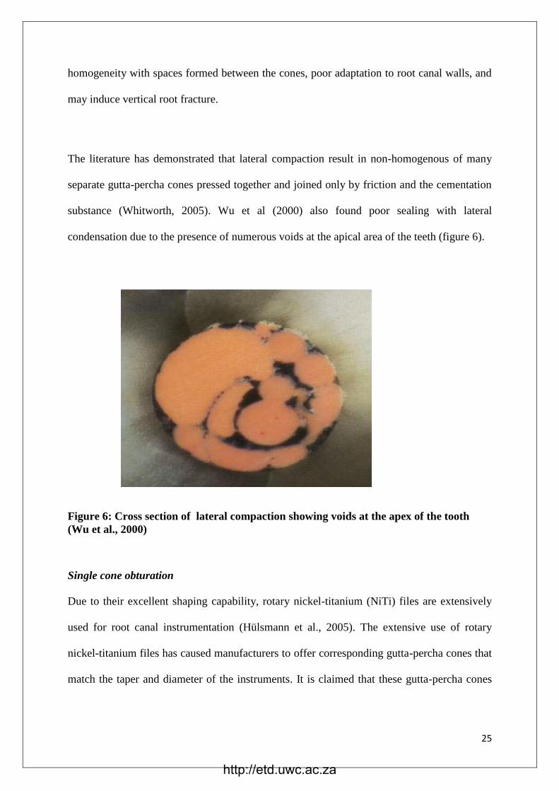
The literature has demonstrated that lateral compaction result in non-homogenous of many
separate gutta-percha cones pressed together and joined only by friction and the cementation
substance (Whitworth, 2005). Wu et al (2000) also found poor sealing with lateral
condensation due to the presence of numerous voids at the apical area of the teeth (figure 6).
Figure 6: Cross section of lateral compaction showing voids at the apex of the tooth
(Wu et al., 2000)
Single cone obturation
Due to their excellent shaping capability, rotary nickel-titanium (NiTi) files are extensively
used for root canal instrumentation (Hülsmann et al., 2005). The extensive use of rotary
nickel-titanium files has caused manufacturers to offer corresponding gutta-percha cones that
match the taper and diameter of the instruments. It is claimed that these gutta-percha cones
http://etd.uwc.ac.za

26
will match the taper and diameter of the canals that are prepared with the rotary NiTi
instruments.
Several authors have evaluated the quality of these single-cone fillings with regards to sealing
ability, bond strength, radiographic quality, and percentage of gutta-percha and sealer-filled
canal area (Nica et al., 2012; Robberecht et al., 2012; Wu et al., 2006; Yilmaz et al., 2009).
Some studies reported comparable results obtained with single-cone obturation compared
with the lateral compaction technique or methods that used thermoplasticized gutta-percha
(Gordon et al., 2005; Nica et al., 2012; Schäfer et al., 2012; Somma et al., 2011; Taşdemir et
al., 2009), whereas other studies found single-cone obturation to result in inferior
obturation (Pommel & Camps, 2001b; Whitworth, 2005; Yücel & Çiftçi, 2006). The root
canal obturation technique utilizing single cone with less taper exhibit less ability to seal
(Monticelli et al., 2007).
The Protaper single-cone obturation was introduced by Dentsply (USA) to modify gutta-
percha root canal sealer balance. With the diameter of the cone corresponding to the final
shaping instrumentation, superior quality of obturation is expected. The root canal is prepared
with the ProTaper files utilizing F1, F2, F3, F4 or F5 as the master files depending on the size
of the root canal (Robberecht et al., 2012). The corresponding single cone which is F1, F2,
F3, F4 or F5, which must always correspond to the master file used, is utilized to obturate the
root canal (figure 7). The system offer great advantage because it provides the gutta-percha
that adapts better to the walls of the root canal system. The closer adaptation of the gutta-
percha to the walls of the root canal helps to push the sealer into the lateral and accessory
canals and thereby provide sealing of these canals. The system is very quick because there are
no lateral cones needed and there is no requirement for the use of a spreader (Robberecht et
al., 2012).
http://etd.uwc.ac.za

27
Figure 7: ProTaper single obturators (Dentsply)
Root canal obturation with these cones used as a single cone technique is alleged to provide a
three dimensional obturation in less time than traditional obturation techniques and to
guarantee a high volume of gutta-percha in the canal (figure 8) (Schäfer et al., 2013)).
Particularly the latter aspect is of clinical significance because gutta-percha is dimensionally
stable, and thus a maximum amount of gutta-percha packed into the canal should be intended
at (Wu & Wesselink, 1997).
.
Figure 8: ProTaper single-cone obturation (Schäfer et al., 2013)
http://etd.uwc.ac.za
28
The combination of single cone and endodontic cement produced a uniform mass which
prevents failures detected among multiple cones (Gomes et al., 2005). The gutta-percha
points of the ProTaper single-cone system were launched into the market highlighting that
they are simpler and result in quicker obturation. In this system, root canals are shaped with
ProTaper instruments and filled with the gutta-percha point size matching the size of the last
instrument used. Their manufacturer claims that the ProTaper single-cone gutta-percha points
fit perfectly within the root canals shaped with the instruments of the same system (Inan et
al., 2009). By using the single-cone and lateral condensation techniques in single-rooted
teeth, Holland et al (2004) assessed the effect of the type of endodontic cement and of the
filling technique on the apical marginal micro-leakage. They found that the single-cone
technique attained the better sealing of the root canal compared to lateral condensation. The
authors concluded that the single-cone technique displayed less marginal leakage than the
lateral condensation technique, but it may be characterized by overfilling, which did not
occur with the lateral condensation technique (Holland et al., 2004).
Inan et al (2009) evaluated the apical sealing among the single-cone, Thermafil and cold
lateral condensation techniques, in mandibular premolars, utilising fluid filtration technique.
After instrumenting the teeth with F3 ProTaper single-cone system, they showed that,
although the lateral condensation group presented a higher leakage than single-cone and
Thermafil techniques, however this difference was not statistically significant. The authors
came to a conclusion that the apical sealing through the use of the single-cone technique is
similar with both the lateral condensation and Thermafil techniques.
http://etd.uwc.ac.za

29
Warm obturation
Warm obturation fulfils the requirement of root canal filling, because homogeneity is
provided throughout the entire length of the filling. Warm obturation is regarded by many
authors as the best form of obturation with good adaptation of gutta-percha to the root canal
walls (Zhang et al., 2011). The apical 2 to 3 mm is well sealed with warm obturation
compared to cold obturation. The majority of canal irregularities, including fins, deltas and
lateral canals, are located in the apical third of the root canal and are often not well filled.
Warm obturation material is able to negotiate through these irregularities because of its flow
characteristics and seal them off. The compaction process can utilize two different methods
namely the downpack method and the backfill method (Zhang et al., 2011).
Downpack method
In the down-pack method, compaction occurs in different waves as heat is apically driven by
pluggers (figure 9).
Figure 9: Downpack method utilising heated probes to obturate apical part of the root
canal (Buchanan, 1994)
http://etd.uwc.ac.za

30
The largest plugger is utilized to compact the gutta-percha apically utilizing finger pressure
(figure 10). The plugger must not contact the canal walls as it can transmit heat to the root.
The next plugger is used to condense the gutta-percha until 4 to 5 mm of the canal is
compacted. This helps in sealing all the canal divisions and accessory canals during the
downpacking process (Whitworth, 2005)
Figure 10: Probes that are used to heat the gutta-percha in a downpacking method with
sizes 0 (.25), 1 (.40) and 2 (.70) (Buchanan, 1994)
Backfill method
In the backfill method, warm gutta-percha is injected into the root canal system by utilizing a
gun (figure 11). The gutta-percha is placed in increments of 3 to 4 mm and compacted by
hand to avoid cooling contraction
http://etd.uwc.ac.za
31
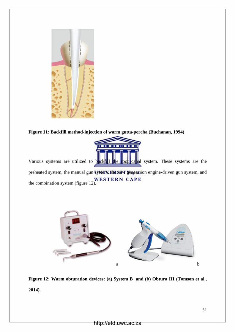
Figure 11: Backfill method-injection of warm gutta-percha (Buchanan, 1994)
Various systems are utilized to backfill the root canal system. These systems are the
preheated system, the manual gun system, the new generation engine-driven gun system, and
the combination system (figure 12).
a b
Figure 12: Warm obturation devices: (a) System B and (b) Obtura III (Tomson et al.,
2014).
http://etd.uwc.ac.za
32
The preheated carpule system is the form of backfill method that utilizes a mini oven to warm
the gutta-percha and delivers viscous gutta-percha, but offers little working time and it is
inexpensive. The manual gun system is also another backfill method that heats the gutta-
percha pallets and maintains the temperature until the gutta-percha is released. In the manual
gun system, working time does not become an issue since there is unlimited working time.
The engine driven system is a new generation system where the heat and the rate of flow are
regulated (Whitworth, 2005).
Combination system
The combination system incorporates both the downpacking and backfilling devices to
obturate the root canal. The example of this device is the use of System B in root canal
obturation. The gutta-percha is deposited in increments and compacted. There is
downpacking of the apical part first, which is then followed by backpacking of the coronal
part of the root canal. The warm obturation system has a disadvantage of extruding the sealer
beyond the apex hence zinc oxide eugenol sealers are mostly recommended, since they do not
cause any damaging effects to the apical tissue (Whitworth, 2005).
The carrier system
The carrier based systems consist of a rigid core which normally guides the soft gutta-percha
through the root canal during root canal obturations. Thermafil and GuttaCore are the two
popular carrier based systems that are available in the market. Thermafil and GuttaCore have
higher rigidities than conventional gutta-percha. This can be attributed to the strengthened
central cores of the former two types of gutta-percha. Both Thermafil and GuttaCore rely on
heating to make their circumferential gutta-percha flowable during canal insertion (Patel &
Owen, 2016). Carriers for core-based techniques can be made-up using different materials;
http://etd.uwc.ac.za

33
Thermafil small size obturators (up to size 40) (Tulsa Dental Dentsply, Tulsa, OK, USA) are
made of Vectra, which is a liquid crystal polymer and larger sizes are made of polysulfone,
whereas GuttaCore carriers (Tulsa Dental Dentsply) are made of cross-linked gutta-percha.
These materials are covered with alpha-phase gutta-percha. One disadvantage of a carrier-
based root filling system is denudation of the core with shedding of the gutta-percha coating
(Alhashimi et al., 2014a). Shedding of gutta-percha from the carrier might occur during the
insertion of the carriers into the root canal system, particularly in constricted or rigorously
curved canals. This would lead to voids and insufficient filling of the root canal space (Weller
N, Kimbrough F, 1997). Earlier studies have demonstrated that the most common causes of
shedding of the gutta-percha coating are winding the carrier during insertion into the root
canal space and insufficient amounts of sealer placed prior to insertion of the obturators in the
root canal (DuLac et al., 1999; Levitan et al., 2003).
Connection between the carrier and gutta-percha coating is therefore a vital aspect in the
choice of a core-based obturation system and would aid in avoidance of stripping of the
gutta-percha coating, producing a root canal filling with lesser voids. Another disadvantage
of presently available carrier-based obturation systems is that the volume of gutta-percha is
not consistently distributed around the carrier. This might cause shedding of the gutta-percha
from the carrier material when the obturator is inserted into the root canal space also leading
to possible voids (Alhashimi et al., 2014). The frictional forces present among the gutta-
percha and the root canal walls may generate an extrusion effect, whereby the filling material
is retained at the orifice of the canal (Bertacci et al., 2007).
http://etd.uwc.ac.za

34
Thermafil
Thermafil is the warm gutta-percha that contains a central core of carrier (figure 13).
Thermafil has an advantage over lateral compaction due to its three dimensional root canal
obturation. There is build-up of excess gutta-percha at the apical area, and Thermafil was
found to have advantages especially in straight canals (Juhlin et al., 1993). In curved canals,
the carrier cannot follow the curvature of the root canal walls leading to perforation of the
gutta-percha (Juhlin et al., 1993). This lead to the root canal walls becoming in contact with
the carrier, leading to inadequate obturation (Juhlin et al., 1993). According to Gutmann
(1993), the obturation of curved root canals with Thermafil resulted in a denser and well
adapted root canal filling throughout the entire root canal system. The author noted that even
though Thermafil produced good results, there was a significant amount of material extrusion
beyond the apex in Thermafil obturations (Gutmann et al., 1993).
Figure 13: Thermafil gutta-percha with carrier (Tomson et al., 2014)
Thermafil also comes in various sizes which are colour coded and labelled as 20, 25, 30, 35
and 40 which corresponds to the files of ProTaper Universal system. Thermafil gutta-percha
is heated in thermoprep oven prior to placement in the root canal system (figure 14). The
original carrier Thermafil system was a tapered helix made of stainless steel coated with
gutta-percha. The newer Thermafil carrier is made of titanium or thermoplastic polymer,
Polysulfone and Vectra (Sutow et al., 1999).
http://etd.uwc.ac.za

35
Figure 14: Thermoprep oven for heating Thermafil (Tomson et al., 2014)
GuttaCore
Recently a cross-linked gutta-percha carrier, GuttaCore has been developed (figure 15).
Figure 15: GuttaCore carrier system (Gutmann, 2008)
GuttaCore was introduced in order to provide the clinician with better root canal filling
technique, material advances and its chemistry allow for the development of a superior core
http://etd.uwc.ac.za
36
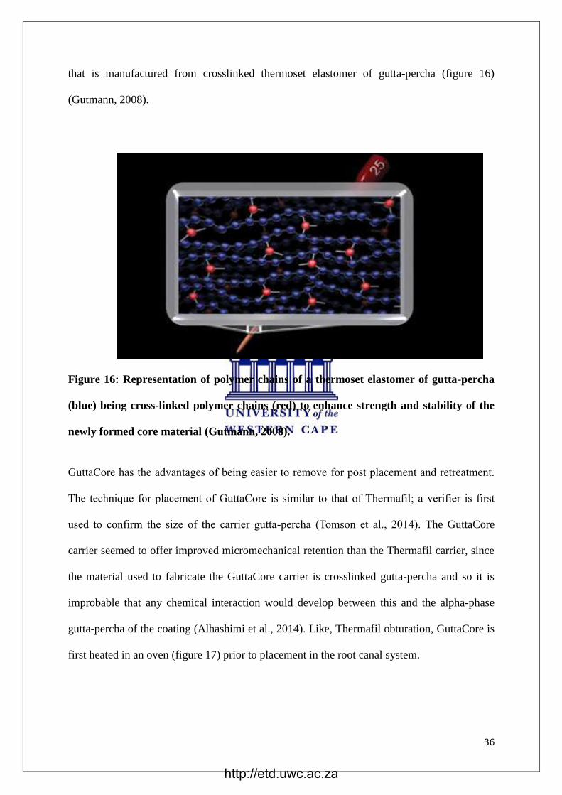
that is manufactured from crosslinked thermoset elastomer of gutta-percha (figure 16)
(Gutmann, 2008).
Figure 16: Representation of polymer chains of a thermoset elastomer of gutta-percha
(blue) being cross-linked polymer chains (red) to enhance strength and stability of the
newly formed core material (Gutmann, 2008).
GuttaCore has the advantages of being easier to remove for post placement and retreatment.
The technique for placement of GuttaCore is similar to that of Thermafil; a verifier is first
used to confirm the size of the carrier gutta-percha (Tomson et al., 2014). The GuttaCore
carrier seemed to offer improved micromechanical retention than the Thermafil carrier, since
the material used to fabricate the GuttaCore carrier is crosslinked gutta-percha and so it is
improbable that any chemical interaction would develop between this and the alpha-phase
gutta-percha of the coating (Alhashimi et al., 2014). Like, Thermafil obturation, GuttaCore is
first heated in an oven (figure 17) prior to placement in the root canal system.
http://etd.uwc.ac.za

37
Figure 17: A GuttaCore oven is used to simplify the heating of the core carrier. Two
cores may be heated simultaneously (Gutmann, 2008)
Summary of filling techniques in a 3D view
Various forms of obturations are demonstrated in figure 18, where lateral compaction (figure
18c) and single cone obturation (figure 18b) are found not to provide good apical seal since
they do not provide the obturation of the isthmus of the canal. Thermal obturation (figure
18d) and paste only obturation (figure 18a) provided excellent obturation when viewed in a
three dimension. The disadvantage of paste only system is that with dissolution of the paste
by apical fluid will result in re-infection of the root canal. The paste system has a poor
reputation in endodontics due to the present of the toxic elements like formaldehyde within
the paste (Whitworth, 2005).
Figure 18: Spectrum of filling techniques, showing (A) paste only, (B) single cones with
paste, (C) cold lateral condensation, (D) thermoplastic compaction (Whitworth, 2005).
http://etd.uwc.ac.za

38
Apical and coronal seal
Apical seal
It is known that the gutta-percha does not bond to tooth structure unless it is resin infiltrated.
A sealer is used to seal the apical area to avoid infection by micro-organisms. It has been
accepted that the ‘triad’ of preparation, disinfection and canal obturation is the key to success
in endodontics. Without apical seal, apical fluids diffuse into the empty canal space, stagnate,
undergo degradation, and then act as physiochemical irritants when they diffuse back into the
periapical tissue (Machtou, 2002).
Coronal seal
The canal may be contaminated because of contact between oral microbial flora and root
canal inlets. Sometimes it may occur due to the loss of the temporary filling or inadequate
sealing of the restoration. The lack of apical and coronal seal has been proven by several in
vitro studies to lead to most endodontic failures (Machtou, 2002).
Root canal sealers
The sealers are responsible for the principal functions of the final root filling. They are
helpful in sealing the core and irregularities in the root canal. Various types of root canal
sealers are shown in table 2 below. The sealers are classified according to the type, brand,
principle composition and the manufacturing company. All these root canal sealers are useful
in providing an excellent apical seal. They seal the gap between the gutta-percha and the root
canal wall at the apical part of the tooth (Ørstavik, 2005).
http://etd.uwc.ac.za
39
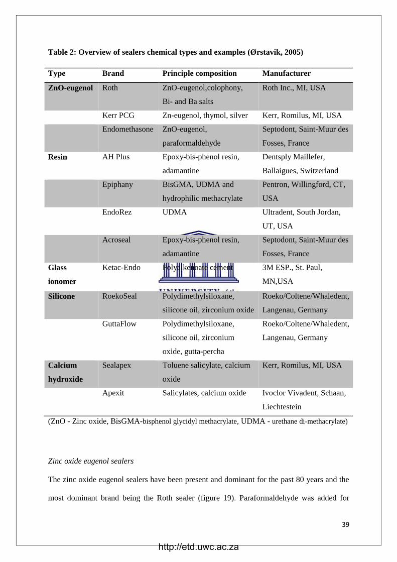
Table 2: Overview of sealers chemical types and examples (Ørstavik, 2005)
Type Brand Principle composition Manufacturer
ZnO-eugenol Roth ZnO-eugenol,colophony,
Bi- and Ba salts
Roth Inc., MI, USA
Kerr PCG Zn-eugenol, thymol, silver Kerr, Romilus, MI, USA
Endomethasone ZnO-eugenol,
paraformaldehyde
Septodont, Saint-Muur des
Fosses, France
Resin AH Plus Epoxy-bis-phenol resin,
adamantine
Dentsply Maillefer,
Ballaigues, Switzerland
Epiphany BisGMA, UDMA and
hydrophilic methacrylate
Pentron, Willingford, CT,
USA
EndoRez UDMA Ultradent, South Jordan,
UT, USA
Acroseal Epoxy-bis-phenol resin,
adamantine
Septodont, Saint-Muur des
Fosses, France
Glass
ionomer
Ketac-Endo Polyalkenoate cement 3M ESP., St. Paul,
MN,USA
Silicone RoekoSeal Polydimethylsiloxane,
silicone oil, zirconium oxide
Roeko/Coltene/Whaledent,
Langenau, Germany
GuttaFlow Polydimethylsiloxane,
silicone oil, zirconium
oxide, gutta-percha
Roeko/Coltene/Whaledent,
Langenau, Germany
Calcium
hydroxide
Sealapex Toluene salicylate, calcium
oxide
Kerr, Romilus, MI, USA
Apexit Salicylates, calcium oxide Ivoclor Vivadent, Schaan,
Liechtestein
(ZnO - Zinc oxide, BisGMA-bisphenol glycidyl methacrylate, UDMA - urethane di-methacrylate)
Zinc oxide eugenol sealers
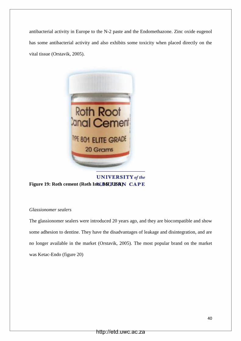
The zinc oxide eugenol sealers have been present and dominant for the past 80 years and the
most dominant brand being the Roth sealer (figure 19). Paraformaldehyde was added for
http://etd.uwc.ac.za
40
antibacterial activity in Europe to the N-2 paste and the Endomethazone. Zinc oxide eugenol
has some antibacterial activity and also exhibits some toxicity when placed directly on the
vital tissue (Orstavik, 2005).
Figure 19: Roth cement (Roth Inc., MI, USA)
Glassionomer sealers
The glassionomer sealers were introduced 20 years ago, and they are biocompatible and show
some adhesion to dentine. They have the disadvantages of leakage and disintegration, and are
no longer available in the market (Orstavik, 2005). The most popular brand on the market
was Ketac-Endo (figure 20)
http://etd.uwc.ac.za

41
Figure 20: Ketac-Endo glass ionomer sealer (3M ESP., St Paul, MN, USA)
Resin bonded sealers
The resin bonded sealers are bis-phenol resin that use the methamine for polymerization, and
the example of these sealers is the AH Plus (figure 21).
Figure 21: AH Plus resin bonded sealer (Dentsply Maillefer, Ballaigues, Switzetland)
The resin bonded sealers give off some formaldehyde during setting since it is a methamine.
AH plus sealer was developed to counter the release of formaldehyde during polymerization.
Another example of this group is the Bakelite resin, which has strong antibacterial properties,
but it shrinks and leaves a reddish hue on the surrounding tooth structure.
http://etd.uwc.ac.za

42
It can be used without gutta-percha core, and set to a very hard and insoluble mass. The teeth
that are obturated with this material result in difficult endodontic retreatment (Orstavik,
2005). Another example of this group is EndoRez which is a urethane dimethacrylate sealer
which has hydrophilic properties. It has good performance in the presence of moisture,
together with resin-coated gutta-percha provide enhanced adhesion and seal throughout the
filling mass. The combination of a sealer, primer and the core, give rise to a concept of
‘monoblock’ (Orstavik, 2005).
Calcium hydroxide sealers
The most popular brand in calcium hydroxide sealers is Sealapex, which has a complex and
an inhomogeneous setting reaction (figure 22).
Figure 22: Sealapex root canal sealer (Kerr, Romilus, MI, USA)
A hard surface is produced through contact with moisture, but the deeper mix may remain in
dough-like consistency. The material has a disadvantage of lacking physical sturdiness
(strength or hardness) (Orstavik, 2005).
http://etd.uwc.ac.za
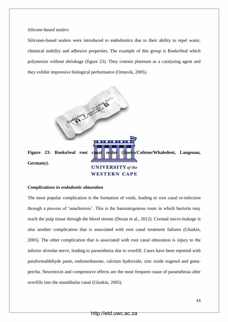
43
Silicone-based sealers
Silicones-based sealers were introduced to endodontics due to their ability to repel water,
chemical stability and adhesive properties. The example of this group is RoekoSeal which
polymerize without shrinkage (figure 23). They contain platinum as a catalysing agent and
they exhibit impressive biological performance (Orstavik, 2005).
Figure 23: RoekoSeal root canal sealer (Roeko/Coltene/Whaledent, Langenau,
Germany).
Complications in endodontic obturation
The most popular complication is the formation of voids, leading to root canal re-infection
through a process of ‘anachoresis’. This is the haematogenous route in which bacteria may
reach the pulp tissue through the blood stream (Dezan et al., 2012). Coronal micro-leakage is
also another complication that is associated with root canal treatment failures (Gluskin,
2005). The other complication that is associated with root canal obturation is injury to the
inferior alveolar nerve, leading to paraesthesia due to overfill. Cases have been reported with
paraformaldehyde paste, endomethasone, calcium hydroxide, zinc oxide eugenol and gutta-
percha. Neurotoxin and compressive effects are the most frequent cause of paraesthesia after
overfills into the mandibular canal (Gluskin, 2005).
http://etd.uwc.ac.za
44
The adaptation of the gutta-percha to the root canal wall
For the filling material to be effective, it must have a close adaptation to the walls of the root
canals. This will ensure the sealing of all lateral and accessory canals that are frequently
found on the root canal walls. The other benefit is the ability to seal the canal thereby
avoiding colonization of micro-organisms in the spaces within the root canal system. The
adaptation of gutta-percha to the walls of the root canal depends on the successful removal of
the dentine smear layer (Gengoglu et al., 1993). Lack of a perfect fitting cone due to
manufacturing specifications may also lead to gutta-percha not sealing the apical area of the
canal. The gutta-percha alone, with tug-back at the apical area of the canal, does not
necessary have a perfect seal without a root canal sealer (Silva-filho et al., 2013).
The other problem that was encountered in obturation of root canals was the sealing of lateral
and accessory canals. It was demonstrated in one study that the combination system produced
better results in the sealing of lateral and accessory canals (Robberecht et al., 2012). Other
authors evaluated the apical sealing of lateral compaction and warm obturation system during
root canal obturation (ElAyouti et al., 2009; Silva-filho et al., 2013). In one study, there was
no difference in apical seal between cold lateral compaction and warm obturation during root
canal obturation (Silva-filho et al., 2013).
Vertical compaction demonstrated good adaptation with minimal sealer, and in GuttaFlow,
voids were more abundant in coronal section. In Thermafil obturation, the carrier contact
dentinal walls (figure 24) (ElAyouti et al., 2009). At 1mm from apex, Thermafil
demonstrated less adaptation with abundance of voids. The increase in voids at 1mm were
thought to be due to blocking of canal orifice with softened Thermafil obturator causing
entrapment of air which could not escape through the apical foramen
http://etd.uwc.ac.za

45
Figure 24: Adaptation of apical cone, vertical compaction, GuttaFlow and Thermafil
(ElAyouti et al., 2009)
Putting a cone in warm water first and then inserting it into the canal was recommended
(Chohayeb, 1992). This will ensure that the cone record the impression of the apical part of
the root canal in three dimensions The literature has demonstrated that Thermafil produce
good adaptation to the walls of the canals as compared to lateral compaction and single-cone
obturation (Chohayeb, 1992).
Warm obturation also produced better obturation density when comparing continuous wave
of condensation with cold lateral compaction (Lea et al., 2005). Poor adaptation of gutta-
percha to the root canal wall was observed due to the presence of smear layer (figure 25).
According to Haapsalo et al (2010), the smear layer may prevent penetration of root canal
filling to the dentinal tubules and affects the adaptation of the filling material to the wall of
the root canal.
http://etd.uwc.ac.za

46
Figure 25: Lateral compaction with smear layer, D, dentine; S, sealer (Gengoglu et al.,
1993).
Presence of smear layer obstruct proper adaptation of gutta-percha to the walls of the root
canal. Proper adaptation of lateral compacted gutta-percha can be seen in the absence of
smear layer (figure 26) (Gengoglu et al., 1993).
Figure 26: Lateral compaction without smear layer, D, dentine; S, sealer (Gengoglu et
al., 1993).
There was good adaptation of thermoplastic gutta-percha to the walls of the root canal in the
presence of smear layer compared to lateral compaction (figure 27) (Gengoglu et al., 1993).
http://etd.uwc.ac.za

47
Figure 27: Thermoplastic gutta-percha in presence of smear layer, D, dentine; G, gutta-
percha (Gencoglu et al., 1993).
Methods used to study adaptation of obturation materials
Dye penetration
Various types of dyes like eosin, methylene blue; Black Indian Ink and Procion brilliant blue
are used to evaluate adaptation and micro-leakage of the gutta-percha (Veríssimo & do Vale,
2006). The dye penetration test is a relatively easy test to perform. Methylene blue is the most
commonly used test due to its high penetration and tinting ability. The roots are sectioned
after dye penetration which may be time consuming because the time needed for each section
is between 20 and 30 minutes (ElAyouti et al., 2009). The teeth are submerged into a dye and
the dye penetrates into the spaces between the walls of the canals and the restorative material.
The teeth are then sectioned and the linear dye penetration is recorded. Microscopy can be
used at various magnifications to check adaptation of gutta-percha to the walls of the root
canal.
A clearing technique was recommended as a method to be used in dye penetration method.
The teeth become transparent after being demineralized, dehydrated and immersed into a
http://etd.uwc.ac.za

48
solution of methyl salicylate (Veríssimo & do Vale, 2006). The transparent teeth will then
provide a three dimensional view of the internal anatomy of the root canal. The clearing
technique is a fast and a simple technique that prevents the loss of dentine substance. The
dye penetration is one of the oldest methods used to evaluate gutta-percha leakage and
adaptation but sample preparations are technique sensitive and may be damaged during the
process (Tripi et al., 2001).
Fluid filtration or transportation methodology
The fluid filtration method was modified by Wu et al in 1994. In this technique, the sealing
capacity is measured by movement of air bubbles inside the capillary tube. The coronal
portion of the filled canal is connected to a tube that is filled with water under atmospheric
pressure and the apex is connected to a 20µL glass capillary tube that is 170mm long with
uniform calibre and filled with water (Wu et al., 1994). An atmospheric pressure is applied
coronally and forces the water to pass through the spaces within the root canal. This method
has the advantage over dye penetration as samples are not destroyed and is useful in assessing
both the apical and coronal seal. There is an elimination of an operator error as results are
recorded automatically. This technique is not standardised as there might be pressure that
may range from 10 to 20 psi and also the time to measure this can be between 1 min to 3
hours (Pommel & Camps, 2001a).
Dye extraction method
In the dye extraction method, an acid is utilised to release the dye from the teeth. The teeth
are dissolved in an acid and the dye is released from the interface and the optical density is
measured utilising a spectrophotometer. The dye extraction method is quick and the
equipment is available at most institutions. This method is far more superior to the fluid
http://etd.uwc.ac.za
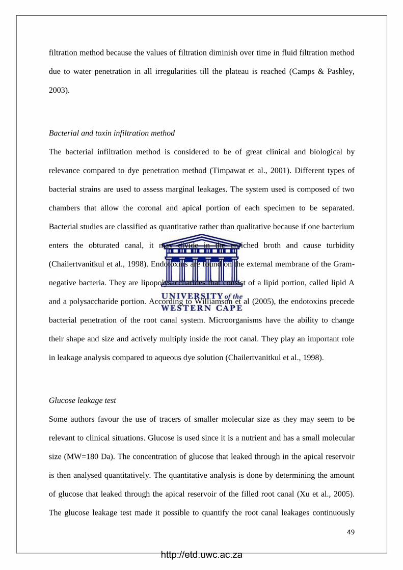
49
filtration method because the values of filtration diminish over time in fluid filtration method
due to water penetration in all irregularities till the plateau is reached (Camps & Pashley,
2003).
Bacterial and toxin infiltration method
The bacterial infiltration method is considered to be of great clinical and biological by
relevance compared to dye penetration method (Timpawat et al., 2001). Different types of
bacterial strains are used to assess marginal leakages. The system used is composed of two
chambers that allow the coronal and apical portion of each specimen to be separated.
Bacterial studies are classified as quantitative rather than qualitative because if one bacterium
enters the obturated canal, it may divide in the enriched broth and cause turbidity
(Chailertvanitkul et al., 1998). Endotoxins are found on the external membrane of the Gram-
negative bacteria. They are lipopolysaccharides that consist of a lipid portion, called lipid A
and a polysaccharide portion. According to Williamson et al (2005), the endotoxins precede
bacterial penetration of the root canal system. Microorganisms have the ability to change
their shape and size and actively multiply inside the root canal. They play an important role
in leakage analysis compared to aqueous dye solution (Chailertvanitkul et al., 1998).
Glucose leakage test
Some authors favour the use of tracers of smaller molecular size as they may seem to be
relevant to clinical situations. Glucose is used since it is a nutrient and has a small molecular
size (MW=180 Da). The concentration of glucose that leaked through in the apical reservoir
is then analysed quantitatively. The quantitative analysis is done by determining the amount
of glucose that leaked through the apical reservoir of the filled root canal (Xu et al., 2005).
The glucose leakage test made it possible to quantify the root canal leakages continuously
http://etd.uwc.ac.za

50
over time. In this method a low pressure is used and this helps in ruling out entrapped air of
fluid and is sufficient for a device with high sensitivity (Xu et al., 2005).
Periapical radiographs
The role of periapical radiographs before and after treatment cannot be overemphasised. The
diagnosis of the pulp and periapical conditions, the anatomy of the root canal, preparation and
obturation of the root canal, all play a role in the success of treatment in endodontics. Intra-
oral radiographs are also widely used to evaluate adaptation of obturation materials but have
the disadvantage of showing a two dimensional view of the image. A proper radiation
technique which includes the use of paralleling technique and film holders is advocated in
other to have a clear image of the tooth. Radiation images are supposed to be free of
distortion and superimposition of adjacent anatomical landmarks which can lead to difficult
interpretation (Fava and Dummer, 1997).
Tuned aperture computed tomography (TACT)
In tuned aperture tomography, 8 to 10 series of radiographic images are taken at different
projection geometries. An imaging unit with specialised software is used to reconstruct a
three dimensional data set which may be viewed slice by slice (Patel et al., 2009). The
advantage of TACT is that the images produced show less super imposition of noise over the
region of interest. There is less radiation dose in TACT compared to conventional periapical
radiographs. There is also an absence of artefacts resulting from radiation interaction with
metallic fillings. TACT is still a research tool but is a promising radiographic technique for
the future (Patel et al., 2009).
http://etd.uwc.ac.za

51
Cone beam computed tomography (CBCT)
In radiographic x-ray images, the three dimension object is compressed into a two dimension
image. This leads to superimposition of image and limits the aspect of a three dimension
anatomy. To overcome this, the CBCT was developed in the late 1990 by the Italian and
Japanese group (Arai et al., 1999; Mozzo et al., 1998). The two groups were working
independently and developed computed tomography (CT) specifically for maxillofacial and
dental use. The CT scanner was known as cone beam computed tomography (CBCT) or
digital volume tomography (DVT). The image is acquired in the course of a single sweep of
the scanner and the x-ray beam is cone shaped. Some cone beam CT scanners can be adjusted
to capture only the maxilla or the mandible (Patel, 2009). Images can be scanned in 10 to 40
seconds in CBCT which makes CBCT dose less compared to conventional CT scanners
(Patel et al., 2007).
The effective dose of CBCT is high compared to conventional periapical or panoramic
radiographs (Scarfe & Farman, 2008). The costs of CBCT are much less than that of
conventional CT and there is 60% more periapical radiolucency detection in CBCT compared
to periapical radiographs (Patel et al., 2007). The drawback of CBCT is its spatial resolution
which is about 2 line pairs per millimetre compared to 10 to 15 line pairs per millimetre for
conventional and digital radiographs. The limitations of CBCT are in cases where metal
restoration, metal posts and root fillings, and adjacent dental implants because they cause the
artefact to the reconstructed image (Scarfe & Farman, 2008).
Microcomputed tomography (Micro-CT)
Micro-computed tomography (Micro-CT) has been described as a new and exciting tool in
endodontics to assess the geometry of the root canal (Peters et al., 2001). High resolution
http://etd.uwc.ac.za

52
micro-CT is an emerging technology with several promising application in different fields of
dentistry. It is highly accurate and non-destructive method for in-vitro evaluation of root
canal fillings (Zogheib et al., 2013). Micro-CT has been used as a research tool in
endodontics to evaluate root canal anatomy and assessment of root canal morphology after
instrumentation (Hammad et al., 2009). Micro-CT provides a three dimensional view of the
root canal system by providing an undistorted image of the tooth. It has the highest resolution
at a very low exposure compared to a conventional CT scan. Micro-CT can be a useful tool to
check for gap formation between the root canal filling and the dentine walls because it is a
less complicated technique compared to conventional methods, specimens are not damaged
and images can be viewed in 3 dimensions. Most of the studies on adaptation of root canal
filling material to the walls of the canal utilized dyes and scanning electron microscopy as a
form of evaluation.
Micro-CT is new and advanced technology that can provide instant and accurate three
dimensional results of root canal obturation. This nondestructive imaging tool overcomes the
limitations of the previously used models like dye penetration, fluid transport, and cross-
section analyses, which are valuable techniques, but the results do not always corroborate
with each other (Zogheib et al., 2013).
A brief historical review on material adaptation
Several studies were done to assess the adaptability of thermal obturations to the wall of the
root canal. Chohayeb et al (1992) found more leakages on teeth obturated with Thermafil
compared to those obturated with lateral condensation. They also noted some degree of
overfill in teeth that were obturated with Thermafil. They attributed the leakages to the
composition of alpha phase gutta-percha. Their results differed with those performed by
http://etd.uwc.ac.za
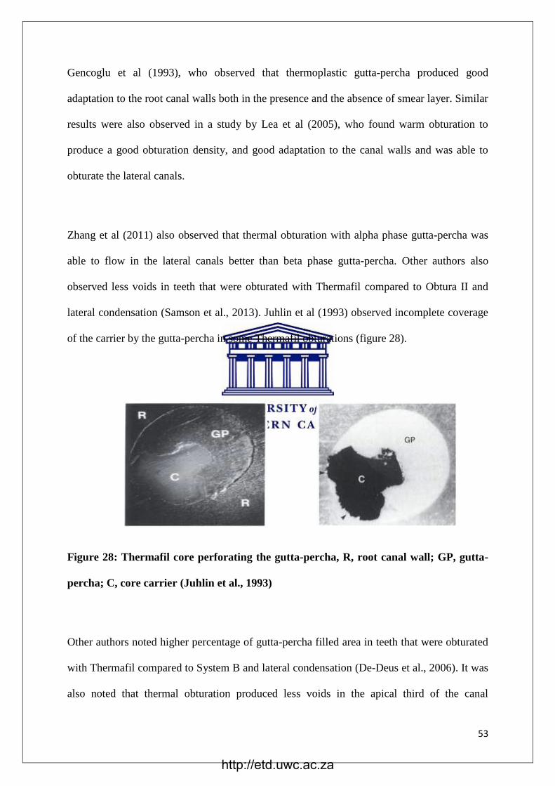
53
Gencoglu et al (1993), who observed that thermoplastic gutta-percha produced good
adaptation to the root canal walls both in the presence and the absence of smear layer. Similar
results were also observed in a study by Lea et al (2005), who found warm obturation to
produce a good obturation density, and good adaptation to the canal walls and was able to
obturate the lateral canals.
Zhang et al (2011) also observed that thermal obturation with alpha phase gutta-percha was
able to flow in the lateral canals better than beta phase gutta-percha. Other authors also
observed less voids in teeth that were obturated with Thermafil compared to Obtura II and
lateral condensation (Samson et al., 2013). Juhlin et al (1993) observed incomplete coverage
of the carrier by the gutta-percha in some Thermafil obturations (figure 28).
Figure 28: Thermafil core perforating the gutta-percha, R, root canal wall; GP, gutta-
percha; C, core carrier (Juhlin et al., 1993)
Other authors noted higher percentage of gutta-percha filled area in teeth that were obturated
with Thermafil compared to System B and lateral condensation (De-Deus et al., 2006). It was
also noted that thermal obturation produced less voids in the apical third of the canal
http://etd.uwc.ac.za

54
(Hammad et al., 2009). While ElAyout et al (2009) reported contradicting result in teeth
obturated with Thermafil. They found Thermafil to produce more voids and less homogenous
obturation at apical level of the canals compared to GuttaFlow, apical cone and vertical
compaction. With regards to sealer distribution and sealer extrusion, warm obturation
produced better results. There was less sealer distribution especially at the apical level on
teeth obturated with warm obturation compared to lateral condensation and single-cone
obturation (Wu et al., 2000). ElAyouti et al (2009) also observed less sealer distribution in
teeth that were obturated with Thermafil. Samson et al (2013) observed sealer extrusion in
canals that were obturated with Thermafil.
Schäfer et al (2013) did a study on crosslinked gutta-percha utilising GuttaCore as one of the
crosslinked materials. The crosslinked carrier systems produced constant obturation of
tapered preparations. The obturation with GuttaCore was associated with fewer voids when
compared to lateral compaction during obturation of canals that were enlarged with hand
instruments. There were a considerable high number of void free specimens in canals that
were obturated with GuttaCore. GuttaCore also produced a lower percentage of sealer filled
areas than lateral compaction. GuttaCore also demonstrated good obturation of lateral canals
(figure 29). Their results demonstrated good material adaptation of crosslinked gutta-percha.
http://etd.uwc.ac.za
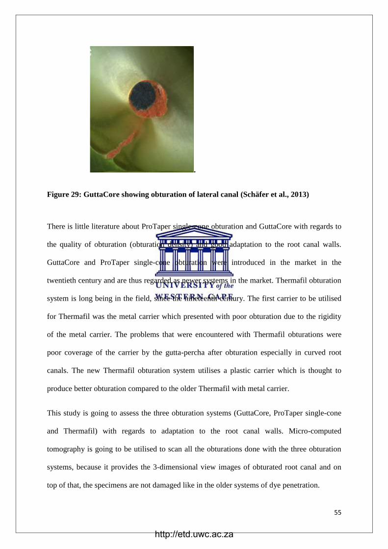
55
.
Figure 29: GuttaCore showing obturation of lateral canal (Schäfer et al., 2013)
There is little literature about ProTaper single-cone obturation and GuttaCore with regards to
the quality of obturation (obturation density) and good adaptation to the root canal walls.
GuttaCore and ProTaper single-cone obturation were introduced in the market in the
twentieth century and are thus regarded as newer systems in the market. Thermafil obturation
system is long being in the field, since the nineteenth century. The first carrier to be utilised
for Thermafil was the metal carrier which presented with poor obturation due to the rigidity
of the metal carrier. The problems that were encountered with Thermafil obturations were
poor coverage of the carrier by the gutta-percha after obturation especially in curved root
canals. The new Thermafil obturation system utilises a plastic carrier which is thought to
produce better obturation compared to the older Thermafil with metal carrier.
This study is going to assess the three obturation systems (GuttaCore, ProTaper single-cone
and Thermafil) with regards to adaptation to the root canal walls. Micro-computed
tomography is going to be utilised to scan all the obturations done with the three obturation
systems, because it provides the 3-dimensional view images of obturated root canal and on
top of that, the specimens are not damaged like in the older systems of dye penetration.
http://etd.uwc.ac.za

56
Chapter 3
Aims and objectives
Aims
The aim of this study was to evaluate the adaptation of gutta-percha of three obturation
systems within the root canal system using microcomputed tomography (micro-CT).
Objectives
1. To determine and measure the gap formation at three different levels of the root
following obturation with GuttaCore, Thermafil and ProTaper
2. To determine and compare the apical sealing ability of the obturation techniques
following obturation with GuttaCore, Thermafil and ProTaper single-cone obturation
techniques.
3. To calculate percentage of volume of voids in root canals obturated with three
different obturation methods, GuttaCore, Thermafil and ProTaper single-cone
obturation.
4. To compare the adaptation of the three obturation techniques namely GuttaCore,
Thermafil and ProTaper single-cone obturation
Null hypothesis
The null hypothesis tested was that there are no differences in the percentage of interfacial
gaps and voids in single-rooted canals when obturating with the GuttaCore core-carrier
technique, ProTaper single-cone gutta-percha obturation technique and Thermafil core-carrier
technique when obturating with the same root canal sealer.
http://etd.uwc.ac.za

57
Ethics statement
This research proposal was presented to the Research Committee of the Faculty of Dentistry
of the University of Western Cape and to the Senate Research Committee for ethics approval
and for registration as a research project (Project no SHD 2014/12, approved on 22/10/2014).
This research is a laboratory study that involves the use of extracted teeth (see appendix for
consent form). This research was not supported by any research grant from any foundation or
company, and the researcher declares that there is no conflict of interest.
http://etd.uwc.ac.za
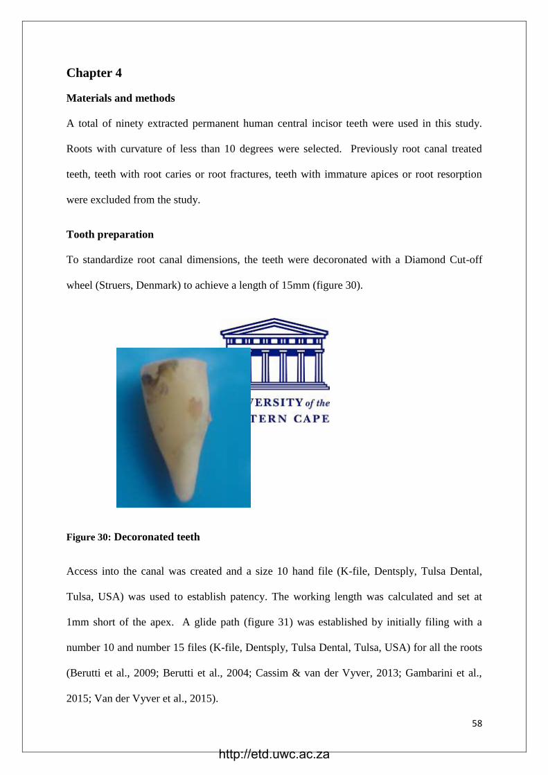
58
Chapter 4
Materials and methods
A total of ninety extracted permanent human central incisor teeth were used in this study.
Roots with curvature of less than 10 degrees were selected. Previously root canal treated
teeth, teeth with root caries or root fractures, teeth with immature apices or root resorption
were excluded from the study.
Tooth preparation
To standardize root canal dimensions, the teeth were decoronated with a Diamond Cut-off
wheel (Struers, Denmark) to achieve a length of 15mm (figure 30).
Figure 30: Decoronated teeth
Access into the canal was created and a size 10 hand file (K-file, Dentsply, Tulsa Dental,
Tulsa, USA) was used to establish patency. The working length was calculated and set at
1mm short of the apex. A glide path (figure 31) was established by initially filing with a
number 10 and number 15 files (K-file, Dentsply, Tulsa Dental, Tulsa, USA) for all the roots
(Berutti et al., 2009; Berutti et al., 2004; Cassim & van der Vyver, 2013; Gambarini et al.,
2015; Van der Vyver et al., 2015).
http://etd.uwc.ac.za

59
Figure 31: Glide path preparation with a No. 15 K-file showing file upright in canal
The ProTaper Universal files (Dentsply Tulsa Dental, Tulsa, USA) were used to clean and
shape the root canal up to the master file (figure 32).
Figure 32: ProTaper rotary files (Dentsply Tulsa Dental, Tulsa, USA)
Rotary ProTaper Universal files (Dentsply Tulsa Dental, Tulsa, USA) were used with wave
one rotary hand-piece (figure 33).
http://etd.uwc.ac.za
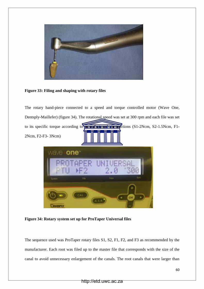
60
Figure 33: Filing and shaping with rotary files
The rotary hand-piece connected to a speed and torque controlled motor (Wave One,
Dentsply-Maillefer) (figure 34). The rotational speed was set at 300 rpm and each file was set
to its specific torque according to manufacturer’s instructions (S1-2Ncm, S2-1.5Ncm, F1-
2Ncm, F2-F3- 3Ncm)
Figure 34: Rotary system set up for ProTaper Universal files
The sequence used was ProTaper rotary files S1, S2, F1, F2, and F3 as recommended by the
manufacturer. Each root was filed up to the master file that corresponds with the size of the
canal to avoid unnecessary enlargement of the canals. The root canals that were larger than
http://etd.uwc.ac.za

61
the standard F3 file were excluded from the study. During preparation and between each file,
1ml of 5.25% sodium hypochlorite (figure 35) was used as an irrigant to debride the canal
(Palazzi et al., 2012).
Figure 35: Sodium hypochlorite irrigation solution (Vista)
After completion of instrumentation, all specimens were flushed with 5ml of 17% ethylene-
diamine-tetra-acetic acid (EDTA) (figure 36) and a final rinse with sodium hypochlorite to
rinse off the EDTA from the root canal walls to remove the smear layer (Aslantas et al.,
2014).
http://etd.uwc.ac.za

62
Figure 36: Smear clear irrigation solution (17% EDTA) (Kerr)
All the canals were dried with paper points and were ready for obturation. The roots in all the
three groups were treated by a single operator and all files were replaced after five uses.
Root canal obturation
The roots were randomly divided into three groups of thirty each and each group was
obturated with a different obturation technique (n=30). The roots in Group 1 were obturated
with GuttaCore obturation system (figure 37).
http://etd.uwc.ac.za
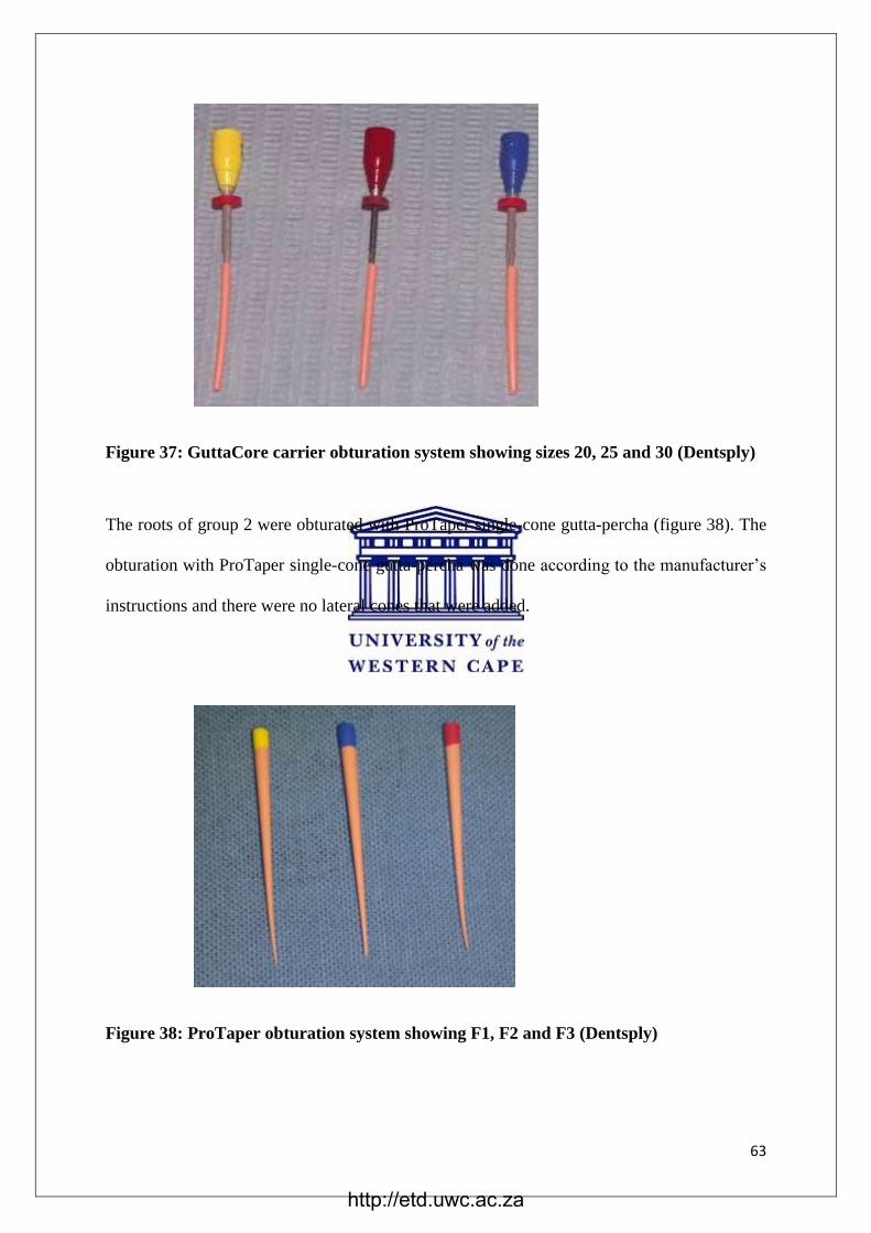
63
Figure 37: GuttaCore carrier obturation system showing sizes 20, 25 and 30 (Dentsply)
The roots of group 2 were obturated with ProTaper single-cone gutta-percha (figure 38). The
obturation with ProTaper single-cone gutta-percha was done according to the manufacturer’s
instructions and there were no lateral cones that were added.
Figure 38: ProTaper obturation system showing F1, F2 and F3 (Dentsply)
http://etd.uwc.ac.za
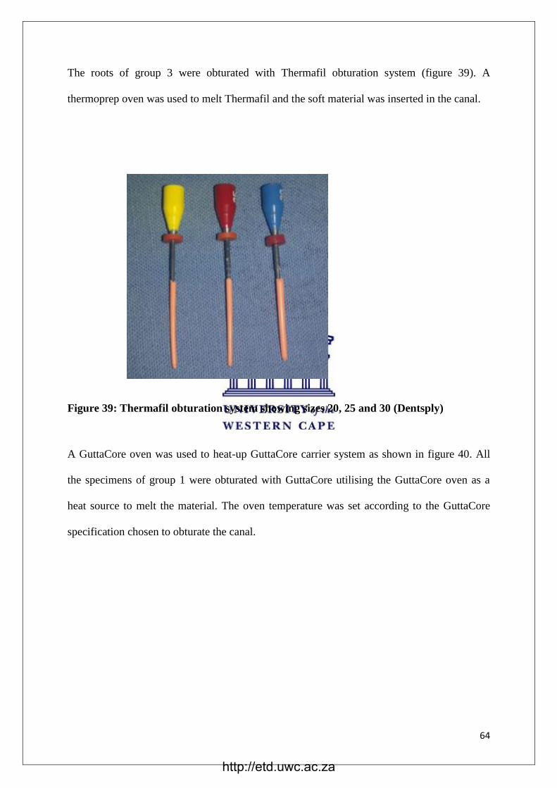
64
The roots of group 3 were obturated with Thermafil obturation system (figure 39). A
thermoprep oven was used to melt Thermafil and the soft material was inserted in the canal.
Figure 39: Thermafil obturation system showing sizes 20, 25 and 30 (Dentsply)
A GuttaCore oven was used to heat-up GuttaCore carrier system as shown in figure 40. All
the specimens of group 1 were obturated with GuttaCore utilising the GuttaCore oven as a
heat source to melt the material. The oven temperature was set according to the GuttaCore
specification chosen to obturate the canal.
http://etd.uwc.ac.za

65
Figure 40: GuttaCore oven that was used in this study
A thermoprep oven (figure 41) was used to heat-up Thermafil carrier system during warm
obturation in group 3 obturation. The temperature was set according to the system of the
Thermafil gutta-percha chosen
Figure 41: Thermoprep oven that was used in this study
http://etd.uwc.ac.za

66
AH Plus root canal sealer (Dentsply Tulsa Dental, Tulsa, USA) was used for apical sealing
during obturation in all the three groups. All roots were stored in an incubator at a
temperature of 37oC and at 100% relative humidity for a period of 24 hours to allow complete
setting of root canal sealer. The three groups of obturated specimens were now ready for
scanning with microcomputed tomography (figure 42).
Figure 42: Obturated specimens representing each of the three groups
Micro-CT Imaging
A high-resolution micro-CT scanner model A v|tome|x 240D (General Electric, MA, USA)
was used to scan the teeth (figure 43). Each root was positioned on the specimen stage and
scanned with a very high isotropic resolution of 15μm, rotational step of 0.6° and a rotational
angle of 360°. With a fully automated CT scan reconstruction and analysis process (datos|x
2.0 software) with high precision and reproducible 3D metrology, images obtained from the
scan were reconstructed to show two-dimensional slices of the inner structure of the roots.
Accelerated 3D CT reconstruction technique by velo| CT software was constructed for
volume visualization in 3D rendered mode analysis, and measurements of the volume of the
root canal filling material and percentage of gaps and voids present in the canals.
http://etd.uwc.ac.za

67
Figure 43: Micro-CT machine that was used to scan the root model A v|tome|x 240D
(General Electric, MA, USA)
Axial sections at 1mm, 3mm, and 6mm from apex were made and two different parameters
were measured: Total area of voids/gaps in square micrometers, the ratio between voids/gaps
and the total canal area in the section were calculated. In 3D surface-rendered
reconstructions, the volume of voids in cubic micrometers, the ratio between volume of
voids/gaps and the total canal volume were then calculated.
Statistical Analysis
Since the dataset from either parameter was not normally distributed even after data
transformation, each dataset was analysed using the non-parametric test; Kruskal–Wallis
http://etd.uwc.ac.za

68
analysis of variance. Statistical significance was pre-set at alpha = 0.05 for all analyses
to explore significant differences between GuttaCore, ProTaper and Thermafil. The
following outcome measures were assessed:
1. Total volume of voids between gutta-percha and the root canal walls
2. Total volume of cement within the canal space
3. Percentage of volume of voids to the total volume of canal space on 3D surface
rendered reconstructions within the entire root canal space following obturation;
4. The presence or absence of voids/gaps between the gutta-percha/cement and
dentine interface was assessed on axial sections at 1mm, 3mm, and 6mm from
the apex coronally.
http://etd.uwc.ac.za
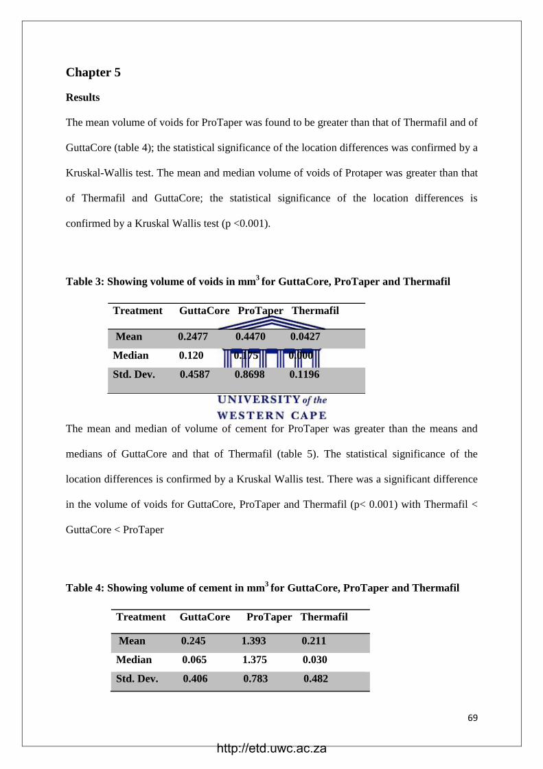
69
Chapter 5
Results
The mean volume of voids for ProTaper was found to be greater than that of Thermafil and of
GuttaCore (table 4); the statistical significance of the location differences was confirmed by a
Kruskal-Wallis test. The mean and median volume of voids of Protaper was greater than that
of Thermafil and GuttaCore; the statistical significance of the location differences is
confirmed by a Kruskal Wallis test (p <0.001).
Table 3: Showing volume of voids in mm3
for GuttaCore, ProTaper and Thermafil
The mean and median of volume of cement for ProTaper was greater than the means and
medians of GuttaCore and that of Thermafil (table 5). The statistical significance of the
location differences is confirmed by a Kruskal Wallis test. There was a significant difference
in the volume of voids for GuttaCore, ProTaper and Thermafil (p< 0.001) with Thermafil <
GuttaCore < ProTaper
Table 4: Showing volume of cement in mm3
for GuttaCore, ProTaper and Thermafil
Treatment GuttaCore ProTaper Thermafil
Mean 0.245 1.393 0.211
Median 0.065 1.375 0.030
Std. Dev. 0.406 0.783 0.482
Treatment GuttaCore ProTaper Thermafil
Mean 0.2477 0.4470 0.0427
Median 0.120 0.175 0.000
Std. Dev. 0.4587 0.8698 0.1196
http://etd.uwc.ac.za

70
The percentage volume of voids was higher for roots that were obturated with ProTaper
single-cone obturation, followed by the GuttaCore. Thermafil had less percentage of volume
of voids which demonstrate superior obturation
Table 5: Showing percentage volume of voids in mm3
for GuttaCore, ProTaper and
Thermafil
Treatment GuttaCore ProTaper Thermafil
Mean 1.931 4.596 0.452
Median 1.091 2.009 0.000
Std. Dev. 2.292 6.783 1.171
Figure 44 shows a Bar graph representing the volume of voids and volume of cement for
GuttaCore, ProTaper and Thermafil. ProTaper single-cone had the highest percentage volume
of voids, followed by GuttaCore and Thermafil was the least.
Figure 44: Total volume of voids and volume of cement for GuttaCore, ProTaper and
Thermafil
0
0.2
0.4
0.6
0.8
1
1.2
1.4
1.6
GuttaCore ProTaper Thermafil
Volume of Voids
Volume of Cement
http://etd.uwc.ac.za

71
Figure 45 shows a Bar graph representing percentage volume of voids for GuttaCore,
ProTaper single-cone and Thermafil. Thermafil demonstrated the least percentage of voids
followed by GuttaCore and then ProTaper single-cone.
Figure 45: Percentage voids for GuttaCore, ProTaper and Thermafil
At the level of 1mm from the apex, GuttaCore and Thermafil produced good adaptation to the
walls of the root canal and there was also sealing of canal aberrations. There was less cement
in the obturations that were done by the two carrier system at 1mm level (GP-gutta-percha,
D-dentine, C-cement, CC-core-carrier and V-voids). Whereas with ProTaper single-cone
obturation, there was a lot of cement utilized and there were also voids within the cement
itself (figure 45).
Figure 46: GuttaCore: 1mm ProTaper: 1mm Thermafil: 1mm
0
1
2
3
4
5
GuttaCore ProTaper Thermafil
% of Voids
http://etd.uwc.ac.za

72
There was good adaptation of GuttaCore and Thermafil gutta-percha at the level of 3mm
from the apex. Both the systems utilised less cement (root canal sealer) and the obturation
with these systems followed the anatomy of the root canal. ProTaper single-cone obturation
was surrounded by more cement than the core-carrier systems and there was also the presence
of voids within the cement (figure 46).
Figure 47: GuttaCore: 3mm ProTaper: 3mm Thermafil: 3mm
At the level of 6mm from the apex, the root canals get wider and as a result present with
difficult obturation. The two carrier systems followed the canal anatomy providing good
material adaptation. There was less cement and fewer voids present in the obturation with the
two core-carrier systems. Whereas the ProTaper single-cone obturation presented with voids
and more cement was used to seal the root canals (figure 47)
Figure 48: GuttaCore: 6mm ProTaper: 6mm Thermafil: 6mm
http://etd.uwc.ac.za

73
Chapter 6
Discussion
Under the conditions of the present study, all three obturation systems exhibited void
formation when observed using the micro-CT scanned images set at 15µm resolution.
As there was a significant difference in the percentage of void formation between the
systems tested when obturated with the GuttaCore carrier technique, ProTaper single-
cone technique and Thermafil carrier technique, the null hypothesis that stated there
was no differences in the percentage of interfacial voids had to be rejected.
Thermafil demonstrated the best adaptation when compared to GuttaCore and ProTaper
gutta-percha. When the volume of voids were evaluated, Thermafil was significantly
lower than GuttaCore and ProTaper (Kruskal–Wallis analysis of variance p<0.001). This
indicated good material adaptation for specimens obturated with Thermafil. When the
percentage of volume of voids to the canal volume was compared, ProTaper gutta-
percha had a higher percentage when compared to Thermafil and GuttaCore (p<0.001).
The review of the literature has shown superior obturation with thermal obturation
compared to lateral condensation both in the presence or absence of smear layer
(Gencoglu et al., 1993).
This study was conducted in the absence of smear layer and Thermafil produced
superior obturation when compared with ProTaper single-cone gutta-percha and
GuttaCore. In the current study, the smear layer was removed from all the root canal
walls using EDTA thus enhancing adaptation of gutta-percha to the walls of root canal.
The smear layer also plays an important role with regards to proper sealing of the apical
area (Veríssimo & do Vale, 2006).
http://etd.uwc.ac.za

74
Apical leakage is one of the more common complications encountered in endodontic
failures. The chemical properties of a sealer are very important in sealer selection. AH
Plus root canal sealer was used to apically seal off all the roots in the three groups. It is
classified as a resin bonded sealer and uses methamine for polymerization. The presence of
large amount of root canal sealer usually leads to root canal failure due to the
dissolution of the root canal sealer by cellular fluids. The sealer coverage is influenced by
the closeness of the adaptation of the gutta-percha to the walls of the root canal (Wu et al.,
2000).
In the current study the sealer coverage was found to be reduced apically with Thermafil and
GuttaCore indicating good fit of the two carrier systems to the walls of the root canal. There
was more of sealer material apically with ProTaper gutta-percha indicating poor adaptation of
the gutta-percha to the root canal walls leaving large amount of sealer between the gutta-
percha and root canal wall (figure 47). This may also account for more voids present with
single-cone ProTaper. The presence of voids within the cement may lead to reinfection of the
root canal. Poor adaptation of the gutta-percha can also be demonstrated by the presence
of large volume of sealer material itself within the obturated root canals. The gap
between the gutta-percha and the walls of the root canal is usually occupied by a large
amount of root canal sealer when there is poor adaptation of the gutta-percha to the root
canal walls. There was a higher volume of cement in teeth obturated with ProTaper
single-cone gutta-percha technique than GuttaCore carrier technique and Thermafil
carrier technique (Kruskal-Wallis, p<0.001).
This study also assessed critical areas of the root canal which is the apical third of the
root canal. Proper obturation of this area is crucial as most canal aberrations like apical
deltas and lateral canals are located in this area. Because of the incidence of lateral
http://etd.uwc.ac.za

75
canals and apical deltas in this area, proper obturation in this area may be difficult. The
study divided the apical area into three sections, 1mm, 3mm and 6mm short of the apex
and assessed the sealing ability of each technique to determine the best fit of the gutta-
percha. The three areas were evaluated for voids formation and gutta-percha adaptation
to the root canal walls.
Thermafil produced better adaptation, followed by GuttaCore and then ProTaper gutta-
percha as indicated by reduced amount of cement around the gutta-percha in all three
axial sections namely 1mm, 3mm and 6mm. There was a large volume of cement in
ProTaper gutta-percha compared with Thermafil and GuttaCore. Thermafil and
GuttaCore showed very little or no cement at all levels studied. Carrier-based root
fillings, in particular, have a mean sealer thickness of 2μm, considerably less than the
7μm resolution capability of the micro-CT scanner (Zogheib et al., 2013). This may
explain why in the majority of Thermafil obturators fillings sealer was indistinguishable
from gutta-percha.
GuttaCore was introduced into the market as cross-linked gutta-percha which has the
advantage of easier removal in the root canals for post placement and during retreatment
(Tomson et al., 2014). Since GuttaCore is a fairly new product in the market, there are few
studies that evaluated its adaptation to the root canal wall and its ability to seal all canal
abberations. GuttaCore produced superior obturation when compared to ProTaper single-cone
gutta-percha but not superior for Thermafil obturations. GuttaCore demonstated good
adaptation to the root canal wall much better when compared to ProTaper gutta-percha
(figure 46). Although the mean volume of cement for GuttaCore and Thermafil showed better
results, GuttaCore exhibited more voids when compared to Thermafil as a result of GuttaCore
http://etd.uwc.ac.za

76
having a cross-linked gutta-percha. The two systems are classified as carrier based obturation
systems and as we can see from the results of the volumes of voids and cement, they
produced superior obturation compared to ProTaper obturation.
Thermafil obturation demonstrated good adaptation to the root canal wall at 1mm, 3mm and
6mm from the apex of the root (figure 46-48). There were also lesser volume of cement in all
the three sections. At 1mm section, the canal curvature was ovoid but Thermafil obturation
was able to negotiate all the canal aberrations (figure 46). The quality of obturation
achieved by Thermafil in single-rooted canals was superior to that achieved by
GuttaCore and the single cone obturation using ProTaper.
The quality of obturation can be assessed through laboratory studies. Several in vitro
techniques were developed to determine the sealing ability of root fillings: Dye
penetration, fluid transport, and scanning electron microscopy analyses. All have
limitations of measuring voids by analysis of sectioned roots. In the past various types
of dyes like eosin, methylene blue; black Indian ink and Procion brilliant blue were
used to evaluate adaptation and micro-leakage of the gutta-percha (Verissimo and do
Vale, 2006). Microscopy can be used at various magnifications to determine adaptation
of gutta-percha to the walls of the root canal. It is one of the oldest methods used to
evaluate gutta-percha leakage and adaptation but sample preparations are technique
sensitive and may be damaged during the process (Tripi et al., 2001).
Micro-computed tomography (micro-CT) has been described as a new and promising tool in
endodontics to assess the geometry of the root canal (Peters et al., 2000, 2001; Hammad et
al., 2009). High resolution micro-CT is an emerging technology with several promising
application in different fields of dentistry. It is highly accurate and non-destructive method
http://etd.uwc.ac.za

77
for in-vitro evaluation of root canal fillings (Zogheib et al., 2013). However, micro-CT
sections lack the resolution when compared with scanning electron microscopy (SEM), for
the use of studying interfacial gaps and intracanal voids but SEM is a more destructive
method of investigation and is labour intensive (Li et al., 2014).
Micro-CT provides a three dimensional view of the root canal system by providing an
undistorted image of the tooth. It has the highest resolution at a very low exposure compared
to a conventional CT scan. The micro-CT can be a useful tool to check for gap formation
between the root canal filling and the dentine walls because it is a less complicated technique
compared to conventional methods, specimens are not damaged and images can be viewed in
3 dimensions. Micro-CT imaging tool overcomes the limitations of the previously used
model like dye penetration, fluid transport, and cross-sectional analyses, which are
valuable techniques, but the results do not always corroborate with each other (Zogheib
et al., 2013). Therefore in this study, micro-CT was used as an imaging tool of choice
based on its advantages that are demonstrated by the literature. In clinical situations,
radiographs are frequently used to analyse root canal treatment. Intra-oral radiographs are
widely used to evaluate adaptation of obturation materials but have the disadvantage of
showing a two dimensional view of the image.
The results of this study show a high variation in the data especially for cold
condensation ProTaper single-cone technique. However, in general, the high variance of
the data acquired is an indirect indicator of the unpredictability of commonly used
obturation techniques in perfectly adapting to the canal walls and root filling materials
(Li et al, 2014). A similar conclusion was reached in a stereomicroscopy study to
examine voids present in central incisors that were obturated with cold lateral
http://etd.uwc.ac.za

78
compaction (Keçeci et al., 2005). The warm obturation system produced better results in
the sealing of lateral and accessory canals in previous studies (Robberecht et al., 2012). The
literature has also demonstrated that Thermafil produced good adaptation to the walls of the
canals when compared to lateral compaction and single-cone obturation (Lea et al., 2005).
The results of the current study correlate with the previous studies with regards to adaptation
of warm obturation system to the root canal walls. The study also emphasises the endodontic
protocol, which starts from access cavity preparation, patency and glide path preparation,
filing and shaping (including irrigation protocol) and obturation. With regards to access
cavity, a straight line access is always advocated for proper vision and access to all the root
canal orifices. The straight line access also help in preventing the fracture of endodontic
instruments by avoiding the contact of the files with the walls of the access cavity. In the
current study, the straight line access was obtained easily since all the teeth were decoronated
at the cemento-enamel junction. There was no instrument fracture that was encountered
during filing and shaping of canals. In the current study, canal patency was obtained by using
a number 10 K-file as this allowed some of the root canal cement to extrude through the canal
and ensure best possible apical seal.
There are various methods of preparing a glide path in the literature which ensures smooth
filing of root canal. In the current study, the manual glide preparation utilising standard size
10 and size 15 K-files applied in a ‘watch winding’ motion was used (West, 2010). This is
the cost effective and easy method of glide path preparation since the K-files are always
available on the market and the technique of manual glide path preparation is not complicated
compared to rotary methods of glide path preparation (Cassim & van der Vyver, 2013). Filing
and shaping of all the ninety specimens was efficient and smooth after the preparation of
http://etd.uwc.ac.za

79
glide path and there was no canal obstruction in all the specimens that were used in the
current study.
A standard irrigation protocol of using sodium hypochlorite and EDTA was followed in the
current study. Sodium hypochlorite was used in filing and shaping of all the specimens and it
was also used as an irrigation solution due to its tissue dissolving effects and antibacterial
activity. After filing and shaping, there is usually accumulation of dentine mud (smear layer)
which may clog the canal and prevent proper adaptation of obturation material. In the current
study, EDTA was utilised to remove the smear layer on the dentine surface to allow better
obturation. The obturation protocol advocates irrigation with sodium hypochlorite after the
use of EDTA to prevent continuous demineralisation of root canal wall which might lead to
weakening of the walls. In the current study, sodium hypochlorite was use as a final irrigation
solution to flush off EDTA thus preventing unnecessary demineralization of dentine walls.
Gencoglu et al (1993), demonstrated that thermal obturation adapt well in the presence and
also in the absence of smear layer. In the current study all specimens were irrigated with
EDTA to remove the smear layer. The removal of smear layer allowed all the three obturation
systems to be standardized in the current study.
In carrier based obturation, the gutta-percha must be heated in an oven in other to make it to
flow easily in the canals. In the current study both GuttaCore and Thermafil were placed in
the warm oven before being placed in the root canals. Heated gutta-percha can easily
negotiate passage through the canal effortlessly. Obturations with GuttaCore and Thermafil
resulted in obturation of lateral canals and apical deltas (Zhang et al., 2011). Obturation of
canal aberrations is essential for prevention of root canal reinfection since it is very difficult
to obturate such canals. With ProTaper obturation, it was very difficult to obtain obturation of
http://etd.uwc.ac.za
80
lateral canals and apical deltas due to rigidity of the cones that normally occur in cold
obturations. Therefore it can be concluded that warm gutta-percha technique produced better
adaptation than cold obturation technique.
The obturation density (quality of obturation) that was produced by the carrier based systems
was better than the obturation that was produced by ProTaper obturation. The results obtained
from obturation with carrier based systems in the current study are similar to those obtained
by Lea et al in 2005, where they found thermal obturation to produce superior obtuation
density when compared to cold lateral obturation system.
http://etd.uwc.ac.za
81
Chapter 7
Conclusions and recommendations
Conclusions
Within the limitations of the present study, both carrier based techniques allowed for
better sealing ability in root canals compared to single cold gutta-percha obturation
technique although none of the materials were gap free especially at 1mm from apex.
This study shows the efficiency of carrier-based obturation systems in filling root canals
hermetically compared to the cold condensation of ProTaper single-cone obturation
technique. Thermafil produced good adaptation to canal walls with least amount of
voids/gap formation, followed by GuttaCore and the ProTaper single-cone obturation.
Good sealing ability of carrier-based techniques makes them appropriate to use in daily
endodontic obturations.
Recommendations
We recommend the use of thermal obturation in order to obtain better adaptation with
minimum number of voids within the obturated teeth. Both the GuttaCore and Thermafil
obturations are likely to enhance the survival of obturated teeth due to superior
obturation observed in the current study. Obturation with ProTaper gutta-percha
produced poor results as measured by the presence of a large number of voids and the
obturation relied more on root canal sealer to seal off the apical part of the root canal.
This may be overcome by the placement of more lateral cones in order to eliminate void
formation when using ProTaper single-cone technique to obturate the teeth.
http://etd.uwc.ac.za

82
Limitations of the study
This study is an in-vitro study, hence may be different from clinical situation. The results
might be affected in clinical situation as there are other factors like presence of saliva, tongue
movement, use of rubber dam, and limited space due to the size of the oral cavity. The study
focussed on single canals with a curvature of less than 10 degrees, the results might be
different when treating multiple rooted teeth and also when treating teeth with curvature of
more than 10 degrees.
http://etd.uwc.ac.za

83
Chapter 8
References
Adams, N. & Tomson, P.L. 2014. Access cavity preparation. British Dental Journal, 216(6):
333–339.
Adcock, J.M., Sidow, S.J., Looney, S.W., Liu, Y., Mcnally, K., Lindsey, K., Tay, F.R. &
Hons, B. 2011. Histologic Evaluation of Canal and Isthmus Debridement Efficacies of
Two Different Irrigant Delivery Techniques in a Closed System. Journal of
Endodontics, 37(4): 544–548. .
Alhashimi, R.A., Foxton, R., Romeed, S. & Deb, S. 2014. An In Vitro Assessment of Gutta-
Percha Coating of New Carrier-Based Root Canal Fillings. The Scientific World
Journal, 2014. 1-6.
Arai, Y., Tammisalo, E., Iwai, K., Hashimoto, K. & Shinoda, K. 1999. Development of a
compact computed tomographic apparatus for dental use. Dentomaxillofacial Radiology,
28: 245–248.
Aslantas, E.E., Buzoglu, H.D. & Altundasar, E. 2014. Effect of EDTA , Sodium Hypochlorite
, and Chlorhexidine Gluconate with or without Surface Modifiers on Dentin
Microhardness. Journal of Endodontics, 40(6): 876–87.
Basrani, B. & Haapasalo, M. 2012. Update on endodontic irrigating solutions. Endodontic
Topics, 27: 74–102.
Bertacci, A., Baroni, C., Breschi, L., Venturi, M. & Prati, C. 2007. The influence of smear
layer in lateral channels filling. Clinical Oral Investigations, 11(4): 353–359.
http://etd.uwc.ac.za

84
Berutti, E., Cantatore, G., Castellucci, A., Chiandussi, G., Pera, F., Migliaretti, G. &
Pasqualini, D. 2009. Use of Nickel-Titanium Rotary PathFile to Create the Glide Path :
Comparison With Manual Preflaring in Simulated Root Canals. Journal of Endodontics,
35(3): 408–412.
Berutti, E., Negro, A.R., Lendini, M. & Pasqualini, D. 2004. Influence of manual preflaring
and torque on the failure rate of ProTaper rotary instruments. Journal of Endodontics,
30(4): 228–230.
Bogen, G., Kim, J.S. & Bakland, L.K. 2008. Direct Pulp Capping With Mineral Trioxide
Aggregate: An Observational Study George Bogen, Jay S. Kim and Leif K. Bakland.
Journal of American Dental Association, 139: 305–315.
Buchanan, L.S. 1994. System B TM
Obturation Continuous Wave of Condensation. Dentistry
Today, 13(10): 4310.
Cameron, J.A. 1983. The use of ultrasonics in the removal of the smear layer: A scanning
electron microscope study. Journal of Endodontics, 9(7): 289–292.
Camps, J. & Pashley, D. 2003. Reliability of the dye penetration studies. Journal of
Endodontics, 29(9): 592–4.
Cassim, I. & van der Vyver, P.J. 2013. The importance of glide path preparation in
endodontics: a consideration of instruments and literature. SADJ : Journal of the South
African Dental Association, 68(7): 322, 324–7.
Castellucci, A. 2003. Access cavity and endodontic anatomy. Endodontics, 1: 244–329.
http://etd.uwc.ac.za

85
Chailertvanitkul, P., Saunders, W.P., Saunders, E.M. & MacKenzie, D. 1998. Polymicrobial
coronal leakage of super EBA root-end fillings following two methods of root-end
preparation. International Endodontic Journal, 31(5): 348–53.
Chana, H., Briggs, P. & Moss, R. 1998. Degradation of a silver point in association with
endodontic infection. International Endodontic Journal, 31(2): 141–6.
Chohayeb, A.A. 1992. Comparison of conventional root canal obturation techniques with
thermafil obturators. Journal of Endodontics, 18(1): 10–12.
Combe, E., Cohen, B. & Cumming, K. 2001. Alpha- and beta- forms of gutta-percha in
products for root canal filling. International Endodontic Journal, 34: 447–451.
Cunningham, W.T. & Joseph, S.W. 1980. Effect of temperature on the bactericidal action of
sodium hypochlorite endodontic irrigant. Oral Surg Oral Med Oral Pathol, 50(6): 569–
571.
Darcey, J., Taylor, C., Roudsari, R.V., Jawad, S. & Hunter, M. 2015. Modern Endodontic
Planning Part 2: Access and Strategy. Dental Update, 42(11): 709–720.
De-Deus, G., Gurgel-Filho, E.D., Magalhães, K.M. & Coutinho-Filho, T. 2006. A laboratory
analysis of gutta-percha-filled area obtained using Thermafil, system B and lateral
condensation. International Endodontic Journal, 39(5): 378–383.
Dezan, E.J., Holland, R., Consolaro, A., Ciesielski, F.I.N. & Jardim, E.G.J. 2012.
Experimentally Induced Anachoresis in the Periapical Region After Root Canal Filling.
International Journal Odontostomatology, 6(1): 5–10.
Dhingra, A. 2014. Glide path in endodontics. Endodontology, 26(1): 217–222.
http://etd.uwc.ac.za

86
DuLac, K. a, Nielsen, C.J., Tomazic, T.J., Ferrillo, P.J. & Hatton, J.F. 1999. Comparison of
the obturation of lateral canals by six techniques. Journal of Endodontics, 25(5): 376–
380.
ElAyouti, A., Kiefner, P., Hecker, H., Chu, A., Löst, C. & Weiger, R. 2009. Homogeneity
and adaptation of endodontic fillings in root canals with enlarged apical preparation.
Oral Surgery, Oral Medicine, Oral Pathology, Oral Radiology and Endodontology,
108(3): e141–e146.
Fava, L. R. G. and Dummer, P.M.H. 1997. Periapical radiographic techniques during
endodontic diagnosis. International Endodontic Journal, 30: 250–261.
Gambarini, G., Plotino, G., Sannino, G.P., Grande, N.M., Giansiracusa, A., Piasecki, L., da
Silva Neto, U.X., Al-Sudani, D. & Testarelli, L. 2015. Cyclic fatigue of instruments for
endodontic glide path. Odontology, 103(1): 56–60.
Gengoglu, N., Samani, S. & Gimday, M. 1993. Dentinal Wall Adaptation of
Thermoplasticized Gutta- percha in the Absence or Presence of Smear Layer : A
Scanning Electron Microscopic Study. Journal of Endodontics, 19(11): 558–562.
Gilhooly, R.M.P., Hayes, S.J., Bryant, S.T. & Dummer, P.M.H. 2001. Comparison of lateral
condensation and thermomechanically compacted warm ??-phase gutta-percha with a
single cone for obturating curved root canals. Oral Surgery, Oral Medicine, Oral
Pathology, Oral Radiology, and Endodontics, 91(1): 89–94.
Gluskin, A.H. 2005. Mishaps and serious complications in endodontic obturation. Endodontic
Topics, 12(1): 52–70.
http://etd.uwc.ac.za
87
Gomes, B.P.F.A., Jacinto, R.C., Pinheiro, E.T., Sousa, E.L.R., Zaia, A.A., Ferraz, C.C.R. &
Souza-Filho, F.J. 2005. Porphyromonas gingivalis, Porphymmonas endodontalis,
Prevotella intermedia and Prevotella nigrescens in endodontic lesions detected by
culture and by PCR. Oral Microbiology and Immunology, 20(4): 211–215.
Gordon, M.P.J., Love, R.M. & Chandler, N.P. 2005. An evaluation of .06 tapered gutta-
percha cones for filling of .06 taper prepared curved root canals. International
Endodontic Journal, 38(2): 87–96.
de Gregorio, C., Estevez, R., Cisneros, R., Paranjpe, A. & Cohenca, N. 2010. Efficacy of
different irrigation and activation systems on the penetration of sodium hypochlorite into
simulated lateral canals and up to working length: An in vitro study. Journal of
Endodontics, 36(7): 1216–1221.
de Gregorio, C., Paranjpe, A., Garcia, A., Navarrete, N., Estevez, R., Esplugues, E.O. &
Cohenca, N. 2012. Efficacy of irrigation systems on penetration of sodium hypochlorite
to working length and to simulated uninstrumented areas in oval shaped root canals.
International Endodontic Journal, 45(5): 475–481.
Grigoratos, D., Knowles, J., Ng, Y. & Gulabivala, K. 2001. Effect of sodium hypochlorite
and calcium hydroxide on the modulus of elasticity and flexural strength of dentine.
International Endodontic Journal, 34(2): 113–119.
Gutmann, J.L. 2008. The Future Of Root Canal Obturation. Famdent Practical Dentistry
Handbook, 9(1): 1–4.
Gutmann, J.L., Saunders, W.P., Saunderst, E.M. & Nguyen, L. 1993. An assessment of the
plastic Thermafil obturation technique Parti Radiographic evaluation of adaptation and
placement. International Endodontic Journal, 26: 173–179.
http://etd.uwc.ac.za

88
Haapasalo, M., Shen, Y., Qian, W. & Gao, Y. 2010. Irrigation in Endodontics. Dental Clinics
of North America, 54(2): 291–312.
Hammad, M., Qualtrough, A. & Silikas, N. 2009. Evaluation of Root Canal Obturation: A
Three-dimensional In Vitro Study. Journal of Endodontics, 35(4): 541–544.
Holland, R., Murata, S.S., Silva, M.N. Da, Dezan Junior, E., Souza, V. De & Bernabé, P.F.E.
2004. Influence of the sealer and a plug in coronal leakage after post space preparation.
Journal of Applied Oral Science : 12(3): 223–6.
Hülsmann, M., Peters, O. a. & Dummer, P.M.H. 2005. Mechanical preparation of root canals:
shaping goals, techniques and means. Endodontic Topics, 10(1): 30–76.
Inan, U., Aydin, C., Tunca, Y.M. & Basak, F. 2009. In Vitro Evaluation of Matched-Taper
Single-Cone. Journal of Canadian Dental Assaciation, 75(2): 5–8.
Juhlin, J.J., Walton, R.E. & Dovgan, J.S. 1993. Adaptation of Thermafil Components to
Canal Walls. Journal of Endodontics, 19(3): 130–135.
Keçeci, A.D., Çelik Ünal, G. & Şen, B.H. 2005. Comparison of cold lateral compaction and
continuous wave of obturation techniques following manual or rotary instrumentation.
International Endodontic Journal, 38(6): 381–388.
Lea, C.S., Apicella, M.J., Mines, P., Yancich, P.P. & Parker, M.H. 2005. Comparison of the
obturation density of cold lateral compaction versus warm vertical compaction using the
continuous wave of condensation technique. Journal of Endodontics, 31(1): 37–39.
http://etd.uwc.ac.za
89
Levitan, M.E., Himel, V.T., Luckey, J.B., Davis, S., Brayton, S., Goldman, M., Zillich, R.,
Dorson, J., Pomeranz, H., Fishelberg, G., Brayton, S., Davis, S., Goldman, M., Schilder,
H., Larder, T., Prescott, A., Brayton, S., Kuttler, Y., Friedman, C., Sandrick, J., Heuer,
M., Rapp, G., Luks, S., Goldman, M., Schilder, H., Goodman, A., Aldrich, W., Schilder,
H., Goodman, A., Aldrich, W., Wolcott, J., Himel, V., Powell, W., Penny, J., Budd, C.,
Weller, N., Kulid, J., Clinton, K., Himel, V. & Natrella, M. 2003. The effect of insertion
rates on fill length and adaptation of a thermoplasticized gutta-percha technique. Journal
of Endodontics, 29(8): 505–8.
Li, G., Niu, L., Selem, L.C., Eid, A.A., Bergeron, B.E., Chen, J., Pashley, D.H. & Tay, F.R.
2014. ScienceDirect Quality of obturation achieved by an endodontic core-carrier
system with crosslinked gutta-percha carrier in single-rooted canals. Journal of
Dentistry, 42(9): 1124–1134.
Machtou, P. 2002. Clinical Apical Seal versus Coronal Seal. Interntional Dentistry South
Africa, 8(3). 20-26
Maniglia-Ferreira, C., Gurgel-filho, E.D., Silva-jr, J.B.D.A., Célia, R., Paula, M. De, Pessoa,
J., Feitosa, D.A. & Sousa-filho, F.J. De. 2013. Chemical composition and thermal
behavior of five brands of thermoplasticized gutta ‑ percha. European Journal of
Dentistry, 7(2). 201-206.
Monticelli, F., Sadek, F.T., Schuster, G.S., Volkmann, K.R., Looney, S.W., Ferrari, M.,
Toledano, M., Pashley, D.H. & Tay, F.R. 2007. Efficacy of Two Contemporary Single-
cone Filling Techniques in Preventing Bacterial Leakage. Journal of Endodontics, 33(3):
310–313.
http://etd.uwc.ac.za

90
Moore, W.R. & Wesselink, P.R. 1982. Factors promoting the tissue dissolving capability of
sodium hypochlorite. International Endodontic Journal, 15(4): 187–196.
Mounce, R.E. 2008. Rotary Nickel Titanium Instrumentation Revolutionized : The Twisted
File. Oral Health, (5): 6–9.
Mozzo, P., Procacci, C., Tacconi, A., Martini, P.T. & Andreis, I.A.B. 1998. Original article A
new volumetric CT machine for dental imaging based on the cone-beam technique :
preliminary results. European Radiology, 1564: 1558–1564.
Nahmias, Y., Cassim, I. & Glassman, G. 2013. ‘ Own the Canal ’– The Importance of a
Reproducible Glide Path. Oral Health Journal: 74–82.
Nica, L.M., Didilescu, A., Rusu, D., Bacila, A. & Stratul, S.I. 2012. Photomicrographic
evaluation of the apical sealing capacity of three types of gutta-percha master cones: An
in vitro study. Odontology, 100(1): 54–60.
O’Connell, M.S., Morgan, L. a, Beeler, W.J. & Baumgartner, J.C. 2000. A comparative study
of smear layer removal using different salts of EDTA. Journal of Endodontics, 26(12):
739–43.
Ørstavik, D. 2005. Materials used for root canal obturation : technical , biological and clinical
testing. Endodontic Topics, 12(3): 25–38.
Palazzi, F., Morra, M., Mohammadi, Z., Grandini, S. & Giardino, L. 2012. Comparison of the
surface tension of 5.25% sodium hypochlorite solution with three new sodium
hypochlorite-based endodontic irrigants. International Endodontic Journal, 45(2): 129–
135.
http://etd.uwc.ac.za

91
Patel, E. & Owen, C.P. 2016. The use of textural analysis to test the hardness and
penetrability of three types of gutta percha cones when exposed to two endodontic
solvents. SADJ : Journal of the South African Dental Association: 346–350.
Patel, S. 2009. New dimensions in endodontic imaging : Part 2 . Cone beam computed
tomography. International Endodontic Journal, 42: 463–475.
Patel, S., Dawood, A., Ford, T.P. & Whaites, E. 2007. The potential applications of cone
beam computed tomography in the management of endodontic problems. International
Endodontic Journal, 40: 818–830.
Patel, S., Dawood, A., Whaites, E. & Ford, T.P. 2009. New dimensions in endodontic
imaging : Part 1 . Conventional and alternative radiographic systems. International
Endodontic Journal, 42: 447–462.
Peters, O.A., Schönenberger, K. & Laib, A. 2001. Effects of four Ni – Ti preparation
techniques on root canal geometry assessed by micro computed tomography.
International Endodontic Journal, 34: 221–230.
Peters, O. a & Barbakow, F. 2000. Effects of irrigation on debris and smear layer on canal
walls prepared by two rotary techniques: a scanning electron microscopic study. Journal
of Endodontics, 26(1): 6–10.
Pommel, L. & Camps, J. 2001a. Effects of pressure and measurement time on the fluid
filtration method in endodontics. Journal of Endodontics, 27(4): 256–258.
Pommel, L. & Camps, J. 2001b. In vitro apical leakage of system B compared with other
filling techniques. Journal of Endodontia, 27(7): 449–451.
http://etd.uwc.ac.za

92
Portenier, I., Waltimo, T., Ørstavik, D. & Haapasalo, M. 2005. The susceptibility of starved,
stationary phase, and growing cells of Enterococcus faecalis to endodontic medicaments.
Journal of Endodontics, 31(5): 380–386.
Rafter, M. 2005. Apexification : a review. Dental Traumatology, 21: 1–8.
Robberecht, L., Colard, T. & Claisse-Crinquette, A. 2012. Qualitative evaluation of two
endodontic obturation techniques: tapered single-cone method versus warm vertical
condensation and injection system An in vitro study. Journal of Oral Science, 54(1): 99–
104.
Ruddle, C.J. 2005. ‘ Tsunami ’ Endodontics. International Dentistry SA, 11(4). 6-18
Samson, E., Kulkarni, S., Kumar, S. & Likhitkar, M. 2013. Evaluation and Comparison of
Apical Sealing Ability….Samson E et al An In-Vitro Evaluation and Comparison of
Apical Sealing Ability of Three Different Obturation Technique -Lateral Condensation,
Obtura II, and Thermafil. Journal of International Oral Health, 5(2): 35–43.
Scarfe, W.C. & Farman, A.G. 2008. What is Cone-Beam CT and How Does it Work ? Dental
Clinics of North America, 52: 707–730.
Schäfer, E., Köster, M. & Bürklein, S. 2013. Percentage of gutta-percha-filled areas in canals
instrumented with nickel-titanium systems and obturated with matching single cones.
Journal of Endodontics, 39(7): 924–928.
Schäfer, E., Nelius, B. & Bürklein, S. 2012. A comparative evaluation of gutta-percha filled
areas in curved root canals obturated with different techniques. Clinical Oral
Investigations, 16(1): 225–230.
http://etd.uwc.ac.za
93
Silva-filho, J.M., Souza-gabriel, A.E., Bianchi, G., De-bem, S.H.C., Alfredo, E. & Gariba, R.
2013. Comparison of Two Techniques for Selection of Master Gutta-Percha Cone Using
Micro-Computed Tomography. Brazilian Dental Journal, 24(4): 367–370.
Somma, F., Cretella, G., Carotenuto, M., Pecci, R., Bedini, R., De Biasi, M. & Angerame, D.
2011. Quality of thermoplasticized and single point root fillings assessed by micro-
computed tomography. International Endodontic Journal, 44(4): 362–369.
Susin, L., Liu, Y., Yoon, J.C., Parente, J.M., Loushine, R.J., Ricucci, D. & Bryan, T. 2010.
Canal and isthmus debridement efficacies of two irrigant agitation techniques in a closed
system. International Endodontic Journal, 43: 1077–1090.
Sutow, E.J., Foong, W.C., Zakariasen, K.L., Hall, G.C. & Jones, D.W. 1999. Corrosion and
cytotoxicity evaluation of thermafil endodontic obturator carriers. Journal of
Endodontia, 25(8): 562–566.
Taşdemir, T., Er, K., Yildirim, T., Buruk, K., Çelik, D., Cora, S., Tahan, E., Tuncel, B. &
Serper, A. 2009. Comparison of the sealing ability of three filling techniques in canals
shaped with two different rotary systems: A bacterial leakage study. Oral Surgery, Oral
Medicine, Oral Pathology, Oral Radiology and Endodontology, 108(3): 129–134.
Tay, F.R. & Pashley, D.H. 2007. Monoblocks in Root Canals: A Hypothetical or a Tangible
Goal. Journal of Endodontics, 33(4): 391–398.
Timpawat, S., Amornchat, C. & Trisuwan, W.R. 2001. Bacterial coronal leakage after
obturation with three root canal sealers. Journal of Endodontics, 27(1): 36–39.
Tolerance, T., Silver, O.F. & Fillings, C. 1941. Damaged Periapical. Journal of Dental
Research, 20(4): 355–361.
http://etd.uwc.ac.za

94
Tomson, R.M.E., Polycarpou, N. & Tomson, P.L. 2014. Contemporary obturation of the root
canal system. Nature Publishing Group, 216(6): 315–322.
Tripi, T.R., Bonaccorso, A., Tripi, V., Condorelli, G.G. & Rapisarda, E. 2001. Defects in GT
rotary instruments after use: an SEM study. Journal of Endodontics, 27(12): 782–5.
Veríssimo, D.M. & do Vale, M.S. 2006. Methodologies for assessment of apical and coronal
leakage of endodontic filling materials: a critical review. Journal of Oral Science, 48(3):
93–98.
Violich, D.R. & Chandler, N.P. 2010. The smear layer in endodontics – a review.
International Endodontic Journal, 43: 2–15.
Van der Vyver, P. 2011. Creating a glide path for rotary NiTi instruments: part two.
International Dentistry South Africa, 13(2): 14–25.
Van der Vyver, P., Paleker, F. & Jonker, C. 2015. Comparison of preparation times of three
different rotary glide path instrument systems. South African Dental Journal, 70(4):
146–9.
Walsch, H. 2004. The hybrid concept of nickel – titanium rotary instrumentation. Dental
Clinics of North America, 48: 183–202.
Wang, Z., Shen, Y. & Haapasalo, M. 2012. Effectiveness of Endodontic Disinfecting
Solutions against Young and Old Enterococcus faecalis Biofilms in Dentin Canals.
Journal of Endodontics, 38(10): 1376–1379.
Weller N, Kimbrough F, A.R. 1997. A Comparison of Thermoplastic Obturation Techniques:
Adaptation to the Canal Walls. Journal of Endodontics, 23(11): 703–6.
http://etd.uwc.ac.za

95
West, J. 2006. Endodontic update 2006. Journal of Esthetic and Restorative Dentistry, 18(5):
280–300.
Whitworth, J. 2005. Methods of filling root canals: principles and practices. Endodontic
Topics, (13): 2–24.
Wu, M. K, Ozok, A. R, Wesselink, P.R. 2000. Sealer distribution in root canals obtured by
three techniques. International Endodontic Journal, 33: 340–345.
Wu, E.G.K.O.N.M. & Wesselink, P.R. 1997. Effect of sealer thickness on long-term sealing
ability : a 2-year follow-up study. International Endodontic Journal, 30: 307–312.
Wu, M. ???K, De Gee, A.J. & Wesselink, P.R. 1994. Fluid transport and dye penetration
along root canal fillings. International Endodontic Journal, 27(5): 233–238.
Wu, M.K., van der Sluis, L.W.M. & Wesselink, P.R. 2006. A 1-year follow-up study on
leakage of single-cone fillings with RoekoRSA sealer. Oral Surgery, Oral Medicine,
Oral Pathology, Oral Radiology and Endodontology, 101(5): 662–667.
Wu Mk, Ozok AR, & W.P. 2000. Sealer distribution in root canals obtured by three
techniques. International Endodontic Journal, 33: 340–345.
Xu, Q., Fan, M.W., Fan, B., Cheung, G.S.P. & Hu, H.L. 2005. A new quantitative method
using glucose for analysis of endodontic leakage. Oral Surgery, Oral Medicine, Oral
Pathology, Oral Radiology, and Endodontics, 99(1): 107–111.
Yilmaz, Z., Deniz, D., Ozcelik, B., Sahin, C., Cimilli, H., Cehreli, Z.C. & Kartal, N. 2009.
Sealing efficiency of BeeFill 2in1 and System B/Obtura II versus single-cone and cold
lateral compaction techniques. Oral Surgery, Oral Medicine, Oral Pathology, Oral
Radiology and Endodontology, 108(6): e51–e55.
http://etd.uwc.ac.za
96
Yücel, A.Ç. & Çiftçi, A. 2006. Effects of different root canal obturation techniques on
bacterial penetration. Oral Surgery, Oral Medicine, Oral Pathology, Oral Radiology and
Endodontology, 102(4): 88–92.
Zhang, C., Huang, W., Sun, Z. & Hou, B. 2011. A comparison of two gutta-percha master
points consisting of different phases in filling of artificial lateral canals and depressions
in the apical region of root canals when using a warm vertical compaction technique.
International Endodontic Journal, 44: 1041–1046.
Zogheib, C., Naaman, A., Sigurdsson, A., Medioni, E., Bourbouze, G. & Arbab-Chirani, R.
2013. Comparative micro-computed tomographic evaluation of two carrier-based
obturation systems. Clinical Oral Investigations, 17(8): 1879–1883.
Zou, L., Shen, Y. & Li, W. 2010. Penetration of Sodium Hypochlorite into Dentin. Journal of
Endodonotics, 36(5): 793–796.
http://etd.uwc.ac.za

97
Appendix
Appendix 1: Ethical clearance letter
Oral & Dental Research Institute
Faculty of Dentistry and WHO Oral Health Collaborating Centre
University of the Western Cape, Cape Town
Patient Information Sheet to be given to the patient to take home
I, Dr Shadrack Kabini, am a qualified dentist involved in research and training at the
University of the Western Cape, Faculty of Dentistry. I am doing research about materials
that we use to restore the root canals. Generally, after the removal of your teeth, they are
either discarded or given to the students to practice on. I wish to use your teeth to be able to
determine which material can seal the root canal better. I will be using the teeth for a single
research project after which the teeth will be discarded and will not be used in future research
projects.
Donating your teeth in this study is on a voluntary basis. Donating your teeth for this study or
refusing to participate will not harm or prejudice you in any way. You retain the right to
withdraw your consent at any time without any consequences and the samples will then be
immediately destroyed. There will be no physical risk to you as related to the collection of
the teeth you donate.
There will be no payment made for the use of your teeth and your teeth will not be sold or
supplied to any other facility. All information will be kept strictly confidential. The teeth
supplied to me will not have your name on it as well as I will not be able to identify you in
any way. Therefore, you will not be identified by the teeth you donate and all tooth samples
will remain anonymous.
http://etd.uwc.ac.za
98
All teeth will be stored and used at the Oral and Dental Research Institute, Faculty of
Dentistry, Tygerberg Campus, University of the Western Cape which is a secure laboratory
and locked when not in use. Upon completion of this study all teeth will be discarded
immediately. It is my intention to publish the results of my findings in a scientific journal
however; the results will in no way identify you.
Thanking you.
-------------------.
Dr Shadrack Kabini
Registrar
Oral & Dental Research Institute; Oral Health Centre Tygerberg
Contact details: Tel: (021) 937 3170
Mobile: 0731358183
I, (Patient name)............................................................................................., fully understand
the information supplied to me by Dr S. Kabini in this information sheet and wish to/ do not
wish to participate in this research project.
Signature: .....................................................................................
Date: .............................................................................................
http://etd.uwc.ac.za