an in vivo study of the penetrability of endodontic ... · endodontic practitioners that the...
TRANSCRIPT

Loyola University ChicagoLoyola eCommons
Master's Theses Theses and Dissertations
1972
An In Vivo Study of the Penetrability ofEndodontic RestorationsDonald L. ScoralleLoyola University Chicago
This Thesis is brought to you for free and open access by the Theses and Dissertations at Loyola eCommons. It has been accepted for inclusion inMaster's Theses by an authorized administrator of Loyola eCommons. For more information, please contact [email protected].
This work is licensed under a Creative Commons Attribution-Noncommercial-No Derivative Works 3.0 License.Copyright © 1972 Donald L. Scoralle
Recommended CitationScoralle, Donald L., "An In Vivo Study of the Penetrability of Endodontic Restorations" (1972). Master's Theses. Paper 2525.http://ecommons.luc.edu/luc_theses/2525
P"'l- ....... - ·~---
AN IN VNO STUDY OF THE PENETRABILrrY
OF ENDOOONTIC RES'IDRATIONS
by
Donald L. Scoralle; D.D.S.
A Thesis Submitted. to the Faculty of the Graduate School
of Loyola University in Partial Fulfillment of
the Requirements for the Degree of
Master of Science
May, 1972

AUTOBIOGRAPHY
Donald Lawrence Scoralle was born in Aberdeen,
Washington on the first day of July, 1935. There, he
attended elementary schools and graduated from Weatherwax
High School in 1953.
His college education was obtained at both Grays
Harbor Community College in Aberdeen and The University of
Puget Sound in Tacoma, Washington. He then entered Chicago's
Loyola University School of Dentistry in 1958. His degree
of Doctor of Dental Surgery was conferred in June, 1962.
Since graduating, he has served continuously on
active duty in The United S~ates Navy Dental Corps. For his
first year he was designated to receive a rotating dental
internship at The Great Lakes Naval Hospital. His service
career has also taken him aboard The u.s.s. Sperry and The
Miramar Naval Air Station, both in San Diego, California.
After spending a year in Vietnam, he returned to the San Diego
area for further duty.
Beginning in August, 1969, the author embarked on
a three year period of graduate training. The first year was
at The Naval Dental Schpol in Bethesda, Maryland. In 1970
he was selected by the Navy and the Graduate School of his
i

dental alma mater, Loyola University, to pursue a dual
graduate program leading to the didactic degree of Master
of Science in Oral Biology and a Certificate of Specialty
Training in Endodontics.
Dr. Scoralle is a career officer and holds the
rank of Lieutenant Commander in The United States Navy.
Shortly, he will assume the duties as the staff endodontist
at Treasure Island's Naval Station in San Francisco.
ii .._ ______________________________________________________________________ ~=-'
-DEDICATION
To Betty, my wife and best friend, and to each of
my children. Their love, loyalty and encouragement have
served as an inspiration to achieve otherwise unreachable
goals.
To the memory of my father, Jose~h V. Scoralle,
whose personal sacrifices cannot be matched but will never
be forgotten.
To a compelling credo which echoes that it only
takes a little more--to go first class.
iii

-AC.KN:)WLEDGEMENTS
Although many more have contributed to my Loyola
education, I wish to publically recognize:
John v. Madonia, D.D.S., Ph.D., Chairman, Department of Microbiology and Coordinator of Graduate Education, my classmate, teacher, and thesis advisor, for his advice and thoughtfulness.
Michael A. Heuer, D.D.S., M.S., for his support, guidance and constructive criticism in my Thesis and seminars.
Henry Kahn, D.D.S., without his sincere effort and assistance, this Thesis would be but a mere skeleton of its present construction.
Harold Gerstein, D.D.S. and Franklin s. Weine, D.D.S., M.S., for their counsel which has helped to form the foundation of my professional position.
Marshall H. Smulson, D.D.S., for putting the above together as the Chairman, Department of Endodontics.
Fred Buddingh, D.V.M., Ph.D., and his staff at Loyola's Animal Research Facility, for the pleasant working conditions and willing hands.
u. s. Navy Dental Corps for making it all possible.
Mrs. Aurelia Minor and Mrs. Joan Smith, my mother and sister, for being who they are.
I iv -------..---

Chapter
I
II
III
IV
VI
VII
VIII
I
TABLE OF CONTENTS
INTRODUCTION • • • • • •
REVIEW OF THE LITERATURE •
A. Methods of Testing Dental Margins •
B. Methylene Blue . . . . . . . . . . . c. Dogs in De~tal Research . . . . METHODS AND MATERIALS . . . . A. Phase I Technics - Intravenous Dye
1
3
3
34
40
49
56
B. Phase II Technics - Intradental Dye • • • 59
64 RESULTS . . . . . . . . . A. Phase I Findings . . . . . . . . . . . . 64
B. Endodontic Observations of Dog Teeth 100
SUMMARY . . . . . . . . . . . . . . . . 110
115 REFERENCES • • . . . . . . . APPENDIX • • • . . . . 127
P~. Tables . . . . . . . . . . . . . 128
139 B. Figures • • . . . . . . . . . . . . .
v
L------------.------------------------------------
-CHAPTER I
INTRODUCTION
It is the accepted view of the preponderance of
endodontic practitioners that the ultimate goal of root canal
therapy is to seal the root canal system from the periodontal
ligament. Clinical practice and research has shown that all
spaces of the root canal must be filled with some form of
stable, inert, yet manageable material or materials. This
necessity has been pointed out repeatedly since Dr. Hudson
of Philadelphia reputedly filled the .eirst "fang" with gold
foil around 1830. 86 Since that time practitioners have
sought a variety of methods to cremate, extirpate and/or
obliterate the root canal and its contents. 2 Dentists have
tried waxes, cotton, numerous varieties of cement, different
metals, wood, resins, and countless technics involving gutta
percha. Each contributor implied or stated unequivocably that
his was the 11 best 11 or "easiest" method and surely offered
the most complete manner of solving the problem of the
unfilled root canal.
The early pioneers of dentistry empirically
envisioned that ·the tissue "fluids and gases" must be excluded
from the canals. This insight is expressed in the current
1

2 CH I
view that the tissues surrounding the root react pathologically
when extracellular fluids do not enjoy a total biologic
exchange of nutriments and wastes.
, _____________ -ii
CHAPTER II
REVIEW OF THE LITERATURE
A. Methods of Testing Dental Margin§
Incensed by the poor quality of the endodontic
treatment being rendered by many of his colleagues, McQuillen
soundly admonished them in 1867 11 for the slovenly character
of their operations 11•87 Many men were still leaving the
canals unfilled after removing the pulpal remains. Some
years earlier he had shown in experiments on extracted teeth
that water could penetrate into the pulp chamber. By sealing
the forarnina with wax, and placing the teeth in water for a
few hours, removed them, and the previously dry pulp chamber
could be found to be saturated with water. McQuillen stated,
11 these experiments were instituted to prove that the liquor
sanguinous and other fluids in this way could enter the pulp
cavities of teeth which were left unfilled as in the plan
proposed above, and then decomposing become more prolific
sources of trouble to the patient". These experiments clin-
ically portrayed the thinking of many of the early practition
ers and established hermetic sealing of the root canal as a
pillar of endodontic success. 2
3
-------------------------------------------------

p
·4 CH II
Following these early efforts of testing, a variety
of methods have been employed to test the sealing ability and
marginal adaptation of dental filling and cementing materials.
Essentially, six methods have been tried by investigators:
1. Direct Observations
2. Bacteriologic Method
3. Dye Method
4. Fluorescence Method
5. Air Pressure Method
6. Isotope Method
1. Direct Observat:i.Qn
Individually, as well as collectively, dentists are
an ingenious and inventive band. One only has to quickly
leaf through any dental supply catalogue, new or old, and
browse among the many instruments and devices to be assured
of their ingenuity. The efforts of the early pioneers of
dentistry, i.e., McQuillen, Rhein, Trego, Farrar, and Black,
must be especially appreciated. They had little or no
scientific precedent to guide them. There were no sophisti-
cated testing devices nor today's modern technology at their
disposal. The status and success we enjoy today has been <
made possible as they sought to shed light on dentistry's
enigmas. Indeed, current efforts have been predicated by
their ventures. In a large measure we have simply improved
i;.....------·--------------------------------------------------.,_--------.... ---.....~

pt
5 CH II
or embellished upon their basic ideas.
Because of the gross instability of the restorative
materials of the McQuillen and Rhein era, marginal discrepancies
were commonly measured with a micrometer. As the adaptability
of dental materials improved, the micrometer could not be
relied upon to reveal minute cavo-surface defects. It simply
provided a linear measure of the expansion or contraction of
the bulk filling rather than the adaptation along its periphery.
During a presentation before the Illinois State
Dental Society, in 1895, Cattell displayed the roots of six
teeth he had filled by utilizing separate technics. 24 After
sectioning the roots and examining his results, Cattell humbly
admitted that it was much easier to write about filling a
root canal than it was to perform the task of filling a root
canal.
Later that same year Harlan invited four dental
practitioners to fill root canals exercising their favorite
technics and materials. 57 Afterward, the external root
surf ace was filed in the vertical plane until the root canal
was exposed. The quality of each man's efforts was visually
judged and it was decided that 11 roots can be filled very
satisfactorily out of the mouth by different men using their
own methods".
L_. ______________________________________________ ---J
p
6 CH II
Evaluation of root canal fillings continued to be
made by direct observation. In 1931, Buchbinder filled glass
tubes with a sealer which he formulated and observed them for
one year. 20 Lantz and Perrson devised a means to take
serialized x-rays over a protracted period of time to observe
experimental endodontic procedures in dogs. 76 A Japanese
dental school reported in 1970 that teeth endodontically
restored in the laboratory by students were evaluated by
115 roentgenograms. What appeared to be poorly obturated
canals roentgenographically were later substantiated by black
ink penetration.
2. ~acteriologic Method
Quite logically, the bacteriologic method was one
of the first to be applied since the teeth are perpetually
emersed in a myriad of organisms. Van Leeuwenhoek, with his
microscope, described the bacteria that he found in scrapings
from his own teeth in 1683. 22 Their implication with dental
caries was not established until 1880 by w. D. Miller. 65
Evaluations of the efficiency of the tooth/restoration
margin historically dates back to at least 1900 when dentists
became concerned about their amalgam formula. It was generally
felt that the life expectancy of an amalgam restoration was
four years before signs of recurrent dental caries became
evident. Thus, dental scientists were alerted to the necessity
.,, 7 CH II
of attaining impenetrable margins. Hewett proposed that
capillary attraction (a result of a combination of the proposed
factors in dental caries) be added to the list of the known
causes of dental caries.59
He showed how resin varnish could
enhance the seal of amalgam and gold in a test solution of
"artificial saliva 11•
In 1902, Webster averred that the claims for or
against the use of different root canal fillings lacked
scientific reason. 1361137 He utilized salivary bacteria in
a series of experiments to test the sealing qualities of
several temporary access cavity sealers and root canal filling
materials placed in the ends of drawn glass tubes. He found
that ail the sealants tested could be penetrated by normal
oral flora. It was concluded that few, if any, root canal
fillings are resistant to either moisture or bacteria.
Due to the lack of anatomical uniformity found in
the pulpal spaces, a desirable property of root canal filling
material is that they be plastic or moldable upon insertion.
It is also desirable that once the material is manipulated
by physical force, heat or solvents, it will remain stable in
its final molded form, free of elastic strains. Little, if
any controlled research has been done to establish the physical
characteristics of root filling materials since Price presented
his findings at the National Dental Association meeting in

p
8 CH II
Chicago in 1918.106
He asked several dentists to fill the
root canals of pre-sterilized extracted human teeth with
different materials, employing different methods and then
placed them in a streptococcus media. He observed that none
of the materials or methods used could adequately exclude the
organisms from penetrating between the restoration and tooth.
Since then, specific microorganisms have been used
to evaluate the marginal integrity of temporary fillings useful
to dentists. Fraser used Escherichia coli, alpha hemolytic
streptococci, and Bacillus proteus while Parris and his co
workers used Sarcina lutea and Serratia marcescens. 4 l,lOO,lOl
Grossman employed roughened glass capillary tubes to simulate
root canals in order to study the comparative seals of
temporary stopping, base plate gutta percha, zinc oxyphosphate
cement, 11 Pro-Tem 11, and zinc oxide-eugenol cement in the
53 presence of common oral bacteria, saliva and dyes. The
glass allowed him to view the results directly. His obser-
vations led him to suggest the placement of a "double seal"
between endodontic appointments with zinc oxide-eugenol cement
as the surface sealant.
The marginal adaptation of amalgam, silicate and
resinous coronal restorations have also been assayed according
to their relative penetrability by a variety of organisms
including Pseudomonas aeruginosa, Serratia marcescens,

9 CH II
Bacillus globegii and Staphylococcus aureus. 1101120, 74 , 9 3
While bacteriologic methods of appraising the tooth-
restoration interface are valid, Kapsimalis, et al, have
cautioned that the sterilization technics employed with
extracted teeth can lead to false positive or false negative
findings. 72 They suggest three factors that could result in
such erroneous conclusions: 1) The act of sterilizing the
tooth surface, although necessary, may alter the internal
tooth environment rendering it incapable of sustaining
bacterial growth~ 2) Bacteria already present on the surface
may contaminate the results unless completely eradicated7 and
3) The filling substance under investigation may exhibit anti-
bacterial activity and destroy the test organisms.
3. pye Methods
Dyes have been widely employed as a means of
indexing and comparing the sealing ability of dental restorative
materials. Many investigators hypothesize that if a material
will seal against a dye not only can the character of penetra-
tion be easily observed, but it may also be expected to
marginally oppose the ingress of saliva and bacteria.
Prior to using saliva and bacteria, Webster employed
a dye in his penetration experiments. 135 He fabricated fine
glass tubing and filled them with bulk root canal fillings
_w_i_d_e_l_y_u_s_e_d_a_t_t_h_a_t_t_i_m_e_,_i_._e ... ·-~--.,=-~-o-r ... o_p_e_r_c_h_a_p_l_u_s ___ g __ ~_:_t-~-=J
p
10 CH II
cones, gutta percha alone, chloropercha plus cotton, and
three different cements. 'The ends of the tubes were placed
in a "red colored solution 11 for 24 to 72 hours. Although
the chloropercha plus gutta percha gave the best results, all
the materials as tested leaked at their periphery. He also
observed that his root canal fillings made by heating and
packing gutta percha cones exhibite~ considerable leakage
due to contraction upon cooling.
In 1939 Grossman also used glass tubes to test a
number of temporary fillings against the penetration of
several dyes. 53 Of the materials he included in this study,
zinc oxide-eugenol was the only one which did not leak. He
tried methylene blue, gentian violet, carbol fuchsin and
Sudan III and noted that the choice of dye had no effect on
his results.
In 1950 Tremblay felt that the cold-curing acrylics
offered possibilities as a restorative material. 130 This
brought about much C'Ontroversy since the instability of resins
to thermal changes was widely accepted. Fisher, Castagnola,
Buchanan, Kornfeld, Hirsch and Weinreb used dyes to evaluate
. . 40 23 19 73 62 the early resin formula known during the 1950's. ' ' ' '
Although not as effective a sealant as amalgam, they generally
found the self-curing resin superior to silicate against
prontosil soluble red, methylene blue, barium sulfide and
i:.....-------------------------------------·------
11 CH II
aniline blue dyes.
Massler and Ostrovsky also packed temporary and
permanent type restorative materials into glass tubes having
an inner diameter of 5 mm. 83 Above each material, cotton was
seated. The tube's ends were submerged into solutions of
gentian violet or methylene blue. The length of time it took
to stain the cotton was then recorded for each tube. The
gutta percha, temporary stopping, zince oxyphosphate cement
and self-curing plastic leaked within a matter of hours while
amalgam sealed the best, followed by zinc oxide-eugenol cement,
then gold inlays.
Later in the same decade, Schroeder and Stewart
assessed commonly used root canal sealers a.gainst a solution
Of th 1 bl 117,118,123 me y ene ue. They reported that AH 26,
Diaket, Kerr's and Grossman's sealers all gave good results.
In 1960 Going preferred a combination of dye
(crystal violet) and radioactive sodium iodide (r131 ) when
comparing commonly used restorations in operative dentistry. 46
He also sought to determine if molecular size made a difference
in the diffusion of solutions. Although both solutions
provided satisfactory conditions for study, the isotopes
demonstrated greater penetrability than the dye. The develop-
ment of autoradiographic technics allowed the detection of
minute amounts of tracer ions which would not otherwise b_e_._ .. .,J
12 CH II
evident.
Parris and Kapsimalis attempted to evaluate the
relative efficiency of temporary stopping gutta percha, base
plate gutta percha, zinc phosphate cement, zinc oxide-eugenol
cement, cavit and amalgam as temporary sealing agents in
99· extracted anterior teeth. The teeth were subjected to 10
cycles of 60° c. water for one minute followed by a 4° C.
media of 2% aqueous .aniline blue dye. Following these abrupt
changes in temperature, only cavit and amalgam exhibited leak-
proof cavity seals. Also trying aniline blue, Kakar and
Subramanian filled roughened glass tubes 4.5 mm in diameter
and extracted teeth with cohesive gold foil, amalgam, gutta
percha, zinc oxide-eugenol, zinc oxyphosphate and silicate
69 cements. Nearly all of their restorations and seals showed
leakage within 48 hours, however, amalgam and zinc oxide-
eugenol appeared to be best. In addition, their feeling was
that glass tubes provided unreliable results under these
conditions.
Taking another tack, Pinto and Buonocore endeavored
to magnify their results by testing the effects of having a
base or cavity liner under the commonly used operative '
dentistry materials placed in bovine teeth. The penetration
105 of Basic Fuchsin or 0.5% chromotrope dye was measured.
They found that marginal permeability increased or remained

p -
13 CH II
the same when liners or bases were used except when placed
under silicate cement restorations • •
The type of solution that the teeth are stored in
prior to a penetration study may affect one's results. Lyell,
Barber and Massler deposited extracted teeth in saliva, sodium
sulfide, tap water, distilled water and moist air for varying
time periods and concentrations. 80 Initially, unlined and
unbased Class V amalgam restorations had been placed in the
extracted teeth. This was followed by storage in the different
solutions. Next, the restorations were exposed to combinations
of 3.8% toluidine blue plus s 35 as sodium sulfate and the
toluidine blue combined with ca45 as calcium chloride. They
reported that the in-vitro submersion of restored teeth in
sodium sulfide or saliva will chemically block the ingress of
dyes and isotopes. They also agreed with Going's observation
that isotopes penetrate further than dyes. Unfortunately
their work did not include the affect that solutions of
formalin or saline may have had under the same experimental
conditions.
While seeking to evaluate the clinical possibilities
of zine oxide-creosote { 11 Nobudyne 11), Sekine and coworkers
also used a glass tube method of testing marginal integrity. 119
They compared its seal to gutta percha and copper cement against
methylene blue. The "Nobudyne" fared the best of these three

jiP
14 CH II
materials as a sealant and was recommended for use as an
11 ideal direct pulp capping material" or as a root canal
filling.
The tenor of nearly all penetration studies performed
in research laboratories 'WOUld lead us to believe that all
dental restorative materials leak to one degree or another.
Yet daily, as we clinically remove coronal restorations which
may be even 5 to 10 years old, we see that the underlying
cement base appears to be completely dry. These were the
thoughts of Roydhouse, Weiss and Leonard as they proposed to
explain why laboratory research failed to reflect the clinical
"t t" 112,113 si ua ion. Three sets of experiments were performed
on extracted bovine and human teeth restored with acrylics,
silicates and amalgam. These were evaluated by several dyes
(basic fuchsin, methyl violet and chromotrope 2R). The
interpretation of their results led them to a number of
profound observations. Among them were: 1) Success is
affected by a. the ability of an operator, b. the restorative
material, c. the test solution's ionic charge, molecular size
and solvent, and d. presence of viable pulp (which influences
osmotic, gaseous and vapor pressures): 2) The significance of
laboratory research must be looked upon as a 11 potential 11
rather than a clinical reality.

p
l
15 CH II
Antoniazzi; Mjor, and Nygaard-Ostby focused their
attention on the in vitro sealability of Kloroperka N-0,
AH 26, and Kerr's sealer with a 0.5% aqueous solution of
methylene blue adjusted to pH 7. 4 Similar to Roydhouse, they
concluded that in vitro studies offered little information
which could be applied biologically.
In 1968, Curson and Kirk assessed various aspects
of root canal cements including their seals. 35 Pairs of 1 mm
diameter glass capillary tubes filled with each sealer were
suspended for different lengths of time in methylene blue.
They found that ?DE, Rickert's, Grossman's, AH 26, Diaket and
Tubliseal sealers were all satisfactory. It was also observed
that the seal obtained by Grossman's formula degenerated after
30 days. Quite obviously, determinations based on prolonged
test periods is in order if dye studies are to have validity.
4. Fluoresence Method
The applications of the principles of fluorescence
in industry and in biologic research have been known for many
years. In 1929, the dye flourescein was given orally to
mark the extent of cancerous tissues which were to receive
therapeutic radiation. 33 Since it is non-toxic, it has been
widely administered intra-venously to detect and combat
neoplastic growths throughout the body. 75196
It is quite probable that Nelson, Wolcott and
IP 16 CH II
Paffenbarger were the first to use flourescein in dentistry. 95
Wolcott's thesis was to determine the clinical aspects of
marginal percolation around dental restorations. Using a
pyrometer on live subjects they found that coffee ingested
0 at 60 c. would raise the temperature under cold-cure resin
fillings to 52° c. Likewise, a cold beverage (4° C.) could
0 lower the intaglio surface temperature to 9 c. Hence, a
43° c. range of temperature is possible during a meal beneath
a resin restoration. Since the linear coefficient of expansion
of dental restorations is generally greater than that of tooth
structure, marginal discrepancies must occur daily wherever
such restorations exist. For example, the coefficient of
thermal expansion for quick-cure acrylic has been approximated
at seven times that of tooth structure. Thus, resin fillings
could be expected to leak marginally on an expansion-contraction
basis alone. To test this hypothesis Wolcott put crystals of
fluorescein dye on the pulpal floor of a prepared cavity.
After filling the cavity with a direct resin material and
allowing it to set, the tooth was subjected to 10 cycles of
chilling and warming. With the aid of ultra-violet light and
a microscope, he noticed fluid exuding from margins showing
the yellow-green fluorescence characteristic of the dye.
Although pertinent data was not included in the article, the
authors concluded that temperature changes cause fluids to
~---------------------------·..,._----~---·---------------------------------------
17 CH II
escape around the margins of restorations of gutta percha,
zinc oxide-eugenol cement, silicate cement, zinc phosphate
cement, amalgam, cast gold, gold foil and acrylic resin.
Christen and Mitchell have delved extensively into
the background and application of ultraviolet radiation and
26 27 fluorescence. ' Three fluorescent dyes (rhodamine B,
fluorescein, and demethylchlortetrac~cline)·were used to
determine microleakage potentials around amalgam, with and
without cavity varnish liners, as well as zinc oxide-eugenol
fillings and gutta percha restorations in bovine teeth. An
amalgam lined with cavity varnish was the most resistant to
penetration while the zinc oxide-eugenol restoration could
not be evaluated by fluorescent dyes. In a preliminary trial,
fluorescein was mixed separately with eugenol, then with zinc
oxide powder. The inherent fluorescence of the dye was not
visible. Therefore, it would appear that the use of fluorescent
materials to detect marginal discrepancies for any material
containing zinc oxide powder or eugenol may be of limited
value if Christen's observations are correct.
Holliger has devised an elaborate photography
arrangement to observe f~uorescent microleakage around
63 silicates and amalgams. He has been able to obtain macro-
photographs ( 5 x magnification) of his tooth sections for
detailed scanning. He used an interesting combination of
, 18 CH II
orange and red rhodamine called "Red Water Glow".
Gross, Goldberg, Loiselle and Stuever used a 2%
solution of f luorescein dye to penetrate amalgam restorations
in a series of experiments involving animal and human
. 52 79 subJects. ' Their purpose was to compare in vitro and
in vivo data to determine whether in vitro findings may be
looked upon as having clinically valid significance. Unlined
amalgams were placed on the buccal aspect of molars in live
young Syrian hamsters. Four weeks later, the animals were
re-anesthetized in order to apply the dye locally over the
teeth for five minutes duration. In addition, amalgam
restorations were placed in extracted teeth from hamsters
and similarly bathed in f luorescein. This allowed the authors
to evaluate the restorations placed: 1) in vivo and tested
in vivo, 2) in vivo and tested in vitro and 3) in vitro and
tested in vitro. In their human study, unlined amalgam
restorations were manipulated into bilaterally paired teeth
in 10 patients_ On one side of the mouth the teeth were
bathed in the dye after allowing the amalgam to age for two
weeks. From the remaining side the teeth were first extracted,
then placed in the dye. From this they determined that the
mean leakage score for fillings placed and tested in both
animals and humans was 0.2. In vitro, the mean human
penetration score was 2.5 compared to the hamster's 2.3.
·-----------· ",.-g;,~~.,.--. ......... ,_ ____________ _
-
, 19 CH II
Thus, the marginal penetration of amalgam restorations is
about twelve times greater in extracted teeth than in viable
ones when tested with fluorescein.
In Messing's view, commercially prepared fluorescein
leads to false impressions because of its high penetrability
due to its low molecular we·ight. 91 He produced a high molecular
weight dye by dissolving fluorescein isothiocyanate in rabbit
gamma globulin. He then filled pairs of canals with silver
tips and/or gutta percha in combination with chlorapercha,
AH 26 or Rickert sealers. The comparative seals affected by
these root canal filling technics were judged by placing his
conjugated dye either within the root canal or dipping the
apices of the teeth into it. Under these conditions, all of
his root canal fillings effectively resisted the dye. This
prompted Messing to hypothesize that regardless of the
filling method preferred, a good hermetic seal will result
when a careful filling technique is adopted following adequate
biomechanical canal preparation. Here, one must also speculate
on the reliability of fluorescein in view of Christen's
observation and the zinc oxide-eugenol contents of many root
canal sealers.
Another means of evaluating canal obliteration was
tried by Ainley on extracted human teeth. 1 He questioned
whether chilling gutta percha and silver cones would produce
_J

~~·------------------
--
20 CH II
superior marginal fittings. He filled the apical 5 mm of
each canal with the test filling material with and without
Diaket root canal sealer. It is of interest to note that in
a preliminary study, Ainley decided to use Diaket rather than
Roth 801 (a Grossman formula) as his sealant because the
latter fluoresced when mixed with his test dye rhodamine B.
Apparently the Roth 801 formula did not mask the fluorescence
like unadulterated zinc oxide-eugenol did for Christen. 26
Ainley carefully sealed the rhodamine B in juxta-
position to the various fills. He endeavored to detect any
fluorescence in the distilled water surrounding his specimens
with a fluorometer. He stated that: 11 No determination could
be made of a superior obturating technique because of the
overlapping range of leakage values between the test groups
and the broad inconsistent range of values within a given
test group. 11
I-bst recently, Geiser and Jensen have compared the
relative seals of Kerr's sealer, Kerr tubli-seal, Diaket, and
AH 26 by exposing them to a 2% f luorescein solution and a
42 bacteriophage for seven days. The dye was.placed either
within the canal or at the tooth's apex. It was concluded
that none of these canal cements provided a perfect seal
against fluorescein. However, Diaket and AH 26 were at
97% effective with the bacteriophages. When these
; * "* ......

,...-----------------------------------------------------------------.
21 CH II
results are viewed in the light of present day clinical
success, the authors claim that a fluid tight seal is
apparently not necessary in endodontic practice.
5. Air Pressure Method
William E. Harper was typical of the early imaginative
breed of dental investigator. Harper devised a method of
testing cavity retention features and insertion techniques
58 for amalgams by the use of air pressure. He filled steel
test tubes with amalgam. An air-tight connection was made
with the underside of this tube connecting it to an air pressure
guage and air tank. The amalgam/tube was immersed in a water
bowl while the air valve was opened. The amount of air
pressure needed to evol<:e air bubbles was recorded for 400
samples. Lil<:e Roydhouse, Harper concluded that if he had to
restrict his opinion on the basis of his experiments alone,
amalgam would be condemned! However, the history of amalgam's
clinical dependability directly refuted this temptation. The
amount of air pressure a filling must withstand to remain
11 air-tight 11 under the conditions of the mouth would have to
be established.
Fiasconaro and Sherman adopted Harper's method to
test the sealing properties of acrylic compared to gold foils
39 and inlays, amalgam, silicate and zinc oxy-phosphate cement.
Class V preparations of standardized dimensions were cut in

,
--
22 CH II
the crowns of extracted human molars and then filled with the
selected materials. The root portion was resected so a brass
tube could be sealed into the pulp chamber. A control valve
allowed an increase of one pound of pressure every 10 seconds
at the pulpal surface of the restorations. The acrylic
fillings collectively leaked when 2.8 to 10.0 p.s.i. of air
pressure were applied. The authors admitted that a material
may be effective even though not able to survive pressures
of less than one pound since no standard is available to
indicate minimum pressures which could be expected to safely
seal a cavity. In an addendum study, gutta percha was also
similarly tested as a coronal restoration. It showed no sign
of leakage up to 50 p.s.i. of air pressure.
In England fifty years later, Pickard and Gayford
scorned the use of dyes and isotopes to evaluate operative
restorations because they felt it would be difficult to
interpret their mode of penetration. 104 They devised a
clever method of studying leakage in extracted teeth with air
pressure. The authors developed a formula which enabled them
to calculate the radius of leakage paths that appear over a
period of time. From this they could compute the total
leakage at the periphery of a restoration.

p
--
23 CH II
6. Isotope Method
A technique of tracing a chemical element in living
tissues was revealed by Chievitz and Hevesy in 1935. 25 They
injected a solution containing radioactive phosphorus uptake
and distribution in bones and teeth. Their efforts helped
to broaden the avenues of biologic experimentation. It can
be assumed that stalking isotopes is one of the methods most
widely used today for probing the biochemistry of living
tissue.
Wasserman, Blayney, Groetzinger a.nd DeWitt used the
same idea as above, but confined their search to the different
possible pathways of phosphorous into and within the dentition~ 33
Their work revolved around three postulated metabolic pathways
for phosphorus in teeth: 1) The pulp (its role in nutrition
was firmly established)7 2) The cemento-dentinal junction
(the cementum was considered as part of the periodontal tissues,
leaving the cemento-dentinal junction as a tooth boundary)r
and 3) the outer enamel surface (substances which may be
absorbed from the saliva).
Maxillary anterior teeth in dogs served as their
model. The pulp (as a pathway) was isolated by either injuring
it or completely removing it and packing the canal with gutta
percha. To eliminate the enamel surface from the system,
specific teeth were covered with temporary crowns. The
p 24 CH II
cemento-dentinal junction was considered highlighted when a
tooth's canal was hermetically sealed and its crown covered
by a cemented temporary form. A radioactive solution of
sodium phosphate containing P32 was injected intravenously
following the dental procedures. The animals were sacrificed
after 24 hours and the teeth surgically removed.
The amount of radioactivit_y of each crown or root
was measured by a Geiger-Mueller counter and contrasted with
untouched adjacent teeth. Their data indicated that the
pulp was the primary locus of tooth metabolism. Also, when
the pulp is non-viable, minerals can not only permeate the
cemento-dentinal boundary, but ca.n be found in the enamel as
well. So, the uptake of phosphorus from saliva by the enamel
is negligible compared to that taken from dentin. This
finding was verified in 1943 by McCauley and Gilda, who
prepared autoradiograms after injecting dogs with a P32
1 t. 84 so u ion.
The original autoradiograms were probably made at
139 about the turn of this century. Although they are widely
used to solve the mysteries of histochemistry, they remain an
imperfect tool inspite of recent advances in photography '
and biological imprints.
When "reading" radioactivity in teeth, several factors
should be considered. The radioactive form of numerous elements

,.-: ________________ _
--
25 CH II
exists naturally in minute arrounts in the teeth. Naturally
occurring potassium (K40 }, carbon (c14}, and tritium (H 3 }
64 have been found in autoradiograms of human teeth. Their
presence may taint the findings of an experiment since radio-
isotopes of different elements are not readily distinguished
on a autoradiogram.
In penetration studies both chemical exchanges and/or
biologic absorbtions take place. Also, in all living tissues
we can expect an exchange of elements to occur. For example,
calcium, phosphorous, hydrogen and sulfur are incorporated
in a tooth's matrix, existing in a dynamic state of equilibrium.
An injection of a solution containing any one such isotope
would be absorbed throughout the calcified structure. It is
evident that this phenomena would encumber attempted in vivo
penetration studies of radicular margins.
Selecting an appropriate isotope requires some
"d t. 15 consi era ion. Not only must molecular size and half-life
be considered, but also the type and range of the emitted
particles is important. Radiation in the form of short acting
beta particles will give sharper autoradiograms in contrast
to long range beta particles and gamma rays which produce
"halos and shadows 11• Having limited exposure range, it is
necessary that the entire specimen be intimate with the
emulsion for accurate measurements. ____________________________________ .._.....,__,.,..._.,,,,,,!
~-----------------26 CH II
Other related factors in producing legible
radioautograms have been investigated by Kapsimalis, Evans,
71 and Tuckerman. These men compared various concentrations
of different isotopes, length of exposure time, grain size in
an emulsion, and specimen thickness to determine how each
affected the sharpness of a picture. Anyone contemplating
radioautogram studies would benefit from reading their
articles.
The effects of tissue fixation and preparation must
be kept in mind. They may interact with the "fixed" isotope
and leach it from the specimen. Furtherrrore, the interpreta-
tion of autoradiograms when testing marginal sealing is limited
to the plane in which the tooth is cut. The autoradiograph
must therefore be considered as valid only for the plane in
which the restoration is sectioned.
Among the more mundane facets of isotope research,
we should mention that the cost is high for concentrated
solutions of suitable isotopes for penetration studies. To
inject a laboratory animal of modest size with sufficient
concentrations may well be prohibitively expensive. Another
consideration is that the Atomic Energy Commission requires '
that all animal waste products be monitored and properly
disposed of. If an animal is to be maintained for prolonged
I
observation periods, suitable waste control measures must be
--------.J

~----------~ 27 CH II
available. These are but a few of the complications which
must be dealt with when using radionuclide substances.
Various isotopes have been utilized to analyze
dental materials which have added to our store of knowledge.
Radioactive calcium (ca45 ) has been the isotope of choice for
several authors seeking to evaluate the marginal stability of
t . t t' t . 1 5,18,34,88,103,116,125,126 rou ine res ora ive ma eria s. · The
basic approach of these scientists was to restore extracted
teeth, coat the root surf ace to avoid absorption through the
cementum, then, bathe the restoration in calcium solution for
48 hours or less. In summary, their conclusions were: 1) In
time, ca45 will penetrate between the tooth and any filling
material: 2) Amalgam restorations become less penetrable as
they age: 3) The leakage pattern varied from silicate restora-
tion to silicate restoration1 4) 24 hours should pass before
finishing silicate restorations: 5) The best margin for resin
restorations is obtained by the combined bulk-pack/flow
technic: 6) It is not necessary to wait 24 hours to finish
acrylics1 7) Use of cavity varnish beneath restorations
enhances their marginal seal: 8) Thick mixes of zinc phosphate
cement are less penetra~le than mixes of thin consistency:
9) Zinc oxide-eugenol cement is a very effective seal
initially but deteriorates after one month: and 10) Less
penetration occurs when the prepared cavity walls have

I
I --
28 CH II
roughened surfaces.
ca45 has also been used to test root canal sealers.
Higgenbotham studied the sealing ability of Rickert's formula,
Tubli-seal, Diaket, Procosol, and Klorperka N-0 when used with
60 gutta percha. He tried each combination as a coronal
restoration and as a root c·anal filling. The Kerr and
Procosol formulas provided the best barriers to ca45 and it
improved with time. The Kloroperka sealant allowed the
greatest ingress of ionic fluid.
Wainwright and his associates have accomplished
numerous dental projects with isotopes. Similar to Messing,
he produced his own test solution by incorporating radioactive
iodine (r131 ) with human serum albumin in an effort to
increase its molecular structure. 132 He compared its penetrat-
ing ability to a lower molecular weight solution .of sodium
. d 'd 1 t . . r 131 1 t t. . io i e, a so con a1n1ng , on ama garn res ora ions in
extracted teeth. Although rnicroleakage was evident with both
iodized liquids, that of the serum solution was definitely
less pronounced.
A classic article in the endodontic literature was
written by Dow and Ingle in 1955. 38 They wanted to determine
if r 131 could seep into root canals whether they were care-
fully or poorly obturated by the lateral condensation technic.
Their autoradiograms showed that a root canal should be

29 CH II
completely sealed if it is to preclude apical fluids.
Talim and Singh also utilized radioactive iodine to
evaluate endodontic fillings manipulated l:oth in vitro and
in vivo. 1281129 Extracted anterior teeth were filled with
gutta percha alone or combined with zinc oxide-eugenol cement
or chloropercha; silver cones with zinc oxide-eugenol, and
N2
alone. Six of each type were immersed in the isotope
solution immediately after restoration or 1 week later. In
an additj.onal experiment teeth were similarly treated in
situ and left for 1 week before extraction. Agreeable
results were obtained autoradiographically from both studies.
Increased penetration took place on specimens permitted to
sit for one week regardless if they were filled in vitro or
in vivo. Gutta percha or silver points with zince oxide-
eugenol and N2 alone effected good seals. Gutta percha by
itself or combined with chloropercha were the least effective
of those tested.
Brannstrom, Bergman and Soremark utilized Na22 in
saline to explore the margins of lined and unlined amalgam
restoration. 10116 Their observations differed from Phillip's
and Swartz's findings in that their copalite-lined restorations '
did not oppose the Na22 adequately. A dentin coating of a
Chember-type varnish consisting of 5 parts calcium hydroxide,
~ parts zinc oxide, 2 parts polystyrene and q.s. chloroform

~---------~--~! Y,:·'
30 CH II
resisted or reduced the penetrating sodium ions even when the
amalgam restorations were subjected to temperature changes.
This also suggests the possibility that ca45 and Na22 do not
have equal penetration capabilities as Going and Massler
pointed out in a later article.
Going, Massler, and Dute tried a number of different
d . t. . t ( 8 35 p32 N 22 Rb86 d C 45) ra 1oac 1ve iso opes , , a , ; an a to
decide whether ionic charge and chemical activity affected
their ability to invade the restoration/tooth interface. 44
Class V preparations were cut in 147 extracted teeth, restored
with different materials, then immersed for 24 hours in the
radioactive solutions. Their findings were declared after
observing the individual autoradiograms and comparing them to
the penetrations seen in unrestored preparations. The authors
discovered that, of the isotopes selected, P32 was the only
one which did not reach the pulp chamber when placed directly
35 45 . into the open cavities. The s and Ca resulted 1n the
greatest degree of penetration and also produced the most
sharply detailed autoradiograms. In general, the negatively
charged isotopes, 8 35 and P32 , were absorbed in greater
abundance on the restoration surf ace. Depending on the
isotope selected, the order of leakage among silicate, zinc
phosphate and zine oxide-eugenol cements could be reversed.
t__::__::s concluded that the ionic charge and chemical reactivity

31 CH II
of the isotope, as well as the physical and chemical activity
of the restorative material influence the character of the
marginal penetration. In a later paper with Massler, Going
postulated: 11 Penetration appeared to be a function of
chemical reactivity rather than of particle size or ionic
charge, since positively charged sodium (Na22 ) with an atomic
weight of 22 was the deepest penetrator, whereas calcium (ca45 )
with an atomic weight of 45 and also positively charged, was
the poorest penetrator. ,.4S Yet, sulfur and iodine, with atomic
weights of 99 and 131, were the most reliable penetrants.
Several different isotopes were further studied by
Marshall and Massler to evaluate various endodontic filling
t . 1 d t h . 82 ma eria s an ec nics. Gutta percha and silver points were
packed solely or in combination with Kerr's, Wach's Kloroperka
N-0, or Grossman's cements into the apical third of extracted
human teeth. The canals of these teeth had been instrumented
to where a size 6 Kerr file could pass through the apical
foramen. The integrity of these endodontic fills were checked
by placing the tagged solutions either within the canals or
outside their apex. 35 As in Going's work, Marshall found sulfur
provided the best autograms. With only minor differences, all
his sealers and root canal fillings defied entry to the
activated ions.
32 CH II
Kapsimalis and Evans carefully selected radiosulfur
(s35 ) because it was an anion, tritiated glucose since it
is nonpolar and tritiated proline because of its chemical
palarity and ability to act as an acid or base depending on
the pH of its solution. 70 These were utilized to compare the
sealing properties of commonly used endodontic materials.
Extracted teeth were prepared to a #90 file, then filled with
gutta percha or silver points alone or with one of several
root canal sealers. In their study the tooth surfaces were not
coated and the teeth directly submerged into the tracer solu-
tions for 48 hours. Horizontal ·sections, 300 microns thick,
were cut and autoradiographs made of these serial segments.
Gross leakage appeared when the canals were filled with gutta
percha cones or silver points alone. Kloroperka N-0, Kerr's
sealer, Diaket and PCA sealer leaked with all of the isotopes
used in this study. Only Procosol and AH 26 root canal
cements showed no leakage against these three radioactive
forms.
More recently, Going, Myers, and Prussin used
radioactive manganese (Mn56 ) to formulate a means of
quantitating microleakage of non-metallic dental restorations. 47
Manganese was selected because its presence would not be
masked by other isotopes potentially present when measured by
a scintillation detector. They compared temporary stopping,

33 CH II
resins, and silicate cements placed in human teeth both in
vivo and in vitro. In general, the authors found the in vivo
uptake to be greater than in vitro uptake at the margins. Of
interest here was the authors' observation that in vitro,
temporary stopping provided a good initial seal. The same
material in vivo absorbed approximately six times more tracer
solution (higher values indicated a poorer·seal). Since the
coefficient of thermal expansion of gutta percha is greater
than that of the tooth, a fact which may be related to the
lack of marginal seal in vivo.
32 Radioactive phosphorus (P ) was used most recently
in Italy by Barletta, Negro, and Roccia. 7 They compared
zinc oxide-eugenol, Wach's, Kerr's, Grossman's and chloropercha
pastes using a variety of filling methods. They confirmed
the finding of others before them, that none of those tested
totally precluded the entry of the test solution.
Combinations of isotopes with dyes have been
concocted to study the adaptation of dental restorations in
order to determine how molecular size influences the degree
of penetration. Going and his associates tried r 131 combined
with crystal violet dye to check all the coronal restorative
46 materials commonly in use today. Lyell's group tried
mixing toluidine blue with either ca45 or s 35 to analyze
amalgam restorations. 80 Both studies agreed that the

34 CH II
isotopes penetrated much deeper than the dyes.
B. Methylene Blue
A dye was selected for this investigation because
its molecular size and structure is rrore realistically
applicable than smaller structured ionic tracers. It
was postulated that if chemical attraction occurred
between the dye solution and the tested area and/or
material, that combination of rrolecules would possibly
be visible. More importantly, if it did take place, it
"VX>uld represent potential penetration by a molecular
structure ten times the size of what presently appears
to be the rrost suitable isotopic marker, radioactive
sulfur ( s 35 ).
Methylene blue chloride was chosen in view of
its relative molecular size. Its empirical formula,
'~ s
TETRAMETHYLTHIONINE CHLORIDE
{METH,YLENE BLUE)
-----------------,------------------·--· ---------------------------------·

' '
l
35 CH II
As an organic salt it enjoys a high degree of staining ability,
yet is relatively non-toxic. The positive and detracting
attributes have been broadly documented in the literatur~
similar documentation for other dyes is comparatively scanty.
Bacteria were ruled out as a yardstick because of
their relative size. The average diameter of commonly found
oral microorganisms is 2-4 microns which is approximately
. 35 56 10,000 times that of S • Air tests appeared to be not
only technically impossible to set up in vivo, but open to
personal interpretation since there is no comparable situation
in the mouth with which to draw conclusions.
From the preceding, it is readily apparent that
merely the selection of a test method presents a dilemma.
Selection of a single test solution compounds this dilemma
tenfold. The ultimate choice is the filtrate of a series of
compromises. The drawbacks of the current margin-testing
methods have briefly been mentioned. An in vivo experiment
to evaluate endodontic materials presents special problems.
When introduced into a laboratory animal, the substance should
go to the periodontium and be able to diffuse into the extra-
cellular fluid in sufficient quantities to be detectable in
._...._"--------------.-.---------------·-p·-~-...... ---~-·------------.._ ________________ ..... , __ ~
36 CH II
some manner. It must also have the potential to permeate
the apical foramina, yet not become complexed as part of the
tooth structure in the process. Ideally, such a test agent
must be capable of crossing the capillary barrier into the
connective tissue spaces and extravascular fluids.
Bergman compared ·the dif fuseability of positively
charged (cationic) dyes (toluidine blue, methylene blue,
crystal violet and methyl green) with anionic dyes (congo
red, Evans blue, and methyl orange). 9 Their diffusion through
cartilage and a dialyzing membrane was examined. As a group,
the cationic dyes and methylene blue proved to be the most
reliable penetrants.
Compared with other frequently used disclosing
solutions, methylene blue has fared extremely well when
scrutinized in vivo or in vitro within dentin or enamel. 3114
The works of Bodecker and Lefkowitz in this regard are widely
known and held. They were among the first to point out the
different character of diffusion between in vivo and in.vitro
situations.
Methylene blue may be referred to as Tetramethyl-
thionine chloride, methylene blue chloride, or Swiss blue.
It is available in two other forms commercially. Methylene
blue thiocyanate is used by the dairy industry to check the
rate of reduction by bacteria in milk. A zinc form of

37 CH II
methylene blue is known but finds little applicability due to
its highly toxic nature. Conn states that the pathologist and
bacteriologist would have a difficult time trying to get along
30 without this dye. Next to hematoxylin, it is employed for
a greater variety of purposes than any other biologic stain.
In the laboratory it may find use as: 1) a nuclear stain
histologically~ 2) a bacterial stain in identifying diphtheria
organisms~ 3) a vital stain for nervous tissue~ 4) a blood
stain~ and 5) an oxidation-reduction indicator in milk.
Medically, it is used to treat methemoglobinemia in humans
. 13 89 and animals. ' It may also find use as an antidote for
cyanide poisoning. 43111 Generally, it is kept on hand during
open heart surgery to check the patency and integrity of
vessels or ducts.51
Methylene blue (Tetramethylthionine chloride) was
89 reputedly compounded in 1876 by Caro. As early as 1890
it was utilized therapeutically as an antiseptic in the
intestinal and genitourinary tracts. Since then, it has
also been recorded as an oral rinse in Vincent's infections
because of this mild germicidal activity.98
In veterinary circles methylene blue is the drug '
f h . . t . th 1 b. . 11, 29, 90 F . 1 o c oice in reating me emog o inemia. arm anima s
are subject to this malady due to the intake of nitrate or
nitrite salts in their forage or well water. The farmer and
·.a-~~-·-------·-------------------.---------.1

;~
k
38 CH II
his family are also susceptible because of the common water
supply. Internally, these salts oxidize ~he (normally)
divalent iron in hemoglobin to methernoglobin, the trivalent
state. This decreases the oxygen-carrying capacity of red
blood cells. If acutely affected, an animal may die due to
the resulting hypoxia. Because of its oxidation-reduction
capacity, methylene blue is given parenterally or intra-
venously to reduce the methemoglobin back to hemoglobin.
Superficially the use of methylene blue to treat
methemoglobinemia seems to present a paradox since early
investigators also injected this dye to experimentally induce
this condition. Depending on the dosage, some workers did
and some did not achieve this end. 171941124 We now know that
when perfused in high concentrations, the ferrous ion of
hemoglobin is converted to the ferric form in methemoglobine-
mia. Low concentrations (1-4 mg/kg) induces the reversion
back to the physiologic ferrous state. Bodansky has authored
an excellent article covering the aspects of methemoglobinemia
including a comprehensive review of the literature. 12
In laboratory animals this dye may be employed to
demonstrate oxidation-reduction phenomina. Anemia may also
be studied since prolonged administration of this dye produces
. . 51 107 92 an erythrocytopenia. ' '
l::-_ --------------'

39 CH II
Experimentally, methylene blue has been given to a
variety of animals in a wide range of doses. Rentsch and
Wittekind administered it to guinea pigs, dogs, cats, rats,
mice, and rabbits in concentrations ranging from 20 to 200
mg/kg in each category of animal.
It has similarly been used in man and its physiologic
d d 17,131,50
effects recor e • Nadler injected 50 cc. of a 1%
solution intravenously into 18 human volunteers to evaluate
its clinical possibilities with cyanide poisoning. Only minor
side effects were reported. However, Goluboff and Wheaton
diagnosed toxic effects in the form of hemolytic anemia when
using methylene blue to treat well water-induced cyanosis in
2 to 4 week old infants.
Macht and Harden were interested in its toxicity
when they compared the "methylene blue" as produced by several
81 different manufacturers. Of the five brands he tested, all
differed chemically and toxicologically. In addition, he
concluded that the minimal lethal dose of chemically pure
methylene blue was 41 mg per kilogram of body weight for
cats. Nadler thought this figure was high and estimated 7
mg/kg for man and dogs. As cited earlier, others have given
much higher concentrations to animals experimentally and
rarely reported any untoward reactions. I I . I
...__~..-.--~--------·--------------------------------------------------------~

40 CH II
Attempts have been made to recover or observe
methylene blue at different sites in the living
. 50,92,81,131,109 D 't h b 1 t d · th organism. eposi s ave een oca e in e
saliva, gall bladder, stomach, small intestine, urine, feces,
and aqueous extracts of muscle tissue when given intravenously
or by mouth. Intralymphatically and intravenously it has
been used to delineate tumor growths and has been noted to
enter the interstitial fluid. 48 Regardless of its route of
administration, it is excreted within 24-48 hours via the
intestine, kidneys and liver in four forms: methylene blue,
methylene azure, and the leuco (reduced) modes of these
131 compounds.
With the above in mind, the adoption of methylene
blue chloride appeared to be a reasonable choice for an
in vivo pilot study of endodontic filling technics.
c. Dogs in Dental Research
The choice of animals as research models is rather
extensive. A particular species most likely will be selected
a.ccording to the experimental design of the proposed project.
An in vivo comparison of endodontic·filling technics requires
teeth not only large enough, but whose root canals are
similar in internal anatomy and physiology as those in man.
As a research animal in endodontics the dog has produced a
long and varied history.

L
--41 CH II
Grove was another one of the adventuresome breed of
early dental scientists. Curious about the effects of certain
root canal medicaments, he placed different antiseptics in
dog root canals before filling them utilizing an 11off ice"
routine. 55 He observed a destructive response to the
formalin compounds histologically and radiographically. This
prompted Grove to vehemently denounce formaldehyde as a
therapeutic agent. He found severe inflammatory changes and
abscesses forming in the periapical tissues as a result of
its use. 54 This takes on a historical interest since this
report, plus Grove's earlier published comments, directly
opposed the views of another dental giant of that era, Dr.
J. P. Buckley. The popular "Buckley Method 11 of treating
gangrenous pulps included the use of a paste made from
tricresol and formaldehyde which Buckley had introduced in
1904. 21 In the aftermath, both publically exchanged critical
remarks and charges of professional ineptitude in the 1911,
1913 and 1914 journals of Dental Cosmos and Dental Review.
After extirpating the pulps in dog teeth, Coolidge
placed fifteen different endodontic medicaments into separate
31 canals for a period of 21 days. Following a histologic
study, he announced that all were irritating in some degree
to the dog's periapical connective tissues. To avoid improper
extrapolation, he qualified his findings by stating: "This
L
42 CH II
experiment reveals only the reaction to the drugs used on
living, healthy connective tissue of dogs in the periapical
region of the teeth. Human teeth treated in a similar manner
might present a different reaction to the same drugs. 11
However, Coolidge's work was later interpreted by
orban, his colleague at Chicago College of Dental Surgery,
to imply that the periapical tissues of dogs were "more
sensitive than human tissues 11•
97 From this it was postulated
that if satisfactory results are obtained following root canal
therapy in dogs, then we may accept it as a fair means of
evaluation and extend its use to humans.
Working independently, Stein and Hill inocculated
. 61 122 living dog root canals with streptococcal organisms. '
They produced granulomas which by roentgenogram and tissue
examination were deemed comparable to those in man. Dixon
and Rickert assayed the periapical tissues' response to a
37 new sealer in both human patients and dogs. They too
concluded that both reactions were identical and that the
histologic response in dogs is analogous to humankind.
Although with a microscope both look the same, bone tissue
following extractions appearsto heal faster than that in '
h b . 28 uman eings.
More recently, Barker and Lockett have related a
number of valid observations concerning the dog's dentition. 6

43 CH II
With their evidence, they believe that dog pulp tissue
responds to pulp capping procedures with a higher rate of
morbidity. This may suggest that canine pulpal tissue is
not as hardy as its mankind counterpart. These men also
noted that calcif ic repair did not take place beneath their
pulpcaps.
There is a scarcity of literature· allocated to the
detailed morphology of canine teeth. Even recent veterinary
texts devote very little space to this subject. What informa-
tion that is available is generally concerned with the external
anatomy of the teeth. The pulpal tissues and canal conf igura-
tions are ignored or neglected. Apparently there is no demand
for this knowledge in a veterinary practice. Curously enough,
in 1889 "a professorship of dental surgery" was installed at
36 the Chicago Veterinary College. The school gave a course
in veterinary dentistry which embraced the anatomy, physiology,
and pathology in domestic animals. The demise of veterinary
dentistry possible could have been anticipated by the author's
remarks when he introduced this course to the dental profession:
"We cannot adjust the rubber dam, devitalize the pulp and fill
the root as the operate~ in dentistry can. Even when filling
"WOuld be practicable we are not given an opportunity to perform
the work. Perhaps it would be too much to expect it, when the
probabilities are that if the owner has any money to spend on
L--------------------~

-44 CH II
teeth, it will be on his own. 11 Within his corrunents quite
likely lie the feeling of today's veterinarians too~ thus
the paucity of dental knowledge in their circles. His
concluding statements may well have been an epitath.
In the current veterinary literature, St. Clair and
Jones describe the occlusion and external anatomy of both
deciduous and permanent posterior teeth in the dog. 114
77 Lawson and his colleagues also review the dental anatomy.
In addition, they cover the histology of dog enamel, dentin,
cementlli~, pulp and periodontal ligament. In a brief offering,
Persson expounds on dental procedures in dogs. 102 Here, he
discloses how to extract intact teeth, retained deciduous
teeth and fractured teeth. He explains how periodontal
treatment may be rendered and a malocclusion relieved through
cuspal reduction.
Routine endodontic treatment has been performed on
dogs, especially on the cuspid teeth of "working" or sentry
dogs. These articles are usually the result of a combined
venture by a veterinarian and a dentist.
One team successfully filled a mandibular cuspid
78 using a lateral condensation technic with gutta percha.
In 1967, Ramy and Segreto wrote of their success in filling
d . d . h . f . . d 1 107 og cuspi s wit a mix o zinc oxi e-eugeno • In addition
they removed the tooth's apex to ensure that all pathologic

45 CH II
tissue was eliminated. Out of ten cases two failed within
3 to 4 months postoperatively. Since their method relies
on zinc oxide-eugenol to seal the apiaoectomized tooth, it
hints that the sealing potential of this cement deteriorates
with time. This would be consistent with the observation of
. . 35 126 129 other investigators. ' '
In 1970 Ross and Myers revealed the procedure they
111 have followed in root filling over 200 cases. They injected
zinc oxide-eugenol paste into the canals after performing an
apicoectomy. Their treatment also required the insertion of
a retrograde amalgam. These researchers admit to having no
failures while following their cases for 18 months.
In order to be clinically applicable to humans, the
instruments and materials should be similarly applied where
possible. For this study, mice, rats, hamsters, guinea pigs,
rabbits, and cats were quickly ignored because of their wide
dental variability and lack of size. Obviously, canal instru-
mentation on such small creatures would be arbitrary when one
keeps in mind the design of available endodontic instruments.
The Hormel or Pitman-Moore strain of miniature
134 swine has recently been proposed for dental research.
Their dental development and tissue response have been closely
paralleled with that of mankind's. Unfortunately, their
v initial cost and daily upkeepplaces them in a economic class ,..
~-----------------~
46 .CH II
with monkeys, chimpanzees and baboons. Neither can one
overlook the arduous task of anesthetizing swine.
The great majority of information concerning dental
restorations has come about through their service in extracted
teeth~ The value of this source of knowledge cannot be
overstated. However, a more meaningful evaluation will evolve
from their rehearsal in situ. Many practitioners concur with
the view that the ultimate input of professional wisdom may
be gleaned from their own clinical failures.
The dental and periapical tissues of animals provide
a biologic media unequaled by any contrived non-viable model
designed to judge endodontic technics. Animal selection for
intra-dental .experiments, of necessity, places great emphasis
on tooth dimensions as well as physiologic applicability.
As yet, definitive criteria for animal selection have not been
established. Unfortunately the pick of animal does not account
for possible differences in tissue responses between species.
To eliminate this variable, hopefully a contrast will be made
in the near future of the tissue reactions between useable
candidates. The .same approach, technic, and materials would
be employed in each instance, varying only the type of animal.
Presently, our animal preference is based on supposition, not
scientific fact.

l
47 CH II
Expanding Tagger•s criteria as a basis for choosing
the ideal test animal, we may seek the following attributes
for endodontic research:127
1. Morphogenically the teeth should be similar.
2. The periapical tissues should resemble that of the
human anatomically ·and histologically.
3. The susceptability to peridontal infections should
be comparable.
4. The inflammatory response should be applicable and
consistent.
5. The animal must not be allergic to the implanted
materials.
6. The external and internal tooth dimensions ought to
approximate that of humans.
7. Routine dental armamentarium should be adaptable to
the animal.
8. The animal must be able to withstand the procedures.
9. The diet must be controllable.
10. The availability, cost and care of the animal should
be reasonable.
An overall view of the cited literature would ,
support employment of the dog as a research model. A match-up
with the enumerated criteria lend further support, making

......
48 CH II
them an appropriate prototype for an in vivo comparison of
endodontic filling techniques.
l' "

CHAPTER III
METHODS AND MATERIALS
This project was carried out in two distinct phases:
1) Exploring the possibility of evaluating root canal fillings
following the in vivo intravenous administration of 1%
methylene blue dye.
2) Evaluating endodontic materials and technics in vivo with
a solution of 1% methylene blue placed intradentallly.
A total of 114 root canals in ten dogs were treated
to accomplish these experiments. Four dogs and 26 root canals
were devoted to the pilot phase of this study where the
feasibility of intravenous injections of 1% methylene blue
was explored. The remaining six dogs and 88 root canals
served as models for the second phase of this project. In
~ the latter phase the same dye was placed in a reservoir above ... ,f .• ;:_
~ the filling material and sealed within the tooth for a minimum
of 48 hours.
The animals selected ranged in weight from 8 to 26
kilograms (Kg.) and were of mixed breed backgrounds. ·The
primary criteria for their selection was that they have a
full complement of mand~bular and maxillary premolars. The
teeth were also examined for freedom from gross caries and
~ roronal fractures. All operatj_ons on the animals were
49 _____ .... ________________________ -.l

50 CH III
performed in an operating room of the Animal Research Facility
at Loyola University Medical Center.
Regardless of the route or method of dye placement,
the dogs were all consistently handled in the following
manner. General anesthesia was achieved through the intra-
venous injection of l cubic· centimeter (cc.) of sodium
pentobarbital for each 2.5 kilograms of animal body weight.*
Each animal was weighed immediately preceding every operating
session.
Roentgenograms were taken at the same points of
~ progress as customarily taken in a routine clinical endodontic
case. This included films to portray the pre-operative
condition, establish the working length, and determine the
post-operative results. A portable hospital x-ray unit
(General Electric-15 Ma) was adapted for dental use by taping
a 9 11 extension cone over the collimator (see Figure 2). It
was found that 90 KVP at 10 Ma for 0.5 seconds produced
satisfactory films.
Before each scheduled operation, all instruments to
be used were completely sterilized. A hot bead sterilizer
was ready for use if any other instrument required spontaneous
' sterilization. In the operating suite masking, gowning and
gloving by those participating followed the surgical protocol.
*Holmes Serum Co. Inc., Springfield, Ill.
l

L
. 51 CH III
Jaw retraction was achieved with a self-retaining,
spring-type retractor. A rubber dam was applied to appropriate
teeth in the arches and stablized with rubber dam clamps.
The teeth to be operated on were also ligated with heavy waxed
dental floss to aid in gingival retraction and control local
hemorrhage where it occurred. The dam surface and exposed
teeth were then swabbed with a 70% alcohol solution.
A standardized method of preparing each tooth
evolved through preliminary trials. The occlusal surface was
reduced with diamond or heatless·stones to a flat horizontal
plane for reference measurements. This not only completely
reduced the tooth out of occlusion but also exposed the
central pulp horn which guided the unroof ing of the pulp
chamber. A path 1 mm. in depth was cut into this flattened
surface with a fissure bur producing an occlusal (Class I)
preparation resembling that prepared in a human posterior
tooth. The remainder of the access preparation was completed
with a # 4 round bur to expose the chamber completely and
create undercuts for the future temporary cement.
The mesial and distal canal orifices could now be
readily located with an endodontic explorer. Each canal was
first instrumented with a # 30-35 file to initially flare
the orifices of the root canals which persistently were found
to be constricted. This aided in broaching the pulpal contents
52 CH III
and later instrumentation within the canal. Endodontic files
(# 25), with rubber stops set flush with the flat occlusal
surface, were seated to the depth of each canal to determine
its length radiographically. While this roentgenogram was
being processed the canal contents were further broached of
all soft tissue. With the ·11 working length" established, the
canals were totally cleansed and shaped according to the type
of root canal filling they were to receive. An attempt was
made to simulate the clinical preparation common with each
technic; i.e., for the gutta percha procedures a funnel-
shaped preparation was made by lateral filing action plus the
11 step" method of canal instrumentatj_on. The preparation for
silver cones was made by a direct reaming action. Throughout
the preparation procedure the canals were generously irrigated
with a sodium hypochlorite solution. Before filling, each
canal was thoroughly dried with cotton pellets and paper
points.
Separate canals were filled with the following
materials:
1) segmented gutta percha cones plus chloropercha
solution.*
2) segmented gutta percha cones plus Kloroperka N-0
solution.**
* Thin solution of gutta percha cones dissolved in chloroform. · ** Union Broach Co., Inc., Long Island City, N. Y.
'-------------------------------------...-------------------------------------i

53 CH III
3) lateral condensation of gutta percha cones
with varied consistencies of Proco-Sol
radiopaque silver cement.*
4) silver cones with Proco-Sol radiopaque silver
cement.
5) thick Proco-Sol radiopaque silver cement alone.
6) thin Proco-Sol radiopaque silver cement alone.
The access opening was finally sealed with zinc oxide-eugenol . cement. In all, each fill session lasted approximately five
hours from the time of anesthetic injectlon until the
temporary fillings were inserted.
Another facet of this project was to evaluate
chloropercha and Kloroperka N-0 root canal fillings and
technics performed by an individual who was experienced in
their manipulation. If an evaluation of materials is to be
valid the material must be placed in a consistent fashion and
by an operator competent with its use. 68 Dr. Henry Kahn of
the Loyola University School of Dentistry's endodontic
department has routinely utilized these materials for forty
years in his own dental practice. He gained the technic as
a student and young practitioner from Loyola's Dr. Edgar D.
Coolidge. 33 Dr. Kahn placed all of the chloropercha and
f Kloroperka root canal fillings tested in this study. l * Proco-Sol Chemical Co., Inc., Philadelphia, Pa.

L
54 CH III
Dr. Kahn's technic is to adapt the apical end of
a non-standardized gutta percha cone to the working depth
of the prepared canal. Approximately three millimeters of
this cone's tip is heat-tacked onto the end of a suitable
long--handled Luk' s plugger. * This gutta percha segment is
dipped into the chloropercha or Kloroperka solution for five
seconds which softens the outer cone surf ace while a small
amount of the solution clings to it as well. The initial
segment is seated to the working distance in the fully dried
canal. Apical pressure is maintained with the plugger while
it is rotated, locking the gutta percha into place. This
motion also frees the segment from the plugger. Smaller
diameter Luk's pluggers are then utilized to vertically
condense this initial segment of gutta percha. Additional
3 mm. pieces of gutta percha are dipped in the particular
solution and similarlyadded as before until the full length
and bredth of the canal is filled.
A method of surgical extraction was devised to
recover the premolar teeth intact (see Figure 1). An
"envelope" type of mucogingival flap is retracted, assisted
by relieving incisions anteriorly and posteriorly. This
tissue flap extended to an additional tooth on either side
*Union Broach Co., Inc., Long Island City, N. Y.

55 CH III
of the teeth to be removed. The mesial segment of the tooth
is separated from its distal member by cutting through the
furca in a buccal-to-lingual direction with a # 560 fissure
bur. This cut is continued inferiorly, bisecting the angle
between the two roots to about their suspected apical level.
This channel is extended buccally and lingually completely
through both cortical plates. A di~stema normally exists
between the mandibular premolars. Other vertical channels
are cut through this interdental space. These grooves serve
i: a twofold function. They reduce the 11grip 11 on the tooth by
' one entire wall and also provide convenient purchase points.
~: Now, a periosteal elevator, bone chisel, or broad-tipped
elevator (i.e. # 34) may be used to lever the buccal cortical
bone segments from between the perpendicular channels. A
gentle rocking rnbtion is used to luxate the individual roots
with the # 34 elevator. The use of forceps alone is not
advised since very little rotational movement is normally
possible. The path of exit must be in the long axis of the
roots. Immediately following the extractions the animals
were sacrificed, while still anesthetized, using Totaltox.*
After extraction, the teeth were again x-rayed and
photographed for permanent recording of their appearance.
* Chicago Veterinary Supply Co., Chicago, Illinois
L-----------------.,,,, .....

56 CH III
The root portions from Pilot dogs # 1 and 3 {I.D. ~· ~· ~··· # 713 and # 143) were then sectioned serially. These ground
sections were cut 1 mm. thick at the American Dental
Association optical research laboratory. These discs were
studied under a dissecting microscope at 20 to 40 X magnif i
cation. Any discoloration ·at each 1 mm. level was noted.
This technic was found t.o be not only laborious but the dye
stain was very difficult to discern at the root canal filling/
tooth junction. Beginning with pilot dog #4 (I.D. # 163)
and continuing through the remainder of the study, the teeth
were sectioned vertically through the root canal so that the
full extent of the canal wall and filling material could be
seen unencumbered (described later).
A. Phase I Technics - Intravenous Dye
Four dogs were used for preliminary studies.
Pilot Dog # l (I.D. # 713)*:
Under general anesthesia, trial endodontic fillings were manipulated into the mandibular 2nd, 3rd, and 4th premolars and the mandibular left 1st molar. A temporary cement of zinc oxide-eugenol was inserted into the access preparations once the canals were filled. The right mandibular 3rd and 4th premolars were splinted tightly together with # 20 gauge stainless steel wire. The purpose of the wire was to produce periapical inflammation and hopefully induce the dye to ~scape from the capillaries of the periodontal ligament space.8,48
* Animal identification number
.......

·57 CH III
A solution of 1% Methylene Blue Injection, u.s.P.* was given in a concentration of 20 mg/kg. The dye was further diluted to 0.25% in normal saline which had been previously warmed to body temperature. A total of 180 mg. of dye was administered by the intravenous (I.V.) drip method. A buretrol I.V. unit** was used to control the dilutions and perfusion rate for all intravenous studies. The perfusion took approximately 90 minutes. According to other investigators, the pre-warming, the dilution and prolonged injection would substantially reduce the possibility of any toxic reaction due to the elevated concentration levels (1-2 mg./kg. is the usual therapeutic dose).81,124
The animal mysteriously expired approximately 2 hours after beginning the infusion. One maxillary premolar was extracted shortly thereafter to determine if any dye had reached the alveolus. Since no trace of stain was seen on the cementum surf ace, holes were drilled ::i.nto the cancellous bone just beyond the apices of the right maxillary and mandibular premolars. Undiluted 1% methylene blue was deposited into these holes for 24 hours, after which the maxillary premolars and trial-filled teeth were extracted.
Pilot Dog # 2
A trial injection of 20 mg./kg. methylene blue was given intravenously to a 10 kilogram dog. The concentration was diluted 1:4 with sterile saline and controlled in the Buretrol intravenous unit. The animal began to show signs of angioneurotic edema after approximately 25 milliliters (125 mg.) had been infused. The intravenous unit was stopped and the dog returned to his cage.
Pilot Dog # 3 (I.D. # 143)
A trial injection of 10 mg./kg. was given to a 15 kilogram mongrel dog without invoking toxic effects. At a later date, endodontic fillings were inserted in the mandibular premolars. The animal then received 3 cycles of the dye intravenously at a concentration of 10 mg./kg. of boQy weight, diluted to 0.25% with injectable saline. These injections were given 48-72
..
___ * __ A_m_e_r,~i-c_a_n __ Q_u_.i_·n __ i_n_e.._C_o_. __ ' __ I_n_c_._' __ P_l ....... a_i_n_v_i_e_w __ , __ N_. __ Y_. __________________ Ji **Travenol Lab., Inc., Morton Grove, Illinois _
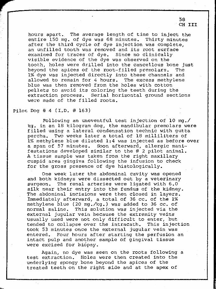
"=----------------~
58 CH III
hours apart. The average length of time to inject the entire 150 mg. of dye was 68 minutes. Thirty minutes after the third cycle of dye injection was complete, an unfilled tooth was removed and its root surf ace examined for traces of dye. Since no clinically visible evidence of the dye was observed on the tooth, holes were drilled into the cancellous bone just beyond the apices of the root-f i.lled premolars. The 1% dye was injected directly into these channels and allowed to remain for 4 hours. The excess methylene blue was then removed from the holes with cotton pellets to avoid its coloring the teeth during the extraction process. Serial horizontal ground sections were made of the filled roots.
Pilot Dog # 4 (I.D. # 163)
Following an uneventful test injection of 10 mg./ kg. in an 18 kilogram dog, the mandibular premolars were filled using a lateral condensation technic with gutta percha. Two weeks later a total of 18 milliliters of 1% methylene blue diluted 1:4 was injected as before over a span of 57 minutes. Soon afterward, allergic manifestations developed similar to the # 2 pilot animal. A tissue sample was taken from the right maxillary cuspid area gingiva following the infusion to check for the gross presence of dye histologically.
One week later the abdominal cavity was opened and both kidneys were dissected out by a veterinary surgeon. The renal arteries were ligated with 6.0 silk near their entry into the fundus of the kidney. The abdominal incisions were then closed in layers. Immediately afterward, a total of 36 cc. of the 1% methylene blue (20 mg./kg.) was added to 36 cc. of normal saline. This solution was injected via the external jugular vein because the extremity veins usually used were not only difficult to enter, but tended to collapse over the intracath. This injection took 53 minutes once the external jugular vein was entered. Four hours after starting the perfusion an intact pulp and another sample of gingival tissue were excised for biopsy.
Again, no dye was seen on the roots following a test extraction. Holes were then created into the underlying spongy bone beyond the apices of the treated teeth on the right side and at the apex of

the left mandibular 2nd premolar. 'I'he holes were saturated with the 1% dye for an additional hour before the teeth were removed.
59 CH III
Thus, these teeth could potentially receive the dye in at least 2 and in some cases 3 routes:
1) I.V. - 10 mg./kg. 2 weeks prior to extraction. 2) I.V. - 20 mg./kg. for 4 hours with renal
arteries tied. 3) directly from a subjacent bony reservoir.
These teeth were sectioned longitudinally and opened by fracturing them through the _canal for complete visualization.
Phase II Technics - Intradental Dye
A total of 88 root canal fillings from six dogs
were evaluated. As in phase I, technics and materials were
handled as they 'W'Ould be clinically.
Where silver points were to be employed, all canals
were prepared to a # 70 or # 80 file to the depth of the
canal. This was necessary because the internal anatomy of
the dog premolar root canal is somewhat 11 cigar~shaped 11 • The
apex of the canal is blunt-ended rather than tapered as
usually found in humans. In order to produce a reasonable
silver cone preparation in such a canal, instrumentation to
larger sizes was indicated.
All of the plastic type root canal fillings were
trimmed of excess f illi~g material leaving ab:>ut 7 mm. of
filling substance in the root canal. A crypt was created in
the coronal portion of the canal and chamber by warm instru-
ments and/or small round burs where the dye could be housed.
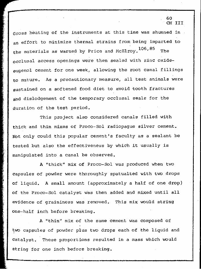
60 CH III
Gross heating of the instruments at this time was shunned in
an effort to minimize thermal strains from being imparted to
the materials as warned by Price and McElroy.106185
The
occlusal access openings were then sealed with zinc oxide-
eugenol cement for one week, allowing the root canal fillings
to mature. As a precautionary measure, all test animals were
sustained on a softened food diet to avoid ·tooth fractures
and dislodgement of the temporary occlusal seals for the
duration of the test period.
This project also considered canals filled with
thick and thin mixes of Proco-Sol radiopague silver cement.
Not only could this popular cement's faculty as a sealant be
tested but also the effectiveness by which it usually is
manipulated into a canal be observed.
A "thick" mix of Proco-Sol was produced when two
capsules of powder were thoroughly spatualted with two drops
of liquid. A small amount (approximately a half of one drop)
of the Proco-Sol catalyst was then added and mixed until all
evidence of graininess was removed. This mix would string
one-half inch before breaking.
A "thin" mix of the same cement was composed of
~wo capsules of powder plus two drops each of the liquid and
catalyst. These proportions resulted in a mass which would
string for one inch before breaking.
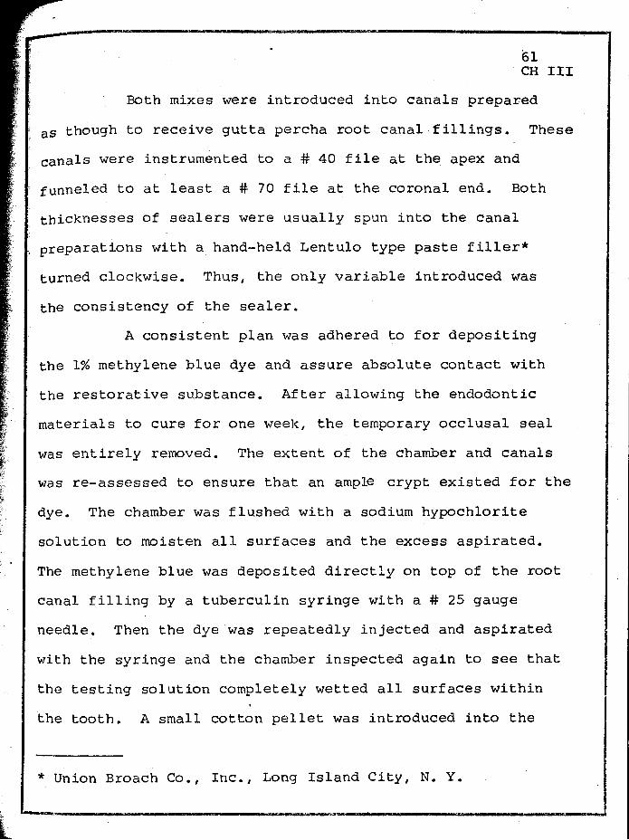
L
61 CH III
Both mixes were introduced into canals prepared
as though to receive gutta percha root canal fillings. These
canals were instrumented to a # 40 file at the apex and
funneled to at least a # 70 file at the coronal end. Both
thicknesses of sealers were usually spun into the canal
preparations with a hand-held Lentulo type paste filler*
turned clockwise. Thus, the only variable introduced was
the consistency of the sealer.
A consistent plan was adhered to for depositing
the 1% methylene blue dye and assure absolute contact with
the restorative substance. After allowing the endodontic
materials to cure for one week, the temporary occlusal seal
was entirely removed. The extent of the chamber and canals
was re-assessed to ensure that an ample crypt existed for the
dye. The chamber was flushed with a sodium hypochlorite
solution to moisten all surfaces and the excess aspirated.
The methylene blue was deposited directly on top of the root
canal filling by a tuberculin syringe with a # 25 gauge
needle. Then the dye was repeatedly injected and aspirated
with the syringe and the chamber inspected again to see that
the testing solution completely wetted all surfaces within
the tooth. A small cotton pellet was introduced into the
*Union Broach Co., Inc., Long Island City, N. Y.

62 CH III
crypt to 11 hold" the dye. Before sealing the occlusal access
again with zinc oxide-eugenol, the cotton pellet was re-
saturated with more dye. T'WO days later these teeth were
surgically removed, then radiographed and photographed again.
The only departure from this routine was in one
case (I.D. # 298) where the dye was placed on the same day
that the canals were filled. This allowed a comparison of
11 new 11 versus 11 aged 11 root canal fillings.
After the photographic records were taken, the
roots were ready for sectioning. Aided by Loupes, the
coronal end of the root was carefully removed with a high-
speed handpiece and crosscut fissure bur to the level where
the restorative material filled the prepared canal throughout
its breadth. The occlusal's periphery was then beveled leaving
a narrow ring of dentin around the filling material. A
narrow, vertical groove was cut with a # ~ round bur along
the entire length of the remaining root portion. A second
groove was similarly cut 180° around from the first groove.
Both vertical grooves were cautiously made so as to avert
perforating the underlying root canal wall. The final
layer of dentin was fractured by fitting these longitudinal . .
grooves onto the leading edge of side-cutting Rongeur forceps.
The entire root canal filling, root canal preparation, and
any dye penetration could now be completely examined. As ,.
L------------------------------~

63 CH III
the root canal filling material was removed its characteristics
and appearance were also noted.
Penetration was 11 read 11 by measuring the greatest
length of linear invasion by the dye. Any bluish discolora-
tion on the canal wall was interpreted as being due to a
discrepancy between the filling matter and the prepared dentin
surface. The extent of dye penetration was measured with a
Boley-Guage micrometer, aided by Loupe's magnification. Each
measurement was taken at least twice. If successive readings
differed by more than 0.3 mm., it was re-measured at a later
time.
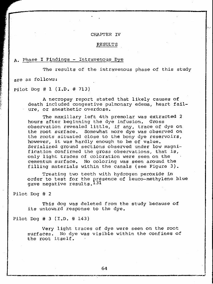
CHAP'rER IV
RESULTS
A. Phase I Findings - Intravenous Dye
The results of the intravenous phase of this study
are as follows:
Pilot Dog # l (I.D. # 713)
A necropsy report stated that likely causes of death included congestive pulmonary edema, heart failure, or anesthetic overdose.
The maxillary left 4th premolar was extracted 2 hours after beginning the dye infusion. Gross observation revealed little, if any, trace of dye on the root surf ace. Somewhat more dye was observed on the roots situated close to the bony dye reservoirs, however, it was hardly enough to be of value. Serialized ground sections observed under low magnification confirmed the gross observations, that is, only light traces of coloration were seen on the cementum surf ace. No coloring was seen around the filling materials within the canals (see Figure 3).
Treating two teeth with hydrogen peroxide in order to test for the presence of leuco-methylene blue gave negative results.131
Pilot Dog # 2
This dog was deleted from the study because of its untoward response to the dye.
Pilot Dog # 3 (I.D. # 143)
Very light traces of dye were seen on the root surfaces. No dye was visible within the confines of the root itself.
64

65 CH DJ
Pilot Dog # 4 (I.D. # 163}
The right mandibular 3rd premolar shattered during the extraction process and was eliminated from the study. A pre-existing ankylosis was suspected because no lateral movement was gained through the use of broad bladed straight elevators.
Visual examination of the root surf aces revealed no evidence of blue coloration on any of the treated teeth. The entire cementum surface of both mandibular 2nd premolars (whose apices were near the dye reservoir holes) was lightly coated with the dye.
No evidence of the dye was clinically obvious upon vertical sectioning of the roots (see Figure 4).
The histologic search for the gross presence of methylene blue in the gingiva and pulp tissue was negative.
In all dogs comprising the intravenous phase, the
saliva assumed a distinct blue-green hue within thirty minutes
after initiating the dye flow. Also, within that length of
time the entire tongue and oral mucosa became bluish-purple
in color. This coloration was not perceptible after 24 hours.
B. Phase II Findings - Intradental Dye
When viewed individually, the results appear to be
consistently inconsistent. This is especially remarkable
since the treated premolars were all anatomically uniform
in configuration, size, and number.. In addition, the same
operator accomplished every filling in each grouping and
each group of root cana1 fillings was manipulated by the
same individual at a single sitting in one animal. Both
operators would be considered as completely familiar with

~--------"- .
' 66 CH J.V
the materials and technic each wielded. It is reassuring
to realize that others have confessed an equivalent
. 4 7 I 112 I 113 I th . . H . t 1 diary. n is same vein arper poignan y
remarked that, 11 the inconsistency of the results has been a
revelation and mortification to me 11•58
The degree of penetration varied from tooth to
tooth and from root canal filling to root canal filling. If
anything, our measurements could be generous to a fault.
In some cases the qualified discoloration was so slight that
its actual presence was suspect. The prevailing attitude
was that regardleas of the intensity of the dentin's foreign
coloration, any bluish discoloration would represent
penetration. Intensity is difficult to convert to a numerical
value because the judgment itself is a subjective process.
The estimated value when plugged into a graph or table
hopefully yields an unbiased, relatable message (see Tables
9 and 10).
In all cases where the root canal was fully prepared
to receive a root canal filling but was left unfilled, the
dye penetrated to the apex. As it was applied in Phase II of
this project, 1% methylene blue is a reliable test agent.
The method used to apply the dye was equally reliable. This
dye was not seen penetrating into the mass of any of the .. bulk filling materials studied in this thesis, however, it

67 CH r.v
does penetrate an estimated thickness of 1.5 mm. of the
proco-Sol sealer. Our preliminary work with both human and
dog teeth as well as glass capillary tubes showed that methy-
lene blue will permeate Proco-Sol sealer to an approximate
depth of 15mm.
On the day the dye was sealed within the dog's
teeth some of it was incorporated into the mass of the
occlusal zinc oxide-eugenol temporary cement turning it
bright blue in color. After 48 hours, the color was no
longer visible. The reason for the disappearance of this
coloring from the temporary access sealant is not understood
at this point, but the color appeared to transfer from the
zinc oxide-eugenol cement into the dentin and enamel
surrounding the occlusal access preparation. Coronally, the
methylene blue dye pervades the entire dentin and enamel
fabric staining it a brilliantblue. The external pattern of
coloration is not a precise indication of the internal
distribution of methylene blue. The dye will extend further
along the internal dentin surf aces than is apparent when the
tooth is scrutinized externally. This may be due in part to
the distribution and histology of canine dentin tubules.
When the dye penetrates the margins of a root canal
filling, it does so unevenly. We generally see irregular
projections of discoloration along the root canal wall

68 CH IV
resembling pseudopodia. Also, the intensity of this staining
is not usually uniform around 360° at any level of the root
canal wall. When we see a band of blue color on this inner
dentin wall projecting apically, it represents a void in the
margin only at that site. That void may be due to operator
failure or it may be indicative of innate inadequacies in
the physical properties of the endodontic materi itself.
our preliminary studies indicated that a 48 hour period gave
sufficient time to reveal any marginal discrepancies.
# 166 - Segmented Gutta Percha Cones with Kloroperka N-0:
One control tooth was lost during the extraction
process. The remaining three controls contained no root
canal filling materials. The canal wall became intensely
stained from crown to apex. The dye also permeated the dentin
in toto.
The gutta percha with the Kloroperka solution acting
as a sealer produced a smooth, homogenous mass as a root canal
filling material. This mass does not appear to form a
complete peripheral bond with the inner dentin wall. It
adheres to the prepared root canal wall in small patches
only. The primary material tended to be brittle and only
three root canal fillings could be removed intact. The path
of the dye was evident on both the dentinal wall and surface
of the material itself. The intensity of dentin discoloration

f
69 CH J.V
was 11 heavy" when assaying this aspect. The intensity was
greatest coronally and gradually decreased toward the apex
(see Figure 8).
While filling the distal root of the right mandibular
third premolar, a void was observed according to a radiograph.
The root canal filling was left unaltered to observe whether
the methylene blue could reach that level. The depth of
penetration was 3.8 mm. in one half of this root canal wall
while measuring only 1.9 mm. in its other half section (see
Table 1). Dye diffusion for the Kloroperka method was the
severest of all the substances and technics included in this
study, averaging 4.14 mm. penet~ation per root canal filling.
The mesial root of the right mandibular premolar
was omitted from consideration as the dye appeared to have
been improperly placed.
#166 - Proco-Sol (thick):
The dentin is stained 11 moderately heavy 11 in general
around the entire perimeter of the thickened sealer. The
intensity of dentin discoloration is remarkably uniform at
the uppermost level. The fade-out of this coloration ends
quite abruptly as one follows it toward the apex (see Figure
9). The average depth of dye seepage was 1.77 mm. here.
In the mesial root of the left maxillary third
bicuspid, a crater (depression) or a tiny hole persisted after

70 CH IV
removing the Lentulo during the root canal filling procedure.
If the crater or hole remained while the sealer hardened,
the dye could be expected to explore the defect. This
appeared to be the case as this root canal exhibited the
greatest linear penetration of the seven evaluated in this
group.
# 298 - _§?qmented Gutta Percha Cones with Chloropercha:
In this animal the dye was sealed over the root
canal filling immediately after the obturations were completed.
The control teeth (prepared but not root canal filled) were
darkly stained the entire length of the preparation. The
dye fully permeated the dentin also. The filling material
is a homogenous mass but fractures readily. Where dye stain
occurred in the marginal dentin it is "very light" in general.
Its mean linear penetration was 1.49 mm. The brilliance of
the discoloration fades somewhat rapidly toward the apex (see
Figure 10).
# 301 - Segmented Gutta Percha Cones with Chloropercha:
The dye here was placed one week after the teeth
had been treated and root canal filled. The mandibular right
second premolar roots we~e initially intended for controls
but were fractured by the dog on his cage bars, and had to be
deleted. Without any endodontic material in the mandibular
left second premolar (controls), the dye diffused unimpeded

71 CH IV
the full length of the root canals. Compared to the
Kloroperka sealer, the chloropercha did not cling to the
walls nearly as well.
The physical consistency of these root canal fillings
appears to be h:>mogenous in nature. Their surface is very
smooth indicating that the prepared root canal walls were
well-planed. Although the filling masses-were somewhat
brittle, four of them could be retrieved from the root canals.
"Moderately heavy" stained dentin was observed around the
entire circumference of the root canal filling. After being
allowed to age for one week, there was more than 200% deeper
penetration than that seen when the testing solution was
placed on the day of the root canal filling (1.49 mm. vs.
3.63 mm.).
For the matured chloropercha the greated amount of
marginal diffusion was observed in both roots of the right
mandibular fourth premolar (see Figure 11). On the day of
op~ration, a notation was made that a great deal of hemorrhage
exuded from this tooth. Success in the chloropercha and
Kloroperka technics relies on absolutely dry dentinal walls.
The presence of moisture acts as a physical deterrent, not
allowing these materials to intimately contact the dentinal
surface.

f· ,.
72 CH IV
# 301 - Proco-Sol (thin):
When sectioning these teeth, this root canal sealer
was extremely brittle and did not adhere well to the dentin.
This material tended to shatter while the teeth were being
fractured with the Rongeurs. Therefore, hand instruments
were utilized in place of the Rongeurs to eliminate the
crushing force and retain as much of the root canal sealer
as possible in its applied position.
Multiple voids existed in the body of the sealer
mass although a conscientious effort had been made to fill
the canals in their entirety. Only one c~nal (distal, right
maxillary second premolar), was judged to be well filled
while two others were thought to be fairly well filled by
this sealer alone when examined immediately after sectioning.
The dye was '!Light 11 in intensity at the occlusal
1 mm. level then quickly paled toward the apex (see Figure
12). In only two of the eight fills was the dentin penetration
considered "heavy". The comparative depth of dye penetration
for similarly inserted thick or thin consistencies of Proco-Sol
sealer is notably close (1.77 mm. vs. 1.79 mm.).
# 322 - Gutta Percha Con~s with Proco-Sol (thin):
Here, control canals were prepared to a # 50 file
at the apex and funneled to at least a size # 70 file at their
orifice. A handpiece-driven Lentulo paste filler spun the

73 CH IV
thin mixture (as recommended by the manufacture) into the
four root canals using them as controls. The large size of
the preparation allowed the Lentulo to freely deposit the
cement without binding. It is noteworthy that these four
controls produced the least linear penetration by the
methylene blue (l.25 mm.) in this investigation. The "light"
dentin coloration existed completely around the prepared
walls and ended abruptly with very little fading (see Figure
13).
The lateral condensation technique employed here
resulted in a homogenous mass of gutta percha which appeared
to be well adapted to the smooth walls of these preparations.
Evidence of dye was seen on the gutta percha surface in five
of the six fillings placed in this ~nimal. The dye was
especially visible in roots where the longest penetrations
also took place. In the one case where the dye was not seen
on the gutta percha surface, the linear penetration was but
1.2 mm., compared to the overall group mean of 3.65 mm. The
mesial roots of both third premolars were eliminated from
evaluation when the animal fractured them prior to their
extraction. In three of the remaining roots, the longest
extent of discoloration was so pale that their actual penetra-
tion was reasonably doubtful. Therefore the average length
of linear penetration here is deceptively excessive •
.... ----·~----------...... ---------------------...... -------.,_,----------------,.,_..._ __ ~ .... ..&

~------~·.
f f ,.
. 74 CH IV
# 326 - Silver Cones with Proco-Sol (thick):
The root canals used as controls were prepared to
a size # 60 file, while those to receive test root canal
fillings were instrumented to a # 70 or # 80 at their apex.
Two of the controls were fitted with silver cones and wedged
into place without sealer. Heavy deposits of dye were seen
around the entire circumference of this prepared surf ace.
The remaining two control canals were filled with thick
Proco-Sol radiopaque root sealer by turning a root canal
file counter-clockwise. Large voids were seen in both controls
filled with the thick sealer. In this manner, the relatively
deep penetration was quite likely due to poor manipulation and
flow characteristics rather than due to the material's lack
of sealability.
Although a ring of stain was generally observed
here, the intensity of discoloration was "very light".
Discoloration was noted on the surf ace of four of the eight
silver points. In restorations where the dye appeared on the
metal's surfa~e, there was considerable linear penetration
also (note the distal root of the mandibular right third
premolar and both roots of the third premolar plus the
' distal root of the fourth premolar on the left side). It is
interesting to observe that the average straight-line pene-
tration for these four was 3.43 mm. whereas the average for
r 75 CH IV
the remaining four where the dye was not evident on the
silver point surface averaged 1.15 mm., or three times the
length of discoloration. This finding was similar to that
seen in the case # 322 with laterally condensed gutta percha
with root canal sealer. There, five of the six laterally
condensed gutta percha masses had dye on their surface. Their
average marginal penetration being 4.16 mm. whereas the sixth
gutta percha filling exhibits merely a 1.2 mm. depth of dye
diffusion (see Figures 13 and 14).
Small voids were observed in the film of sealer
surrounding the silver cones. This may be the same "laking"
which Weiner has reported from his in vitro studies of
endodontic sealers. 138 In the pres~nt study, silver cones
cemented with Proco-Sol gave the best overall results of
those methods commonly used in endodontic practices today.
# 331 - Gutta Percha Cones with Proco-Sol (thick):
Two controls were filled with this consistency of
sealer by using root canal files. After sectionin:J, it
could be seen that both canals were incompletely filled. The
linear penetration in one was 1.6 mm. while the other was
5~6 mm. One reasonable explanation for their broad difference <
in penetration is that the thicker mix seemed to display
poorer flow properties. A single gutta percha cone was
adapted and seated into the remaining two control canals.
76 CH DI
The methylene blue readily stained the full extent of the
prepared canal in each instance (see Figure 15).
Unfortunately, the bulk filling materials from
this aspect of the study were discarded. It would have been
interesting to search their surf ace for traces of dyes and
compare the findings with those of animals # 322 and # 326.
It is rational to speculate that the higher average of
penetration seen here (3.95 mm.) may also represent some
diffusion through the sealer and onto the dentin wall by dye
that creeped between the gutta percha/sealer interface.
Of interest here was the observance that the
intensity of the dentinal staining was interpreted as either
"very light" (in 2 test teeth) or "heavy" (in the remaining
6 trials) and always encircled the entire canal. Although
the two were noted to be "very light", they averaged 5.15 mm.
of linear penetration whereas the six decreed to be "heavy"
averaged 3.55 mm. or 1.50 mm. less! This observation agrees
with the gutta percha root canal fillings laterally condensed
in animal #322. There, 6 trials were 11 light 11 and averaged
4.55 mm. in linear diffusion while the 2 classed as "very
heavy" bore a 3.20 mm. linear average of dye. In the latter
animal, "moderately heavy" deposits of dye extended 1.35 mm.
less than when the root canal filling was 11 lightly 11
penetrated.

17 CH TV
With the silver cone, chloropercha, and Kloroperka
experimental root canal fillings the heaviest dispersion of
methylene blue dye correspondcdwith the deepest linear extent
of penetration.

CHAPTER V
DISCUSSION
A. General Observations Involving this Study
Through the years a multitude of tests have been
employed to assess the marginal integrity of restorative
materials used in dentistry. Vast amounts of scientific
knowledge have been made available allowing the profession
to rapidly progress. More directly, scientific· research has
helped to enhance the public's confidence in dentistry while
raising the level of professionalism. The experimental
methods used have had varying degrees of merit.
Because of expediency, time, and availability, many
tests have been performed primarily on extracted human teeth. *
When testing the margins of root canal fillings, additional
problems must be confronted and solved. The ultimate and
only valid test of any root canal filling (including
substances used, operator ability, technic, etc.) must take
place in an acceptable biologic model.
Many aspects of endodontics have been investigated
through the use of laboratory animals. These generally
involved the assessment of how a particular material and/or
medicament effected or affected tissues. The prime concern
78

79 CH V
was, "how will the tissues be affected if that substance
invades them during the course of treatment or soon there-
after?".
The bane of the endodontist has been root canal
failure and how to avoid it. Hence, the constant search for
new materials and improved methods which will help us seal
the root canal system from the body. When performing the
root canal filling procedure we can not be sure if we have
sealed off all the internal pulp channels despite our best
radiographic evaluations and clinical acumen.
To test the sealing ability of root canal fillings,
ingenious experimental designs have been implemented. Many
test solutions have been pitted against the margins of root
canal fillings including dyes, inks, bacteria, isotopes, and
fluorescent substances. But, all have been applied to the
non-functioning, non-living tooth! We now must know the fate
of these substances and procedures in vivo. Such information
is analagous to the terms "statistically significant" and
"clinically significant". Certainly the knowledge gained
from the laboratory may be statistically significant - in
the laboratory situation, yet may be found to be clinically
insignificant.
In the 1930 1 s while studying tooth metabolism by
means of "dental lymph 11 Bodecker and Lefkowitz were among
-·· P:SCWWU J
r:=-~------80 CH V
the first to point out that dentin permeability differs in
vital and pulpless teeth. 14 They demonstrated that a dye
placed in an empty root canal will exhibit greater diffusion
than in one where the pulp is present. Also; this penetration
will differ in sound and old (sclerotic) teeth. Loiselle and
his colleagues found that in vitro hamster teeth exhibited
greater marginal permeability when amalgam restorations were
compared under in vivo and in vitro conditions. 79 Going
discovered that temporary stopping placed in vitro teeth
provided a good initial barrier but when he repeated the
experiment in vivo its penetrability increased sixfold. 47
Going also has emphasized that the same restorative materials
tested in the laboratory gave different results when similarly
tested in a living organism. It becomes evident that we can
no longer ignore the difference in response of the same dental
materials under morbid and vital conditions. Furthermore,
we are rapidly exhausting the variety of tests to which we
can subject teeth in the laboratory, therefore we must explore
means of evaluating our endodontic treatment aspects within
viable tissue. Failure and loss of the tooth from the mouth
becomes "clinically significant 11 regardless of whether bench
tests of a procedure are "statistically significant" or not.
The possibility of an intravenously placed dye
reaching the dental tissues seemed remote, yet had intriguing

81 CH V
p0tential. The location of tumorous tissues with vital dyes
is well known. A number of dyes have been employed to accom-
plish this, including methylene blue. Some authors stated
that the I.V. dye would reach the interstitial tissues.48149150
Therefore because of its contrasting color, non-toxicity,
documented broad previous use, economy, and availability,
methylene blue was chosen.
The decision of injecting the dye on three separate
days 48 to 72 hours apart was made with full knowledge that
the dye was normally eliminated in 24 hours. Actually, the
eliminiation begins soon after entry of the dye into the body.
If it could reach the interstitial spaces its concentration
would be highly dilute. Therefore, a supposition was made
that a three-cycle - perfusion would permit adequate exposure
and accumulation. To determine if renal excretion of methylene
blue prevailed, the kidneys were excluded from the circulation
in one pilot dog since it is one of the major organs for the
dye's elimination. These proposed measures proved to be of
no avail.
The teeth were carefully examined for the dye's
presence following the injections. Little, if any, was
apparent. Because of its oxidation-reducation capacity, a
portion of the dye in circulation is said to exist in the
leuco - or "invisible" (oxidized) form. To rule out the
82 ~· CH V
r i possibility that the dye was indeed present, but not in its
original form, some extracted teeth were placed in an oxidizing
solution to reduce any unseen dye back to its visible (reduced)
131 form. This act not only removed the precious traces of
dye present and evident on the root surf ace, but produced a
well bleached tooth!
For the sake of consistency the dye was always
injected intravenously. The possibility exists that an
injection into the carotid artery may have achieved our goal
as Spector avers that the arterial end of the capillary is
121 more permeable. Drugs could also have been given to
increase the capillary permeability. In the present project,
a localized increase in capillary permeability was attempted
by the production of a mild localized inflammation induced by
tightly splinting two adjacent teeth together. This step
did not achieve the goal of attracting additional dye to the
area. Perhaps the injury must be more severe to be effective
for this purpose.
Except for the mandibular second premolars in dog
# 163, the only traces of dye observed were on the apical
one-fourth of the roots ~see Figure 4). Whether these deposits
represented true diffusion (dye was also placed close to their
apices) or if the dye was picked up in the extraction process
cannot be positively confirmed. Regardless, the amount was
83 CH V
insufficient to act as a guage for in vivo apical penetration
studies.
In animal experiments regarding endodontics a number
of preliminary questions must be answered: 1) Are the chosen
animals and human teeth comparable? 2) What is the morphology
of the teeth and the root canal? 3) Are the canals amenable
to reasonable preparation and restoration with available
armamentarium? 4) Do these teeth respond comparably to the
testing agent? 5) Is the agent compatible with life when
used intravenously? 6) Is the intravenous route feasible
or could the agent be more opportune if used locally? 7) How
much of the agent must be injected to get a "reasonable"
reading? 8) What is the optimal time period between injection,
dispersion and animal sacrifice for that agent? 9) What
physical condition will the animal be in while dye dispersion
takes place? 10) Does the agent have an affinity for other
structures as well? 11) What is the in vivo permeability of
the outer tooth structure to the agent used?
It becomes readily apparent to anyone so inclined,
that an in vivo study requires a great deal of cerebration
in order to come up with a reasonable experimental design • .
While perfusing the animals with 1% methylene blue
injectable, u.s.P., angioneurotic edema occurred in three
84 CH V
different dogs, a circumstance which to our knowledge has
not previously been reported (see Figure 6). After the
first episode, the dogs used in Phase I were given trial
injections intravenously and observed for a reasonable length
of time before commencing with the endodontic treatments.
Unfortunately this technique offers no absolute assurance of
immunity. In pilot dog # 163 these same symptoms did not
develop with the trial injection, yet were manifest with a
subsequent injection. Ironically these reactions may have
r been due to the fact we were using a chemically pure and r i
sterile form of methylene blue. Macht concluded that the
more refined this dye is, the rrore potentially toxic it
81 becomes. In Conn it is stated that a 11 pure 11 form must be
used for vital staining, however, the pure form is a poor
h . t 1 . t . 30 is o ogic s ain. If micrometric quantities of the dye
reached the periodontium, methylene blue lacked the gross
staining capability needed to act as a detectable indicator
for the evaluation of endodontic procedures.
Another possible explanation of the toxic incidents
and the peripheral vascular collapse we experienced, may
have been due to not using injectable glucose as a physiologi-<
cal dilutant for the dye (see Figure 7). Brooks felt this
was not necessary because she believed that a healthy body's
natural glucose content was adequate. Glucose apparently

85 CH V
acts as the catalytic agent in the redox cycle which re-converts
roethemoglobin (Fe+++) back to hemoglobin (_Fe++), thereby
avoiding an accumulation of the methemoglobin. In addition,
many investigators (cited earlier) injected this dye intra-
venously without any specified dilution or precautions in
most cases - even when concentrations of 100-200 mg./kg. were
t i injected. Some have mentioned the peripheral collapse, but
~ this was not C'Onsidered to be a major deterrent or contra-
indication for its use.SO,Bl
There is a need for in vivo research with dyes.
Their employment offers considerable promise as a dental
detective agent. The medical, biological, toxicological and
chemical literature carry much pertinent information. While
in reality there is very little known concerning dye toxicity,
elimination routes, target organs, and natural tissue barriers.
Also, it should be noted that dyes not readily available in
the U.S.A. are being used in other countries to demonstrate
the location of cancers. In vivo dyes may prove to be
advantageous in future dental research as well.
Placing a dye within a tooth is a legitimate test of
the sealing capability of a dental material. Here, the entire
filling/tooth junction is subject_ to the dye for penetration.
This magnifies the sealing ability (or lack of it) because a
large marginal surface is exposed to the dye.
''( ,. .. , .

86 CH V
Unless a canal is prepared completely through the
apex, tests of root canal filling materials against apical
penetration cannot be considered valid. Where the root canal
preparation is short of the apex, the apex becomes inadver-
tantly packed with dentin filings in most instances which
3-will inhibit penetration. Also, the entire margin is not
exposed to the testing agent, therefore, a portion of the
"seal 11 is provided from the surrounding cementum, dentin, or
predentin. Having a closed or narrow apex provides an
excellent apical stop and is not wholly a test of the root
canal filling material's ability to seal the root. We can
see how an evaluation of a material which is vertically
condensed against a closed apex will inadvertantly embrace
the ability of the technic rather than the material to seal
a cavity in the root.
A popular test method is to fill the root canals
of anterior teeth, then cover the entire outer surface of
the tooth, except for the apical 2 - 3 mm. with a barrier
compound such as wax. Unfortunately, this still allows the
dye solution to penetrate the multiple accessory canals
usually present in the apex and, if left long enought, the
dye will eventually pervade the dentin through the unwaxed
cementum. Undetected gaps in the apical cementum will allow
the intrusion of a test agent. Such penetration (by way of

87 CH V
the cementum and dentin) possibly could be interpreted as
a failure of the restorative material or technic.
A number of other authors have placed their testing
agent within the tooth. 114142182191 Here, we are assured of
assaying the marginal integrity of the material or technic
employed in its placement as the tooth acts as its own dye
reservoir and naturally confines penetration to any interface
between tooth and seal it may reach. Preliminary studies
have revealed that in the case of extracted human teeth
methylene blue will penetrate any patent opening, diffuse
through cementum in time, through minute abrasions in the
cementum, and areas of developmental grooves which are
especially vulnerable in dog teeth. Once the dye reaches the
dentin tubules it passes unimpeded to the root canal making
the perception of its derivation dubious.
With in vivo studies temperature is a built-in
control. While trying to simulate the dental environment, in
vitro studies are subject to temperature fluctuations and
dehydration problems which, in spite of conscientious efforts,
are difficult to compensate for. The temperature within the
dog's root is constant ~y comparison. A dog is not normally
fed hot meals so the teeth are not subject to as wide a range
of temperature changes while feeding. They do, however,
drink cool water which may provide some temperature variation.

r:--------.. t .
88 CH V
In addition, occlusal forces are experienced in the dog
comparable in many respects to those occurring in humans.
A review of the significant studies of tooth-
restoration margins reveals that: 1) results depend on the
operator who places the restorations; 2) results vary accord-
ing to the material being employed; 3) results rely on the
depth and range of the particular dye or isotope selected;
4} results will differ when performed in vivo and in vitro;
5) results are affected if a restoration is tested immediately
after placement or following the lapse of a period of time;
6) conflicting results are reported by separate authors
evaluating the same materials; and 7) testing solutions
invariably penetrate all restorations.
Isotopes have been shown to penetrate further than
dye solutions. This is possible because the autoradiogram is
able to demonstrate an unseen isotope whereas a dye must be
present in sufficient amounts to be visible. This does not
rule out the rationale of dyes in margin studies. In many
respects dyes are considered superior to radioactive ions,
i.e., molecular size, differences in chemical structure,
chemical inertness, and ,their propensity to combine
histochemically with a wide assortment of healthy or patho-
48 49 logical tissues and organs. '
r:= :' ------------------
It may be safely assumed that many of today's
89 CH V
philosophic disagreements over materials and techniques are
due in part to the diversity of experimental designs and
results in penetration studies. Although the argument for
unfettered freedom in this regard is sound, it is also
accurate to assert that some standardization of procedure is
in order. The following might be considered:
1. The status of the teeth to be treated. BrieflyL
this would cover extracted, pulpless, and vital
tooth data from experimental animals, and man, in
both young and mature dentitions.
2. The diffusion ability of the test solutions in
experimental animals and man, in both young and
mature dentitions.
3. The isotopic or dye solution, its pH and temperature
during the experiment as well as molecular size.
4. Competent knowledge of the che~ical reactivity of
the testing media with the substances to be
evaluated.
5. Selection of a controlled material and root canal
filling technic.
6. Waiting a specified length of time to allow the
materials to fully set.
L----~----------------------....;..,------------------...J

f 90 CH V
In today's endodontic literature there is a dearth
of knowledge concerning the physical characteristics and
predictable properties of gutta percha and silver points under
different environmental and working conditions. This is but
one of the items which create gaps in our present knowledge.
Current dental articles are replete with "good,
better, or best" being stressed when comparisons are made of
marginal seals afforded by the clinical methods of ·individual
operators. The assumption by the reader is that the lower
rated materials in any given study is not worthy to be used
in his practice. The clinician may fail to appreciate that
all materials and penetrants have not been tested under the
same conditions, have not been placed under standardized
routines, that different investigators have varying technical
abilities, that the skill and familiarity of the research
analyst varies with each study and/or material, and certainly,
that the clinician's own capacity to duplicate a study's
results is purely speculative. If such testing methods were
infallible, we'd have abandoned gold foil, amalgam and
certainly root canal therapy long ago.
Restorations knowingly placed for a research project
conceivably may be rrore carefully done than those routinely
done in everyday dental practice. It is equally unfair to
condemn a material or technic when it is not manipulated by
someone familiar with its working qualities. For this
91 CH V
reason, warm gutta percha and diffusion technics used to seal
root canals were purposely excluded from this thesis. Without
knowledge or skill in the use of the intracacies of their
management, they might have been unjustly surveyed.
Investigations of· the margins produced by various
root canal fillings will be informative, but without standard-
ization they must be considered as being valid only for those
operators placing the root canal fillings. Rather than
adopting a technic which a particular article has deemed
11 best 11 under certain circumscribed conditions, we might better
test our own ability utilizing the various technics. It is
a simple matter to prepare and endodontically seal root
canals with recommended or personal innovative rrodes, then
seal the occlusal access opening, coat the tooth with wax and
immerse the tooth in a vegetable dye or ink for 24-48 hours
in an off ice incubator. The teeth can be longitudinally
sectioned and examined. This -would serve as a 11 spot-check"
of our own mastery of a particular technic.
Endodontic root canal filling technics involving
gutta percha with Kloroperka or chloropercha solutions were
included for study since they are frequently condemned by
penetration studies. Their condemnation has compelled many
endodontists to use them almost furtively, as if they were

92 CH V
clandestine operations. Both materials are often resorted to
by the endodontic community in particular 11 problem11 situations.
Their use may be indicated in: 1) narrow, ledged, or perfor-
ated canals, or canals obstructed by calcification, or broken
instruments. They may also find use where an accessory canal
or internal resorption is suspected. Some operators have
found them helpful when adapting a master gutta percha cone
or when oblitering ribbon - shaped canals, often encountered
in mandibular anterior teeth. A tapered preparation, unviolate:l
apical constriction and well dehydrated wall are emphasized
as being fundamental to their effectiveness.
Although the Kloroperka technic had the highest
mean linear penetration (4.14 mm.), its median penetration
value was less than that for gutta percha, laterally condensed
with thick or thin mixes of sealer (see Table 10).
The most obvious observation with the chloropercha
technic is that when the methylene blue was placed on the
same day as the filling procedure, it was able to penetrate
less than half as far as when the dye was placed one week
after sealing the root canal (l.49 mm. vs. 3.63 mm.). This
finding is in agreement with that of Roydhouse and
others. 112111311281129 Therefore, the results of dog# 298
lacked clinical reality and were omitted from the academic
ranking in Table 10 of the modern methods of filling root

canals. Of the basic materials used to fill canals as
93 CH V
measured by the standards of this study, chloropercha ranked
second following the silver cone method.
The reason for the vivid increase in dye penetration
in the root canals filled with chloropercha and allowed to
age in situ, is openly speculative. Assuming that evaporation
of the excess chloroform takes place, we might speculate that:
a) Reopening the tooth to place the dye allowed the
evaporation to take place, or if these teeth had remained
coronally sealed after root canal filling would their initial
root canal seal be retained? b) How is evaporation accomplish-
ed in a completely sealed tooth, if it is? c) What is the
coefficient of contraction or expansion of chloropercha upon
setting or thermal changes?
Few authors have compared results of root canal
fillings tested immediately upon filling versus those tested
after remaining in situ for a period of time. Most studies
were content to examine the comparative sealing potential of
freshly condensed materials. In experiments where the
investigator allowed the endodontic material to mature,
increased penetration was generally observed. However, one
study reported a decrease in penetration for a dye inserted . 4
after pausing for 48 hours. The same authors also
observed greater diffusion of their dye in zinc oxide-eugenol

94 CH V
cement than in Kloroperka. These are unique findings in the
articles dealing with endodontics however.
Both the Kloroperka and chlorpercha fillings appeared
to form a smooth, homogenous mass. Clinically, they appeared
to adapt very well to the root canal's internal discrepancies.
Its value in narrow, curved or unnegotiable canals where
contraction is negligible may therefore be justified. Claims
of these root canal fillings being good adherents to the
dentinal walls are probably overstated as both poorly adhered.
Of the two, Kloroperka root canal fillings seemed to cling to
dentin somewhat better than chloropercha. Its mode of
attachment was characteristically seen in patches. In
extracted human teeth, the same observation was made during
the preliminary tests of this project.
The intensity of the dentin stain penetrating
beneath Kloroperka was judged as 11 heavy 11 while that beneath
the chloropercha method was a bit less and deemed "moderately
heavy".
For root canals filled solely with thick or thin
mixes of Proco-Sol cement, the character of dentin staining
is fairly.consistent. The dentin stains 11 light 11 to
"moderately heavy" at the superior reaches of the sealer.
This intensity fades abruptly as one follows the stain
apically. This contrasts with the pattern of fading produced

95 CH V
by the dye with Kloroperka and chloropercha restorations
which usually fade gradually making the terminal penetration
difficult to read.
When the root canals were filled with thick or thin
Proco-Sol cement, they appeared to be clinically well-filled
with the sealer up to the level of the orifice of the root
canal. Rather remarkable differences in penetration occurred
in those root canals filled with a hand-held Lentulo (1.77 mm.
and 1.79 mm.) contrasted with the four controls in dogs# 331
and# 326 (3.83 mm.). The latter cement root canal fillings
were placed by the commonly used means of turning a reamer or
file counterclockwise in the root canal. In·all these cases,
the roots appeared to be satisfactorily and fully filled by
the cement. Of interest here is that the four canals filled
with the engine driven Lentulo experienced the least dye
penetration of all the root canals in this study (1.25 mm.).
They also were seen to be more compactly filled and embodied
fewer voids. Where a canal is prepared large enough to
accomodate it, the engine-operated Lentulo would appear to
offer an excellent means of introducing root canal sealer into
a tooth.
The penetrations of laterally condensed gutta percha
employing thick or thin mixtures of root canal cement are
close. There is a 0.30 mm. less linear penetration visible

L
when a thin mix was employed with gutta percha.
96 CH V
Upon re-examination of all our root canal fillings
another item repeatedly entered the picture. Closer
inspection of the preserved root canal fillings revealed that
where the silver point filling method was used, 4 of the eight
silver cones had distinct traces of dye well down their
surfaces. Of these 4 restorations the average linear penetra-
tion was 3.43 mm. The mean pen.etration of the remaining 4
silver~filled canals was only 1.15 mm. In dog # 322 where a
routine lateral condensation root canal filling technic was
employed with gutta percha, strikingly similar results were
observed. Here, 5 of the 6 bulk masses of retrieved gutta
percha fillings had deposits of methylene blue on the surface
of the gutta percha core. Again, gross penetration was also
comr:lonly seen in the dentin walls of these five canals. The
average penetration of dye was 4.16 mm. compared to 1.2 mm.
dye penetration in the lone root canal whose core of gutta
percha was free of visible dye stain on the surface.
Coupled with the earlier observation that the dye
appears to be able to penetrate only the surface 1.5 mm. of
Proco-Sol cement, "penetration" may be a dual phenomenon
where a bulk material or core is cemented into a root canal
with a root canal sealer. The dye would appear to be able
to 11penetrate 11 through the silver/Proco-Sol and gutta percha/

Proco-Sol sealer interfaces. The deepest dye pattern on
the dentin walls may therefore be a result of the dye's
97 CH V
ability to permeate outward through narrow, adjacent thicknesses
of. the sealer (as would exist between the dentin wall and the
well-condensed gutta percha). Here the dye's primary route
of access was between the silver or gutta percha cone/Proco-
Sol sealer interface. The lateral diffuslon of the dye
through the root canal sealer and onto the root canal wall
may be termed "secondary penetration 11 in such cases.
An enigma was created when an attempt was made to
equate or relate the intensity of the dentin's stain with the
depth of linear penetration. When chloropercha and Kloroperka
technics were examined, 11 heavy 11 dye penetration was directly
associated with a greater linear dye penetration. In general,
this was not the case when a Proco-Sol sealer was involved
in the root canal filling. With the lateral condensation
technic the dye penetration was 11 light 11 but extended further
toward the apex. Thus, when Proco-Sol sealer was a part of
a root canal filling material, the intensity of methylene
blue penetration was not directly proportional to the extent
of linear penetration. No attempt was made to decode these '
notations, however, it is possibly related to the "secondary
penetration" seen when Proco-Sol sealer is used with gutta
percha or silver cones.

r= f. ' f:
98 CH V
It is within the realm of possibility that dye was
able to penetrate between the bulk materials and sealer if a
less than adequate volume of sealer were used to coat the
walls initially. This supposition ~~uld be more plausible
if but a single cone of gutta percha were used. With multiple
cones of gutta percha being laterally condensed, this
supposition seems remote.
The controls where a single gutta percha or silver
cone were adapted but seated without root canal sealer, were
"very heavily" penetrated by the dye. Any attempt to restore
a canal with a single cone, inspite of its sincere adaptation,
would likely lead to failure.
Curiously enough, the most consistent dye-repelling
root canal filling was Proco-Sol cement when used by itself.
To avoid misinterpretation of this finding, we are reminded
of the endodontic sealer studies of Curson and Kirk as well
as those of Weiner and Schilder.351138
All, independently
found that in time, the zinc oxide-eugenol based sealers
deteriorate or volumetrically contract. Their solitary use
as a complete sealant could also be expected to fail eventually.
It seems quite reasonable to believe that where
root canal fillings are concerend, we are dealing with t'WO
interfaces: 1) the sealer/dentin interface and 2) the sealer/
bulk filling material (core) interface. Non-union between

either one will admit apical tissue fluid into the canal
99 CH V
system. Indigenous to root canal core materials and sealers
as we know them today, there are known, inherent discrepancies
in long term volume stability which complicate the
bl 35,85,106,126,138 pro em. An interpret~tion of those studies
'WOUld imply that, upon setting, the Proco-Sol sealer contracts
three-dimensionally (into itself). The interface becomes
vulnerable as each root canal filling material (core and
sealer) responds to:
1. The forces involved in the root canal filling
technique.
2. Their innate properties (separate and/or in combina-
tion).
3. The influence imparted by the surrounding hard and
soft tissue environment.
Future research would undoubtedly be rewarding if
we knew more of the particulars of interface adhesion from
studies of root canal filling materials,, other dental materials
and dentinal surfaces~ from the inherent physical character-
istics of root canal sealers in general and gutta percha in
particular~ and alternative materials and methods to isolate
the root canal system from the periodontal ligament.

B. Endodontic Aspects of Dog Teeth
100 CH V
Very little information is at hand covering the
clinical endodontic aspects of dog teeth. A number of
observations were made based on the animals treated endodont-
ically during the course of this project and its preliminary
studies. These corrunents are intended to help fill the broad
gaps in the chasm of available clinical data, and hopefully,
will assist the research of others in the future. The
remarks are not intended to be a comprehensive treatise on
canine endodontics but will be restricted to the maxillary and
mandibular premolar therapy.
General Observations
It goes without saying that a preliminary study of
a dog skull will be helpful to familiarize the investigator
with its general anatomic features and dental anatomy. At
this time he may practice applying the rubber dam and trying
various rubber dam clamps. This·will reduce operating room
time and preclude the possibility of not having suitable
rubber dam clamps on hand. Practicing on extracted dog teeth
prior to the main project will reap irruneasurable benefits.
These teeth may be obtained from schools of veterinary
medicine, local veterinarians and from research facilities~
i.e., hospitals, medical school, dental schools, etc.

101 CH V
During the preparatory dental exam when selecting
a dog for endodontic research the occlusal surf aces should
be inspected for wear patterns, chipped enamel, etc.
severely worn coronal surfaces are often associated with
root canals which are comparatively small in size. At least
four roots were deleted from this study due to inter-treatment
tooth fractures. Tooth fractures due to food mastication
was ruled out as a possible etiology in view of the fact
that all of the test animals were fed soft diets during the
evaluation period.
It may be that dogs confined to cages tend to gnaw
to relieve their frustration or some may gnaw as extensions
of a natural habit. Clues of a gnawing habit may be gained
by examining the dentition and also the exposed bars of the
cage for tooth marks. The act of. gnawing contributes to the
build-up of secondary dentin along the canal wall. This new
dentin is very dense and was found to be intrinsically very
hard making root canal preparation more difficult. This
habitual grinding may also increase the difficulty of
extracting. these teeth and possibly· contribute to ankylosis.
Clinically, ankylosis was diagnosed as the cause of a
minimum of three root fractures during the extraction process.
In one case the entire root appeared to be rigidly attached
to bone. I

102 ~v
Inclusion of roentgenographs for instrumentation
and case documentation is valuable. A po~table type x-ray
machine, manufactured and equipped specifically for dentistry
is advisable. A 15 Ma portable hospital unit, however, may
be adapted for dental use by adding a cone over the collimator.
A few minutes taken before starting the operation will be well-
spent on trial x-rays, varying the ~illiamperage and voltage
settings and exposure times. This is especially advisable
when different machines are provided. From one x-ray unit
satisfactory roentgenographs were obtained with 90 Kvp at 10
Ma for 0.5 seconds. It is interesting to note that each
article concerned with canine dentistry suggests different x-ray
tt . 107,111,134 se 1ngs.
One reason for favoring mandibular premolars in
endodontic research is the inherent difficulty of taking
maxillary roentgenographic views. The dog's palate is flat,
therefore maxillary premolar and molar views must be taken
from exaggerated angulations resulting in distorted images.
This also applies to films of the mandibular cuspids and first
and second premolars where the contour of the mandible's
surface prevents routine film placement. An osseous shelf
projects lingually from the inferior surface interfering
with parallel film placement in these mandibular areas.
Acceptable roentgenographic views of the maxillary and
103 CH V
mandibular incisors plus those of the mandibular premolar
and molar areas may be obtained by following the usual dental
x-ray methods. In these areas little anatomical interference
is encountered.
Because of the difficulty in film placement, the
packets are hand-held in hemostats. The body may be protected
from direct radiation exposure by wearing protective aprons
and gloves. For this technic the manufactured lead--lined
mitten provides awkward hand control of the hemostat. The
hand may be shielded from the direct rays by draping a
protective cover over it. Another means would be to sew
two or three strips of ~ inch flat, elastic ribbon on one
side of the mitten which will stabilize it to the back of
the hand leaving the fingers unencumbered.
External Premolar Anatomy
_The dental formula for the dog's permanent dentition
3 1 4 2 is normally 2(I3 , c1 , P4, M3) for a total of 42 teeth.
However, some variation is seen according to the muzzle
length of the animal. Brachycephalic (short-headed) dogs
may have a decreased number of molar teeth. Carnassial
(sectorial) is a term frequently applied to the maxillary
fourth premolars and mandibular first molars.
The first premolars of either arch are usually
single-rooted, peg-shaped teeth. They are small in stature,

~-------
l L
averaging less than 14.0 mm. in length. The crown is
104 CH V
rudimentary in appearance, of·ten projecting less than 2 .o mm.
into the dental arch. In older dogs the first prerrolars
are frequently missing.
The second premolars were generally observed to
have two roots. In all cases, this maxillary second premolar
had two separate, divergent roots. In two ·of the ten animals,
the roots of the mandibular second premolars were fused.
Each segment, however, housed a separate pulp canal. In both
instances the morphology was identical in the left and right
quadrants, which also connotes bilateral symmetry. our
observation differs from that of Barker and Lockett who
remarked that thi.s tooth was "usually single rooted 11•
6
The maxillary and mandibular third premolars, as
well ·as the mandibular fourth premolars were found to
continually possess two separate roots. From a dentist's
point of view the maxillary fourth premolar appears to be
misclassified (see Figure 17). It has large mesial and
distal cusps, in addition to another c_uspal projection on the
mesiolingual portion of the crown. The root configuration
of thi_s carnassial tooth has three similarly corresponding
roots. Clinically, the crown and root morphology of this
upper fourth premolar suggests that it might be rrore
appropriately included as a molar tooth.
r=_· _______________ __
In general, the distal roots of premolars are
105 CH V
slightly shorter and rrore bulky in stature compared to the
mesial roots. This contrast is especially noticeable in the
mandibular premolars, however, is also true in the maxilla.
As a general statement, it may be said that the root length
increases with each premola·r as one goes posteriorly {see
Figure 18).
Internal Premolar Anatomy
Morphologically there appears to be little
variability in the configuration of the premolar root canals
from dog-to-dog and tooth-to-tooth. No individual root
was seen to have more than a single canal. Unlike human
teeth, no bizarre canal paths were discovered in canine
premolars, for example, where a single canal bifurcates and
later rejoins itself forming an island of dentin within the
root canal. Although not encountered during the course of
this project, the possible existence of diverse canal systems
in canine premolars cannot be ruled out. The customary
finding was a bulbous, sausage-shaped canal. Where the crown
was chipped, heavily abraided and in older dogs, the lumen
of the canals often appeared to be narrowed. In these cases
the predentin or dentin was remarkably resistant to filing.
Prior to exiting, the apical pulp stump arborizes
through multiple tiny foramina. As many as eight to ten

rr---------1-06 f rnv ~ ' were counted in the horizontal ground sections. As few as ~
l
two were noted, but less than four foramina was the
exception rather than the rule. Unless one of the foramina
was incidentally threaded, intentional perforation of the
apex was arduous with hand filing. Barker and Lockett
suggest the utilization of an engine-powered reamer for this
6 purpose.
Extractions
Compared to man, the extraction of dog premolars
presents an onerous task when the desire is to recover them
intact. However, removal of teeth from younger dogs or
those afflicted with periodontal disease does seem to require
less physical effort. The extraction problem is partially
due to the fact that very little lateral rrovement is possible '
with surgical elevators. The premolar roots have a longitu-
dinal developmental groove on the interradicular surface.
This groove becomes rrore pronounced as one proceeds posteriorly
in the mouth and is especially evident on the distal roots.
This groove acts as a 11 keyway 11 with the enveloping alveolar
bone making a very stable niche for the tooth. This "keyway"
also acts as a deterrant to rotational luxating moves.
The non-yielding nature of the alveolar bone
surrounding the roots may prove to be the crux of the luxation
difficulties. This conveys several potential facets. The
107 CH V
attachment apparatus may be more tenacious than in man. In
addition, the histochemical composition of this bone may be
more dense. The mandibular alveolar bone appears to contain
only a modest amount of cancellous bone interposed between
the buccal and lingual plates of cortical bone. Another
observation was that the periapical roentgenograms generally
revealed a broader, diffuse lamina dura cir-cumscribing these
roots. The lack of alveolar resiliency may be due in part
or totally to these empirical notations.
In the surgical extraction process (see Figure 1)
# 34 elevators are used to thoroughly loosen or expel the
separated roots. The direction of this avulsion must be in
the long axis of the teeth. Generally speaking, the maxillary
premolars are easier to remove unscathed than those in the
mandible. The reasons for this are not clear, however, it is
conjectured that their shorter average root length (1 to 2 mm.
less) and thinner buccal plate is implicated. Because of the
existence of a flattened palate it can also be assumed that
more cancellous-type bone surrounds the premolar roots of
the upper arch.
Preliminary studies showed that, if intact, the
dog's cementum will exclude methylene blue from the dentin
for at least two days in vitro. It cannot be stated thatthis
would be true in vivo. This observation may be of signif i-
cance in future in vivo endodontic studies because if the
cementum is permeable to a dye, its penet~ation, although
108 CH V
through the dentin, could readily be interpreted as due to
"marginal penetration". Early tests in preparation for this
thesis also indicated that cementum covering the roots of
extracted human teeth is more readily penetrated with
methylene blue than the cementum of ~og's 1n vitro. Another
observation from those early tests was that when this dye
diffused through the cementum, in dog teeth, the area of the
developmental groove was more susceptible to its entry. The
dye's invasion into the dentin surrounding the groove usually
assumed a wedge-shaped pattern (see Figure 19).
Longi.tudinal Sectioning
Sectioning the root in half vertically provides a
superior means of examining the internal aspect of a root
canal and/or its contents. It is also an excellent means of
extracting the pulp for soft tissue studies. When this type
of root division was adopted, longitudinal paths were cut as
near to 180° circumferentially to one another as possible.
The depth of both channels was cautiously prepared in order
to penetrate close, to b~t not into/ the root canal. It was
discovered that when the long-axis grooves were not situated
opposite to one another or close enough to the root canal,
the blades of the side-cutting Rongeur (used to effect the j
L
109 CH V
root's fracture) were directed obliquely through the root,
producing an unfavorable split alongside the canal.
occasionally this resulted in three root fragments, the third
piece being the tubular frame of the canal. If the teeth
originated from mature dogs, it could be theorized that
cleavage occurred at the interface between the primary and
secondary dentin. However, this cou.ld be averted by cutting
two "starter 11 notches at the 12 and 6 o'clock positions
occlusally and at the apex then simply uniting the two cuts
on each side.

r':-------. CHAPTER VI
SUMMARY
The purpose of this in vivo project was twofold:
1) to explore the possibility of utilizing intravenous
injections of methylene blu_e for evaluating endodontic
procedures; 2) to evaluate the relative penetrability of
commonly used root canal filling materials and technics in
vivo with 1% methylene blue place intradentally.
The experimental design involved two separate
and distinct phases: PHASE I - Selected teeth were treated
endodontically. A 1% methylene blue injection u.s.P. was
given intravenously into four dogs. Several controlled
injection technics were attempted in an effort to attract
the disclosing solution to the alveoli of the teeth. This
included inducing mild periapical inflammation, multiple
injection cycles, varying the concentration of the dye, by-
passing the kidneys, and placing the dye near the apices in
intraboney reservoirs. Histologic samples of pulp and
gingival tissues were examined for their gross content of
dye following the various injection technics. The teeth
were scrutinized externally and internally for the presence
of the dye. Insufficient quantities of the 1% methylene blue
110

111 CH VI
reached the dental apparatus to validly judge endodontic
aspects of dental treatment.
In PHASE II a total of 88 root canals in six dogs
were endodontically treated to observe the response of several
materials and technics manipulated in the teeth of a living I
model. These methods and treatments were utilized in the
same manner as they would be clinically. The compositions
were allowed to mature in situ for one week. The 1% methylene
blue injection, u.s.P. was then sealed in an intradental
crypt atop the endodontic restorations for 48 hours. In one
dog the dye was deposited on the same day as the filling
procedure. The teeth were surgically extracted in order to
recover them intact. The roots were then sectioned vertically
in half. The root canal prepartion, basic filling material,
root canal sealer and character of the dye penetration, could
be visually inspected throughout the breadth and length of
the root canal.
In addition to the biologic model having root canals
similar to man's and following procedures as used in the
off ice, the experiments were controlled with the following
parameters: The specific root canal filling material and
filling technics were performed by persons clinically
familiar with their nuancesr the same type of root canal
filling was placed in a single animal~ each type of root

112 CH VI
canal filling was inserted by one individual on a given day 1
the dye was systematically deposited7 and the entire root
canal filling and its sealing ability could be circumspected
by vertically sectioning through each root canal.
From the data of this study the following observa-
tions may be made:
1) Intravenous injections of methylene blue as attempted in
this investigation is an inadequate means of evaluating
endodontic procedures.
2) Untoward reactions may occur in animals even when "non-
toxic 11 dyes are employed intravenously. These responses
have occurred with the use of refined or "medicinal grades"
of dye such as 1% methylene blue injection, u.s.P., in dogs.
3) These results represent a "potential" sealing ability of ·
the root canal filling materials. Each person cannot
expect to get precisely the same results because of varying
innate personal abilities. The manner of employing each
root canal filling material will vary from one individual
to the next. Clinical results reflect an individual's
ability and conscience as well as the potential quality of
a root canal filling material and technic. Many discrepan-
cies and poor performance attributed to a material in
penetration studies are just as likely to be due to the
operator as the root canal filling materials.

r= _______________ __ 113 CH VI
4) Variable results may be realized from the same root canal
filling material and technic even when ~anipulated by the
same operator. The quality of one individual's root canal
fillings will vary throughout a given day.
5) The consistency has little to do with the seal provided by
Proco-Sol radiopaque silver cement. The finding that
relatively good sealing results a~e obtained even when
root canals are poorly filled, voids exist, etc., suggests
' that this sealer may possess a partial chemical inhibition
toward methylene blue. This study has seen that the dye
will penetrate nearly 1.5 mm. of the sealer's surface.
Another clue that a chemical barrier may be operating is
the observation that the vertical stain pattern fades
abruptly rather than gradually when the dentin surf ace is
protected with Proco-Sol cement.
6) In endodontics we are often dealing with two interfaces:
a. Between the root canal sealer and tooth surf ace.
b. Between the root canal sealer and bulk filling
material (gutta percha or silver cones}.
A clinical case may fail due to penetration through either
route. These interfaces become penetrable as a result of:
manipulation, differences in volumetric stability and
surf ace characteristics peculiar to each substance.

F
114 CH VI
7) To realistically evaluate any root canal filling material
or technic in vivo or in vitro, ample time must be given
to allow the root canal filling materials to throughly
set before exposing it to a test design.
8) A solution of 1% methylene blue injection, u.s.P., is
capable of penetrating any root canal filling materials
or techniques as utilized during this investigation.
9) None of the root canal filling techniques as evaluated by
this investigation may be justly endorsed nor renounced
as good, better or best, worst, etc. Each root canal
filling technique has its own rate of success according
to the capability of the clinician. Judgement of the
sealability of root canal filling.materials and techniques
cannot be based on numerical values alone.
10) As yet, dentistry has not seen the "ideal" endodontic root
canal filling material or technique. If it has, man and
nature have expunged its perfection.

REFERENCES
1. Ainley, J. E.: Fluorimetric assay of ·the apical seal of root canal fillings. Oral Surg. 29:753-762, 1970.
2. American Dental Convention (author unknown): Filling teeth and roots~ what is gained by fang filling, and average of success. Dent. Cosmos. 3:79-81~ 1861, 62.
3. Anderson, D. J. and Ronning, G. A.: Dye diffusion in human dentin. Arch. Oral Biol. 7:505-512, 1962.
4. Antoniazzi, J. H., Mjor, I. A. and Nygaard-Ostby, B.: Assessment of the sealing properties of root filling materials. Odont. T 76:261-271, 1968.
5. Armstrong, W. D. and Simon, W. J.: Penetration of radiocalcium at the margins of filling materials~ a preliminary report. J. ~..mer. Dent. Assn. 43:684-686, 1951.
6. Barker, B. c. w. and Lockett, B. C.: Utilization of the mandibular premolars of the dog for endodontic research. Aust. Dent. J. 16:280-286, 1971.
7. Barletta, c., Negro, G. c. and Roccia, G. M.: Controllo con isotopi radioattivi (Ph32) di alcuni tra i pin comuni materiali per otturazione canalare. Minerva Stomat 19:159-163, 1970.
8. Baumrind, s. and Buck, D. L.: Rate changes in cell replication and protein synthesis in the periodontal ligament incident to tooth movement. Amer. J. Orthod. 57:109-131, 1970.
9. Bergman, B.: Diffusion of some cationic and anionic dyes through mandibular disk and cartilage in vitro: Acta Odont. Scand. 26:103-110, 1968.
10. Bergman, B. and Soremark, R.: Studies on permeability of certain dentin~l sealing agents. An invitro investigation using radiosodium. Odont. T. 69:113-117, 1961.
115

. 116
11. Blood, D. C. and Henderson, J. A. Veterinary Medicine. Baltimore, Md.: The Williams and Wilkins Co. 1960, pp. 851-859.
12. Bodansky, O.: Methemogloginemia and Methemoglobinproducing compounds. Pharm. Rev. 3:144-196, 1951.
13. Bodansky, o. and Gutman, H.: Treatment of methemoglobinemia. J. Pharm. Exp. Ther. 90:46-56, 1947.
14. Bodecker, c. F. and Lefkowitz, W.: Further observation on vital staining of· dentin and enamel. J. Dent. Res. 25:387-399, 1946.
15. Boyd, J. D.: isotope.
Dental autoradiography, I.Choice of radioJ. Dent. Res. 36:274-285, 1957.
16. Brannstrom, M. and Soremark, R.: The penetration of Na22 ions around amalgam restorations with and without cavity varnish. Odont. Rev. 13:331-336, 1962.
17. Brooks, M. M.: Does methylene blue form methemoglobinemia? Proc. Soc. Exp. Biol. Med. 31:1134-1137, 1934.
18. Brown, D. W.: The cavity-sealing properties of autopolymesizing acrylic and zinc phosphate cement. 31st Gen. Mtg. I.A.D.R., Abstract#-49, 1953.
19. Buchanan, G. s.: Resistance of self-curing acrylic resin to the passage of dye between filling and cavity wall. J. Canad. Dent. Assn. 17:131-138, 1951.
20. Buchbinder, M.: A non-shrinking root canal material. Dent. Cosmos. 73:14-16, 1931.
21. Buckley, J. P.: A review of the literature on dental therapeutics for the past sixty years~ with special emphasis on the treatment of the dental pulp and filling root canals. Dental Items Int. 60:926-936, 1938.
22. Burnett, c. w. and Scherp, H. W.: Oral Microbiology and and Infectious Di~ease. Baltimore: The Williams and Williams, Co., 1968, ed. 3~ Chapter 2.
117
23. Castagnola, L.: Experimentielle untersuchungen iiber im munde selbe-polymerisierende Kunstharz fullungen. Schweiz Mschr. Zahnheilk 60:880-886, 1950.
24. Cattell, D. M.: Results of experimental root canal fillings. Dent. Review 9:386-387, 1895.
25. Chievitz, o. and Hevesy, G.: Danske Vidensk Selsk Biol. Medd, Translated by Wasserman, F., Blayney, J. R., Groetzinger, G. and DeWitt, T. G.: Studies on the different pathways of exchange of minerals in teeth with the aid of radioactive phosphorus. J. Dent. Res. 20:389-398, 1941.
26. Christen, A. G.: Ultraviolet radiation and fluorescence in diagnosis, therapy, and research. J. Oral Ther. 3:62-79, 1966.
27. Christen, A. G. and Mitchell, D. F.: A fluorescent dye method for demonstrating leakage around dental restorations. J. Dent. Res. 45:1485-1492, 1966.
28. Clafin, R. s.: Healing of disturbed and undisturbed extraction wounds. J. Amer. Dent. Assn. 23:945-959, 1936.
29. Clarke, E. G. c. and Clarke, Myra L. Garner's Veterinary Toxicology. Baltimore, Md.: The Williams and Wilkins Co. 1967, 3rd ed. pp. 108-110.
30. Conn, H. J. Biological Stains~ Baltimore, Md.: The Williams and Wilkins Co., 7th ed. pp. 100-103.
31. Coolidge, E. D.: Reaction of dog tissue to drugs used in root canal treatment. J. Amer. Dent. Assn. 19:747-759, 1932.
32. Coolidge, E. D. and Kesel, R. G.: A Textbook of Endodontology. Philadelphia: Lea and Febiger, 1956, ed. 2; Chapter 15.
33. Copeman, s. M., Coke, F. and Gouldesbrough, C.: "Activated" (irradiated) fluorescein in the treatment of cancer. Brit. Med. J. 2:233-236, 1929.

118
34. Crawford, W. H., and Larson, J. H.: Fluid penetration between fillings and teeth using Ca45. J. Dent. Assn. 85:518-522, 1956.
35. Curson, I. and Kirk, sealing cements.
E. E.: An assessment of root canalOral Surg. 26:229-236, 1968.
36. Ditewig, G.: Veterinary Dentistry. Dent. Review 9:705-710, 1895.
37. Dixon, c. M. and Rickert, u. G.: Histologic verification of results of root canal therapy in experimental animals. J. Amer. Dent. Assn. 25:781-1803, 1938.
38. Dow, P. R. and Ingle, J. I.: Isotope determination of root canal failure. Oral Surg. 8:1100-1104, 1955.
39. Fiasconaro, J. and Sherman, H.: Sealing properties of acrylics. New York Dent. J. 18:189-198, 1952.
r: t 40. Fischer, M.: Experirnentiel le untersuchungen iiber die f abschlussdick tigkeit der zahnheilkude gebrauchlichen ~ ~ fullungs materielien. Schweiz Mschr. Zahnheilk 59:
595-611, 1949.
41. Fraser, c. J.: A study of the efficiency of dental fillings. J. Dent. Res. 9:507-517, 1929.
42. Geiser, M. w. and Jensen, J. R.: Comparative sealability of four root canal cements. SOth Gen. Mtg. I.A.D.R., Abstract~713, 1972.
43. Gesell, R., Krueger, H., Nicholson, H., Brassfield, C. and Pelecovich, M.: A comparison of" the response of the anesthetized dog to intravenous administration of sodium cyanide during uniform artificial ventilation and during normally controlled ventilation with additional observations on the effects of methylene blue. Amer. J. Physiol. 100:227-240, 1932.
44. Going, R. E., Massler, M. and Dute, H. L.: Marginal penetration of dental restorations by different radioactive isotopes.• J. Dent. Res. 39:273-284, 1960.
45. Going, R. E. and Massler, M.: Influence of cavity liners under amalgam restorations on penetration by radioactive isotopes. J. Prosth. Dent. 11:298-312, 1961.
______________________________ __.___,,_:llll'P ________________________________ , ___________ ,

r-______________ __ 46. Going, R. E., Massler, M. and Dute, H. L.: Marginal
penetrations of dental restorations as studied by crystal violet dye and rl31. J. Amer. Dent. Assn. 61:28S-300, 1960.
119
47. Going, R. E., Myers, H. M. and Prussin, s. G.: Quantitative method for studying microleakage in vivo and in vitro. J. Dent. Res. 47:1128-1132, 1968.
48. Goldacre, R. J. and Sylven, B.: On the access of bloodborne dyes to various tumor regions. Brit. J. Cancer 16:306-322, 1962. .
49. Goldacre, R. J. and Sylven, B.: A rapid method for studying tumorous blood supply using systemic dyes. Nature (London) 184:63-64, 19S9.
SO. Goluboff, N. and Wheaton, R.: Methylene blue induced cyanosis and acute hemolytic anemia complicating the treatment of methemoglobinemia. J. Pediat. S8:86-89, 1961.
Sl. Goodman, Louis s. and Gilman, Alfred, The Pharmacological Basis of Therapeutics. New York, N. Y.: The Macmillan Co. 1967,pp.1047-1048.
S2. Gross, R. L., Goldberg, A. F. and Loiselle, R. J.: Microleakage around silver alloy restorations in a biologic model. J. Dent. Res. 47:1183-1186, 1968.
S3. Grossman, L. I.: A study of temporary fillings as hermetic sealing agents. J. Dent. Res. 18:67-71, 1939.
S4. Grove, c. J.: Why formaldehyde preparations are contraindicated in septic root canals with a practical scientific method for the treatment of putrescent pulps. Dent. Review 27:996-lOOS, 1913.
SS. Grove, c. J.: infections.
Some important causes of periapical Dent. Cosmos. S8:1333-1352, 1916.
S6. Handbook of Chemistry and Physics. Cleveland, Ohio: ~~Chemical Rubber Publishing Co. (19SS) 36th ed.
pp; 3S7-387, 394-447.
57. Harlan, A. w.: Experimental root filling. Dent. Review 9:746-748, 1895.
'---------------~ ! ~
120
58. Harper, w. E.: The character of the adaptation of amalgams to the walls of cavities attained by present methods of instrumentation and the use of the best known alloys, as indicated by the air pressure test. Dent. Review 26:1179-1198, 1912.
59. Hewett, A. C.: Capillary attraction - A factor in gold and amalgam filling. Dent. Review 16:845-852, 1902.
60. Higgenbotham, T. L.: A comparative study of the physical properties of five commonly used root canal sealers. Oral Surg. 24:89-101~ 1967.
61. Hill, T. J.: Experimental granulomas in dogs. J. Amer. Dent. Assn. 19:1389-1398, 1932.
62. Hirsch, L. and Weinreb, M. M.: Marginal fit of direct acrylic restorations. J. Amer. Dent. Assn. 56:13-21, 1958.
63. Hollinger, H. H.: Penetration of restoration margins.
I 64.
Dent. Radiogr. Photogr. 49:9-14, 1967.
Hunt, A. M.: Distribution of naturally occuring isotopes in human teeth as seen in autoradiograms. J. Dent. Res. 39:715, 1960. Abstract.
65. Ibid., Chapter 22.
66. Ingle, J. I.: Endodontics. Philadelphia: Lea and Febiger, 1969, pp. 216-221.
67. Jones, L. Meyer. Veterinary Pharmacology· and Therapeutics. Ames, Ia.: Iowa State Univ. Press, 1970, pp. 978-981.
68. Kahn, H.: Mechanical technic in root canal therapy. Dent. Survey 22:295-299, 1946.
69. Kakar, R. C. and Subramanian, V.: various restorative materials. 13:156-165, 1963.
Sealing qualities of J. Prosth. Dent.
I 70. Kapsimalis, P. and Evans, R.: Sealing properties of endodontic filling materials using radioactive polar I and nonpolar isotopes. Oral Surg. 22:386-393, 1966.
I f ____________________________________ _.. __________________________________ --..1
~------------------~-------------------------------1-2_1 ____ __ .0 • ~'
71. Kapsimalis, P., Evans, R. and Tuckerman, M. M.: Modified autoradiographic technique for marginal penetration studies. Oral Surg. 20:494-504, 1965.
72. Kapsimalis, P., Parris, L., Evans, R., and Cobe, H. M.: Improved bacteriologic techniques for marginal penetration studies. Oral Surg. 19:93~100, 1965.
73. Kornfeld, B.: Study of clinical behavior of resins in operative dentistry. J. Dent. Res. 32:714-715, 1953. Abstract.
74. Lamstein, A. and Blechman, H.: Marginal seepage around acrylic resin veneers in gold crowns. J. Prosth. Dent. 6:706-709, 1956.
75. Lange, K. and Boyd, L. J.: Use of fluorescein method in establishment of diagnosis and prognosis of peripherol vascular disease. Arch. Intern. Med. 74:175, 1944.
76. Lantz, B. and Perrson, P. A.: perforation in dogs• teeth. Rev. 16:238-257, 1965.
Experimental root A roentgen study. Odont.
77. Lawson, D. D., Nixon, G. s., Noble, H. w., and Weipers, w. L.: Dental anatomy and histology of the dog. Res. Vet. Sci. 1:201-204, 1960.
78. Leopold, s. and Spinella, D. root canal restorations. 13 0: 2 9- 3 3 I 195 7 •
J.: A technic for canine J. Amer. Vet. Med. Assn.
79. Loiselle, R. J., Goldberg, A. F., Gross, R. L. and Steuver, c. H.: Marginal microleakage - an in vivo assessment. J. Amer. Dent. Assn. 78:758-760, 1969.
80. Lyell, J., Barber, D. and Massler, M.: Effects of saliva and sulfide solutions on the marginal seal of amalg~m restorations. J. Dent. Res. 43:375-379, 1964.
81. Macht, D. and Harden, W. C.: Toxicology and assay of methylene blue. Ann. Intern. Med. 7:738-745, 1933.
82. Marshall, F. J. and Massler, M.: The sealing of pulpless teeth evaluated with radioisotopes. J. Dent. Med. 16:172-184, 1961.

. 122
83. Massler, M. and Ostrovsky, A.: various filling materials. 234, 1954.
Sealing qualities of J. Dent. Child. 21:228-
84. McCauley, H. B. and Guilda, J. E.: In· vivo distribution of radiophosphorus (p32) in vital and pulpless teeth of a dog as indicated by radioautographs. J. Dent. Res. 22:200-203, 1943.
85. McElroy, D. L.: Physical properties of root canal filling materials. J. Amer. Dent. Assn. 59:433-440, 1955.
86. McQuillen, J. H.: Who first filled nerve cavities? Dent. Cosmos. 3:556-557, 1862.
87. McQuillen, J. H.: Necessity of root filling. Brit. J. Dent. Sci. 10:507-508, 1867.
88. Mengale, c., Swartz, M. L. and Phillips, R. W.: Adaptation of restorative materials as influenced by roughness of cavity walls. J. Dent. Res. 39:825-835, 1960.
89. The Merck Index o~ Chemicals and Drugs. Rahway, N. J.: Merck and Co., Inc. (1960) 7th ed. p. 676.
90. The Merck Veterinary Manual. Rahway, N. J.: Merck and Co. Inc. (1967) 3rd ed. pp. 1018-1021, 1623.
91. Messing, J. J.: An investigation of the sealing properties of some root filling materials. J. Brit. Endodont. Soc. 4:18-22, 1970.
92. Mitchell, R. A. and Struck, H. C.: Production of anemia in dogs with methylene blue. Fed. Proc. 10:93, 1951, Abstract.
93. Mortensen, D. w., Boucher, N. E. and Ryge, G.: A method of testing for marginal leakage of dental restorations with bacteria. J. Dent. Res. 44:58-63, 1965.
94. Nadler, J. E., Green, H., and Rosenbaum, A.: Intravenous injection of methylene blue in man with reference to its toxic symptoms and effect on the electrocardiogram. Amer. J. Med. Sci. 188:15-21, 1934.

123
95. Nelsen, R. J., Wolcott, R. B. and Paffenbarger, G. c.: Fluid exchange at the margins of dental restorations. J. Amer. Dent. Assn. 44:288-295, 1952.
96. Newton, K. A.: The distribution of dyes and fluorescent substances by the blood stream within tumors. Brit. J. Radial. 38:224-226, 1965.
97. Orban, B.: The problem of root canal treatment. J. Amer. Dent. Assn. 19:1384-1388, 1932.
98. Osol, Arthur; Pratt, Robertson; and Altschule, Mark D.: The Urrited States Dispensatory and Physicians Pharmacology. Philadelphia, Pa.; J. B. Lippincott Co. 1967, 26th ed. pp. 725-726.
99. Parris, L. and Kapsimalis, P.: The effect of temperature on the sealing properties of temporary filling materials, I. Oral Surg. 13:982-989, 1960.
100. Parris, L. and Kapsimalis, P.: The effect of temperature change on the sealing properties of temporary filling materials, I. Oral Surg. 13:982-989, 1960.
101. Parris, L., Kapsirnalis, P., Cobe, H. H. and Evans, R.: The effect of temperature on the sealing properties of temporary filling materials, II. Oral Surg 17:771-778, 1964.
102. Persson, w. D.: Canine dentistry. Canad. J. Comp. Med. 22:162-163, 1958.
103. Phillips, R. w., Gilmore, H. W., Swartz, M. L., and Schenker, s. I.: Adaptation of restorations in vivo as assessed by ca45. J. Amer. Dent. Assn. 62:9-20, 1961.
104. Pickard, H. M. and Gayford, of amalgam restorations. July 20, 1965.
J. J.: Leakage at the margins Brit. Dent. J. 119:69-77,
105. Pinto, J. and Buonocore, M.: Effect of bases and cavity liners on marginal leakage of filling materials. New York Dent. J. 29:199-207, 1963 •
.. ·~-&Wr-------------------""'---~-,------------------.....-

. 124
106. Price, w. A.: Report of laboratory investigations on the physical properties of root filling materials and the efficiency of root fillings for blocking infection from sterile tooth structure. J. Nat'l. Dent. Assn. 5:1260-1280, 1918.
107. Ramy, c. T. and Segreto, v. A.: Apicoectomy and root canal therapy for exposed pulp canal i.n the dog. J. Amer. Vet. Med. Assn. 150-977-983, 1967.
108. Rentsch, G. and Wittekind, D.: Methylene blue and erythrocytes in the living animal. Contribution to the toxicology of methylene blue and formations of Heinz Bodies. Toxic Appl. Pharmaco~. 11:81-87, 1967.
109. Rewbridge, A. G., Hanke, M. T. and Halpert, B.: Further observations on the function of the gall bladder. Amer. J. Physiol. 95:511-518, 1930.
110. Rose, E.E., Joginder, L., Williams, N. B., and Falcetti, J. P.: The screening of materials for adhesion to human tooth structure. J. Dent. Res. 34:577-596, 1953.
111. Ross, D. L. and Myers, J. W.: Endodontic therapy for canine teeth in the dog. J. Amer. Vet. Med. Assn. 157:1713-1718, 1970.
112. Roydhouse, R. H.: Penetration around the margins of restorations: 2. Nature and significance. J. Canad. Assn. 34:21-28, 1968.
113. Roydhouse, R. H., Weiss, M. E. and Leonard, L.: Penetration around the margins of restorations: 1. Review and experiments. J. Canad. Dent. Assn. 33:680-689, 1967.
114. St. Clair, L. E. and Jones, N. D.: Observations on the cheek teeth of the dog. J. Amer. Vet. Med. Assn. 130:275-279, 1957.
115. Sanjo, D., Inomata, H. and Karibe, H.: The e~aluation of root canal fillings by radiography. Jap. J. Conservative
.. Dent. 13:42-48, 1970. Oral Res. Abst. #3349, 1971. Abstract.
. 125
116. Sausen, R. E., Armstrong, w. D. and Simon, w. J.: Penetration of radiocalcium at margins of acrylic restorations made by compression and noncompression technics. J. Amer. Dent. Assn. 47:636-638, 1953.
117. Schroeder, A.: Gewebsvertraglichkeit des wurzel fullmittels AH 26. Zahnaerztl Welt 58:563-567, 1957.
118. Schroeder, A.: Zurn problem der baktenemdhten wurzelkanalversorgung. Zahnzerztl Welt 58:531-537, 1957.
119. Sekine, N., Ishikawa, T. and Imanishi, T.: Clinical availabilities of creosote in the endodontic treatment. Bull. Tokyo Dent. Coll. 5:85-115, 1964.
120. Seltzer, S.: Penetration of microorganisms between the tooth and direct resin fillings. J. Arner. Dent. Assn. 51:560-566, 1955.
121. Spector, w. G.: permeability.
Substances which affect capillary Pharrnacol. Rev. 10:475-505, 1958.
122. Stein, G.: Experirnentelle wurzelbehandlung. Ztschr F. Stomatol. 29:744-748, 1931.
123. Stewart, G. sealers.
G.: A comparative study of three root canal Oral Surg. 11:1029-1041, 1174-1178, 1958.
124. Stossel, T. P. and Jennings, R. B.: Failure of methylene blue to produce methemoglobinemia in vivo. Amer. J. Clin. Path. 45:600-604, 1966.
125. Swartz, M. L. and Phillips, R. W.: In vitro studies on the marginal leakage of restorative materials. J. Arner. Dent. Assn. 62:141-151, 1961.
126. Swartz, M. L. and Phillips, R. W.: Influence of manipulative variables on the marginal adaptation of. certain restorative materials. J. Dent. Res. 12:172-181, 1962.
127. Tagger, M.: Endodontics: A review of the past and its present status. ·Alpha Omegan 60: 107-118, 196 7.
128. Talirn,. s. T. and Singh, I.: Sealing of root canal filling in vivo conditions as assessed by radioactive iodine. J. Indian Dent. Assn. 39:198-202, 1967.
-

. 126
129. Talim, s. T., Singh, I. and Kher, V. W.: Sealing of root canal fillings in vitro conditions as assessed by radioactive iodine. J. Indian Dent. Assn. 39:131-133, 1967.
130. Tremblay, V.: Use of "fast curing" acrylic material in operative dentistry. J. Canad. Dent. Assn. 16:249-251, 1950.
131. Underhill, F. P. and Closson, o. E.: The physiological behavior of methylene blue and methylene azure: A contribution to the study of the oxidation and reduction processes in the animal organism. Amer. J. Physiol. 13:358-371, 1905.
132. Wainwright, W.W., Stowell, E. C. and Taylor, J. B.: Microleakage of in vitro amalgam fillings to rl31 labeled human serum albumin and Narl31. J. Dent. Res. 38:749, 1959, Abstract.
133. Wasserman, F., Blayney, J. R., Groetzinger, G. and DeWitt, T. G.: Studied on the different pathways of exchange of minerals in teeth with the aid of radioactive phosphorus. J. Dent. Res. 20:384-398, 1941.
134. Weaver, M. E., Sorenson, F. M. and Jurap, E. B.: The miniature pig as an experimental animal in dental research. Arch. Oral Biol. 7:17-24, 1962.
135. Webster, A. E.: Some experimental root canal fillings. Dominion Dent. J. 12:109-113, 1900.
136. Webster, A. E.: The comparative value of ordinary sealings for root canal dressing as excluders of bacteria. Dent. Cosmos. 44:925-929, 1902.
137. Webster, A. E.: Temporary and permanent fillings as barriers to bacteria. Dent. Review 17:275-286, 1903~
138. Weiner, B. H. and Schilder, H.: A comparative study of important physical properties of various root canal sealers. II Evaluation of dimensional changes. Oral
· Surg. 32:928-937, .1971.
1
139. Yagoda, H.: Radioactive Measurements with Nuclear Emulsions.j Translated by Myers, H. M., Jennings, F. and Becks, H.: j
I, A rapid radioautographic technique for combined calcified i
and soft tissues. J. Dent. Res. 31:416-420, 1952. l I
l----------------------------------------_,.,---------------------------------~
CHAPTER VIII
APPENDIX
A. Tc;i.ble3i ------·
B. Figures -----
127

. 128
TABLE l
I.D. # 166 - SEGMENTED GUTTA PERCHA WITH KLO ID PERKA N-0
Prep. Linear % Tooth Restoration _!,ength (mm.) Pentr. (mm.) Pentr.
Right Mand. 2nd M Prep. only 7.3 7.3 100.00
(Control) D Prep. only 6.5 6.5 100.00
(Control)
3rd M G.P. + Kloro. 7.6 3.6 47.37 D II 6.8 3.8 55.88
4th M II
D II 7.4 2.6 35.14
Left Mand. 2nd M Prep. only
(Control) D Prep. only 6.5 6.5 100.00
(Control)
3rd M G. P. + Kloro. 6.8 3.9 57.35
D II 7.3 5.4 73.97
4th M II 6.8 3.0 44.12 D II 8.5 6.5 78.82
Total # Rests = 8 Total # Controls = 3 Total Length - Rests. 59.9 29.0 48.41
II II Controls 20.3 20. 3· 100.00 Ave. II - Rests. 8.56 4.14 28.36
II II Controls 6.77 6.77 100.00 Total % Penetr. - Rests.· 392.65
II II II Controls 300.00
Ave. II II Rests. 56.09 II II II Controls 100.0~.J
I I.
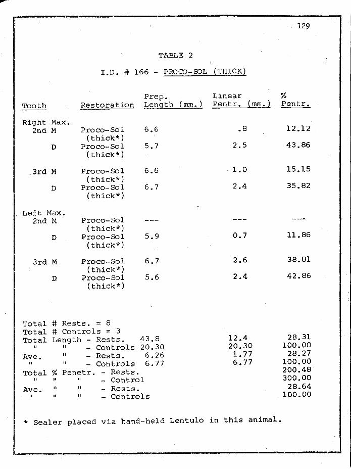
. 129
TABLE 2
I.D. # 166 - PROCO-SOL (THICK)
Prep. Linear % Tooth Restoration Leng!:.!:!_l mm. ) Pentr. (mm.) Pentr.
Right Max. 2nd M Proco-Sol 6.6 .8 12.12
(thick*) D Proco-Sol 5.7 2.5 43.86
(thick*)
3rd M Proco-Sol 6.6 1.0 15.15 (thick*)
D Proco-Sol 6.7 2.4 35.82 (thick*)
Left Max. 2nd M Proco-Sol
(thick*) D Proco-Sol 5.9 0.7 11.86
(thick*)
3rd M Proco-Sol 6.7 2.6 38.81 (thick*)
D Proco-Sol 5.6 2.4 42.86 (thick*)
Total # Rests. = 8 Total # Controls = 3 Total Length - Rests. 43.8 12.4 28.31
ti ti - Controls 20.30 20.30 100.00
Ave. ti - Rests. 6.26 1.77 28.27 ti II - Controls 6.77 6.77 100.00
Total % Penetr. - Rests. 200.48 II II ti Control 300.00
Ave. II II - Rests. 28.64 II ti It Controls 100.00
* Sealer placed via hand-held Lentulo in this animal.

. 130
TABLE 3
I.D. # 298 - SEGMENTED GUTTA PERCHA WITH CHLORJPERCHA
Prep. Linear % Tooth Restoration Length (mm. ) Pentr. (mm.) Pentr.
Right Mand. 2nd M Prep. only 7.1 7.1 100.00
(control) D Prep. only 7.6 7.6 100.00
(control)
3rd M G.Pu + Chloro. 6.6 0.7 10.61 D II 6.3 0.9 14.29
4th M II 7.4 0.6 8.11 D II 8.2 3.1 37.80
Left Mand. 2nd M Prep. only 6.7 6.7 100.00
(control) D Prep. only 6.8 6.8 100.00
(control)
3rd M G.P. + Chloro. 5.4 1.7 31.48 D II 5.8 1.7 29.31
4th M II 7.4 1.7 22.97 D II 7.8 1.5 19.23
Total # Rests. = 8 Total # Controls = 4 Total Length -·Rests. 54.90 11.90 21.68
II II Controls 28.20 28.20 100.00
Ave. II - Rests. 6.86 1.49 21.68 II II - Controls 7.05 7.05 100.00
Total % Penetr. - Rests. 173.80 II II II Controls 400.00
Ave. II II Rests. 21.73 II ti II Controls 100.00

131
TABLE 4
I.D. # 301 - SEGMENTED GUTTA PERCHA WITH CHLOR)PERCHA
Prep. Linear % Tooth Restoration Length (mm.) Pe~!.:r. {mm.) Pentr.
Right Mand. 2nd M Prep. only
(control) D Prep. only
(control}
3rd M G. p. + Chloro. 7.3 3.8 52.05 D II 6.0 2.4 40.00
4th M II 8.1 4.8 59.26 D II 8.7 7.5 86.21
Left Mand. 2nd M Prep. only 8.5 8.5 100.00
(control) D Prep. only 7.3 7.3 100.00
(control)
3rd M G.P. + Chloro. 6.4 3.6 56.25 D II 6.0 3.1 51.67
4th M II 7.0 1.8 25.71 D II 7.6 2.0 26.32
Total # Rests. = 8 Total # Controls = 2 Total Length - Rests. 57.1 29.0 50.78
II II Controls 15.8 15.8 100.00
Ave. II - Rests. 7.14 3.63 50.84 II II Controls 7.9 7.95 100.00
Total % Penetr. - Rests.· 397.47 II II II Controls 200.00
Ave. II II Rests. 49.68 II II II Controls 100.00
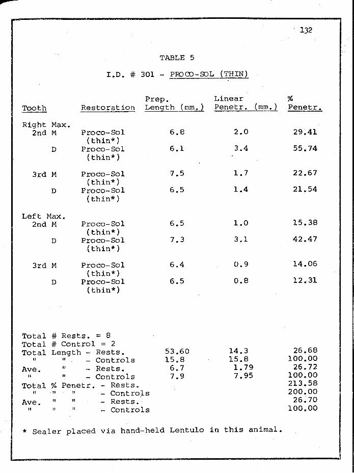
. 132
TABLE 5
I.D. # 301 - PROCO-SOL (THIN)
Prep. Linear % Tooth Restoration Length (mm.) Penetr. (m~-~ Penetr.
Right Max. 2nd M Proco-Sol 6.8 2.0 29~41
(thin*) D Proco-Sol 6.1 3.4 55.74
(thin*)
3rd M Proco-Sol 7.5 1.7 22.67 (thin*)
D Proco-Sol 6.5 1.4 21.54 (thin*)
Left Max. 2nd M Proco-Sol 6.5 1.0 15.38
(thin*) D Proco-Sol 7.3 3.1 42.47
(thin*)
3rd M Proco-Sol 6.4 0.9 14.06 (thin*)
D Proco-Sol 6.5 0.8 12.31 (thin*)
Total # Rests. = 8 Total # Control = 2 Total Length - Rests. 53.60 14.3 26.68
II II Controls 15.8 15.8 100.00 Ave. II Rests. 6.7 1.79 26.72
II II Controls 7.9 7.95 100.00 Total % Penetr. - Rests. 213.58
II . ti II Contro;J..s 200.00 Ave. II II - Rests. 26.70
II II II Controls 100.00
* Sealer placed via hand-held Lentulo in this animal.
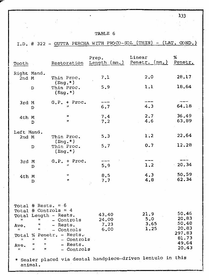
133
TABLE 6
I.D. # 322 - GUTTA PERCHA WITH PROQ)-SJL (THIN) - (LAT. Q)ND.)
Prep. Linear % Tooth Restoration Length _( mm.J. Penetr. (mm.} Penetr.
Right Mand. 2nd M Thin Proc. 7.1 2.0 28.17
{Eng.*) D Thin Proc. 5.9 1.1 18.64
{Eng.*)
3rd M G.P. + Proc. D II 6.7 4.3 64.18
4th M II 7.4 2.7 36.49 D II 7.2 4.6 63.89
Left Mand. 2nd M Thin Proc. 5.3 1.2 22.64
{Eng.*) D Thin Proc. 5.7 0.7 12.28
(Eng.*)
3rd M G.P. + Proc. D II 5.9 1.2 20.34
4th M II 8.5 4.3 50.59 D II 7.7 4.8 62.34
Total # Rests. = 6 Total # Controls = 4 Total Length - Rests. 43.40 21.9 50.46
II II Controls 24.00 5.0 20.83
Ave. II - Rests. 7.23 3.65 50.48 II II Controls 6.00 1.25 20.83
Total % Penetr. - Rests•' 297.83 II II II Controls 81.73
Ave. II II Rests. 49.64 II II II Controls 20.43
* Sealer placed via dental handpiece-driven lentulo in this animal.

134
TABLE 7
I .D. # 326 - STLVER POINTS WITH PROOO-SOL (THICK)
Prep. Linear % Tooth Restoration Length (mm.) Pentr. (mm.) Pentr. ---Right Mand.
2nd M Thick Proc. 6.5 5.0 76.92 (control*)
D Ag. Pt. only 7.1 6.1 85.92 (control)
3rd M Ag. Pt. + Proc. * 7.4 0.9 12.16 D " 7.4 3.7 50.00
4th M II 7.4 2.8 37.84 D II 7.7 0.3 3.90
Left Mand. 2nd M Thick Proc. 6.9 3.3 47.83
(control*) D Ag. Pt. only 7.1 3.8 53.52
(control)
3rd M Ag. Pt. + Proc* * 7.4 4.4 59.46 D II 7.3 2.6 35.62
4th M II 7.7 0.6 7.79 D II 7.0 3.0 42.86
* sealer placed via hand-held root canal file in this animal.
Total # Rests. = 8 Total # Controls = 2 + 2 Total Length - Rests. 59.3 18.3 30.86
II II Proc. (thick) 13 ~4 8.3 61.94 II II Ag. Pt. (only) 14.2 9.9 69.72
Ave. II - Rests. 7.41 2.29 30.87 II II Proc. (thick) 6.7 4.15 61.94 II II Ag. Pt. (only) 7.1 4.95 69.72
Total % Penetr. - Rests. 249.63 II II II - Proc. (thick) 124.75 II II II - Ag. Pt. (only) 139.44 Lve. % Penetr. - Rests. 31.20
II II II - Proc 62.38 II II II - ACJ,• !'.t:..J..C2nJ.~ ... 69.72 -

. 135
TABLE 8
I.D. # 331 - GUTTA PERCHA WITH PROCO-SOL (THICK) - (LAT. CDND.)
Tooth Restoration
Right Mand. 2nd M Thick Proc.
(control*) D G.P. (control)
3rd M G.P. + Proc. (thick*)
D G.P. + Proc. (thick*)
4th M G.P. + Proc. (thick*)
D G.P. + Proc. (thick*)
Left Mand. 2nd M Thick Proc.
(control*) D G.P. (control)
3rd M G.P. + Proc. (thick*)
D G.P. + Proc. (thick*)
4th M G.P. + Proc. (thick*)
D G.P. + Proc. (thick*)
Prep. Le_!lgth (mm.)
7.4
5.3
6.5
6.5
7.6
8.1
6.5
5.1
7.3
7.0
7.7
8.2
Linear % Pen tr. (mm.) Pen tr.
5.6
5.3
2.6
5.6
3.4
4.5
1.6
5.1
2.3
2.8
5.8
4.6
75.68
100.00
40.00
86.15
44.74
55. 5_6
24.62
100.00
31.51
40.00
75.32
56.10
* Sealer placed via hand-held root canal file in this animal.
Total Total Total
II
II
Ave. II
II
# Rests. = 8 # Controls = 2 + 2 Length - Rests.
11 Proc. (thick) 11
· G.P. (only) 11
- Rests. 11 Proc. (thick) 11 G.P. (only)
58.9 13.9 10.4
7.36 6.95 5.2
31.6 53.65 7.2 51.79
10.4 100.00 3.95 53.67 3.6 52.79 5.2 100.00
r---------------------------------------------c_o_n_t_i_n_u_e_a __ o_n __ n __ ex: pag: .• J

. 136
TABLE 8 (cont' a.)
Total % Penetr. Rests. 429.38 II II II Proc. (thick) 100.30 II II II G.P. (only) 200.00
Ave. II II - Rests. 53.67 II II II Proc. (thick) 50.15 II II II G.P. (only) 100.00
l
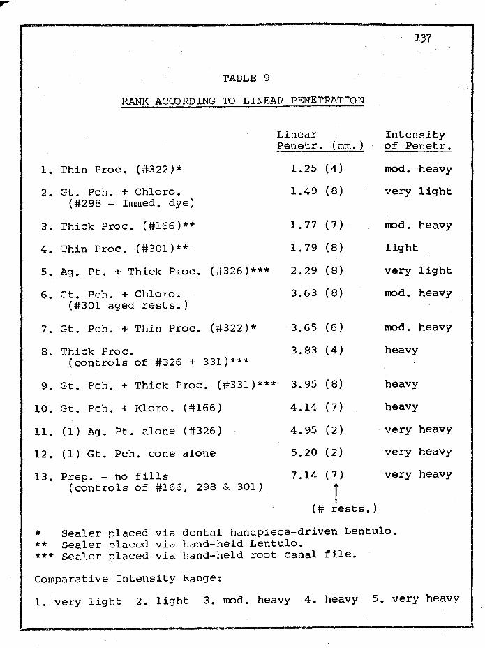
1.37
TABLE 9
RANK ACCDRDING TO LINEAR PENETRATION
l. Thin Proc. (#322)*
2. Gt. Pch. + Chloro. (#298 - Immed. dye)
3. Thick Proc. (#166)**
4. Thin Proc. (#301)** ·
5. Ag. Pt. + Thick Proc. (#326)***
6. Gt. Pch. + Chloro. (#301 aged rests.)
7. Gt. Pch. + Thin Proc. (#322)*
8. Thick Proc. (controls of #326 + 331)***
9. Gt. Pch. + Thick Proc. (#331)***
10. Gt. Pch. + Kloro. (#166)
11. (1) Ag. Pt. alone (#326)
12. (1) Gt. Pch. cone alone
13. Prep. - no fills (controls of #166, 298 & 301)
Linear Penetr. (mm. )
Intensity of Penetr.
1.25 (4) mod. heavy
1.49 (8) very light
1.77 (7) mod. heavy
1.79 (8) light
2.29 (8) very light
3.63 (8) mod. heavy
3.65 (6) mod. heavy
3.83 (4) heavy
3.95 (8) heavy
4.14 (7) heavy
4.95 (2) very heavy
5.20 (2) very heavy
7 .14 ( 7) very heavy
i {#rests.)
* Sealer placed via dental handpiece-driven Lentulo. ** Sealer placed via hand-held Lentulo. *** Sealer placed via hand-held root canal file.
Comparative Intensity Range:
1. very light 2. light 3. mod. heavy 4. heavy 5. very heavy

. 138
TABLE 10
RANK -- BY RESTORATION TECHNIQUE
Range of Median Mean # of Linear Linear Linear Rests. Penetr. (mm.) Penetr. (mm.) Penetr. (mm.)
1. Ag. Pt. 8 0.3 - 4.4 2.7 2.29 + Proc. (#326)
2. Gt. Pch. 8 1.8 - 7.5 3.35 3.63 + Chloro. ( #301)
3. Gt. Pch. 6 1.2 - 4.8 4.3 3.65 + Proc. (thin) (#322)
4. Gt. Pch. 8 2.3 - 5.8 3.95 3.95 + Proc. (thick) (#331)
5. Gt. Pch. 7 2.6 - 6.7 3.8 4.14 + Kloro. (#166)

~-----------------~ l39
FIGURE l
SURGICAL EXTRACTION TSCHNIQUE
A
p,
B
A. Facial View B. Occlusal View

FIGURS 2
X- Rl\.Y. ADAPTED FOR D"SNTAL USE

11+1
I.D. #713: PILOT DOG ;Jl - 90 MINUT E PERFUSION
Note : 1. Dog died 2 hours after dye p".' rfus i on b ~t;a.."1. 2. Hax. l eft p r emol a r ext r acted 30 ninute s afte r death. 3. "Clouds " i ndic at e dye r ese rvoi rs creat ed in bone.
Dye plac ~: d in r eservoirs for 24 hours.

FIGURE 4
I.D. //163: 2 DYE PE!i.FUSIONS, KI DNEYS BYPASSED
Note : 1. Can:.l.ls filled \ria l at c r;i.l condensation. 2. 11 Clouds 11 :indicate the approx. position of the
dye reservoir to the 'pex. 3. De gr ee of stain on root surface.

FI GUR."., 5
I.D. //163: DISTRIBUTION OF DYE IN 0 '.l\L TISSUES
Note: 1. Author r e r;r et s poor color r eproduction from slide. 2 . Coloration of mar gi nal gingiva, inferior surf;3.Ce
of ton f;lH"! and tons i llar a rea. 3. Fh.t ·pal at e i n do g .

FIGURE 6
I.D. #163: ANGi rnJEUROTIC ED;~·'iA
Note : 1. Allergic >·;he als arising from dermi s on inner aspect of hi nd l egs.

~...a'.r~x:c::acmwa li!M"!ll!~.nc-..nom:,..,,• -'""""""""w:1~·--=---••T"QP"7MrOlr:""""" ___ ,,.,.,..._,_,,...,,_.,.,.,.=-·,_• .... t.~
FIGURE 7
I. D. #163: PSlIPHERAL TJASCULAR COLLAPSE
Note : 1. Intr~cath in External Jugul~r Vein . 2. Collapse caused cessation of dye flow with lScc.
solution r emaining in Buretrol.
.145 ~

FIGURE 8
I.D. //166 : p I 'rE'T'RAT IOll OF KLO~OPERKA ROOI' CA.JUL FILLINGS
Note: 1. Dye distribution in ~ ach h2.lf scetio!i. . 2. 2nd premol a rs prepared but not fill ed . 3. Dye patt~m in left ht h premolar (D) h. Rj_ ght ~th p r l"' r.iolar (H) del et ed from study because
of improper dye plac ~:nent .
146 .

FIGURE 9
I. D. #166 : PENE"fRATION OF PROCO-SOL (thick )
•
1166 PROCOSOL ( _._.,.,,. ..
Ri ta \
ft:
2 dBi M D
Note: 1. Extent of stain on toot C;ln al wall. 2. Rapid fading of dye's intensity,
147

FIGUR.S 10
I. D. /)298 : P"SNE'I'RABILITY OF CHLOROPKRGHA HOOT CANAL FILLHJ~
Note: 1. Dye placed on the s ame day as the root canal filling. 2. Relative l ack of dye's vertical (linear) penetration. 3. Dye intensely pe rvaded con+,rol t ePth. 4. Apical cement um relatively free of dye.

FIGURE 11
I. D. 11 301: PENET11.ABILITY OF CHLOROPERCHA ROOT CAN AL FILLrnG
Note: 1. Mand. ri_ght 2nd premolar fractured on c:a.ge bars and ri_elet~d from sturiy.
2. Identical dye s t aining in control tooth here as with //298.
3. Increased linear penetration compar~d to #298. Root canal filling here was allowed to 11 age 11 for 1 we~k.
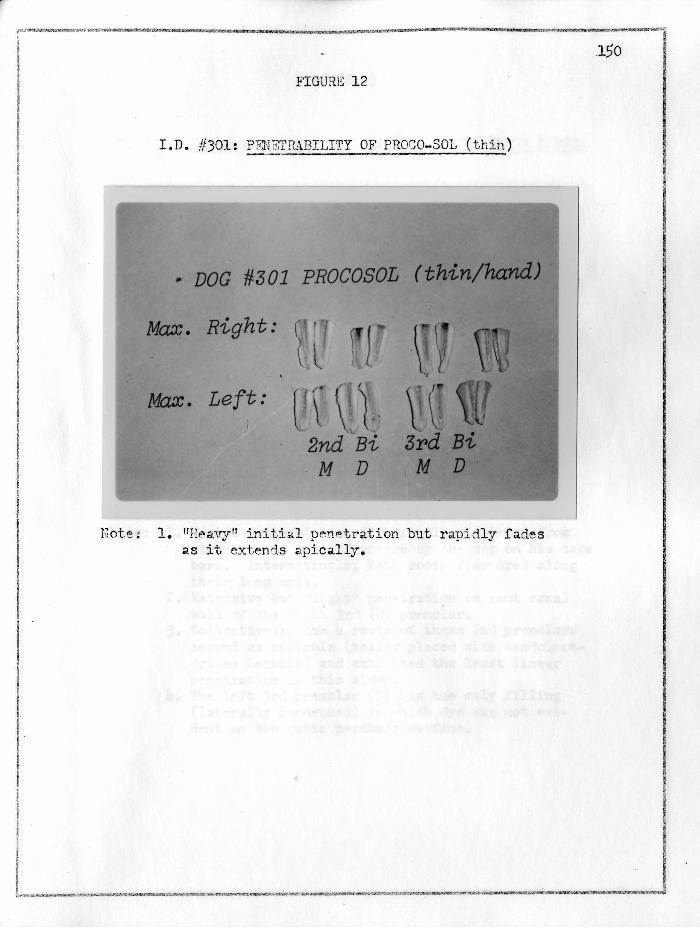
FIGURE 12
I.D. //301: PENETRABILITY OF PROCO- SOL (thin)
• DOG #301 PROCOSOL (t
/tb:c. Right:
M;Jz. Left:
a•..-.---.....-1 150
Note~ 1. 11Heav;y1: ini th.l p~n<-!tration but r apidl y fades •s it extends apically.
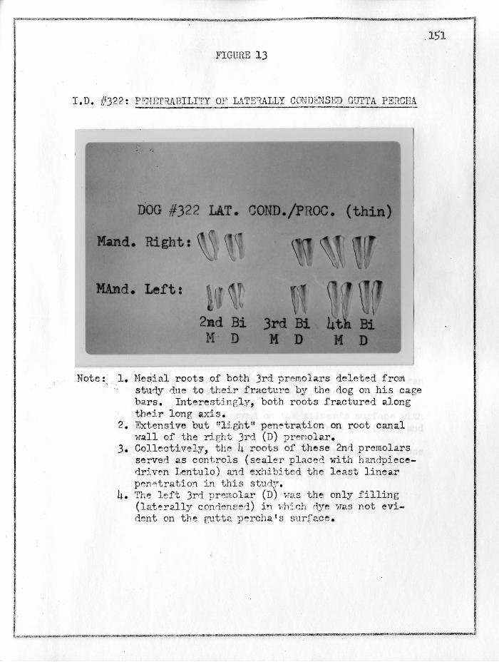
FIGURE 13
I. D. /,!322: PF.NETRABILITY Of LATE'tALLY CONDENSED GUI'TA PE:i.CHA
d. Left:
Note: 1. Hesial roots of both 3rd premolars deleted from study due to their fract11re by the dog on his cage bars. Interestingly, both roots fractured ai.long th eir long axis.
2. Extensive but "light" pen~tration on root canal wall of the ri ght Jrd (D) premolar.
3. Collectively, the 4 roots of these 2nd premolars served as controls (sealer placed with h<cndpiecedri ven Len tu lo) :c'l.d exhibited the l e<tst linear pcn ,~ tration in this study .
4. Th~ l eft Jrd premoh.r (D) was the only filling (la.t~r.,_lly conrl.ensed ) i :t wh:i.. r.:h rl.~re was not evident on t he gutt<'. percha 1 s surf2.ce.
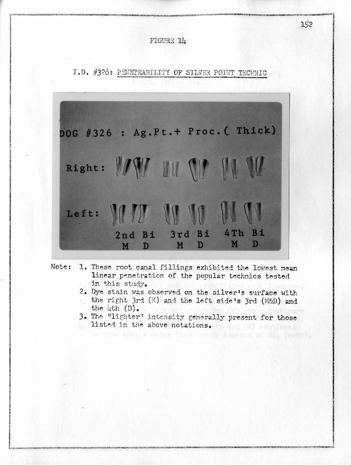
FIGURS 14
I.D. //326: P EirnTRABI LITY OF SILV~R POINT TSCENIC
Note: 1. These root canal fillings exhibited the loFest :neCl.Il linear p!":netr;ition of the- popular technics tested in this study.
2. Dye stain was obse rv~d on the silver's surf~ce with the ri ght 3rd (H) and the l~ft side's 3rd (H&D) and the 4th (D).
3. Tho 11li r:ht cra inten si ty generally present for those listr: rl i n the above not-.tions.
I .
l ~·---__... ................... ~~n-.~~'!S~,~~.~~ . .

FIGURE 15
I. D. #331: P'~NETRABILITY OF LATE:?.ALLY CONIYSNSED GUTTA PEH.CHA (thick Proc.)
Note : 1. Wirie vari:..tions in th~ ext<>nt of linear penetration from one sp~ ci~en to anoth~ r.
2. The l eft anrl. ri eht 2nd (D) were filled with 1 adapted gi_1 tta pe rch2. con~ , but without s eal~r.
J. The rl.iff~rP.nc e s in cha r acter of th 0 dve 's diffusion p<?.t t~rn in th~ l~ft and r ight 2n'i (H) · whe re both were fill ed th rou gh th" u se of hand-held root c._'1al fil es .
4. rr'fest tube !f fragrn.entation of l eft 3rd (H) mentioned in this t he s i s under 11 Sr.rl.oiontic Aspects of Dog Teeth".

FIGURE 16
I.D. #322 : OCGLUSAL '.'I'~AR PAT1'"<~RN
Noti:; : 1. Patt"'rn eYiri.~nt on ri.e sh.1 inclin·~s of both :'.'fa.x. l.tth premol ars. An expost"d l1orizontal bar on this :mimal 1 s cage bore obvious tooth marks .
2. Althon gh the 2.bove max . 2nd 211d 3rd premolars were intended to be a part of this study, the animal's hab t ual gnawing p roduced their obvious fractures. All we r e excluied from this inve s tigation.
1.54

I L
FJGTBE 17
CARN ASSB.L T SSTH OF DOGS
Note : 1. The uppe r specj ffi""'.'1 is a Max:. 4th premoh.r. 2. The l owe r tooth is a ?rand. 1st moloi.r. 3. D~velopmental grooves on interradicular surf;;i.ce
of root.

FIGUrtE 18
DOG HAXILLARY AND d ANDi f.:'. ULAR PRSMOLA.RS
st Bi
) Bi
Not~: 1. An i!lcreas~ :i_n root length with each succeeding postf: rior tooth .
2. Th~ mand . ?.nd p r erriolar roots m~y be less divergent than the roots of the mand . 3rd premolar. An example of fusei ~1;mdibular 2nd premolar root~ is inclur:le-:i.
J. The dist.,_l root is usually shorte r (l-2mm .) and has more bulk t han the m~sial root on t r." sam~ tooth.
I ~Mliiltl_~~·-"-N'fl ____ ~~~~~~-~'"M>•~W~~~
·' .. "- .... ·. ·,
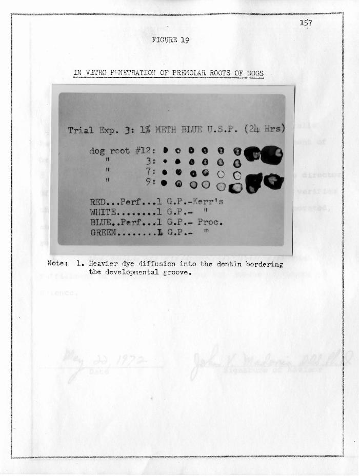
______________ ._... __________ ...._. _____ ,_ ________ ,_.~
f:'IGUR.E 19
rn VTI'RO Pr.:NSTRATIW OF P!lE;'10LAR ROOTS OF DOGS
Trial Exp. ): 1% M , H BLl . . ~ .• ( 24 Hr )
dog root #12: • 0
" 3: • • • II 7: • • 0 " 9: • Oo
RED ••. P rt ..• l G.P.-K~rr' s
WHITE •••••••• 1 G.P .- '' BLUE •• P rt •. .-1 G.P .- Proc. GREEJJ........ G.P.- "
Note: 1. Heavier dye dif.ft::si on into the dentin borderir!g the developrn~ntal groove.
157

APPROVAL SHEET
The thesis submitted by Dr. Donald L. Scoralle
has been read and approved by members of the Department of
Oral Biology.
The final copies have been examined by the director
of the thesis and the signature which appears below verifies
the fact that any necessary changes have been incorporated,
and that the thesis is now given final approval with
reference to content, form, and mechanical accuracy.
The thesis is, therefore, accepted in partial
fulfillment of the requirement for the degree of Master of
Science.
J.c}J I? 1 ;L- 11 m Date Signature
______________ , ... _______ . ___ _,. __ J