an intermediate along the recovery stroke of myosin vi revealed … · an intermediate along the...
TRANSCRIPT
An intermediate along the recovery stroke of myosinVI revealed by X-ray crystallography andmolecular dynamicsFlorian Blanca,b,c,d, Tatiana Isabeta,b, Hannah Benistya,b, H. Lee Sweeneye,f, Marco Cecchinic,d,1, and Anne Houdussea,b,1
aStructural Motility, Institut Curie, Paris Sciences et Lettres (PSL) Research University, CNRS, UMR 144, F-75005 Paris, France; bSorbonne Universités,Université Pierre et Marie Curie (UPMC) University Paris 06, CNRS, UMR 144, F-75005 Paris, France; cInstitut de Science et d’Ingénierie Supramoléculaires(ISIS), UMR 7006 CNRS, Université de Strasbourg, F-67083 Strasbourg Cedex, France; dInstitut de Chimie de Strasbourg, UMR 7177 CNRS, Université deStrasbourg, F-67083 Strasbourg Cedex, France; eDepartment of Pharmacology & Therapeutics, University of Florida College of Medicine, Gainesville, FL32610-0267; and fMyology Institute, University of Florida College of Medicine, Gainesville, FL 32610-0267
Edited by Michael L. Klein, Temple University, Philadelphia, PA, and approved May 7, 2018 (received for review June 27, 2017)
Myosins form a class of actin-based, ATPase motor proteins thatmediate important cellular functions such as cargo transport andcell motility. Their functional cycle involves two large-scale swingsof the lever arm: the force-generating powerstroke, which takesplace on actin, and the recovery stroke during which the lever armis reprimed into an armed configuration. Previous analyses of theprerecovery (postrigor) and postrecovery (prepowerstroke) statespredicted that closure of switch II in the ATP binding site precedesthe movement of the converter and the lever arm. Here, we reporton a crystal structure of myosin VI, called pretransition state (PTS),which was solved at 2.2 Å resolution. Structural analysis and all-atom molecular dynamics simulations are consistent with PTS beingan intermediate along the recovery stroke, where the Relay/SH1elements adopt a postrecovery conformation, and switch II re-mains open. In this state, the converter appears to be largelyuncoupled from the motor domain and explores an ensemble ofpartially reprimed configurations through extensive, reversiblefluctuations. Moreover, we found that the free energy cost ofhydrogen-bonding switch II to ATP is lowered by more than10 kcal/mol compared with the prerecovery state. These resultssupport the conclusion that closing of switch II does not initiatethe recovery stroke transition in myosin VI. Rather, they suggesta mechanism in which lever arm repriming would be mostlydriven by thermal fluctuations and eventually stabilized by theswitch II interaction with the nucleotide in a ratchet-like fashion.
molecular motors | chemomechanical transduction | myosin |recovery stroke | molecular dynamics simulations
Myosins are a wide superfamily of molecular motor proteinsinvolved in a number of vital processes as diverse as in-
tracellular cargo transport, endocytosis, muscle contraction, andcell motility (1). Defective myosins were found to be implicatedin severe pathologies in humans such as hypertrophic cardio-myopathy (2) and deafness (3), while others, including myosinVI, were shown to have a role in cancer cell proliferation andmetastasis (4). Recent studies highlighted the therapeutic po-tential of small-molecule inhibitors (5) and activators (6–8) tar-geting myosin, demonstrating that a detailed knowledge of theforce production mechanism in this motor family would facilitatethe rational design of drug candidates.Myosin motors work through a complex cycle of conforma-
tional transitions that couple ATP hydrolysis with force pro-duction on actin (Fig. 1). Previous analyses characterized theconformational states of the motor domain during the cycle andthe kinetics of the transitions between them (reviewed in refs. 9and 10; see also refs. 11–13). These studies, along with mea-surements of the stroke size, are consistent with the swinginglever arm hypothesis, in which the structural changes in the ATP-binding site or the actin-binding site are amplified into a largeswing of the extended lever arm region through the rotation of the
converter subdomain (14). In this framework, two major eventsoccur: a force-generating step taking place on actin, which corre-sponds to the large-amplitude swing of the lever arm termedpowerstroke, and an off-actin reverse transition called recoverystroke in which the motor and the lever arm return to their primedconfiguration. This latter is crucial for chemomechanical trans-duction, as it couples the repriming of the lever arm with ATPhydrolysis. Also, this step occurs entirely off-actin and thereforerepresents an interesting target for pharmacological regulation (5).Early crystallographic studies on Dictyostelium discoideum
myosin II (Dd Myo2) and other myosins with various ATP an-alogs have trapped the motor domain in the prerecovery (alsocalled postrigor state, PR) and the postrecovery (also calledprepowerstroke state, PPS) conformations. Their comparisonrevealed that key structural changes accompany the reverseswing of the lever arm: (i) closure of the inner cleft via theformation of critical interactions near the active site (e.g., switchII closure on the γ-phosphate of ATP) and (ii) a major confor-mational change of the flexible connectors between the motordomain and the converter (i.e., the Relay and SH1 helix). Im-portantly, the latter rearrangement involves the formation ofa kink in the Relay helix. Computational studies started fromthese high-resolution structures were instrumental for the
Significance
Myosins are motor proteins involved in the transport of cellularcargoes and muscle contraction. Upon interaction with actin,the motor domain undergoes a conformational transition,called powerstroke, in which the lever arm is swung to gen-erate force and directional motion. The recovery stroke repri-mes the motor by coupling the reverse swing of the lever armto ATP hydrolysis. Using X-ray crystallography and molecularsimulations, we characterize a putative intermediate along therecovery stroke of myosin VI, which challenges existing modelsof myosin chemomechanical transduction. Intriguingly, thenew structure suggests that the repriming of the lever armwould be uncoupled from ATPase activity until the very end ofthe recovery stroke and mostly driven by thermal fluctuations.
Author contributions: F.B., M.C., and A.H. designed research; F.B., T.I., and H.B. performedresearch; F.B., T.I., H.B., H.L.S., M.C., and A.H. analyzed data; and F.B., M.C., and A.H.wrote the paper.
The authors declare no conflict of interest.
This article is a PNAS Direct Submission.
Published under the PNAS license.
Data deposition: The atomic coordinates and structure factors have been deposited in theProtein Data Bank, www.wwpdb.org (PDB ID code 5O2L).1To whom correspondence may be addressed. Email: [email protected] or [email protected].
This article contains supporting information online at www.pnas.org/lookup/suppl/doi:10.1073/pnas.1711512115/-/DCSupplemental.
Published online May 29, 2018.
www.pnas.org/cgi/doi/10.1073/pnas.1711512115 PNAS | June 12, 2018 | vol. 115 | no. 24 | 6213–6218
BIOPH
YSICSAND
COMPU
TATIONALBIOLO
GY
Dow
nloa
ded
by g
uest
on
May
29,
202
0

development of mechanistic models of the transition betweenthe initial PR state and the final PPS state. Based on variouscomputational strategies (15–24), several models were proposed(SI Appendix, Supplementary Text 1). A common feature of thesemodels [with the notable exception of Cui and coworkers (17,18)] is that switch II closure is presented as the initiating event ofthe recovery stroke, which triggers the large-amplitude rotationof the converter. Although the details of the coupling betweenswitch II closure and converter repriming are still under debate,the most accepted view [first proposed by Fischer et al. (15)] isthat closing of switch II exerts strain on the Relay helix thatbends and kinks in response, driving the converter rotation.Importantly, none of the existing models predicts the occurrenceof intermediates where the converter is uncoupled from themotor domain.Here, we report on the structural and dynamic characteriza-
tion of a putative intermediate along the recovery stroke ofmyosin VI by X-ray crystallography and molecular dynamics(MD), which we call pretransition state (PTS); see Fig. 1. Thestructure, solved at 2.2 Å resolution, reveals a configuration ofthe motor domain in which the Relay/SH1 elements adopt anearly postrecovery (PPS-like) configuration while switch II isopen as in PR. Using molecular simulations, we explore theimplications of the PTS structure for the recovery stroke mech-anism. Our results indicate that, if PTS were on-path to thepostrecovery state, switch II closure would occur at the end ofthe recovery stroke with the lever arm being essentially reprimedby thermal fluctuations. The isolation of the PTS structure thussuggests the existence of statistical, rather than mechanical,coupling between ATP hydrolysis and the backward swing of thelever arm, in contrast with existing models of the recovery stroke.
ResultsOverall Description of the PTS Myosin VI Crystal Structure. Fromextensive crystallization screens, a previously uncharacterizedconformation of the motor domain of myosin VI has been de-termined at 2.2 Å resolution (SI Appendix, Table S1). Crystals ofthis structural state were produced with the ATP analogADP.BeFx and could not be obtained with the ADP.Pi ana-logs ADP.VO4 or ADP.AlF4. The crystal structure (Fig. 2) revealsa conformation of the motor domain that differs significantly fromthe PR [Protein Data Bank (PDB) ID code 2VAS] and the PPS(PDB ID code 2V26) states previously reported for myosin VI.Most importantly, although the converter is partially reprimed, thestructural features around the nucleotide, in particular switch II,are not in position to promote hydrolysis of ATP (Fig. 2B).
In the active site, switch II is slightly shifted toward the“closed” position found in the PPS state but still exhibits thestructural features of an “open” state; the distance betweenthe Beryllium atom of the -BeFx group and the amide nitrogen ofG459 (7.0 Å) is too large for hydrogen bonding, and the criticalsalt bridge R205–E461, which is required to promote ATP hy-drolysis (25), is not formed. The U50/L50 actin-binding cleft iswide open and exhibits minimal deviation from the PR confor-mation (see also SI Appendix, Fig. S1). However, the position ofthe converter indicates that the motor is in a partially primedconfiguration (Fig. 2D). Finally, the Relay/SH1 elements stronglyresemble the conformation adopted in the PPS structure, mostprominently because of the kinked Relay helix (Fig. 2C). Sincethis new structure is compatible with an ATP bound state, it islikely to represent a state that myosin adopts in complex with ATPwhen the motor is detached from actin. Furthermore, its structuralfeatures are consistent with an intermediate state on the way tothe hydrolysis-competent PPS state. Since PPS was referred to asthe transition state of myosin hydrolysis, we name this structure ofmyosin VI the pretransition state (PTS).However, important differences from PPS still exist. Although
the internal RMSD of the Relay/SH1 elements between PTS andPPS is quite small (0.75 Å excluding the Relay loop), structuralalignment onto the N-terminal subdomain reveals that these ele-ments undergo a rigid-body motion to complete the recoverystroke. This global movement, which brings the N-terminal regionof the Relay helix toward the inside of the nucleotide-binding site, isconsistent with the “seesaw” motion originally proposed by Fischeret al. (15); see Fig. 2B. Also, the converter subdomain adopts anintermediate position between PR and PPS and displays the ca-nonical R fold, which was observed in the PR and Rigor structuresof myosin VI and virtually every crystal structure for other myosinisoforms; i.e., the converter adopts an unconventional P fold only inthe reprimed PPS and Pi Release structures of myosin VI (11, 26).Interestingly, most of the contacts between the converter and themotor domain in either PR or PPS of myosin VI are not formed inthe PTS structure, suggesting that this latter might represent a“decoupled converter” state (SI Appendix, Tables S2 and S3).In summary, the PTS structure exhibits a mostly open switch II
(PR-like) in a motor with nearly rearranged Relay/SH1 elements(PPS-like) and a converter in an intermediate position. Thesefeatures are consistent with a conformational state of the motorrepresentative of a previously undescribed intermediate alongthe recovery stroke of myosin VI.
Unbiased MD Simulations Reveal a Dynamic Converter in PTS. To ex-plore the significance of the PTS structure, we performed sub-microsecond MD simulations (>1.4 μs of cumulated simulationtime) with an explicit treatment of the solvent starting from thePR (2 × 100 ns, 1 × 200 ns), PTS (1 × 306 ns, 2 × 100 ns), and PPS(3 × 100 ns with ATP, 2 × 100 ns with ADP.Pi) structures ofmyosin VI; see SI Appendix, Table S4. The resulting trajectorieswere analyzed by monitoring structural observables that de-scribe the conformation of the various elements involved in therecovery stroke. The results of the analysis follow.The projection of the center of geometry of the converter on
the plane defined by the two transverse principal axes of themotor domain (defined in SI Appendix, Supplementary Text 2 andFig. S2) shows that the PTS converter is highly dynamic andexplores a significantly larger volume than in PR or PPS, where itis confined in proximity to the crystallographic position by spe-cific interactions with the N-terminal domain; see Fig. 3 and SIAppendix, Figs. S3–S5 and Table S2. In the 306-ns PTS simulation,the time series of the longitudinal component of the converterfluctuations shows that, from 50 ns to 70 ns, the converter un-dergoes a spontaneous swing toward a new position that is closerto PPS (Fig. 3). After the swing, the converter appears to be asconfined as in PR and PPS, although the new position is notequivalent to a PPS state. This is due to the formation of newcontacts between the converter and the N-terminal domain, someof them being absent in both the PR and PPS states (SI Appendix,
Fig. 1. Overview of the actomyosin cycle. When ATP is bound, the motorundergoes a fast and reversible transition known as the recovery strokethat reprimes the lever arm in preparation for force production. The redstar materializes the putative position in the cycle of the PTS intermediatereported in this study.
6214 | www.pnas.org/cgi/doi/10.1073/pnas.1711512115 Blanc et al.
Dow
nloa
ded
by g
uest
on
May
29,
202
0
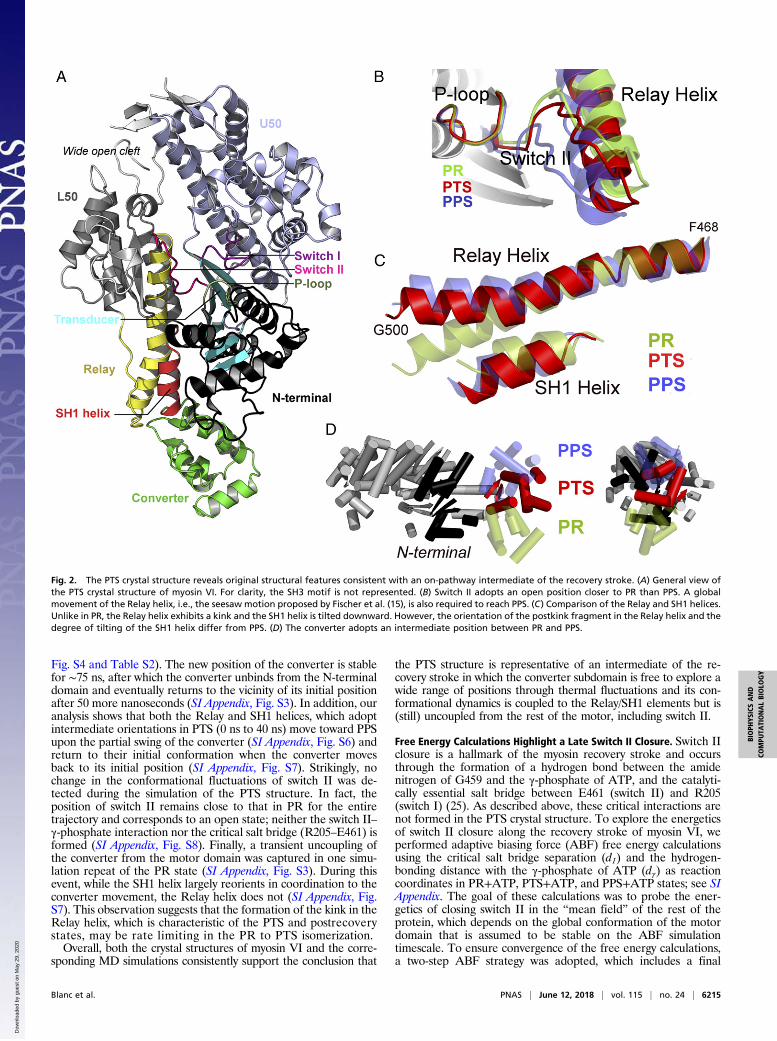
Fig. S4 and Table S2). The new position of the converter is stablefor ∼75 ns, after which the converter unbinds from the N-terminaldomain and eventually returns to the vicinity of its initial positionafter 50 more nanoseconds (SI Appendix, Fig. S3). In addition, ouranalysis shows that both the Relay and SH1 helices, which adoptintermediate orientations in PTS (0 ns to 40 ns) move toward PPSupon the partial swing of the converter (SI Appendix, Fig. S6) andreturn to their initial conformation when the converter movesback to its initial position (SI Appendix, Fig. S7). Strikingly, nochange in the conformational fluctuations of switch II was de-tected during the simulation of the PTS structure. In fact, theposition of switch II remains close to that in PR for the entiretrajectory and corresponds to an open state; neither the switch II–γ-phosphate interaction nor the critical salt bridge (R205–E461) isformed (SI Appendix, Fig. S8). Finally, a transient uncoupling ofthe converter from the motor domain was captured in one simu-lation repeat of the PR state (SI Appendix, Fig. S3). During thisevent, while the SH1 helix largely reorients in coordination to theconverter movement, the Relay helix does not (SI Appendix, Fig.S7). This observation suggests that the formation of the kink in theRelay helix, which is characteristic of the PTS and postrecoverystates, may be rate limiting in the PR to PTS isomerization.Overall, both the crystal structures of myosin VI and the corre-
sponding MD simulations consistently support the conclusion that
the PTS structure is representative of an intermediate of the re-covery stroke in which the converter subdomain is free to explore awide range of positions through thermal fluctuations and its con-formational dynamics is coupled to the Relay/SH1 elements but is(still) uncoupled from the rest of the motor, including switch II.
Free Energy Calculations Highlight a Late Switch II Closure. Switch IIclosure is a hallmark of the myosin recovery stroke and occursthrough the formation of a hydrogen bond between the amidenitrogen of G459 and the γ-phosphate of ATP, and the catalyti-cally essential salt bridge between E461 (switch II) and R205(switch I) (25). As described above, these critical interactions arenot formed in the PTS crystal structure. To explore the energeticsof switch II closure along the recovery stroke of myosin VI, weperformed adaptive biasing force (ABF) free energy calculationsusing the critical salt bridge separation (d1) and the hydrogen-bonding distance with the γ-phosphate of ATP (dγ) as reactioncoordinates in PR+ATP, PTS+ATP, and PPS+ATP states; see SIAppendix. The goal of these calculations was to probe the ener-getics of closing switch II in the “mean field” of the rest of theprotein, which depends on the global conformation of the motordomain that is assumed to be stable on the ABF simulationtimescale. To ensure convergence of the free energy calculations,a two-step ABF strategy was adopted, which includes a final
Fig. 2. The PTS crystal structure reveals original structural features consistent with an on-pathway intermediate of the recovery stroke. (A) General view ofthe PTS crystal structure of myosin VI. For clarity, the SH3 motif is not represented. (B) Switch II adopts an open position closer to PR than PPS. A globalmovement of the Relay helix, i.e., the seesaw motion proposed by Fischer et al. (15), is also required to reach PPS. (C) Comparison of the Relay and SH1 helices.Unlike in PR, the Relay helix exhibits a kink and the SH1 helix is tilted downward. However, the orientation of the postkink fragment in the Relay helix and thedegree of tilting of the SH1 helix differ from PPS. (D) The converter adopts an intermediate position between PR and PPS.
Blanc et al. PNAS | June 12, 2018 | vol. 115 | no. 24 | 6215
BIOPH
YSICSAND
COMPU
TATIONALBIOLO
GY
Dow
nloa
ded
by g
uest
on
May
29,
202
0

stratification over 56 nonoverlapping windows; see SI Appendix.The completeness of sampling (SI Appendix, Fig. S10), the smoothconvergence of the free energy gradient per window (SI Appendix,Fig. S11), and the small statistical errors on the resulting PMF (SIAppendix, Fig. S12) all suggest converged free energy results. Theresults in Fig. 4 A and C show that the position of the converterand/or the conformation of the Relay/SH1 elements effectivelyshift the equilibrium from an open switch II in PR+ATP to aclosed switch II in PPS+ATP. Also, they indicate that a “partiallyclosed” switch II state with a formed G459–ATP hydrogen bondbut an open salt bridge may be stabilized in PTS, which remainscatalytically inactive. Visual inspection of the ABF trajectory in
PTS shows that the partially closed state with a formed G459–ATPhydrogen bond is reached by uncoupling switch II from the Relayhelix, which involves the breaking of a pair of hydrogen bondsbetween N474 on the Relay helix and the backbone of E461 onswitch II (Fig. 4D) as well as the extraction of the side chain ofF460 from a hydrophobic cavity belonging to the L50 subdomain(SI Appendix, Fig. S15). However, since the formation of thecritical salt bridge is still disfavored in PTS, the free energy resultsin Fig. 4B indicate that supplementary rearrangements are re-quired to complete the recovery stroke. Given that in PTS the“seesaw” motion of the Relay helix is incomplete (see OverallDescription of the PTS Myosin VI Crystal Structure), we infer that
Fig. 3. Positional dynamics of the converter in MD. (A) Geometric observables to monitor the position of the converter in simulation. By projecting the centerof geometry of the converter Cα atoms on the principal axes of the motor domain, the components X′, Y′, and Z′ provide a convenient representation of theconverter position relative to the motor domain; see SI Appendix for details. (B) Positional dynamics of the converter on the transverse plane X′Y′. Data pointsfor PTS correspond to the first 125 ns; see SI Appendix, Fig. S3 for the complete data. Crosses indicate the crystallographic values. The data show the existenceof two positional states for the converter in PTS: one widely distributed and centered on (−12 Å, −6 Å) and one more confined and in slight overlap with PPScentered on (−15 Å, 0 Å). (C) Time series of the Z′ component. The decrease in Z′ starting at t = 50 ns in the PTS simulation corresponds to a partial reprimingtoward the PPS position. For clarity, the running average over 2 ns is plotted.
Fig. 4. State-dependent free energy landscape of switch II closure in the myosin VI motor domain. (A) PR state. (B) PTS state. (C) PPS state. Crosses indicatevalues from MD-equilibrated structures, which are very similar to the crystal structures. All free energies are given in kcal per mole. (D) Representativeconfiguration of the partially closed switch II state sampled by the ABF simulation of PTS. Compared with the PTS crystal structure (in red), switch II uncouplesfrom the Relay helix and undergoes a large motion to form the hydrogen bond with ATP. Interestingly, this configuration is distinct from PPS (in blue),notably because the critical salt bridge is disfavored.
6216 | www.pnas.org/cgi/doi/10.1073/pnas.1711512115 Blanc et al.
Dow
nloa
ded
by g
uest
on
May
29,
202
0
this global movement is crucial to produce an ATPase competentstate. Thus, the present ABF calculations suggest that two distinctpathways exist to reach the final PPS state: one in which the switchII–ATP hydrogen bond is formed in PTS via the uncoupling ofswitch II from the L50 subdomain and another one in which theseesaw motion of the Relay helix with a fully coupled switch IIresults in the formation of the critical salt bridge interaction withswitch I. Although we cannot conclude which pathway is kineti-cally preferred for the PTS to PPS transition, we note that both ofthem are consistent with a late closure of switch II during therecovery stroke transition, which is the most important resultemerging from the simulations. Finally, the ABF results (Fig. 4B)suggest that the partially closed switch II with a formed G459–ATP hydrogen bond would be most favored in the PTS state,which is actually not observed in the crystal structure. Since thefree energy difference between the broken and formed hydrogenbond configurations probed by ABF along dγ is small (∼2 kcal/mol;see SI Appendix, Fig. S9B), both states are likely populated in PTSwith the fully open state possibly selected on crystallization.
DiscussionBiomolecular motors like myosin harness and transduce thechemical energy of ATP by cycling through a series of complexconformational transitions. The structural characterization of allof the relevant steps with atomic resolution is critical for theelucidation of the mechanism that steers function. Nonetheless,it is not sufficient. High-resolution dynamical and, most impor-tantly, energetic information is needed to assess the significanceof the structural states, infer the sequence of events, and explainwhy alternative and potentially meaningful pathways are actuallynot explored. By focusing on the recovery stroke of myosin VI,we demonstrate that the synergistic use of X-ray crystallographyand all-atom MD provides a powerful approach to exploreprotein function with atomic resolution.The recovery stroke is a critical step of the myosin cycle in which
the repriming of the lever arm is coupled to ATP hydrolysis.Providing a detailed understanding of this large isomerization ofthe motor domain is of fundamental importance, in particular toelucidate how chemical energy may be stored in preparation forthe powerstroke. However, its characterization by solution ex-periments is challenging. First, this motor isomerization occurs onthe millisecond timescale (27), which makes it difficult to beprobed by time-resolved experiments. Second, this transitioncorresponds to the largest isomerization of the motor domain,which cannot be easily correlated with a unique biophysical signalsuch as ATP binding, which precedes it, or ATP hydrolysis, whichoccurs after it. Last, it is a reversible process.In this work, we report on the structural and dynamical char-
acterization of a putative intermediate along the recovery strokeof myosin VI, which we term PTS. Comparison of the PTSstructure with the PR and the PPS states reveals a previouslyunreported configuration of the motor domain in which the Relay/SH1 elements adopt a nearly postrecovery (PPS-like) configura-tion, the converter is in an intermediate position, and switch II isopen. Corresponding MD simulations support the conclusion thatswitch II and the converter are not mechanically coupled in PTS,with the motor domain remaining catalytically inactive even if theconverter has departed from the initial prerecovery position. Mostimportantly, the discovery of the PTS structure suggests a mech-anism for the recovery stroke in myosin. In the emerging scenario,the repriming of the motor head to the armed prepowerstrokeconfiguration would be mediated by (i) the spontaneous isomer-ization (kinking/tilting) of the Relay/SH1 elements coupled with aconverter swing to an intermediate position, (ii) closing of switchII over the nucleotide via the seesaw motion of the Relay helix,and (iii) completion of the converter swing. Intriguingly, this in-terpretation is consistent with a mechanism in which lever armrepriming would be initiated by thermal fluctuations and proceedthrough a restricted random search, with the converter probing anensemble of configurations compatible with a kinked Relay helixuntil it finds its way to the postrecovery binding interface.
Free energy calculations on the closure of switch II in PR, PTS,and PPS provide additional information. The results indicate thatspontaneous closure of switch II is essentially impossible in PR,because it is thermodynamically disfavored, such that a transitiontoward an intermediate state similar to PTS would be required atthe beginning of the recovery stroke. Also, they indicate that theformation of the catalytically essential salt bridge is still unfavor-able in PTS. Therefore, our analysis supports the conclusion thatswitch II closure is a late event of the recovery stroke, which re-quires an additional rearrangement of the motor domain that isnot sampled yet in PTS. Finally, the results indicate that the for-mation of a hydrogen bond between switch II and the γ-phosphateof ATP is energetically favorable in PTS and can be formed uponbreaking of interactions between switch II and the Relay helix.Hence, these free energy results are consistent with the existenceof two distinct pathways to close switch II, which involve or not anuncoupling of switch II from the L50 subdomain. Assuming thatthe PTS structure is on-path to the postrecovery state, these re-sults provide an understanding of the recovery stroke mechanismin myosin VI. Whether or not the emerging scenario is specificto myosin VI is presently unclear. We note, however, that themechanism above is consistent with our recent finding that smoothmuscle myosin II can be effectively trapped in a prehydrolysis stateby binding of an allosteric inhibitor (5), whose negative modula-tory activity may precisely block the conformational transition ofthe Relay/SH1 elements at the beginning of the recovery stroke;see SI Appendix, Supplementary Text 1.The mechanistic interpretation of PTS emerging from X-ray
crystallography and MD simulations is in clear disagreement withexisting models of the recovery stroke (15, 21, 22) which wereobtained for Dd Myo2; see SI Appendix, Supplementary Text 1. Inthe most accepted view, the recovery stroke starts with the spon-taneous closure of switch II via the formation of the critical saltbridge with switch I, which promotes a 60° rotation of the converterby pulling on the Relay helix (15). This model assumes strong,mechanical coupling between the configuration of the active site (inparticular, the position of switch II) and the converter swing, withthe Relay helix acting as a mechanical connector. In sharp contrast,our analysis of myosin VI supports the existence of statisticalcoupling between the reorientation of the converter and ATP hy-drolysis, suggesting a mechanism in which the repriming of theconverter is mostly driven by thermal fluctuations and ultimatelystabilized by closing of switch II over the nucleotide in a “ratchet-like” fashion. Since these two scenarios involve the same elemen-tary subtransitions, albeit with different timing, discriminating be-tween the two would require time-resolved experiments able todeconvolute the sequence of structural events with atomic resolu-tion, which are currently unavailable. Note, for instance, that themutagenesis experiments in support of Fischer’s interpretation (28)cannot really distinguish between the “strongly coupled” and the“ratchet-like” models because both of them involve the same see-saw motion of the Relay helix; see SI Appendix, Supplementary Text1. To the best of our knowledge, only advanced simulation tech-niques for path optimization in free energy space, such as the stringmethod in collective variables (29, 30), would allow for sufficienttime and space resolution to determine which pathway is kineticallypreferred. These challenging calculations are left for the future.Finally, a striking peculiarity of myosin VI is the existence of
two stable conformations for the converter (26, 31). As the PRstructure of myosin VI exhibits the canonical R-fold converter,an internal conformational transition of the converter must takeplace during the recovery stroke of myosin VI. The presence ofan R-fold converter in the PTS structure is consistent with thepicture that the converter isomerization takes place at the end ofthe recovery stroke, as previously suggested (32, 33). Also, itsuggests that the P fold is unstable when the converter does notoccupy a fully reprimed PPS position. Whether the isomerizationto the P fold is required to complete switch II closure and/orto have a fully reprimed converter is presently unclear and re-quires further investigation.
Blanc et al. PNAS | June 12, 2018 | vol. 115 | no. 24 | 6217
BIOPH
YSICSAND
COMPU
TATIONALBIOLO
GY
Dow
nloa
ded
by g
uest
on
May
29,
202
0
Materials and MethodsExpression Constructs, Production, and Purification. Recombinant DNA ofporcine myosin VI was generated to express a truncated myosin VI constructcontaining the motor domain using the baculovirus expression system. A C-terminal truncation was made at I789, creating the motor domain construct.This truncation is at the end of the first (proximal) helix of insert 2. In addition,the construct had a Flag tag (encoding DYKDDDDK) appended via a glycine tothe N terminus to facilitate purification. Expressed myosin molecules werepurified as previously described (26, 34).
Crystallization and Data Collection. Crystals of myosin VI in the PTS state wereobtained with the motor domain construct incubated with 2 mMMgADP-BeFXusing the hanging-drop vapor diffusion method. Spontaneous nucleationoccurred at 277 K with equal amounts of reservoir solution (containing 7%polyethylene glycol [PEG] 8000, 50 mM Tris, pH 7.5, 1 mM TCEP, 15% glycerol)and stock solution of the protein (10 mg/mL in 10 mM Hepes, pH 7.5, 50 mMNaCl, 1 mM TCEP, 1 mM NaN3 with 1 mM EDTA). The best crystals wereobtained using seeding. Crystals of proteins were cryocooled before datacollection at the European Synchrotron Radiation Facility (ESRF). The datasetswere processed with XDS (35). Statistics on the data collection and the finalmodels are given in SI Appendix, Table S1. The myosin VI motor domain PTSwas solved by molecular replacement with the myosin VI motor domain PPSmodel (PDB ID code 2V26) using the program Phaser (36). Refinement wasperformed at 2.20 Å resolution using Coot (37) and BUSTER (38). The atomiccoordinates and structure factors have been deposited in the Protein DataBank, https://www.wwpdb.org/, with accession number 5O2L.
Explicit Solvent Unbiased MD Simulations. PR, PTS, and PPS structural modelswere solvated in orthorhombic boxes of TIP3P water (supplemented with150 mM NaCl) and minimized under harmonic restraints. Minimized, re-strained systems were heated up to 300 K for 1 ns at constant volume. Then,
2-ns equilibration dynamics were run at constant pressure during which theharmonic restraints were smoothly turned down. Production dynamics werelaunched from the resulting coordinates and velocities. Simulations were runwith NAMD 2.10 (39) using the CHARMM36 force field (40). Short-rangeelectrostatics and Van der Waals interactions were cut off at 12 Å. Long-range electrostatics was treated by the Particle Mesh Ewald method. Thelength of bonds involving hydrogen atoms was constrained with RATTLE,and a 2-fs integration time step was used. See SI Appendix for details.
Potential of Mean Force Calculations with the ABF Method. Bidimensionalpotentials of mean force were computed along the distances d1 betweenR205CZ and E461CD (critical salt bridge), and dγ between G459N andATPO1G (Switch II/ATP hydrogen bond) using the ABF algorithm (41) asimplemented in NAMD 2.10 (42). See SI Appendix for details.
ACKNOWLEDGMENTS. We thank Dr. Karl Petersen and Dr. Julien Robert-Paganin for critical reading of the manuscript. We thank the beamline sci-entists of the beamline ID23-1 (ESRF synchrotron) for excellent support dur-ing data collection. This work was granted access to the High PerformanceComputing (HPC) resources of Centre de Calcul Recherche et Technologie(CCRT)/Centre Informatique National de l’Enseignement Supérieur (CINES)under Allocation 2016-[076644] made by Grand Equipement National deCalcul Intensif. H.L.S. was supported by National Institutes of Health GrantDC009100. The A.H. and M.C. teams were jointly supported by the Fondationpour la Recherche Médicale (Grant DBI20141231319). M.C. was supported bythe Agence Nationale de la Recherche (ANR) through the LabEx Chemistry ofComplex Systems (Project CSC-MCE-13), and the International Center forFrontier Research in Chemistry. A.H. was supported by a grant from theAssociation Française Contre les Myopathies 17235. The A.H. team is part ofLabEx CelTisPhyBio:11-LBX-0038, which is part of the Initiative d’ExcellenceParis Sciences et Lettres (Grant ANR-10-IDEX-0001-02 PSL). F.B. received sup-port from the French Ministry of Higher Education and Research.
1. Schliwa M (2003) Molecular Motors (Wiley-VCH, Weinheim, Germany).2. Geisterfer-Lowrance AAT, et al. (1990) A molecular basis for familial hypertrophic
cardiomyopathy: A β cardiac myosin heavy chain gene missense mutation. Cell 62:999–1006.
3. Melchionda S, et al. (2001) MYO6, the human homologue of the gene responsible fordeafness in Snell’s waltzer mice, is mutated in autosomal dominant nonsyndromichearing loss. Am J Hum Genet 69:635–640.
4. Makowska KA, Hughes RE, White KJ, Wells CM, Peckham M (2015) Specific myosinscontrol actin organization, cell morphology, and migration in prostate cancer cells.Cell Rep 13:2118–2125.
5. Sirigu S, et al. (2016) Highly selective inhibition of myosin motors provides the basis ofpotential therapeutic application. Proc Natl Acad Sci USA 113:E7448–E7455.
6. Malik FI, et al. (2011) Cardiac myosin activation: A potential therapeutic approach forsystolic heart failure. Science 331:1439–1443.
7. Pylypenko O, et al. (2015) Myosin VI deafness mutation prevents the initiation ofprocessive runs on actin. Proc Natl Acad Sci USA 112:E1201–E1209.
8. Planelles-Herrero VJ, Hartman JJ, Robert-Paganin J, Malik FI, Houdusse A (2017)Mechanistic and structural basis for activation of cardiac myosin force production byomecamtiv mecarbil. Nat Commun 8:190.
9. Geeves MA, Holmes KC (1999) Structural mechanism of muscle contraction. Annu RevBiochem 68:687–728.
10. Sweeney HL, Houdusse A (2010) Structural and functional insights into the myosinmotor mechanism. Annu Rev Biophys 39:539–557.
11. Llinas P, et al. (2015) How actin initiates the motor activity of myosin. Dev Cell 33:401–412.
12. von der Ecken J, Heissler SM, Pathan-Chhatbar S, Manstein DJ, Raunser S (2016) Cryo-EM structure of a human cytoplasmic actomyosin complex at near-atomic resolution.Nature 534:724–728.
13. Wulf SF, et al. (2016) Force-producing ADP state of myosin bound to actin. Proc NatlAcad Sci USA 113:E1844–E1852.
14. Warshaw DM (2004) Lever arms and necks: A common mechanistic theme across themyosin superfamily. J Muscle Res Cell Motil 25:467–474.
15. Fischer S, Windshügel B, Horak D, Holmes KC, Smith JC (2005) Structural mechanismof the recovery stroke in the myosin molecular motor. Proc Natl Acad Sci USA 102:6873–6878.
16. Woo H-J (2007) Exploration of the conformational space of myosin recovery stroke viamolecular dynamics. Biophys Chem 125:127–137.
17. Yu H, Ma L, Yang Y, Cui Q (2007) Mechanochemical coupling in the myosin motordomain. I. Insights from equilibrium active-site simulations. PLOS Comput Biol 3:e21.
18. Yu H, Ma L, Yang Y, Cui Q (2007) Mechanochemical coupling in the myosin motordomain. II. Analysis of critical residues. PLOS Comput Biol 3:e23.
19. Mesentean S, Koppole S, Smith JC, Fischer S (2007) The principal motions involved inthe coupling mechanism of the recovery stroke of the myosin motor. J Mol Biol 367:591–602.
20. Koppole S, Smith JC, Fischer S (2007) The structural coupling between ATPase acti-vation and recovery stroke in the myosin II motor. Structure 15:825–837.
21. Elber R, West A (2010) Atomically detailed simulation of the recovery stroke in myosinby milestoning. Proc Natl Acad Sci USA 107:5001–5005.
22. Baumketner A, Nesmelov Y (2011) Early stages of the recovery stroke in myosin IIstudied by molecular dynamics simulations. Protein Sci 20:2013–2022.
23. Baumketner A (2012) Interactions between relay helix and Src homology 1 (SH1)domain helix drive the converter domain rotation during the recovery stroke ofmyosin II. Proteins 80:1569–1581.
24. Baumketner A (2012) The mechanism of the converter domain rotation in the re-covery stroke of myosin motor protein. Proteins 80:2701–2710.
25. Onishi H, et al. (1998) Functional transitions in myosin: Formation of a critical salt-bridge and transmission of effect to the sensitive tryptophan. Proc Natl Acad Sci USA95:6653–6658.
26. Ménétrey J, Llinas P, Mukherjea M, Sweeney HL, Houdusse A (2007) The structuralbasis for the large powerstroke of myosin VI. Cell 131:300–308.
27. Trivedi DV, et al. (2015) Direct measurements of the coordination of lever arm swingand the catalytic cycle in myosin V. Proc Natl Acad Sci USA 112:14593–14598.
28. Kintses B, Yang Z, Málnási-Csizmadia A (2008) Experimental investigation of theseesaw mechanism of the relay region that moves the myosin lever arm. J Biol Chem283:34121–34128.
29. Maragliano L, Fischer A, Vanden-Eijnden E, Ciccotti G (2006) String method in col-lective variables: Minimum free energy paths and isocommittor surfaces. J Chem Phys125:24106.
30. Pan AC, Sezer D, Roux B (2008) Finding transition pathways using the string methodwith swarms of trajectories. J Phys Chem B 112:3432–3440.
31. Ménétrey J, et al. (2012) Processive steps in the reverse direction require uncouplingof the lead head lever arm of myosin VI. Mol Cell 48:75–86.
32. Ménétrey J, et al. (2008) The post-rigor structure of myosin VI and implications for therecovery stroke. EMBO J 27:244–252.
33. Ovchinnikov V, Cecchini M, Vanden-Eijnden E, Karplus M (2011) A conformationaltransition in the myosin VI converter contributes to the variable step size. Biophys J101:2436–2444.
34. Sweeney HL, et al. (1998) Kinetic tuning of myosin via a flexible loop adjacent to thenucleotide binding pocket. J Biol Chem 273:6262–6270.
35. Kabsch W (2010) XDS. Acta Crystallogr D Biol Crystallogr 66:125–132.36. McCoy AJ, et al. (2007) Phaser crystallographic software. J Appl Cryst 40:658–674.37. Emsley P, Cowtan K (2004) Coot: Model-building tools for molecular graphics. Acta
Crystallogr D Biol Crystallogr 60:2126–2132.38. Bricogne G, et al. (2011) BUSTER Version 2.11.2 (Global Phasing Ltd, Cambridge, UK).39. Phillips JC, et al. (2005) Scalable molecular dynamics with NAMD. J Comput Chem 26:
1781–1802.40. Huang J, MacKerell AD, Jr (2013) CHARMM36 all-atom additive protein force field:
Validation based on comparison to NMR data. J Comput Chem 34:2135–2145.41. Comer J, et al. (2015) The adaptive biasing force method: Everything you always
wanted to know but were afraid to ask. J Phys Chem B 119:1129–1151.42. Fiorin G, Klein ML, Hénin J (2013) Using collective variables to drive molecular dy-
namics simulations. Mol Phys 111:3345–3362.
6218 | www.pnas.org/cgi/doi/10.1073/pnas.1711512115 Blanc et al.
Dow
nloa
ded
by g
uest
on
May
29,
202
0