an introduction to diffraction and...
TRANSCRIPT
An Introduction to Diffraction and Scattering
Brendan J KennedyBrendan J. KennedySchool of Chemistry
The University of Sydney
Types of ForcesTypes of ForcesTypes of ForcesTypes of Forces1) Strong forces2) Weak forces3) Electromagnetic forces3) Electromagnetic forces4) Gravity
Types of MatterTypes of Matter1) Atoms2) Molecules)3) Crystals4) Particles Solids Surfaces Liquids Glasses4) Particles, Solids, Surfaces, Liquids, Glasses,
Gases
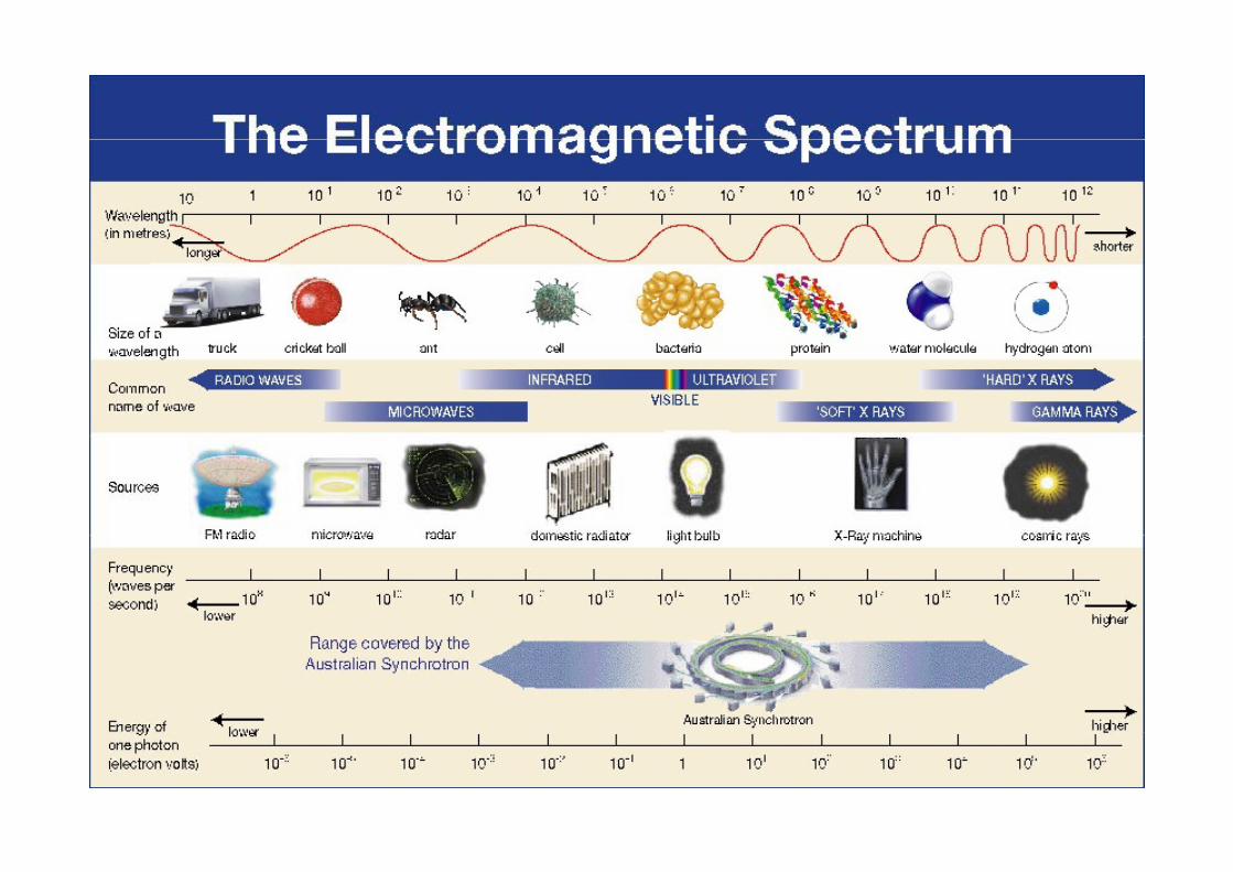

Electromagnetic Radiation at a Synchrotron
Energy transmitted in the form of waves or particles
γ rays X rays UV Vis IR μ waves
higher energy lower energy
γ-rays X-rays UV Vis IR μ-waves
For particles:
d B li l th Pl k’ t / t [λ h/ ]de Broglie wavelength = Planck’s const./momentum [λ = h/p]
For an electron accelerated through 100 Volts, λ = 1.2 Å,

Why is wavelength important?Why is wavelength important?
T b l d di i i h l h f i ilTo probe a sample you need radiation with a wavelength of similar, or smaller, magnitude to the size of the “object” under investigation.
sample sample
Visible light X raysVisible light X-rays
To investigate atomic/molecular structure, we use X-rays, electrons,To investigate atomic/molecular structure, we use X rays, electrons, and neutrons, since they can have wavelengths about the sizes of atoms.
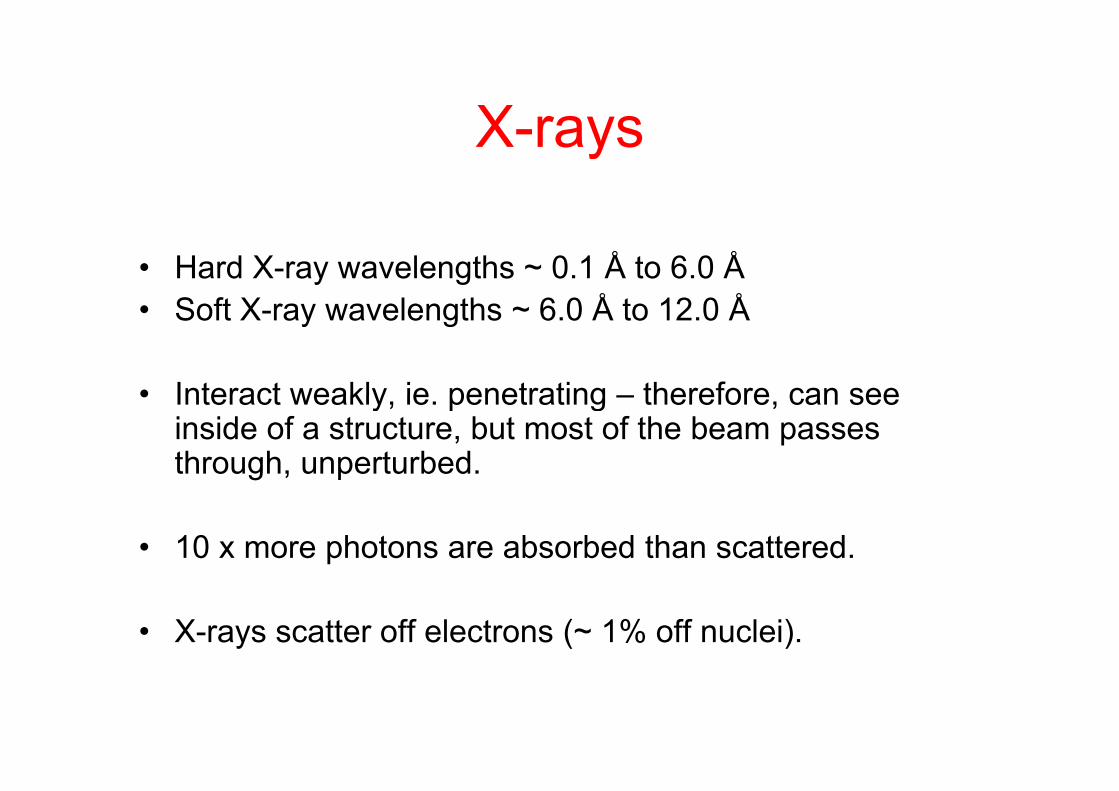
X raysX-rays
• Hard X-ray wavelengths ~ 0.1 Å to 6.0 Å• Soft X ray wavelengths 6 0 Å to 12 0 Å• Soft X-ray wavelengths ~ 6.0 Å to 12.0 Å
• Interact weakly ie penetrating – therefore can see• Interact weakly, ie. penetrating – therefore, can see inside of a structure, but most of the beam passes through, unperturbed.
• 10 x more photons are absorbed than scattered.
• X-rays scatter off electrons (~ 1% off nuclei).
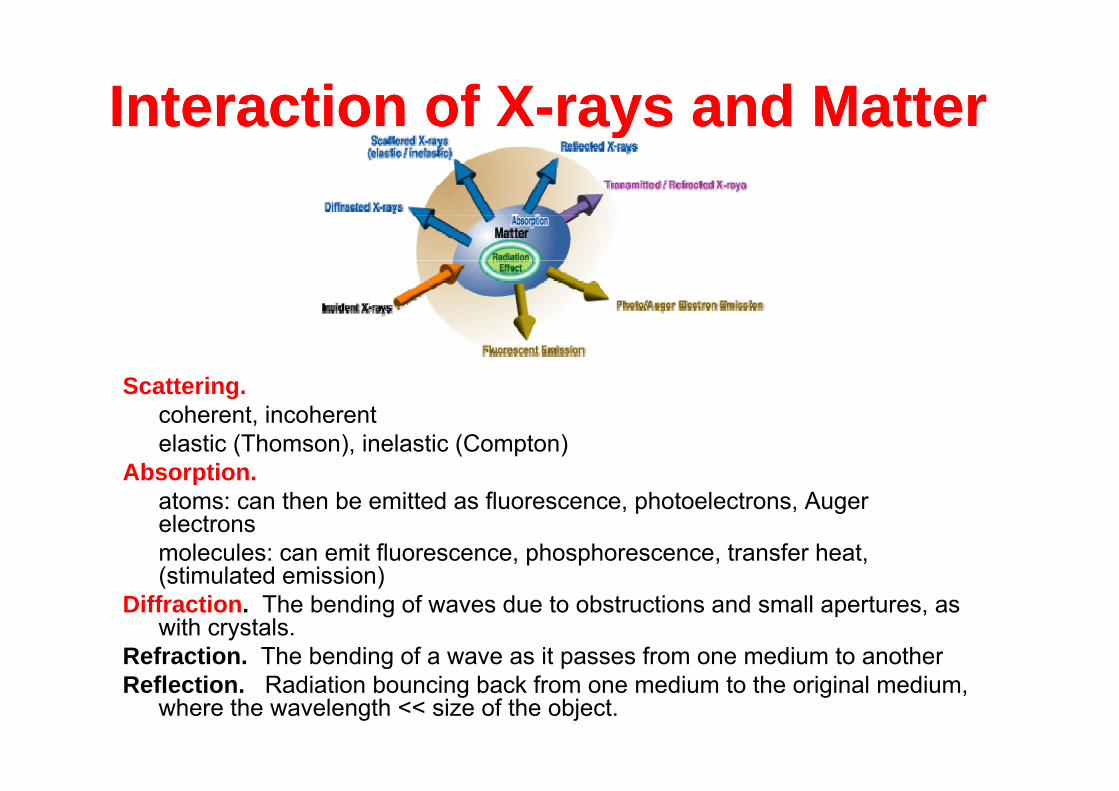
Interaction of XInteraction of X--rays and Matterrays and Matterte act o ote act o o ays a d atteays a d atte
Scattering.gcoherent, incoherentelastic (Thomson), inelastic (Compton)
Absorption.atoms: can then be emitted as fluorescence, photoelectrons, Auger electronsmolecules: can emit fluorescence, phosphorescence, transfer heat, (stimulated emission)(stimulated emission)
Diffraction. The bending of waves due to obstructions and small apertures, as with crystals.
Refraction. The bending of a wave as it passes from one medium to anotherg pReflection. Radiation bouncing back from one medium to the original medium,
where the wavelength << size of the object.
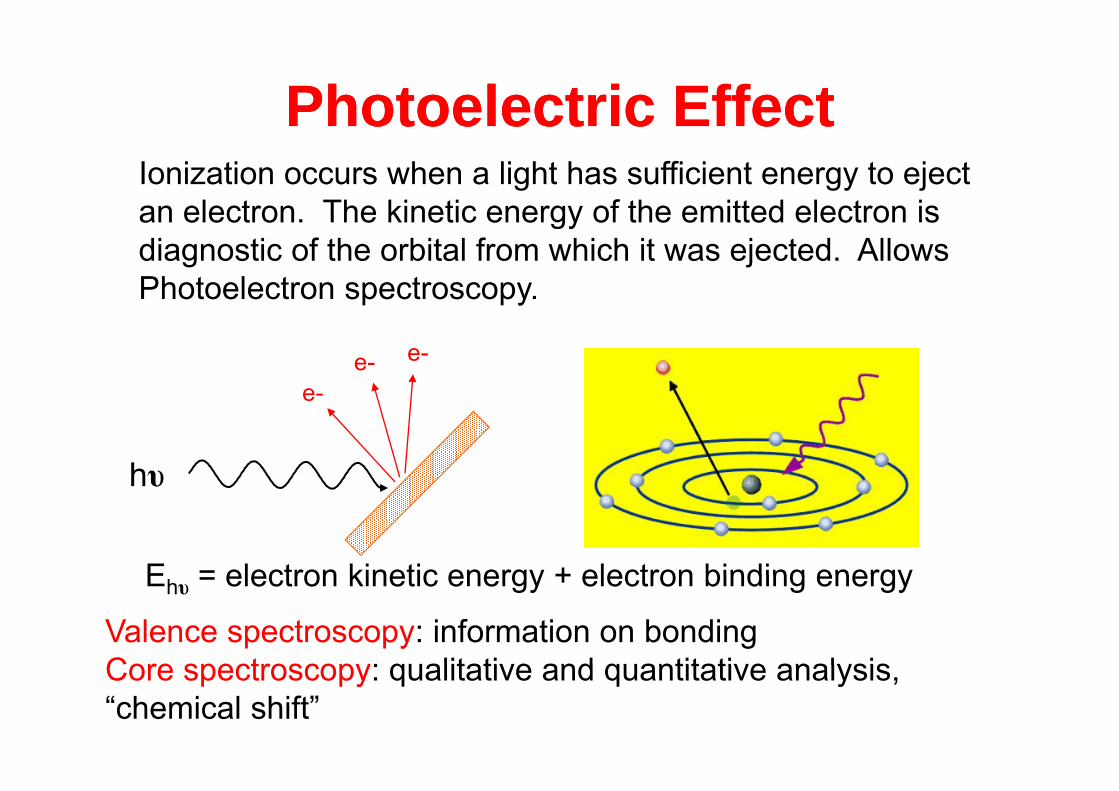
Photoelectric EffectPhotoelectric EffectIonization occurs when a light has sufficient energy to eject an electron The kinetic energy of the emitted electron is
otoe ect c ectotoe ect c ectan electron. The kinetic energy of the emitted electron is diagnostic of the orbital from which it was ejected. Allows Photoelectron spectroscopy.
e-e- e-
hυ
e-
E = electron kinetic energy + electron binding energy
hυ
Ehυ = electron kinetic energy + electron binding energy
Valence spectroscopy: information on bondingCore spectroscopy: qualitative and quantitative analysisCore spectroscopy: qualitative and quantitative analysis, “chemical shift”
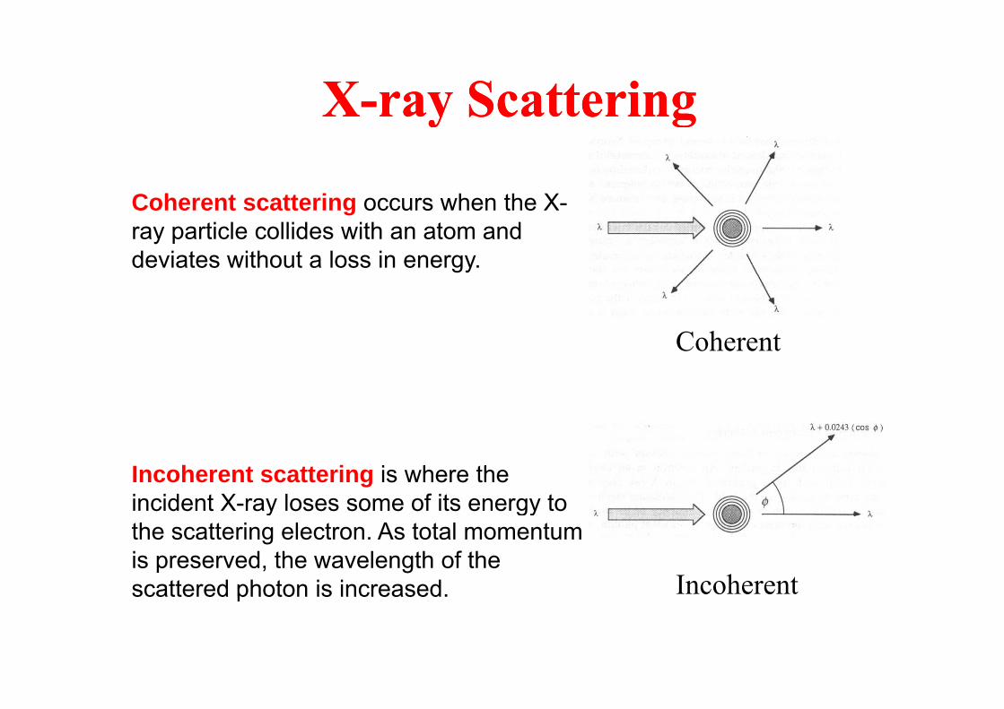
XX--ray Scatteringray ScatteringXX ray Scatteringray Scattering
C h i h h XCoherent scattering occurs when the X-ray particle collides with an atom and deviates without a loss in energy.
Coherent
Incoherent scattering is where the incident X-ray loses some of its energy to y gythe scattering electron. As total momentum is preserved, the wavelength of the scattered photon is increased. Incoherentscattered photon is increased.
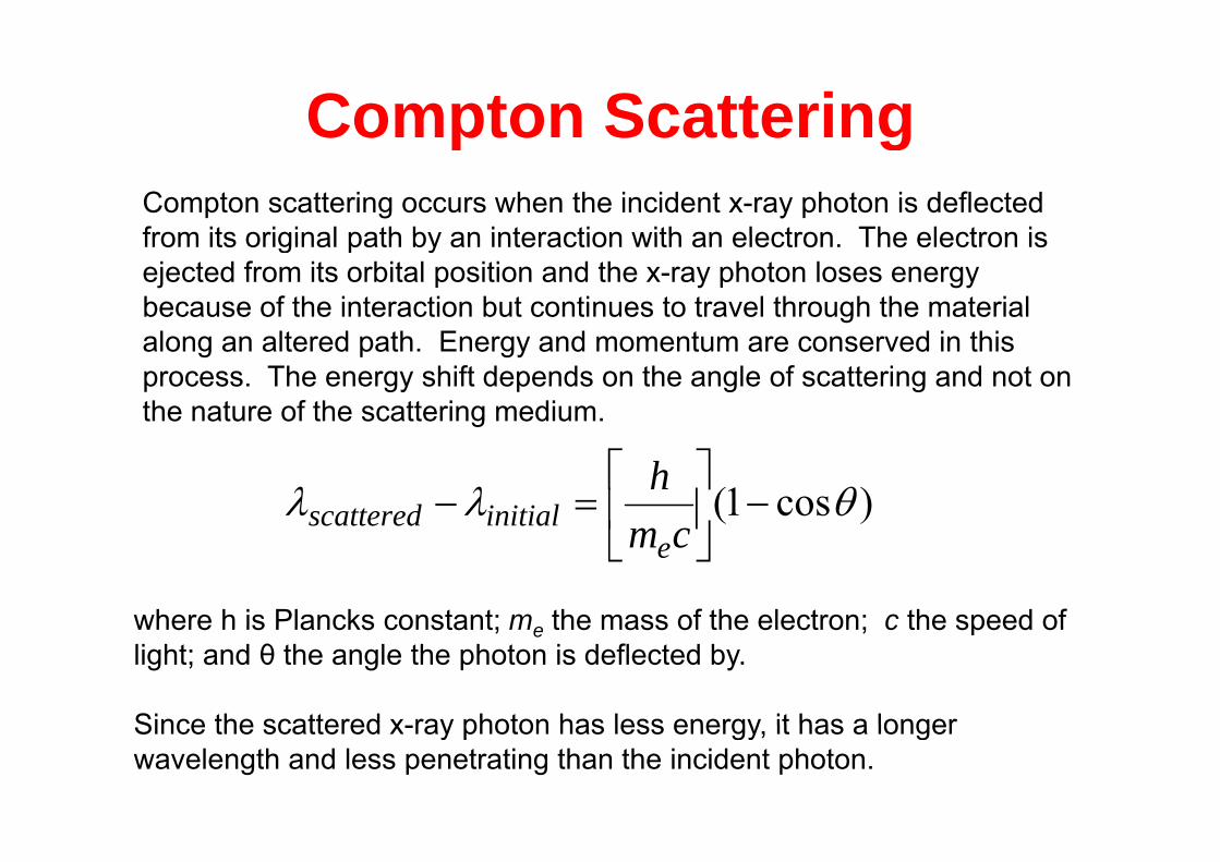
Compton ScatteringCo pto Scatte gCompton scattering occurs when the incident x-ray photon is deflected from its original path by an interaction with an electron The electron isfrom its original path by an interaction with an electron. The electron is ejected from its orbital position and the x-ray photon loses energy because of the interaction but continues to travel through the material l lt d th E d t d i thialong an altered path. Energy and momentum are conserved in this
process. The energy shift depends on the angle of scattering and not on the nature of the scattering medium.
)cos1(
hinitialscattered
cme
where h is Plancks constant; me the mass of the electron; c the speed ofwhere h is Plancks constant; me the mass of the electron; c the speed of light; and θ the angle the photon is deflected by.
Since the scattered x ray photon has less energy it has a longerSince the scattered x-ray photon has less energy, it has a longer wavelength and less penetrating than the incident photon.
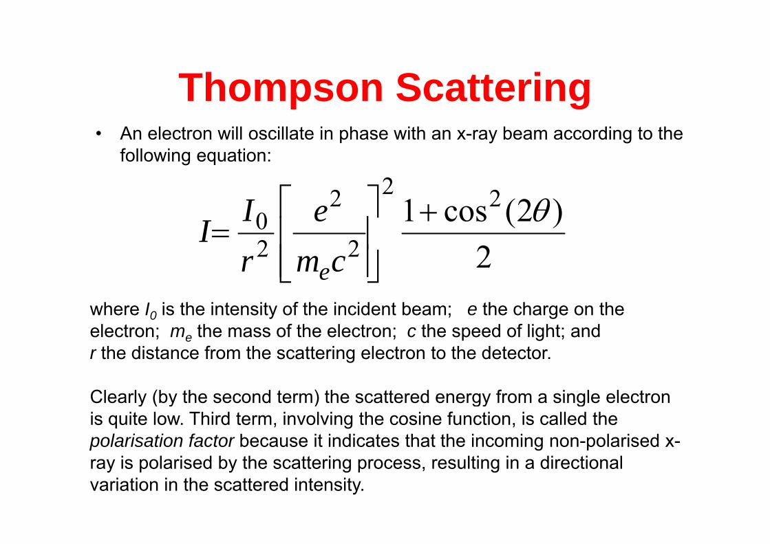
Thompson ScatteringThompson Scattering• An electron will oscillate in phase with an x-ray beam according to the
f ll i ifollowing equation:
)2(cos1 222 eI2
)2(cos122
0
cme
rII
e ewhere I0 is the intensity of the incident beam; e the charge on the electron; m the mass of the electron; c the speed of light; andelectron; me the mass of the electron; c the speed of light; and r the distance from the scattering electron to the detector.
Clearly (by the second term) the scattered energy from a single electronClearly (by the second term) the scattered energy from a single electron is quite low. Third term, involving the cosine function, is called the polarisation factor because it indicates that the incoming non-polarised x-
i l i d b th tt i lti i di ti lray is polarised by the scattering process, resulting in a directional variation in the scattered intensity.
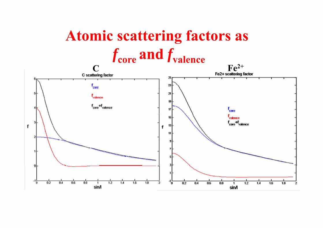
Atomic scattering factors asAtomic scattering factors asfcore and fvalencecore valence
C Fe2+
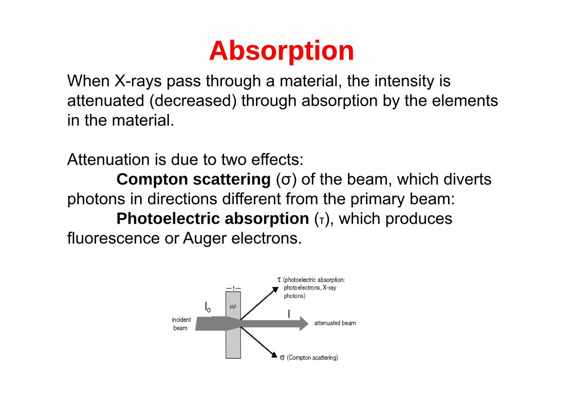
AbsorptionAbsorptionppWhen X-rays pass through a material, the intensity is attenuated (decreased) through absorption by the elements in the material.
Attenuation is due to two effects:Attenuation is due to two effects: Compton scattering (σ) of the beam, which diverts
photons in directions different from the primary beam:photons in directions different from the primary beam: Photoelectric absorption (τ), which produces
fluorescence or Auger electrons.

AbsorptionAbsorptionppThese two effects may be combined into a single bulk mass absorption coefficient μ). This coefficient is different for each element and wavelength and is defined as a the sum of photoelectric and Compton scattering:
Where τ = photoelectric absorption coefficient and
σ is the Compton Scattering coefficientIn many materials photoelectric absorption accounts for about 95 percent of the absorption and Compton scattering can be ignored Further Compton scattering is not importantcan be ignored. Further, Compton scattering is not important at wavelengths greater than 1 Å.
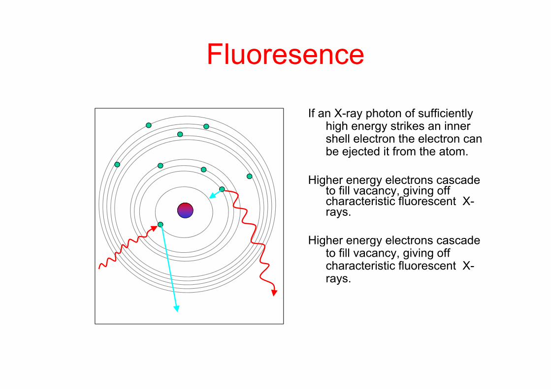
FluoresenceFluoresence
If an X-ray photon of sufficiently high energy strikes an inner shell electron the electron can be ejected it from the atombe ejected it from the atom.
Higher energy electrons cascade to fill vacancy, giving off to aca cy, g g ocharacteristic fluorescent X-rays.
Higher energy electrons cascadeHigher energy electrons cascade to fill vacancy, giving off characteristic fluorescent X-rays.y
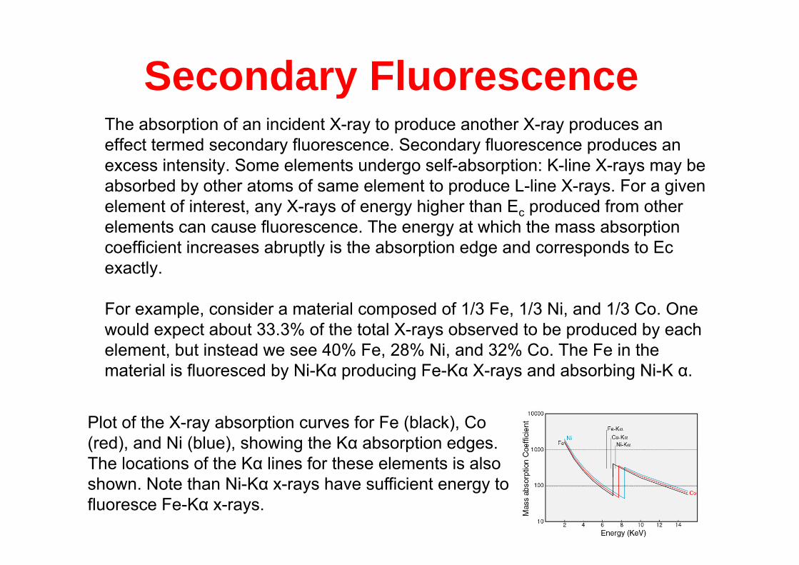
Secondary FluorescenceThe absorption of an incident X-ray to produce another X-ray produces an effect termed secondary fluorescence. Secondary fluorescence produces an
Seco da y uo esce ce
excess intensity. Some elements undergo self-absorption: K-line X-rays may be absorbed by other atoms of same element to produce L-line X-rays. For a given element of interest, any X-rays of energy higher than Ec produced from other elements can cause fluorescence. The energy at which the mass absorption coefficient increases abruptly is the absorption edge and corresponds to Ec exactly.
For example, consider a material composed of 1/3 Fe, 1/3 Ni, and 1/3 Co. One would expect about 33.3% of the total X-rays observed to be produced by each element b t instead e see 40% Fe 28% Ni and 32% Co The Fe in theelement, but instead we see 40% Fe, 28% Ni, and 32% Co. The Fe in the material is fluoresced by Ni-Kα producing Fe-Kα X-rays and absorbing Ni-K α.
Plot of the X-ray absorption curves for Fe (black), Co (red), and Ni (blue), showing the Kα absorption edges. The locations of the Kα lines for these elements is also shown. Note than Ni-Kα x-rays have sufficient energy to fluoresce Fe-Kα x-rays.

Scattering and Diffraction TechniquesScattering and Diffraction Techniquesg qg q
3-Dimensional X-ray crystallographyPhasing methods g
2-Dimensional Grazing incidence diffraction
L l ttLow-angle scatter
DynamicsDynamicsEquilibrium: B factors, diffuse scatterNon-equilibrium: Laue methodq
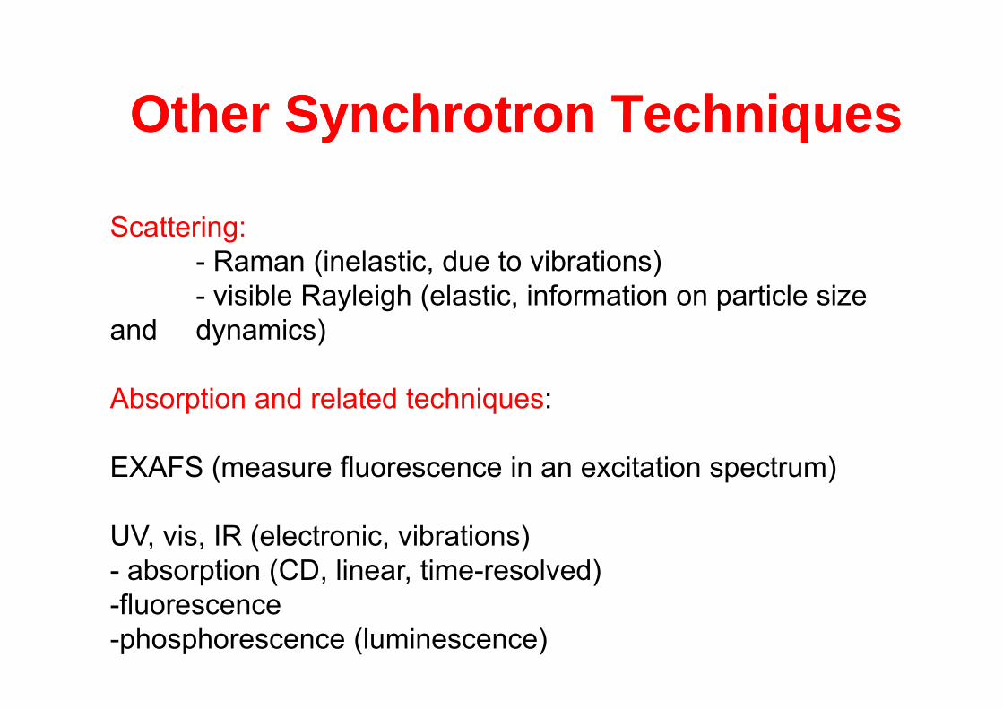
Other Synchrotron TechniquesOther Synchrotron TechniquesOther Synchrotron TechniquesOther Synchrotron Techniques
Scattering:- Raman (inelastic, due to vibrations)( , )- visible Rayleigh (elastic, information on particle size
and dynamics)
Absorption and related techniques:
EXAFS (measure fluorescence in an excitation spectrum)
UV, vis, IR (electronic, vibrations)- absorption (CD, linear, time-resolved)fl-fluorescence
-phosphorescence (luminescence)
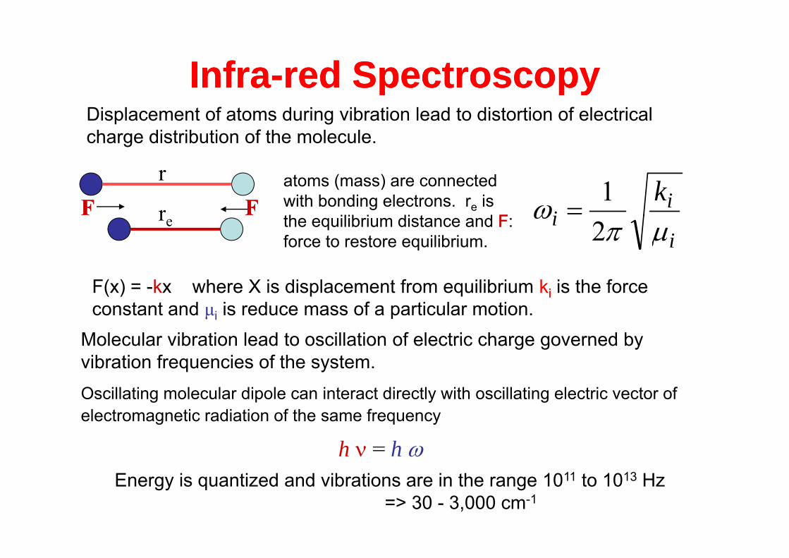
InfraInfra--red Spectroscopyred Spectroscopyp pyp pyDisplacement of atoms during vibration lead to distortion of electrical charge distribution of the molecule.
r
r FFFFatoms (mass) are connected with bonding electrons. re is th ilib i di t d FF
ii
k 1re the equilibrium distance and FF:
force to restore equilibrium. ii
2
Molecular vibration lead to oscillation of electric charge governed by
F(x) = -kx where X is displacement from equilibrium kii is the force constant and μi is reduce mass of a particular motion.
Molecular vibration lead to oscillation of electric charge governed by vibration frequencies of the system.Oscillating molecular dipole can interact directly with oscillating electric vector of
h = h electromagnetic radiation of the same frequency
Energy is quantized and vibrations are in the range 1011 to 1013 Hz => 30 - 3,000 cm-1
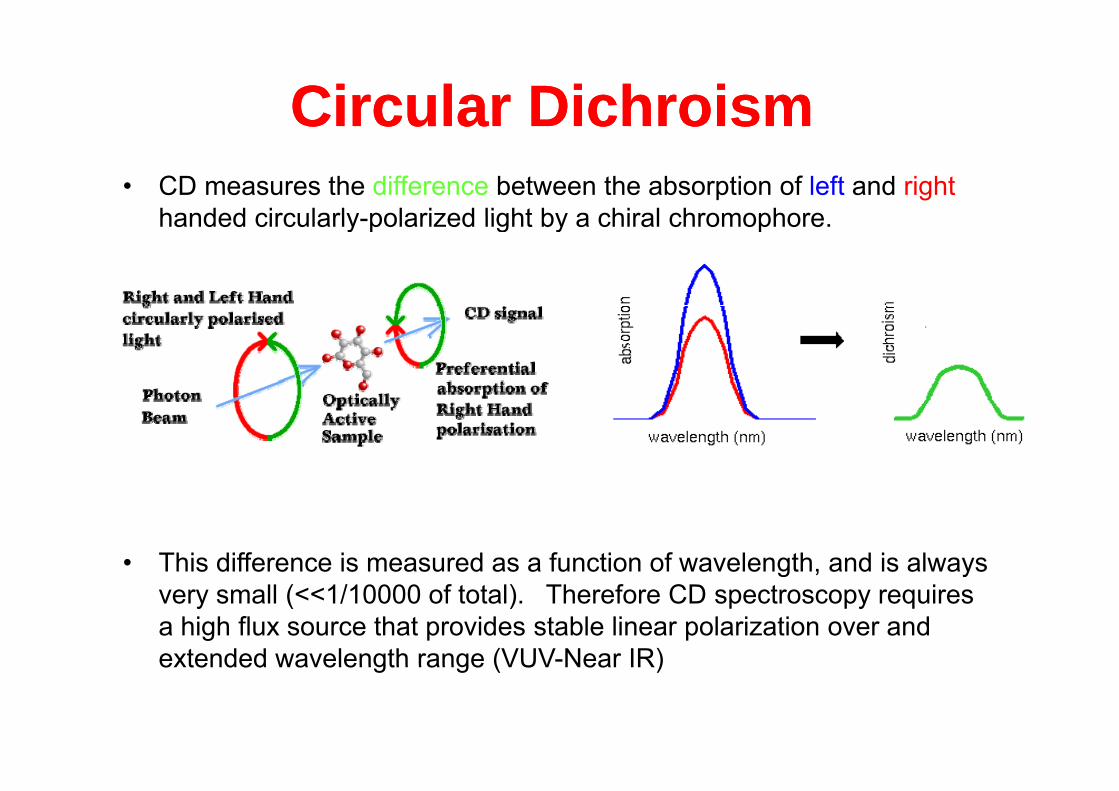
Circular DichroismCircular Dichroism• CD measures the difference between the absorption of left and right
handed circularly polarized light by a chiral chromophore
C cu a c o sC cu a c o shanded circularly-polarized light by a chiral chromophore.
• This difference is measured as a function of wavelength, and is always g , yvery small (<<1/10000 of total). Therefore CD spectroscopy requires a high flux source that provides stable linear polarization over and extended wavelength range (VUV-Near IR)extended wavelength range (VUV Near IR)
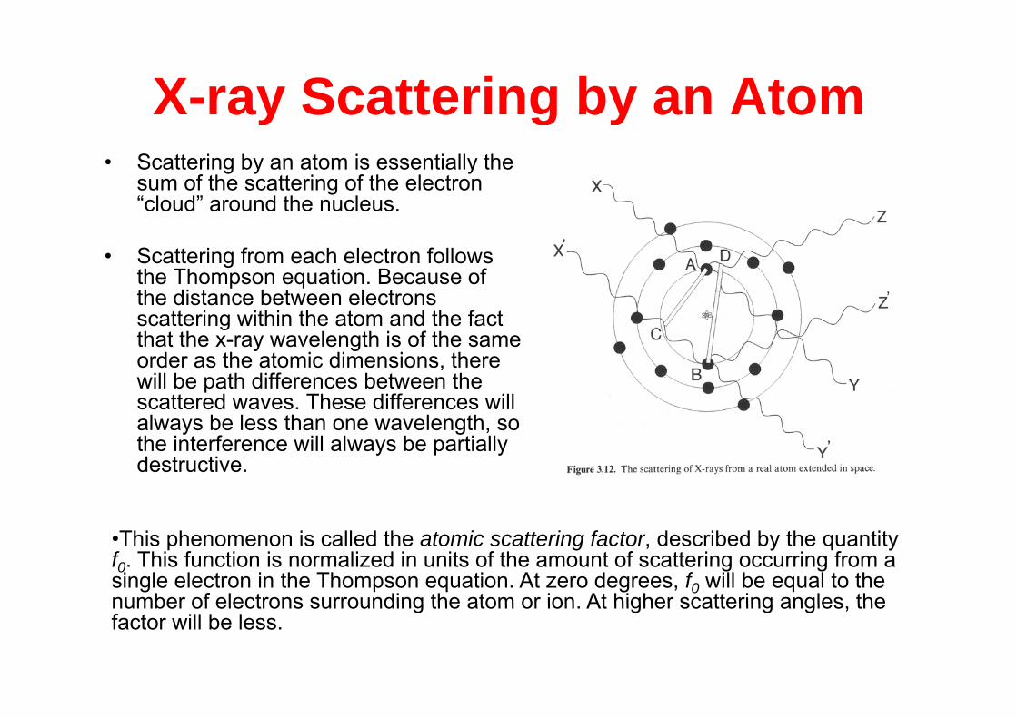
X-ray Scattering by an Atom• Scattering by an atom is essentially the
sum of the scattering of the electron “ l d” d th l
ay Scatte g by a to
“cloud” around the nucleus.
• Scattering from each electron follows the Thompson equation Because ofthe Thompson equation. Because of the distance between electrons scattering within the atom and the fact that the x-ray wavelength is of the same order as the atomic dimensions thereorder as the atomic dimensions, there will be path differences between the scattered waves. These differences will always be less than one wavelength, so the interference will always be partiallythe interference will always be partially destructive.
•This phenomenon is called the atomic scattering factor, described by the quantity f0. This function is normalized in units of the amount of scattering occurring from a single electron in the Thompson equation. At zero degrees, f0 will be equal to the number of electrons surrounding the atom or ion At higher scattering angles thenumber of electrons surrounding the atom or ion. At higher scattering angles, the factor will be less.
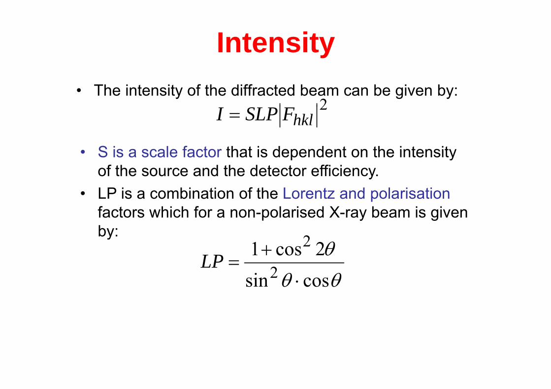
Intensity• The intensity of the diffracted beam can be given by:
2
• S is a scale factor that is dependent on the intensity
2hklFSLPI
• S is a scale factor that is dependent on the intensity of the source and the detector efficiency.
• LP is a combination of the Lorentz and polarisation• LP is a combination of the Lorentz and polarisationfactors which for a non-polarised X-ray beam is given by: 2y
cossin2cos1
2
2
LP
cossin

The Structure Factor Fhklhkl• For a reflection from a plane hkl. The structure factor
Fhkl is:Fhkl is:
lli 2 ]2exp[sinexpF
cellunit
1 2
2hkl i
i
hklii xihklBf
( ) th f ti l di t f t i i
1i
• xI = (xiyizi) are the fractional coordinates for atom i inthe cell
• hkl are the Miller indices for the reflection• hkl are the Miller indices for the reflection• Bi is the Debye-Waller displacement (thermal) factor
for atom (in Å2)for atom (in Å )• fi. Is the scattering factor.
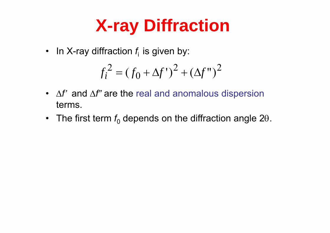
X-ray Diffraction• In X-ray diffraction fi is given by:
f’ d f” th l d l di i
220
2 )"()'( ffffi
• f’ and f” are the real and anomalous dispersionterms.The first term f depends on the diffraction angle 2• The first term f0 depends on the diffraction angle 2.
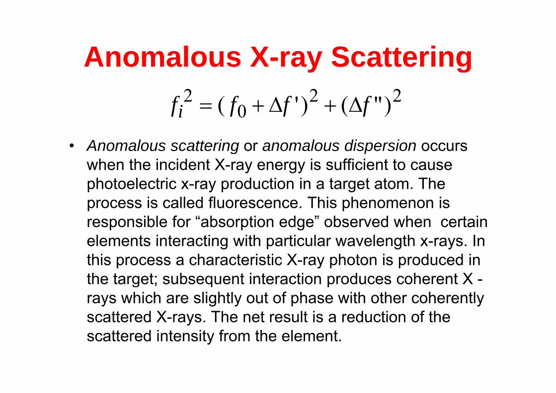
Anomalous X-ray ScatteringAnomalous X ray Scattering22
02 )"()'( ffffi
• Anomalous scattering or anomalous dispersion occurs
0 )()( ffffi
g pwhen the incident X-ray energy is sufficient to cause photoelectric x-ray production in a target atom. The
fprocess is called fluorescence. This phenomenon is responsible for “absorption edge” observed when certain elements interacting with particular wavelength x rays Inelements interacting with particular wavelength x-rays. In this process a characteristic X-ray photon is produced in the target; subsequent interaction produces coherent X -g ; q prays which are slightly out of phase with other coherently scattered X-rays. The net result is a reduction of the
tt d i t it f th l tscattered intensity from the element.
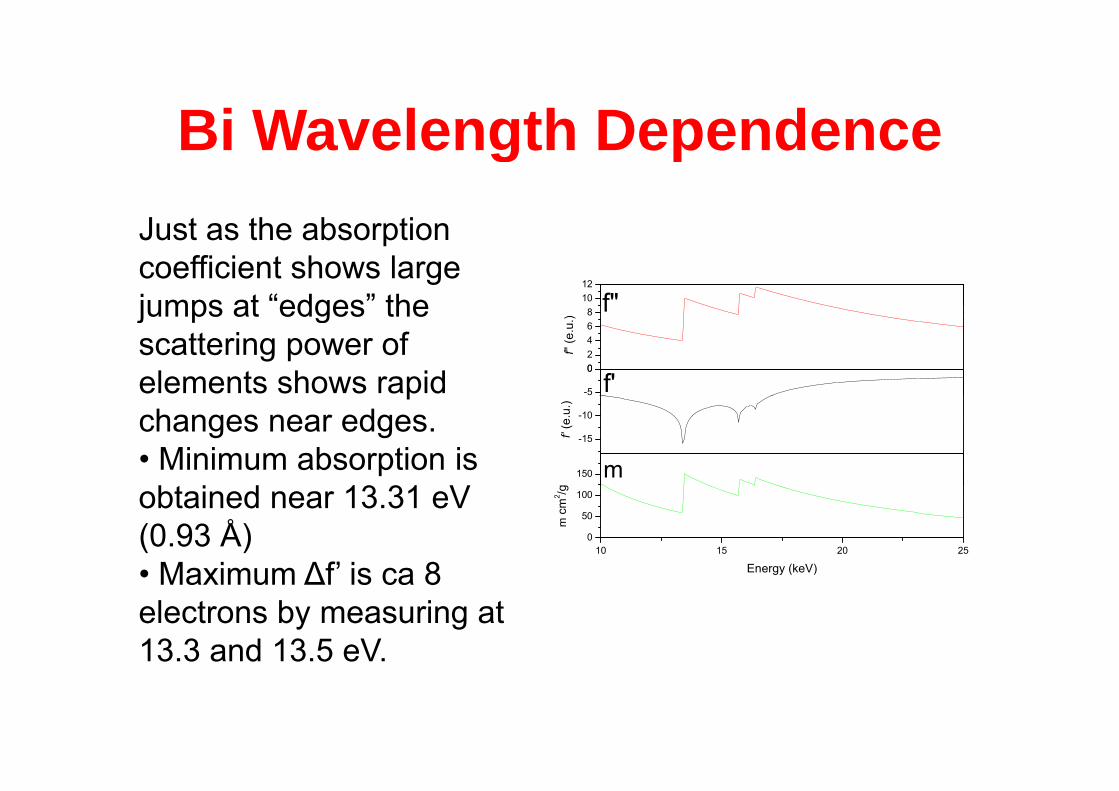
Bi Wavelength DependenceBi Wavelength Dependence
1012
f"
Just as the absorption coefficient shows large jumps at “edges” the
-5
002468
f'
f" (e
.u.)
fjumps at edges the scattering power ofelements shows rapid
150
-15
-10
m
f' (e
.u.)
pchanges near edges.• Minimum absorption is b i d 13 31 V
10 15 20 250
50
100
m c
m2 /g
Energy (keV)
obtained near 13.31 eV (0.93 Å)• Maximum ∆f’ is ca 8• Maximum ∆f is ca 8 electrons by measuring at 13.3 and 13.5 eV.
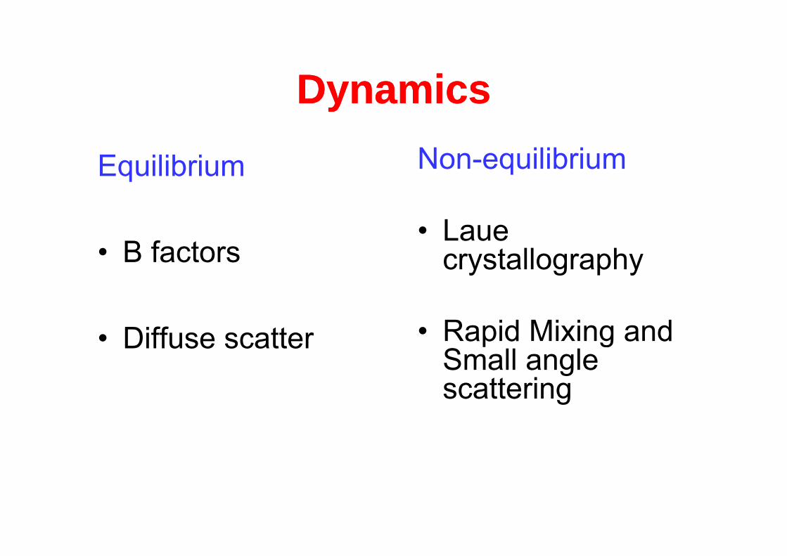
DynamicsDynamicsDynamicsDynamicsN ilib iEquilibrium Non-equilibrium
• B factors• Laue
crystallography
• Diffuse scatter • Rapid Mixing and • Diffuse scatter ap d g a dSmall angle scatteringg
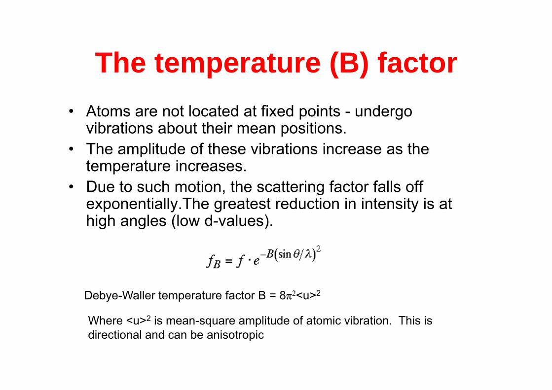
The temperature (B) factorThe temperature (B) factorAt t l t d t fi d i t d• Atoms are not located at fixed points - undergo vibrations about their mean positions.
• The amplitude of these vibrations increase as theThe amplitude of these vibrations increase as the temperature increases.
• Due to such motion, the scattering factor falls off gexponentially.The greatest reduction in intensity is at high angles (low d-values).
Where <u>2 is mean-square amplitude of atomic vibration. This is
Debye-Waller temperature factor B = 8π2<u>2
directional and can be anisotropic
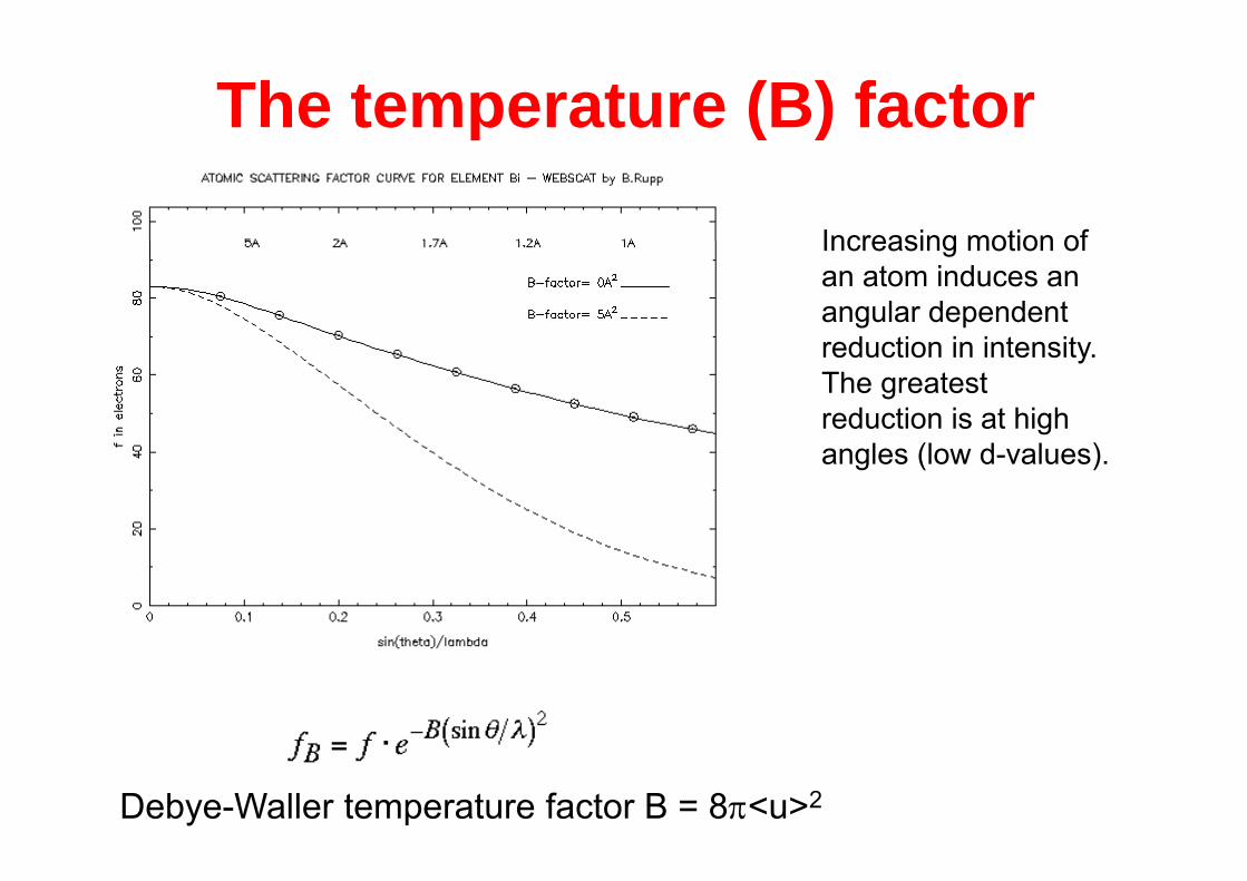
The temperature (B) factor( )
Increasing motion ofIncreasing motion of an atom induces an angular dependent
d ti i i t itreduction in intensity. The greatest reduction is at high angles (low d-values).
Debye-Waller temperature factor B = 8<u>2
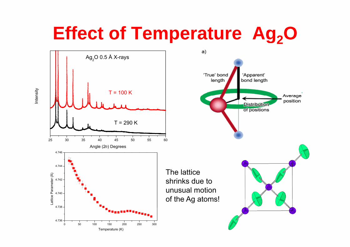
Effect of Temperature Ag2Oect o e pe atu e g2OAg2O 0.5 Å X-rays
T = 100 Knsity
T = 290 K
Inte
n
25 30 35 40 45 50 55 60
T 290 K
Angle (2) Degrees4 746
4 742
4.744
4.746
er (Å
) The lattice h i k d t
4.738
4.740
4.742
Latti
ce P
aram
ete shrinks due to
unusual motion of the Ag atoms!
0 50 100 150 200 250 3004.736
4.738
Temperature (K)
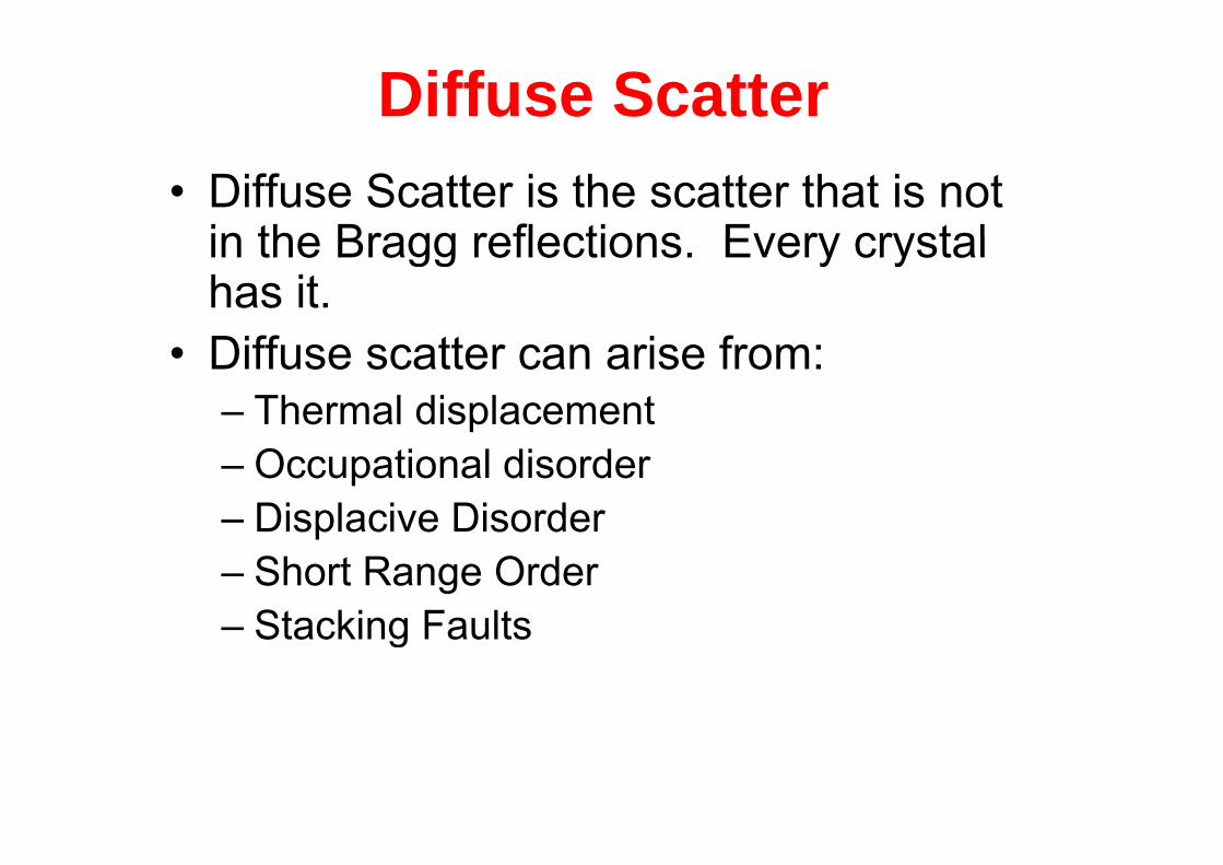
Diffuse Scatter• Diffuse Scatter is the scatter that is not
in the Bragg reflections Every crystalin the Bragg reflections. Every crystal has it.
• Diffuse scatter can arise from:– Thermal displacement– Occupational disorder– Displacive DisorderDisplacive Disorder– Short Range Order– Stacking Faults– Stacking Faults
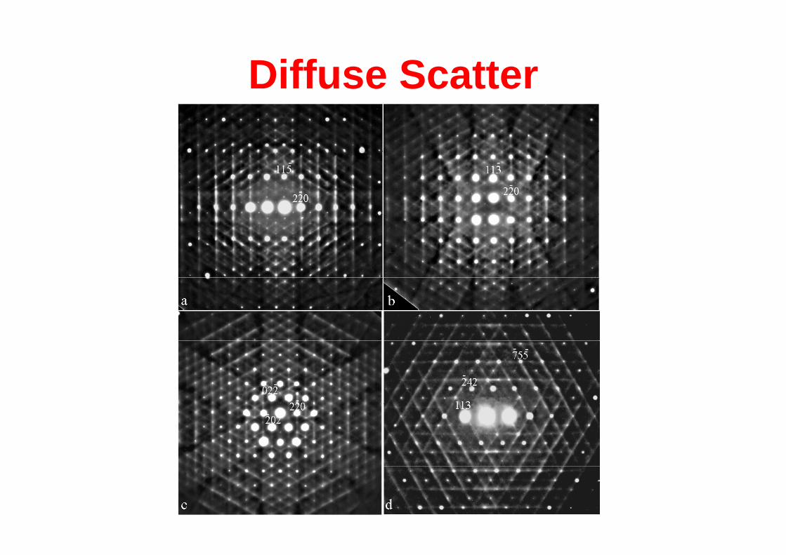
Diffuse Scatteruse Scatte