an investigation into possible mechanisms involved in...
TRANSCRIPT
AN INVESTIGATION INTO POSSIBLE MECHANISMS INVOLVED
IN THE PRACTOLOL INDUCED "OCULOMUCOCUTANEOUS SYNDROME.
Submitted in fu l f i lm e n t fo r the Degree o f Doctor o f Philosophy.
Graham R .E l l io t t .
Surrey University - September 1984.
S °l o|t+=?-o
ProQuest Number: 10798428
All rights reserved
INFORMATION TO ALL USERS The quality of this reproduction is dependent upon the quality of the copy submitted.
In the unlikely event that the author did not send a com p le te manuscript and there are missing pages, these will be noted. Also, if material had to be removed,
a note will indicate the deletion.
uestProQuest 10798428
Published by ProQuest LLC(2018). Copyright of the Dissertation is held by the Author.
All rights reserved.This work is protected against unauthorized copying under Title 17, United States C ode
Microform Edition © ProQuest LLC.
ProQuest LLC.789 East Eisenhower Parkway
P.O. Box 1346 Ann Arbor, Ml 48106- 1346
ABSTRACT.
Experiments aimed at e luc idating the id e n t i ty o f antigenic metabolites
of p ra c to lo l , using in v i t ro generated practo lo l metabolites and
sera from practo lo l pa tien ts , were unsuccessful as none of the sera
tested contained measurable concentrations of antimetabolite a n t i
bodies. Collaborative experiments with workers who had o r ig in a l ly
established the technique also fa i le d to detect such antibodies. I t
was concluded, a f te r fo llow up studies, tha t the active sera must
have been used up or damaged during the o r ig ina l inves tiga tions .
Rabbits and guinea-pigs, in jected with the prote in bound practo lo l
metabolites did not respond by synthesising antjmetabolite antibod
ies. The probable reason fo r the lack of response was the low concen
t ra t io n of hapten binding (1 metabolite molecule/6 prote in molecules).
A ra t io o f at least 10:1 is normally used in such experiments and
ra t ios o f greater than 100:1 are not uncommon.
Neither in v i t r o generated metabolites o f practo lo l nor chemical ana
logues, had any e f fe c t on human skin f ib ro b la s t growth or collagen
synthesis in v i t r o . In contrast, p ra c to lo l, propranolol and paracetamol
all inh ib ited these f ib rob la s ts functions in a dose re la ted fashion.
Cells from uninvolved skin o f a psoriasis pa tient were more se n s it
ive to the in h ib i to ry actions o f the two 3-receptor blocking drugs
than f ib ro b la s ts obtained from a control volunteer but were less sen
s i t iv e to paracetamol ind ica ting a va r ia t ion in the response to
3-receptor blocking drugs and tha t such changes in s e n s i t iv i t y need
not be para lle led by chemically re la ted compounds such as paracetamol.
Practolol was not taken up by the f ib ro b la s ts to any great extent

ind ica ting tha t i t s s i te o f action was the plasma membrane. Uptake
o f leucine was in h ib ite d to the same extent as collagen synthesis
suggesting that practo lo l may in te r fe re with protein synthesis by
l im i t in g substrate a v a i la b i l i t y .
In v i t r o morphological studies are consistent w ith the idea tha t the
three drugs act by d i f fe re n t mechanisms although fu r th e r studies are
necessary to confirm th is .
The fo llow ing conclusions can be drawn from the experimental f ind ings .
- The side effec ts o f practo lo l are more l ik e ly to have been due
to the parent molecule than to a metabolite.
- The action o f practo lo l is l i k e ly to have been an in h ib i to ry ,
rather than a s tim u la tory , one.
- Susceptible patients have an increased s e n s i t iv i t y to practo lo l
which could be re flec ted in the response o f f ib ro b la s ts from
such patients in v i t ro .
ACKNOWLEDGEMENTS.
I wish to thank the Medical Research Council fo r f inanc ia l
support and the B r i t is h Indus tr ia l B iologica l Research Association
fo r chemicals, bench space, access to photocopy machines etc. etc.
I am g ra te fu l fo r the encouragement and help o f Dr.H.E.Amos and
Prof. J.W.Bridges during the time i t took both to carry out these
experiments and to w rite th is thesis.
I am also happy to acknowledge the help and support o f the s ta f f at
BIBRA who could not have been more cooperative, in p a r t ic u la r Dr K.
M il l .r who provided guidance during the i n i t i a l phases of the
f ib ro b la s t experiments, Dr.J.Evans, fo r making space on the electron
microscope, Dr B.Veilleux fo r helping to cu ltu re tfre Staplococcus aureus
bacteria , Alan Dagnell fo r photographs and Dr.B.Lake fo r advice on
preparing the practo lo l m etabo lite /prote in complexes and the co-valent
binding studies.

CONTENTS. Page.
T i t le page.
Abstract. 1
Acknowledgements. 3
Contents. 4
M ate ria ls . 6
Chapter 1. In troduction to the Thesis. 9
Part One. Experiments to fu r th e r characterise the chemical nature 50
of the antigenic metabolites o f practo lo l using an
immunological approach.
Chapter 2. Experiments aimed at evaluating previously published 51
methods o f detecting antibodies to practo lo l metabolites
and confiming e a r l ie r resu lts .
Chapter 3. Investigations to determine possible reasons why a n t i - 82
bodies to in v i t r o generated practo lo l metabolites
could not be detected in the serum of practo lo l treated
pati en ts.
Chapter 4. Experiments aimed at ra is ing antibodies to in v i t r o
generated metabolites o f practo lo l in animals.
95

Part Two. Experiments to investigate the e f fe c t o f p ra c to lo l ,
Chapter 5.
Chapter 6 .
Chapter 7.
Chapter 8 .
Discussion.
in v i t r o generated metabolites o f p racto lo l and
chemical analogues on human skin f ib ro b la s t functions.
The e ffe c t of p ra c to lo l, in v i t ro generated metabolites
o f practo lo l and chemical analogues on the rate o f growth
o f human skin f ib rob la s ts in v i t ro .
The e f fe c t o f p ra c to lo l, in v i t r o generated metabolites
of practo lo l and chemical analogues on human skin
f ib ro b la s t collagen synthesis.
Experiments aimed at investiga ting thfe e f fe c t p racto lo l
on production o f a fac to r by hamster peritoneal macro
phages capable o f in fluencing human skin f ib ro b la s t
collagen synthesis.
The e f fe c t o f p ra c to lo l, paracetamol and propranolol
on DT P j morphology a f te r in v i t ro exposure.
References.
MATERIALS.
Animals. New Zealand White Rabbits. Ranch Rabbits, Sussex.
Syrian Hamsters, Albino Guinea-pigs. Torcellus AB Ltd.
F ib rob las ts . and PS human skin f ib ro b la s ts were a g i f t from
the Cell Mutation U nit, MRC., Sussex U n ivers ity .
Chapter 2.
Chapter 3
125 I human serum albumin.(25pci/mg) Amersham In ternationa l
7-ethoxy and hydroxy coumarin. Sigma Fine Chemicals.
is o c i t ra te dehydrogenase (type 4 pigs heart) . "
i s o c i t r i c acid (type 1) "
nicotinamide adenine dinucleotide phosphate (yeast)(NADP). "
Niacin (free acid) "
thiamine (HC1) "
3-glycerophosphate (grade 1) "
ovalbumin (grade 2 ) "
bovine gamma globulin ( f ra c t io n 2 ) "
bovine serum albumin (BSA) "
penassy broth Difco l t d .
yeast ex trac t "
Sheep anti human serum albumin Wellcome
Horse anti human serum "
Fluorosceine isothiocyanate labelled sheep anti human IgG. ( FITC IgG) Wei 1 come
Chapter 4 .
Freunds complete adjuvant Difco

Chapter
Chapter 6
Chapter 7
Earls minimal essential medium
L-glutamine (lOOx)
non-essential amino acids (lOOx)
p e n ic i l l in streptomycin ( 10,000 un its 'm l
foeta l c a l f serum
tryps i n
trypan blue
phosphate buffered saline ta b le ts .
Calf thymus deoxyribonucleic acid (type 1
L-pro line
hydroxyproli nedi phenyl ami ne
di phenyl ami ne
chloramine T
ascorbic acid
dimethyl ami ne-benzaldehyde
donnor c a l f serum 14C leucine
1idnocain
sheep red blood ce lls
rabb it anti sheep red blood ce lls (Ambroceptor)
Bouins reagent
Mayers haemotoxyline
eosin
Flow Laboratories.
II
II
)II
ll
George T. Gurr l t d
Oxoid
). Sigma Fine Chemicals
ll
ll
ll
II
ll
Fise>r\s.
Gibco b io c u lt
Arnersham
Sigma Fine Chemicals
Wei 1 come
II
Raymond A.Lam.
II
Chapter 8 .
o i l - re d 0 Raymond A.Lam
periodic acid "
ce lestine blue "
cacodylate bu ffe r E.M.Scope UK.
epon 812 "
a ra ld ite 502 "
uranyl acetate "
14Radiolabelled practo lo l ( C ring labelled 17.l lgc i/m g , 99.5%pure)
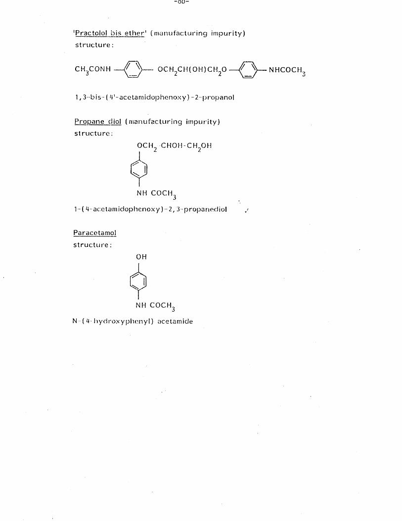
unlabelled p ra c to lo l, propranolol, paracetamol and the various
chemical analogues of practo lo l (chemical names and structures are
given on page 8a/b)ware a g i f t of IC I, Pharmaceuticals D iv is ion ,
Macclesfield.
Other chemicals used were o f standard reagent grade.
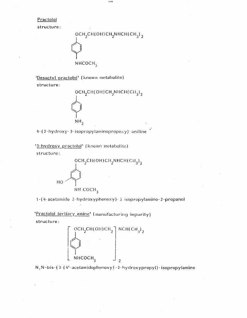
Practolol
s t r u c t u r e
OCH C H ( O H ) C H N H C H (C H )
NHCOCH.3
' Desac ty l p r a c t o l o l 1 ( k n o w n metabo l i te )
s t r u c t u r e :
OCH C H ( O H ) C H N H C H (C H )
4-( 2- h y d r o x y - 3 - i sop ro p y lam i n o p r o p o x y ) a n i l in e
13 - h y d r o x y p r a c t o l o l 1 ( k n o w n metabo l i te )
s t r u c t u r e :
OCH C H ( O H ) C H 2N H C H (C H )
HO '
NHCOCH..
1 - ( 4- acetamido 3- h y d r o x y p h e n o x y ) - 3 i s o p r o p y !a m in o - 2 - p r o p a n o l
'P rac to lo l t e r t i a r y am in e1 ( m a n u f a c t u r i n g i m p u r i t y )
s t r u c t u r e :
OCH C H ( O H ) C H NC H (C H .<) 2
NHCOCH.
N , N - b i s - ( 3 ( 4 ' - a ce ta m id o p h e n o xy ) - 2 - h y d r o x y p r o p y l ) - i s o p ro p y la m in e
- O D “
1 Prac to lo l b is e t h e r 1 ( n ia n u fa c tu r i n g im p u r i t y )
s t r u c t u r e :
C H 3CONH — P \ — OCH C H ( O H ) C H O — r ^
1, 3--bis- ( 4 ' - a c e ta m id o p h e n o x y ) - 2 -p ro pa n o l
Propane cJiol ( m a n u f a c t u r i n g im p u r i t y )
s t r u c t u r e :
OCH --CHOH-CH OH
NH COCH
1-( 4 - a e e ta m id o p h e n o x y ) ~2, 3 - p ro pa n ed io l
Paracetamol
s t r u c t u r e :
OH
NH CO C H3
N- ( 4- h y d r o x y p h e n y l ) acetamide
N H C O C H ,3
CHAPTER 1.
INTRODUCTION TO THE THESIS
INTRODUCTION
The terms adrenergic and cho linerg ic were introduced by Dale (1933)
to describe nerve f ib res tha t release, respective ly , a sympathetic
transm itte r and ace ty lcholine, but the terms have since been extended
to include the corresponding neurotransmitter receptors.
Langley (1905) suggested that there were two types o f t issue recep
to r , exc ita to ry and in h ib i to ry . This concept was supported by Dale
(1906) who showed tha t an ergot preparation paralysed the s tim u la to r
actions o f the adrenergic system without a ffec t ing in h ib i to ry impul
ses. The concept of independent receptors received support from Ahlqu is t
(1948) who investigated the e f fe c t o f f iv e catecholamines on adrenergic
actions and showed tha t although they were q u a l i ta t iv e ly s im ila r
there were qu an tita t ive differences. He explained th is f ind ing by
suggesting that the two receptors had d i f fe re n t a f f in i t i e s fo r the
compounds tested. The order o f potency fo r one set o f responses was,
adrenaline, nor-adrenaline, methylnor-adrenaline and isoprenaline. This
potency order correlated with contraction o f the rabb it u terine myomet
rium, dog and rabb it uterus in s i tu and fo r in h ib i t io n o f atropine ind
uced gut contractions. The second class o f responses, ty p i f ie d by
vasod ila tion , in h ib i t io n o f myometrial contraction and a pos it ive iono-
tro p ic e f fe c t had a catecholamine potency order o f isop rena line , adren
a l ine , methyl adrenaline, methyl nor-adrenaline and nor-adrenaline.
Ahlquist designated the receptors as alpha (a) and beta (B) respective ly
and emphasised that the c la s s i f ic a t io n was based on agonist potency.
The adrenergic receptor blocking agents ava ilab le at tha t time acted
only at a-receptors and th is lead to c o n f l ic t in g reports on th e i r s i te
o f action and e f f ic ie n c y , (Ahlquist 1967).
The use o f di-chloroisoprenaline (Powell and S la ter 1958), a compound
which was shown to block 3-adrenergic functions, f i n a l l y established
the two receptor concept. Later work by Lands et al (1967), Furchgott
(1967), Levy (1966) and Moran (1966) demonstrated tha t the 3-receptor
could be divided in to subtypes. Stimulation o f 3i-receptors produced
e ffec ts on the heart and 1ipo lys is w h i ls t s tim u la tion o f 32-receptors
produced changes in bronchiolar and blood vessel tone. Furchgott (1967)
studied the a f f in i t y constants o f various tissue receptors fo r pro-
netholol and concluded that the 3-receptor could be even fu r th e r sub
divided. Bristow et al (1970) extended Furchgotts work and measured
the receptor a f f in i t i e s o f the 3-receptor blocking drugs propranolol
and p rac to lo l. They found tha t the a f f in i t y constant fo r the blocking
action o f practo lo l on the heart v/as f i f te e n tim es'greater than fo r
tracheal muscle and s ix hundred and s ix ty times greater than fo r a o r t ic
muscle. They in terpreted these quan tita t ive differences as evidence
fo r the presence o f 3-receptor subtypes in the tissues studied.
Furchgott (1972) discussed the problems involved in measuring the a f
f i n i t y constants o f 3-receptors fo r agonists and antagonists. For ex
ample sympathetic nerves ac t ive ly accumulate nor-adrenaline w h i ls t
propranolol can in te r fe re with i t s uptake. S im ila r ly measurement o f
the blood drug level may give a value unrelated to the free drug con
c e n t ra t io n - i f binding to plasma proteins occurs. The d i f f i c u l t i e s in
volved in c lass ify ing 3-receptor subtypes are fu r th e r underlined by the
f ind ing that schemes based on a comparison o f agonist potency do not
wholly agree with those derived from a comparison of antagonist ac t ion ,
(Bristow et al 1970, Furchgott 1972). Despite the doubt surrounding
the d iv e rs i ty o f 3-receptor subtypes, the two receptor concept has
proved useful in defin ing tissue s e le c t iv i t y o f 3-adrenergic receptor
blocking drugs.
Structure function re la tionsh ips of beta adrenergic receptor blocking
drugs.
The discovery of d ich lorisoprenaline (Powell and S later 1958) stimu
lated the development of other 3-receptor blocking drugs and resulted
in a number o f compounds with a wide spectrum of pharmacological actions.
For example propranolol ( f i g l , l ) , a non spe c if ic 3-receptor blocking drug,
was shown to possess membrane s ta b i l is in g a c t iv i t y (Prichard 1978) w h i ls t
practo lo l ( f ig 1,1), which is spec if ic fo r 3 i - re ce p to rs , has no membrane
s ta b i l is in g a c t iv i t y but does posses in t r in s ic sympathomimetic ac t
i v i t y , (Prichard 1978). These associated properties provided the basis
fo r a system of c lass ify ing the drugs and can be corre lated w ith
functional groups on the molecule, (F itzgera ld 1969, Prichard 1978).
Most 3-receptor blocking drugs are analogues o f isoprenaline ( f i g l , l ) .
The 3-receptor s p e c i f ic i t y has been shown to reside with the iso
propyl substitu ted secondary amine while the a f f in i t y is re la ted to
the size and degree o f branching o f the a lky l side chain, (Goodman
and Gilman 1975 , Dollery et al 1969). This re la t ionsh ip also applies
to 3-blocking drugs based on pronethalol ( f i g 1,1) and d ich lo ro isoprena line ,
( f i g l , l ) . Hydroxyl sub s titu t io n at the 3-carbon confers sterio isom-
erism on the molecule resu lt ing in D and L forms of which the L
form is the morefKarmacologically active . This applies to agonists
and antagonists. Antagonist and agonist properties have been shown
to be dependent on the type o f su b s t itu t io n on the benzene nucleus.
For example isoprena line ,.a potent agonist, has hydroxyl groups sub
s t i tu te d at the 3 and 4 positions w h i ls t d ich lo ro isopro te reno l, an
antagonist, has chlorine atoms substitu ted across the same carbon atoms.
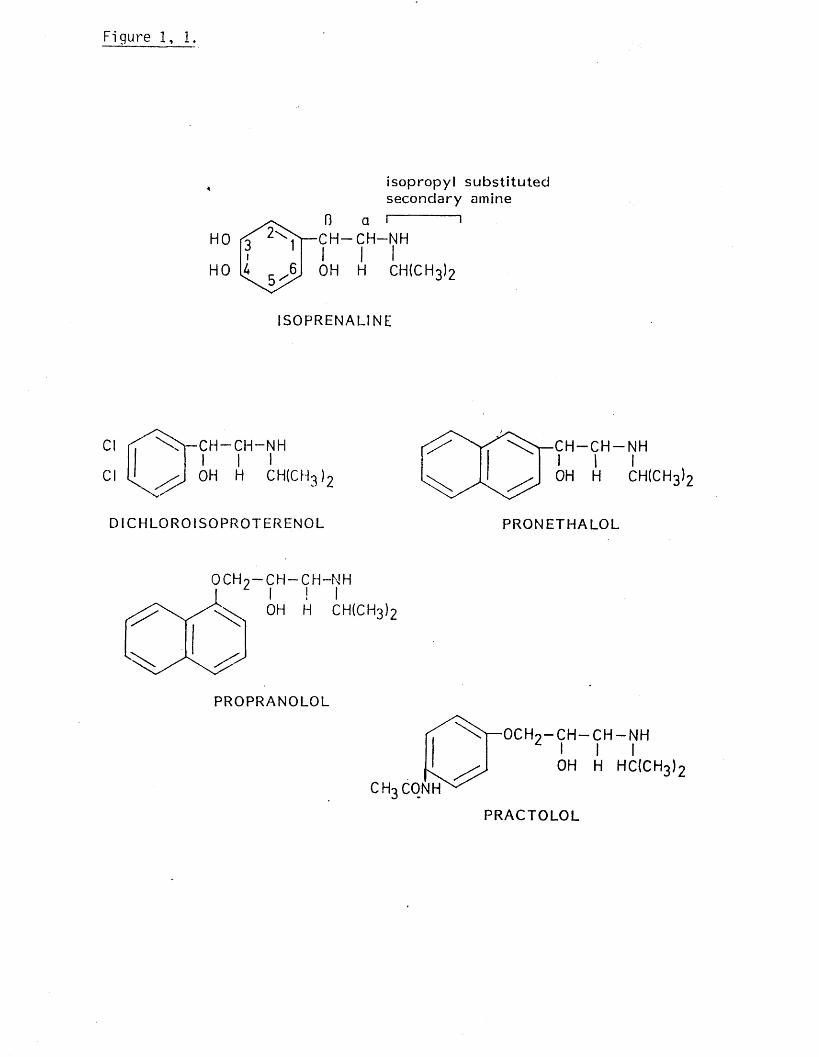
Figure 1, 1.
i s o p r o p y l s u b s t i t u t e d s e c o n d a ry amine
13 a I C H - C H - N HI I IOH H CH(CH3)2
I 5 0 P R E N A L IN E
Cl
Cl
■CH-CH-NH I I IOH H CH(CH3 )2
D IC H L O R O ISOPROTERENOL
- C H - N H1 I
OH H CH(CH3)2
P R O N E T H A L O L
OCH! I
OH H CH(CH3)2
PROPRANOLOL
c h 3 CONH
•o c h 2- c h - c h - n h I I I OH H HC(CH3)2
P R A C T O L O L

C lin ica l uses o f beta-receptor blocking drugs.
The 3-receptor blocking drugs have been used to t re a t a va r ie ty of
c l in ic a l cond it ions, ( table 1,1). but the main therapeutic use is fo r
the treatment of cardiovascular disease, p a r t ic u la r ly hypertension
where long term treatment resu lts in a gradual reduction in blood
pressure, (Prichard 1964, Prichard and Gil lam 1969). The mechan
ism by which the blood pressureis reduced is not known. Various
hypotheses have been put forward to account fo r th is hypotensive
e f fe c t including an action on the central nervous system, a block
ade of adrenergic neurones, a lte ra t io ns in renin leve ls , an action
on the barroceptors and an e f fe c t secondary to cardiac output
such as a reduction in plasma volume. There is evidence fo r and
against each o f these but probably a l l are involved to varying
degrees,(Prichard 1978).
Adverse side e ffec ts o f treatment with beta blocking drugs
Adverse side e ffec ts common to beta blocking drugs.
A ll 3-blocking drugs in use are capable o f giv ing r ise to adverse
e ffec ts (Sanders 1978) the severity o f which can be influenced by the
dose, 3-receptor s p e c i f ic i t y , presence or absence o f in t r in s ic a c t iv i t y
and physical properties such as l i p id s o lu b i l i t y (Prichard 1978,
Carruthers 1980), Side e ffec ts are reported by 40-50% of a l l patients
on 3-blocking drugs (Dany et al 1979), although reducing the dose or
changing to another drug can a l le v ia te the symptoms (Zacharias et al
1972). About 20% of patients have to be withdrawn from treatment
( Petrie et al 1976) w h i ls t with others the side e ffec ts are dose
l im i t in g or can be to lerated (Dany et al 1979, Prichard 1978,
Zacharias et al 1972). Deliberate or massive overdoses are seldom

Table 1.
Current therapeutic uses fo r beta receptor blocking drugs.
Angina pectoris
Hypertension
Cardiac arrhythmias
Thyrotoxicosis
Hypertrophic cardiomyopathy
Schizophrenia w
Anxiety state
Phaeochromocytoma
Tetralogy o f F a l lo t
Parkinson’ s disease
Dissecting a o rt ic aneurysm
V io l i n i s t ’ s bow tremor
Glaucoma
Familial benign tremor
(data modified from M il la r-C ra ig 1979 and van Joost et al 1979).
fa ta l (Dukes 1979).Practo lo l (9gm) and metoprolol (lOgm) have been
taken without causing death (Faverel-Garrigues et al 1977) although
large oral doses o f 3-blocking drugs can prove le th a l , eg oxprenolol
(4.5gm) and sota lo l (3gm) (Faverel-Garrigues e t al 1977, Montagna and
Groppi 1980).*
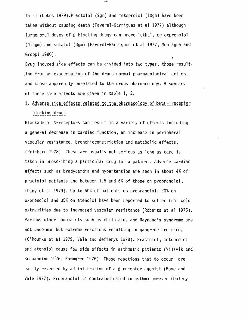
Drug induced side e ffec ts can be divided in to two types, those re s u l t
in g from an exacerbation o f the drugs normal pharmacological action
and those apparently unrelated to the drugs pharmacology. A summary
o f these side e ffec ts are given in table 1 , 2 .
1 - Adverse_side_effects_related_to_the_pharmacology_gf_beta_-_receptorikl29king_drugs
Blockade o f 3-receptors can re su lt in a va r ie ty o f e ffec ts including
a general decrease in cardiac function , an increase in peripheral
vascular resistance, bronchioconstriction and metabolic e f fe c ts ,
(Prichard 1978). These are usually not serious as long as care is
taken in prescribing a p a r t ic u la r drug fo r a pa tien t. Adverse cardiac
e ffec ts such as bradycardia and hypertension are seen in about 4% o f
practo lo l patients and between 1.5 and 6% of those on propranolol,
(Dany et al 1979). Up to 60% o f patients on propranolol, 20% on
oxprenolol and 35% on atenolol have been reported to su ffe r from cold
extrem ities due to increased vascular resistance (Roberts e t al 1976).
Various other complaints such as ch i lb la in s and Raynaud’ s syndrome are
not uncommon but extreme reactions resu lt ing in gangrene are rare,
(O’ Rourke et al 1979, Vale and Jefferys 1978). P rac to lo l, metoprolol
and atenolol cause few side e ffec ts in asthmatic patients (V i ls v ik and
Schaanning 1976, Formgren 1976). Those reactions tha t do occur are
eas ily reversed by administration o f a 3-receptor agonist (Boye and
Vale 1977). Propranolol is contraindicated in asthma however (Dolery
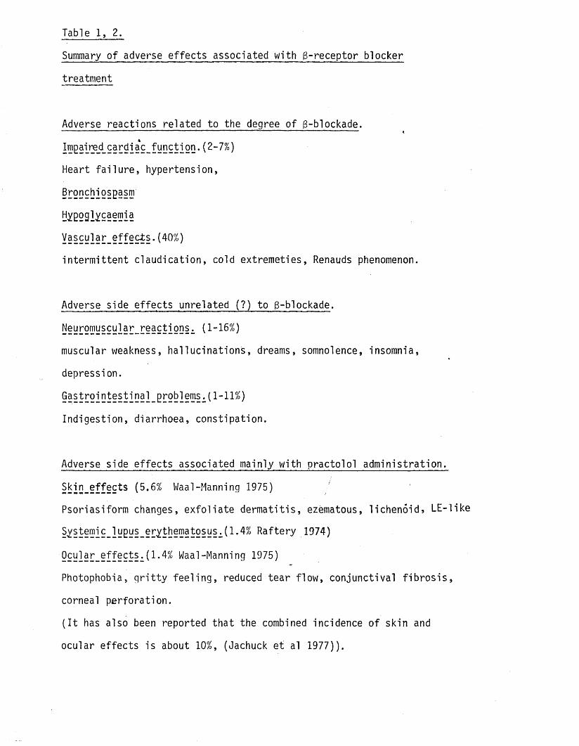
Table 1, 2.
Summary o f adverse e ffec ts associated with g-receptor blocker
treatment
Adverse reactions related to the degree o f 3-blockade.-------------- ' - T - - - ^
yo2il20 • ( 2- 7%)
Heart fa i lu re , hypertension,
Bronchiospasm
Hygoglycaemia
y§§22l2r_§ff§2*§-(40%)
in te rm it te n t c laud ica tion , cold extremeties, Renauds phenomenon.
Adverse side effec ts unrelated (?) to 3-blockade.
Neuromuscular_reactions^ (1-16%)
muscular weakness, ha lluc ina t ions , dreams, somnolence, insomnia,
depression.
Ind igestion, diarrhoea, constipation.
Adverse side e ffec ts associated mainly with practo lo l adm in is tra tion .
Skin_effects (5.6% Waal-Manning 1975)
Psoriasiform changes, ex fo l ia te de rm a tit is , ezematous, l icheno id , LE-
Raftery 1974)
922l2T_2ff§222i(1-4% Waal-Manning 1975)
Photophobia, g r i t t y fe e l in g , reduced tear f low , conjunctival f ib ro s is
corneal perforation .
( I t has also been reported tha t the combined incidence o f skin and
ocular e ffects is about 10%, (Jachuck et al 1977)).
l ik e
,
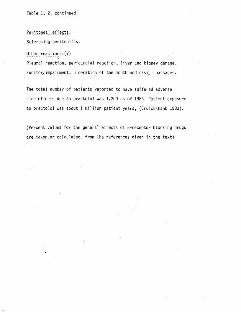
Table 1, 2. continued.
Sclerosing p e r i to n i t is .
Q^her_ react i 20§ ^ )*
Pleural reaction, pericard ia l reaction, l i v e r and kidney damage,
auditory impairment, u lceration o f the mouth and nasal passages.
The to ta l number o f patients reported to have suffered adverse
side effec ts due to practo lo l was 1,300 as o f 1983. Patient exposure
to practo lo l was about 1 m i l l io n pa tien t years, (Cruickshank 1983).
(Percent values fo r the general e ffec ts o f 3-receptor blocking drugs
are taken,or calcu lated, from the references given in the te x t )

et al 1969). Both glycogenolysis and l ip id metabolism can be d is
turbed by 3-receptor blockade. Disruption o f glycogenolysis may res
u l t in hypoglycaemia although th is occurs in frequently (Prichard
1978). Insu lin induced hypoglycaemia can be enhanced however, p a r t ic
u la r ly by those drugs with membrane s ta b i l is in g properties and l i t t l e
3-receptor s p e c i f ic i t y (Deacon et al 1977). An increase in blood
t r ig ly c e r id e s , but not glucose or cholesterol leve ls , has been rep
orted during metoprolol, a teno lo l, pindolol and propranolol therapy
(Shaw et al 1978) although other authors have not been able to con
firm th is change (Nilsson et al 1977).
2. Effects unrelated to the pharmacological action of beta-receptor_______ —_—____L ____ _ _ _ _ ______ —__ * __ — _____ _
b]ocking_drugs.
A number o f reactions,apparently unrelated to the pharmacology of
3-receptor blocking drugs,can be seen with a l l members o f the group,
w h i ls t other reactions occur ra re ly or are apparently associated with
a p a r t ic u la r drug. L ip id soluble compounds such as propran
o lo l , pindolol and oxprenolol enter the C.N.S. re la t iv e ly eas ily and
can give r ise to a number o f psychotic disturbances such as nightmares,
v iv id dreams, ha lluc inations and muscular weakness (Robinson 1978).
Isolated examples occure with most 3-receptor blocking drugs
(Berglund 1977, Ashton 1976). Estimates of the number o f patients
affected vary. For example Dany e t al (1979) compared publications
which showed tha t between 0.6 and 4.3% o f patients on propranolol had
trouble sleeping. Gastro intestina l complaints are common and in d i
gestion, diarrhoea, nausea, cramps and constipation have been assoc
iated with a l l 3-blocking drugs although treatment ra re ly needs to
be discontinued (Robinson 1978). I t is not always possible to gauge
accurately the numbers o f patients affected as the methods o f obta in
ing information can bias the resu lts (Dany et al 1979, Roberts e t al
1976). Estimates o f the percentage o f patients taking propranolol
chron ica lly who su ffe r from in te s t in a l complaints vary from 0 . 9%
to 11.2% (Dany et al 1979).
Occasional and s ingular adverse reactions have been reported fo r
a l l the drugs in use, fo r example hyperpigmentation associated with<
oxprenolol (Hcirrower and Strong 1977). In some cases a causal
re la tionsh ip has been established by challenging with the drug but
th is is not always the case and some reports re ly on the coin
cidence of drug and e f fe c t . The most serious and s ig n if ic a n t series
o f adverse e ffec ts occured as a re su lt o f practo lo l adm inistra tion.
Isolated reaction found with other 8-blocking drugs w i l l be discussed
in re la tionsh ip with the practo lo l e f fe c t as there is concern tha t
they could also give r ise to aspects o f the practo lo l syndrome (Scowen
1978, Sutherland and Wilson 1976).
Adverse side e ffec ts associated mostly with practo lo l adm in is tra tion .
Practolol was introduced in 1970 as a BiSpecific antagonist.
During i t s c l in ic a l t r i a l s no adverse e ffects were noted although
only small numbers o f patients were treated fo r short periods o f
time (George et al 1970, Sandler and Clayton 1970) Soon a f te r i t s
in troduction however Wiseman (1971) and Zacharias (1972) described
a v a r ity of skin rashes associated with i t s use and in 1973 a p ract
o lo l induced systemic lupus erythematosus-type syndrome was reported
(Raftery and Denman 1973, Assem and Banks 1973). During the next
year i t became apparent tha t a complex o f adverse reactions could be
caused by practo lo l invo lv ing the sk in , eyes and serosal surfaces.
The term "oculomucocutaneous syndrome" was suggested by Wright (1975)
to cover the var ie ty of lesions reported. Symptoms included a p so r i
asiform skin eruption (Fe lix et al 1974), u lceration o f the mouth and nasal
passages, impairment o f hearing and vis ion (Wright 1975) and a
po lyseros it is which resulted in e ith e r f ib ro s is or effus ion in to
the serous cav ity . More recently reports o f l i v e r damage (Brown et
al 1978) and damage to the lungs (Marshall e t al 1977b) and Kidneys
(Farr et al 1975) have been published. The reactions out!ined above
occured both s ing ly and in combination and are now discussed in
more d e ta i l .
A Skin_effects.
Practolol re lated e f fe c ts .
Fe lix et al ^ 1974) described 21 practo lo l patients seen over a
period o f two years with e ith e r psoriasiform (14 patients) excematous
(1 pa tien t) lichenoid (2 patients) lupus erythematosus l ik e (1 pat
ien t) eruptions or e x fo l ia t iv e derm atit is (3 pa tien ts ). The rashes
cleared on withdrawal of the drug and reappeared a f te r oral challenge.
The development and h isto logy o f the psoriasiform lesion is thought
to best ty p i fy the practo'lol reaction although h is to log ica l find ings
were most marked in those patients with the lichenoid type eruption.
The rash developed over a period o f months s ta r t in g w ith a scaling
and thickening o f the skin o f the palms and soles, associated with
p s o r ia s is - l ike plaques over the knees, elbows and other bony prom
inences. I t la te r spread to other areas o f the limbs and then to the
trunk,as an erythematous rash with scaly edges. The h is to log ica l
appearance was s im ila r in each case, varying only in degree depending
on the lesion. There was a marked epidermal atrophy and c o l lo id
bodies could be seen migrating from the epidermis to the outer horn^
layer. There was patchy swelling o f the basement membrane, which was
disrupted in some places. Deposits o f immunoglobulin (Ig)G,IgM and
complement fac to r C3 were found at the dermo-epidermal junc tion in
6 out o f 8 patients examined and antinuclear antibodies (ANA) in the
serum of 5 out o f 18 patients investigated. Only one pa tien t tested
fo r ANA had a psoriasiform rash however.
Effects associated with other beta blocking drugs.
Rashes have been associated with a l l 3-blocking drugs in use (Scowen
1978, Neumann and van Joost 1981), but i t is not certa in tha t these
represent examples o f the practo lo l induced rashes as noneof these
patients developed the cha rac te r is t ic thickening o f the palms and soles
described by Fe lix et al (1974). The h isto logy of some lesions how
ever is s im ila r to tha t found with practo lo l induced eruptions.
Damage to the epidermis resu lt ing in i t s th inn ing , epidermal oedema,
1iq u it ra c t iv e degeneration and foc i o f 1iq u it ra c t iv e damage have been
described in a va r ie ty o f rashes associated with oxprenolol (Gange and
Levene 1979, Holt and Waddington 1978) labeto lo l (F in lay and
Waddington 1978, Savage et al 1978, Gange and Wilson-Jones 1978) and
propranolol (Cochran et al 1976). More im portantly , degenerating and
necrotic ce l ls associated with c o l lo id body formation have also been
described in the epidermis o f patients on the above drugs, a f ind ing
considered to be cha rac te r is t ic o f the practo lo l induced rashes
(Fe lix et al 1974). F ib r in and fibrinogen deposits were detected at
the dermo-epidermal junction in the labeto lo l pa tien t o f Fin lay and
Waddington (1978) and Savage et al (1978) respective ly and IgM and
complement were s im i la r ly detected by Holt and Waddington (1978) in
th e i r oxprenolol treated patient*.The pa tien t o f Gange and Levene
(1979) was found to have c irc u la t in g antibodies to an epidermal
in te rc e l lu la r substance but i t s s p e c i f ic i t y was not fu r th e r de ter
mined to see i f i t resembled the pemphigus in te rc e l lu la r antibody
or tha t described by Amos et al (1975) which was strongly associated
w ith practo lo l induced ocular damage.

The h isto logy o f 3-blocking drug induced lesions seems to be d im ila r
(Holt and Waddington 1978) and the evidence suggests tha t non-pract-
o lo l drugs can occasionally give r ise to a rash with a cha ra c te r is t ic
histopathology d i f fe r in g from the appearance of the practo lo l induced
lesion only in degree.
JL _0_c_uJ_ar_ A f/_ e_c_t_s_.
Practolol re lated e f fe c ts .
In 1975 Wright described 27 patients su ffe r ing from ocular e ffec ts .
These started ty p ic a l ly with an " i tch y foreign body"sensation in
the eyes followed by a reduction in tear f low , lysozyme and secret
ory IgA content o f the tears were also reduced, which resulted in
a cha rac te r is t ic conjunctival f ib ro s is and scarring. In some patients
corneal opacities and u lceration developed. ANAs were present in
the serum of a l l patients and 25 had c irc u la t in g antibodies to
the in te rc e l lu la r region o f the epidermis. This autoantibody d if fe re d
in s p e c i f ic i ty from a s im ila r pemphigus autoantibody (Amos e t al
1975). In contrast o n ly -1 out o f 3 patients reported by Fe lix (1974)
had both ANA and eye trouble. Rahi et al (1976) described in de ta i l
the histopathology o f practo lo l induced ocular to x ic i t y in 6 pa tien ts ,
including 2 necropsy cases. They reported destruction o f the lacr im
al gland, thickening and acanthosis o f the conjunctival epithelium
and loss o f goblet c e l ls . These chronic inflammatory changes resulted
in the f ib ro s is o f the underlying stroma. The corneal epithelium
was less obviously a f f l ic te d but e p i th e l ia l and stromal u lcera tion
lead to perforation in 2 cases. Immunoglobulin was found bound to
e p ith e l ia l ce l l membranes and in the subcapsular e p i th e l ia l region
o f the lens but complement f ix a t io n could not be demonstrated in
e ith e r case.
Effects associated w ith other beta blocking drugs.
Occurence o f dry eyes have been reported with a l l 3-blocking
drugs (Scowen 1978). Dollery et al (1977) reported tha t 25% o f a
to ta l o f 483 patients seen,who had been taking various 3-blocking
drugs, complained o f a g r i t t y fe e l in g , sore or red eyes and/or photo-
phobia. Fraunfelder (1976) l is te d the side e ffects o f propranolol as
invo lv ing the eyelids and conjunctiva. Reports purporting to describe
incidences s im ila r to the practo lo l syndrome are rare. Bevis e t al
(1975) reported a pa tien t w ith propranolol induced ocular changes
and Knapp and Galloway (1975) Holt and Waddington (1975) and Clayden
(1975) have reported adverse e ffec ts a f te r administration o f oxprenolol.
The top ica l administration o f atenolol has been reported to give r ise
to conjunctiv itis and reduced tear secretions (Rebeller and Mureau
1979) and metoprolol to conjunctival oedema, hyperaemia and p e r i
ocular derm atit is (van Joost et al 1979). The s ign if icance o f any o f
these reports is unclear and i t is too soon to assess i f the changes
observed a f te r top ical treatment could re su lt in more serious damage
as th is is a re la t iv e ly new use fo r 3-blocking drugs.
In the absence o f a d e f in i t iv e report i t would seem u n like ly tha t
oral 3-blocker therapy, using drugs other than p ra c to lo l, would
re su lt in serious ocular damage.
C Sclerosing p e r i to n i t is .
Practolol related e f fe c ts .
Id iopath ic sclerosing p e r i to n i t is is a condition ra re ly seen and
Geisthovel and Kalfhaus (1965) have reviewed the l i te ra tu re from
1942 when p la s t ic p e r i to n i t is was f i r s t described by Hartmann.
Brown et al (1974) described 3 female patients with d is t in c t rad io
log ica l features showing d i la t io n confined to the small bowclbut
extending from the duodenum to the ileum. At laparotomy the v iscera l

and parie ta l peritoneum were seen to be thickened, with massive
peritoneal adhesions. The re troperitoneal space was not affected.
H is to lo g ica l ly the thickening was seen to be due to a non-specific
inflammatory response,with a layer o f compact,laminated,fibrosis
beneath the mesothelium composed o f collagen bundles. There was no
sign o f tubercu li or foreign p a rt ic le s . The histo logy d if fe re d from
id iopath ic and methysergide associated re troperitoneal f ib ro s is in
tha t the deep adipose tissue and large blood vessels were not effected.
Biopsies obtained from patients who underwent a second operation fo r
removal o f the peritoneal adhesions showed that a vigorous, granu
la t io n , tissue response had developed with a f ib r inous exudate ever-
ly ing the mesothelial surface. Later scanning electron microscopic
and h is to log ica l studies suggested that the mesothelium had in fa c t
been destroyed in patients with sclerosing p e r i to n i t is , (Myllarniemi
and Lappaniemi 1981). As the association between practo lo l and the
sclerosing condition was not d e f in i te the authors carried out a
survey,via the Committee on Safety o f Medicines, which revealed four
s im ila r cases. Other reports followed describing the ch a ra c te r is t ic
peritoneal adhesions and histology,(Meyboom. 1975, Windsor e t al
1975 and Marshall et al 1977a) Withdrawal o f the drug did not prev
ent the development of the f ib r o t i c condition which could express
i t s e l f 8 months or 12 months or even 18 months a f te r a pa tien t had
stopped taking p ra c to lo l, (Brown etal 1974, Hailey and Goodman 1975,
Allan and Cade 1975). Approximately 25% o f ind iv iduals w ith s c le r
osing p e r i to n i t is suffered from inflammation o f other serosal mem
branes , (Harty 1978). Pleural e ffus ions, jo in t e ffus ion s ,p leu r
isy and c o n s tr i t iv e p e r ic a rd i t is have also been reported to occur, in
association with the sclerosing condition (Flemming and H ick ling 1975,
MacKay and Axford 1976, Fraser and Irv ine 1976, Dyer and Vorley 1975).
Dermatitis and con junc tiv it is occured in over 50% of such pa tients .
Marshall et al (1977b) reported in de ta il on 6 patients who devel
oped resp ira tory disease a f te r surgical treatment fo r sclerosing
p e r i to n i t is . Findings included extensive f ib r o t ic pleural thickening
and lesions fn the lung parenchyma. Radiological changes always
occured during practo lo l administration but, as with the p e r i to n i t is
(Marshall et al 1977a), these could preceed c l in ic a l symptoms by as
much as 20 months (0-20 months, mean 9.3months). In some cases dyspnoea
was s t i 11 increasing several years a f te r the pleural th ickening was
diagnosed. . Laminated collagen bundles and signs of a mild in flam
matory reaction were found in biopsies o f the thickened pleural
t issue , almost iden tica l in appearance to specimens from patients
with sclerosing p e r i to n i t is .
Effects associated with other beta blocking drugs.
Marshall et al (1977a) have presented evidence tha t appears to im p l i
cate other 3-blocking drugs in the formation o f small bowel abnorm
a l i t i e s . They studied 54 patients who had been on 3-blocking drugs
fo r more than a year, using a rad io log ica l technique. 2 patients
on p ra c to lo l, 2 patients on propranolol and 3 patients on oxprenolol
had s im ila r rad io log ica l small bowel abnormalities and 1 pa tien t who
remained on propranolol fo r a fu r th e r 6 months developed abdominal
pain and nausea. Radiological investigations revealed tha t the changes
had progressed in th is pa tien t. On withdrawal o f the drug(s) the
signs regressed. In contrast Marigold e t al (1982) could f in d no ind
ications^ in a group of 32 patients taking propranolol (25) and
oxprenolol (7 ), o f s im ila r rad io log ica l changes. Oxprenolol and t im
o lo l have both been associated with sclerosing p e r i to n i t is , (Kennedy
and Ducrow 1977, Baxter-Smith et al 1978) but a causal re la t io n

ship is doubtful and Nancarrow (1978) suggested that the lesions
were more compatible with e ith e r a secondary p e r i to n i t is or an id io
pathic collagen disease. Clark and Terris (1983) however reported
tha t metoprolol caused sclerosing p e r i to n i t is in a female pa tien t
who had been taking the drug fo r 2 years. Findings on laparotomy
were consistent with those described by Brown et al (1974) fo r p ract
o lo l induced changes. Harty (1978) described a patient who devel
oped itchy eyes, a p u r i t ic scaly eruption on his legs and fee t and
asc ites, over a period o f 2 or 3 months, w h i ls t taking propranolol.
The small bowel was found to be encased in a dense fibrous tissue.
Histology o f the peritoneal biopsy showed an intense f ib ro p la s t ic
and inflammatory response tha t extended to the mesothelial l in in g
and affected the mesenteric adipose tissue. The h isto logy o f the
lesion was d i f fe re n t from tha t described fo r practo lo l as the adipose
tissue was involved. The fa c t tha t the patient had taken propranolol
fo r such a short time, in conjunction with other drugs, makes i t
un l ike ly tha t the disease was due to the 8 -blocking drug. A case o f
sclerosing p e r i to n i t is in a man who had been taking propranolol fo r
2 years was reported by Ahmed (1981) so that iso lated cases may
occur . There are 2 reports o f re troperitoneal f ib ro s is in patients
who had taken atenolol fo r 9 months (Doherty e t al 1978) and 1\ months
(Johnson and McFarland 1980). The histo logy o f drug induced (eg methy-
sergide) and id iopa th ic retoperitoneal f ib ro s is are the same (Graham
et al 1966) so tha t i t was not possible from biopsies to be sure i f
atenolol was the causative agent or not. The patient o f Johnson and
McFarland (1980) had been hypertensive fo r 5 years before an operation
to free his r ig h t ureter. A fte r th is treatment his blood pressure
returned to normal. This change led Gavin et al (1980) to suggest
tha t the hypertension had been caused by the re troperitoneal f i b
ro s is , although they did not comment on the fac t that the pa tien t
would have had to have had the f ib r o t ic condition fo r 5 years.
Timolol and metoprolol have also been associated with re troperitonea l
f ib ro s is (Rimmer et al 1983, Thomson and Julian 1983). Bullimore
(1980), using*data published by the Committee on Safety o f Medicine,
suggested tha t 8-blocking drugs predominated in causing re t ro p e r i t
oneal f ib ro s is and tha t some members o f th is series o f drugs are more
active than others in th is respect. Castle et al (1980) however
quoted a persgnal comunication reporting tha t 16 patients who had
changed from practo lo l to atenolol 6 years previously had not only
lo s t symptoms o f the oculomucocuaneous syndrome but had no signs of
a f ib r o t i c lesion.
A case o f pulmonary f ib ro s is occuring in a pa tien t who had taken
pindolol fo r 7 years was reported by Musk and Pollard (1979). H is t
ology was s im ila r to tha t described fo r practo lo l induced pulmon
ary f ib ro s is (Erwtman e t al 1977).
Sclerosing conditiqns are rare with 8“ blocking drugs other than prac t
o lo l and i t is d i f f i c u l t to ru le out the coincidence o f drug therapy
and id iopa th ic disease. The various aspects of discussions on the
incidence of drug induced f ib ros ing e ffec ts are ty p i f ie d by the
correspondence of Bullimore (1980) and Castle e t al (1980) over the pos
s i b i l i t y of atenolol induced re troperitoneal f ib ro s is .
D Systemic Lupus Erythematosus (SLE).
Practolol re lated e f fe c ts .
Raftery §nd Denman (1973) described 3 patients with a r th ra lg ia , fever
and rashes. They had raised erythrocyte sedimentation rates, lupus
erythematosus ce lls and ANA but no antibodies to native deoxyribo-
nucleic acid (DNA). Serum complement levels and gamma globulin
levels were normal and there was no evidence o f renal involvement.
A fte r withdrawal o f the drug the c l in ic a l symptoms improved but
s tero id treatment was needed before the ANA dropped and the lupus
ce l ls disapeared. The c l in ic a l appearance and laboratory f ind ings
were consisteht w ith a diagnosis o f drug induced SLE.
Effects associated with other beta blocking drugs.
Both pindolol and labeta lo l have been reported to induce a SLE
syndrome (Bensaid et al 1979, G r i f f i t h s and Richardson 1979, Brown
et al 1981). Labetalol is also associated with the presence o f a n t i -
mitochondrial antibodies (AMA). Douglas-Wilson et al reported tha t
7 out of 90 patients (8%) on labeta lo l had AMA t i t r e s o f between 1/8
and 1/126 w h ils t only 0.7% o f untreated hypertensives and 0.6% of
patients treated with other 3-blocking drugs had a pos it ive t i t r e .
The AMA usually appeared months a f te r commencing treatment w ith
labeta lo l but did not appear to be associated with any abnormality
o f the sk in , l i v e r or pericardium although the presence o f AMA was
reported to be strongly co-related with b i l ia r y c irrh os is (90%) and,
to a lesser extent, autoimmune diseases (8%).
Summary.
There is evidence tha t some reactions associated with the practo lo l
syndrome can occasionally occur w ith other 3-adrenergic receptor
blocking drugs. These can cause skin lesions, the h isto logy o f which
is s im ila r to those conditions observed in practo lo l treated pa tien ts ,
(F in lay and Waddington 1978, Gange and Levene 1979). Propranolol
and oxprenolol can induce ocular e ffec ts s im ila r to those induced
by practo lo l but d i f fe r in g in in te n s ity . Reports o f sclerosing reactions
associated with non practo lo l 3-receptor blocking drugs are rare and
causal re la tionships were not well established although 3-adrenergic
receptor blockade may be associated with an increased r is k o f re tro -
peritoneal f i& ros is (Bullimore 1980). No drug however has given r ise
to the cha rac te r is t ic pattern o f adverse reactions observed with pract
o lo l and a l l the evidence available suggests tha t i t is unique in
th is respect. Some patients have been reported to cross react with
other 3-receptor blocking drugs on trans fe r from practo lo l (Furhoff
et al 1976, Assem 1975) but these reports would not appear to be
typ ica l o f such patients ( Fe lix et al 1975, Furhoff et al 1976) and
would not appear to j u s t i f y a lte r in g the opinion expressed above.
Possible mechanisms of the practo lo l induced adverse e ffec ts .
Pharmacological mechanisms.
A. § f f§ 2 5 § -9 D > f i5 !T 2 ^ ]§ § L fy ! ]9 i l2 D i .Several authors have suggested tha t a pharmacological mechanism may
be involved in the adverse e ffec ts associated with practo lo l admin
i s t r a t io n , ( fo r a summary o f these see table 1,2). Nistrup-Madsun and
S0ndergaard (1977) suggested tha t the psoriasiform eruption seen in
such patients was the re su lt o f 3-blockade which resulted in a
d isruption of^the balance between cyc lic adenosine Si^'-monophosphate
(cAMP) and c y c l ic guanine 3;5'-monophosphate (cGMP) which 1e«£ to a
loss of control of f ib ro b la s t growth and a p ro l i fe ra t iv e response.
Human f ib rob la s ts in cu ltu re (Rao et al 1971, Berg et al 1981) and
human keratinocytes (Harper and Flaxman 1975) w i l l respond to cate
chol iwturves with an increase in cAMP content which resu lts in
m ito t ic in h ib i t io n , possibly at the G phase o f the ce l l cycle
(Harper et al 1974). Propranolol, but not p ra c to lo l, can block th is
in h ib i t io n suggesting that the receptor involved has 32s ra ther than
3X, cha rac te r is t ics , (Voorhees et al 1974). Propranolol has also been
shown to block the in h ib i t io n o f collagen synthesis induced by iso-
prenaline in acute (6h) in v i t r o experiments, (Berg et al 1981). Cells
incubated with isoprenaline fo r longer periods however (72h) became
desensitised and there was no in h ib i t io n o f collagen synthesis and no
increase in cAMP leve ls , (Saltzman 1982).
B_ Effects_on_the_skin
There is-some evidence tha t to p ic a l ly applied 3-blockers can have a
d ire c t action on apparently normal skin in v ivo . For example Gaylarde
et al (1978) demonstrated that propranolol could induce psoriasiform
changes in guinea-pig skin and van Joost et al (1979) tha t glaucoma
patients treated to p ic a l ly w ith metoprolol or atenolol developed
derm atit is . Existing or in c ip ie n t lesions can be exacerbated by
3-b locker therapy. Wiley and Weinstein (1977) demonstrated tha t in t ra -
dermal in jec t ions o f propranolol induced a s ix fo ld increase in the
p ro l i te ra t io n 'o f skin in an uninvolved area o f a psoriasis pa tien t
and van Joost and Srnitt (1981) tha t a pa tien t who developed derm
a t i t i s a f te r oral administration of propranolol had a pos it ive ep i-
cutaneous te s t to propranolol, atenolol and oxprenolol. The s ig n i f
icance o f these reports with regard to the practo lo l syndrome is not
c lear however as top ica l or intradermal challange with practo lo l fa i le d
to produce psoriasiform changes in patients who gave such a reaction
to oral adm in istra tion , (F e i l ix et al 1974, Mikkelsen et al 1975).
A number o f 3-blocking drugs can also exacerbate ex is ting lesions
a f te r oral adm in is tra tion , practo lo l (S0ndergaard e t al 1976,V1
Ridley 1.974), oxprenolol (Cumberpatch 1974) and labeta lo l (Staughton
et al 1980). Dunn et al. (1975) described a pa tien t in which practo lo l
caused expression o f anti cedent Sjogrens syndrome.
A Effects_pn_secretpry_organs^
Daily in jec t ions o f propranolol have been shown to induce a decrease
in weight,and ce ll s iz e ,o f ra t submaxillary glands, in some cases with
the formation o f atrophic f igu res , (Fukuda 1968). S im ilar changes
were also reported to occur a f te r oral adm in is tra tion , (Smith and
Butler 1978). These authors also reported that there was a loss
o f secretions from the parotid and submaxillary glands.and i n f i l
t ra t io n o f lymphocytes around the duct and parenchyma with resu ltan t
destruction o f the e p ith e l ia l c e l ls . Some animals also developed
granulomas on the serosal surface o f the colon and duodenum and
changes in the in te s t in a l serosa. These changes were only observed
a f te r s ix to eighteen months so tha t propranolol would have to be
taken fo r a long period by humans before any s im ila r e f fe c t would
be expected. Tanaka et al (1983) have shown tha t practo lo l can in
duce a reduction in tear flow in beagles given the drug o ra l ly .%
Lymphocyte i n f i l t r a t i o n in to the lacrymal gland was also observed.
As only two animals per dose were used however these find ings need
to be confirmed. Practolol accumulates in the lacrymal gland (Scales
and Cosgrove 1970) so that i t is possible tha t damage to th is gland
observed in humans (Rahi et al 1976) could have been due to a
d ire c t e f fe c t on the 3-receptors. Recently Okine e ta l (1982) have
reported tha t oral administration o f p ra c to lo l, but not propranolol,
acebutalol, atenolol or pronetha lo l, inh ib ited the synthesis o f
gas tro in tes tina l mucosal glycoprotein synthesis in the ra t . I t was
suggested by the authors tha t an impaired formation o f mucus could,
in tu rn , re su lt in in te s t in a l adhesion and dry eyes. This f in d in g ,
together with tha t o f Tanaka et al (1983), would suggest tha t p ra t-
o lo l is capable, in some circumstances, o f inducing changes which,
i f they occured in humans, could re su lt in the adverse e ffec ts found.
However the lack o f any adverse e ffec ts in animals, including the
ra t and beagle, fed practo lo l chron ica lly makes the in te rp re ta t io n
o f the above reports d i f f i c u l t (Scales and Cosgrove 1970).
P_ §ffe c ts_ o n _ ]eukocyte_]ysosomal_enzYme_release^
Brown et al (1974) suggested that the sclerosing p e r i to n i t is could
be due to a stim ulation o f leucocyte lysosomal enzyme release which
could, if i tu rn , cause tissue damage and in i t i a t e an inflammatory
reaction. Release o f 3-glucuronidase (GUR) from human leukocytes
is enhanced by phagocytosis o f immune complexes (L ichtenstein and
Margolis 1968, Weissmann et al 1971a,Ignarro e t al 1974), cGMP
and agents causing an increase in cGMP (Zurie r e t al 1974). Enzyme
release is depressed and the above increase blocked by cAMP and chem
ic a ls , such as 8-agonists, which stimulate cAMP formation, (Zurie r
et al 1974, Weissmann et al 1971a). Propranolol reduces the blocking
action o f cAM and 8-agonists but has no d ire c t e f fe c t i t s e l f ,
(Zurie r et al 1974). Human lymphocytes and f ib rob la s ts are thought
to have 82 ra ther than 81 receptors, (Conolly and Greenacre 1977,
Voorhees et al 1974). There appears to be no paper published des
cr ib ing the 87receptor subclass of human macrophages but ra t macro
phages have been reported to posses 82 receptors, (Schenkelaars and
Bonta 1984). Using carrageenin e l ic i te d ce lls they showed tha t the
s tim ulation o f GUR secretion by leukotriene could be blocked by
isoprenaline and tha t th is block could be reversed by propranolol
but not by p rac to lo l. Neither practo lo l nor propranolol had any
d ire c t e f fe c t on GUR release.
I t seems u n like ly therefore that practo lo l would have any e f fe c t ,
d ire c t or in d ire c t , on lysosomal enzyme release. This view is sup
ported by the find ing that practo lo l can stimulate cAMP formation by
human lymphocytes in v i t r o , (Lima and Turner 1981). I t appears th a t,
at least in the above model, practo lo l is a p a r t ia l agonist and
would be expected, i f anything, to depress GUR release via the
increase in cAMP synthesis.
E Effects on the immune system.
In 1971 A llison et al proposed tha t B -ce lls , capable o f synthesising
autoanttbodies, were under the control o f a suppressor T -ce ll pop
u la tion. This concept le ^ Raftery and Denman (1973) to
suggest tha t 3-blockade could d i f f e r e n t ia l l y in te r fe re with th is group

o f regulatory lymphocytes allowing expression o f autoantibodies
and development o f a lupus type syndrome. Patients with active SLE
have an impaired antigen spec if ic T-suppressor ce ll a c t iv i t y ,
(Gladman et al 1980). Further, anti DNA antibody production by B-cells
from SLE patients in v i t ro can be prevented by allogeneic normal,c
but not SLE, *T-cells, (Clough et al 1980). S im ilar extensive lymph
ocyte function tests have not been carried out using ce l ls from
practo lo l patients so that i t is not possible to say i f T -ce ll are
p re fe re n t ia l ly suppressed in adversely affected patients. There are
data available which show tha t adrenergic agents can modulate
lymphocyte metabolism, p ro l i fe ra t io n and function , (Strom et al
1972, Hadden et al 1972) and tha t these changes could be blocked by
B2-receptor blocking drugs, (Conolly and Greenacre 1977). Propranolol has
been shown to enhance IgE formation in the rabb it (Homer and Cain
1979), t o ta l , but not antigen sp e c if ic , IgE formation in the ra t ,
(Pauwels et al 1980) and antibody formation to heterogenous prote in
in the ra t (Benner et a.l 1968) and mouse (Nakazawa et al 1976).
Buckley and McGregor (1977) also reported tha t propranolol increased
the number of survivors and decreased the duration of disease in ethanol
suppressed rats infected with type I I I pneumococci. As these e ffec ts
are probably mediated via 32 receptors (Conolly and Greenacre 1977) i t
is un like ly tha t practo lo l would have a s im ila r e f fe c t unless, l ik e
the stim ula tion of growth in psoriasis pa tien ts , there was a pre
disposing fac to r. Bjorkholm et al (1980) suggested tha t ischemic heart
disease could be associated with an immunological disrangement.
This concept received support from the work o f Takeichi et al (1980).
These workers reported tha t spontaneously hypertensive rats have a
reduced number o f thymocytes at b i r th and a depressed T -ce ll

dependent immune response, a condition which worsens with age.
I t is not certa in as yet i f the immune changes and the hypertension
are fu n c t io n a lly re lated.
A var ie ty o f autoantibodies have been found in the sera o f practo lo l
patients. I t is possible therefore that these patients had a suppressor
ce l l defect, feehan et al (1976) found antibodies to the nucleus,
thyro id , thyrog lobu lin , smooth muscle, sa liva ry duct, immunoglobulin,
gas tr ic parie ta l c e l ls , an in te rc e l lu la r substance and basement
membranes, mostly in patients who had been taking the drug fo r
more than one^year or who had adverse e ffec ts . In te rc e l lu la r a n t i
bodies and ANA were commonly associated with ocular damage, (Amos
et al 1975). Rahi et al (1976) detected immunoglobulin deposits
between the conjunctival epithelium and in the lens and Fe lix et a l (1974)
reported the presence o f IgG,IgM and complement at the dermo-epid-
ermal junction in some patients su ffe ring from skin rashes. S im ilar
deposits in the dermo-epidermal junction o f skin from patients with
practolo l-associated psoriasiform changes were detected by Dahl et al
(1975). Whilst there appears to be a re la tionsh ip between practo lo l
and the presence of autoantibodies there is no c lear re la t ionsh ip
between the presence o f autoantibodies and the d if fe ren t side e f
fects . A summary o f the various findingsare given in table 1,3.
There is l i t t l e evidence to suggest tha t e ith e r the immunoglobulin
deposits or autoantibodies were cyto tox ic . Rahi et al (1976) came to
the conclusion tha t the deposits in the lens and e p ith e l ia l t issue
were formed as a resu lt o f tissue damage rather than being responsible
fo r the damage. Amos e t al (1975) suggested tha t the in te rc e l lu la r
antibody could be binding non-spec if ica lly and tha t there was in
s u f f ic ie n t evidence to ascribe cytotoxic properties to i t .
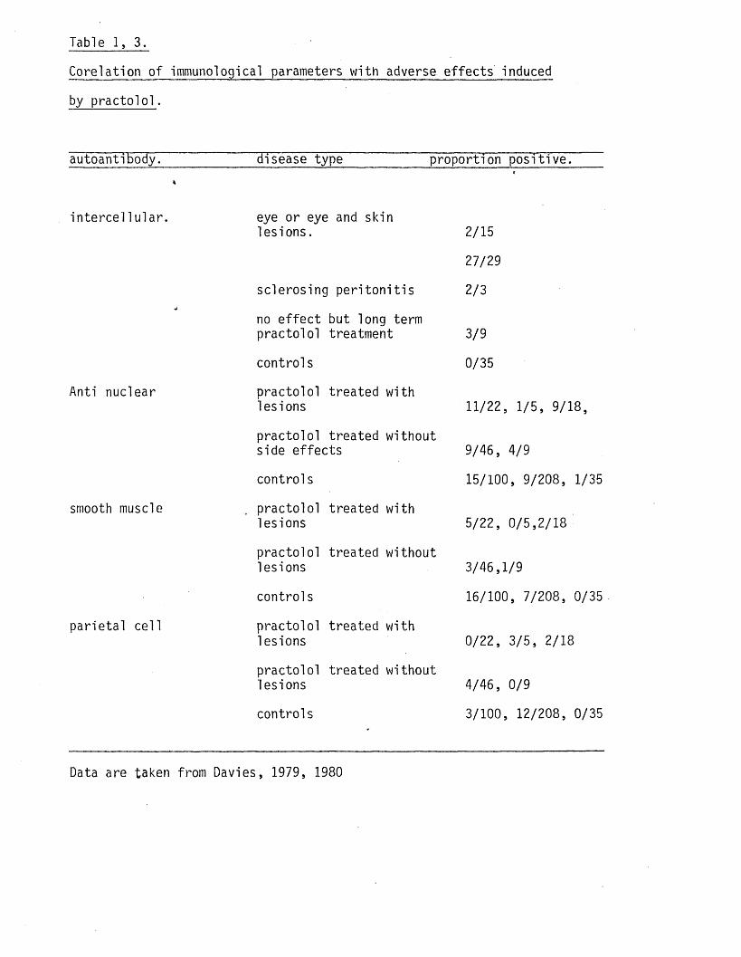
Table 1, 3.
Corelation o f immunological parameters w ith adverse e ffec ts induced
by p rac to lo l.
autoantibody. disease type proportion po s it ive .4
in te rce l 1u la r. eye or eye and skin les ions .
<
2/15
27/29
sclerosing p e r i to n i t is 2/3*
no e f fe c t but long term practo lo l treatment 3/9
controls 0/35
Anti nuclear practo lo l treated with lesions 11/22, 1/5, 9/18,
practo lo l treated without side effec ts 9/46, 4/9
controls 15/100, 9/208, 1/35
smooth muscle practolo l treated with lesions 5/22, 0/5,2/18
practo lo l treated without lesions 3/46,1/9
controls 16/100, 7/208, 0/35
parie ta l ce ll practo lo l treated with lesions 0/22, 3/5, 2/18
practo lo l treated without lesions 4/46, 0/9
controls 3/100, 12/208, 0/35
Data are taken from Davies, 1979, 1980

S im ila r ly the high incidence o f ANA found in some patients would
appear to be secondary to tissue damage and not causally re lated
(Wilson et al 1978, Raftery et al 1973).
Behan et al (1976) tested d i f fe re n t aspects o f the immune system in
practo lo l patients to see i f there was any evidence of a dysfunction.
Those patients who had adverse e ffects and those who had been taking
practo lo l fo r long periods (greater than 12 months) showed a s im ila r
pattern o f responses which d if fe red from the control group and from
patients who had taken practo lo l fo r short periods. The former
group of patients showed a decreased skin response to Candida and
streptok inase/stre ’ptodornase, decreased lymphocyte protein synthesis
and a depressed lymphocyte transformation response to mitogenic stim
u la t ion , s im ila r in some respects to the find ings o f Takeichi et al
(1980) using hypertensive ra ts . Amos and Brigden (1976) could not
confirm the impaired transformation response however and suggested
that th is was due to differences in the control patients used by the
two groups. Pugh et al .(1976) followed the lymphocyte a c t iv i t y in
practolo l patients over a course o f treatment and concluded tha t ab
normalities in lymphocyte response to mitogens did not precede the
formation o f autoantibodies but did re f le c t disease a c t iv i t y in
general. I t appears l i k e ly therefore tha t practo lo l has no d ire c t
e f fe c t on the immune system.
Summary.
There is s u f f ic ie n t data available to show tha t propranolol can
a f fe c t ce l ls o f the sk in , sa liva ry glands and the immune system.
P racto lo l, in most systems where i t has been tested, is w ithout
a c t iv i t y as would be expected as most ce l ls appear to posses 32
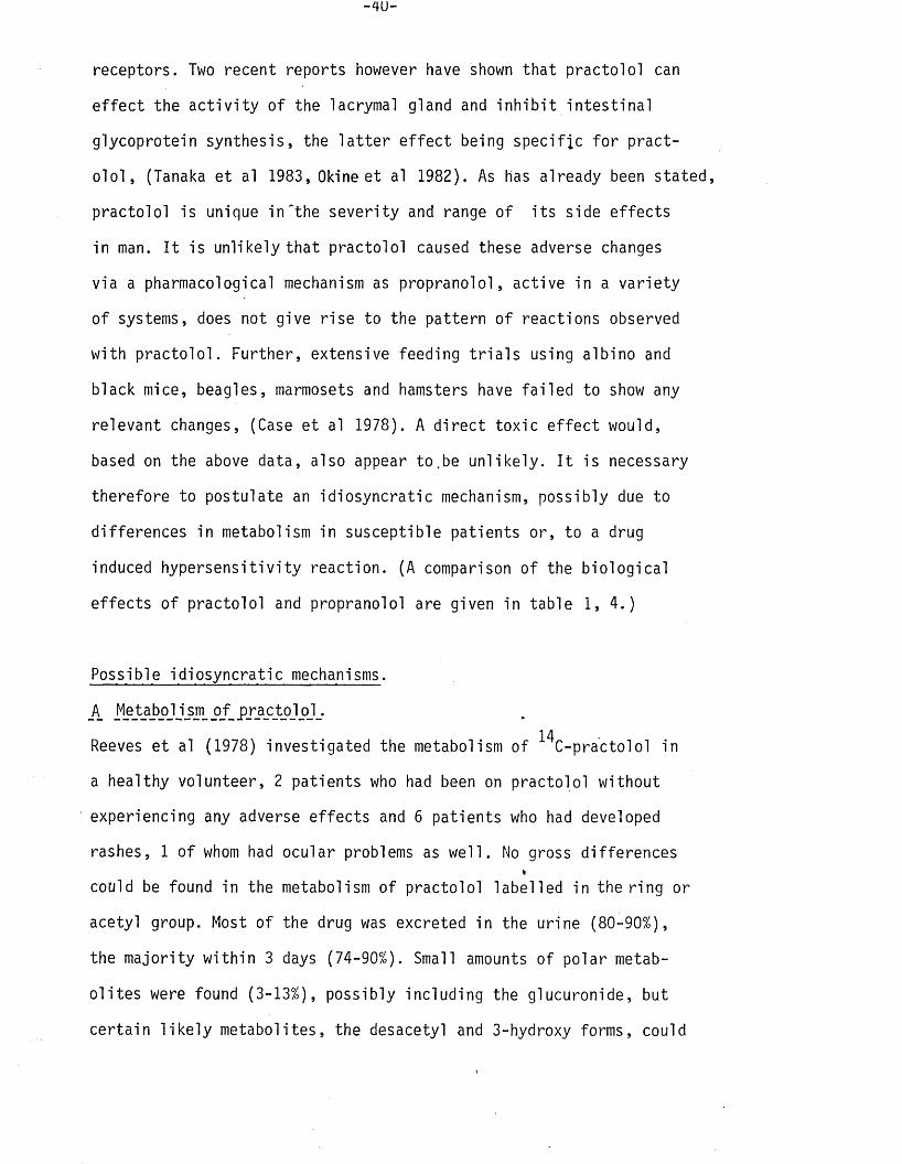
receptors. Two recent reports however have shown that practo lo l can
e f fe c t the a c t iv i t y o f the lacrymal gland and in h ib i t in te s t in a l
glycoprotein synthesis, the la t te r e f fe c t being spec if ic fo r p ract
o lo l , (Tanaka et al 1983, Okine et al 1982). As has already been stated,
practo lo l is unique in 'th e severity and range o f i t s side e ffects
in man. I t is unlikely tha t practo lo l caused these adverse changes
via a pharmacological mechanism as propranolol, active in a va r ie ty
o f systems, does not give r ise to the pattern o f reactions observed
with p rac to lo l. Further, extensive feeding t r ia ls using albino and
black mice, beagles, marmosets and hamsters have fa i le d to show any
relevant changes, (Case et al 1978). A d ire c t tox ic e f fe c t would,
based on the above data, also appear to.be un like ly . I t is necessary
therefore to postulate an id iosyncra t ic mechanism, possibly due to
differences in metabolism in susceptible patients or, to a drug
induced hypersens it iv ity reaction. (A comparison o f the b io log ica l
e ffec ts o f practo lo l and propranolol are given in table 1, 4 .)
Possible id iosyncra t ic mechanisms.
A AeAaA°Jj-SOLp_tPJActoJoJ.!4
Reeves et al (1978) investigated the metabolism of C-practolol in
a healthy volunteer, 2 patients who had been on practo lo l without
experiencing any adverse e ffec ts and 6 patients who had developed
rashes, 1 o f whom had ocular problems as w e ll. No gross differences%
cotild be found in the metabolism of practo lo l labelled in the r ing or
acetyl group. Most o f the drug was excreted in the urine (80-90%),
the m a jority w ith in 3 days (74-90%). Small amounts o f polar metab
o l i te s were found (3-13%), possibly including the glucuronide, but
certa in l i k e ly metabolites, the desacetyl and 3-hydroxy forms, could
-41-
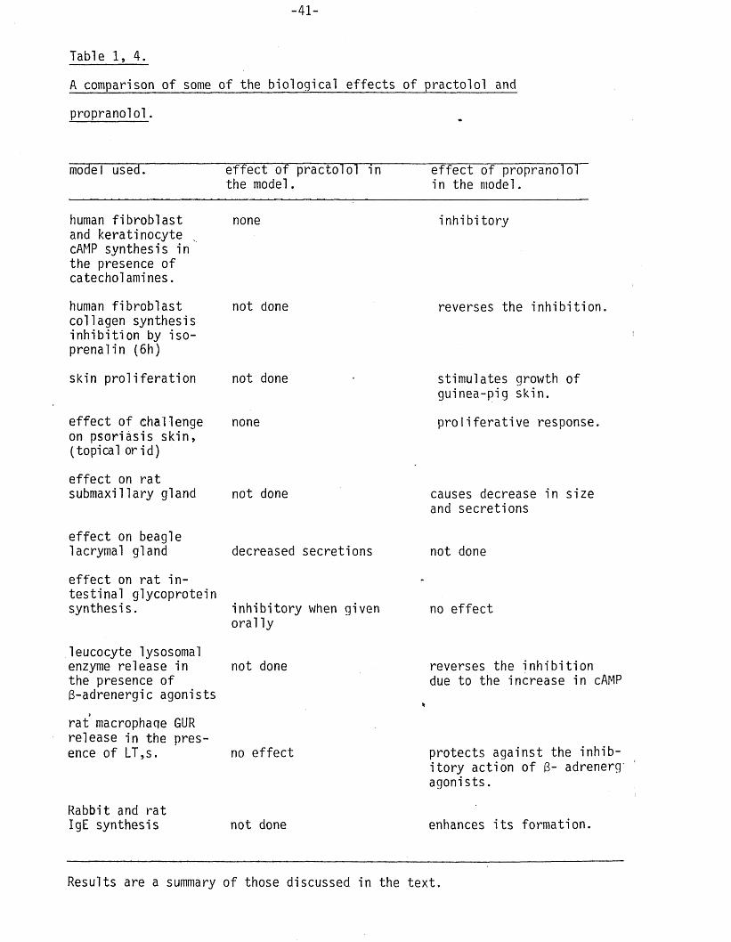
Table 1, 4.
A comparison o f some o f the b io log ica l e ffec ts o f practo lo l and
propranolol.
e f fe c t o f practo lo l in the model.
model used. e f fe c t o f propranolol in the model.
human f ib ro b la s t noneand keratinocyte cAMP synthesis in the presence o f catecholamines.
human f ib ro b la s t not donecollagen synthesis in h ib i t io n by iso- prenalin (6h)
skin p ro l i te ra t io n not done
e ffe c t o f challenge none on psoriasis sk in ,(topical or id )
in h ib i to ry
e ffe c t on ra t submaxillary gland
e ffe c t on beagle lacrymal gland
e ffe c t on ra t in te s t in a l glycoprotein synthesis.
leucocyte lysosomal enzyme release in the presence o f 3-adrenergic agonists
not done
decreased secretions
in h ib i to ry when given o ra l ly
not done
ra t macrophaqe GUR release in the presence o f LT,s. no e ffe c t
Rabbit and ra tIgE synthesis not done
reverses the in h ib i t io n .
stimulates growth o f guinea-pig skin.
p ro l i fe ra t iv e response.
causes decrease in size and secretions
not done
no e f fe c t
reverses the in h ib i t io n due to the increase in cAMP
protects against the in h ib i to r y action o f 3- adrenerg' agonists.
enhances i t s formation.
Results are a summary o f those discussed in the te x t .
not be id e n t i f ie d . Deacetylation did occur and accounted fo r
approximately 5% o f the metabolism of p ra c to lo l , s im ila r to that
found in the ra t and beagle. The metabolic fa te o f practo lo l in
various species is outlined in f i g . 1. 2.
Plasma levels o f practo lo l were o f the order o f lug/ml a f te r 3h and
decayed mono-exponentially with a h a lf l i f e o f 12-16h. Saliva levels
varied from 0.45-2.0ug/ml and decreased at the same rate as serum
leve ls . The authors concluded that i f the mechanism leading to the
adverse reactions involved differences in metabolies o f k ine tics
then these must be re la t iv e ly small and not read ily demonstrable
Individual phase I or phase I I metabolites were not id e n t i f ie d
however in the plasma or urine so tha t there could have been
differences which were not detected.
Reeves et al (1979) investigated the metabolism o f practo lo l in the
marmoset, hamster, ra t , mouse, guinea-pig and rabb it. The marmoset
showed extensive deacetylation (57%) and the hamster appeared to p re f
e re n t ia l ly synthesise the 3-hydroxy metabolite (11%) ora conjugate
o f 3-hydroxy p rac to lo l. The other species metabolised the drug s im i la r ly
to man. I t is u n l ike ly tha t gross differences in the metabolism of
p ra c to lo l , as re flec ted by analysis of ur ine, faeces and expired a i r ,
can be used to pred ic t s u s c e p t ib i l i ty to practo lo l as these animals
exhibited none of the signs associated with the practo lo l syndrome
during chronic feeding studies (Case et al 1978). Scales and Cosgrove (1970)%
reported that enterohepatic c irc u la t io n played an important part in
the metabolism o f practo lo l in the dog, ra t and mouse and Reeves e t al
(1979) tha t desacetyl practolo l could become reacetylated to p racto lo l
in the rabb it. There are mechanisms therefore which would allow
extensive metabolism of the drug, which may re su lt in s ig n if ic a n t
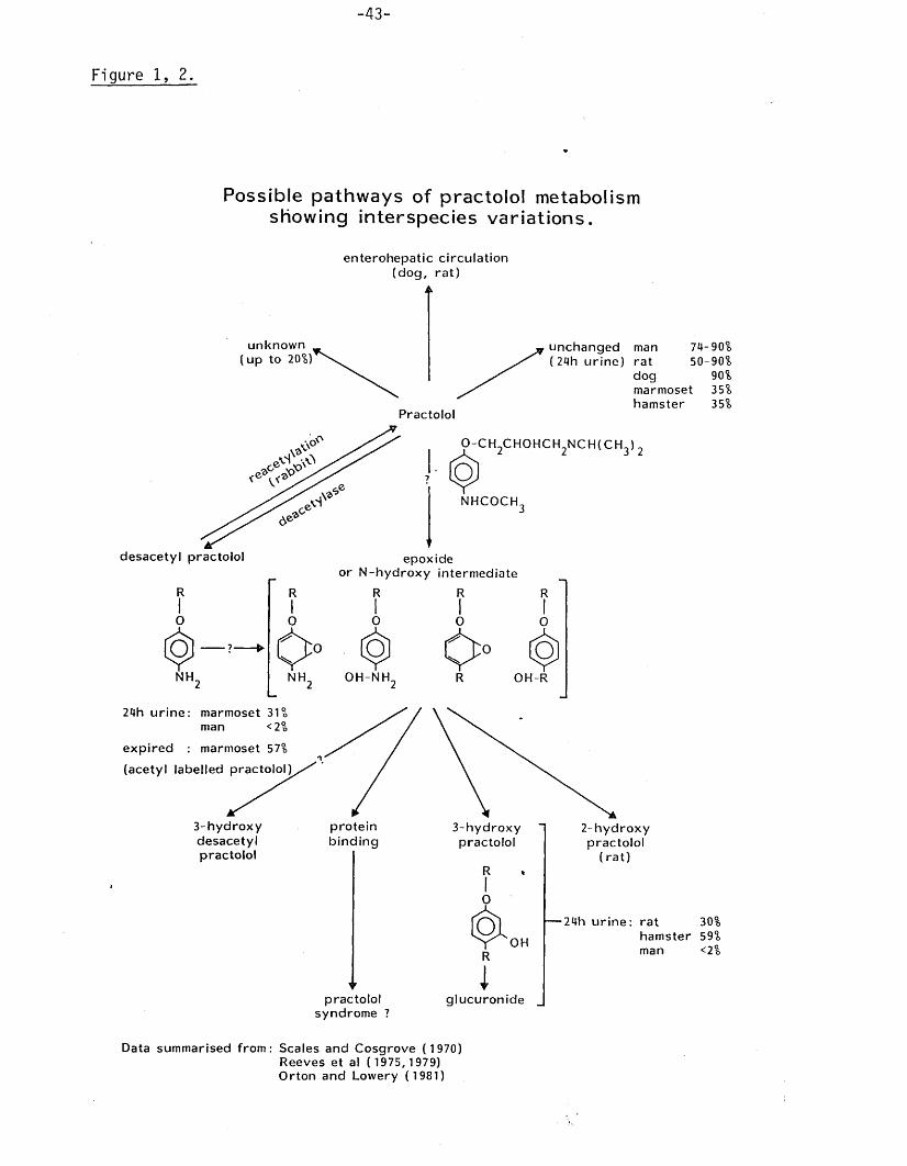
Figure 1, 2.
Possible pathways of practolol metabolism showing interspecies variations.
enterohepatic circulation (dog, ra t)
unknown (up to 20%)’
unchanged man ( 24h urine) rat
dog
Practolol
marmosethamster
74-90 50-90
90 35 35
o - c h 2c h o h c h 2n c h ( c h 3) 2
desacetyl practolol
RIo
¥NH.
7 £ ) ,NHCOCH,
epoxide or N -hydroxy intermediate
R R
O
NH,
RIo
OH-NH.
O O° <$]R OH-R
24h urine: marmoset 31 man < 2
expired : marmoset 57%
(acetyl labelled practolol)
3-hydroxydesacetylpractolol
proteinbinding
▼practolol
syndrome ?
3-hydroxypractolol
2-hydroxypractolol
(ra t )
30% hamster 59%
< 2 %
24h urine: rat
OH manR
glucuronide
Data summarised from: Scales and Cosgrove ( 1970)Reeves et al ( 1975, 1979) Orton and Lowery (1981)
0\O
0\O
0\O
Q\0
Q\0

-44-
differences in humans, which would not be detected during routine
ana ly tica l procedures.
A va r ie ty o f animal species have been used in to x ic i t y ^ t r ia ls and
some have been shown to metabolise practo lo l extensively. In no
case however has an untoward e f fe c t , re lated to the practo lo l syn
drome, been reDorted. Drug metabolism in practo lo l patients with
and without side e ffec ts and a control was very s im ila r (Reeves et
al 1978) and th is information, together w ith the animal data, sug
gests tha t practo lo l does not give r ise to p a r t ic u la r metabolites
which are responsible fo r the adverse reactions.
B_ Drug_ hypersen si t i v j ty_ re ac ti oru
The a lte rna tive to an ind iosyncra tic response due to differences in
drug metabolism is a drug induced hypersens it iv ity reaction. There
are certa in c h a ra c te r is t ic s ,o f a drug induced a l le rg ic reaction,which
suggest such a diagnosis. A fte r the primary exposure a la te n t period
is required before a reaction is seen. This is not the case a f te r sub
sequent ones. The reaction occurs in a m inority o f patients and is
not l in e a r ly re lated to dosage or to pharmacological properties of
the chemical (F i l ip e k 1979). These requirements are typ ica l o f ther
practo lo l induced e ffec ts . Such reactions are usually o f four main
types (Gel! and Coombs 1968), Type I , anaphylactic, IgE mediated;
Type I I , cy to tox ic , IgG mediated; type I I I Arthus and type IV, ce l l
mediated, delayed. The practo lo l reaction does not seem to f i t in to
any o f these categories (Amos et al 1978). Neverthe\eS5 some signs
o f immunological a c t iv i t y were seen in oracto lo l pa tien ts , p a r t icu
la r ly the lupus syndrome described by Fe lix et al (1974) and the auto
antibodies reported by Amos et al (1975) and Behan et al (1976). In
order to d e f in i te ly show tha t practolo l can cause a hyp erse ns it iv i ty
reaction i t is necessary to demonstrate a spec if ic in te rac tion between

the drug, or a metabolite, and e ith e r sensitised lymphocytes or drug
spec if ic antibodies. Assem (1975) reported tha t 2 out o f 3
practo lo l patients and 1 o f 2 propranolol patients had.a pos it ive
lymphocyte transformation te s t to lug/ml p ra c to lo l ; one o f the pract
o lo l patients also giv ing a pos it ive response to lug/ml propranolol.
A number o f authors have t r ie d to repeat these experiments using
p ra c to lo l, practolo l/a lbumin complexes, desacetyl practo lo l linked
to sheep red blood ce lls and two possible metabolites o f practo lo l
but without success, (Behan et al 1976, Amos and Brigden 1976, Amos
1979). Raftery and Denman (1973) were equally unsuccessful in detecting
antibody receptors to practo lo l on lymphocytes obtained from pract
o lo l patients. I t is not altogether suprising tha t immunological
investigations using the parent molecule should be negative. In order
to provoke an immunological response a chemical should have a molec
u la r weight o f at least 6000 or, i f not, be f i rm ly bound to some
macromolecule forming a hapten/carrier complex, (Landsteiner and
Jacobs 1936). Most drugs have a molecular weight below 6000, fo r ex
ample practo lo l (264) and p e n ic i l l in BT (346) and must therefore form
such high molecular weight complexes during t h e i r rmetabolism in order
to provoke an immune response. Drug antibodies have been reported fo r
hydralazine (Heine and Friedman 1962) and procainamide (Russell and
Z i f f 1968) but a survey by Nakatsu and Scully (1980), using a double
antibody technique, fa i le d to reveal IgG antibodies to acetyl s a l ic y l ic%
acid, erythromycin, l idnocain , te t ra c y c l in , p e n ic i l l in , a m p ic i l l in
or procainamide. This lack o f success could have been due to the fa c t
that they did not look fo r IgM antibodies, the most common class o f
drug antibody (Whittingham and MacKay 1976) or to the fa c t tha t hapten/
ca r r ie r complexes were e ithe r not formed or the hapten was not bound
-46-
at a concentration s u f f ic ie n t to provoke an immune response. I t was
o r ig in a l ly thought tha t the hapten/protein bond had to be covalent but
Plescia and Palczik (1964) and Boyd and Pert (1968) have published
data suggesting tha t strong associates are s u f f ic ie n t fo r the production
of antibodies to small b io log ica l molecules although i t is not c lear
i f th is concept also applies to drugs. The presence and s p e c i f ic i t y
o f drug antibodies are usually investigated using model compounds.
This is not a problem i f the parent molecule is the antigen but can
pose a problem i f the active species is a metabolite as the metabolism
of the drug may not have been studied, the active form may be present
in small amounts,or be unstable, and therefore not detected,or model
compound may not be ava ilab le . In order to overcome these d i f f i c u l t i e s
with p ra c to lo l ,Amos et al (1977, 1978) used an ingenious system to
generate metabolites o f practo lo l in v i t r o and then trap them using a
scavenger macromolecule. Practolol metabolites were generated by in c
ubating the drug in v i t r o with a microsomal mixed functional oxidase
system iso lated from ra t or hamster l iv e rs . In one set o f experiments
the metabolites produced were allowed to couple to non-agglutinating
rabb it antibodies to human 0 red blood ce l ls which were then used top*
passively sensitise human 0 red blood c e l ls . This reagent was then
titrated against practo lo l patients serum in an agglu tination assay.
(Amos e t al 1977). In a second series o f experiments practo lo l metab-125o l i te s were allowed to couple to I-human serum albumin (HSA) which
*was* then used in a modified Farr assay, (Amos et al 1978). This idea
was based on the work o f Orton and Lowery (1981) who showed tha t
radio!abelled metabolites of p ra c to lo l, generated in v i t r o , would bind
to microsomal protein and tha t th is binding could be blocked by HSA
which scavenged the metabolites. Amos e t al (1977) demonstrated tha t
antimetabolite antibodies o f the IgM class were present in the sera
o f practo lo l patients with severe adverse side e ffec ts but not
control sera. They then went on to further characterise the s p e c i f ic i ty
o f the antibody and to asses i t s s ign if icance , (Amos e t .a l 1977, Amos
1979). The presence o f the antimetabolite antibody could not be r e l
ated to any p a r t ic u la r manifestation o f the oculomucocultaneous syndrome
but there did appear to be an inverse re la tionsh ip between the antimetab
o l i t e antibody and the in te rc e l lu la r antibody and the sclerosing cond
i t io n . They also showed tha t i f susceptible patients were challenged
with practo lo l the level o f the antimetabolite antibody rose rap id ly
to levels seen at the h ight o f the adverse reaction, behaviour typ ica l
o f a secondary a l le rg ic reaction, (Amos et al 1978). The authors sug
gested tha t the antibody was produced early in the development o f the
adverse reaction and th a t, as the damage became more severe, i t bound
to antigenic determinants on the tissue, ie tissue bound metabolite.
This would also explain the low antimetabolite t i t r e in patients with
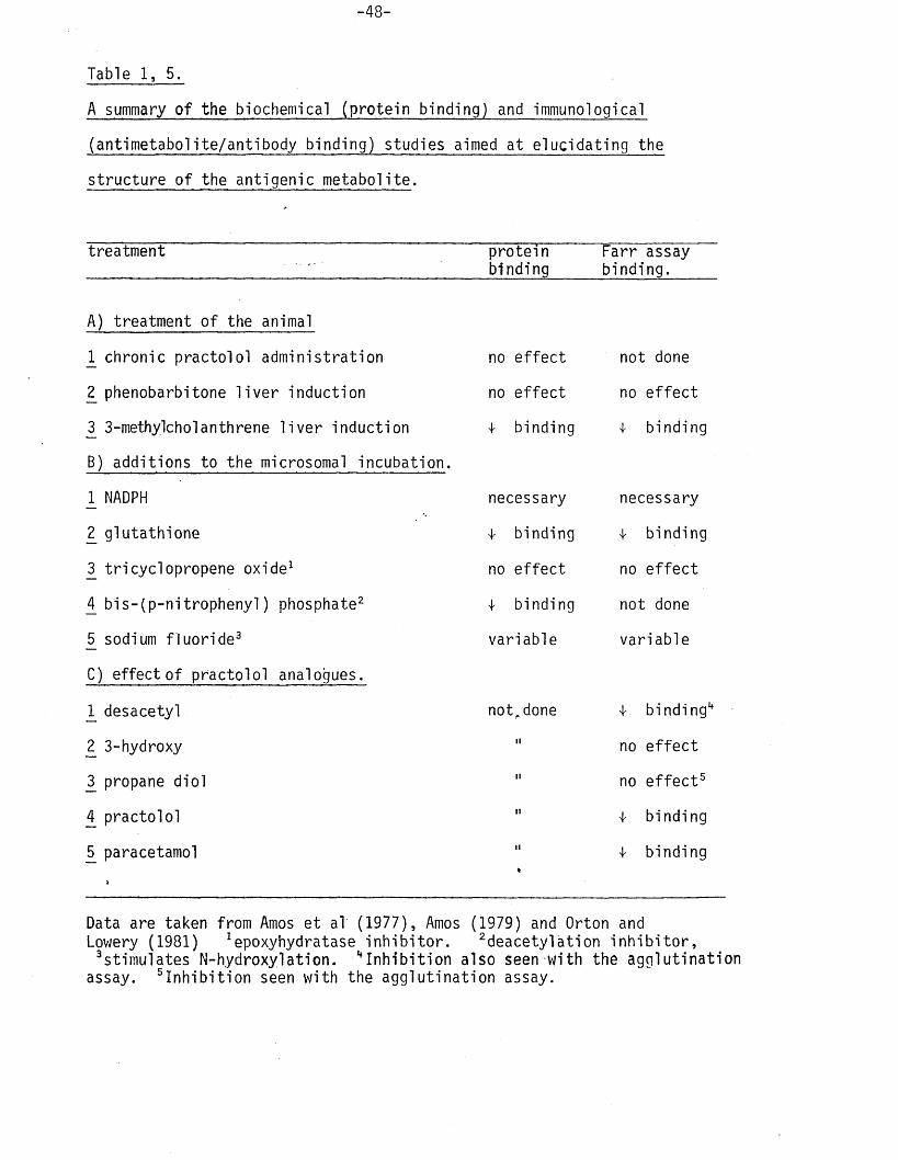
autoantibodies. The resu lts reported by Amos et al (1977), Amos (1979)
and Orton and Lov/ery (1981) are summarised in table 1,5.
Although the s ign if icance o f ' th e above f ind ings , w ith regard to the
practolo l syndrome, are not c lea r, i t is apparent ^hat practo lo l could,
in v ivo , give r ise to metabolites which bound to macromolecules to
form haptens. This binding could have induced a novel type o f hyper
s e n s i t iv i t y reaction as suggested by Amos et al (1978), or d i re c t ly
caused tissue damage. In e ith e r case i t is conceivable tha t a bu ild up%
of the tissue bound metabolite would be necessary to invoke e ith e r
an immune response or overt tissue damage. Lindup et al (1979) have
reported a' time and concentration dependent accumulation o f radio
a c t iv i t y in the eye, skin and small in te s t in e o f male hamsters fed rad io-
labelled p ra c to lo l, ie tissues most associated with the adverse e ffec ts
-48-
Table 1, 5.
A summary o f the biochemical (prote in binding) and immunological
(antimetabolite/antibody binding) studies aimed at e luc idating the
s tructure o f the antigenic metabolite.
treatment proteinbinding
Farr assay binding.
A) treatment o f the animal
1 chronic practo lo l administration no e ffe c t not done
2 phenobarbitone l i v e r induction no e ffe c t no e f fe c t
3 3-methylcholanthrene l iv e r induction 4- binding 4 binding
B) additions to the microsomal incubation.
1 NADPH necessary necessary
2 glutath ione 4- binding 4 binding
3 tricyclopropene oxide1 no e ffe c t no e f fe c t
4 b is -(p -n itro p h e n y l) phosphate2 4 binding not done
5 sodium f lu o r id e 3 variable variable
C) effect o f practo lo l analogues.
1 desacetyl notr done 4 binding4
2 3-hydroxy n no e f fe c t
3 propane d io l H no e f fe c t5
4 practolo l n 4 binding
5 paracetamol n 4 binding
>*
Data are taken from Amos et al (1977), Amos (1979) and Orton and Lowery (1981) ! epoxyhydratase in h ib i to r . 2deacetylation in h ib i to r ,
S tim u la tes N-hydroxylation. ^ In h ib i t io n also seen with the agg lu tina tion assay, i n h ib i t i o n seen with the agg lu tination assay.

in man. Most, 80%, o f the a c t iv i t y was due to practo lo l i t s e l f w h i ls t
the remaining 20% was not characterised. I t is possible tha t such an
accumulation in tissues in susceptible patients could provoke an
adverse reaction.
Research needs.
A cha rac te r is t ic o f the practo lo l syndrome is the extensive f ib ro s is
o f the peritoneum and other mucosal membranes, (see section on the
adverse effec ts due to p ra c to lo l) . I t is possible that th is f ib ros ing
response is due to a metabolite o f practo lo l acting d ire c t ly on the
ce ll to promote i t s growth and/or synthesis o f collagen. Support fo r
th is proposal comes from the work o f Shafer (i960. 1961). He showed
that diphenylhydantoin, in vivo and in v i t r o , and some o f i t s metab
o l i te s 2£ v jlLO j stimulated the rate o f growth and synthesis o f c o l l
agen by human skin f ib ro b la s ts . Fibroblasts have been used to study,
in v i t r o , the e f fe c t o f a va r ie ty o f d i f fe re n t lesions o f both genetic
and chemical o r ig in , (Le Roy e t al 1974, Buckingham et al 1978, V is t ica
et al 1977,Hunt et al 1979, Hassell and G ilbe rt 1983). Stimulation
(Shapiro 1958) and in h ib i t io n (Richards et al 1975) of various func
tions have been demonstrated. This approach appeared to be a va lid
one fo r investiga ting the possible mechanism by which p ra c to lo l , and/or
i t s metabolites, could have induced the f ib ros ing reactions associated
with i t s administration. As a f i r s t step i t was decided to t r y and char-%
acterise , chemically,the nature of the metabolite(s) reacting with
spec if ic antibodies in the sera o f adversely effected patients using
the methods described by Amos et al (1978). This compound(s) could
then be used in in v i t ro experiments aimed at evaluating the co n tr ib
ution o f f ib ro b la s t e ffec ts to the practo lo l syndrome.

EXPERIMENTS TO FURTHER CHARACTERISE THE CHEMICAL
NATURE OF THE ANTIGENIC METABOLITES OF PRACTOLOL
USING AN IMMUNOLOGICAL APPROACH.

CHAPTER 2.
EXPERIMENTS AIMED AT EVALUATING PREVIOUSLY PUBLISHED
METHODS OF DETECTING ANTIBODIES TO PRACTOLOL METABOLITES
AND CONFIRMING EARLIER RESULTS.
INTRODUCTION
Amos et al (1977, 1978) demonstrated, in the serum o f adversely e ffe c
ted pa tien ts , antibodies to i n ' v i t r o generated, protein bound, metab
o l i te s o f practo lo l (the antigen complex). Although the s ign if icance(
of th is f ind ing , with regard to the pathogenesis o f the oculomucocut-
aneous syndrome, is not c lear the fac t tha t practo lo l could be met
abolised in vivo to reactive metabolites, capable o f binding to mac
romolecules to form haptens, might be an important step in the mech
anism by which practo lo l exerted i t s adverse reactions, be i t via a novel
form o f hype rsens it iv ity reaction (Amos e t al 1978) or a d ire c t tox ic
e f fe c t due to macromolecule binding. Amos et al (1977) p a r t ia l ly
characterised the chemical nature o f an antigenic metabolite using
a competitive binding assay. In the system f i r s t used by Amos et al
(1977) practo lo l metabolites were bound to non-agglutinating a n t i
bodies tohuman Ored blood ce lls (HRBC). These were then used to pass
ive ly sensitise HRBC which were f i r s t incubated with patients sera and
then anti human immunoglobulin. Agglutination was observed i f the pat
ients sera contained antimetabolite antibodies. When competitive binding
assays were performed patients sera were f i r s t incubated w ith chem
ica l analoges o f practo lo l before being incubated with the sensitised
SRBC. The greater the s im i la r i t y between the chemical analogue and the
metabolite bound to the anti SRBC antibody the greater the In h ib i t io n
o f agg lu tination . P rac to lo l, des-acetyl practo lo l and des-isopropyl
practo lo l p a r t ly inh ib ited agglu tination but preincubation o f pat
ients sera with the antigen complex resulted in almost to ta l a b o l i t io n .
Such competitive binding assays could also be used with the rad io-
immunological approach la te r used by Amos et al (1978).
Complete e luc idation o f the chemical s tructure o f these reactive
metabolites would be important as the p u r if ie d chemical, or a sim
ple precursor, could then be used in studies aimed at unravelling
the mechanism by which practolo l induced such severe changes in
some patients. The aim o f the experiments described in th is section
was to confirm the resu lts o f Amos e t al (1978) and Amos (1979).
This involved evaluating both the methodology available fo r preparing
the antigen complex and the assays fo r detecting and measuring bind
ing o f th is to antibodies in the patients sera. At the same time i t
was necessary to ensure tha t the assay methods could also be used to
estimate the levels o f anti metabolite antibodies present in any pa rt
ic u la r patients serum and tha t the assays could be used in compet
i t i v e binding experiments aimed at id e n t i fy in g the chemical s t ru c t
ure o f the antigenic metabolite(s).
METHODS.
A Princip les o f methods.
Evaluation o f methods used to generate the antigen complex.
Synthesis o f the antigen metabolite complex as described by Amos e t al
(1978) involved two steps. F i r s t preparation o f the hamster l i v e r
microsomes and second, generation and iso la t io n of practo lo l metab
o l i te s bound to a c a r r ie r protein (Human serum albumin(HSA)).
The a c t iv i t y o f the microsomal preparation used in the experiments
was demonstrated by fo llow ing the de-ethylation o f 7-ethoxycoumarin
to 7-hydroxycoumarin, (Lake e t al 1979).
In order to show that practo lo l metabolites bound to the c a r r ie r molec
ule (HSA), incubations were carried out using radio labelled p ra c to lo l.

Covalent binding o f practo lo l metabolites to HSA was then measured 14by assaying C a c t iv i t y associated with the protein a f te r solvent
extraction or exhaustive equil ib rium d ia lys is o f HSA iso lated from the 14the microsome/ C practo lo l incubation system. Recently the merits
o f solvent extraction and exhaustive equil ib rium d ia lys is as methods(
fo r assessing covalent binding have been investigated by Sun and Dent
(1980). The standard technique involves p re c ip ita t in g the protein
from the medium using 10% tr ic h lo ra c e t ic acid (TCA), removing assoc
ia ted, but unbound, ra d io a c t iv i ty by solvent extraction and measuring
the bound residual a c t iv i t y in the protein p e l le t . This method has
several drawbacks. F i r s t low molecular weight molecules may not
p rec ip ita te with TCA. Second, l ip id s -a re extracted by many organic
solvents which could lead to a low apparent binding i f the drug bound
to a l i p id compartment., fo r example halothane (Van Dyke and Gandolf
1974). Lastly covalent bonds may be cleaved during acid or base t re a t
ment o f the TCA prec ip ita ted p e l le t , (G i l le t te and Pohl 1977). Sun
and Dent (1980) suggested tha t equil ib rium d ia lys is gave a more acc
urate estimation o f covalent binding and reported tha t values obtained
by them fo r binding to ce l ls and microsomes was 2-7 times higher when
the analysis was performed by this technique than by solvent extrac
t io n . As the solvent extraction method had been used by Orton and
Lowery (1981) to demonstrate binding o f practo lo l metabolites to mic
rosomes we also used th is method, as well as equilib rium d ia ly s is ,
to assess covalent binding so that a comparison o f the values obtained
could be made.

Evaluation o f the methods used to measure antigen/antibody binding.
Amos et al (1978) detected antibodies to the antigen complex using
a modified Farr assay (Farr 1958). The.specific antibody in the pat
ients serum bound to the metabolite covalently bound to a radiolab- 125el led c a r r ie r ( I HSA). Separation o f antibody bound and unbound
t
antigen complex was performed by s a l t p re c ip ita t io n o f the gamma
globulin fra c t io n . Only antibody bound metabolite was precip
ita ted and , with i t , the radio!abelled c a r r ie r prote in . The radio
a c t iv i t y in the washed p e l le t was a measure o f the degree o f antigen/
antibody binding. Ammonium sulphate is usually used to s a l t out the
gamma g lobulin f ra c t io n , a 50% solu tion (w/v) p re c ip ita t in g a l l pro
te in bar the albumin f ra c t io n , (Cohn et al 1940).Other chemicals
can be used, fo r example sodium sulphate or ammonium acetate, but
are not used rou tine ly . In the system described by Amos et al (1978)
protein (serum or gamma g lobu lin ) was not added to the assay. This is
often recommended to ensure a good p re c ip ita t io n on addition o f the
p re c ip ita t in g agent, p a r t ic u la r ly when the concentration o f antibody
in the serum is low (Hunter 1978). In order to assess the importance
o f the factors discussed above, and the r e l i a b i l i t y o f the o r ig in a l
method in my hands, i t was decided to use a model system to evaluate
not only the Farr assay but also other assay systems which could be
used. Idea lly i t is necessary to have a characterised, rad io !abe lled ,
antigen and spec if ic antisera in order to establish such binding
assays. Neither o f these was ava ilab le fo r the present study as both
the antigen and antibody were the subject o f the investiga tion .125I t was decided to use I HSA and anti HSA sera to evaluate the
proposed assay systems. Several modifications o f the Farr assay were

used, reducing the incubation volume or adding protein to the inc
ubations, to ensure a good p rec ip ita t ion on addition o f the ammonium
sulphate.
Besides the Farr assay, the Cross Linked Antibody (Keck 1978) and the
Staphylococcus aureus (Jonsson and Kronvall 1974, Kessler 1975) meth
ods were a lso 'inves tiga ted . The former employed an inso lub il ized
crosslinked gamma g lobulin f ra c t io n , formed a f te r sodium sulphate
p re c ip ita t io n . This has the advantage that bound antigen can eas ily
be separated from unbound antigen and i t minimises the r is k o f carry
ing over radioactive HSA, fo r example during a p re c ip ita t io n step.
The la t te r method u t i l is e s the a f f in i t y o f protein A, a major ce ll
wall component o f most s tra ins o f Staphylococcus aureus, fo r the Fc
part o f some immunoglobulin molecules. The bacteria act as an immuno-
absorbant with a high absorbing capacity and bind rap id ly to antigen/
antibody complexes. The bound antigen can eas ily be separated from
the free form by cen tr ifuga tion o f the bacteria , (Kessler 1975). The
a f f in i t y o f protein A varies fo r d i f fe re n t mammalian species. In
humans 90% of IgG^ and IgG^ is bound but hardly any o f the IgM or
IgA classes or the IgG^ sub class (K fonvall and-Williams 1969). I f
the antimetabolite antibody was of the IgM class as suggested by Amos
et al (1977) then a marked difference would be expected in binding to
patients serum using th is method compared with the other methods.
_B Practical de ta ils of the methods.
Preparation o f the microsomes.
Hamsters were k i l le d by cervical d is loca tion , the l iv e rs removed and,
a f te r removal o f the gall bladders, placed in a beaker containing
Tris (50mM)/KCl (154mM) bu ffer,ph7 .4. Livers from several animals
were pooled, washed once in the bu ffe r , b lo tted dry, and homogenised
in 4 volumes (w/v) o f Tris/KCl bu ffe r using a Teflon/glass motorised
Potter type homogeniser. The homogenate was cen tr i f iged in 50ml poly
carbonate tubes at 10,000g using a MSE Highspeed 18 at 4°C fo r 20min.
The postmitochondrial supernatant (PMS) obtained was transfered to
25ml polycarbonate (MSE) tubes and spun at 105,000g fo r 60min at 4°C
in a MSE Superspeed 50TC centrifuge. The supernatant was discarded and
the p e l le t washed once before being suspended in a small volume of
Tris/KCl bu ffe r. The microsomes were stored on ice u n t i l needed.
Protein estimation
Microsomes (0.5ml) were dissolved in 9.5ml o f 0.1N sodium hydroxide
and a liquots fu r th e r d ilu ted to 1/100 and 1/200. Standard solutions
o f bovine serum albumin (BSA) were made up in 0.1N sodium hydroxide
to cover the range 0 to lOOyg/ml. Solution A (5ml) (100ml o f 2%
sodium carbonate, 1ml 1% cupric sulphate, 1ml 2% potassium sodium
ta r t ra te ) was added to 1ml a liquots o f the protein so lu tion in a 20ml
glass centrifuge tube, vortex mixed and l e f t a t room temperature fo r
a fu ther lOmin. Solution B(0.5ml)(Folin-Ciacalteaux reagent) was added,
the so lu tion vortex mixed and l e f t at room temperature fo r 30min a f te r
which i t s absorbance was read at 720nm using a Cecil 272 l in e a r read
out spectrophotometer. A graph of absorbance against ygBSA was con
structed and the concentration of microsomal protein read o f f i t . The
o r ig ina l microsomal preparation was then d ilu ted to the required con
centration.
The de-ethylation o f 7-ethoxycoumarin.
A 2 mMolar stock so lu tion o f 7-ethoxycoumarin was prepared by b o il in g
22.8mg in 60 ml o f Tris/KCl bu ffe r. The solu tion was readjusted to 60ml
on cooling.A O.SmMolar standard so lu tion o f 7-hydroxycoumarin was
prepared by d issolv ing 40.5mg in 20ml ethanol and making up thei
volume to 500ml with d is t i l l e d water. This was fu r th e r d ilu ted to
0.1 mMolar with d is t i l l e d water fo r use during the assay. 1ml o f co
factors containing lu n i t is o c i t ra te dehydrogenase (ICDH), 15y moles
i s o c i t r i c acid (ICA), 1 ymole NADP and 10 ymoles o f magnesium sulphate,
was added to 0.w5ml of microsomes in Severil tubes placed in a shaking
water bath at 37^C. A fte r 5min preincubation 0.5ml o f 2mMolar 7-ethoxy
coumarin was added and incubated fo r the desired time. The tubes were
then placed on ice and protein prec ip ita ted by mixing in 1ml o f 5%
(w/v) zinc sulphate and 1ml saturated barium hydroxide. A fte r lOmin
the p rec ip ita te was spun down at 1500g fo r 15min at 4^C in a MSE .
Cool spin centrifuge. Levels o f 7-hydroxycoumarin in the supernatant
were measured d i re c t ly using a perkin-Elmer MPFgg fluoresence spect
rophotometer (exc ita t ion wavelength 380nni, emission wavelength 425nm)
and comparing the sample fluorescence with tha t obtained fo r a standard
so lu tion.
Preparation o f p ra c to lo l/p ro te in complexes.
Microsomal protein (5mg), 1ml o f 10mMolar practolo l and 12mg HSA in
Tris/KCl bu ffe r and bu ffe r (to 4ml) were added to 25 ml conical f lasks
on ice. The flasks were placed in a shaking water bath set a t 37^C and
incubated J x r 5min before adding 1ml o f cofactors,(NADPH regenerating
system, 0.5mM NADP, 15mM ISA, 5 units ISDH). The incubation was allowed
to proceed fo r the required length o f time a f te r which the microsomes

were spun down as described previously. The supernatants were placed
in glass s c in t i l l a n t v ia ls and stored at -20°C u n t i l needed.
To separate bound practo lo l metabolites from other low molecular
weight components the supernatant was eluted down a lcmx30cm G-25
sephadex column by means o f phosphate buffered saline (PBA) ph 7.2<
using a LKB p e r is ta l t ic pump (0.8x10 speed, 7ml fra c t io n s ) . Fractions
were collected in glass te s t tubes using a LKB 7000 U ltrarac. Protein
levels were monitored continuously using a LKB Uvicord I I (280nm).
The frac tions corresponding to those in which HSA eluted were concen
tra ted under pressure by 50% using a UM-10 Diaflow f i l t e r and th is
solution was used as the radio labelled antigen in antigen/antibody
binding studies.Radiolabelled practolo l (lyCi ring labelled pract-125olo l plus cold practo lo l to 2mM) or I HSA were added as required.
14Measurement of . C practo lo l protein binding to human serum albumin.
§2l ex tract i on_methodJ,
Equal volumes o f the p ra c to lo l/p ro te in complex and 10% TCA in d is t
i l l e d water were vortex mixed in a 20ml glass centrifuge tube.and the
p rec ip ita te spun down at lOOOg fo r 5min at 18°C using a MSE Cool spin
centrifuge. The supernatant was discarded and the p rec ip ita te extrac
ted s ix times with 5ml o f d ie thyl ether. The p rec ip ita te was then
dissolved in 2ml o f 0.1 N sodium hydroxide and aliquots taken to .14to carry out a protein estimation and to determine C a c t iv i t y .
The protein was reprec ip ita ted with 4ml o f 10% TCA, centrifuged to
give a p e l le t , washed once in d ie thyl ether,and protein and ^C lev
els measured as before. I f the spec if ic a c t iv i t y was not constant f u r
ther extractions were carried out u n t i l no fu ther changes occured.
^99 j ] j i r J i/IP_ dj p] s i s_ me t hod_.
A volume o f supernatant was dialysed against 4x41 PBS in a 51
conical f lask placed on a magnetic s t i r r e r in a 4°C cold room. The14buffer was changed ever 48hrs. The protein concentration and C ac t
i v i t y were measured a t ' th e end o f 6 and 8 days. I f the spe c if ic ac t
i v i t y was not constant the supernatant was dialysed fu r th e r.
Preparation o f serum samples.
Serum from practo lo l treated patients was provided by Dr. Amos (ICI
Parmaceuticals d iv is io n , A lderly Edge, Macclesfield, Cheshire).
Guinea-pig serum was prepared from blood taken from ear veins and
control human serum from blood obtained, from the cub ita l vein. The
blood was collected in e ith e r p la s t ic universal containers or LP
p la s t ic tubes ,depending onthe.volume o f blood, and incubated at 37°C
fo r 2h. The c lo ts were then eased from the sides o f the tubes and the
tubes placed in a fr idge at 4°C overnight. The serum was separated out
by cen tr ifug ing at lOOOg fo r 20min at room temperature using a MSE
bench cen tr ifuge, aliqaoted (500yl) and stored at -20°C. Sera to be
used in antibody/antigen binding studies were f i r s t heat inactivated
at 57°C in a water bath fo r 45min and then d ilu ted i f necessary.
Antibody/antigen binding assays.
M pdj f j ed_ Fa r r_ ass ays_.%
Method 1. A 20yl a liquo t o f serum was added to lOOyl o f rad io labelled
antigen solution in a 2.0ml glass te s t tube. The so lu tion was mixed
and incubated fo r 2h at 37°C and 2h at 4°C with occasional mixing. A
0.4ml a liquo t o f PBS and 0.52ml o f a saturated aqueous solu tion o f
ammonium sulphate, ph 7.2, w^e added, vortex mixed and the so lu tion .
l e f t at room temperature fo r 15min to allow the precipitate to form.
This was then centrifuged down fo r 40min at 1500g and room temperature
using an MSE bench centrifuge. The supernatant was discarded and the
p rec ip ita te washed twice with 1ml o f a 40% aqueous solu tion o f amm
onium sulphate. R ad ioactiv ity was e ith e r counted d ire c t ly in a Packard
auto gamma s c in t i l la t io n spectophotometer 5230 or the p rec ip ita te
was dissolved in 1ml of ce llusolve overnight at 37°C, put in a s c in t
i l l a t i o n v ia l ^containing 10ml o f Dimilune s c in t i l la n t and counted
using a Packard T ri-carb l iq u id s c in t i l la t io n spectrophotometer.
Method 2. This assay is s im ila r to the one described above with the
difference that PBS was not added to the serum and antigen solutions
a f te r the 2h incubation at 4°C. Instead 120yl o f a saturated ammonium
sulphate so lu tion was added and the assay continued as above.
Method 3. Serum (50y l), antigen solu tion ( 50yl) 0.1% BSA in PBS (50yl)
and normal guinea-pig serum (20y l) were mixed in LP stoppered tubes
and vortex mixed. A fte r a 2h incubation period at 37°C and 2h at 4°C
170yl o f a saturated ammonium sulphate so lu tion was added and the
solutions mixed. The p rec ip ita te formed was then treated as p re v i
ously described.
The cross linked antibody method.
The method used is based on tha t o f Keck (1978). The gamma g lobulin
fra c t ion o f a serum sample was prec ip ita ted by mixing with 2 volumes
o f 27% aqueous so lu tion o f sodium sulphate in a LP p la s t ic tube. A f t
er cen tr ifuga tion at lOOOg fo r 5min at room temperature the p e l le t
was dissolved in 1 volume of borate buffered saline containing 0 . 2%
bovine gamma globulin (BGG) and d ilu ted out in to LP tubes using the
same bu ffe r. The protein was reprec ip ita ted with 2 volumes o f 27% sod
ium sulphate containing 5y l/ ml o f 25% glutaraldehyde so lu tion . The
tubes were l e f t at room temperature fo r 5min and excess glutaraldehyde
neutralised by adding 1ml o f 1% ethylene diamine. A fte r spinning down,f
the p rec ip ita te was washed twice with the borate/BGG bu ffe r and 50yl
o f antigen solu tion added. The tubes were then capped, the p re c ip i t
ate suspended by vortex mixing and incubated fo r 48h at 4°C clipped
to a ro ta t ing disc set at an angle o f 45°, (12 revo lu tion/m in). The
p rec ip ita te was then washed three times with the borate/BGG buffer
and counted fo r ra d io a c t iv i ty .
Staphlococcus aureus-Protein A assay.
Preparation o f bac te r ia .
The bacteria l absorbant was prepared by the method o f Jonsson and
Kronwall (1974) as modified by Keseler (1975). One loop o f Cowan
I s tra in of Staphlococcus aureus was added to 11 o f aerated penassy
broth,supplemented with 5mg/ml casein, 2.5mg/ml yeast ex trac t, 2.0mg
/ml thiamine, 2.5mg/ml 3-glycerophosphate and 4.0 g/ml n ia c in , in a
41 conical f lask and incubated fo r 24h at 37°C. The bacteria were
spun down a t 2000g at room temperature fo r 20min using a MSE Cool-
spin, washed twice in PBS/10% sodium azide bu ffer (ph 7.2) and sus
pended at 10% (w/v) in PBS/azide bu ffer containing 1.5% formalin.
The ce lls were then incubated fo r l jh at room temperature, washed
once in PBS/azide bu ffe r and resuspended to 10% (w/v). The bacteria
were placed in a large volumetric f lask to form a th in layer on the
bottom, heated at 80°C fo r 5min and then cooled rap id ly on ice. They
were then spun down, washed twice in PBS/azide bu ffe r and resuspended

to 10% (w/v). The reagent was stored at 4°C u n t i l needed.
Within 24h o f being used a volume o f ce l ls was spun down and incu
bated in a T riton X 100 bu ffe r , (0.5% Triton X 100, 150mM sodium
ch lo ride , 5mM EDTA, 50mM T r is , 0.2% sodium azide ph 7.4) at room
temperature fo r 15min. They were then washed once in the t r i t o n bu ffe rr
and resuspended at 10% (w/v) in the same buffer containing O.lmg/ml
ovalbumin and 5mM potassium iodide.
Assay procedure.
Serum (20yl) and antigen solu tion (lOOyl) were added to 1.5ml poly
propylene centrifuge tubes, vortex mixed and incubated fo r 2h at 37°C
and 2h at 4°C. An a liquo t o f the bacteria l reagent (300yl) was added
to each tube, the contents mixed and the ce lls spun down using a
Beckman Microfuge (8730g,5min,room temperature). The p e l le t was washed
twice in tr iton/ovalbumin bu ffer and the ra d io a c t iv i ty counted using
a Packard auto gamma s c in t i l l a t io n spectrophotometer.
RESULTS
Determination o f hamster l i v e r microsomal metabolising a c t i v i t y .
Hamster l i v e r microsomes were prepared as described in the Methods
Section. Microsomal a c t iv i t y was demonstrated by assaying fo r the
de-ethylation o f 7-ethoxycoumarin to 7-hydroxycoumarin using the meth
od of Lake et al (1979). Formation o f 7-hydroxycoumarin was l in e a r
over the f i r s t 2min and increased with microsomal protein concentra
tion ( f i g . 2 , 1 - 2 ) .A c t iv i ty was equivalent to 65nmoles 7-hydroxy
coumarin .formed/ mg microsomal pro te in / min.

Figure 2 , 1.
7-0H coumarin(nmoles)*16 1
0 10 30 50yg protein/incubation
The e f fe c t of increasing protein concentration on the deethyl a t i on o f 7-hydroxycoumarin using hamster l i v e r microsomes. a = 2min incubation. o= 5min incubation and ©= lOmin incubation. (Results are the mean + SD, n=3)
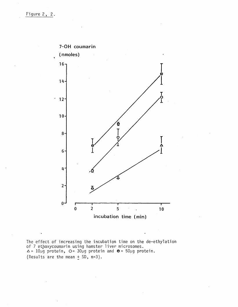
Figure 2 , 2.
7-0H coumarin (nmoles)
11
14-1
12
1 0 -!
8 -
6 -
4"
i
“ Trio
incubation time (min)
The e ffe c t of increasing the incubation time on the de-ethylation o f 7 etjioxycoumarin using hamster l i v e r microsomes. a = lOyg pro te in , 0 = 30yg protein and © = 50pg protein .(Results are the mean + SD, n=3).

Measurement o f practo lo l metabolite binding to human serum albumin.14A mixture of HSA and C practo lo l was passed down a sephadex G25
column and the e f f lu e n t monitored at 280nm. Fractions ( 4x10 speed)
50 drop fra c t ions ) were then counted fo r ra d io a c t iv i ty . Almost a l l
the a c t iv i t y was located in frac tions 15-20, corresponding to those
frac tions in which practo lo l eluted. The small amount o f absorbtion
at 280nm seen was due to practo lo l i t s e l f . HSA eluted in frac tions
6-11.( f ig . 2, 3 .) .14Practolol (2mM, 1 ci C p rac to lo l) was incubated with hamster l i v e r
microsomes and 12mg HSA fo r 30min, with or w ithout cofactors. The mic
rosomes were sedimented by cen tr ifuga tion and the supernatant eluted
down a sephadex G25 column, (0.8x10 speed, 100 drop fra c t io n s ) .
Two peaks were observed ( f ig . 2, 4) and the frac t ions making up the
f i r s t one, corresponding to HSA, were pooled and concentrated under
pressure,using a UM 10 diaflow f i l t e r , to about h a lf the o r ig ina l14volume. Protein bound C a c t iv i t y was measured a f te r solvent ex trac tion
or exhaustive equilib rium d ia lys is o f the concentrated protein f ra c
t ion .
When covalent binding was measured by exhaustive equilibrium d ia lys is
0.54nMol/mg protein was found to have bound in the absence o f co fact
ors and 3.08nMol in the presence o f cofactors, an increase o f 470%.
corresponding values obtained a f te r solvent extraction were O.lnMol and
l . l ln M o l , lower than those obtained by exhaustive equilib rium d ia ly s is
although the percentage increase observed was much greater ( 1000%)
due la rge ly to the lower control values obtained by solvent ex trac t ion .
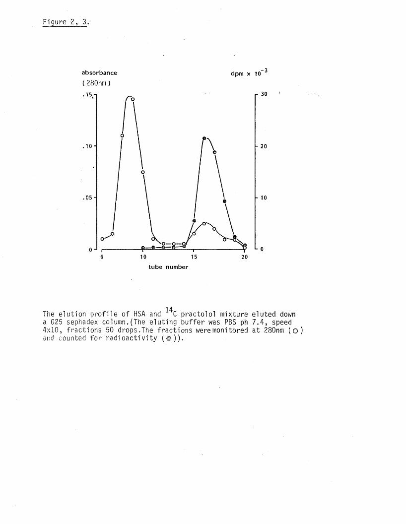
Figure 2, 3.
absorbance
( 280nm)dpm x 10
.05 -
i— -o— <06 10 15 20
r 30
20
10
L 0
tube num ber
14The e lu tion p ro f i le o f HSA and C practo lo l mixture eluted down a G25 sephadex column.(The e lu ting bu ffe r was PBS ph 7.4, speed 4x10, frac tions 50 drops.The frac tions were monitored at 280nm ( o )and counted fo r r a d i o a c t i v i t y ( ©) ) •

Figure 2 , 4.
absorbance.280 nM
7 ml
b
HSA/metabolite fractions
The separation o f HSA bound metabolites o f practolo l from the incubation mixture using a G 25 sephadex column.(The e lu t ing so lu tion was PBS, ph 7.4. speed 0.8x10, fra c t io n volumes 7ml. The e f f lu e n t was monitored at 280nm).
The assesment o f d i f fe re n t radioimmuno assays fo r detecting
antibodies to practo lo l metabolites using a model system.
The modified Farr assay.
A comparison o f d i f fe re n t modified Farr assays.
The r e l i a b i l i t y o f the three d i f fe re n t modifications was compared
(see Methods Section) using radio labelled HSA (1/8 d i lu t io n / 6 . 5yg
assay) and sheep anti HSA (1/10 d i lu t io n ) .A l l three modifications
proved to be equally e ffe c t ive in d iscrim inating between sp e c if ic a n t i -
sera and heat inactivated control sera (table 2 , 1 ) . The system used
by Amos et al (1978) proved, in my hands,to be unre liab le as the prec
ip i ta te formed on addition o f ammonium sulphate did not always give
a good p e l le t on cen tr ifuga tion . I t was therefore decided to use method
3_ (see Methods Section) rou tine ly as i t gave the most reproducable
resu lts .
A_compari_son_of the_ab_iJ i ty o f_di/_ferent_ chemi cal s _t_o_ precip i tate_
the pamma_ _gl obulj n_ fract i on _of_serum.
A comparison was made o f the a b i l i t y o f 50% ammonium sulphate, 27%
sodium sulphate and a saturated so lu tion o f ammonium acetate in ethanol125to se le c t ive ly p rac ip ita te I HSA bound to anti HSA serum. Amm
onium sulphate and sodium sulphate were considerably more e f fe c t iv e125in p re c ip ita t in g bound I HSA than ammonium ace ta te ,(tab le 2 , 2 ) .
A 50% solu tion o f ammonium sulphate gave the lowest control values
when heat inactivated human serum was used. The re la t iv e increase in
ra d io a c t iv i ty in the protein p e l le t comparing control and experimental
sera was*660% using ammonium sulphate, 264% using sodium sulphate and
131% with ammonium acetate.
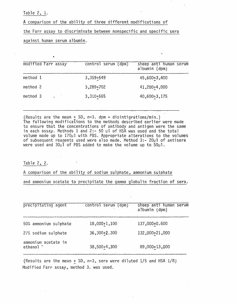
Table 2, 1.
A comparison o f the a b i l i t y o f three d i f fe re n t modifications o f
the Farr assay to discrim inate between nonspecific and sp e c if ic sera
against human serum albumin.
(*
modified Farr assay control serum (dpm) sheep anti human serumalbumin (dpm)
method 1 3,359+549 45,600+3,400
method 2 3,289+702 41,200+4,000
method 3 . 3,310+665 40,600+3,175
(Results are the mean + SD, n=3. dpm = d is in t ig ra t io n s /m in . )The fo llow ing modifications to the methods described e a r l ie r were made to ensure tha t the concentrations o f antibody and antigen were the same in each assay. Methods 1 and 2 :- 50 y l o f HSA was used and the to ta l volume made up to 170pl with PBS. Appropriate a lte ra t ions to the volumes o f subsequent reagents used were also made. Method 3 :- 20pl o f antisera were used and 30pl o f PBS added to make the volume up to 50pl.
Table 2, 2 .
A comparison o f the a b i l i t y o f sodium sulphate, ammonium sulphate
and ammonium acetate to p rec ip ita te the gamma globulin f ra c t io n o f sera.
p re c ip ita t in g agent control serum (dpm) sheep anti human serum albumin (dpm)
50% ammonium sulphate 18,000+1,100 137,000+0.600
27% sodium sulphate 36,300+2.300 132,000+21,000
ammonium acetate inethanol * 38,500+4,300 89,000+13,000
(Results are the mean + SD, n=3, sera were d ilu ted 1/5 and HSA 1/ 8 ) Modified Farr assay, method 3. was used.
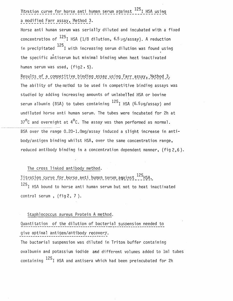
125Titration curve fo r horse anti human serum against I HSA using
a modified Farr assay, Method 3.
Horse anti human serum was s e r ia l ly d ilu ted and incubated with a fixed 125concentration o f I HSA (1/8 d i lu t io n , 6.5 yg/assay). A reduction
, 125in prec ip ita ted I w ith increasing serum d i lu t io n was found using
the spec if ic anti serum but minimal binding when heat inactivated
human serum was used, ( f i g 2 9 5).
Results of _a_competitiv_e_bin_ding_a_s_say_using Farr assay, Method _3.
The a b i l i t y o f the method to be used in competitive binding assays was
studied by adding increasing amounts o f uniabelled HSA or bovine125serum albumin (BSA) to tubes containing I HSA (6. 5pg/assay) and
undiluted horse anti human serum. The tubes were incubated fo r 2h at
37°C and overnight at 4°C. The assay was then performed as normal.
BSA over the range 0.20-1.0mg/assay induced a s l ig h t increase in a n t i
body/antigen binding w h i ls t HSA, over the same concentration range,
reduced antibody binding in a concentration dependent manner, ( f i g 2 , 6 ).
The cross linked antibody method.125J l t r a t i on _c_urve _f o r _ho_r_se_a n t i_huma_n_ serum_ agaj ns t _ HSA._
125 I HSA bound to horse anti human serum but not to heat inactivated
control serum , ( f i g 2, 7 ).
Staphlococcus aureus Protein A method.
Quantitation o f the d i lu t io n o f bacteria l suspension needed to
give optimal antigen/antibody recovery.
The bacteria l suspension was d ilu ted in T riton bu ffer containing
ovalbumin and potassium iodide and d i f fe re n t volumes added to 1ml tubes 125containing I HSA and anti sera which had been preincubated fo r 2h
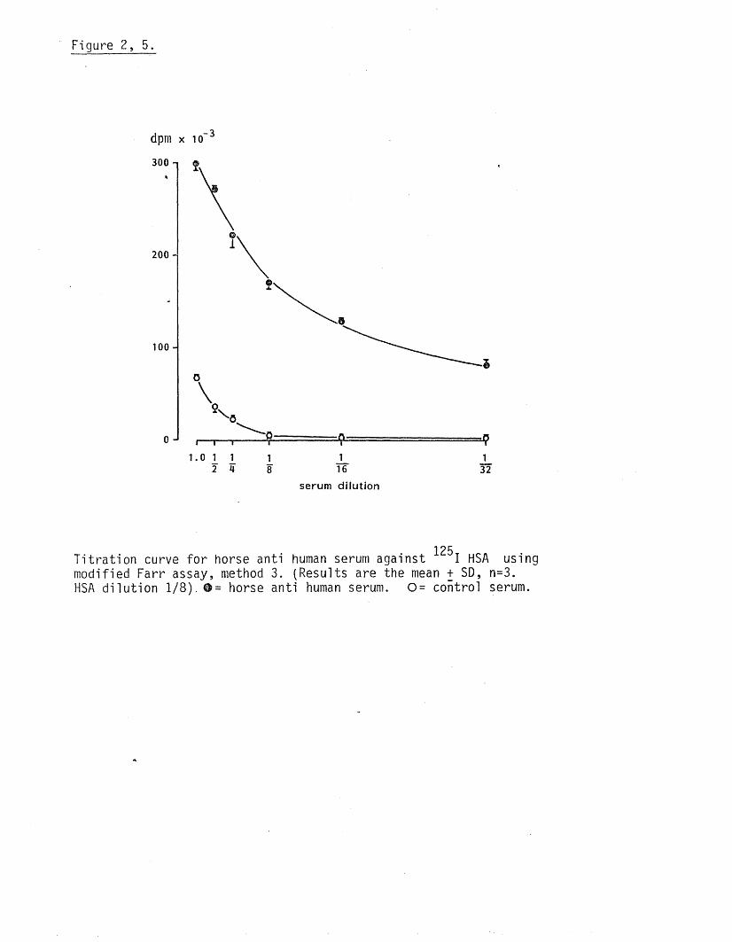
Figure 2, 5.
dpm x i o 3
300
© ,
1200 -
100 -
1.0 1 1 1 J_ J_2 4 8 16 32
serum dilution
125T it ra t io n curve fo r horse anti human serum against I HSA using modified Farr assay, method 3. (Results are the mean + SD, n=3.HSA d i lu t io n 1 / 8 ) . # = horse anti human serum. 0 = control serum.

Figure 2, 6.
_3dpm x 10
120 -i
ioo H
50 H
0 . 2 . 4 . 6 . 8 1 . 0
mg HSA o r B S A / tu b e
Competitive binding assay using modified Farr assay, method 3. 1?{-( Results are the mean + SD, n=3. Horse anti human serum 1/20 d i lu t io n . I HSA 1/8 d i lu t io n ) o=~unlabelled BSA and © = unlabelled HSA.
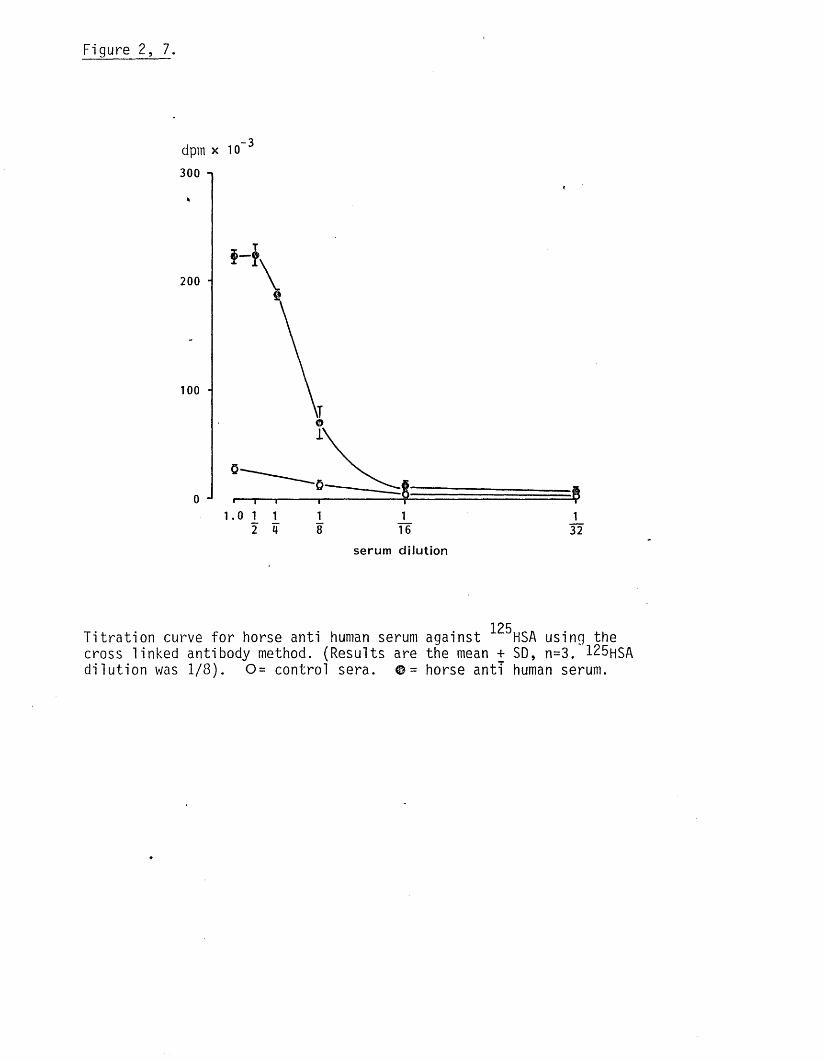
Figure 2, 7.
- 3dpm x 10
300 n
2 0 0 -
100 -
1 .0 1 1 I J_ _1_2 4 8 16 32
serum d ilu tio n
125T it ra t io n curve fo r horse anti human serum against HSA using the cross linked antibody method. (Results are the mean + SD, n=3. 125hSA d i lu t io n was 1/ 8 ). 0 = control sera. ©= horse anti human serum.

at 37°C and 2h at 4°C. Specific and control binding, using d i f fe re n t
volumes o f the bacteria l suspension, was measured as described
in the Methods Section. Optimal binding (sp ec if ic vs con tro l) was found
using 300ylofthe neat suspension (10%w/v) and th is was used fo r sub
sequent assays, (tab le 2, 3 ) .
IItr§tion_of_s'heep_anti_human_serum_a]bumin_using_the_Staph]ocgccus
jyr§y§_ Protein, method..
Specific binding to the sheep anti HSA dropped rap id ly with increasing
serum d i lu t io n . Binding to control serum was very low, ( f i g 2, 8 ).
15 s _o^_ a_ cpmpeti t i ve_bi ndi ng_ass_a_y_ us i ng_ the_ §taphl°coccus_ aureus
.P CQte in _A .method..
Unlabelled HSA added to a 1/20 d i lu t io n o f sheep anti HSA in h ib ite d
binding o f the rad io labelled protein in a dose re lated fa sh io n ,(0 . 2- 1.0
mg/assay) BSA s i ig h t ly enhanced I HSA binding, ( f i g 2, 9) .
A comparison o f the a n t ig e n ic i ty o f the rad io labelled and unlabelled
human serum albumin samples used in the competitive binding assays.
I t was noted that high concentrations o f unlabelled HSA (0.2-l.Omg/
assay) were needed to reduce the binding o f antibody to the radio-
labelled antigen, (6.5 pg/assay). I t appeared tha t the rad io labe lled
HSA was more antigenic than the unlabelled HSA. This p o s s ib i l i t y
was tested by carrying out a double d if fu s io n experiment against
sheep anti HSA. A fte r 2 days p re c ip ita t io n lines were observed w ith a125
1/512 d i lu t io n o f ■“ I HSA but only at a d i lu t io n o f 1/128 w ith the
unlabelled antigen.

Table 2, 3.
Experiment to determine the concentration o f Staph!ococcus aureus
giving optimal antibody/antigen recovery.
volume o f bacteria added control serum (dpm) sheep anti HSA (dpm) c o n tro lx l00experim.
10yl 1,325+1,200 271,700+71,100 0.5%
50yl 1,700+ 600 235,800+51,700 0.7%
lOOyl 2,800+ 300 191,700+25,600 1.5%
200yl „ 4,800+2,400 340,500+36,000 1.4%
300yl 4,200+1,300 345,700+43,200 ( 1.2%
125(Results are the mean + SD, n=3) Sera were not d ilu ted . HSA was d i lu ted 1/ 8 .

Figure 2, 8.
- 3' dpm x 10
400 i
300 -
200 -
100
1 11.0 15 10 20 40 80
serum d i lu t io n
125T it ra t io n o f sheep anti human serum albumin against HSA using the Staphlococcus protein A method.(Results are the mean + SD, n=3. ^H S A was d ilu ted 1/ 8 ).Control serum = 0 . sheep anti human serum albumin =© .
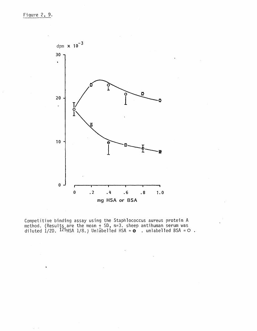
Figure 2, 9.
- 3dpm x 10
30 n
20 -
0 .2 .4 .6 .8 1.0mg H SA o r BSA
Competitive binding assay using the Staph!ococcus aureus prote in A method. (Results are the mean + SD, n=3. sheep anti human serum was d ilu ted 1/20. 125hsA 1/8 .) Unlabelled HSA =© . unlabelled BSA = 0 .

Analysis o f serum from practo lo l treated patients fo r antibodies to
in v i t ro generated practolo l metabolites.
Protein bound metabolites of practo lo l were generated as described
previously and, a f te r iso la t io n and concentration, used as the a n t i
gen complex in Farr, cross linked gamma g lobulin and Staplococcus(
aureus assays.'The resu lts obtained using sera from 12 d i f fe re n t pat
ients are given in table 2, 4. Some o f the pa tients , (7 ), had already
been shown to contain antimetabolite antibodies by Amos e t al (person
al communication) at a t i t r e o f 1/10. I t was not generally possible
to use the same samples as Amos et al but th e i r f ind ings , using serum
from the same patients are given fo r comparison where possible.
No d ifference in binding between control and experimental serum
samples could be detected or between the antigen complex generated in
the presence o f cofactors or the absence o f cofactors using any o f the
assays. Differences in the percentage binding between the three assays
used were seen. The Farr assay gave the highest binding values but
these ra re ly exceeded 5%. o f the to ta l counts added. The cross linked
antibody method gave s l ig h t ly lower values fo r antigen binding and the
Staplococcus aureus protein A method the lowest binding values, less
than 1%.
DISCUSSION
The aim o f the experiments described in th is Chapter was to evaluate
the methods used in generating the p ra c to lo l/p ro te in complex and the
assays fo r measuring bind to spec if ic antibodies present in the serum
of adversely e ffected patients.
The a c t iv i t y o f the microsomal mixed functional oxidase system was
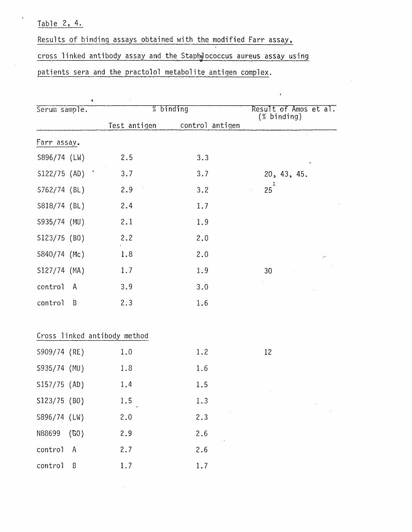
Table 2, 4.
Results o f binding assays obtained with the modified Farr assay.
cross linked antibody assay and the Staphjlococcus aureus assay using
patients sera and the practo lo l metabolite antigen complex.
4
Serum sampled % binding Result o f Amos et a l.(7o binding)
_________________ Test_antigen______control antigen_______________________
Farr assay.
S896/74 (LW) 2.5 3.3
S122/75 (AD) 3.7 3.7 20,
S762/74 (BL) 2.9 3.2i
25
S818/74 (BL) 2.4 1.7
S935/74 (MU) 2.1 1.9
S123/75 (BO) 2.2 2.0
S840/74 (Me) 1.8 2.0
S127/74 (MA) 1.7 1.9 30
control A 3.9 3.0
control B 2.3 1.6
Cross linked antibody method
S909/74 (RE) 1.0 1.2 12
S935/74 (MU) 1.8 1.6
S157/75 (AD) 1.4 1.5
S123/75 (BO) 1.5 1,3
S896/74 (LW) 2.0 2.3
N88699 (130) 2.9 2.6
control A 2.7 2.6
control B 1.7 1.7

Table 2, 4. continued.
Staphlococcus aureus method.
N88699 (GO) 0.23 0.29
S157/57 (AD) 0.30 0.29
S818/74 (BL) ' 0.33 0.16
S47/75 (CA) 0.32 0.51 25
S102/75 (MUL) 0.41 0.38 16.5
S935/74 (MU) 0.35 0.36
S195/75 (ST) . 0.27 0.29 34, 15.6
control A 0.39 0.40
control B 0.30 0.40
Results are the mean o f duplicate assays.
The sera had been stored at BIBRA. Each sample was given a number
at the time i t was taken by Dr Amos. The results o f assays performed,
using serum from the same patient,by Amos et al are given when ava ilab le
A na lys is was performed on the same sample.
demonstrated using the de-ethylation o f 7-ethoxycoumarin as an ind
ica to r of microsomal enzyme a c t iv i t y . Formation o f m etabolite /prote in14complexes was confirmed by measuring covalent binding of C-practo-
lo l to HSA,added to the incubation system,after solvent or exhaustive
equilib rium d ia lys is extraction o f the iso lated HSA fra c t io n . Cova
len t binding to%HSA was about 3 fo ld higher a f te r exhaustive d ia lys is
extraction than a f te r solvent ex trac tion . This find ing is in agreement w ith
an e a r l ie r report o f Sun and Dent (1980). They showed tha t covalent
binding values were between 2 and 7 times greater a f te r exhaustive
d ia lys is tha t a f te r solvent ex trac tion . The large increase in binding
in the presence o f cofactors indicates that much o f the covalently
bound a c t iv i t y was due to practo lo l metabolites. The value o f l . l ln m o l
/mg protein obtained a f te r solvent extraction is in good agreement with
the value published by Orton and Lowery (1977). They reported binding
o f 1.49nmol/mg microsomal protein using s im ila r incubation conditions.
Addition of 12.5mg HSA to the incubation in h ib ite d binding by 96%,
presumably by scavenging the active metabolites formed. A value of
about 1.43nmol/mg HSA would therefore be expected i f a l l the metabol
ites became bound to HSA, approximately 30% more than was found in
th is study. I t is not possible to compare the resu lts gained in the
experiment described above with the resu lts obtained by Amos et al
as they did not measure metabolite binding.
I t was not possible to va lidate the various binding assays using known
antigents or antibodies as characterisation o f these parameters was125the goal o f th is pro jec t. Commercial anti sera against HSA and I HSA
were therefore used as a model system. Using th is approach i t was dem
onstrated that high spec if ic binding, but low binding to control sera,125occured with a l l the assays evaluated and tha t binding of HSA could
be blocked by unlabelled HSA but not by BSA.
Using a l l three techniques, sera from patients who had been treated
with practo lo l were analysed fo r the presence of antibodies against the
antigen complex, including sera from patients known to have had a
pos it ive t i t r e at some time. None o f the sera tested gave a pos it ive
re su lt but because i t was not possible to use the same serum samples
used by Amos et al i t was not possible to compare resu lts d i re c t ly .
Several explanations fo r the lack o f binding suggest themselves. F i r s t
i t is possible that the methods used to generate the antigen complex
were inadequate. This would seem un like ly as the method used in these
studies were the same as those used successfully by Amos et al (1978)
and,in some cases,the antigen complex was prepared by the workers who
had taken part in the o r ig ina l studies. Second, the sera could have
deteriorated during storage. This period was as long as 5 years in
some cases and, as the sera had not been a liquoted, some samples, par
t ic u la r ly the more in te res ting ones, had been frozen and thawed several
times, (e ithe r on purpose or accidently when the deepfreeze broke
down). Third, i t was possible that a l l the active sera had been used
in the o r ig ina l studies and only inactive sera was l e f t . Further stud
ies were undertaken to evaluate these p o s s ib i l i t ie s .

CHAPTER 3.
INVESTIGATIONS TO DETERMINE POSSIBLE REASONS WHY
ANTIBODIES TO IN VITRO GENERATED PRACTOLOL METABOLITES
COULD NOT BE DETECTED IN THE SERUM OF PRACTOLOL
TREATED PATIENTS.
INTRODUCTION
The methods necessary to generate antigen complexes .and assay fo r
spec if ic antibodies to these in the serum o f practo lo l treated
patients were evaluated in Chapter 2. Using these methods however
no metabolite antibodies could be detected, even using serum from
patients previously shown to have synthesised such antibodies by Amos
et a l . I t was suggested tha t e ith e r the methods used were inadequate
or tha t the sera used was, fo r some reason, inac tive . These p o s s ib i l
i t ie s were evaluated. In order to control fo r methodological errors
co llabora tive experiemnts were performed with Dr. B.Lake and Ms 0
A r t is BSc, workers who had performed the o r ig ina l experiments with
Dr. Amos,(1978). In one experiment sera which had been stored at The
B r i t is h Biologica l Research Association (BIBRA) were used and in a sec
ond sera were obtained from ICI Macclesfield. This enabled both a com
parison o f the methodology and of serum samples stored at d i f fe re n t
laboratories.
As a check on the s ta b i l i t y o f the sera, samples were analysed fo r
th e ir autoantibody t i t r e , using guinea-pig oesophageal r ings , and com
pared with t i t r e s obtained o r ig in a l ly using sera collected at about the
same time.
Amos et al (1977) had suggested that there could be a negative re la
t ionship between autoantibody t i t r e and the presence of antimetabolite
antibodies. A number o f serum samples were chosen at random and tested
fo r autoantibodies. I t was intended to perform Farr assays on those
samplesWhich were negative at a 1/10 d i lu t io n .
METHODS
Methods used in th is Chapter fo r the generation o f the antigen complex
and detection, by Farr assay, o f antimetabolite antibodies were the
same as those described in Chapter 2.
Measurement o-f autoantibodies.
A guinea-pig oesophagus was dissected out and pieces frozen onto a
cryostat chuck placed in a cardice/acetone freezing mixture. Sections,
5-6ym, were cut using a Bright microtome and freeze fixed onto 76x26mm
glass microscope slides and l e f t in the microtome u n t i l needed.
Before s ta in ing the s lides were put in racks suspended in 500ml PBS
in a glass tank placed on a magnetic s t i r r e r and washed fo r lh . They
were then removed, drained o f excess bu ffe r and placed in a damp box.
The seruftto be used (0.4ml o f the required d i lu t io n ) was placed on
each s lide and l e f t fo r 30min. The seru#\was then drained, the s lides
washed fo r lh in a change o f bu ffe r before being replaced in the damp
box. The sections were then counter stained fo r 30min using 0.4ml o f
1/10 d i lu t io n of FITC labelled sheep anti human IgG. A fte r a f in a l
wash fo r lh in a change o f bu ffe r the s lides were drained and mounted
in buffered glycerol under a glass covers!ip . The sections were exam
ined using a Zeiss fluorescent microscope set fo r exc ita t ion at 450-
500nm (optimal wavelength 490nm) and the fluorescence ca lib ra ted using
an a rb ita ry scale with reference to a control from which serum had
been ommitted, ( s l ig h t + to ++++).
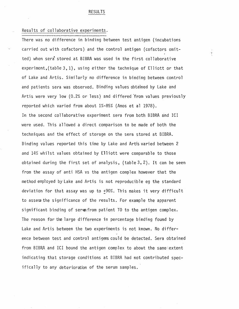
RESULTS
Results o f co llabora tive experiments.
There was no d ifference in binding between te s t antigen (incubations
carried out with cofactors)- and the control antigen (cofactors omit
ted) when sera' stored at BIBRA was used in the f i r s t co llabora tive
experiment,(table 3, 1), using e ith e r the technique o f E l l i o t t or tha t
of Lake and A r t is . S im ila r ly no d ifference in binding between control
and patients sera was observed. Binding values obtained by Lake and
A r t is were very low (0.2% or less) and d if fe red 'from values previously
reported which varied from about l%-85% (Amos et al 1978).
In the second co llabora tive experiment sera from both BIBRA and ICI
were used. This allowed a d ire c t comparison to be made of both the
techniques and the e f fe c t o f storage on the sera stored at BIBRA.
Binding values reported th is time by Lake and Artis varied between 2
and 14% w h ils t values obtained by E l l i o t t were comparable to those
obtained during the f i r s t set o f analysis, (tab le 3, 2). I t can be seen
from the assay of anti HSA vs the antigen complex however tha t the
method employed by Lake and A r t is is not reproducible eg the standard
deviation fo r tha t assay was up to +90%. This makes i t very d i f f i c u l t
to assess the s ign if icance of the resu lts . For example the apparent
s ig n if ic a n t binding o f seromfrom pa tien t TO to the antigen complex.
The reason fo r the large d ifference in percentage binding found by
Lake and A r t is between the two experiments is not known. No d i f f e r
ence between te s t and control antigens could be detected. Sera obtained
from BIBRA and ICI bound the antigen complex to about the same .extent
ind ica ting tha t storage conditions at BIBRA had not contributed spec
i f i c a l l y to any deterioration o f the serum samples.
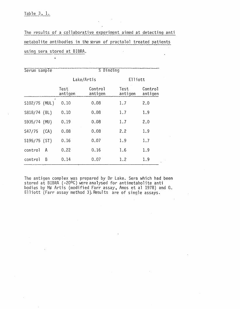
Table 3, 1.
The resu lts o f a co llabora tive experiment aimed at detecting anti
metabolite antibodies in the serum of practo lo l treated patients
using sera stored at BIBRA.
Serum sample % Binding
Lake/Artis El 1i o t t
Test Control antigen antigen
Testantigen
Controlantigen
S102/75 (MUL) 0.10 0.08 1.7 2.0
S818/74 (BL) 0.10 0.08 1.7 1.9
S935/74 (MU) 0.19 0.08 1.7 2.0
S47/75 (CA) 0.08 0.08 2.2 1.9
S195/75 (ST) 0.16 0.07 1.9 1.7
control A 0.22 0.16 1.6 1.9
control B 0.14 0.07 1.2 1.9
The antigen complex was prepared by Dr Lake. Sera which had been stored at BIBRA (-20°C) were analysed fo r antimetabolite anti bodies by Ms A r t is (modified Farr assay, Amos e t al 1978) and 6. E l l i o t t (Farr assay method 3). Results are o f s ing le assays.
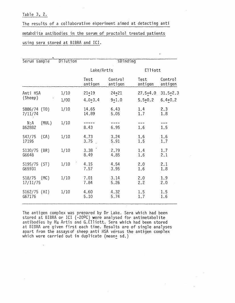
Table 3, 2.
The resu lts o f a co llabora tive experiment aimed at detecting anti
metabolite antibodies in the serum o f practo lo l treated patients
using sera stored at BIBRA and ICI.
*___________________________________________________Serum samp’le D ilu t ion %Binding
Lake/Artis E l l i o t t
Testantigen
Controlantigen
Testantigen
Controlantigen
Anti HSA 1/10 21+19 24+21 27.5+4.0 31.5+2.:(Sheep) 1/00 4.0+3.4 9+1.0 5.5+0.2 6.4+0.2
S886/74 (TO) 1/10 14.65 6.43 1.4 2.37/11/74 14.89 5.05 1.7 1.8
N:A (MUL) 1/10 --------------------- ----------------- — —
B62882 8.43 6.95 1.6 1.5
S47/75 (CA) 1/10 4.73 3.24 1.6 1.617195 3.75 5.91 1.5 1.7
S130/75 (BR) 1/10 3.38 2.79 1.4 1.7G5648 8.49 4.85 1.6 2.1
S195/75 (ST) 1/10 4.15 4.54 2.0 2.1G65931 7.57 3.95 1.6 1.8
S18/75 (MC) . 1/10 7.01 3.14 2.0 1.917/11/75 7.84 5.26 2.2 2.0
S162/75 (HI) 1/10 4.60 4.32 1.5 1.5G67176 5.10 5.74 1.7 1.6
The antigen complex was prepared by Dr Lake. Sera which had been stored at BIBRA or ICI ( - 2 0 ° C ) were analysed fo r antimetabolite antibodies by Ms A r t is and G .E l l io t t . Sera which had been stored at BIBRA are given f i r s t each time. Results are o f s ingle analyses apart from the assays of sheep anti HSA versus the antigen complex which were carried out in duplicate (mean+ sd.)

Analysis of sera from patients treated with practo lo l to id e n t i fy
samples with an autoantibody t i t r e of less than 1/10.
Autoantibody levels were measured in the sera o f 17 patients chosen
at random, including 4 patients where se r ia l samples were ava ilab le ,
(tab le 3, 3 ). A l l had ,a t i t r e o f at least + at a 1/10 d i lu t io n using
FITC labelled sheep anti human IgG serum. An antibody to an in te r c e l l
u la r antigen was found in the serum of a l l patients tested and a
nuclear antibody in the serum of about 50% o f the pa tien ts , ( f ig 3, 1
and 2).There was some evidence tha t the autoantibody t i t r e changed
with time when sera from pa tien t BL was used but no apparent changes
when sera from patients MUL,RE or RY were used. Due to the presence
of autoantibodies in a l l the sera tested Farr assays were not per
formed.
A comparison of autoantibody t i t r e s in stored sera with those
obtained soon a f te r c o l le c t io n .
In order to investigate-the p o s s ib i l i ty that the sera may have lo s t
th e ir a c t iv i t y during storage the autoantibody t i t r e was measured in
6 serum samples and compared with the t i t r e reported at the time o f
co llec t ion by Dr Amos. Dr Amos also standardised the scale used to
assessthe re la t iv e response, + to ++++. None o f the samples analysed
had a t i t r e o f greater than + at 1/80 d i lu t io n . Most o f the patients
had a t i t r e of at least 1/320 when the analysis was performed soon
a f te r the blood was taken, (tab le 3, 4).

The binding of an t ibod ies in the serum of a p rac to lo l t r e a t e d p a t i e n t (S924/74, 1/10 d i l u t i o n ) to the i n t e r c e l 1u la r region of a xenogenic t i s s u e (guinea-pig oesophagus). Counter sta in ing was c a r r i e d out using FITC l abe l l ed sheep antihuman IgG.
Figur e 3, 2.
Binding of an t ibod ies in the serum of a p rac to lo l t r e a t e d p a t i e n t (S122/75, 1/10 d i l u t i o n ) to the nucleus of a xenogenic t i s s u e . Condit ions as f o r f i g . 3, 1.
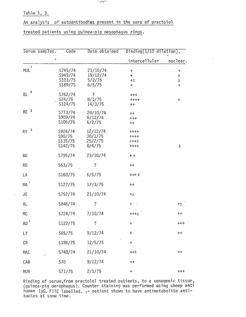
Table 3, 3.
An analysis o f autoantibodies present in the sera of p racto lo l
treated patients using guinea-pig oesophagus r in g s .
Serum samples. Code Date obtained Binding(1/10 d i lu t io n ) .% in te rc e l lu la r nuclear
MUL1 S745/74S949/74S103/75S169/75
21/10/7419/12/745/2/756/5/75
+++±+
++++
BL 1 S762/74S24/75S124/75
?8 / i/75 14/3/75
+++++++++
+
RE 1 S773/74S909/74S105/75
28/10/746/12/746/2/75
+++++++
RY 1 S924/74S90/75S135/75S142/75
12/12/7430/1/7525/2/758/4/75
+++++++++++±+++± +
BO S795/74 23/10/74 + ±
RO S63/75 ? ++
LA S168/75 6/5/75 +++ ±
MA 1 S127/75 17/3/75 ++
JE S752/74 21/10/74 +±
AL S846/74 ? + . +±
MC S724/74 7/10/74 ++++ ++
AD1 S122/75 ? + +++
LY S65/75 9/12/74 + ++
CR S186/75 12/5/75 +
MAC S748/74 21/10/74 ++± ++
CAR S70 9/12/74 ++
MUR S71/75 2/1/75 + +++
Binding o f serum,from practo lo l treated pa tien ts , to a xenogenic t issue , (guinea-pig oesophagus). Counter s ta in ing was performed using sheep anti human IgG. FITC labelled. 1= oatient shown to have antimetabolite a n t i bodies at some time.
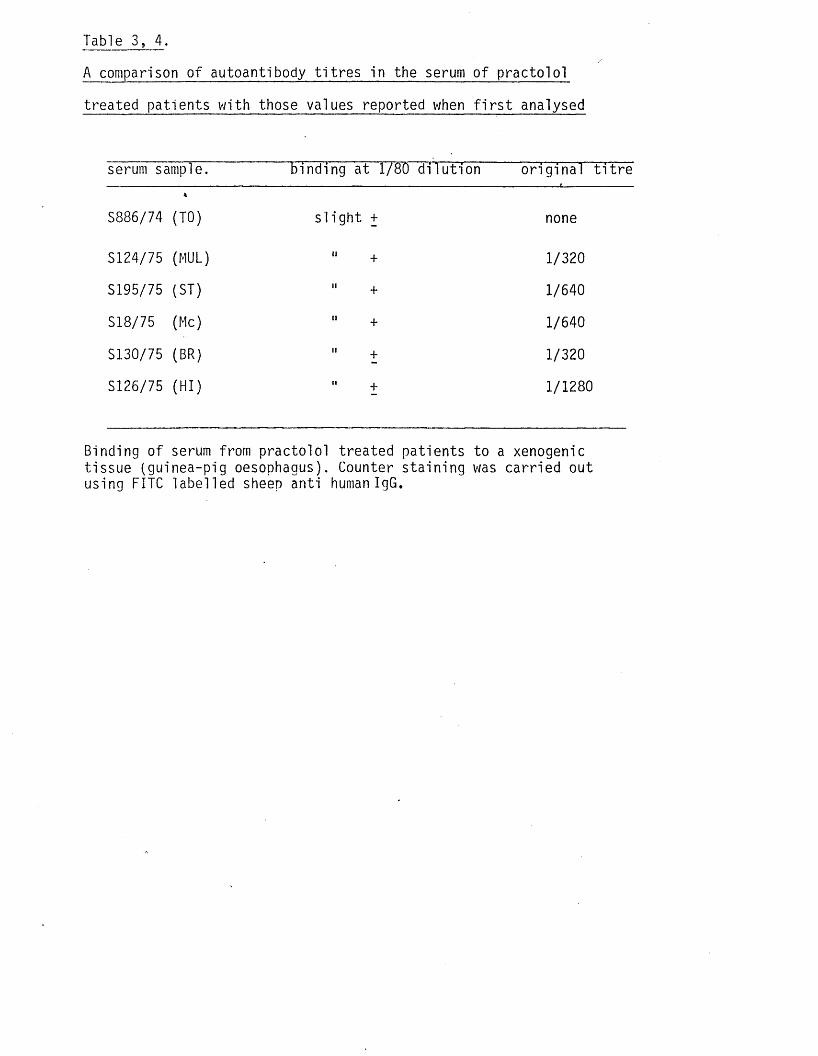
Table 3, 4 .
A comparison of autoantibody t i t r e s in the serum of practo lo l
treated patients with those values reported when f i r s t analysed
serum sample. binding at 1/80 d i lu t io n o r ig ina l t i t r e
S886/74 (TO) s l ig h t + none
S124/75 (MUL) 11 + 1/320
S195/75 (ST) " + 1/640
S18/75 (Me) " + 1/640
S130/75 (BR) 11 + 1/320
S126/75 (HI) " t 1/1280
Binding of serum from practo lo l treated patients to a xenogenic tissue (guinea-pig oesophagus). Counter sta in ing was carried out using FITC labelled sheep anti human IgG.
DISCUSSION
The co llabora tive experiments carried out in cooperation with Dr.B.
Lake and J .A r t is demonstrated that the results given in Chapter 2
were not fa lse negatives. The second experiment also showed tha t *
there was l i t t l e d ifference between sera stored at BIBRA and ICI.
Whilst i t was s t i l l possible tha t the sera had become deactivated
during storage i t appeared to be un like ly that non-standard storage
conditions had contributed to th is process.The most in te res ting f in d
ing to emerge, from the two co llabora tive experiments was the d i f f e r
ences in antigen binding obtained using the o r ig ina l method. In the
f i r s t experiment (tab le 3, 1) binding values v/ere less than 0.5%
where as in the second experiment (table 3, 2) i t varied between 3 and
15%. Antigen binding o f greater than 10% was o r ig in a l ly accepted as
being necessary to demonstrate the presence o f spec if ic antibodies
(Amos et al 1978). Only one patient of the seven tested in the second
experiment had a t i t r e . o f greater than 10%, (pa tien t TO),although four
of the patients had been shown to have synthesised antimetabolite a n t i
bodies previously,(MUL, CA, BR, and ST). There was however considerable
va r ia t ion , not only between the two experiments but also between dup
lica tes (+90% when anti HSA sera was incubated w ith the antigen complex)
which makes i t d i f f i c u l t to in te rp re t th is f ind ing .
I t was possible that a l l the serum containing antimetabolite a n t i
bodies had been used up by the o r ig ina l investiga to rs . A method fo r
test ing fo r active sera was indicated by the proposal o f Amos et al
(1978) that there might be an inverse re la tionsh ip between au toanti
body levels and the presence of antimetabolite antibodies. A number
of samples were therefore analysed fo r autoantibodies using a d i lu t io n

of 1/10. I t was intended to use any sera which were negative at th is
d i lu t io n in Farr assays. Unfortunately a l l the sera tested were pos it ive
and were not analysed fo r the presence o f antimetabolite antibodies.
In order to investigate the idea that the samples had deteriorated
during storage autoantibody t i t r e s o f stored sera were compared with the
t i t r e s obtained soon a f te r they were co llected. As can be seen from
table 3, 4 ., none o f the sera bound strongly to guinea-pig oesophagus
rings at a 1/80 d i lu t io n where as a l l the sera had t i t r e s o f 1/320
or greater at the time o f co l le c t io n . I t is possible therefore tha t the
samples had deteriorated during storage.
The most probable reason fo r the negative resu lts obtained in these
studies is tha t the active sera had a l l been used up in the o r ig ina l
studies. Although autoantibody t i t r e s appear to be maintained in v iv o ,
(see table 3, 3.) anti metabolite antibodies f luc tua te g rea tly w ith in
a short time period. Amos et al (1977) fo r example reported tha t th is
a c t iv i t y dropped by 65% and 75% in two patients in a one month period.
With such a short time .in which to c o l le c t active sera i t is probable
that many samples obtained would be inactive and that the small number
of active samples available would be rap id ly used up. In view o f the
fac t that no fu r th e r serum samples from practo lo l patients would be
forthcoming i t was not considered f r u i t f u l 1 to pursue the problem
any fu r the r.
Incidences of adverse e ffects associated with drug adm in is tra tion , such
as the practo lo l syndrome, can, p o te n t ia l ly , provide a great deal o f
information, not only on the mechanism of the drug induced reaction but
also on the way in which normal control processes, such as the immune
system, are regulated. I t is important therefore tha t samples obtained
from treated patients are stored p roperly , fo r example at -70°C or

f'reeze dryed with a preservative. Samples should ce r ta in ly be aliquoted
as soon as possible and not repeatedly thawed and refrozen.
I t is also important tha t samples known to contain the reported act
i v i t y are not used up so tha t workers wishing to confirm, or extend,"
the o r ig ina l observations have a pos it ive control with which to work.
CHAPTER 4.
EXPERIMENTS AIMED AT RAISING ANTIBODIES TO IN VITRO
GENERATED METABOLITES OF PRACTOLOL IN ANIMALS.
INTRODUCTION.
I t had not proved possible to characterise, chemically, the nature
o f the antigenic practo lo l metabolites using sera obtained from
patients treated with practo lo l (chapters 2 and 3). A d i f fe re n t
approach to the problem is to raise antibodies to in v i t r o gener
ated metabolites o f p ra c to lo l , using animals, and use these antibodies
to define the s tructure o f the immunologically active product(s).
Antibodies to protein bound practo lo l had been raised in rabbits by
Chuong et al (1977a) As there were no reports o f antibodies being
raised to in v i t r o generated metabolites i t was decided to fo llow the
immunization schedule described by Chuong et al (1977a) in these experiments.
Rabbits (4) and guinea-pigs (5) were in jected with metabolite/microsome
complex and the formation of antibodies followed by e ith e r skin te s t -14ing or by carrying out modified Farr assays (method 3) using C pract-
125olo l or the HSA/metabolite complexTletabolites binding to microsomal
protein are probably iden tica l w ith those which bind to HSA. This
approach therefore allowed the HSA/metabolite complex to be used
as the antigen in the Farr assays,as described prev ious ly ,and preempted
problems associated with the generation o f antibodies to the c a r r ie r
protein in jected in to the animals. Amos et al (1977) had reported tha t
practo lo l p a r t ia l ly inh ib ited binding o f the HSA/ metabolite complex
to antibodies in human sera. I t was probable therefore tha t ^C p rac t
o lo l would also bind to antibodies against m e ta b o l i te s , i f only w ith a
reduced a f f in i t y .
METHODS.
HSA/metabol i t e complexes were generated as described in chapter 2.
Microsome/metabolite complexes were formed in the same way with the
exception that HSA was not added to the incubation system:4
Immunisation schedule.
The niicrosomal/metabolite system was homogenised with complete Freunds
adjuvent using a motorised, g la ss / te f lo n , Potter type homogeniser to
give a f in a l concentration o f Img prote in /m l. New Zealand White rabbits
(2.5-3.0 kg)were in jected at m u lt ip le s ites in the leg muscles with a
to ta l of 2mg (10x0.2ml) o f the adjuvant/ antigen mixture and then
every week fo r 5 more weeks with 4x0.5ml subcutaneously. Blood samples
were taken from the ear veins lOdays a f te r the la s t in je c t io n and serum
isolated as described in chapter 2.
Albino guinea-pigs (400-500g) were in jected with a to ta l o f 0.5mg
protein (50pl in each hind footpad and 4x0.1ml subcutaneously) fo r the
f i r s t in je c t io n and then weekly fo r 5 weeks with 5x0.lml subcutaneously.
Blood samples were taken from the ear veins 10 days a f te r the la s t in
je c t io n and serum iso la ted.\
I f the resu lts of the tests fo r antimetabolite antibodies were neg
ative the animals were l e f t fo r 6 months and the schedule repeated.
Analysis o f serum fo r antibodies against practo lo l metabolites.
14Either the HSA/metabolite complex or C practo lo l was used as the
antigen in the modified Farr assay. Serum was heat inactivated before
being used and d ilu ted 1/10 with PBS.
Procedure fo r skin te s t in g .
The flanks o f the animals were shaved and 0.2ml o f each tes t a n t i
gen in jected in trade rm a lly . Each in je c t io n s i te was numbered using
a water proof f e l t t i p pen. The skin thickness at each s i te was measured
a fte r 2, 6 and 23h using a Pocotest d ia l gauge. The antigen te s t so l- %
utions were as fo llow s, 1x10 M p ra c to lo l , microsomal preparation (lmg/ml)
d ilu ted 1/10 with Tris/HCl bu ffe r , 0.5 units/ml ICDH (1/10 d i lu t io n ) ,
and the HSA/metabolite complex. Results are given as the skin thickness
at the s i te o f antigen in je c t io n minus the thickness at the s i te o f
Tris/HCl bu ffe r (mm).
RESULTS.
Experiments aimed at detecting antibody production to practo lo l
metabolites in rabbits and guinea-pigs using a modified Farr assay.
None of the serum samples from the four rabbits or f ive guinea-pigs
contained antibodies to the HSA/metabolite complex or p racto lo l
a t a ■1/10 d i lu t io n (table 4, 1 .) . when assayed a f te r the f i r s t series
of in jec t ions . A fte r the second series there was s t i l l no detectable
a c t iv i t y , (data not given).
Experiments aimed at detecting antimetabolite antibodies by skin te s t in g .
None o f the animals tested showed any signs o f reacting to intradermal
in jec t ions o f the HSA/metabolite complex or practo lo l at any o f the
time periods. A ll the animals reacted to the microsomal preparation
and, most s trong ly , to ICDH. Maximum changes in skin thickness were
found a f te r about 6-7h. Results fo r the rabbits are given in tab le 4,
2 and fo r the guinea-pigs in table 4, 3. S im ilar values were ob ta i-
ed a f te r the second series of in je c t io n s , (data not given).

Table 4, 1.
Binding o f rabb it and guinea-pig sera to the HSA/metabolite complex-
and to C practo lo l using a modified Farr assay.
animal%
125HSA/metabolite complex % binding.
14C practo lo l
% binding.
rabbits.
1 4.9 2.8
2 5.6 3.3
3 6.0 2.9
4 3.7 3.4
controls
1 5.4 4.7
2 3.1 2.5
guinea-pigs
1 6.0 3.3
2 6.7 2.5
3 5.2 3.7
4 3.6 2.9
5 3.8 3.0
controls
1 4.6 3.3
2 5.4 3.7
Results are the mean o f duplicate assays fo r each animal.
1/10 serum d i lu t io n .

Table 4, 2.
Results o f skin tes t carried out on New Zealand White rabb its .
animal. skin thickness (mm), tes t minus control values.
ICDH microsomalpreparation
metabolite/HSA
complex
practo lo l
re su lt ■a fte r 2h.
1 3.2 1.8 0.2 0.1
2 2.7 1.9 0.1 0.2
3 3.0 2.1 0.3 0.1
4 1.8 1.8 0.1 0.0
control animals
1 0.5 0.6 0.1 0.2
2 0.0 0.2 0.0 0.0
results a f te r 6h
1 3.2 1.9 0.1 0.0
2 2.8 1.6 0.0 0.0
3 2.9 1.3 0.1 0.0
4 2.5 1.8 0.0 0.0
control animals
1 0.4 0.1 0.0 0.1
2 0.0 0.2 0.0 0.0
results a f te r 23h
1 2.0 0.8 0.0 0.0
2 1.9 1.2 0.0 0.0
3 2.2 1.0 0.0 0.0
4 . 1.8 0.9 0.0 0.0
control animals.
1 0.6 0.4 0.1 0.0
o<L 0.0 0.0 0.0 0.0

Table 4, 3.
Results of skin tests carried out on albino guinea-pigs.
animal skin thickness (mm), tes t minus control values."ICDH microsomal metabolite/HSA practo lo l
» preparation complex
resu lt a f te r 2h
1 1.6 0.2 0.1 0.0
2 2.4 0.2 0.2 0.3
3 2.0 0.8 0.1 0.0
4 2.5 0.5 0.3 0.1
5 1.1 0.3 0.1 0.0
control animals
1 0.4 0.2 0.2 0.0
2 0.8 0.5 0.1 0.1
results a f te r 7h
1 2.9 1.3 0.0 0.0
2 4.5 2.1 0.2 0.5
3 2.9 0.9 0.0 0.2
4 2.9 1.4 0.1 0.1
5 2.9 0.2 0.0 0.0
control animals
1 0.0 0.3 0.0 0.0
2 0.0 0.2 0.0 0.0
results a f te r 24h
1 1*8 0.1 0.0 0.0
2 2.1 1.3 0.2 0.0
3 1.3 0.0 0.0 0.0
4 1.5 0.0 0.0 0.0
5 0.6 0.0 0.0 0.0

Table 4, 3. continued.
animal skin thickness (mm) tes t minus control value.ICDH. microsomal
preparationmetabolite/HSA
complexpracto lo l
control animals
1 0.1 0.2 0.0 0.0
2 0.7 0.4 0.0 0.0

DISCUSSION.
None o f the animals immunised with the microsome/metabolite complex
synthesised antibodies spe c if ic fo r e ith e r practo lo l or i t s metabolites
using the in je c t io n schedule o f Chuong et al (1977a). A response to *
microsomal p ro te in , and to a much greater extent ICDH, was observed
in a l l animals when challenged intradermally ind ica ting tha t the anim
als were responding to the in jec t io ns .
The negative response to the metabolite haptens is probably not a fa lse
re su lt due to the in a b i l i t y of the methods used to detect antimetabolite
antibodies. I t is presumed tha t the metabolites which bound to microsomes
are identica l to those scavenged by HSA so that antibodies to the
microsomal bound metabolites would be expected to bind to HSA bound
metabolites. This proposal is fu r th e r supported by the lack o f binding 14between C practo lo l and antibodies in the experimental sera. Amos
et al (1977) demonstrated tha t practo lo l could, p a r t ia l ly , in h ib i t
binding between patients, sera and the antigen complex suggesting some
cross re a c t iv i ty . I t is also l i k e ly tha t the metabolite(s) was
antigenic as antibodies to practo lo l were raised by Chuong e t al (1977a)
and such metabolites presumably induced an immune reaction in susceptible
practo lo l patients. The most l i k e ly explanation fo r the lack o f a n t i
metabolite antibodies is tha t the hapten density on the c a r r ie r mol
ecule was too low to induce an immune response. Maximum binding of
practo lo l metabolites to HSA was 3.3nmol/mg protein (chapter 2). Ass
uming a molecular weight fo r HSA o f 50,000 th is is equivalent to 3.3nmol/
20 nmol HSA, ie 1 molecule o f practo lo l : 6 molecules of HSA. I t is
possible tha t bonds other than covalent ones are s u f f ic ie n t to induce
an immune; response to a hapten/protein c a r r ie r and that during the assay
to measure covalent binding these bonds were broken.
Values from covalent binding assays may, therefore, give an a r t i f i c i a l l y
low number o f antigenic determinants. I t would appear u n like ly how
ever tha t so mild a process as equilibrium d ia lys is would
d isrupt linkages capable o f binding a hapten to a protein s u f f ic ie n t ly
strongly to provoke an immune response.
Chuong et al (1977a) used p ra c to lo l/p ro te in complexes with a d rug /ca rr ie r
ra t io of between 40:1 and 140:1 (calculated from th e ir data and not
the values given), when ra is ing antibodies to practolo l and ra t ios o f
between 100:1 and 300:1 when ra is ing antibodies to a sp ir in /p ro te in
conjugates (1977 b). S im ila r ly van Weenmen and Schuurs (1972) raised
antibodies to oestradiol and oe s tr io l using hapten/carrier ra t ios of
up to 40:1. Thus i t would appear tha t the ra t io o f practo lo l metabolites
to c a r r ie r protein used in these experiments was a fac to r o f at least
100-200 too low when compared with ra t ios used to generate antibodies
to s im ila r, low molecular weight,compounds.

PART 2.
*
EXPERIMENTS TO INVESTIGATE THE EFFECT OF PRACTOLOL, IN VITRO
GENERATED METABOLITES OF PRACTOLOL AND CHEMICAL ANALOGUES ON
HUMAN-SKIN FIBROBLAST FUNCTIONS.
INTRODUCTION.
Amos et al (1977,1978) had demonstrated tha t the sera o f patients
taking p ra c to lo l ,and who developed adverse reactions as a re s u lt ,
contained antibodies to in v i t r o generated metabolites o f p rac to lo l.
I t appears that metabolites o f practo lo l could, in v ivo , form hap
tens. These antigenic metabolites could have induced a novel type of
hypersens it iv ity reaction , as suggested by Amos e t al (1978), or have
had a d ire c t e f fe c t on ce l l (eg f ib ro b la s t) function. Some support fo r the
la t te r concept comes from the work o f Lindup e t al (1979). They14demonstrated tha t C a c t iv i t y accumulated in the eye, skin and small
in tes t ine o f hamsters fed radioactive p ra c to lo l, ie tissues associated
with adverse e ffec ts in humans. Both the parent compound (80-84% of
recovered a c t iv i ty ) and metabolites were detected. As f ib ro s is is such
a major feature o f the practo lo l syndrome i t was decided to invest
igate the e f fe c t o f p ra c to lo l , in v i t r o generated metabolites o f p ract
olo l and some chemical analogues on f ib ro b la s t functions.
As discussed in Part 1, i t had not proved possible to f u l l y charact
erise the chemical nature of the practo lo l metabolite(s) which provoked
the immune response in adversely affected patients. Preliminary evid
ence suggested that the metabolite could have been formed by N-hydroxy-
la t io n o f practo lo l or desacetyl practo lo l (Orton and Lowery 1981,
Amos 1979). Some chemical analogues o f p ra c to lo l, including desacetyl
practolo l and 3-hydroxy p ra c to lo l , possible metabolites in v iv o , (Reeves
et al 1978), were available from IC I, Macclesfield however and i t
was decided to compare the a c t iv i t y of these compounds with practo lo l
and in v i t ro generated practo lo l metabolites in the systems chosen.
The chemical structures o f the practo lo l analogues are given on page8a/8b.
Propranolol, a 3i and 32 receptor blocking drug, was used to control fo r
effects mediated via 3-receptors and paracetamol as a control fo r
the acetmido/benzene r ing system o f p ra c to lo l.
Two f ib ro b la s t functions were chosen as being suitable fo r in v e s t i
gation, ce l l growth and collagen synthesis. A number of authors have
shown tha t chemicals, drugs and enviromental po llu tan ts , can cxffect
these parameters with both stim ulatory and in h ib i to ry e ffec ts being
reported. Shafer (1960, 1961) demonstrated that diphenylhydantoin in
vivo and in v i t r o , and some o f i t s metabolites in v i t r o , stimulated
f ib ro b la s t growth w h ils t lead (V is t ica et al 1977) inh ib ited both ce l l
growth and col lagen synthesise and potassium para-aminobenzoate (P r ie s t ly and
Brown 1979) inh ib ited ce ll growth but not collagen deposition.
Philips (1974) and Fry and Bridges (1977) also used changes in the rate
o f f ib ro b la s t growth as an ind ica to r o f c y to to x ic i ty .
Besides a d ire c t e f fe c t upon f ib ro b la s t function i t is possible tha t
the active chemical had an in d ire c t e f fe c t via an intermediary e ffe c to r
ce ll such as the macrophage. Heppleston and Styles (1967), Burrel and
Anderson (1973), Nourse .et al (1975) and A llison et al (1977) have shown that
macrophages can synthesis and secrete a fibrogenic fac to r when stimulated
with mineral dusts such as s i l i c a . I t was envisaged that exposing f i b
roblasts to the supernatant or ce l l contents o f macrophages incubated
with the various chemicals would allow th is p o s s ib i l i ty to be invest
igated.
Practolol has been reported to exacerbate ex is t ing lesions (see in t ro d
uction). and the adverse e ffec ts o f p ra c to lo l, ie f ib r o s is ,psoriasis
type eruptions, can be thought o f as p ro l i fe ra t iv e . I t was decided
therefore to use f ib rob la s ts from an uninvolved area o f a psorias is pat
ien t (PS^), also a p ro l i fe ra t iv e condition, as well as from a healthy
donnor (DT^P^) to see i f there were any differences in the in v i t r o res
ponse of the two ce ll types to the chemicals used.
CHAPTER 5.
THE EFFECT OF PRACTOLOL, IN VITRO GENERATED METABOLITES
OF PRACTOLOL AND CHEMICAL ANALOGUES ON THE RATE OF GROWTH
OF HUNAN SKIN FIBROBLASTS IN VITRO.
INTRODUCTION
As has already been stated in the in troduction to Part 2, changes
in ce ll growth have been used to monitor the e ffec ts of d rugs ,po ll
utants and ind us tr ia l chemicals and to investigate the genetic basis
of diseases involv ing p ro l i fe ra t iv e events such as a r t h r i t i s and
psorias is , (Hassell and G ilbe rt 1983, Hunt et al 1979, Ponec et al
1977, P ries tley and Brown 1979, Nourse e t al 1975, P ries tley 1980,
Goldberg et al 1980, Rowe et al 1977). I t is possible tha t p ra c to lo l,
or one o f i t s m etabolites, had a stimulatory e f fe c t on f ib ro b la s t
growth which could, in v iv o , re su lt in a f ib ros ing reaction.
METHODS
A ll media, glasswear, p las tics and instruments were s te r i l is e d by
autoclaving or f i l t e r in g (0.22ym m il l ip o re f i l t e r ) or were obtained
p re s te r i l ise d . S te r i le procedures were carried out in a Bassair flow
cabinet. Cells were grown in an atmosphere of 5% C0£/95% a i r a t 37°C
in a Flow Laboratories model 3MH tissue cultu re incubator.
Induction o f hamster l i v e r s .
A 29% (w/v) so lu tion o f arochlor 1254 in corn o i l , prepared by fo rc
ing the mixture through a 19 gauge syringe needle, was in jected in to
male Syrian hamsters in trape ritonea ly (500mg/kg). A fte r 5 days the
l iv e rs were removed and induction monitored as an increase in re la t iv e
l i v e r weight.
Preparation o f s te r i le S9 hamster l i v e r f ra c t io n s .
Hamster l iv e rs were excised asep tica lly and washed in 2 volumes o f
0.54mM Tris/HCl bu ffe r , ph 7.4, A 50% homogenate in Tris/HCL bu ffe r
was prepared and the postmitochondrial supernatant (10,000g, S9 f ra c t io n )
iso lated as previously described (page57 ).
Cell growth experiements.2Stock ce lls were grown in 175cm incubation flasks in 100ml Earle ’ s
minimal essential medium (MEM) containing L-glutamine (2mM), non-ess
en tia l amino acids (1% v /v ) , 12,000 units pen ic i l l in /s trep tom yc in
and 10% foeta l c a l f serum, u n t i l l confluent. The ce lls were detached
by incubating them in 30ml 0.25% tryps in in PBS fo r 15min at 37^C./r
A fte r washing the ce lls were suspended in growth medium (0.5x10 /5m l).
V ia b i l i t y was assesed using trypan blue exclusion. Aliquots (5ml) o f2
the ce ll suspension were transfered to 25cm incubation flasks and
allowed to adhere overnight. The material under inve s tiga tio n , or an
equal volume of PBS, was then added and ce ll growth monitored as
fo llows. Using a scalple blade, 2 crosses were cut in to the under side
o f each incubation f lask and used to a lign a g ra ticu le placed in
one eye piece o f a tissue cultu re microscope. A mean count o f ce l ls
in the 2 areas was then determined. The re la t iv e increase in ce l l
growth was obtained by d iv id ing the number o f ce l ls at time X by the
number at time 0. This was then p lo tted against time (h).
The procedure was modified when the e f fe c t o f in v i t r o generated met
abolites o f practo lo l on ce l l growth was investigated. Practolol
(lml of a lOmM so lu t io n ) , S9 fra c t io n (to a f in a l concentration of
10% w /v), cofactors (NADP 0.5mM, ICA 15mM, ICDH 5 un its ) and Tris/HCl
bu ffer (to give a f in a l volume o f 5ml) were mixed in 25ml conical2flasks on ice. The medium was decanted from the 25cm incubation flasks
and the f ib ro b la s t monolayer washed once with PBS. The practo lo l metab
o l i t e generating system was added to the f ib rob la s ts and the c e l ls in c
ubated fo r lh at 37°C in a CO2 incubator. The practolo l/S9 system was
then discarded, the ce ll layer washed twice with PBS, 5ml o f growth
medium added and ce ll growth monitored as described above. Cell counts
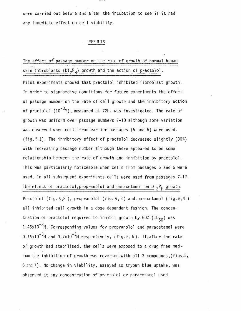
were carried out before and a f te r the incubation to see i f i t had
any immediate e f fe c t on ce ll v ia b i l i t y .
RESULTS.
The e ffe c t of* passage number on the rate o f growth o f normal human
skin f ib rob la s ts (DTqPh) growth and the action of p ra c to lo l .
P i lo t experiments showed tha t practo lo l in h ib ite d f ib ro b la s t growth.
In order to standardise conditions fo r fu ture experiments the e f fe c t
of passage number on the rate o f ce l l growth and the in h ib i to ry action_3
of practo lo l (10 M), measured at 72h, was investigated. The rate o f
growth was uniform over passage numbers 7-18 although some va r ia t ion
was observed when ce l ls from e a r l ie r passages (5 and 6) were used.
( f ig . 5,1). The in h ib i to ry e f fe c t o f practo lo l decreased s l ig h t ly (30%)
with increasing passage number although there appeared to be some
re la tionsh ip between the rate o f growth and in h ib i t io n by p rac to lo l.
This was p a r t ic u la r ly noticeable when ce l ls from passages 5 and 6 were
used. In a l l subsequent experiments ce l ls were used from passages 7-12.
The e f fe c t o f p ra c to lo l , propranolol and paracetamol on PT^P^ growth.
Practolol ( f ig . 5 , 2 ) , propranolol ( f ig . 5, 3) and paracetamol ( f ig . 5 , 4 )
a l l inh ib ited ce l l growth in a dose dependent fashion. The concen
t ra t io n of practo lo l required to in h ib i t growth by 50% (ID^q) was-3
1.45x10 M. Corresponding values fo r propranolol and paracetamol were
0.16x10 and 0.7x10 respective ly , ( f ig . 5, 5 ) . I f , a f t e r the rate
of growth had s ta b i l is e d , the ce lls were exposed to a drug free med
ium the in h ib i t io n of growth was reversed.with a l l 3 compounds,(figs.54
6 and 7). No change in v i a b i l i t y , assayed as trypan blue uptake, was
observed at any concentration o f practo lo l or paracetamol used.

Figure 5 , 1 .
Passage Number
The e ffe c t o f passage number on the rate o f DTgP f ib ro b la s t growth ( o ) and i t s in h ib i t io n by practo lo l ( lx lO “ ^M) ( # ) . <Parameters were measured a f te r 72h. (Results o f increase in ce l l number are the mean o f t r ip l i c a te samples.)
rela
tive
incr
ease
in
cell
num
ber
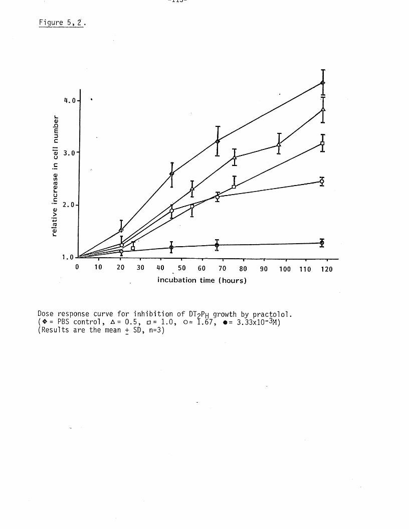
“ 1 X J "
Figure 5 , 2 .
a;AEDC
<uuc<U(0ro0)L.uc
ra<DL.
0 10 20 30 40 50 60 70 80 90 100 110 120in c u b a tio n tim e (h o u rs )
Dose response curve fo r in h ib i t io n o f DT2PH growth by p rac to lo l.( ♦ = PBS con tro l, A = 0.5, □= 1.0, 0= 1.67, •= 3.33xl0"3M)(Results are the mean + SD, n=3)

Figure 5, 3
4 .0 ,
t 1 r100 110 120
in c u b a t io n tim e (h o u r s )
Dose response curve fo r in h ib i t io n o f DT2Ph growth by propranolol.( ♦= PBS contro l. 0 = 0.5, A = l .o and @= 2 .0x l0 “ 4M)(Results are the mean + SD, n=3)
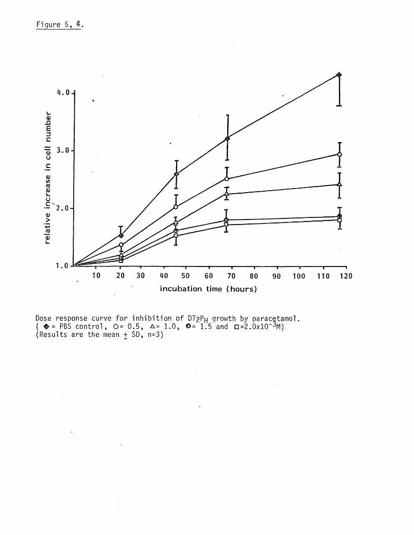
rela
tive
in
crea
se
in ce
ll n
um
ber
Figure 5, 4.
4.0
3.0
2 . 0
10 20 30 60 70 90 100 110 12080incubation time (h o u rs )
Dose response curve fo r in h ib i t io n o f DT2Ph growth by paracetamol.( ^ = PBS con tro l, 0 = 0.5, a = 1.0, ©= 1.5 and n=2.0xl0"^M)(Results are the mean + SD, n=3)
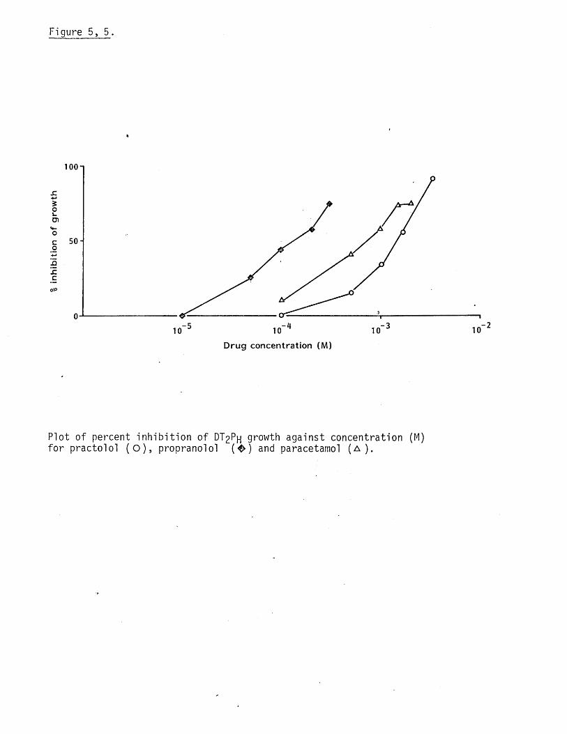
inhi
bitio
n of
gro
wth
Figure 5 , 5 .
100-1
50-
- 2- 3,"4- 5 10101010
Drug concentration (M)
Plot o f percent in h ib i t io n o f DT^Ph growth against concentration (M) fo r practolo l ( O ) , propranolol { & ) and paracetamol ( a ).
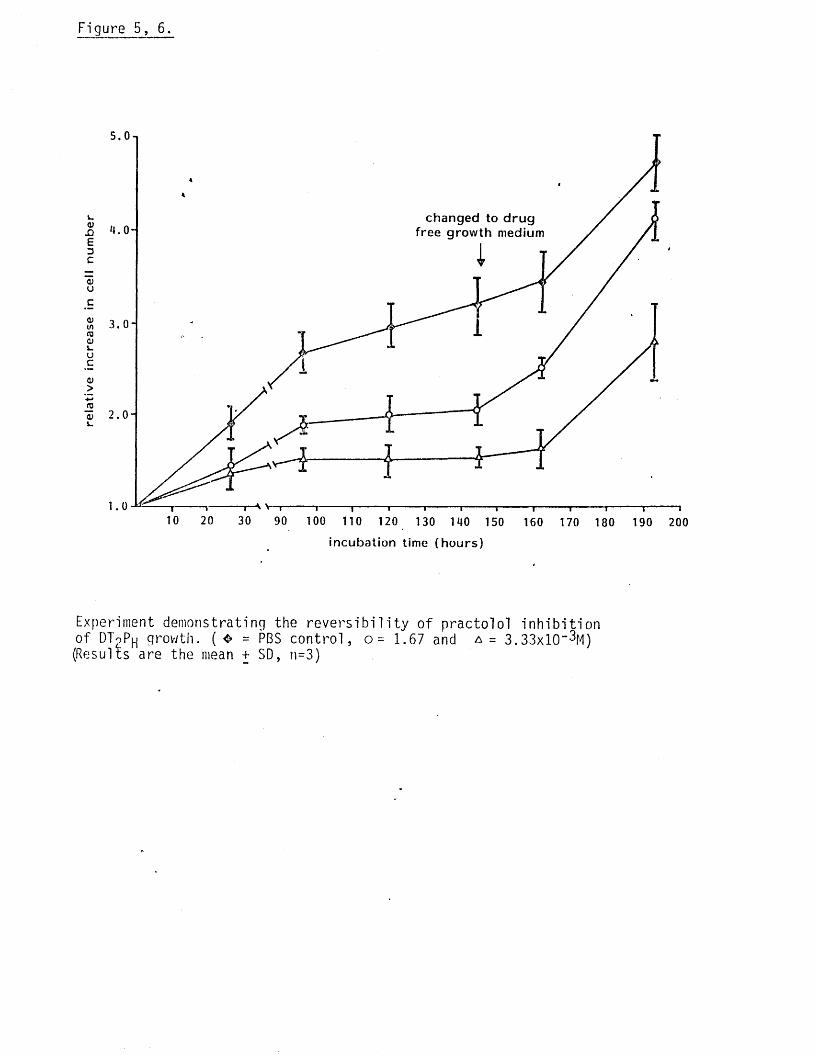
rela
tive
incr
ease
in
cell
num
ber
Figure 5, 6.
5.0
changed to d rug free growth medium
3 .0
2 . 0
10 20 30 90 100 110 120 130 140 150 160 170 180 190 200
incubation time (hours)
Experiment demonstrating the re v e rs ib i l i t y of practo lo l in h ib i t io nof DToP | growth. (❖ = PBS con tro l, 1.67 and a = 3.33x10“ ^M)(Results are the mean + SD, n=3)

Figure 5, 7.
£oL.U)
<Uu
(Ut/)rn<uL-Uc<D>4-»
a;
4
changed to d rug free medium ,3
2
125 50 70 90 110
incubation time (hours)
Experiment demonstrating the re v e rs a b i l i ty o f propranolol andparacetamol in h ib i t io n o f DT9PU growth. ( ® = PBS con tro l, a =
-3 -30.5x10 M paracetamol and n = 0.1x10 M propranolol).(Results are the mean + SD, n=3).
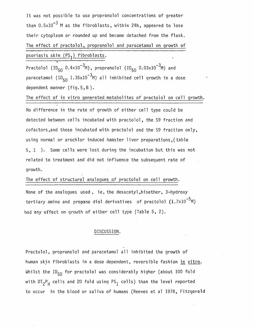
I t was not possible to use propranolol concentrations o f greater_3
than 0.5x10 M as the f ib ro b la s ts , w ith in 24h, appeared to lose
th e i r cytoplasm or rounded up and became detached from the f la sk .
The e ffe c t o f p ra c to lo l , propranolol and paracetamol on growth o f
psoriasis skin (PS^ ) f ib ro b la s ts .
Practolol (ID5Q 0.4xl0~^M), propranolol (ID^q 0.03x10 ^M) and-3paracetamol (ID^g 1.35x10 M) a l l inh ib ited ce l l growth in a dose
dependent manner ( f i g . 5 , 8 ) .
The e f fe c t o f in v i t r o generated metabolites o f practo lo l on ce l l growth.
No difference in the rate o f growth o f e ith e r ce ll type could be
detected between ce l ls incubated with p ra c to lo l, the S9 fra c t io n and
cofactors ,and those incubated with practo lo l and the S9 f ra c t io n only,
using normal or arochlor induced hamster l i v e r prepara tions,(tab le
5, 1 ). Some ce lls were lo s t during the incubation but th is was not
re lated to treatment and did not influence the subsequent rate of
growth.
the e f fe c t o f s truc tu ra l analogues o f practo lo l on ce ll growth.
None of the analogues used, ie , the desacety l,b isether, 3-hydroxy-5te r t ia r y amine and propane d io l derivatives o f practo lo l (1.7x10 M)
had any e f fe c t on growth of e ith e r ce ll type (Table 5, 2).
DISCUSSION.
P rac to lo l, propranolol and paracetamol a l l in h ib ite d the growth o f
human skin f ib rob la s ts in a dose dependent, revers ib le fashion in v i t r o .
Whilst the ID^g fo r practo lo l was considerably higher (about 100 fo ld
with DT P j ce l ls and 20 fo ld using PS c e l ls ) than the level reported
to occur in the blood or sa liva o f humans (Reeves et al 1978, F itzgera ld
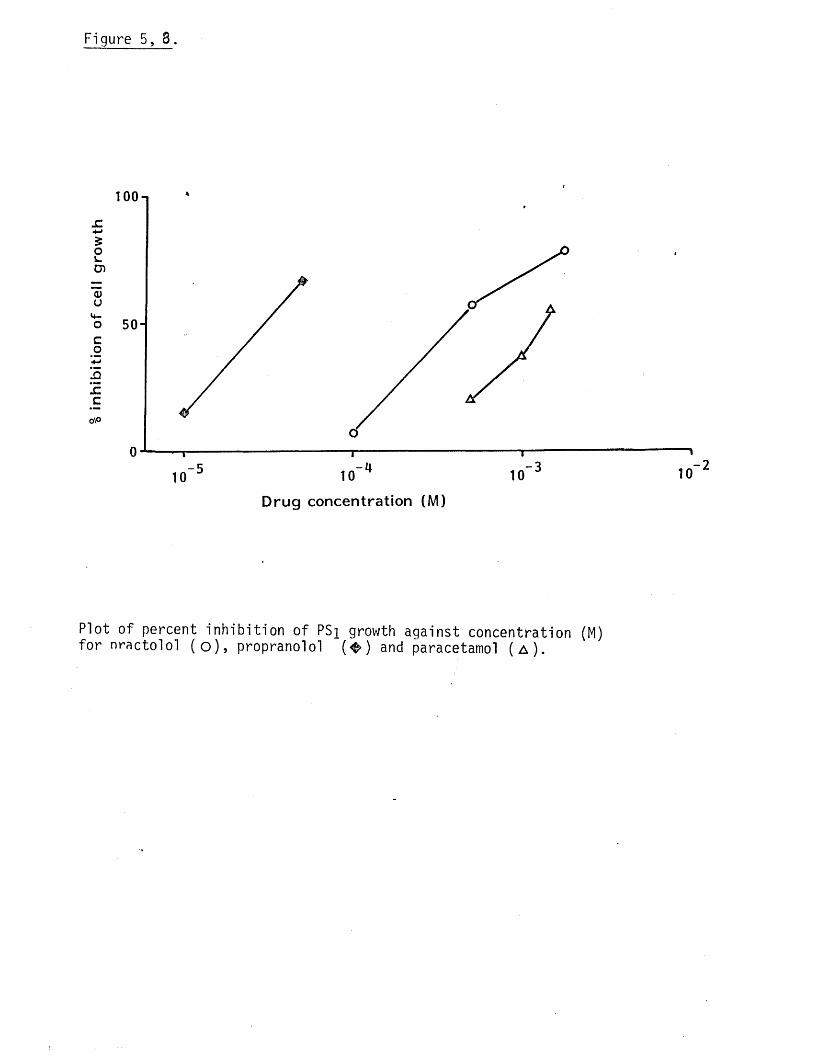
inhi
bitio
n of
ce
ll g
row
th
Figure 5 , 3 .
o\o
1 00 n
50-
T
10-5 -4
10
D rug concentration (M)
10-3 10- 2
Plot o f percent in h ib i t io n o f PSi growth against concentration (M) fo r nracto lo l ( o ) , propranolol ( ^ ) and paracetamol ( a ).
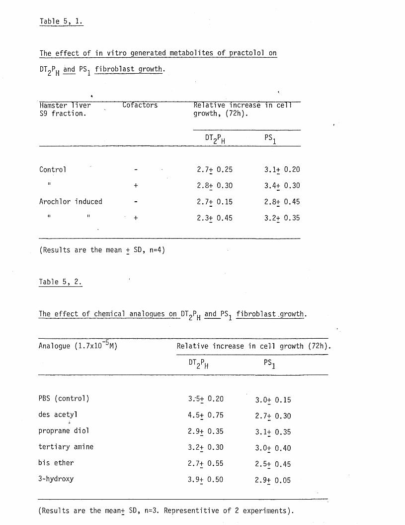
Table 5, 1.
The e f fe c t o f in v i t r o generated metabolites o f practo lo l on
DT2Ph and PS f ib ro b la s t growth.
*Hamster l i v e r S9 fra c t io n .
Cofactors Relative increase growth, (72h).
DT2PH
in cel 1
PS1
Control 2.7+ 0.25 3.1+ 0.20
II + 2.8+ 0.30 3.4+ 0.30
Arochlor induced - 2.7+ 0.15 2.8+ 0.45
n ii + 2.3+ 0.45 3.2+ 0.35
(Results are the mean + SD, n=4)
Table 5, 2.
The e ffe c t o f chemical analogues on DTqP and PS f ib ro b la s t .growth.
Analogue (1.7x10 ^M) Relative increase in ce ll growth ( 72h),
DT2PH PS1
PBS (contro l) 3.'5+ 0.20 3.0+ 0.15
des acetyl 4.5+ 0.75 2.7+ 0.30
proprane dio l 2.9+ 0.35 3.1+ 0.35
te r t ia r y amine 3.2+ 0.30 3.0+ 0.40
bis ether 2.7+ 0.55 2.5+ 0.45
3-hydroxy 3.9+ 0.50 2.9+ 0.05
(Results are the mean+ SD, n=3. Representitive o f 2 experiments).
- 1 2 2 -
and Scales 1968,Bodem et al 1972), tissue levels o f the drug could be
high enough (NB they are 5 fo ld higher in the beagle (Scales and
Cosgrove 1970)) fo r some in h ib i t io n o f f ib ro b la s t growth to occur
in vivo. In contrast, in v i t r o generated metabolites o f p ra c to lo l,
using normal or arochlor induced hamster l iv e rs , had no e f fe c t on
f ib ro b la s t growth implying tha t reactive metabolite formation may
not be necessary fo r growth. Amos et al successfully used hamster l i v e r
preparations, comparable to those used in the present inve s t iga t io n ,
to generate m etabo lite /prote in complexes that bound spec if ic antibodies
in the serum o f practo lo l patients with the oculomucocutaneous syndrome.
(Amos et al 1978). I t seems reasonable to assume tha t metabolites
formed during these experiments were s im ila r q u a l i ta t iv e ly to those
which have been claimed to form haptens in adversely effected patients .
In support o f the contention tha t metabolites of practo lo l are not
responsible fo r the e f fe c t on f ib ro b la s t growth, the s truc tu ra l an
alogues o f practo lo l also had no e f fe c t under the conditions used. A-5concentration o f 1.67x10 M o f the s truc tu ra l analogues was used in
these studies, about 1% of the dose of practo lo l needed to in h ib i t
ce ll growth by 50%. Reeves et al (1978) reported that no sing le met
abo lite o f practo lo l exceeded 2% o f recovered a c t iv i t y in human vo l- 14unteers given C practo lo l o ra l ly . In view o f the l i k e ly blood and
serum levels o f p ra c to lo l, and m e ta b o li te s , i t is probable tha t the
concentration o f analogues used reflected the in vivo s i tu a t io n .*
Whilst an e f fe c t due to a novel metabolite cannot be. ruled out these
resu lts ind icate th a t , in v iv o , metabolites o f p ra c to lo l, e ith e r at
the s i te o f formation or system ica lly , are un like ly to influence
f ib ro b la s t growth.
Fibroblasts derived from skin taken from an uninvolved area o f a
psoriasis pa tien t, a c l in ic a l condition characterised by ce l l
p r o l i te ra t io n , were more sensitive than normal ce lls to practo lo l
and propranolol but were less sensitive towards paracetamol. Whilst
not proving tha t f ib rob la s ts taken from patients with p ro l i fe ra t iv e
skin conditions are always more sensitive to 3-receptor blocking
drugs than ce lls from people without skin complaints these resu lts<
do ind icate that in h ib i ta ry e ffec ts on f ib rob la s ts due to the two
3- receptor blocking agents and paracetamol do not always pa ra l le l
each other and that some people may be more sensitive to such drugs
than others.
CHAPTER 6.
4
THE EFFECT OF PRACTOLOL, IN VITRO GENERATED METABOLITES
OF PRACTOLOL AND CHEMICAL ANALOGUES ON HUMAN SKIN
FIBROBLAST COLLAGEN SYNTHESIS.

INTRODUCTION.
P rac to lo l, propranolol and paracetamol inh ib ited the growth of
human f ib ro b la s ts in a dose dependent, revers ib le , fashion, although
the concentrations needed were higher than would generally be expec-
ted to occur in v ivo . In v i t r o generated metabolites o f practo lo l
and chemical analogues were without e f fe c t suggesting tha t the adverse
e ffects observed in patients were due to the parent compound. PS
ce l ls were more sensitive to in h ib i t io n by practo lo l and propranolol
but less sensitive to paracetamol. This find ing indicated tha t in h ib
i t io n due to the 3-receptor blocking drugs and paracetamol do not
always pa ra l le l each other and tha t some people are more sens it ive to
3-receptor blocking drugs than others.
I t is possible tha t a spe c if ic ce l l function could be e ffected at a
much lower concentration o f the chemicals than ce ll growth. In view
of i t s importance in re la tionsh ip to f ib ro b la s t function , collagen
synthesis is a l i k e ly ta rge t. Goldberg et al (1980) fo r example dem
onstrated tha t in h ib i t io n o f ce ll growth and collagen synthesis are
not always linked or even fo llow the same pattern.
Green and Goldberg (1964) reported tha t human skin f ib rob la s ts in
cultu re re ta in the a b i l i t y to synthesise collagen fo r well over 20
passages. This process was dependent on an adequate supply o f ascorbic
acid, now known to be necessary fo r pro line hydroxylation
(Stassen et al 1973), and pro line . Harrington et al (1973) also added
in su lin to th e ir cultures to aid v ia b i l i t y and transport across the
ce ll membrane, (Trowel 1 1959). Collagens are characterised as having
a high content of g lyc ine, proline and, uniquely, hydroxyproline
derived from pro line . Collagen synthesis can be assayed therefore by

measuring the incorporation o f radio!abelled pro line in to collagenase
sensitive prote in over a fixed incubation period, (Green and Goldberg
1964, V is tica et al 1977). This approach enables the incorporation
of pro line in to the d i f fe re n t soluble peptides and collagens to be
followed. This can be important when investiga ting lesions using
diseased tissue such as inflammed gingival f ib rob las ts (Narayanan
et al 1978) or the e f fe c t o f chemicals on collagen synthesis, fo r
example hydralazine ( Chen et al 1977),as much o f the hydroxyproline
containing proteins remain in the medium (Green and Goldberg 1964).
Layman et al .(1971), however demonstrated tha t collagen extracted
from the ce ll layer o f cu lt iva ted human skin f ib rob la s ts was s im ila r
to tha t extracted from human skin in i t s ra t io o f hydroxyproline
to p ro line , chain composition, s o lu b i l i t y and resistance to proteo
l y t i c d igestion. This was not so fo r hydroxyproline containing pep
tides iso lated from the medium. Changes in collagen synthesis are
re flected in the amount o f collagen deposited in the ce l l laye r,
(Layman et al 1971) and. Harrington et al (1973) measured collagen in
that compartment using a chemical assay fo r hydroxyproline a f te r acid
digestion of the ce l l mat. This method would appear to be sa t is fa c to ry
fo r the measurement o f gross changes in collagen synthesis.
Fibroblasts from patients have been used to measure collagen synthesis
in such conditions as scleroderma (LeRoy et al 1974, Buckingham et al
1978) and rheumatoid a r t h r i t i s (P r ie s t ly 1980) and to investigate
the mode o f action o f such drugs as D-penicillamine and sodium s a l ic y la te
(P r ie s t ly 1980) and sodium gold thiomalate (Goldberg et al 1980).
Normal ce l l l ines have also been used to monitor the e ffec ts o f ind
u s tr ia l chemicals on collagen synthesis, fo r example lead (V is t ica
et al 1977) polyvinyl chloride dust (Richards et al 1975) and drugs

such as local anaesthetics (Eichhorn and Peterkofski 1979), hydral
azine (Chen et al 1977) and des ferioxamine (Hunt et al 1979).
Enhancing and in h ib i to ry e ffec ts have been reported (Goldberg et al
1980, P r ie s t ly 1980) ind ica ting tha t th is system would be su itab le
fo r investiga ting the e ffec ts o f practo lo l and i t s metabolites
on collagen synthesis in v i t r o .
Financial constraints prevented the use o f rad io labelled pro line fo r
measuring collagen formation which was assayed by measuring to ta l
hydroxyproline levels in the ce ll layer a f te r acid hydrolysis.
Although ascorbic acid is needed fo r optimal collagen synthesis Aleo
et al (1974) suggested that in some circumstances differences in c o l l
agen formation might only be observed in an ascorbic acid d e f ic ie n t
medium. Experiments were also carried out therefore using a medium in
which ascorbic acid had been . le ft out.
METHODS.
Routine cu ltu r ing o f ce l ls and preparation o f the hamster l i v e r S9
fractions were performed as described previously (see chapter 5).
DNA ana lys is .
DNA analysis was carried out according to the method of Burton (1956).
The ce ll samples were spun down at 1500g and 4°C fo r lOmin using a
MSE Coolspin cen tr ifuge, washed once in 2ml o f 0.5M perch lo ric acid
and then heated fo r lOmin at 80°C in 1ml o f 1M perch lo ric acid. A
stock solu tion was prepared by d issolv ing a known weight o f c a l f thymus
DNA in 0.t)lM sodium hydroxide. Standard tubes containing 0-20pg o f DNA
and 1ml o f 1M perch loric acid were s im i la r ly heated. Once the samples
had cooled, 2ml o f a 200:1 diphenyl amine/2% acetaldehyde reagent was
added,(diphenyl amine reagent:- 1.5% diphenyl amine w/v and 1.5% 5N
sulphuric acid in g lac ia l acetic acid). The tubes were then l e f t at
room temperature overnight to allow the colour to develop and the
absorbance read at 600nm. The DNA concentration was read d i re c t ly
from the ca lib ra t io n curve.
Hydroxyproline Analys is .
The hydroxyproline assay is based on the method o f Woessner (1961).
The ce ll sample was spun down at 1500g fo r lOmin at 4°C in 2ml glass
centrifuge tubes and the supernatant discarded. Hydrochloric acid
(250 1/ 5N) was added to each tube which was then sealed using an
oxygen/coal gas flame. The tubes were incubated overnight at 120°C,
opened and the contents transferred to 10ml glass te s t tubes. The
hydrolysate was bought to pH 6.0-8.0 by adding 250ul o f 5N sodium hydrox
ide and then adjusting the ph using d ilu ted hydrochloric acid or
sodium hydroxide. Chloramine T (1ml) was added, the solutions vortex
mixed and l e f t at room temperature fo r 20min, (chloramine T reagent:-
0.05 M chloramine T, 20% d is t i l l e d water, 30% methyl ce lluso lve and 50%
hydroxyproline bu ffe r ph 6.0. Hydroxyproline b u f fe r : - 0.238 M c i t r i c
acid monohydrate, 0.96 M anhydrous sodium acetate, 0.85M sodium hydroxide.
1.2% g lac ia l acetic acid v/ v) . Excess chloramine T was then neu tra l
ised by adding 1ml 3.15 M perchloric acid. A fte r a fu r th e r 5min at
room temperature 1ml of 20% p-dimethylamine-benzaldehyde in methyl -
cellusolve (w/v) was added, the solutions mixed and heated at 60°C
fo r 20min to develop the chromophore. The tubes were cooled fo r 5min
in cold water and the absorbance read at 557nm.
Collagen "synthesis experiments.
Stock ce l ls wsre grown as previously described and detached by incu-
bating with tryps in . A liquots o f the ce ll suspension (5ml, 0.5x10 c e l ls ) ,

in growth medium,containing foeta l c a l f serum, were added to 50mm
tissue cu ltu re Petri dishes and allowed to reach confluency,
normally about 3 days. The medium was then changed to one containing
donnor c a l f serum (DCS) and the ce lls allowed to eq u il ib ra te fo r 2
days. Collagen synthesis was in i t ia te d (day 0) by incubating the ce lls
with DCS medium supplemented with lmg/ml L -p ro line , 50/ig/ml ascorbic
acid and 1 unit/m l in su lin BP, (collagen stim ula ting medium). The
medium, plus any compound under inves tiga tion , was renewed every two
days. The material under inves tiga tion , or an equivalent volume of
PBS, was added on day 0 and collagen synthesis assayed on day 9 unless
other wise stated. Collagen synthesis was assayed as fo llows. The
incubation medium was decanted and the P e tr i dish placed over a ten-
p la te. The ce ll layer was divided in h a lf using a scalpel blade and
one h a lf analysed fo r DNA and the other fo r hydroxyproline. Values
are expressed as yg hydroxyproline/lOOyg DNA.
In some experiments collagen synthesis was in i t ia te d on day 0 and the
chemical under investiga tion added some days la te r . These experiments
were designed to see i f p re in i t ia t io n o f collagen synthesis had any
e f fe c t on the drugs action.
The method was modified s l ig h t ly when the e f fe c t o f in v i t r o generated
metabolites o f practo lo l on collagen formation was studied. P rac to lo l,
the S9 fra c t io n and cofactors were mixed in 25ml conical flasks as
previously described (see chapter 5). The growth medium (FCS) was
deccinted from the P e tr i dishes and the ce lls washed once with PBS.
The metabolite generating system was added and the ce lls incubated
fo r lh at 37°C in a CO tissue culture incubator. The medium was then
discarded, the ce lls washed twice with PBS and 5ml of the DCS, collagen
stim ulating medium added.
14The e f fe c t of practo lo l on C leucine uptake by DTpP^ f ib ro b la s ts .
_3Cells were incubated with 0.5x10 M practo lo l fo r 9 days. On day 8 lyCi
o f 14C leucine was added to each Petr i dish and l e f t fo r 24h. On day
9 lml o f the medium was removed,for counting the a c t iv i t y o f the med
ium, and the ce ll layer washed twice with PBS. The ce ll monolayer was4
then divided in h a lf and one h a lf analysed fo r hydroxyproline. The
other h a lf was digested (lml ce lluso lve , 24h,37°C) and added to 10ml14of Dimilune s c in t i l l a t io n f lu id . C a c t iv i t y was counted using a
Packard Tri-carb l iq u id s c in t i l l a t io n spectrophotometer. Uptake o f 14C leucine was expressed as counts per minute (cpm) in the cells/cpm
ce lls + medium.14Uptake and retention o f C practo lo l by DTqP f ib ro b la s ts . •
F ibroblast cultures were established as previously described and14collagen synthesis in i t ia te d on day 0. On days 3 or 6, 0.2 yCi C
-3practo lo l (0.5x10 M) was added and renewed every 2 days w ith the
collagen stim ulating medium. On day 9 lml o f the medium was counted fo r
ra d io a c t iv ity and h a lf the f ib ro b la s t monolayer analysed fo r hydroxy-14proline and the other h a lf fo r C a c t iv i t y as described above.
RESULTS.
Analysis o f DNA.
The DNA ca lib ra t io n curve was l in e a r over the range 0-20yg DNA
( f ig . 6,1 ). The re la tionsh ip between DNA content and ce ll number was
investigated by assaying DNA in known numbers of c e l ls . There was ag
linea r re la tionsh ip up to at least 2x10 ce lls ( f i g . 6,2).
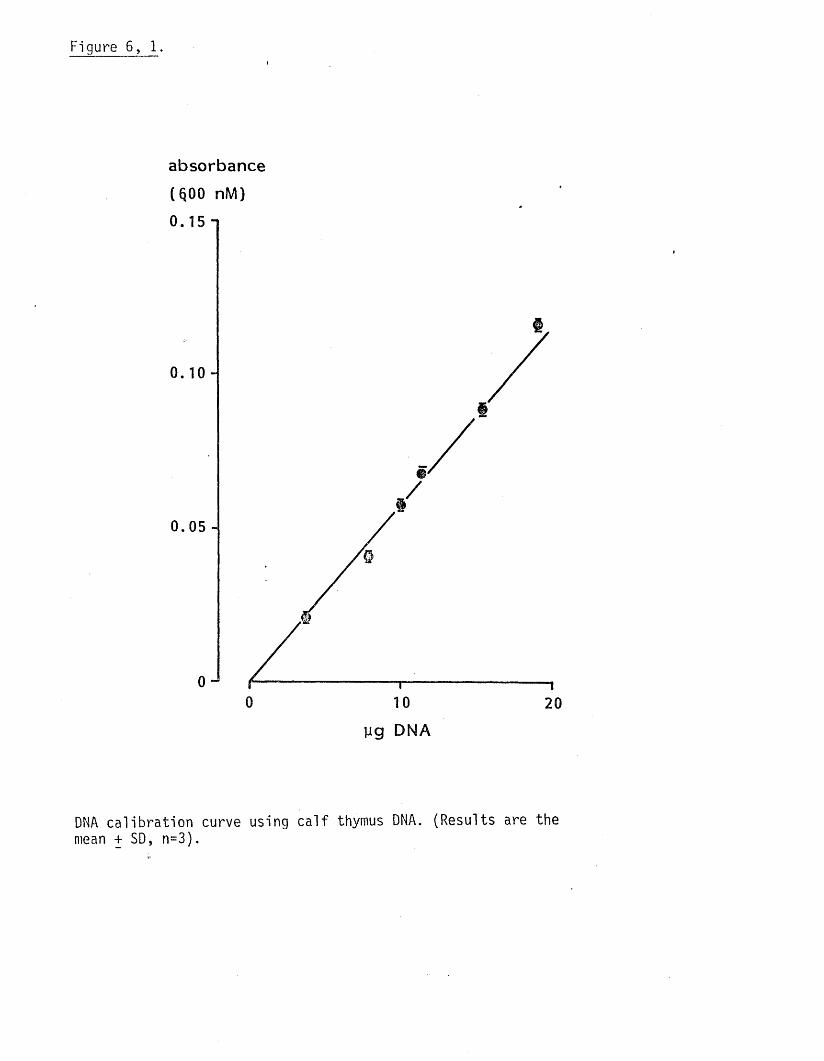
Figure 6, 1.
absorbance (§00 nM)
0.05 -
0
Ug DNA
DNA ca lib ra t io n curve using c a l f thymus DNA. (Results are the mean + SD, n=3).
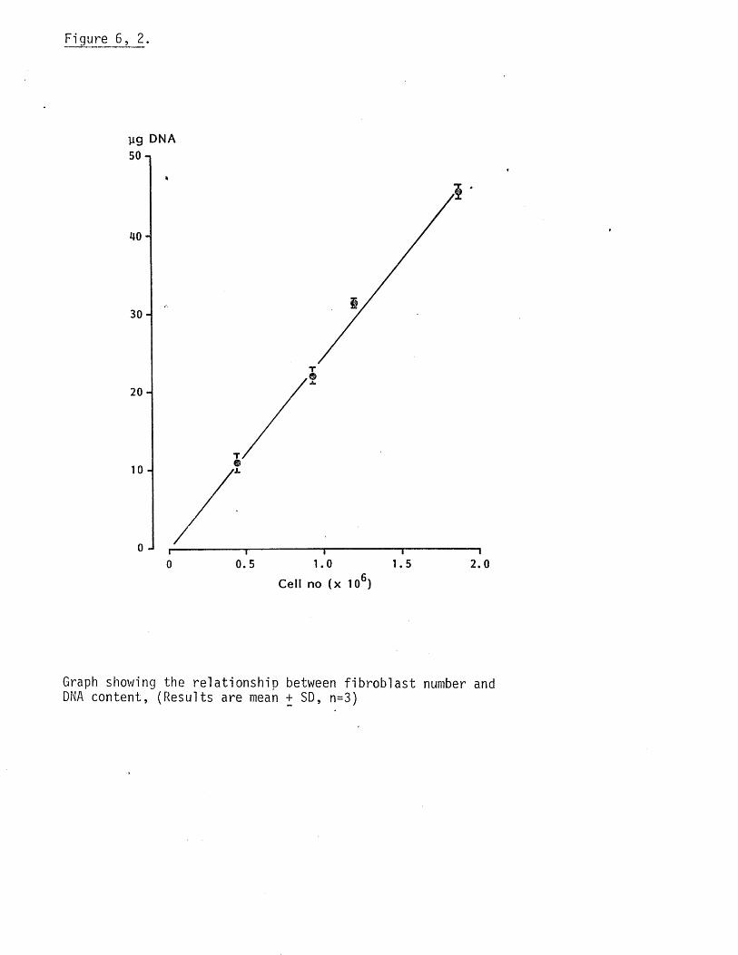
Figure 6, 2.
jig DNA
T
2 0 -
T
'i.
0 0.5 1.0 1.5 2.0
Cell no (x 106)
Graph showing the re la tionsh ip between f ib ro b la s t number and DNA content, (Results are mean + SD, n=3)
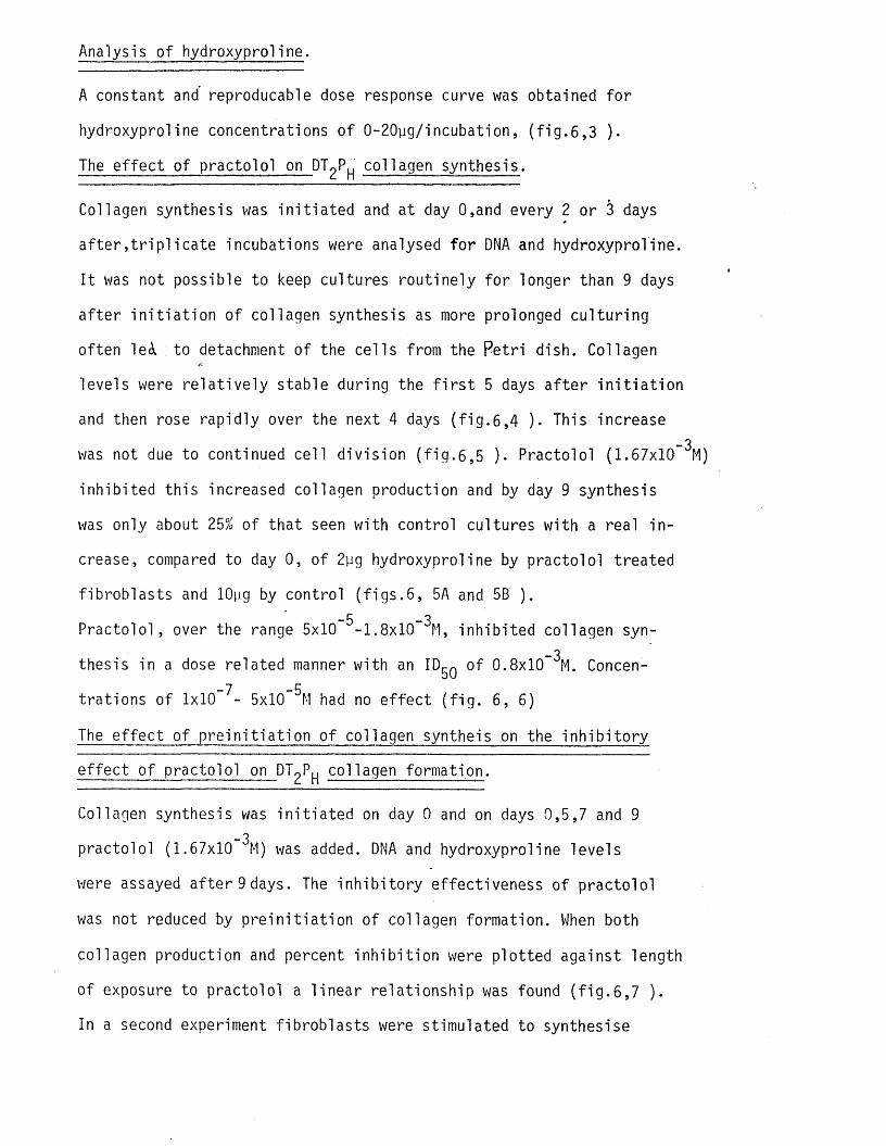
Analysis o f hydroxyproline.
A constant and reproducable dose response curve was obtained fo r
hydroxyproline concentrations o f 0-20pg/incubation, ( f i g . 6,3 ).
The e f fe c t o f practo lo l on DTqP collagen synthesis.
Collagen synthesis was in i t ia te d and at day 0,and every 2 or 3 days
a f t e r , t r ip l i c a te incubations were analysed fo r DNA and hydroxyproline.
I t was not possible to keep cultures rou tine ly fo r longer than 9 days
a f te r in i t ia t io n o f collagen synthesis as more prolonged cu ltu r ing
often le<i to detachment o f the ce l ls from the Petr i dish. Collagen
levels were re la t iv e ly stable during the f i r s t 5 days a f te r i n i t i a t io n
and then rose rap id ly over the next 4 days ( f i g . 6,4 ). This increase_3
was not due to continued ce ll d iv is ion ( f i g . 6,5 ). Practolol (1.67x10
inh ib ited th is increased collagen production and by day 9 synthesis
was only about 25% of tha t seen with control cultures with a real in
crease, compared to day 0, of 2yg hydroxyproline by practo lo l treated
fib rob la s ts and lOpg by control ( f i g s . 6, 5A and 5B ).-5 -3P rac to lo l, over the range 5x10 -1.8x10 M, inh ib ited collagen syn-
_3thesis in a dose related manner with an ID ^ o f 0.8x10 M. Concen
tra t ions o f 1x10 7- 5xl0~^M had no e f fe c t ( f ig . 6, 6)
The e f fe c t of p re in i t ia t io n o f collagen syntheis on the in h ib i to ry
e f fe c t o f practolo l on DTqP collagen formation.
Collagen synthesis was in i t ia te d on day 0 and on days 0,5,7 and 9_3
practo lo l (1.67x10 M) was added. DNA and hydroxyproline levels
were assayed a f te r 9 days. The in h ib i to ry effectiveness of p racto lo l
was not reduced by p re in i t ia t io n of collagen formation. When both
collagen production and percent in h ib i t io n were p lo tted against length
o f exposure to practo lo l a l in e a r re la t ionsh ip was found ( f i g . 6,7 ).
In a second experiment f ib rob la s ts were stimulated to synthesise

Figure 6, 3.
absorbance
( 557 mM)
1 . 0 -}
10 200
]ig O H -pro line
Hydroxyproline ca l ib ra t io n curve. (Results are the mean + SD, n=3)
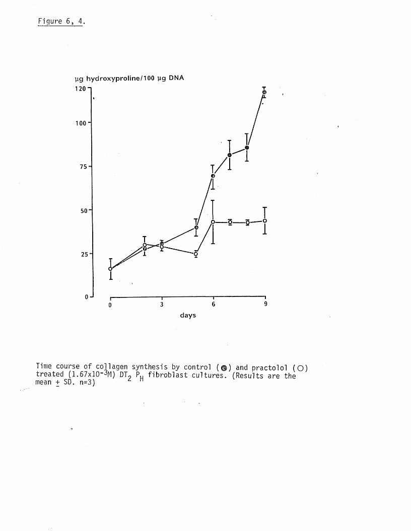
Figure 6, 4.
yg h y d ro x y p ro lin e /100 yg DNA
1 20 1
75-
50"
25"
0 — 5 -------§
days
Time course o f collagen synthesis by control (© ) and practo lo l (O ) treated (1.67xlCMM) DT9 P„ f ib ro b la s t cu ltures. (Results are the mean + SD. n=3)
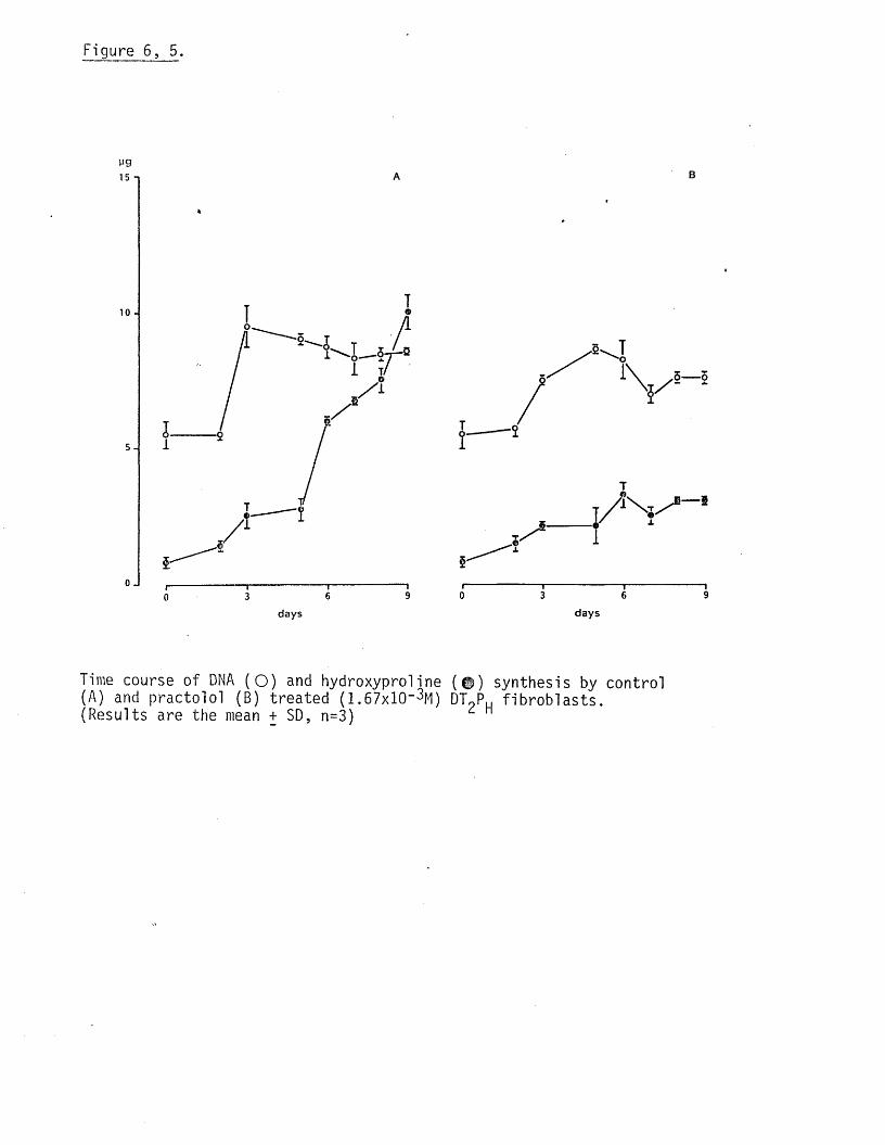
Figure 6, 5.
yg15 n
10 -
5— 5
days days
Time course of DNA (O ) and hydroxyprol ine ( $ ) synthesis by control (A) and practolo l (B) treated (1.67x10“ %) DTpPu f ib ro b la s ts . (Results are the mean + SD, n=3)

Figure 6, 6.
yg hydroxyproline/100 jag DNA
70 n
60-
50 J
40
30
20
10
( 1001)(105%)
t/)QQQ_
(103%)
_ooucok.a
I
oo■MuCOk.Q.
LDIO
XLD
(59%)
oo
■ i- jucok.Q.
Io
Xm
(46%)
oo+-»ucok.a
COIo
Xo
(24%) T
col — ° O
mmmoXA u
0 0 CO . k.
*— o.
Dose response curve fo r in h ib i t io n of Dl^Pf-j collagen synthesis by p rac to lo l. (Results are the mean + SD, n=3)
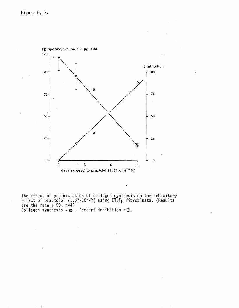
yg hydroxyproline/TOO yg DNA 120
1 0 0 -
75
50-
25
A
30 6 9
% inhibition r 100
75
50
_ 3days exposed to practolol (1 .67 x 10 M)
- 25
L 0
The e ffe c t o f p re in i t ia t io n o f collagen synthesis on the in h ib i to ry e f fe c t o f practo lo l (1.67x10“ 3M) using DT2P f ib ro b la s ts . (Results are the mean ± SD, n«4)Collagen synthesis = © . Percent in h ib i t io n = 0 .
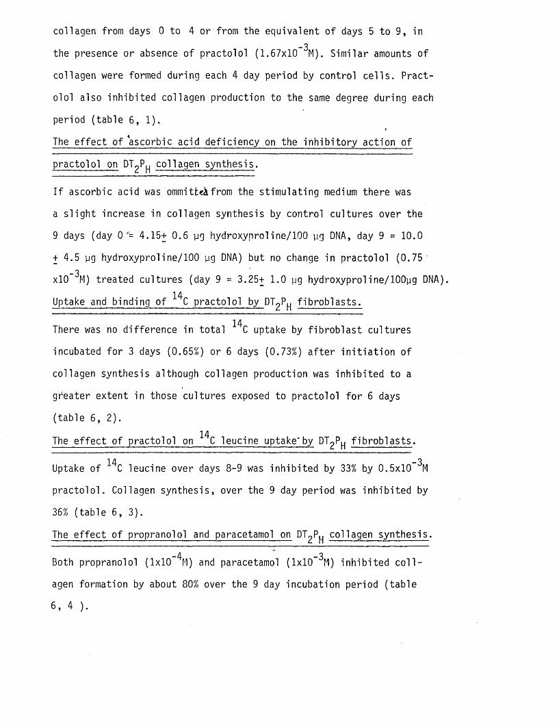
collagen from days 0 to 4 or from the equivalent o f days 5 to 9, in_3
the presence or absence o f practo lo l (1.67x10 M). S im ilar amounts o f
collagen were formed during each 4 day period by control c e l ls . Pract
o lo l also inh ib ited collagen production to the same degree during each
period (table 6, 1).
The e ffe c t o f ascorbic acid defic iency on the in h ib i to ry action o f
practo lo l on DT^P^ collagen synthesis.
I f ascorbic acid was ommitfce^from the stim ula ting medium there was
a s l ig h t increase in collagen synthesis by control cultures over the
9 days (day 0'= 4.15+ 0.6 yg hydroxyproline/100 yg DNA, day 9 = 10.0
+ 4.5 yg hydroxyproline/100 yg DNA) but no change in practo lo l (0 .75 '
xlO ^M) treated cultures (day 9 = 3.25+ 1.0 yg hydroxyproline/lOOyg DNA).14Uptake and binding o f C practo lo l by DTqP f ib ro b la s ts .
14There was no d ifference in to ta l C uptake by f ib ro b la s t cultures
incubated fo r 3 days (0.65%) or 6 days (0.73%) a f te r in i t i a t io n o f
collagen synthesis although collagen production was in h ib ite d to a
greater extent in those cultures exposed to practo lo l fo r 6 days
(tab le 6, 2).
14The e f fe c t o f practo lo l on C leucine uptake'by DT£P f ib ro b la s ts .
14 - 3Uptake o f C leucine over days 8-9 was in h ib ite d by 33% by 0.5x10 M
prac to lo l. Collagen synthesis, over the 9 day period was in h ib ite d by
36% (tab le 6, 3).
The e f fe c t o f propranolol and paracetamol on DT^P^ collagen synthesis.
-4 ~ -3Both propranolol (1x10 M) and paracetamol (1x10 M) inh ib ited c o l l
agen formation by about 80% over the 9 day incubation period (tab le
6, 4 ).

Table 6, 1.
The e f fe c t o f prolonging the e q u i l ib ra t io n stage (before in i t i a t io n
of collagen synthesis) on collagen formation and the in h ib i to ry
e f fe c t o f practo lo l using DT P j c e l l s .
period of collagen y g hydroxyproline/lOOy g DNA ^ in h ib i t io nsynthesis.-------------------------- --------- ---------------------------------------- due to p ra c to lo l.
control practo lo l(1.67xlO-3M)
days 0-4 32.3+4.0 20.8+3.0 36%
(n=5) (n=5)
days 5-9 27.8+4.4 16.1+2.2 41%
(n=6) (n=6)
(Results are the mean + SD, n= number of rep lica tes ).
Table 6, 2.
14The uptake and binding o f C,practolo l by DTqP f ib ro b la s ts .
_ .
days exposed to C uptake. % uptake yg hydroxyproline
practo lo l ( counts/min(0 .2pc i , 0.5x10_3M) x lO '3^.
0 ’ ---- 9.21+0.40 (100%)
3 1.83+0.2 0.73% 7.67+ 0.40 ( 83%)
6 1.63+0.6 0.65% 5.35+ 0.30 ( 58%)
(Results are the mean + SD, n=5).

Table 6, 3.
14The e f fe c t o f practo lo l on uptake of C leucine by DT^P f ib ro b la s ts .
treatment 14C leucine uptake * over days 8-9
_3(counts/min.xlO )
%uptake pg hydroxyproline (formed over 9 days)
control 112.7+11.40 6.35% 8.0+1.50
practo lo l 75.3+3.90 4.25 % 5.0+0.9(0.5x10~3M)
s.
% in h ib i t io n due 33% 36%to practolo l •
(Results are the mean + SD, n=5)
Table 6 , 4 .
The e ffe c t of p ra c to lo l, propranolol and paracetamol on DTgP^
collagen synthesis.
treatment pg hydroxyproline/lOOpg DNA % in h ib i t io n
control 116.25+1.50 0
practolo l 40.15+5.5 65%(1.67x10’ 3M)
Propranolol 21.40+4.0 82%
(1x10~4M).
paracetamol 27.75+5.0 76%(1x10"3M)
(Results are the mean + SD,n=3).
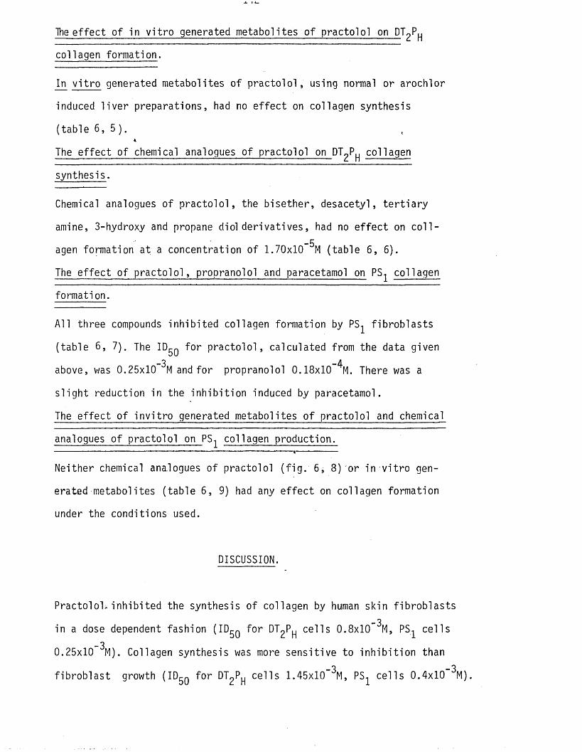
The e f fe c t of in v i t r o generated metabolites o f practo lo l on DTqP
collagen formation.
In v i t r o generated metabolites o f p ra c to lo l, using normal or arochlor
induced l iv e r preparations, had no e f fe c t on collagen synthesis
(table 6 , 5 ) .%
The e ffe c t of chemical analogues of practo lo l on DTgP collagen
synthesis.
Chemical analogues of p ra c to lo l , the b ise ther, desacetyl, te r t ia r y
amine, 3-hydroxy and propane diol d e r iva t ive s , had no e f fe c t on c o l l --5agen formation at a concentration o f 1.70x10 M (table 6, 6).
The e f fe c t o f p ra c to lo l, propranolol and paracetamol on PS col 1agen
formation.
A ll three compounds inh ib ited collagen formation by PS f ib ro b la s ts
(table 6, 7). The ID^q fo r p ra c to lo l, calculated from the data given-3 -4above, was 0.25x10 M and fo r propranolol 0.18x10 M. There was a
s l ig h t reduction in the in h ib i t io n induced by paracetamol.
The e f fe c t o f in v i t ro generated metabolites o f practo lo l and chemical
analogues o f practo lo l on PS collagen production.---------- ■ ■■■-■— ■ ■■— ............ ........... ■ —* ■■ ■- - - i i —......
Neither chemical analogues o f practo lo l ( f ig . 6, 8) or in v i t r o gen
erated metabolites (tab le 6, 9) had any e f fe c t on collagen formation
under the conditions used.
DISCUSSION.
Practolol. inh ib ited the synthesis of collagen by human skin f ib ro b la s ts_3
in a dose dependent fashion ( ID^q fo r DT^P ce l ls 0.8x10 M, PS c e l ls_3
0.25x10 M). Collagen synthesis was more sensitive to in h ib i t io n than
f ib ro b la s t growth (ID^q fo r DT P j ce l ls 1.45x10 ^M, PS c e l ls 0.4x10 ^

Table 6, 5.
The e f fe c t o f in v i t ro generated metabolites o f practo lo l on DTgPp
collagen synthesis.
source o f S9 fra c t ion pg hydroxyproline/lOO g DNA
- cofactors + cofactors
control l iv e rs 63.1+10 .0 61.4+3 .5
arochlo.r induced 65.1+3.5 57.7+4 .01i vers
(Results are the mean + SD, n=4)
Table 6, 6 .
The e f fe c t of chemical analogues o f practo lo l on DT^P collagen
synthesis.
chemical. concentration pg hydroxyproline/lOOpg DNA
experiment 1 experiment 2
PBS control 59 .6+10.0 80 .6+1 .0
practo lo l 1 .67 x1 0 '3M 23.8+6.7 30.8+1 .0
propane diol 1 .70x10‘ 5M 62.3+4.5 85 .1+12 .0
3-hydroxy 11 48.4+1 .0 78 .2+4 .5
des acety l II 43.5+3 .0 77 .3+22.0
t e r t i a r y amine u nd 92.3+7 .0
bis e ther II nd 76.2+1 .0
(Results are the mean + SD, n=3)
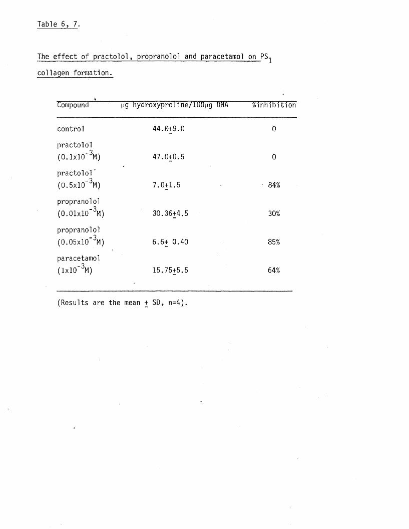
Table 6, 7.
The e f fe c t of p ra c to lo l, propranolol and paracetamol on PS
collagen formation.
Compound yg hydroxyproline/lOOyg DNA %inhib ition
control 44.0+9.0 0
practo lo l( 0 . 1 x 1 0 ' 3M) 47.0+0.5 0
p ra c to lo l“ ( 0 .5 x 1 0 '3M) 7.0+1.5 84%
propranolol( 0 . 0 1 x 1 0 ' 3M) 30.36+4.5 30%
propranolol
( 0 .0 5 x10"3M) 6.6+ 0.40 85%
paracetamol( I x10"3M) 15.75+5.5 64%
(Results are the mean + SD, n=4).
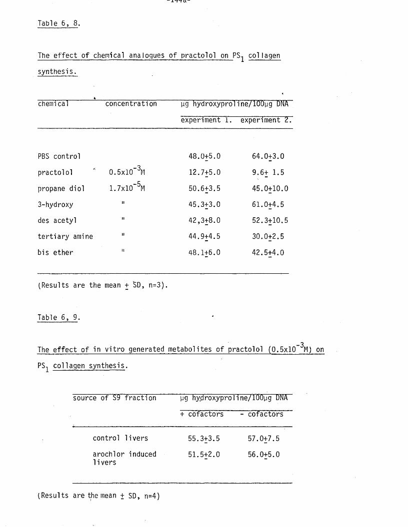
Table 6, 8.
The e f fe c t of chemical analogues o f practo lo l on PS-1 collagen
synthesis.
<chemi cal concentration yg hydroxyproli ne/lOOyg DNA
experiment 1. experiment 2.
PBS control 48.0+5.0 64.0+3.0
practolo l 0.5x10” 3M 12.7+5.0 9.6+ 1.5
propane d io l 1 .7 x1 0 '5M 50.6+3.5 45.0+10.0
3-hydroxy II 45.3+3.0 61.0+4.5
des acetyl II 42,3+8.0 52.3+10.5
te r t ia r y amine II 44.9+4.5 30.0+2.5
bis ether II 48.1+6.0 42.5+4.0
(Results are the mean + SD, n=3).
Table 6, 9 .
_3The e f fe c t o f in v i t r o generated metabolites o f practo lo l (0.5x10 M) on
PSl collagen synthesis.
source o f S9 fra c t io n yg hyjiroxyproline/TOOyg DNA
+ cofactors - cofactors
control l iv e rs 55.3+3.5 57.0+7.5
arochlor induced 51.5+2.0 56.0+5.01i vers
(Results are the mean + SD, n=4)

with a reduction in the ID^q o f about 40% with both ce ll types, pre
in i t i a t io n o f collagen synthesis did not a l te r the in h ib i to ry action
o f practo lo l ind ica ting tha t exposure time and drug concentration were
the main factors in fluencing the degree o f in h ib i t io n . The in h ib i to ry
action of practo lo l was not altered by incubating ce lls in an ascorbic
acid free medium. In contrast to the e ffec ts o f practo lo l i t s e l f , in
v i t r o generated metabolites o f practo lo l had no e f fe c t on collagen
formation supporting the suggestion made in chapter 5 .tha t reactive
metabolites of p ra c to lo l, ie metabolites formed at the s i te o f the
reaction and capable o f binding to p ro te in , probably play no part in
the e ffec ts on f ib ro b la s t function . The find ing tha t chemical analogues
of practo lo l were also inactive at the concentrations used fu r th e r
supports the idea that practo lo l i t s e l f is the active compound. Pract-14o lo l inh ib ited both collagen formation and C leucine uptake by DTgP
f ib rob las ts to the same extent ind ica ting tha t i t may act by a lte r in g
amino acid a v a i la b i l i t y , possibly via a membrane e f fe c t . The re la t iv e ly
low concentrations o f practo lo l tha t accumulated w ith in the c e l ls tend
to support the view that practo lo l may exert i t s main e f fe c t on the
plasma membrane d ire c t ly . Collagen synthesis is only about 55-60% more
sensitive to in h ib i t io n tha t ce l l growth (see above). Thus i t remains
to be seen whether in h ib i t io n o f collagen synthesis is the major ad*-
verse e f fe c t o f low doses o f practo lo l on human f ib ro b la s ts .
Both propranolol and paracetamol inh ib ited collagen synthesis demon
s tra t ing tha t the in h ib i t io n by practo lo l was not spec if ic fo r th is
drug. In common with DT£P c e l ls , PS f ib rob la s ts were also not e ffe c
ted by in . v i t r o generated metabolites or chemical analogues. However
there were more sensitive to practo lo l and propranolol but showed l i t t l e
change in s e n s i t iv i t y towards paracetamol. S im ilar d ifferences in

s e n s i t iv i ty were observed when the e f fe c t o f the drugs on ce ll growth
was investigated. I t is c lear tha t there is some var ia tion in the res
ponse to the two 3-receptor blocking drugs used in th is study and tha t
s e n s i t iv i t y towards drugs whose structures are c losely re lated to tha t
of p ra c to lo l, such as paracetamol, need not pa ra l le l tha t o f the %
3-receptor blocking agents.
CHAPTER 7.
EXPERIMENTS AIMED AT INVESTIGATING THE EFFECT OF PRACTOLOL
ON PRODUCTION OF A FACTOR BY HAMSTER PERITONEAL MACROPHAGES
CAPABLE OF INFLUENCING HUMAN SKIN FIBROBLAST COLLAGEN
SYNTHESIS.
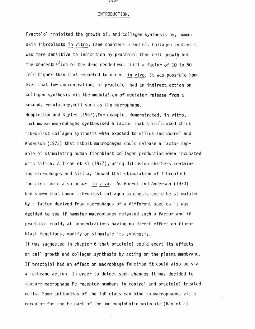
INTRODUCTION.
Practolol inh ib ited the growth o f , and collagen synthesis by, human
skin f ib rob la s ts in v i t r o , (see chapters 5 and 6). Collagen synthesis
was more sensitive to in h ib i t io n by practo lo l than ce ll growth but *
the concentration o f the drug needed was s t i l l a fac to r o f 10 to 50
fo ld higher than that reported to occur in v ivo . I t was possible how
ever tha t low concentrations o f practo lo l had an in d ire c t action on
collagen synthesis via the modulation o f mediator release from a
second, regu la to ry ,ce ll such as the macrophage.
Heppleston and Styles (1967),fo r example, demonstrated, in v i t r o ,
tha t mouse macrophages synthesised a fa c to r tha t stimululated chick
f ib ro b la s t collagen synthesis when exposed to s i l i c a and Burrel and
Anderson (1973) tha t rabb it macrophages could release a fa c to r cap
able o f stim ula ting human f ib ro b la s t collagen production when incubated
with s i l i c a . A ll ison e t al (1977), using d if fu s io n chambers contain
ing macrophages and s i l i c a , showed tha t s tim u la tion o f f ib ro b la s t
function could also occur in v ivo . As Burrel and Anderson (1973)
had shown that human f ib ro b la s t collagen synthesis could be stimulated
by a fac to r derived from macrophages of a d i f fe re n t species i t was
decided to see i f hamster macrophages released such a fa c to r and i f
practolo l could, at concentrations having no d ire c t e f fe c t on f ib r o
b last functions, modify or stimulate i t s synthesis.
I t was suggested in chapter 6 tha t practo lo l could exert i t s e ffec ts
on ce ll growth and collagen synthesis by acting on the plasma membrane.
I f practq lo l had an e f fe c t on macrophage function i t could also be via
a membrane action. In order to detect such changes i t was decided to
measure macrophage Fc receptor numbers in control and practo lo l treated
c e l ls . Some antibodies o f the IgG class can bind to macrophages via a
receptor fo r the Fc part o f the immunoglobulin molecule (Hay et al
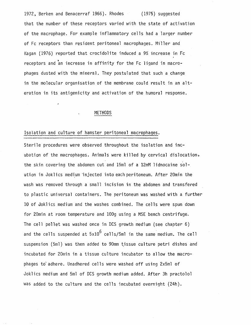
1972, Berken and Benacerraf 1966). Rhodes (1975) suggested
that the number o f these receptors varied with the state o f ac t iva t ion
of the macrophage. For example inflammatory ce l ls had a larger number
of Fc receptors than resident peritoneal macrophages. M i l le r and
Kagan (1976) reported tha t c ro c id o l i te induced a 9% increase in Fc
receptors and an increase in a f f in i t y fo r the Fc ligand in macro
phages dusted with the mineral. They postulated tha t such a change
in the molecular organisation o f the membrane could re su lt in an a l t
eration in i t s an t ig e n ic i ty and ac tiva t ion o f the humoral response.
METHODS
Iso la tion and cultu re o f hamster peritoneal macrophages.
S te r i le procedures were observed throughout the iso la t io n and inc
ubation o f the macrophages. Animals were k i l le d by cervica l d is loca tion*
the skin covering the abdomen cut and 15ml o f a 12mM lidnocaine s o l
ution in Joklics medium 'injected in to each peritoneum. A fte r 20min the
wash was removed through a small inc is ion in the abdomen and transfered
to p la s t ic universal containers. The peritoneum was washed with a fu r th e r
10 of Joklics medium and the washes combined. The ce lls were spun down
fo r 20min at room temperature and lOOg using a MSE bench centrifuge.
The ce ll p e l le t was washed once in DCS growth medium (see chapter 6)g
and the ce lls suspended at 5x10 cells/5ml in the same medium. The ce ll
suspension (5ml) was then added to 90mm tissue cu ltu re pe tr i dishes and
incubated fo r 20min in a tissue cultu re incubator to allow the macro
phages to" adhere. Unadhered ce lls were washed o f f using 2x5ml o f
Joklics medium and 5ml o f DCS growth medium added. A fte r 3h p racto lo l
was added to the culture and the ce lls incubated overnight (24h).
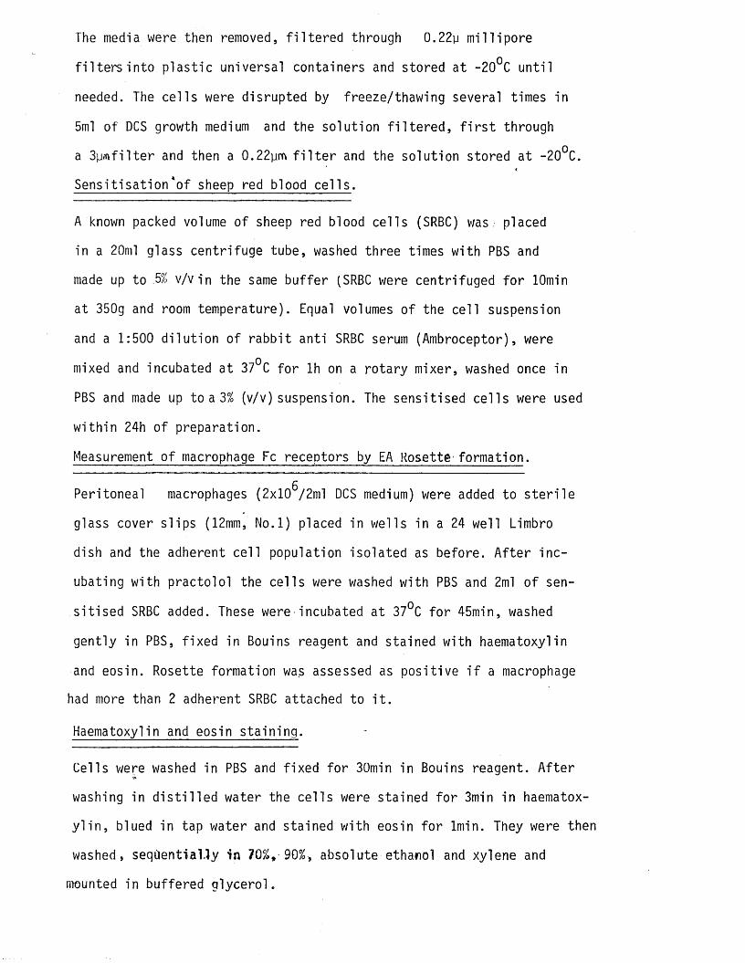
The media were then removed, f i l t e r e d through 0.22y m il l ip o re
f i l t e r s in to p la s t ic universal containers and stored at -20°C u n t i l
needed. The ce lls were disrupted by freeze/thawing several times in
5ml o f DCS growth medium and the solu tion f i l t e r e d , f i r s t through
a 3pm filte r and then a 0.22ym f i l t e r and the so lu tion stored at -20°C.
Sens it isa t ion*o f sheep red blood c e l ls .
A known packed volume o f sheep red blood ce lls (SRBC) was . placed
in a 20ml glass centrifuge tube, washed three times with PBS and
made up to :5% v /v in the same bu ffe r (SRBC were centrifuged fo r lOmin
at 350g and room temperature). Equal volumes o f the ce ll suspension
and a 1:500 d i lu t io n o f rabb it anti SRBC serum (Ambroceptor), were
mixed and incubated at 37°C fo r lh on a ro tary mixer, washed once in
PBS and made up to a 3% (v/v) suspension. The sensitised ce l ls were used
w ith in 24h o f preparation.
Measurement o f macrophage Fc receptors by EA Rosette formation.
gPeritoneal macrophages (2x10 /2ml DCS medium) were added to s te r i le
glass cover s l ips (12mm, No.l) placed in wells in a 24 well Limbro
dish and the adherent ce l l population iso lated as before. A fte r in c
ubating with practo lo l the ce lls were washed with PBS and 2ml o f sen
s it ise d SRBC added. These were incubated at 37°C fo r 45min, washed
gently in PBS, fixed in Bouins reagent and stained with haematoxylin
and eosin. Rosette formation was assessed as pos it ive i f a macrophage
had more than 2 adherent SRBC attached to i t .
Haematoxylin and eosin s ta in in g .
Cells were washed in PBS and fixed fo r 30min in Bouins reagent. A fte r
washing in d is t i l l e d water the ce l ls were stained fo r 3min in haematox
y l i n , blued in tap water and stained with eosin fo r lmin. They were then
washed, seqOentiaUy in 70%, 90%, absolute ethanol and xylene and
mounted in buffered g lyce ro l.

Experiments to investigate the e ffe c t o f control and practo lo l
treated macrophage incubation media and ce ll contents on DT^P
f ib ro b la s t collagen synthesis.
F ibroblast cultures were set up as previously described and c o l l
agen synthesis in i t ia te d on day 0. On day 5 macrophage medium, from
control or practo lo l treated cu ltu res, was added to a f in a l concen
t ra t io n o f 40% v/v. Cell contents were added to a f in a l concentration
o f 20% v/v also on day 5. In su l in , ascorbic acid and pro line were
added to the collagen stim ula ting medium to ensure the correct con-
centrations in the f in a l incubation volume. This was renewed, together
with macrophage medium or ce ll contents, on day 7. On day 9 the ce ll
layer was analysed fo r DNA and hydroxyproline.
RESULTS
The e ffe c t o f hamster macrophage incubation medium and ce ll contents
on collagen synthesis by DT^P^ f ib ro b la s ts , the possible influence
of p ra c to lo l .
Hamster peritoneal macrophages did not secret, or s tore, any fac to r
capable of in fluencing collagen synthesis in v i t r o at the concen
t ra t io n o f ce l l contents or incubation medium used. Practolol (1.25 -4xlO M) did not induce the release or synthesis o f a fib rogenic
fac to r or e f fe c t collagen synthesis in any other way under the
conditions o f the experiment (tab le 7, 1.'). The f in a l concentration-5of practolo l in the f ib ro b la s t cutures was 2.5x10 M (ce ll contents
-5experiments) and 5x10 M (macrophage incubation medium experiments).
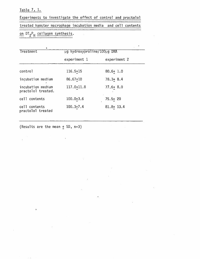
Table 7, 1.
Experiments to investigate the e f fe c t o f control and practo lo l
treated,hamster macrophage incubation media and ce ll contents
on DT2Ph collagen synthesis.
Treatment pg hydroxyproline/lOOpg DNA
experiment 1 experiment 2
control 116.5+15 80.6+ 1.0
incubation medium 86.67+10 78.3+ 8.4
incubation medium 117.0+11.8 77.6+ 8.0practo lo l treated.
ce ll contents 100.0+3.6 75.5+ 20
ce ll contents 100.3+7.4 81.8+ 13.4practo lo l treated
(Results are the mean + SD, n=3)
The e f fe c t o f practolo l on hamster macrophage Fc receptor expression.
Practolol caused a dose related decrease in the number o f Fc receptors,_3
measured by EA rosette formation, with an ID ^ o f 1.3x10 M, ( f ig 7 ,1 . /2 ) .
DISCUSSION.
Although practo lo l had no apparent e f fe c t on the synthesis o f a
fibrogenic fac to r by hamster macrophages, f t did induce a reduction
in the number o f Fc receptors. As mentioned in the Introduction, an
increase in Fc" receptor number is associated with macrophage ac t iva t io n .
The reverse re la tionsh ip has not been shown, ie a decrease in Fc rec
eptor number is correlated with a decrease in macrophage a c t iv i t y , but
such a re la tionsh ip might be antic ipated. In support o f th is concept
M^rland and M0rland (1982) reported tha t chronic consumption o f ethanol
induced a decrease in the phagocytosis o f sheep erythrocytes by ra t
peritoneal macrophages in v i t r o , a process dependent upon Fc receptors,
and that th is change was’ related to an increased s u s c e p t ib i l i ty to
endotoxin. The authors suggested tha t these e ffec ts re flec ted a gen
eral depression o f macrophage a c t iv i t y .
The Fc receptor is important in mediating opsonization, phagocytosis,
antibody mediated c y to to x ic i ty and, possib ly, in determining qual
i ta t iv e and quantative aspects o f local inflammatory responses (Walker
and Demus 1975, G r i f f in et al 1975, Passwell e t al 1980).
Kavai et al (1980) demonstrated tha t human monocyte Fc receptors
regenerated w ith in 4-8h a f te r shedding due to cold treatment. I f these
receptors are con tinua lly turning over then the in h ib i to ry action o f
p ra c to lo l, in depressing Fc receptor number, could be a re f le c t io n of
the general e f fe c t on protein synthesis id e n t i f ie d in the previous
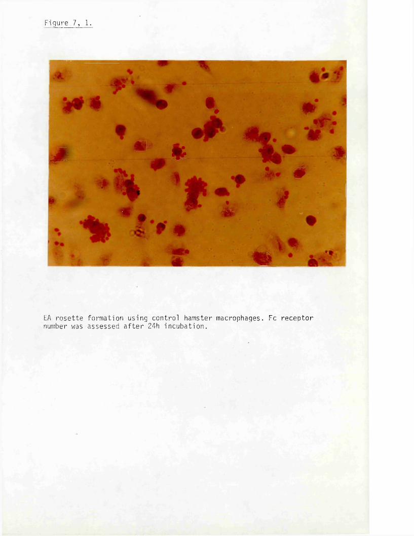
Figure 7, 1.
•.m - *^ 0 #
*
m
EA r o s e t t e formation using contro l hamster macrophages. Fc re c ep to r number was assessed a f t e r 24h incubat ion .
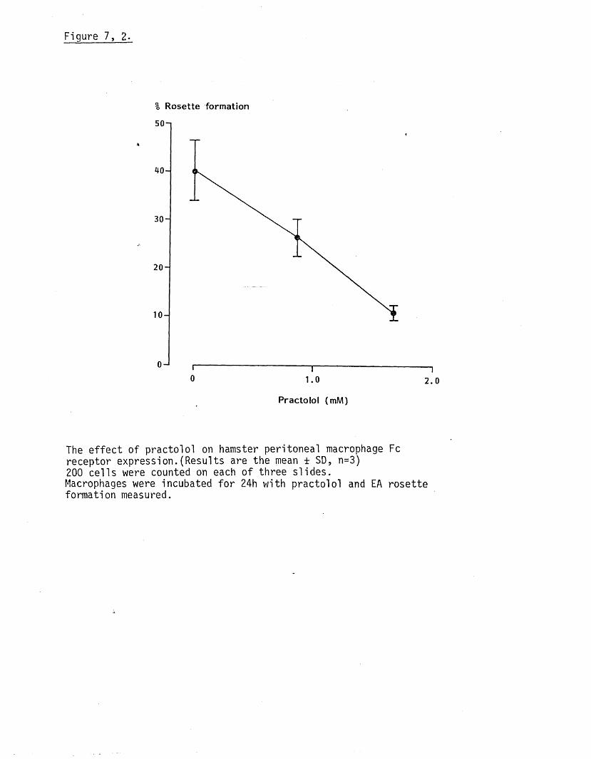
Figure 7, 2.
% Rosette formation
50-1
' H I0 1 . 0 2 . 0
Practolol (mM)
The e f fe c t of practo lo l on hamster peritoneal macrophage Fc receptor expression.(Results are the mean ± SD, n=3)200 ce lls were counted on each o f three s lides .Macrophages were incubated fo r 24h with practo lo l and EA rosette formation measured.

Chapter. Both leucine uptake and collagen synthesis were inh ib ited by_3
about 70% by 1.67x10 M p rac to lo l. I t is notable tha t a s im ila r degree
o f in h ib i t io n of receptor expression was observed in the presence of
the same concentration o f p rac to lo l. However, as the ID^q fo r in h ib
i t io n of hamster macrophage Fc receptor expression was s im ila r to4
that fo r in h ib i t io n o f ce l l growth o f DT^P^ f ib rob las ts (1.3 and 1.4_3
xlO M respective ly) i t is probable that receptor changes re f le c t
gross a lte ra t ions in macrophage function.
CHAPTER 8
4
THE EFFECT OF PRACTOLOL, PARACETAMOL AND PROPRANOLOL
on d i2ph MORPHOLOGY AFTER IN VITRO EXPOSURE.

INTRODUCTION
During experiments to investigate the e f fe c t of p ra c to lo l, pro
pranolol and paracetamol on ce ll growth i t was noted tha t morph
ological changes occured which d if fe red depending on the drug used.
No changes were observed when DT^P^ ce lls were exposed to in, v i t ro
generated metabolites or chemical analogues. The a lte ra t ions in
morphology were investigated in more de ta il and the resu lts reported
in th is Chapter.
METHODS
Fibroblasts were grown in 50mm tissue cu ltu re P e tr i dishes fo r l ig h t
microscopy studies o f growing c e l ls . When ce lls were needed fo r
s ta in ing or electron microscope investigations theij were grown on
mellanex s tr ip s placed in the P e tr i dishes.
Staining fo r neutral l ip id .
The ce ll monolayer was rinsed once in PBS and fixed in Bouins reagent
fo r 30min. A fte r r ins ing in d is t i l l e d water and 60% isopropyl a lc
ohol there were stained fo r lOmin with o i l red 0, rinsed with 60%
isopropyl alcohol, to d i f fe re n t ia te the s ta in , and rinsed with d is t
i l l e d water. Counterstaining was carried out using Mayers haematoxylin
(2min). The ce lls were f in a l l y washed in running tap water and mounted
in buffered g ly c e ro l.
Staining fo r carbohydrate.
Cells were washed in PBS and fixed in a c i t ru s acid bu ffe r fo r 2min,
rinsed in d is t i l l e d water and stained with 1% periodic acid fo r 5min.
A fte r r ins ing with d is t i l l e d water they were stained with Schiffs
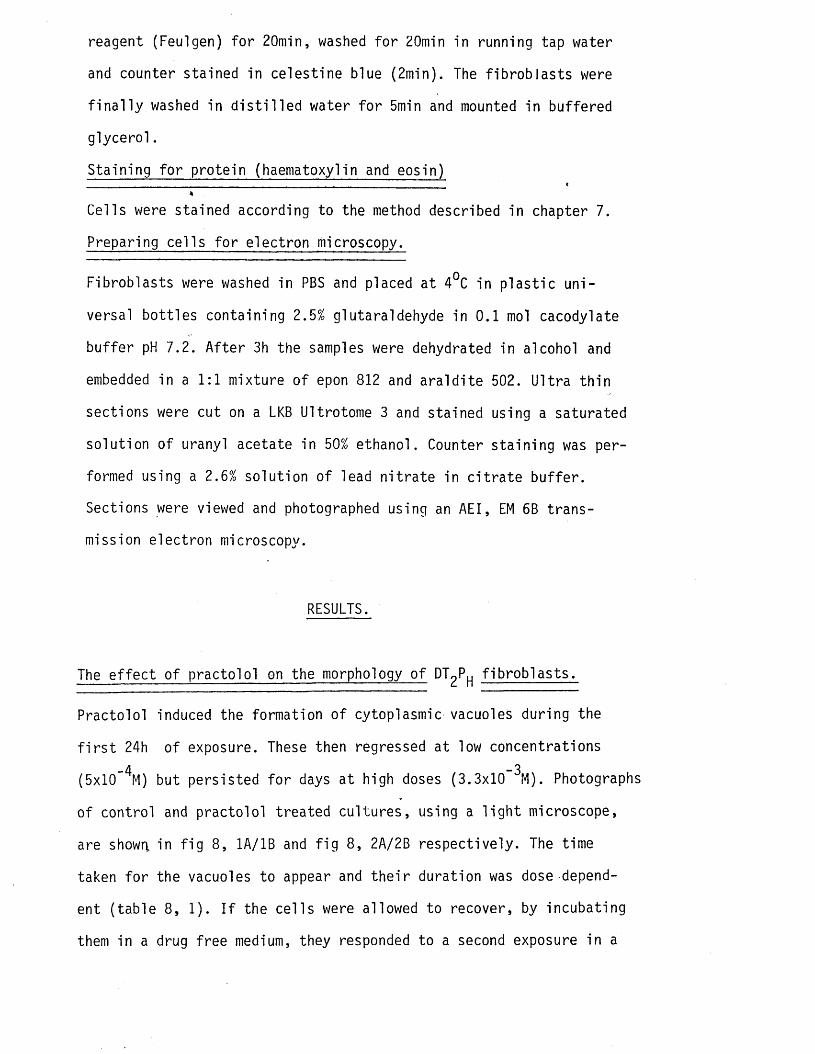
reagent (Feulgen) fo r 20min, washed fo r 20min in running tap water
and counter stained in ce lestine blue (2min). The f ib rob la s ts were
f i n a l l y washed in d is t i l l e d water fo r 5min and mounted in buffered
g lyce ro l.
Staining fo r protein (haematoxylin and eosin)_ c4
Cells were stained according to the method described in chapter 7.
Preparing ce lls fo r electron microscopy.
Fibroblasts were washed in PBS and placed at 4°C in p la s t ic un i
versal bo tt les containing 2.5% glutaraldehyde in 0.1 mol cacodyl ate
bu ffer pH 7.2. A fte r 3h the samples were dehydrated in alcohol and
embedded in a 1:1 mixture o f epon 812 and a ra ld ite 502. U ltra th in
sections were cut on a LKB Ultrotome 3 and stained using a saturated
solu tion o f uranyl acetate in 50% ethanol. Counter s ta in ing was per
formed using a 2.6% solu tion o f lead n i t ra te in c i t ra te bu ffe r.
Sections were viewed and photographed using an AEI, EM 6B trans
mission electron microscopy.
RESULTS.
The e f fe c t of practo lo l on the morphology o f OT^P f ib ro b la s ts .
Practolol induced the formation o f cytoplasmic vacuoles during the
f i r s t 24h of exposure. These then regressed at low concentrations
(5xlO” ^M) but persisted fo r days at high doses (3.3x10 ^M). Photographs
of control and practo lo l treated cu ltu res , using a l ig h t microscope,
are shown, in f ig 8, 1A/1B and f ig 8, 2A/2B respective ly. The time
taken fo r the vacuoles to appear and th e i r duration was dose depend
ent (tab le 8, 1). I f the ce l ls were allowed to recover, by incubating
them in a drug free medium, they responded to a second exposure in a

-101-

Figure 8, 1.
A. Low power photograph o f control DT^P^ ce lls using a
phase contrast lens.
Cells were cultured fo r 24h. Lens:- 10/.25t
4The f ib rob la s ts are mostly spindle shaped with long
processes. An e l ip t ic a l nucleus can ju s t be seen.
B. High power photograph o f control DT^P ce lls using a
phase contrast lens.
Cells were incubated fo r 93h. Lens:- 20/.32
Cellsshow the cha rac te r is t ic spindle shape. The nucleus can
be c le a r ly seen and contains two or three nuc leo li.

1VL

-163-

High power photograph o f practo lo l treated DT^P ce lls
using a phase contrast lens.
Cells were cultured in a drug free medium fo r 93h and*
then fo r 24h with 3.3x10 p ra c to lo l . L e n s 20/.32
Vacuolation caused the ce lls to swell and become round
ed. Although some vacuoles appear to have formed w ith in
the nucleus they are probably in the cytoplasm over-
ly ing i t .
High power photograph o f OT^P ce lls recovering a f te r
practo lo l treatment. Phase contrast lens._3
Cells were incubated with 3.3x10 M practo lo l fo r 24h
and then transfered to a drug free medium, The photo
graph was taken 24h la te r and shows the reduction in vac
uole number and size. Lens:- 20/.32


Table 8, 1.
The e f fe c t o f practo lo l on the formation o f cytoplasmic vacuoles
in f ib ro b la s ts .
time a f te r exposure
to practo lo l (h).
percent vacuole formation 1.67xlO’ 3M 3.3xlO~3Mpractolo l practo lo l
2 5+ 4 20+ 7
16 18+9 100
24 * 57+ 12 100
39 11+9 100
68 0 100
96 0 100
(Results are the mean + SD, n=3). 100% vacuole formation occured when a l l the ce lls contained vacuoles.
Table 8, 2.
The e ffe c t of preexposure to practo lo l on vacuole formation in DTqP
f ib rob las ts on a subsequent exposure to p ra c to lo l .
time a f te r exposure
to practolo l (h).percentage vacuol e formation.f i r s t exposure
to 1.67x10’ 3M
practolo l
second exposure to 1.67x10’ 3M
practo lo l
2 0 4+ 4
,16 12+ 7 17+ 11
24 49+ 15 47+ 13
40 15+ 8 11+ 6
(Results are the mean + SD, n=3. Cells were incubated with practo lo lfo r 96h follou>4 by 24h in a drug free medium and then 40h with p ra c to lo l.Vacuole formation was followed during each incubation period with p ra c to lo l.
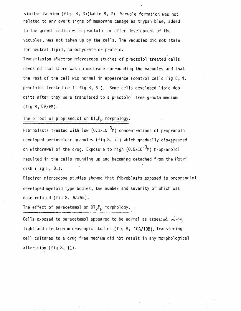
s im ila r fashion ( f ig . 8, 3 ) ( table 8, 2). Vacuole formation was not
related to any overt signs o f membrane damage as trypan blue, added
to the growth medium with practolo l or a f te r development o f the
vacuoles, was not taken up by the c e l ls . The vacuoles did not sta in
fo r neutral l i p id , carbohydrate or protein.
Transmission electron microscope studies o f practo lo l treated ce lls
revealed that there was no membrane surrounding the vacuoles and that
the rest o f the ce ll was normal in appearence (control ce l ls f ig 8, 4.
practolo l treated ce lls f ig 8, 5 . ) . Some ce lls developed l i p id dep
os its a f te r they were transfered to a practo lo l free growth medium
( f ig 8, 6A/6B).
The e f fe c t o f propranolol on DT^P morphology.
_3Fibroblasts treated with low (0.1x10 M) concentrations o f propranolol
developed perinuclear granules ( f ig 8, 7.) which gradually disappeared_3
on withdrawal o f the drug. Exposure to high (0.5x10 M) Propranolol
resulted in the ce lls rounding up and becoming detached from the Petri
dish ( f ig 8, 8 .) .
Electron microscope studies showed tha t f ib rob la s ts exposed to propranolol
developed myeloid type bodies, the number and severity of which was
dose related ( f ig 8, 9A/9B).
The e ffe c t o f paracetamol on DTpP morphology. -
Cells exposed to paracetamol appeared to be normal as assessed
l ig h t and electron microscopic studies ( f ig 8, 10A/10B). Transfering
ce ll cultures to a drug free medium did not re su lt in any morphological
a lte ra t io n ( f ig 8, 11).
Figure 8, 3.
The e f f e c t of preexposure to p rac to lo l on vacuole formation by DT^P^
f i b r o b l a s t s reexposed to p r a c t o l o l .-3Cel ls were incubated with 3.33x10 M p rac to lo l fo r 24h and then in a
drug f ree medium u n t i l the vacuoles had reg ressed . They were then_3
reexposed to 3.3x10 M prac to lo l . The photograph was taken 24h l a t e r .
Lens:- 20/ .32

- 168-
Figure 8 , 4 .
Transmission electron photomicrograph of acontrol DT^P cel 1.
Cells were incubated fo r 93h. Magnification 30,000.
The appearence is tha t o f a quiescent c e l l . There is com
para tive ly l i t t l e rough endoplasmic reticulum (re) with
narrow cisternae. A few mitochondria (m) and an area con
ta in ing golgi apparatus (g) can also be seen. L ip id drop
le ts (1) and lysosomes ( ly ) are scarce. Cytoplasmic f i l a
ments ( c f i ) are present throughout the cytoplasm.
Figure 8, 5.
Transmission electron photomicrograph o f a practo lo l treated
DT2Ph ce11-
Cells were incubated fo r 96h in growth medium and fo r 24h_3
in the presence o f 3.3x10 M p rac to lo l. Magnification 15,000.
Cytoplasmic vacuoles (v) can be seen in the upper o f the
two c e l ls . There is no membrane and the contents are not
electron dense. A small vacuole can be seen (T) in the top
of the lower ce l l which appears to be causing the plasma
membrane to balloon out. I t is not possible to say i f th is
was due to a cytotoxic reaction ie a bleb, or ju s t to the
chance o f a vacuole occuring at the ce ll surface. The rest
o f the ce l l appears normal.

I , ' % !a
fW K y . k 0 M *?
Zkf n • •• ' ;'

— 1 / u —
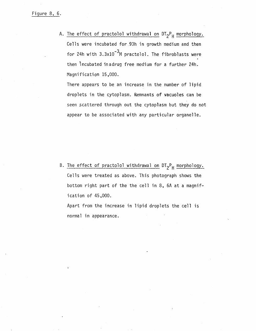
The e ffe c t of practo lo l withdrawal on DT^P^ morphology.
Cells were incubated fo r 93h in growth medium and then_3
fo r 24h with 3.3x10 M p rac to lo l. The f ib rob la s ts were
then incubated inadrug free medium fo r a fu r th e r 24h.
Magnification 15,000.
There appears to be an increase in the number o f l ip id
droplets in the cytoplasm. Remnants o f vacuoles can be
seen scattered through out the cytoplasm but they do not
appear to be associated with any p a r t ic u la r organelle.
The e f fe c t o f practo lo l withdrawal on OT^P morphology.
Cells were treated as above. This photograph shows the
bottom r ig h t part of the the ce ll in 8, 5A at a magnif
ica tion o f 45,000.
Apart from the increase in l i p id droplets the ce ll is
normal in appearance.
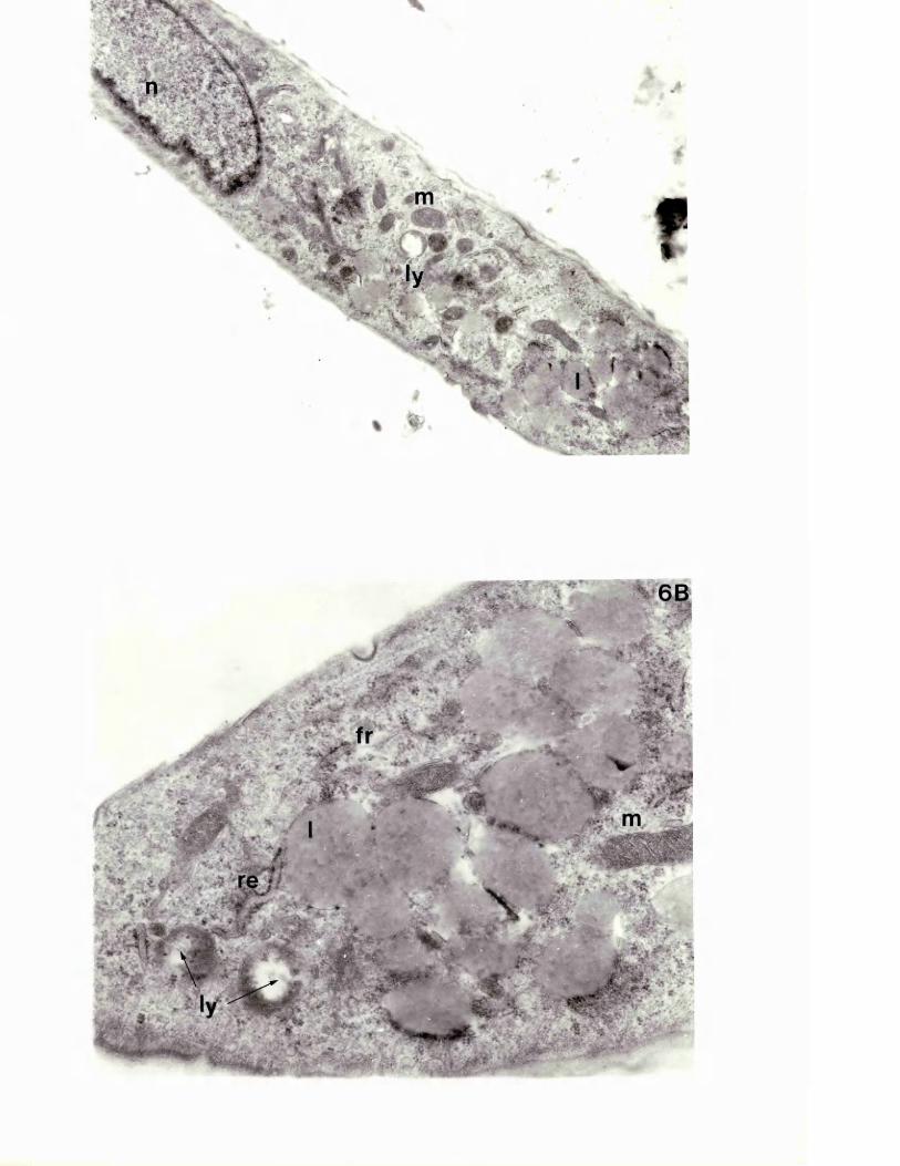
'> • jw

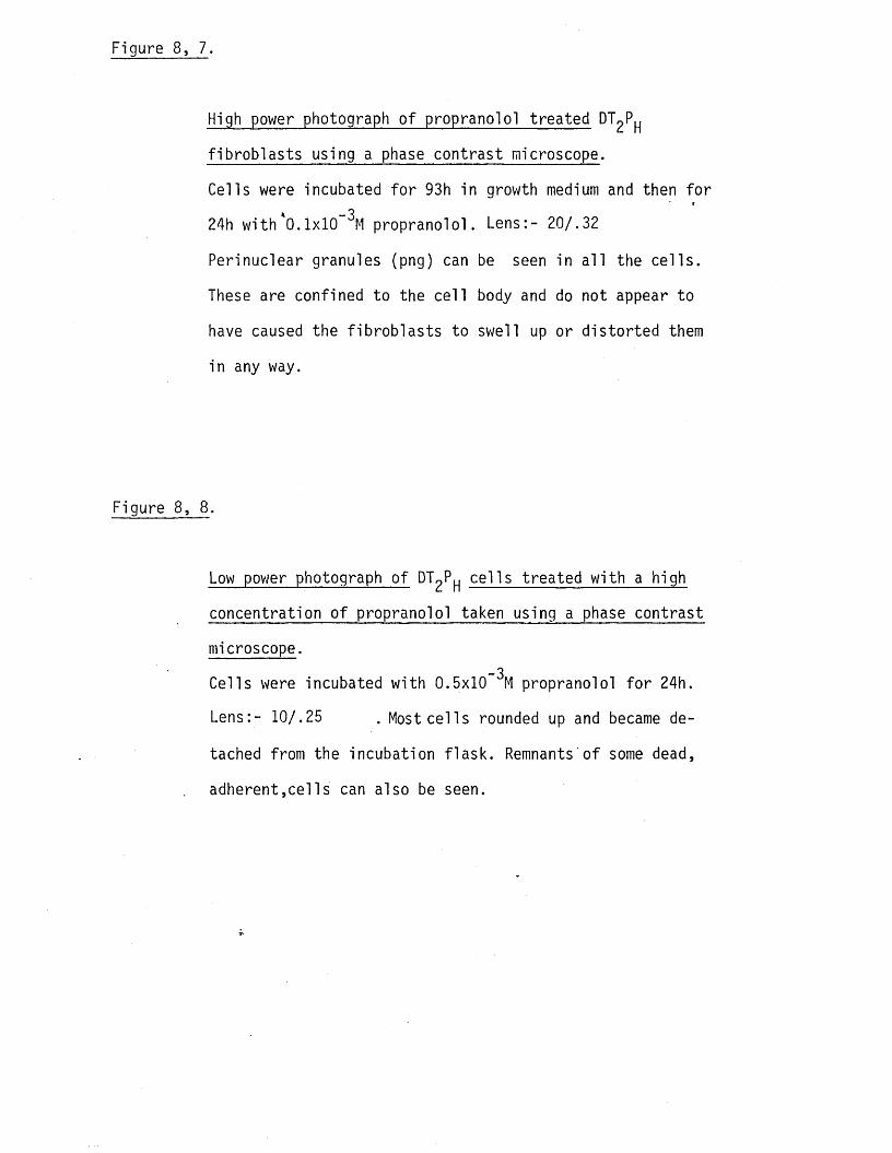
Figure 8, 7.
High power photograph o f propranolol treated Dl^P^
f ib rob la s ts using a phase contrast microscope.
Cells were incubated fo r 93h in growth medium and then fo r
24h with 0.1x10 propranolol. Lens:- 20/.32
Perinuclear granules (png) can be seen in a l l the ce l ls .
These are confined to the ce l l body and do not appear to
have caused the f ib ro b la s ts to swell up or d is to rted them
in any way.
Figure 8, 8 .
Low power photograph of tT^P^ ce lls treated with a high
concentration o f propranolol taken using a phase contrast
microscope._3
Cells were incubated with 0.5x10 M propranolol fo r 24h.
Lens:- 10/.25 . Most ce l ls rounded up and became de
tached from the incubation f lask . Remnants o f some dead,
adherent,cel Is can also be seen.
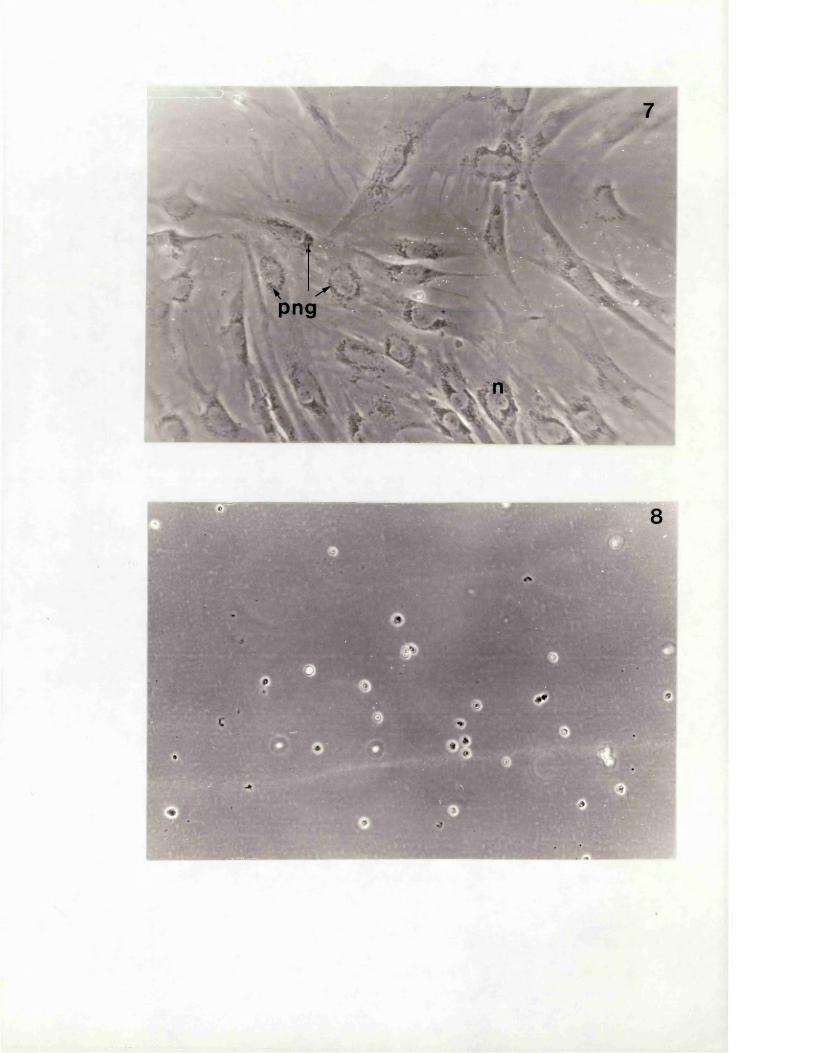

- 1 / 4 -
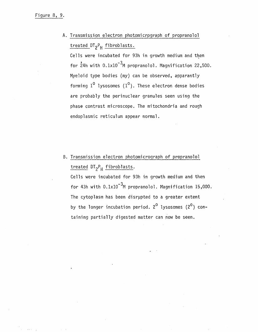
Transmission electron photomicrograph of propranolol
treated_DT2PH f ib ro b la s ts .
Cells were incubated fo r 93h in growth medium and then* -3
fo r 24h with 0.1x10 M propranolol. Magnification 22,500
Myeloid type bodies (my) can be observed, apparantly
forming 1° lysosomes (1°). These electron dense bodies
are probably the perinuclear granules seen using the
phase contrast microscope. The mitochondria and rough
endoplasmic reticulum appear normal.
Transmission electron photomicrograph o f propranolol
treated DT^P^ f ib ro b la s ts .
Cells were incubated fo r 93h in growth medium and then
fo r 43h with 0.1x10 ^M propranolol. Magnification 15,000
The cytoplasm has been disrppted to a greater extent
by the longer incubation period. 2° lysosomes (2°) con
ta in ing p a r t ia l ly digested matter can now be seen.
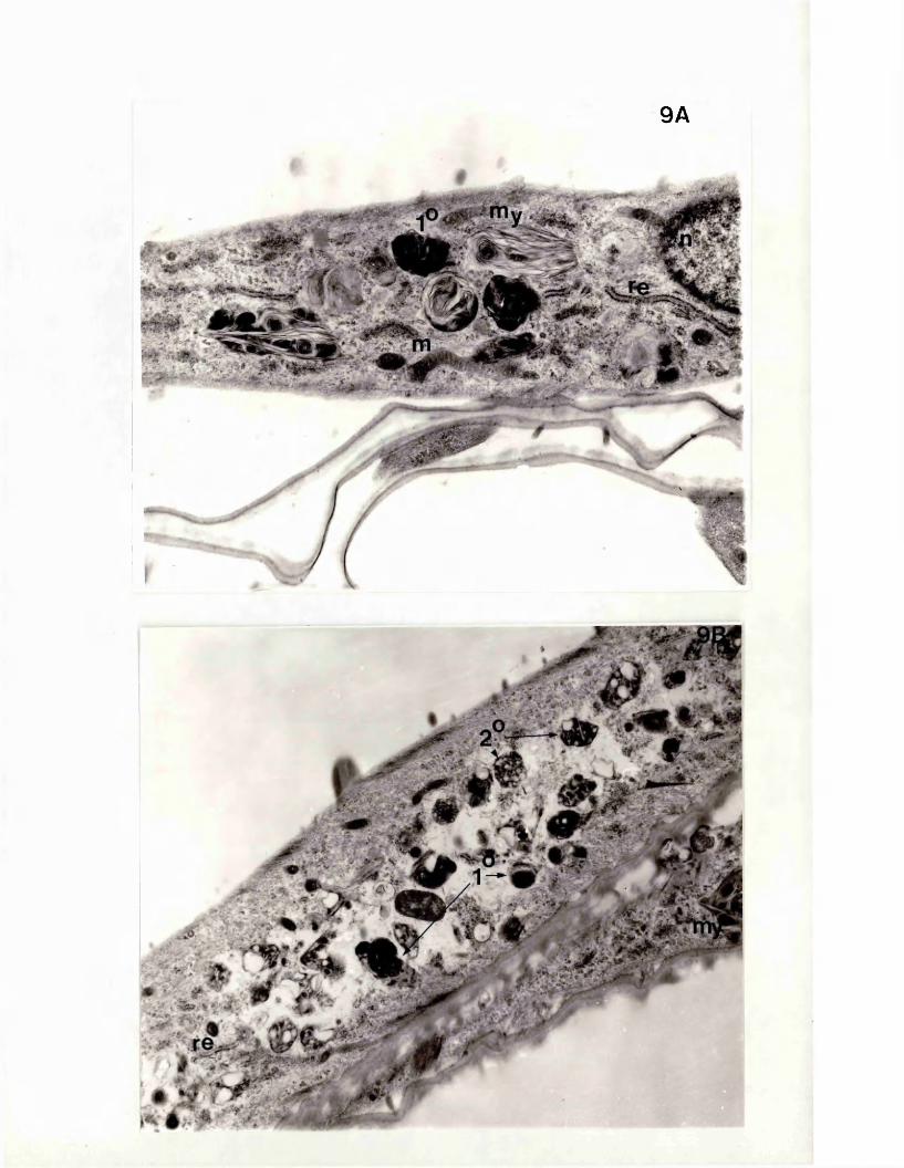
9A
- * £

- 1 / D -
Figure 8, 10.
Transmission electron photomicrograph of propranolol
treated DTgP f ib ro b la s ts .
Cells were grown fo r 93h in growth medium and then fo r * -3
24h with 0.1x10 M propranolol. Magnification 30,000.
Myeloid type bodies consisting o f concentric layers of
membranes and electron dense 1° lysosomes (1 °)occupy
most o f the cytoplasm.
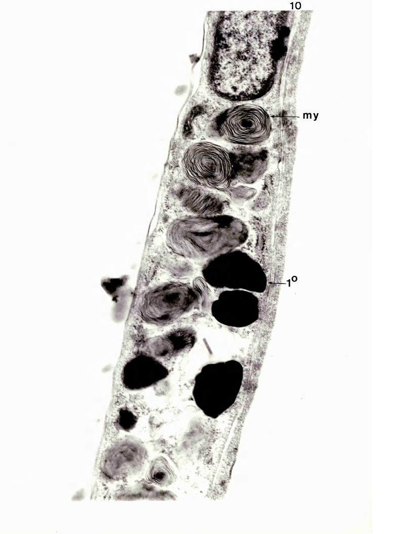
10

- i / a -
Figure 8, 11.
The e f fe c t of paracetamol on morphology._3
Cells were incubated with 2x10 M paracetamol fo r 47h.
Magnification 22,500.
Apart fom the s l ig h t increase in l i p id droplets the ce ll
is s im ila r in appearance to control f ib rob la s ts ( f ig .
8, 4 .) .
Figure 8, 12.
The e f fe c t o f paracetamol on DT^P morphology.-3Cells were incubated fo r 93h with 2x10 M paracetamol.
Magnification 45,000.
The ce lls had a normal morphological appearance.(The
rough endoplasmic reticulum may have p ro l i fe ra ted between
47 and 93h incubation periods but th is is only an im
pression and was never quan t if ied ).
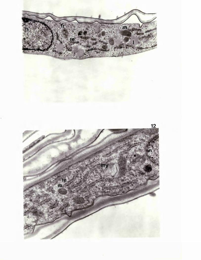

- X / Z ) ~
Figure 8, 13.
Cel ls were t r e a t ed with 2x10 "M paracetamol for 93h and then grown for
a f u r t h e r 24h in a drug f r ee medium. Magnif icat ion 45,000.
These c e l l s d i f f e r e d l i t t l e from those t r e a t e d cont inuous ly with
paracetamol for 47h or 93h.
DISCUSSION.
Control f ib ro b la s ts , using both l ig h t and electron microscopy,
showed charac te r is t ics associated with an inactive c e l l . They were
mostly spindle shaped with an e l ip t ic a l nucleus containing two or
three nu c leo li. Using the electron microscope the nucleus could be
seen to be mainly smooth with occasional indentations containing
d i f fu s e chromatin. The cytoplasm was also typ ica l o f an inactive
ce ll containing few cisternae o f rough endoplasmic reticulum.
There were no clear changes in the morphology o f practo lo l treated
ce lls apart fom the formation of cytoplasmic vacuoles which regressed
on withdrawal o f the drug. S im ila r ly , paracetamol induced no changes
in ce ll morphology which could be observed e ith e r at the l ig h t or
election microscope leve l. Propranolol on the other hand induced the
formation of myeloid type bodies with the accompanying formation of
what appeared to be 1° and 2° lysosomes, probably the perinuclear
particles seen using t h e l ig h microscope.
Vacuole formation in ce l ls exposed to practo lo l was both time and dose
dependent but th is was not a protective response to some adverse e f fe c t
o f practo lo l as a second exposure to the drug, a f te r the ce l ls had
recovered from a f i r s t , e l ic i te d the same pattern of vacuole forma
t ion as the f i r s t exposure. Vacuole formation was not re lated to
membrane damage as trypan blue, added with practo lo l or a f te r devel
opment of the vacuoles, did not enter the c e l ls . I t would also
appear to be un like ly tha t the vacuoles function as storage vesicles
as neither carbohydrate, neutral l i p id or protein could be detected
using normal s ta in ing methods. As f ib ro b la s ts , under the conditions
o f these experiments, synthesiselow levels o f collagen (chapter 6)
i t is also un like ly tha t the ce l ls accumulated collegen precursors.
Eichhorn and Peterkofsky (1979) reported that local anaesthetics
induced vacuole formation in a f ib ro b la s t l in e . The appearance and
disappearance o f the vacuoles followed the same time course and
was associated with a rounding up o f the ce lls and loss o f the
ce l ls processes. Local anaesthetics possess membrane s ta b i l is in g prop
e r t ie s which, i t was suggested,could be important fo r vacuole form
ation. Propranolol, but not p ra c to lo l , possesses such a c t iv i t y so tha t
i t is un like ly tha t the same mechanism is involved in the case o f
practo lo l and,.local a<\cvesthetics.
Futher investigations in to the biochemical basis fo r the d i f fe re n t
morphological actions o f propranolol and practo lo l may help to determine
why treatment with propranolol is not associated with the severe
side e ffects observed in susceptible practo lo l patients.

FINAL DISCUSSION.
L i t t l e is established about the mechanism(s) by which practo lo l
induced severe adverse reactions in some patients . Epidemiological,
immunological and pharmacological studies have been unable to id e n t i fy4
conclusively any fac to r which could provide the basis fo r understand
ing the "oculomucocutaneous syndrome" (Davies 1980).
As discussed in the In troduction , there is no experimental evidence
to support the idea , put forward by a number o f authors (N istrup-
Madsun and S0ndergaard 1977, Brown e t al 1974 and Raftery and Denman
1973), tha t practo lo l could prevent deactivation o f a ta rge t ce ll ie .
the f ib ro b la s t , leukocyte or lymphocyte, by blocking a r ise in cAMP
in the presence o f adrenergic agonists. Indeed the reports demonstrating
tha t such ce lls possess 82 but not 3i-adrenergic receptors suggest tha t
such a pharmacological mechanism would be extremely u n l ik e ly , par
t ic u la r ly as propranolol does not give r ise to such severe side
e ffects as p rac to lo l.
Pharmacokinetic studies revealed no differences between practo lo l
patients with and without side e ffec ts and a control volunteer (Reeves
et al 1978). Extensive animal feeding t r i a l s have fa i le d to demon
s tra te any o f the reactions observed in practolol treated patients
which, taken with the human studies, would suggest tha t a d ire c t tox ic
reaction is not the primary cause o f the practo lo l syndrome.
S im ila r ly , evidence fo r an immune mechanism is inconclusive. Changes
in T -ce ll a c t iv i t y in adversely 'effected patients reported by Behan et
al (1976) could not be confirmed by Amos and Bridgen (1976) and, when
occurring were probably due to the disease and not the cause o f i t
(Pugh et al 1976).
Although autoantibodies were detected in the sera of many practo lo l
treated patients ne ither the antibody t i t r e n o r i t s s p e c i f ic i ty
appeared to be correlated with any p a r t ic u la r manifestation o f the
syndrome (Davies 1980).
The one pos it ive f ind ing was tha t o f Amos et al (1977, 1978) who
reported the presence of antibodies to in v i t ro generated metabolites
o f practo lo l in the sera o f adversely affected practo lo l pa tien ts .
Although the immunological s ign if icance o f these antibodies is not
c lea r, they were not correlated to any p a r t ic u la r disease and were
also detected,in the serum o f patients with no adverse reactions,
the study did show however tha t practo lo l could be metabolised in vivo to
products capable o f binding to macromolecules to form haptens.
I t was postulated in the Introduction tha t the antigenic metabolite(s)
could have had a d ire c t e f fe c t on a ta rget c e l l , inducing changes
which triggered the adverse response. As a f ib ros ing reaction was
such a prominent component o f the practo lo l syndrome the f ib ro b la s t
was a log ica l choice fo r such an inves tiga tion . -
The s tructure o f the antigenic metabolite(s) had been p a r t ia l ly
elucidated by Amos et al (1977) and Amos (1979) and i t was decided to
fu r th e r characterise th is compound using the novel immunological
approach described by Amos e t al (1978). The radioimmunoassay system
used by Amos et al (1978) was evaluated along w ith other possible
methods, using a model system and covalent binding assays carried out
to confirm generation o f protein bound practo lo l metabolites in the
in v i t r o incubation system used. I t did not prove possible however
to measure antimetabolite antibodies in the sera o f practo lo l
pa tients . These negative results were confirmed

by co llabora tive experiments w ith workers who had carried out the
o r ig ina l studies. Follow up investigations provided support fo r
the idea that the sera containing antimetabolite antibodies had been
a l l used up during the o r ig ina l investigations although i t was
also possible tha t the sera had deteriorated during storage.t4
The co llabora tive experiments indicated that the assay system used
in the o r ig ina l studies was not reproducible. The standard deviation
observed between duplicates was very high (+90%) and the percent
binding between the two co llabora tive experiments varied from 0.2%
in the f i r s t experiment to 20% in the second. The o r ig ina l workers
accepted binding values of greater than 10% o f the antigen as in d i
cating spec if ic binding and reported values of up to 80%. Covalent
binding studies, using the equilibrium d ia lys is method, reported here
show a probable maximum binding o f 3nMol/mg prote in . This was calcu
lated as being equivalent to 1 practo lo l metabolite bound per 6 HSA
molecules ie 17% o f the HSA molecules in the in v i t ro microsomal
incubation would bind practo lo l metabolites. As the only rad ioactive
HSA prec ip ita ted in the Farr assay used by Amos et al (1978) was tha t
which was bound to the antigenic metabolite(s) maximum binding
values should be 17% o f the added antigen mixture. I t is possible
tha t metabolites,or practo lo l i t s e l f , complexed non-covalently to HSA
contributed to the antigen/antibody binding but i t is also possible
tha t some o f the reported binding was an a r t i f a c t o f the method used.
The low concentration o f hapten binding was probably the reason why
animals in jected with the protein bound metabolites o f p racto lo l did
not synthesise antimetabolite antibodies although they did respond to
the microsomal protein and is o c i t ra te dehydrogenase present in the
incubation system.
Two metabolites o f p racto lo l had d e f in i te ly been id e n t i f ie d during
animal experiments, the des-acetyl and the 3-hydroxy deriva tives.
(Reeves et al 1979). The des-acetyl metabolite o f practo lo l had also
been shown by Amos (1979) to p a r t ia l ly in h ib i t binding o f the a n t i -
metabolite antibody and the in v i t ro generated antigen complex. I t4
was decided therefore to use the des-acetyl de riva tive and other
analogues of p ra c to lo l, including the 3-hydroxy form,in in v i t r o
experiments to determine the effec ts o f these compounds on f ib ro b la s t
functions. Neither the model compounds nor in v i t r o generated metabolites
had any e f fe c t on e ith e r ce l l growth or collagen synthesis ( a t the
concentrations used) whereas practo lo l inh ib ited both parameters in
a dose dependent manner despite the fa c t tha t i t was not taken up by
the f ib rob las ts to any great extent (0.7%). Practolol also in h ib ite d
the uptake and incorporation o f leucine by these ce lls to the same
extent as the in h ib i t io n o f collagen synthesis. These resu lts are
consistent with the idea tha t practo lo l depresses protein synthesis
by l im i t in g substrate a v a i la b i l i t y , possibly via an e f fe c t on the plasma
membrane. DT^P ce l ls were less sensitive to the in h ib i to ry actions o f
practo lo l than PS ce lls however (5 fo ld ) demonstrating tha t there
is considerable va r ia tion in the response to practo lo l although i t is
not possible to say i f th is va r ia t ion is re lated to s u s c e p t ib i l i ty
to p ro l i fe ra t iv e conditions such as psorias is.
The in h ib i to ry actions on growth and collagen synthesis were also
observed when propranolol and paracetamol were used. However, where
as PSj ce l ls were more sensitive towards propranolol, p a ra l le l in g
the changes observed with p ra c to lo l, they were s l ig h t ly less sens it ive
towards paracetamol. I t appears tha t the two 8-receptor blocking
drugs exert th e ir e ffec ts by a mechanism d if fe re n t to tha t o f paracetamol.
As discussed above i t would appear to be un like ly tha t the actions of
practo lo l and propranolol are mediated via 8-adrenergic receptors.
I t is possible tha t differences in th e ir associated properties, ie
propranolol is l i p id soluble and practo lo l has sympathomimetic a c t iv i t y ,
could play a ro le in th e i r actions. This implies tha t the two 3-
adrenergic receptor blocking drugs in h ib i t ce l l function via d i f fe re n t *
mechanisms and tha t the pa ra lle l responses observed with the two
are co incindenta l. Morphological studies, using both the l ig h t and
transmission electron microscope, provide some support fo r th is idea.
Paracetamol had no obvious e f fe c t on f ib ro b la s t morphology at any
of the concentrations used. Practolol on the other hand induced the
formation of cytoplasmic vacuoles, the number and time course of
which was dose dependent. Vacuole formation did not re su lt in any
appreciable change in f ib ro b la s t s tructure as observed using trans
mission electron microscopy and was not re lated to damage o f the plasma
membrane, assessed using trypan blue, and was not involved with storage
o f neutral l i p id , carbohydrate or prote in . Propranolol treated ce lls
developed perinuclear granules which appeared, using the electron
microscope, to be lysosomes formed from fused myeloid type bodies,
concentric c irc les of membranes. Propranolol was also found to be-3cytocidal at 0.5x10 M whereas practo lo l at concentrations up to
-33.3x10 M, causing almost 100% in h ib i t io n of ce l l growth, appeared to
cause no permanent damage.
The actions o f practo lo l on f ib ro b la s t growth and collagen synthesis
occured at concentrations higher than those reported to occur in the
plasma of patients ( l-2ug /m l) (Reeves et al 1978, F itzgera ld and Scales
1968 and Bodem et al 1972). However practo lo l can accumulate in some
tissues such as the eye, skin and small in te s t ine (Lindup et al 1979)
in a time and dose dependent fashion. Such an accumulation could res
u l t in the formation o f an e ffe c t ive concentration o f p racto lo l
in v ivo . I t was possible however tha t practo lo l exerted i t s main
e ffe c t upon the f ib ros ing response in d ire c t ly via a mediator ce ll
such as the macrophage. The secretion o f a collagen stim u la ting fac to r
by peritoneal macrophages exposed to mineral dusts has been reported
by a number o f authors (Heppleston and Styles 1967, Burrel and
Anderson 1973 and A ll iso n et al 1977). Hamster peritoneal macrophages
did not synthesise any such fac to r however and practo lo l did not
stimulate the synthesis o f one. Practolol did in h ib i t Fc receptor
expression, probably re f le c t in g a general reduction in macrophage
a c t iv i t y and demonstrating that the e ffec ts o f th is drug are not
confined to e ith e r the f ib ro b la s t or to human c e l ls . The ID™ Tor50the e f fe c t on Fc receptor number was s im ila r to tha t fo r in h ib i t io n
of f ib ro b la s t growth. I t is u n l ike ly therefore that th is e f fe c t is
the primary one of p ra c to lo l, ra ther i t probably re f le c ts a common
disruption of macrophage metabolism.
The overall conclusion from the resu lts obtained is tha t the p racto lo l
syndrome is due to the parent compound and not a metabolite* Further,
the action o f practolol- is probably an in h ib i to ry one, not s tim u la tory
as postulated in the Introduction. Moreover, the f ib ro b la s t is probably
not the primary target fo r p rac to lo l. I f these proposals are correct
then several questions remain to be answered. F irs t what is the primary
ta rget fo r practo lo l? Second, what factors could be involved in determ
ining s u s c e p t ib i l i ty to the drug? Third, why are such side e ffec ts not
observed with other 3-receptor blocking drugs? Las tly , why were animal
experiments unable to predict the problems associated with practo lo l
therapy?
Both ocular and peritoneal reactions are associated with loss o f secretory
ce ll function be i t the lacrimal gland, conjunctival goblet c e l ls or
the peritoneal mesothelium (Rahi e t al 1976, Myllarniemi and
Leppaniemi 1981).
Damage to other organs is also characterised by u lceration (nasal
and o ra l) damage to the cochlear and to the pleural membrane (Wright
1975). I t is possible tha t a d ire c t tox ic e f fe c t on secretory ce ll
functions is the primary action of p rac to lo l. The in h ib i t io n o f
leucine uptake by f ib rob la s ts in v i t r o supports th is idea which is' «
strengthened by the report tha t oral administration o f practo lo l to
rats in h ib i ts in te s t in a l mucosal glycoprotein synthesis,(Okine et al
1982). The need fo r practo lo l to accumulate in vivo to an e ffe c t ive
concentration might explain the re la t iv e ly long la te n t period before
adverse reactions became apparent,(eye e ffec ts 23.4 +14 months, Wright
1975)( sclerosing p e r i to n i t is 34+ 15 months, Marshall et al 1977a).
However accumulation o f a tox ic concentration o f practo lo l would not
explain the delayed reactions reported by, fo r example, Allan and
Cade 1975 and Marshall et al 1977b. The in v i t r o studies reported
in th is thesis demonstrate tha t the e ffec ts o f acute exposure to p ract
o lo l (up to 4 days) are revers ib le , however i t appears tha t chronic
practo lo l therapy resu lts in lesions which can be expressed, or
continue to be expressed, a f te r withdrawal o f the drug. That is , there
is some change, analogous to in i t i a t io n in the two stage model o f
carc inogenic ity , which upon appropriate s t im u la tion , allows the f ib ro s i
reaction to manifest i t s e l f . S im ilar ideas have been put forward to
account fo r the action o f hydrocortisone on f ib rob la s ts from patients
with adult or juvenile-onset diabetes m e ll i tus and the action o f
phenytoin on human gingival f ib rob la s ts (Rowe e t al 1977, Hassell and
G ilbe rt 1983).
S u sce p t ib i l i ty could depend on the rate of accumulation o f p racto lo l
and the concentration needed to in h ib i t secretory ce l l function .
Although the work o f Reeves et al (1978) indicates tha t there are no
differences in the pharmacokinetics of practo lo l in d i f fe re n t pa tien ts ,
they administered only one dose o f the drug. Chronic treatment may
re su lt in d i f fe re n t rates o f accumulation,reflecting small d ifferences
in the rates o f uptake and excre tion ,and,possib ly ,varia tions in bind
ing s ites in Ihe d i f fe re n t tissues. I t is also not unreasonable to
assume that the differences in s u s c e p t ib i l i ty reported fo r f ib ro b la s ts
w i l l also be re flected by other ce ll types. Chapman e t al (1980) rep
orted tha t hydrocarbon hydrolase a c t iv i t y in the jejunum, l i v e r and
lesion free epidermis o f psoriasis patients was reduced compared to
control values. I t is possible tha t practo lo l susceptible patients
also have a genetic lesion predisposition to the adverse actions o f
p ra c to lo l, th is genetic defic iency being present in a l l tissues but
the pattern o f adverse reactions re f le c t in g the s ites o f accumulation
o f the drug. In th is conection the work o f Takeichi (1980) and
Bjorkholm et al (1980),suggesting tha t hypertension and ischemic heart
disease could be re lated to an immunological dysfunction ,is pe rt inen t.
Propranolol exhibited the same in h ib i to ry actions on f ib ro b la s t growth
and collagen synthesis in v i t r o as practo lo l ( th is thesis) but did
not in h ib i t in te s t in a l mucosal glycoprotein synthesis in the ra t (Okine
et al 1982). Okine et al suggested tha t the reason practo lo l had an
e f fe c t on glycoprotein synthesis and propranolol ( and also ateno lo l)
had no e f fe c t was that practo lo l is read ily absorbed from the gut and
has a greater b io a v a i la b i l i t y than the other drugs used in th e i r
experiments. S im ilar considerations could be the reason why propranolol
does not /induce adverse reactions in patients despite being more active
than practo lo l i_n v i t r o .
Practolol induced none o f the signs o f the"oculomucocutaneous syndrome"
in animals during chronic feeding t r i a l s (Case et al 1978) using doses
higher than that reported by Okine et al (1982) to cause in h ib i t io n o f

protein synthesis (50-200mg/kg). The s ign if icance o f th e i r find ing
is therefore unclear but could be in terpreted as supporting the
idea that a predisposing fac to r is necessary fo r expression of the
adverse reactions. Experiments using spontaneously hypertensive rats
could help evaluate th is hypothesis as well as the possible ro le
of associated'immunological changes in the development o f the
practolo l syndrome.
Although the work carried out fo r th is thesis has posed more new
questions than i t has answered several aspects o f the mechanism
involved in the practo lo l syndrome have been c la r i f ie d .
F i r s t i t apears tha t p racto lo l and not one o f i t s metabolites is
responsible fo r the adverse reactions associated with i t s adm in istra tion.
Second, the lesion involved is l i k e ly to be one o f in h ib i t io n as opposed
to the stim ulation o f various functions proposed by most authors.
Third, p ra c to lo l, via an e f fe c t on the plasma membrane, could e f fe c t
protein synthesis and hence those ce lls which are most ac t ive , such
as those with a secretory function , would be most effected and, maybe,
more sensitive to the in h ib i to ry actions o f p rac to lo l.
Lastly , the d ifference in the s u s c e p t ib i l i ty o f ce l ls from
d if fe re n t people to the in h ib i to ry e f fe c t o f practo lo l could be one
of the predisposing factors in v ivo .
The p o s s ib i l i ty arises tha t by using f ib rob la s ts from patients with
a va r ie ty of id iopath ic and drug induced cond it ions,predisposing ‘
factors could be id e n t i f ie d . Such a panel of c e l ls , w ith d i f fe re n t
s e n s i t iv i t ie s to drugs could be used to screen new drugs fo r po ten tia l
side effec ts and to id e n t i fy those potentia l patients who would be most
at r is k of developing untoward side e ffec ts .
REFERENCE LIST.

R.P.Ahlquist. Am.J.Physiol 153 586. 1948
R.P.Ahlquist. Annals. NY Acad. S c i. 139 549. 1967.
S.Ahmad. Chest. 79 361. 1981.
J.J.A leo, R.Novack and E.Lavy. Connect. Tiss. Res. 2_ 91. 1974.
D.Allan and D.Cade. Br. Med.J. 4 40. 1975.
A.C .A llison, I.A .C lark and P.Davies. Ann rheum. Dis. 36 8. 1977.
A .C .A llison, A.M.Denman and R.D.Barnes. Lancet i_i_ 135 1971.
H.E.Amos. I n t . J . Immunopharmacol. 1 9. 1979.
H.E.Amos and W.D.Brigden. Lancet ij_ 1298. 1976.
H.E.Amos, W.D.Brigden and R.A.McKerron. Br.Med.J. 1 598. 1975.
H.E.Amos, B.G.Lake and J .A r t is . Br.Med.J. l_ 402. 1978
H.E.Amos, B.G.Lake and H.A.C.Atkinson. C lin . A lle rgy. 7_ 423. 1977.
W.L.Ashton. Curr. Med. Res. Opinion. 442. 1976.
E.S.K.Assem. Br.J.Clin.Pharmacol. 2 184P. 1975.
E.S.K.Assem and R.A.Bank. Proc.Roy.Soc.Med. 66 179. 1973.
D.C.Baxter-Smith, I . J.Monnypenny and N .J .D orr ico tt. Lancet i 149. 1978.
P.O.Behan, W.M.H.Behan,.F.J.Zacharias and J .T .N icho lls . Lancet, i i .
984. 1976.
M.H.Benner, T.Ente, S.Lockeys, S.Makino and C.E.Reed. J .A lle rgy _41
110. 1968.
J.Bensaid, J .C .A ld ig ie r and N.Gaulde. Br.Med.J. _1 1603 1979.
R.A.Berg, J.Moss. B.J.Baum and R.G.Crystal. J .C l in . In ve s t. 67_
1457. 1981.
A.Berken and B.Benacerraf. J.Exp.Med. 123 119. 1966.
R.Bevis, R.B.Cubey and S.H.Taylor. Br.Med.J. 4- 327. 1975.
M.Bjorkholm, U.de Faire and G.Golm. Atherosclerosis. 36 195 1980.
G.Bodem, H.L.Brammel, J.V.Weil and C.A.Chidsey. Cl in.Res. 2C)
169. 1972.
N.P.Boye and J.R.Vale. Euro. J .C l in . Pharmacol. 11_ 11. 1977.
M.Bristow, T.R.Sherrod and R.D.Green. J.Pharmacol.Exp.Thera
171 52. 1970.
P.Brown, H.Baddeley, A.E.Read, J.D.Dadies and J.McGarry. Lancet ij_
1477 1974.
R.C.Brown, J.Gook and M.S.Losowsky. Postgrad. Med. J. 57 189*. 1981.
P.J.E.Brown, M.Lesma, A.N.Hamlyn and C.O.Recod. Br.Med.J. 1_ 1591. 1978.
R.B.Buckingham, R.K.Prince, G.P.Rodnan and F.Taylor. J.Lab.Clin.Med.
92. 5. 1978.
R.M.Buckley and R.R.McGregor. Clin.Res. 25_ 373A (abstract) 1977.
D.W.Bullimore. Br.Med.J. 281 564. 1980.
G.Berglund. Curr.Thera. Res. 2^ 21. 1977.
R.Burrel and M.Anderson. Envir.Res. 6 389. 1973.
K. Burton. Biochem.J. 62 315. 1956.
S.G.Carruthers. Side Effects o f Drugs Annual 4_ 1980. Edited by M.N.G
Dukes. Published by Excerpta Medica.
D.E.Case, W.E.Lindup, C.Lowery, T.C.Orton, P.R.Reeves and S.E.Whittaker
Br.J.Pharmacol. (53 351. 1978.
W.M.Castle, J.M.Cruickshank and J.A.Waycott. Br.Med.J. 281 311. 1980.
P.H.Chapman, P.J.Kersey. B.Keys, S.Shuster and M.D.Rawlins.
Br.Med.J. 281_1315. 1980.
K.H.Chen, M.A.Paz and P.M.Gallop. In V it ro . 13 49. 1977.
P.H.Chuong, C.Labarre, J.Bory and G.Le Moan. Annals Pharmaceutiques
Francaise 35 357. 1977a.
P.H.Chuang, C.Labarre, J.Bory and G.Le Moan. Annals Pharmaceutiques
Francaise. 35 257 1977b
C.V.Clark and R.Terris Lancet i i 937. 1983.
J.R.Claydon Br.Med.J. 2 327 1975.
J.D.Clough, S.A.Frank and L.H.Calabrese. A r th r i t i s and Rheumatism
23 24. 1980.
R.E.I.Cochran, J.Thomson, A.McQueen and R.A.McKerron. Arch Derm
112 1173. 1976.
E.J.Cohen. J.Am.Chem.Soc. 62. 3386. 1940.
M.E.Conolly and J.K.Greenacre. Br.J.Pharmacol. 59 17. 1977.
J.M.Cruickshank. Drugs. 25 331 1983.
J.B.Cumberpatch. Br.Med.J. 4- 528. 1974
M.G.C.Dahl, R.H.Felix, F.A.Ive and D.S.Wilkins. J .Inves t. Derm.
64 299. 1975.
H.H.Dale. J.Physio l. 34 163. 1906.
H.H.Dale. J.Physio l. 80 IIP . 1933.
F.Dany, J.Clementy, J .H i la i re , R.Roudaut and H.Albin. Bordeaux
Medical. 12 527. 1979.
G.E.Davies. In Drugs and Immune Responsiveness. Edited by J.L.Turk
and D.Parker. Published by Macmillan 1979.
G.E.Davies. In Pseudo-Allergic Reactions. Involvement o f Drugs
and Chemicals, Vol. 2 . 1980.
S.P.Deacon, A.Karunanayake and D.Barnett. Br.Med.J. 2 1255. 1977
C.C.Doherty, M.G.McGeown and R.A.Donaldson. Br.Med.J. 2 1786 1978
C.T.Dollery. Br.J.Pharmacol. 4- 295. 1977.
C.T.Dollery, J.W.Paterson and M.E.Conolly. C lin . Pharmac. Ther.
10 765. 1969.
J.Douglas-Wilson, R.J.Booth, T.Y.Bullock and D.G.Campbel1. Lancet
11 312. 1980.
M.N.G.Dukes. Side Effects of Drugs Annual 2 Edited by M.N.G.Dukes.
Published by Excerpta Medica, Amsterdam. 1979.
P.J.Dunn.-> New Zealand Med.J. 82 199. 1975.
N.H.Dyer and C.C.Vorley. Be.Med.J. 2_ 443. 1975.
R.A.van Dyke and A.J.Gandolf. Drug Metab.Disp'. 2 469. 1974.

J.H.Eichhorn and B.Peterkofsky. J.Cell B io l. 21 26. 1979.
T.M.Erwtman, M.C.P.Braat and W.G.van Aken. Br.Med.J. 2 297. 1977.
R.S.Farr. J .In fec t ious Diseases. 103 239. 1958.
J.C.Faveral-Garrigues, D.Alegria, N.Carde and Y.Castaing. Concours
Med. 99 5696. 1977.
M.J.Farr, J.P.'Wingate and J.N.Shaw. Br.Med.J. 2 68. 1975.
R.H.Felix,F.A.Ive and M.G.C.Dahl. Br.Med.J. 4 321. 1974.
R.H.Felix, F.A.Ive and M.G.C.Dahl. Br.Med.J. 1_ 626. 1975.
W .J.F ilipek. Postgrad.Med.J. 65 131. 1979.
A.Y.Finlay and E.Waddington. Br.Med.J. I 987. 1978.
J .D .F itzgera ld . Clin.Pharmaco.Ther. _10 292. 1969.
J.D .F itzgera ld and B.Scales. In t.J .C lin .Pharm acol. I 467. 1968.
H.A.Flemming and P.H ickling. Lancet i i 1202. 1975.
H.Formgren. Br.J.Clin;Pharmacol. _3 1007. 1976.
D.M.Fraser and N .A .Irv ine . Lancet i 89. 1976.
F.T.Fraunfelder. Drug Induced Ocular Side Effects and Drug In te ractions.
Published by Lea and Febiger (Philadelph ia).
J.R.Fry and J.W.Bridges. Biochem. Pharmacol. 26 969. 1977.
M.Fukada. Jap.J.Pharmacol. _18 185. 1968.
R.F.Furchgott. Ann.NY.Acad.Sci. 139 553. 1967.
R.G.Furchgott. Handbook Exp. Pharmak. 33 283. 1972.
A.K.Furhoff, M.Nordlander and C.Peterson. Br.Med.J. 2 831. 1976.
R.W.Gange and G.M.Levene. C lin . Exp.Derm. 4 87. 1979.
R.W.Gange and E.Wilson-Jones. Br.Med.J. I 816. 1978.
M.J.Gavin, W.M.Castle, J.M.Cruickshank and J.A.Waycott. Br.Med.J.
280 1227. 1980.
P.M.Gaylarde, A.P.Brock and I,Sarkany. Clin.Exp.Derm. 3 157. 1978.
W.Geisthovel and G.Kalfhaus. Munch. Med. Wschr. 107. 781. 1965.nrl
P.G.Gell and R.R.Coombs. C lin ica l Aspects o f Immunology. 2 ed it ion
Published by Blackwell S c ie n t i f ic Publications l t d . 1968.

C.F.George, R.E.Nagle and B.L.Pentecost. Br.Med.J. 2 402. 1970
J .R .G i l le t te and L.R.Pohl. J.Toxicol.Enviromental Health. 2
849. 1977.
D.Gladman, E.Keystone,M.Urowitz, D.Cane and L.Poplonski. Clin.Exp.
Immunol. 40 77. 1980.
R.L.Goldberg, D .P.Parra tt, S.R.Kaplan and G.C.Fuller. Biochem.
Pharmacol. 29 869. 1980.
L.S.Goodman and A.Gilman The Pharmacological Basis o f Therapeutics
5 ^ e d i t io n . Published by Macmillan. 1975.
J.R.Graham, H.I.Suby, P.R.LeCompte and N.L.Sadowsky. New Eng.J.Med.
274 359. 1966.
H.Green and B.Goldberg. Proc.Soc.Exp.Biol.Med. 117 258. 1964.
F .M .G rif f in , C.Bianco and S .C .S ilverste in . J.Exp.Med. 141 1269 1975.
I .D .G r i f f i th s and J.Richardson. Br.Med.J. 2 496. 1979.
J.W.Hadden, E.M.Hadden,E.Middelton and R.A.Good. Proc.Nat.Acad.Sci.
USA. 69 3024. 1972.
W.Hailey and I.D.S.Goodman. Br.Med.J. 2 337. 1975.
P.J.A.Holt and E.Waddington. Br.Med.J. 2 539. 1975.
R.A.Harper and B.A.Flaxman. J.Cell Physiol. 8£ 293. 1975.
R.A.Harper, B.A.Flaxman and D.P.Chopra. J .Inves t. Derm. 62 384. 1974.
J.S.Harrington, M.Ritchie, P.C.King and K .M il le r . J.Path. 109
21. 1973.
A.D.B.Harrower and J.A.Strong. Br.Med.J. 2_ 296. 1977.
W.Hartmann. Dt. Z.Chir. 255 173. 1942.
R.F.Harty. Arch.Intern.Med. 138 1424. 1978.
T.M.Hassel and G.H.Gilbert. Am.J.Path. 112 218. 1983.
F.C.Hay, G .Torrig iani and I.M .R o it t . Eur.J. Immunol. 2 257. 1972.
W.I.Heine and H.Friedman. JAMA. 182 726. 1962.
A.G.Heppleston and J.A .Styles. Nature. 2^4 521. 1967.
J.T.Homer and W.A.Cain. In t.A rch .A lle rgy Appl. Immunol. 59 121. 1979.
J.Hunt, R.J.Richards, R.Harwood and A.Jacobs. Br.J.Haematol. 41
69. 1979.
W.M.Hunter. In Handbbok o f Experimental Immunology. 3rc*edition.
Edited by D.NLWeir. Published by Blackwell S c ie n t i f ic Publishing 1978.
L .Ignarro, T .L in t and W.George. J.Exp.Med. 139 1395. 1974.
S.J.Jachuck, J.Stephenson, T .B ird , F.S.Jackson and F.Clark.
Postgrad. Med.J. 53 75. 1977.
J.N.Johnson and J.McFarland. Br.Med.J. 280 864 1980.
S.Jonsson and G.Kronvall. Europ.J.Immunol. 4_ 29. 1974.
T.van Joost, J.M.Hup and F.E.Ros. Br.J.Derm. 101 171. 1979.
T.van Joost and J.H.S.Smitt. Arch.Derm. 117 600. 1981.
M.Kavai, K.Lukacs, I.Sonkoly, I.Jokay, A.Berndt and Gy.Szegedi. C lin .
Exp. Immunol. 40 66. 1980.
K.Keck. J , Immunol.Methods. 20 287. 1978.
S.C.Kennedy and M.Ducrow. Br.Med.J. _1 1598. 1977.
S.W.Kessler. J.Immunol. 115 1617. 1975.
M.S.Knapp and N.R.Galloway. Br.Med.J. 2 557. 1975.
G.Kronvall and R.C.Williams. J.Immunol. 103 828. 1969.
B.G.Lake, M.A.Collins, R.A.Harris and S.P.Gangoli. Xenobiotica. 9
723. 1979.
A.M.Lands, A.Arnold, J .P .M cA u lif f , F.P.Luduena and T.G.Brown. Nature
214. 597. 1967.
K.Landsteiner and J.Jacobs. J.Exp.Med. 64 625. 1936.
J.N.Langley. J.Pysiology 33 374. 1905.
D.L.Layman, E.B.McGoodwin ANd G.R.Martin. Proc.Nat.Acad.Sci. 68
454. 1971.
C.C» LeRoy, M.McGuire and N.Chen. J .C l in . Invest. 54 880. 1974
B.Levy. J . Pharmacol. Exp.Ther. 151 413. 1966.
L.M.Lichtenstein and S.Margolis. Science. 161 902. 1968.
D.R.A.Lima and P.Turner. Br.J.Pharmacol. _11 521. 1981.
W.E.Lindup, S.Longshore, T.C.Orton and S.E.Whittaker. Br.J.Pharmacol.
66 504P. 1979.
A.D.MacKay and A.T.Axford. Lancet i 89. 1976.%
J.H.Marigold, R.E.Pounder, J Pemberton and R.P.H.Thomson. Br.Med.J.
284. 870. 1982.
A.J.Marshall, H.Baddley,D.W.Barritt, J.D.Davies, R.E.Lee, T.S.Low-Beer
and A.E.Read. Quarterly J.Med. 181. 135. 1977a.
A.J.Marshallj W.K.E1tringham, D .W .Barritt , J.D.Davies, D .A .G r if f i th s
L.K.Jackson and A.E.Read. Lancet, i i 1254. 1977b.
R.Meyboom. Lancet, i . 334. 1975.
H.I.Mikkelsen, H.A.Jensen, S.Wadskov, A.Lerche and J.S0ndergaard.
Cl in.Pharmacol.Ther. _3 152. 1975.
M.W.Millar-Craig. J.Soc.Occup.Med. 29 107. 1979. .
K .M il la r and E.Kagan. J.Retie.End.Soc. 20 159. 1976.
M.Montagna and A.Groppi. A rch.Toxicol. ^3 221. 1980.
N.C.Moran. Pharmacol Rev. 18 503. 1966.
B.Mdrland and J.M0rland. Acta.Pharmacol.et Toxicol. 50 221. 1982.
A.W.Musk and J.A .Pollard . Br.Med.J. 2 581. 1979.
H.Myllarniemi and A.Leppaniemi. Acta.Chir.Scand. 147 137. 1981.
K.Nakatsu and K.Skully. Res.Comm.Chem.Path.Pharmacol. 27 203. 1980.
H.Nakazawa, R.Adolphson, E.Chaperon, J.Hobday, R.Townley and J .P au li .
J .A lle rgy Clin.Immunol. 57 200 (abs trac t). 1976.
J.F.Nancarrow. Lancet. 2 525. 1978.
A.Narayanan, R.C.Page and F.Kuxan. Lab. Invest. 391 61. 1978.
H.A.M.Neumann and T.van Joost. Dermatologia 162 330. 1981.
A.Nilsson, B.G.Hansson and B.Hokflet. Br.Med.J. 2 126. 1977.

S.Nistrup Madsen and J.S0ndergaard. Lancet, i 1055. 1977.
L.D.Nourse. P.N.Nourse, H.Botes and H.M.Schwartz. Enviromental Res.
9 115. 1975.
L.K.N.A.Okine, C.Ionnides and D.Parke. Biochem.Pharmacol. 31 2263.
1982.%
T.C.Orton and C.Lowery. J.Pharmacol.Exp.Ther. 219 207. 1981.
J.H.Passwell, H.R.Colten, E.L.Scheeberger, Z.Marom and E.Merler.
Immunology. 41 217. 1980.
R.Pauwels, H.Bazin, B.Platteau and M.van der Straeten. In t.A rch.
A lle rgy Appl. Immunol. 6_1 347. 1980.
J .C .Petr ie , D.B.Galloway, T.A.Jeffers and J.Webster. Postgrad. Med.J.
52 63. 1976.
B .J .P h i l l ip s . Biochem.Pharmacol. 23 131. 1974.
O.J.Plescia, N.C.Balczuk, E.Cora-Figueroa, A.Mukherjee and W.Braun.
Proc.Nat.Acad.Sci. USA 54 1281. 1965.
O.J.Plescia, W.Braun and N.C.Palczuk. Proc.Nat.Acad.Sci. USA.
52 279. 1964.
M.Ponec, J.A.Kempenaar, G.A.van der Meulen-van Harskamp and B.N.Bachra.
Biochem.Pharmacol. 28 2777. 1979.
C.E.Powell and I .H .S la te r. J.Pharmacol.Exp.Ther. 122 480. 1958.
B.N.C.Prichard. Br;Med.J. _1 1227. 1964
B.N.C.Prichard. Br.J.Clin.Pharmacol. 5_ 379. 1978.
B.N.C.Prichard and P.M.S.Gillam. Br.Med.J. j. 7. 1969.
G.C.Priestley. J . Invest.Derm. 74 413.' 1980.
G.C.Priestely and J.Brown. J.Invest.Derm. 72 161. 1979.
S.Pugh, B.Pelton, E.B.Raftery and A.M.Denman. Ann.Rheum.Dis. ^5 344
1976.
E.B.Raftery. Br.Med.J. 4 653. 1974.
E.B.Raftery and A.M.Denman. Br.Med.J. 2 452. 1973.
A.H.S.Rahi, C.M.Chapman, A.Garner and P.Wright. Br.J.OpthaV. 60
312. 1976.
G.J.S.Rao, M.del Monte and H.L.Nadler. Nature 232 253. 1971.
M.J. Rebeller and M.Mureau. Bordeaux Medical J. 12 533. 1979.
P.R.Reeves, D.E.Case, H.T.Jepson, D.J.McCormick,J.T.NicholIs,
R .H .Fe lix ,P .K \L .Holt, F.J.Zacharias and R.W.Fluke. J.Pharmacol.
Exp.Ther. 205 489. 1978.
J.Rhodes. J.Immunol. 114 976. 1975.
R.J.Richards, R.Desai, P.M.Hext and F.A.Rose. Nature. 256 664. 1975.
C.M.Ridley. Br.Med.J. 4 229. 1974.
E.Rimmer, A.Richens, M.E.Forster and R.W.M.Rees. Lancet, i 300. 1983.
C.J.Roberts, A.J.Marshall and D.W.Barrit. Topics in Cardiovascular
Disease. 179. 1976.
B.F.Robinson. Side Effects o f Drugs Annual 2nc* e d it ion . Edited by
M.N.G.Dukes. Published by Excerpta Medica. 1978.
D.A.O’ Rourke,.M.F.Donohue and J.A.Hayes. Med.J.Australia. 2 88. 1979.
D.W.Rowe, B.J.Starman, W.Y.Fujimoto and R.H.Williams. Diabetes 26
284. 1977.
A.S.Russel and M .Z if f . C lin . Exp.Immunol. 3 901. 1968.
L.Saltzman, J.Moss, R.A.Borg, B.Hom and R.G.Crystal. Biochem.J. 15_ 25. 1982.
G.L.Sanders. Adverse Drug Reaction B u l le t in . 68 240. 1978.
Edited by D.M.Davies. Published by Adverse Drug Reaction Research
Unit. Durham.
G.Sandler and G.A.Clayton. Br.Med.J. 2 399. 1970.
R.L.Savage, M.Lesna, J.C.Mucklow and C.J.Stevenson. Br.Med.J. 1_
987. 1978.
B.Scales and M.B.Cosgrove. J.Pharmacol. Exp.Ther. 175 335. 1970.
E.J.Schenkelaas and I.L.Bonta. Euro.J.Pharmacol. In Press.
E.Scowen. Br.Med.J. 1 1344. 1978.
W.G.Shafer. Proc.Soc.Exp.Biol.Med. 104 198. 1960.
W.G.Shafer. Proc.Soc.Exp.Biol.Med. 106 205. 1961.
M.Shapiro. Exp.Med.Surgery. _16 41. 1958.
J.Shaw, J.D.F.England and A.S.P.Hua. Br.Med.J. I 968. 1978.
B.Smith and M. Butler. J.Path. 124 185. 1978.
J.S0ndergaard%, S.Wadskov, H.4E. Jensen and H. I.Mikkelsen. Acta.
Dermatoverner. 56 239. 1976.
F.L.H.Stassen, G.J.Cardinale and S.Udenfriend. Proc.Natl. Acad.Sci
USA. 70 1090. 1973.
R.Staughton, R.Sutton and M .Farre ll. Lancet, i i 581. 1980.
T.Strom, A.Deisseroth, J.Morganroth, C.B.Carpenter and J .P .M e rr i l l
Proc.Natl.Acad.Sci. USA. 69 295. 1972.
J.D.Sun and J.G.Dent. Chem.Biol. In te r . 32 41. 1980.
D.C.Sutherland and J.D.Wilson. New Zealand Med.J. 84 282. 1976
N.Takeichi, K.Suzuki, T.Okayasu and H.Kobayashi. Cl in.Exp.Immunol
40 120. 1980.
N.Tanaka, T.Ohkawa, T.Hiyama and A.Nakajima. J.Pharmacol.Exp.Ther
224 424. 1983.
J.Thompson and D.G.Julian. Br.Med.J. 284 83. 1982.
0 .A.Trowel!. Exp.Cell Res. 16 118. 1959.
J.Vale and D.Jefferys. Lancet i 1216. 1978.
J .S .V ilsv ik and J.Schaanning Br.Med.J. 2 453. 1976.
D .T .V is tica , F.A.Ahrens and W.R.Ellison. Archiv. Biochem.Biophys.
179 15. 1977.
J.J.Voorhees,E.A.Duell, M.Stawiski and E.R.Harrel. Advances in
Cyclic Nucleotide Res. 4 117. 1974.
H.J.Waal-Manning. Drugs (Switzerland) JJ3 336. 1975.
W.S.Walker and A.Demus. J.Immunol. 114 765. 1975.
B.K.van Weenmen and A.H.W.M.Schuurs. FEBS Letters. 24 77. 1972

G.Weissman, P.Dukor and R.B.Zurier. Nature. 231_ 131. 1971a.
G.Weissman, R.Zurier, P.Spieler and I,Go ldste in . J.Exp.Med. 134
149S. 1971b.
S.Whittingham and I.R.Mackay. Med.J.Australia. _1 486. 1976.
H.Wiley and G.Weinstein. J .Inves t. Derm. 68 239. 1977.
J.D.Wilson J.Y .Bullock, D.C.Sutherland, C.Main and K.P.O’ Brian.
Br.Med.J. 1 14. 1978.
W.O.Windsor, F.Kerrin and N.H.Dyer. Br.Med.J. 2 68. 1975.
R.A.Wiseman. Postgrad. Med.J. 47 16. 1971.
J.F.Woessner. Arch.Biochem.Biophys. 93 440. 1961.
P.Wright. Br.Med.J. _1 595. 1975.
F.J.Zacharias, K.J.Cowen, J.Vickers and B.G.Wall. Am.Heart J.
83 755. 1972.
R.B.Zurier , S .Hoffste in and G.Weissmann. J.Cl in . In ve s t. 53 297. 1974.