an investigation of the ecology and bioactive compounds of ... · compounds of pittosporum...
TRANSCRIPT

University of Southern Queensland
An investigation of the ecology and bioactive
compounds of Pittosporum angustifolium
endophytes
A Thesis Submitted by
Michael Thompson
Bachelor of Science USQ
For the Award of
Honours in Science
2014

2
Abstract
Endophytes are microorganisms that reside in the internal tissue of living plants without
causing any apparent negative effects to the host. Endophytes are known to produce
bioactive compounds and are looked upon as a promising source of novel bioactive
compounds. There is currently limited knowledge of Australian endophytes regarding the
species diversity, ecological roles and their potential as producers of antimicrobial
compounds. The plant Pittosporum angustifolium was used medicinally by Indigenous
Australians to treat a variety of conditions such as eczema, coughs and colds. In this study
the diversity of endophytic species, host-preference of endophytes and antimicrobial
potential of the resident endophytes is investigated in P. angustifolium. During this study a
total of 54 endophytes were cultured from leaf samples of seven different P. angustifolium
plants. Using molecular identification methods, the ITS-rDNA and SSU-rDNA regions of
fungal and bacterial endophytes respectively were sequenced and matched to species
recorded in GenBank. This approach, however, could not identify all isolates to the species
level. Analysing the presence/absence of identified isolates in each of the seven trees found
no evidence to indicate any host-specific relationships. Screening of each isolated
endophyte against four human pathogens (Staphylococcus aureus, Serratia marcescens,
Escherichia coli and Candida albicans) found two species displaying antimicrobial activity.
Limitations narrowed the project to focus on one species which was identified as
Pseudocercospora fuligena. P. fuligena was found to inhibit S. marcescens. Antimicrobial
testing found that a crude extract of the fungal endophyte displayed bactericidal activity
with a minimum bactericidal concentration of 2.5mg/ml. Bioassay-guided fractionation of
the crude extract yielded five fractions. Two fractions displayed inhibition of S. marcescens

3
both with a minimum inhibitory concentration of 125 µg/ml. The two fractions were not
found to be bactericidal at any of the concentrations assayed. This study demonstrates the
potential of P. angustifolium as a source of undiscovered endophytic species and
antimicrobial compounds.

4
Declaration
I certify that the work reported in this thesis is entirely my own effort, except where
otherwise acknowledged. I also certify that the work is original and has not previously been
submitted for assessment in any other course of study at any other University.
Signature of candidate Date
Michael Thompson
Endorsements
Supervisor: Dr. John Dearnaley
Signature Date
Dr John Dearnaley
Associate Supervisor: Dr. Mark Lynch
Signature Date
Dr. Mark Lynch

5
Acknowledgements
I would like to acknowledge the following people for the support shown throughout my
honours year. First of all, my supervisor Dr John Dearnaley, who has given me advice and
assistance throughout the year whenever it was needed, as well as providing
encouragement during the uncertainty of the experimental outcomes. Thank you for the
time spent out in the field searching for plant samples, some days longer than others. Thank
you to my associate supervisor Dr Mark Lynch, who was also willing to help out when asked.
I would like to thank Rachel Mapperson, who’s guidance both before and during the project
helped me to complete the project and achieve the results I have. Thank you to Morwenna
Boddington who’s advice and support also helped me through this year.
I would also like to thank Karren Beattie for allowing me to your HPLC equipment and for
assisting me with using it. Thank you to Rachel King for her consultancy on the statistical
side of the project.
I would also like to thank my examiners for their critique of the assessment items, which
allowed me to think about other aspects of the research I previously hadn’t thought of.
I would lastly like to thank my family and friends for all their support and encouragement
during my honours year.
Thank you to all the people I have just mentioned.

6
Table of Contents
Abstract ...................................................................................................................................... 2
Declaration ................................................................................................................................. 4
Acknowledgements .................................................................................................................... 5
Table of Contents ....................................................................................................................... 6
List of figures ............................................................................................................................ 11
List of tables ............................................................................................................................. 12
1.0 Introduction ....................................................................................................................... 13
1.1 Techniques for endophyte isolation .............................................................................. 14
1.2 Biodiversity of endophytes ............................................................................................ 16
1.3 Host preference and specificity...................................................................................... 18
1.4 Secondary metabolites ................................................................................................... 20
1.5 Endophytes as a source of bioactive compounds .......................................................... 22

7
1.5.1 Antimicrobial bioactive compounds ........................................................................ 25
1.5.2 Antiviral bioactive compounds ................................................................................ 28
1.5.3 Anticancer bioactive compounds ............................................................................ 29
1.6 Pittosporum angustifolium (Pittosporaceae) ................................................................. 31
1.7 Research questions and objectives ................................................................................ 34
2.0 Materials and Methods ...................................................................................................... 35
2.1 Sample collection ...................................................................................................... 35
2.2 Endophyte isolation .................................................................................................. 36
2.3 Identification of endophytic isolates .............................................................................. 37
2.3.1 Molecular Identification .......................................................................................... 37
2.3.2 Morphological identification ................................................................................... 39
2.4 Analysis for host preference .......................................................................................... 39
2.5 Primary screening of endophytic isolates ...................................................................... 40

8
2.6 Extraction of bioactive compounds ............................................................................... 40
2.7 Bioassay-guided fractionation ........................................................................................ 42
2.7.1 General experimental procedures........................................................................... 42
2.7.2 Analytical HPLC ........................................................................................................ 42
2.7.3 Bioassay-guided fractionation ................................................................................. 42
2.8 HPLC fraction and crude extract analysis ....................................................................... 43
2.8.1 Minimum Inhibitory Concentration (MIC) ............................................................... 43
2.8.2 Minimum Bactericidal Concentration (MBC) .......................................................... 45
3.0 Results ................................................................................................................................ 46
3.1 Endophyte isolation ....................................................................................................... 46
3.2 Identification of endophytic isolates .............................................................................. 47
3.3 Phylogenetic Analysis ..................................................................................................... 51
3.4 Host preference .............................................................................................................. 54

9
3.5 Primary Screening for Antimicrobial Activity ................................................................. 55
3.6 Bioassay-Guided Fractionation ...................................................................................... 56
3.7 HPLC fraction and crude extract analysis ....................................................................... 58
3.7.1 Minimum Inhibitory Concentration ........................................................................ 58
3.7.2 Minimum Bactericidal Concentration ..................................................................... 59
4.0 Discussion ........................................................................................................................... 60
4.1 Endophyte isolation ....................................................................................................... 60
4.2 Identification of endophytic isolates .............................................................................. 62
4.3 Host preference .............................................................................................................. 66
4.4 Primary Screening for Antimicrobial Activity ................................................................. 68
4.5 Bioassay-Guided Fractionation ...................................................................................... 70
4.6 Future Directions ............................................................................................................ 72
5.0 Conclusion .......................................................................................................................... 74

10
6.0 References ......................................................................................................................... 76
Appendices ............................................................................................................................... 96
Appendix A ........................................................................................................................... 96
Appendix B ........................................................................................................................... 97

11
List of figures
Figure 1. Pittosporum angustifolium (centre) from Pittsworth, QLD ...................................... 32
Figure 2. Locations of sites sampled (labelled A – G) located in Southeast Queensland (Map
obtained from Google Maps) ................................................................................................... 36
Figure 3. 1% (w/v) agarose gel of both successful and unsuccessful PCR products. .............. 50
Figure 4. Neighbour-joining phylogenetic tree of ITS rDNA of fungal endophytes. ............... 52
Figure 5. Neighbour-joining phylogenetic tree of SSU rDNA of bacterial isolates. ................. 53
Figure 6. Bar graph showing the amount of P. angustifolium plants that each species was
isolated from. ........................................................................................................................... 54
Figure 7. Primary screening of endophytic isolates. ............................................................... 56
Figure 8. Analytical HPLC chromatogram for P. fuligena crude extract showing fractions
collected for antimicrobial screening. ..................................................................................... 57
Figure 9. HPLC fractionation chromatogram of P. fuligena crude extract showing fractions
collected for antimicrobial screening. ..................................................................................... 57

12
List of tables
Table 1. Sampling sites of Pittosporum angustifolium in Southeast QLD ............................... 35
Table 2. Total number of endophytes isolated per site. ......................................................... 47
Table 3. Identification of fungal contaminants. ...................................................................... 48
Table 4. GenBank matches of isolated endophytes. ............................................................... 49
Table 5. Dilutions of fractions for use in MIC and MBC assays. .............................................. 58
Table 6. Dilutions of crude extract for use in MIC and MBC assays. ....................................... 58
Table 7. MIC of test samples against S. marcescens. .............................................................. 59

13
1.0 Introduction
The need for new medically and industrially useful compounds continues to increase in
order to solve problems facing society. Such problems include combatting drug resistance in
bacteria, treating fungal and viral infections, treating patients with conditions such as cancer
and diabetes and providing alternatives to synthetic agricultural pesticides.
Endophytes are a significant source of new bioactive compounds that have attracted the
attention of researchers and may provide solutions to various problems that society faces.
Endophytes are described as the microorganisms residing in the internal tissue of living
plants without causing any apparent negative effects (Tran et al., 2010). Endophytes have
been classified into two groups, the clavicipitalean and the non-clavicipitalean (Sieber, 2007).
The clavicipitalean endophytes are those that form symbioses with grasses and tend to
colonize the host shoot system (Sieber, 2007).
It is believed that each individual plant on earth is host to one or more endophytes (Strobel
and Daisy, 2003), and these spend all or part of their life cycle residing asymptomatically
within the host plants tissues (Debbab et al., 2012). When inside the host tissue, fungal
endophytes enter a quiescent (latent) state either for the whole of the infected plant tissues
lifetime or for an extended period of time, which may be until environmental conditions are
favourable for the fungus or the phase disposition of the host changes to the advantage of
the fungus (Sieber, 2007). Plants seem to have been associated with endophytic fungi for
over 400 million years as indicated by fossil records (Rodriguez et al., 2009).
14
Endophytes are transmitted between plants by both vertical (through host seeds) or
horizontal transmission (through spores or mycelium) or a combination of both (Gundel et
al., 2012, Bihon et al., 2011). Some factors that endophytes confer to their host includes
higher antioxidant levels, plant hormone production and anti-herbivore alkaloids as well as
enhanced photosynthesis, which is likely to increase the fitness of the host plant (Gundel et
al., 2012, Sanchez-Azofeifa et al., 2011). However, the ecological roles of most endophytes
are still unclear and are yet to be studied.
1.1 Techniques for endophyte isolation
The plants that are chosen for study of their endophytes usually have properties which
make them of interest to researchers including unique biology, age, endemism,
ethnobotanical history, and/or environmental setting (Strobel, 2003). Using such criteria
removes the random aspect of the selection process and thus allows researchers to narrow
down the selection to only those plants they believe will be useful to their current study.
As endophytes reside within plant tissue it is essential to use techniques that allow the
isolation of the endophyte from the host plant for identification or biochemical analysis.
After a plant is selected, sections of the plant are removed and are later processed in the
laboratory. Plant samples are then surface sterilized to ensure only endophytic microbes are
cultured. Techniques for isolating fungal endophytes and not epiphytes commonly utilize
70% (v/v) ethanol immersion which is followed by rinsing in sterile water, and in some cases
using other sterilants such as sodium hypochlorite followed by sterile water. The plant
samples are then sectioned with scalpel blades or hole punching devices and commonly

15
placed on nutrient agar plates and incubated. Control plates may also be made using plant
samples which have not been surface sterilized to check for epiphytic contaminants (Puri et
al., 2006, Liu et al., 2008, Kusari et al., 2009a, Kjer et al., 2009). The length of time and
strength of sterilant required differs depending on the leaf thickness, host species or type of
organ being sterilized. Long sterilization times may reduce the number of endophytes
isolated by damaging the endophytes within while short times may not remove all epiphytes
present (Hyde and Soytong, 2008). The amount of time determined to be too long or too
short for sterilization may differ between leaves of different species.
Researchers interested in only obtaining fungal endophytes and not bacterial endophytes,
typically carry out initial isolations on agar plates containing antibiotics that suppress
bacterial growth (Giridharan et al., 2012, Kjer et al., 2009, Kusari et al., 2009a). Once fungal
growth has begun, the growing tips of the fungal mycelium are removed and placed onto
plates with fresh potato dextrose agar (Liu et al., 2008, Kusari et al., 2009a, Giridharan et al.,
2012) or other media such as malt agar (Kjer et al., 2009) in order to obtain pure cultures of
each endophyte.
Bacterial endophytes are isolated in a similar manner, using 70% ethanol and/or sodium
hypochlorite (% varies around 1 – 5%) for surface sterilization (Rashid et al., 2012, Reiter
and Sessitsch, 2006, West et al., 2010). The sterilized plant tissue can then be placed onto
media such as nutrient agar and incubated to allow growth of bacterial colonies, similar to
fungal endophyte isolation. The bacteria are then aseptically streaked onto new individual
nutrient agar plates (West et al., 2010) to grow pure colonies.

16
Bacterial endophytic isolation may involve the homogenization of the plant tissue instead of
placement of intact tissue on an agar medium (Andreote et al., 2009). Homogenization
often involves a mortar and pestle and may occur in various solutions such as phosphate
buffered saline or Ringer’s solution. The resulting homogenate is then diluted and plated on
various media such as R2A agar, tryptic soy agar, Luria agar or tryptone soya broth agar
(Andreote et al., 2009, Rashid et al., 2012, Chen et al., 2010) depending on the type of
bacteria that is selected for. After incubation, the resulting bacterial colonies are then
aseptically streaked onto the respective media until pure cultures are obtained (Andreote et
al., 2009, Chen et al., 2010, Rashid et al., 2012).
1.2 Biodiversity of endophytes
The biodiversity of organisms throughout the planet varies along with the variation in
ecosystem. Endophytic species diversity appears to be greater in ecosystems which have
overall high biodiversity (Strobel et al., 2004). Studying the endophytes of plants located in
ecosystems which display a high diversity of overall species could potentially increase the
odds of researchers discovering new endophytic species. Endophytes of such areas may also
produce novel compounds as biological diversity can lead to chemical diversity (Strobel et
al., 2004)
In order to identify the extent of a plant’s endophytic species diversity, successful isolation
of endophytes is necessary. However, culture-dependent methods only favour fast-growing
microbes while the unculturable or slow-growing microbes may not be isolated (Duong et al.,

17
2006). Therefore the diversity of endophytes found in a study may not accurately represent
the true diversity of the host plant’s endophytic community.
Ascomycetes have been the dominant fungal endophytes isolated with few basidiomycetes
being reported as endophytes (Pinruan et al., 2010) where most research has relied on
culture methods. Pinruan et al. (2010) found that the majority of endophytes isolated from
the oil palm Elaeis guineensis were ascomycetes or their anamorphs (320 strains) while only
20 strains were basidiomycetes. Basidiomycetes have equal ability to colonize diverse
habitats as other fungi so it is unclear why so few basidiomycetous endophytes have been
found (Pinruan et al., 2010).
The composition of endophytic communities has been found to vary depending on the
ecosystem their host plant is found in. For example, a study found that there was an
increase observed in the incidence, diversity and host breadth of endophyte communities
when moving from arctic to tropical sites (Arnold and Lutzoni, 2007). Arnold and Lutzoni
(2007) also found that identification of 1403 endophytic strains isolated over arctic/boreal,
temperate and tropical sites revealed that the majority of species found in each area were
specific to that area. This study shows how species richness can vary depending on the
ecosystem.
Plant endophytic richness also seems to be affected by the age of the plant (Asraful Islam et
al., 2010). It was suggested that in the plant Coccoloba cereifera that the variation in
diversity due to age may be caused by nutritional or defence properties that occur with
different stages of leaf development (Sanchez-Azofeifa et al., 2011). For tropical plants,

18
there are often high levels of anthocyanins in the leaves of young plants, which may act as
antifungals. Chemical defences against fungi decline in older and mature leaves where more
species of endophytes are found (Sanchez-Azofeifa et al., 2011).
The species richness of the host plant’s endophytic community is also affected by the water
content of the plant tissue due to its effect on the endophytes growth, frequency of
emergence and interaction with other symbiotic fungi (Sanchez-Azofeifa et al., 2011).
Growth of endophytes can also be stimulated by production of flavonols, CO2, volatile
substances and other substances by the host plant (Sanchez-Azofeifa et al., 2011).
Endophytic richness may change depending on plant tissue type. For example Sun et al.
(2012) found that twigs of Betula platyphylla (Betulaceae), Quercus liaotungensis (Fagaceae)
and Ulmus macrocarpa (Ulmaceae) harboured more endophytic fungal taxa than the leaves.
The vast majority of plants have undocumented endophyte communities. As interest in this
area grows, more plant species are likely to be studied which may bring about discoveries of
new fungal and bacterial species along with novel bioactive compounds of potential benefit
to society.
1.3 Host preference and specificity
Although most endophytic fungi associate with a wide variety of host plants some species
are host specific, only associating with a single host plant species (Liu et al., 2012). To a
lesser degree, endophytes may show a host preference where they are not entirely
restricted to a particular plant species but have significant differences in their frequency of
occurrence in individual plants (Cannon and Simmons, 2002). It may be the case that

19
coevolution plays a role in the association of endophytes and their host plant species.
Evidence for this is found with the lack of plant defences against endophytes in some
species as well as the connection between the reproductive systems of both symbiotic
partners with vertical transmission of endophytes (Aly et al., 2011).
Fungal endophytes seem to have a preference for certain tissue types such as branch, bark
or leaf. This preference may be due to the endophyte’s capacity for utilizing or surviving in
the conditions of the tissue (Wu et al., 2013). As discussed previously, tissue water content
and the host plant’s chemical products affect the growth of endophytes; the difference of
these factors between tissue types may affect the preferences of endophytes. In a study on
Betula platyphylla (Japanese White Birch), Quercus liaotungensis (Oak) and Ulmus
macrocarpa (Elm), the host species was found to have a greater effect on endophytic
community composition (caused 30.1% of variance in community composition) than tissue
specificity (15.1% of variance in community composition) (Sun et al., 2012). Research needs
to be undertaken to explain why host species had a greater impact than tissue type. Indeed
host preference or specificity may be more affected by the environment the plant lives in
rather than the environment within the plant itself.
Nissinen et al. (2012) found endophytic communities to be host-plant specific among plants
from the Arctic and found that Sphingomonas spp. displayed host preference with Oxyria
digyna and Diapensia lapponica. Differences in the endophytic communities between the
three plants studied (Oxyria digyna, Diapensia lapponica and Juncus trifidus) could be
attributed to the habitats in which they grow, the main factors being differences in snow
cover and pH which result in different soil microbes. However, as similar endophytic taxa

20
were found in each plant species growing in wide pH ranges, it is likely that host species is of
higher importance than the host plants habitat (Nissinen et al., 2012).
Endophytes isolated from the hosts Heisteria concinna and Ouratea lucens have also
provided evidence of host preference. Arnold et al. (2001) found that of the endophytes
sampled from their first site, 62% of non-singleton fungal DNA sequences occurred in either
H. concinna or in O. lucens, but not in both species. However, they also found that spatial
heterogeneity of endophytes may be the cause of the presence of certain endophytes
within H. concinna as when the host plant was sampled from two different sites; they found
that 48% of non-singletons occurred in only one of two sites but not in both (Arnold et al.,
2001). This suggests that location can have an effect on the presence of endophytes within a
host as well as the host plant itself.
With the small amount of research into host preferences and specificity of endophytes, it
cannot be definitively confirmed that such relationships exists. More research into the
ecology of host-endophyte relationships may allow us to understand the role these
relationships played in the evolution of both organisms.
1.4 Secondary metabolites
Secondary metabolites are described as metabolic products of an organism that are not
essential for the normal growth, development or reproduction but may have other roles in
areas such as interspecies competition and providing defensive mechanisms (Vaishnav and
Demain, 2011). Secondary metabolites are chemically and taxonomically diverse low
molecular weight (MW < 3000) compounds and many have shown promise as antibacterial

21
or antifungal agents, anticancer drugs, cholesterol-lowering agents, immunosuppressants,
antiparasitic agents, herbicides, diagnostics, and tools for research (Bérdy, 2005, Vaishnav
and Demain, 2011). Use of secondary metabolites by humans spans many areas including
medicine, veterinary science, agriculture and pure scientific research among others (Bérdy,
2005). Secondary metabolites are produced by most types of living organisms. Both
prokaryotes and eukaryotes produce these compounds, however, some organisms produce
secondary metabolites more frequently and with greater variety than others (Bérdy, 2005).
The organisms with the most frequent and versatile production are often found to be
bacteria, fungi and filamentous actinomycetes (Bérdy, 2005).
Many secondary metabolites are produced by pathogenic fungi. These metabolites may be
crucial to allow the fungus to establish disease in the host, especially host-specific toxins
which may allow the fungus to overcome a specific resistance mechanism of the host.
However, nonspecific toxins contribute only partially to virulence and some mycotoxins only
take effect after the death of the fungus, which is not beneficial to the fungal producer of
the metabolite (Fox and Howlett, 2008).
Generally the set of genes that code for the successive steps of antibiotic secondary
metabolite production are clustered together, along with other related genes coding for
gene regulators and resistance against the antibiotic produced. This clustering implies that
at least some of the evolution of the genes has occurred as a group (Stone and Williams,
2006).

22
Production of secondary metabolites is often associated with sporulation of the
microorganism. Some metabolites activate sporulation, others may be pigments of
sporulation structures (e.g. melanins (Yu and Keller, 2005)) and others may be toxic
metabolites secreted upon sporulation (Calvo et al., 2002). The sporulation-associated
pigment melanin is required for the formation or integrity of sexual and asexual spores and
overwintering bodies (Calvo et al., 2002).
The nuclear protein LaeA is a global regulator of secondary metabolism in Aspergillus spp.
regulating multiple genes. In one study, deletion of the laeA gene blocked the expression of
genes including sterigmatocystin (carcinogen), penicillin (antibiotic), and lovastatin (an
antihypercholesterolemic agent) gene clusters (Bok and Keller, 2004). Penicillin and
lovastatin production was increased with overexpression of laeA (Bok and Keller, 2004). The
veA gene is a regulator of secondary metabolism in many fungal species. In Aspergillus
nidulans, veA is involved in regulation of genes for the synthesis of the mycotoxins
sterigmatocystin and aflatoxins (Calvo, 2008).
1.5 Endophytes as a source of bioactive compounds
The idea of endophytes being used as a source of natural bioactive compounds has
increasingly gained the attention of biologists and chemists as the demand for new
compounds continues to grow in the medical field (Aly et al., 2010). Many endophytes
produce secondary metabolites that benefit the host plant by defending against pathogens
and pests (Taechowisan et al., 2005). Studies have shown that some of these compounds
are useful for drug development (Joseph and Priya, 2011). Endophytes are also thought to
be a novel source for industrial enzymes (Zaferanloo et al., 2013). Demand for new enzymes

23
that can cover the thermostability and pH profiles of different applications are increasing
and microbes have so far been the dominant organisms used for discovery of them
(Zaferanloo et al., 2013).
As previously mentioned it is important to select plants suspected to have a high likelihood
of isolating endophytes capable of producing novel bioactive compounds. Such plant species
include those living in unique environments and having novel strategies for survival; having
an ethnobotanical history and which the traditional use relates to the interest of the study;
plants that have occupied an ancient land mass or are an endemic species with unusual
longevity as well as plants growing in areas of high biodiversity (Strobel and Daisy, 2003).
Throughout history various plants have been used for medical purposes and the traditional
medicinal plants of various cultures may be an important source of endophytes to study
(Kaul et al., 2012). Traditional Chinese medicine (TCM) utilizes many plant species, some of
which have been used in the discovery of modern drugs, and are now being used to isolate
endophytes which produce bioactive compounds (Miller et al., 2012).
Indigenous Australians have traditionally used a variety of plants medicinally prompting
researchers to investigate the endophytes within these plants (Miller et al., 2010). In a study
on Snakevine (Kennedia nigricans), a novel class of antibiotic called munumbicins were
isolated. The munumbicins were isolated from Streptomyces - NRRL 3052, a bacterial
endophyte of the plant. The compounds showed activity against many human as well as
plant pathogenic fungi and bacteria, and a Plasmodium sp. (Castillo et al., 2002).

24
Endophytic fungi have been found to produce a number of plant secondary metabolites.
Taxol (paclitaxel) is one such example of an important anticancer drug that is found to be
produced by multiple endophytes. Originally found in the bark of the pacific yew tree (Taxus
brevifolia), taxol is known to be a potent chemotherapeutic agent, used for a variety of
cancers including ovarian and breast cancers (Lin et al., 1996). The first case of taxol
production by endophytic sources was Taxomyces andreanae, showing that organisms other
than Taxus spp. could produce taxol (Strobel et al., 1996). Since this discovery, other
endophytes have been found to produce taxol, including Pestalotiopsis microspora (Strobel
et al., 1996), Ozonium spp., Mucor spp., Alternaria spp. (Zhou et al., 2007).
Podophyllotoxin is a lignin produced by Podophyllum species and has been found to be
produced from the fungal endophytes Trametes hirsuta and Phialocephala fortinii which
were isolated from Podophyllum hexandrum and Podophyllum peltatum (Puri et al., 2006,
Eyberger et al., 2006). Podophyllotoxin is an important compound being a precursor to
three anticancer drugs: etoposide, teniposide and etoposide phosphate. These compounds
inhibit the enzyme topoisomerase II, thus disrupting the cell cycle due to the cell’s inability
to replicate DNA (Eyberger et al., 2006). The endophytic fungus Aspergillus fumigatus was
found to produce the compound deoxypodophyllotoxin, also produced by the host plant
Juniperus communis. Deoxypodophyllotoxin is a lignin with anticancer, antiproliferative and
broad spectrum insecticidal activity (Kusari et al., 2009a).
Camptothecin is a pentacyclic quinoline alkaloid first found in the plant Camptotheca
acuminata which inhibits topoisomerase I and has two semisynthetic derivatives; topotecan
and irinotecan. Camptothecin is also produced by the endophytes Fusarium solani and

25
Entrophospora infrequens from the tree Apodytes dimidiata and the twigs of Nothapodytes
foetida respectively (Shweta et al., 2010, Kusari et al., 2009b, Amna et al., 2006).
In addition to the production of plant secondary compounds, some endophytes have been
found to produce unique bioactive secondary compounds which are not produced by plants.
Such bioactive compounds isolated have been found to have a range of properties including
antimicrobial, antiparasitic, antiviral, anticancer, insecticidal, cytotoxic, neuroprotective,
antioxidant, insulin mimetic, and immunosuppressant properties (Aly et al., 2011, Strobel et
al., 2004).
The prospects for finding novel bioactive compounds from endophytic sources are high.
Many compounds have so far been discovered and with only a small portion of the earth’s
plants having been studied, many more compounds may be found. Below are further
examples of bioactive compounds derived from endophytes grouped into those with
antimicrobial, antiviral and anticancer activity.
1.5.1 Antimicrobial bioactive compounds
Antimicrobial bioactive compounds produced by fungal endophytes include terpenoids,
alkaloids, phenylpropanoids, aliphatic compounds, polyketides, and peptides (Mousa and
Raizada, 2013). Antibiotics are defined as being low-molecular-weight organic natural
products made by microorganisms that are active at low concentration against other
microorganisms (Guo et al., 2011).

26
Phomopsis sp. strain E02018, isolated from a dead twig of Erythrina crista-galli synthesized
the polyketide lactone named phomol (Weber et al., 2004). Phomol exhibited antibacterial
and antifungal activity, inhibiting a variety of bacteria and fungi including Arthrobacter
citreus, Pseudomonas fluorescens, Aspergillus ochraceus and Fusarium fujikuroi. Phomol
showed cytotoxic effects with proliferation of the cell lines used (L1210, Colo-320, MDA-MB-
231) being reduced 50% between 20 μg/ml (L1210) and 50 μg/ml (Colo-320, MDA-MB-231)
(Weber et al., 2004).
A Monochaetia sp. endophyte isolated from Taxus wallichiana as well as the endophyte
Pestalotiopsis microspora, isolated from the rainforest plants; Taxus baccata, Torreya
taxifolia, Wollemia nobelis and Dendrobium speciosum were found to produce the
cyclohexenone called ambuic acid. Ambuic acid exhibited antifungal activity against Diplodia
natelensis, and Cephalosporium gramineum. Ambuic acid was also active against Pythium
ultimum with a minimum inhibitory concentration (MIC) of 7.5 μg/ml. The cyclohexenone
moiety of the compound is similar to tetracycline (Li et al., 2001).
Pestalotiopsis jesteri is an endophyte isolated from Fragraea bodenii which synthesizes the
cyclohexenone epoxides jesterone and hydroxy-jesterone. Both compounds possess
antifungal activity. Jesterone had relatively low MIC values when tested against the
oomyceteous fungi Pythium ultimum, Aphanomyces sp., Phytophthora citrophthora and P.
cinnamomi compared to the high MIC values of hydroxyl-jesterone (Li and Strobel, 2001).
The endophyte Xylaria sp.YX-28 of the host plant Ginkgo biloba L. synthesized the
compound 7-amino-4-methylcoumarin which inhibited the growth of the 13

27
microorganisms tested in a study including S. aureus, E. coli, S. typhia, S. typhimurium, S.
enteritidis, A. hydrophila, Yersinia sp., V. anguillarum, Shigella sp., V. parahaemolyticus, C.
albicans, P. expansum, and A. niger. Due to the broad spectrum activity the compound may
be effective as a natural preservative in food (Liu et al., 2008).
The endophytic fungus Alternaria sp., isolated from the mangrove plant Sonneratia alba
from China yielded two new compounds called xanalteric acids I and II. The two compounds
were tested against a variety of multiresistant bacterial and fungal strains and showed weak
antibacterial activity against Staphylococcus aureus with MIC values of 250 -125 µg/ml (Kjer
et al., 2009).
Acremonium zeae, isolated from the maize kernels of Zea maydis produced two
antimicrobial compounds pyrrocidines A and B. These two compounds were found to have
antifungal activity against Aspergillus flavus and Fusarium verticillioides with both
compounds inhibiting F. verticillioidesi more than A. flavus. Pyrrocidine A also showed high
inhibition against most Gram-positive bacteria (Wicklow et al., 2005).
Ecomycins are a family of antimycotic lipopeptides produced by the bacterium
Pseudomonas viridiflava found in the leaves of lettuce (Lactuca sativa) and many grass
species. The ecomycins affect a wide range of human and plant pathogens. Ecomycin B had
a MIC of 4 µg/ml against Cryptococcus neoformans and 31 µg/ml against Candida albicans
(Miller et al., 1998).

28
1.5.2 Antiviral bioactive compounds
Novel drugs are also needed to treat the viral diseases that affect humanity and endophytes
may be a source for new antiviral drugs. There is scant literature on the effects of
endophytic bioactive compounds on viruses compared to the effects on bacterial pathogens,
though some compounds with antiviral activity have been discovered.
Xiamycin is a novel pentacyclic indolosesquiterpene found to be produced by
Streptomyces sp. GT2002/1503, an endophyte isolated from the mangrove plant Bruguiera
gymnorrhiza. Xiamycin was found to have moderate antiviral activities against HIV. It
specifically blocked CCR5 (R5) tropic HIV-1 while it had no effect on CXCR4 (X4) tropic HIV-1
(Ding et al., 2010).
A solid state fermentation extract of the endophytic fungus Cytonaema sp. revealed two
novel compounds called cytonic acids A and B. These compounds are inhibitors of human
cytomegalovirus (hCMV) protease. MS and NMR methods revealed their structures as p-
tridepsides (Guo et al., 2000).
The endophyte Alternaria tenuissima isolated from a stem of the Sonoran desert plant
Quercus emoryi was found to produce four novel compounds. These secondary products,
called compounds DK, DL, DM and DP, inhibited HIV-1 replication almost completely at the
highest non-cytotoxic dose possible (0.5 μg/ml for compound DL and 1.5 μg/ml for
compounds DK, DM and DP) (Wellensiek et al., 2013).

29
1.5.3 Anticancer bioactive compounds
Cancer is currently one of the leading causes of death worldwide and it has been estimated
that there will be more than 1.6 million new cases of invasive cancer throughout the year
2013 (Siegel et al., 2013). As such, it is critical to find new drugs or technologies capable of
treating the disease. There have been cases of anticancer compounds being produced by
endophytes, most notably taxol (mentioned previously). Other cases of endophytic
anticancer products are mentioned below.
Anticancer effects were found in compounds produced by two endophytic strains of
Fusarium oxyporum isolated from the root tissue of host plant Ephedra fasciculata. The
compounds were identified as beauvericin and bikaverin by NMR and were found to be
cytotoxic when evaluated against four sentinel human cancer cell lines, NCI-H460 (non-
small-cell lung), MIA Pa Ca-2 (pancreatic), MCF-7 (breast), and SF-268 (CNS glioma). The
concentrations resulting in 50% inhibition of cell proliferation/survival were found to range
between 0.01 and 1.81μM (Zhan et al., 2007).
Cajanol is an isoflavone produced by Cajanus cajan that has been described as a novel
anticancer agent. It has also demonstrated other properties including antiplasmodial,
antifungal and antimicrobial activity. It has been found to be produced by the endophytic
fungus Hypocrea lixii isolated from the roots of the host plant pigeon pea (Cajanus cajan).
The level of cytotoxic activity towards A549 cell lines is greater for fungal-produced cajanol
than plant-produced cajanol (Zhao et al., 2013).
Santos et al. (2012) isolated many compounds (not yet identified) with anticancer activity
from endophytes of the Brazilian medicinal plant Combretum leprosum. Extracts of the

30
fungus Aspergillus oryzae CFE108a showed significant cytotoxic effects against cell lines
causing histiocytic sarcoma (J774) with IC50 of 0.80 and Leukemic T-cell lymphoblast (Jurkat)
with IC50 of 0.89. The greatest inhibition was against bladder carcinoma (ECV304) with
IC50 of 3.08 and cervical cancer cells (HeLa) with IC50 of 2.97. Extracts from Fusarium
oxysporum had high rates of inhibition of cell lines causing lymphoid leukemia (P388) with
IC50 of 2.14 and histiocytic sarcoma (J744) with IC50 of 2.98 (Santos et al., 2012).
Fourteen anthracenedione derivatives were isolated from the mangrove endophytic fungus
Halorosellinia sp. (No. 1403) and Guignardia sp. (No. 4382). Growth of KB and KBv200 cells
were strongly inhibited with the strongest of the fourteen compounds displaying
cytotoxicity with IC50 values of 3.17 and 3.21 μM to KB and KBv200 cells, respectively. Each
compound possessed varying R groups which suggest the cause for the varying levels of
cytotoxicity found for each compound is the structure and R groups (Zhang et al., 2010).
Ergoflavin is a compound of the class ergochromes with anticancer and anti-inflammatory
properties. Ergoflavin was originally reported as the major secondary metabolite of
Claviceps purpurea but has since been isolated from the endophyte designated PM0651480,
found in the Indian medicinal plant Mimosops elengi (bakul). Ergoflavin significantly
inhibited human TNF- α and IL-6 with IC50 values of 1.9 0.1 and 1.2 0.3 mm respectively
and induced cytotoxicity in ACHN, H460, Panc1, HCT116, and Calu1 cancer cell lines with IC50
values of 1.2 0.20, 4.0 0.08, 2.4 0.02, 8.0 0.45, and 1.5 0.21mm, respectively
(Deshmukh et al., 2009).

31
Sclerotiorin is a potent anti-proliferative compound effective against different cancer cells
which was isolated from the endophytic fungus Cephalotheca faveolata found in the leaves
of Eugenia jambolana (Giridharan et al., 2012). Incubating the cancer cells at 37oC along
with sclerotiorin demonstrated that sclerotiorin displays effects of time dependent down
regulation of the anti-apoptotic protein BCL-2 whereas it showed time dependent up
regulation of the pro-apoptotic protein BAX, both within the range of 6 to 24 hours.
Sclerotoiorin also promoted over expression of caspase-3 from 12 to 24 hours after
treatment (elevated caspase-3 expression being an indicator of apoptosis) (Giridharan et al.,
2012).
1.6 Pittosporum angustifolium (Pittosporaceae)
The tree species Pittosporum angustifolium belongs to the Pittosporaceae family which
consists of 9 genera and approximately 250 species (Linnek et al., 2012). Species of
Pittosporum have been found in Australia, New Zealand, Norfolk Island, the Society and
Sandwich Islands, the Moluccas, China, Japan, Madeira and Africa. Pittosporum was
introduced into Europe and America last century for horticultural purposes (Cayzer et al.,
2000). Seven of the nine genera are entirely endemic to Australia, although one may extend
into Malesia (Chandler et al., 2007).
32
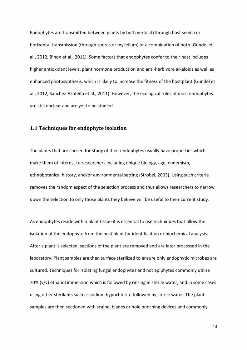
Figure 1. Pittosporum angustifolium (centre) from Pittsworth, QLD
Although not a common species, P. angustifolium is widespread throughout Australia. It was
previously wrongly named as Pittosporum phillyreoides. It is found in habitats of open
eucalypt woodlands and moister areas near inland lakes and drainage lines on sandy soils in
arid zones (Cayzer et al., 2000). P. angustifolium can be described as having pendulous
branches, falcate and glabrous leaves in a weeping canopy with yellow flowers (Cayzer et al.,
2000).
The Australian Aboriginals are known to have used various parts of the plant for different
purposes. A drink was made using the seeds, fruit pulp, leaves or wood in order to relieve

33
pain and cramps while a decoction of the fruit was used to treat eczema and pruritus
(Cayzer et al., 2000). In some areas of Australia, the Aboriginals utilized the fruits to prepare
a concoction that was drunk for coughs, colds or as a lactagogue, however, it is noted that
not all Aboriginals utilized the fruits, as the Pitjantjatjara tribe did not consume the fruits of
the plant at all (Sadgrove and Jones, 2013).
A recent study by Sadgrove and Jones, (2013) extracted the essential oils of Pittosporum
angustifolium and assessed their inhibitory activity against various microbial species. The
oils from the fruits and leaves of two P. angustifolium plants showed moderate
antimicrobial activity against the three microbes that were tested (Staphylococcus aureus,
Staphylococcus epidermidis and Candida albicans), while the fruit of another P.
angustifolium plant showed relatively high inhibition. The difference in antimicrobial activity
of each plant may be due to different chemical compositions found in plants of different
geographical locations. Analysis of the chemical composition of P. angustifolium essential oil
extracts revealed 51 different chemicals, the composition of which differed with each
geographically distinct sub species as well as between the leaves and fruits of the plants. For
example, leaf essential oils showed greater quantities of esters and sesquiterpenols than the
oils of the fruit. Chemical screening of the extracts revealed the presence of saponins,
phenols (both soluble and insoluble), flavonoids (pre- dominantly in the methanol and
hexane extracts), triterpenoids and tannins (Sadgrove and Jones, 2013). Sadgrove and Jones
(2013) suggested that the essential oil components limonene, sabinene, terpinenes, α-
pinene and bicyclogermacrene may be the cause of the antimicrobial activity in the study.

34
1.7 Research questions and objectives
There is currently limited knowledge of Australian endophytes regarding species diversity,
ecological roles as well as their potentials as producers of antimicrobial compounds. This
project seeks to expand this knowledge by examining the endophytes of the plant species P.
angustifolium and asks the questions: ‘What are the endophytes of P. angustifolium’, ‘Do
endophytes of P. angustifolium exhibit a host-specific relationship’ and ‘Do endophytes of P.
angustifolium produce bioactive compounds capable of inhibiting strains of human
pathogenic bacteria and fungi (Staphylococcus aureus, Serratia marcescens, Escherichia coli
and Candida albicans).’
There were three main objectives in this project:
1) To identify both fungal and bacterial endophytes in the leaves of P. angustifolium.
2) To determine if the fungal and or bacterial endophytes of P. angustifolium display host
preference, that is if the same endophytes are present in hosts at multiple plant
locations.
3) To detect and isolate bioactive compounds produced by the endophytes of P.
angustifolium.

35
2.0 Materials and Methods
2.1 Sample collection
Leaves of P. angustifolium were sampled across seven sites located in South East
Queensland in 2013 (Table 1). One plant was sampled from each site and the leaf samples
were taken from three different heights on each plant to gain a better representation of the
overall endophyte community within the plants leaves. Samples were placed in a plastic bag
and stored on ice until they could be processed within the laboratory. Processing of samples
occurred within three hours of collection.
Table 1. Sampling sites of Pittosporum angustifolium in Southeast QLD
Site Location
A Felton
B Biddeston
C Mount Tyson
D Oakey
E Gowrie Junction
F Chinchilla
G Pittsworth

36
Figure 2. Locations of sites sampled (labelled A – G) located in Southeast Queensland (Map obtained from Google Maps)
2.2 Endophyte isolation
Leaf samples were washed by partially filling each sample bag with tap water and shaking
vigorously. This was repeated twice. Samples were then moved to a biohazard safety
cabinet. Leaves were surface sterilized to eliminate any epiphytic microbes and ensure
isolation of only the leaf endophytes. This involved first soaking each leaf for 5 minutes in
sterile water. The leaves were then transferred into 95% ethanol (EtOH). Samples from sites

37
A, B and C were submerged in 95% EtOH for 70 seconds, however, later samples were
submerged in 95% EtOH for 60 seconds in order to reduce over-sterilizing the samples. The
samples were then passed through a blue flame to remove the residual EtOH. Leaves were
then pressed onto a Potato Dextrose agar (PDA) petri dish which acted as a means to
determine successful surface sterilization. A sterile hole punch was then used to remove
sections from each leaf. Eight sections were prepared per leaf and these were placed onto a
petri dish containing PDA. The procedure was repeated for each leaf sample taken. Seven
plates were prepared per plant which included one control plate and duplicate plates for
each of the three location samples taken per plant. Each plate was sealed with parafilm and
incubated in the dark at 23oC. Plates were checked daily for growth of any bacteria or fungi
growing from the edge of the leaf segments. For each fungal colony that grew, the hyphal
tips were subcultured onto a separate PDA plate by cutting out a small section of the
hyphae containing agar with a scalpel blade. Bacterial colonies were subcultured onto PDA
plates by the use of an inoculation loop and the streak plate method. All pure culture
isolates were incubated in the dark at 23oC.
2.3 Identification of endophytic isolates
2.3.1 Molecular Identification
Endophytic isolates were identified by the sequencing of important taxonomic regions
within their rDNA. Fungal isolates were identified via internal transcribed spacer (ITS)
sequencing while bacterial isolates were identified via small subunit (SSU) sequencing. The
DNA of each isolate was extracted using a Sigma-Aldrich XNAP-1KT REDExtract-N-Amp Plant

38
PCR Kit. Fungal ITS-rDNA and bacterial SSU-rDNA were amplified via polymerase chain
reaction (PCR). The fungal isolates utilized the fungal specific primer ITS1F (Gardes and
Bruns, 1993) and the primer ITS4 (White et al., 1990). Bacterial isolates utilized the primer
pair 27F and 1492R (Yu et al., 2013). PCR was set up using a Sigma-Aldrich XNAP-1KT
REDExtract-N-Amp Plant PCR Kit. Each reaction occurred with a total volume of 20µl
containing 4µl distilled water, 10µl PCR ReadyMix, 4µl extracted DNA and 1µl of each primer.
A Thermo Hybaid PCR Express Thermal Cycler was used to perform PCR reactions. DNA was
amplified with 35 cycles of 95C for 1 min, 50C for 1 min and 72C for 1 min, with a final
incubation at 72C for 10 min. All reactions were performed in duplicate along with a
negative control containing water instead of DNA. All PCR products were then purified using
Diffinity RapidTip 2 tips as per the manufacturer’s instructions. All purified products were
then electrophoresed in a 1% (w/v) agarose gel with RedSafe and visualized under UV light.
Molecular weight markers were electrophoresed alongside PCR products and gels were run
for 30 min at 100 volts. Sequencing reactions of purified DNA were performed at the
Brisbane laboratory of the Australian Genome Research Facility (AGRF) in 12l volumes
containing approximately 20ng/µl (10-11µl) of purified DNA and 1-2µl of primer. Sequencing
of fungal isolates used the ITS1F primer while bacterial isolates used the 27F primer.
Reactions utilized 11µl of purified DNA for PCR products showing a faint band after UV
visualization and 10µl DNA for those showing a bright band. Returned sequences were
analysed using the BLASTn search tool from GenBank to identify the closest match for each
isolate.
Phylogenetic analysis was performed with Molecular Evolutionary Genetics Analysis (MEGA)
version 6. The GenBank BLASTn closest matches (more than 97%) were included in the

39
sequence analysis. Sequences were aligned using the ‘align by Muscle’ option with default
settings. The aligned sequences were modified so that each was of the same size. Gaps
within the sequences which were common to all sequences were removed. Phylogenetic
analysis was carried out with a neighbour-joining tree using the Maximum Composite
Likelihood model and bootstrapping of 1000 replicates (Mapperson et al., 2014). The
suitability of the data being analysed was checked by using the compute overall mean
option. A neighbour joining tree was constructed for both the fungal endophytic isolates
and the bacterial isolates. For each of the species identified to the species level, sequences
of the same species were taken from GenBank and included in the analysis.
2.3.2 Morphological identification
Microscopy was used to distinguish bacterial isolates from yeast isolates. Using an
inoculation loop, a sample of bacterial/yeast cells were suspended in a drop of water on a
microscope slide and examined under a microscope.
2.4 Analysis for host preference
Each endophyte which was identified to the species level was analysed to determine
whether they showed a host preference for P. angustifolium. The presence or absence of
each endophyte among each of the seven sampled host plants was noted. A bar graph was
constructed showing the number of host plants colonized by each of the endophytes
identified to the species level.

40
2.5 Primary screening of endophytic isolates
Sensitest agar plates were inoculated with each endophytic isolate. For fungal isolates,
approximately 0.5cm3 of pure fungal culture was removed from its original plate with a
scalpel blade and transferred to the centre of the Sensitest agar plate. For bacterial isolates
the bacteria was transferred to the Sensitest agar by an inoculation loop and streaked in the
centre of the plate. Each isolate was cultured on two Sensitest agar plates to setup duplicate
screenings. Plates were stored in the dark at 23oC. Once each culture grew to at least 2cm in
diameter, the test pathogens were streaked via an inoculation loop from the margin of the
fungal/bacterial colony towards the edge of the plate. Four ATCC type strain pathogens
were used consisting of a gram positive bacterium: Staphylococcus aureus (ATCC 25923),
two gram negative bacteria: Serratia marcescens (ATCC 14756), Escherichia coli (ATCC
25922) and the fungus Candida albicans (ATCC 14053). Plates were incubated in the dark at
37oC and checked after 24-48 hours. Endophytes which showed inhibition on both duplicate
plates were recorded and used for further investigation.
2.6 Extraction of bioactive compounds
10ml of Malt Extract Broth (MEB) was prepared in McCartney bottles. The MEB was
inoculated with an approximately 0.5cm3 portion of mycelia containing agar and incubated
at 23oC for 1 week. The McCartney bottles were swirled by hand daily. One bottle of MEB
was prepared without the inoculum as a control.

41
After 1 week the bottle of fungal inoculum was added into a conical flask containing 500ml
of MEB. One McCartney bottle was prepared for each conical flask and multiple conical
flasks were prepared in order to increase the yield of any compounds extracted from the
MEB. Cotton stoppers were placed in the conical flasks and covered with alfoil to prevent
contamination. All conical flasks were incubated in the dark at 23oC.
After 5 days static growth, 500µl of autoclaved pathogens prepared in saline were added to
the conical flasks. The pathogen added was the same as that which the endophyte had
inhibited during the primary screening. The flasks were again incubated at 23oC. Once
substantial growth was observed, the temperature was increased to 25oC.
After 4-6 weeks growth, the mycelia was filtered from the broth through Chux wipes into a
1L beaker. The filtered mycelia was soaked in 100% ethyl acetate in a separate beaker and
broken up with a pipette tip. 250ml of filtered broth was poured into a separatory funnel
along with an equal amount of ethyl acetate. The separatory funnel was shaken and the
broth layer released into a 500ml beaker. The ethyl acetate layer was filtered through
Whatman filter paper into a separate 500ml beaker. The remaining broth was also put
through the separatory funnel with ethyl acetate. The separatory funnel steps were
repeated with the same broth to increase the yield of compound extracted from the broth.
The ethyl acetate used to soak the filtered mycelia was then added to the ethyl acetate used
with the separatory funnel. The ethyl acetate was then left to evaporate leaving the dried
crude extract.

42
2.7 Bioassay-guided fractionation
2.7.1 General experimental procedures
For HPLC fractionation of the crude extract, Alltech Davisil 40–60 μm 60 Å C18 bonded silica
was used for pre-adsorption work (Alltech, Deerfield, IL, USA). A Shimadzu LC-20AD pump
equipped with a Shimadzu SPD-M20A PDA detector and a Shimadzu SIL-20A autosampler
were fitted to the HPLC machine. A Phenomenex C18 Onyx Monolithic semi-preparative
column (10 mm 100 mm; Phenomenex, Torrance, CA, USA) and a Phenomenex C18 Onyx
Monolithic analytical column (4.6 mm 100 mm) were used for compound separation. All
solvents used for chromatography, were Lab-Scan HPLC grade (RCI Lab-Scan, Bangkok,
Thailand), and the H2O was Millipore Milli-Q PF filtered (Millipore, Billerica, MA, USA). All
synthetic reagents were purchased from Sigma Aldrich and used without further purification.
2.7.2 Analytical HPLC
A portion of the crude extract obtained from the ethyl acetate extraction (13 mg) was
resuspended in methanol. Isocratic HPLC conditions of H2O-ACN-CF3COOH (90:10:0.1) were
initially employed for the first 5 min, then a linear gradient to H2O-ACN (0.1% CF3COOH;
5:95:0.1) was run over 15 min, followed by isocratic conditions of H2O-ACN (0.1% CF3COOH;
5:95:0.1) for a further 5 min, all at a flow rate of 1 mL/min and at 40°C.
2.7.3 Bioassay-guided fractionation
A portion of the crude extract (67 mg) was pre-adsorbed to C18-bonded silica (1 g) then
packed into a stainless steel guard cartridge (10 × 30 mm) that was subsequently attached

43
to a C18 semi-preparative HPLC column. Isocratic HPLC conditions of H2O-ACN-CF3COOH
(90:10:0.1) were initially employed for the first 5 min, then a linear gradient to H2O-ACN
(0.1% CF3COOH; 5:95:0.1) was run over 15 min, followed by isocratic conditions of H2O-ACN
(0.1% CF3COOH; 5:95:0.1) for a further 5 min, all at a flow rate of 4 mL/min. Five fractions
were collected manually at appropriate intervals (Figure 9) from the start of the run, then
prepared for bioassay testing.
2.8 HPLC fraction and crude extract analysis
2.8.1 Minimum Inhibitory Concentration (MIC)
The bacteria Serratia marcescens (ATCC 14756) and Staphylococcus aureus (ATCC 25923)
were subcultured onto Sensitest agar plates and incubated at 37oC for 18 hours prior to
antimicrobial testing. S. marcescens was used as the target microbe for antibacterial testing
while S. aureus was used as a control due to it not being inhibited during the primary
screening and being a Gram positive bacterium in contrast to S. marcescens.
HPLC fractions were weighed and dissolved in 25% EtOH/0.7% saline to a concentration of
1mg/ml. The fractions were diluted by half four times in microcentrifuge tubes to produce
five different concentrations (1: 1mg/ml, 2: 500µg/ml, 3: 250µg/ml , 4: 125µg/ml and 5:
62.5µg/ml). The crude extract was also diluted into five concentrations beginning at
10mg/ml in 40%EtOH/0.7% saline (1:10mg/ml, 2: 5mg/ml, 3: 2.5mg/ml, 4: 1.25mg/ml, 5:
625µg/ml)

44
An antibiotic solution of ciprofloxacin (Sigma-Aldrich) was prepared as a positive control
against S. marcescens and S. aureus. The solution was made at a concentration of
approximately 12µg/ml in sterile H2O.
Suspensions of both S. marcescens and S. aureus were prepared in Mueller-Hinton (MH)
broth to an approximate concentration of a 0.5 McFarland standard. The suspensions were
used within 15 minutes of preparation.
50µl of sterile MH broth was transferred into the wells of a 96 well microdilution tray.
Aliquots of 50µl of fraction dilutions were transferred into the wells. Each fraction was
tested against both S. marcescens and S. aureus with dilutions being tested in duplicate
wells. 50µl of the appropriate bacteria was inoculated into all experimental wells. Columns
11 and 12 were reserved for negative, positive, contamination and solvent controls. The
negative control contained 50µl of sterile water along with 50µl of bacteria. The positive
control contained 50µl of antibiotic solution along with 50µl of bacteria. The contamination
control contained 100µl of sterile 0.7% saline and the solvent control contained 50µl of 25%
EtOH/0.7% saline and 50µl of bacteria.
The microdilution tray was incubated at 37oC, checking for bacterial growth at 18, 21 and 24
hours post incubation. The MIC was recorded as the concentrations in the first wells that
showed no visible growth after incubation.

45
2.8.2 Minimum Bactericidal Concentration (MBC)
Non-growing samples in the microdilution tray were used to determine the minimum
bactericidal concentration. 10µl was transferred from each well which showed no visible
growth onto separate Sensitest agar plates and spread with an inoculation spreader. Plates
were incubated at 37oC for 24 hours. Plates were checked for bacterial growth after
incubation. Fractions which allowed growth of bacteria were recorded as bacteriostatic
while those which displayed no bacterial growth were recorded as bactericidal.

46
3.0 Results
3.1 Endophyte isolation
Endophytes were successfully isolated from each of the 7 host plants sampled. Both fungal
and bacterial species were isolated, with plant samples from sites A – E contributing both
fungal and bacterial isolates and samples from sites F and G contributing only fungal and
bacterial isolates respectively. Endophytes did not grow from all leaf sections plated. Of the
sections displaying endophytic growth, some yielded only a single isolate while others
yielded up to three isolates. A total of 54 isolates were obtained across the 7 plants with
varying numbers of endophytes being isolated from each plant sample. There was a mean of
5.4 fungal and 2.3 bacterial isolates obtained for a single plant. The plant sample from site D
had the highest number of isolates (16 fungal, 2 bacterial) (Table 2). One bacterial isolate
was lost due to the agar within the petri dish drying up. As such no further analysis could be
done on the isolate.
Fungal growth was observed on the sterilisation control plate of the plant samples from site
A, indicating incomplete surface sterilization or contamination of the sterilisation control
plate. The endophyte on the control plate was identified via DNA sequencing and the
epiphytic contaminant was subsequently eliminated from further study.
Isolates were designated a code based on the site of the plant sample which they were
isolated from and the order in which each was isolated (Table 4), for example A1 for the first
endophyte isolated from site A.

47
Table 2. Total number of endophytes isolated per site.
Sample site
Number of fungal isolates obtained
Amount of Bacterial Isolates Obtained
A 7 2
B 7 1
C 1 1
D 16 2
E 2 6
F 5 0
G 0 4
Total 38 16
Mean 5.4 2.3
3.2 Identification of endophytic isolates
All isolates obtained (including the fungal isolate observed on the control plate), were
identified to species or genus level via DNA sequencing. PCR amplification of the ITS regions
of fungal isolates was successful for all isolates except for three (D6, D16 and E6). For the 16
bacterial isolates, PCR amplification of the SSU regions was unsuccessful for five isolates
(D15, E5, E8, G3 and G4) (Figure 3).
The returned sequences from the AGRF revealed that two isolates (C2 and D5) were unable
to be sequenced. After repeated sequencing failures it was decided to leave both isolates
without being identified. Successfully sequenced isolates were analysed with Chromas Lite
version 2.1 to check for contamination. Using the BLAST search tool 27 of the isolates were
identified to the species level and six isolates identified to the genus level. Other isolates
were found to belong to the Dothideomycetes class (one isolate), Sarcosomataceae family

48
(three isolates) and Sordariomycetes class (three isolates). Two isolates were identified to
their closest match to be uncultured bacterium clones. Two isolates from site A were found
to be the same species as that of the epiphytic contaminant growing on the site A control
plate (Table 3). These two isolates were thus not included as endophytic isolates from site A.
All of the successfully sequenced fungal isolates were found to be Ascomycetes while all of
the successfully sequenced bacterial isolates belonged to the phylum Firmicutes.
Table 3. Identification of fungal contaminants.
Isolate A0 was found as a contaminant on the control plate for site A. The two isolates A1 and A2
were found by a BLAST search to be the same species as that of the fungal contaminant on the
control plate.
Isolate Code Closest match GenBank Accession No. Query Cover (%) Identity (%)
A0 Nigrospora oryzae JN211105.1 100 98
A1 Nigrospora oryzae JN211105.1 100 99
A2 Nigrospora oryzae KC937039.1 100 100

49
Table 4. GenBank matches of isolated endophytes.
A total of 54 endophyte isolates were obtained from seven P. angustifolium plants. Table 4 lists the
species identified as the closest matches by the BLAST tool. Not all isolates could be successfully
sequenced and matched via BLAST and are thus not shown in Table 4.
Isolate Code
Closest match GenBank Accession No.
Query Cover (%)
Identity (%)
A3 Pseudocercospora fuligena GU214675.1 100 99
A4 Pseudocercospora fuligena GU214675.1 99 99
A5 Guignardia mangiferae AY816311.1 100 100
A6 Guignardia mangiferae EU677814.1 100 94
A7 Uncultured bacterium clone HM676109.1 100 99
A8 Dothideomycetes sp. JQ760353.1 98 98
A9 Uncultured bacterium clone HM332406.1 100 99
B1 Bacillus subtilis JN366795.1 100 87
B2 Guignardia mangiferae KF381072.1 99 99
B3 Xylaria sp. AB512404.1 100 100
B4 Sarcosomataceae sp. KF128806.1 98 96
B5 Preussia minima AY510425.1 95 96
B6 Sarcosomataceae sp. KF128803.1 100 100
B7 Coniochaeta sp. KF128810.1 100 99
B8 Xylaria sp. JN225909.1 99 95
D1 Pseudocercospora fuligena GU214675.1 100 99
D2 Pseudocercospora fuligena GU214675.1 100 99
D3 Pseudocercospora fuligena GU214675.1 100 99
D4 Pseudocercospora fuligena GU214675.1 99 99
D7 Pseudocercospora fuligena GU214675.1 100 99
D8 Pseudocercospora fuligena GU214675.1 99 99
D9 Pseudocercospora fuligena GU214675.1 99 99
D10 Pseudocercospora fuligena GU214675.1 100 99
D11 Pseudocercospora atromarginalis JX901780.1 100 100
D12 Pseudocercospora fuligena GU214675.1 100 99
D13 Pseudocercospora fuligena GU214675.1 100 99
D14 Pseudocercospora fuligena GU214675.1 100 99
D17 Lecythophora sp. HE863327.1 100 97
D18 Xylaria hypoxylon AY327476.1 99 95
E1 Bacillus pumilus JX645203.1 99 99
E2 Bacillus pumilus KJ410678.1 100 99
E3 Bacillus pumilus AM887694.1 99 99
E4 Bacillus sp. FJ596550.1 99 93
E7 Sarcosomataceae sp. KF128806.1 96 97

50
Table 4 continued.
Isolate Code
Closest match GenBank Accession No.
Query Cover (%)
Identity (%)
F1 Sordariomycetes sp. JQ760129.1 99 98
F2 Xylaria hypoxylon AY327476.1 97 96
F3 Xylaria hypoxylon AY327476.1 97 95
F4 Sporormiella sp. HQ130664.1 99 99
F5 Pyronema sp. KF128839.1 99 99
G1 Bacillus megaterium KF933685.1 100 99
G2 Bacillus megaterium HF584868.1 100 100
Figure 3. 1% (w/v) agarose gel of both successful and unsuccessful PCR products.
Bands indicate successful amplification of PCR products which include both fungal (A2, D5,
D17) isolates and bacterial (A7, B1, E4, G1) isolates.

51
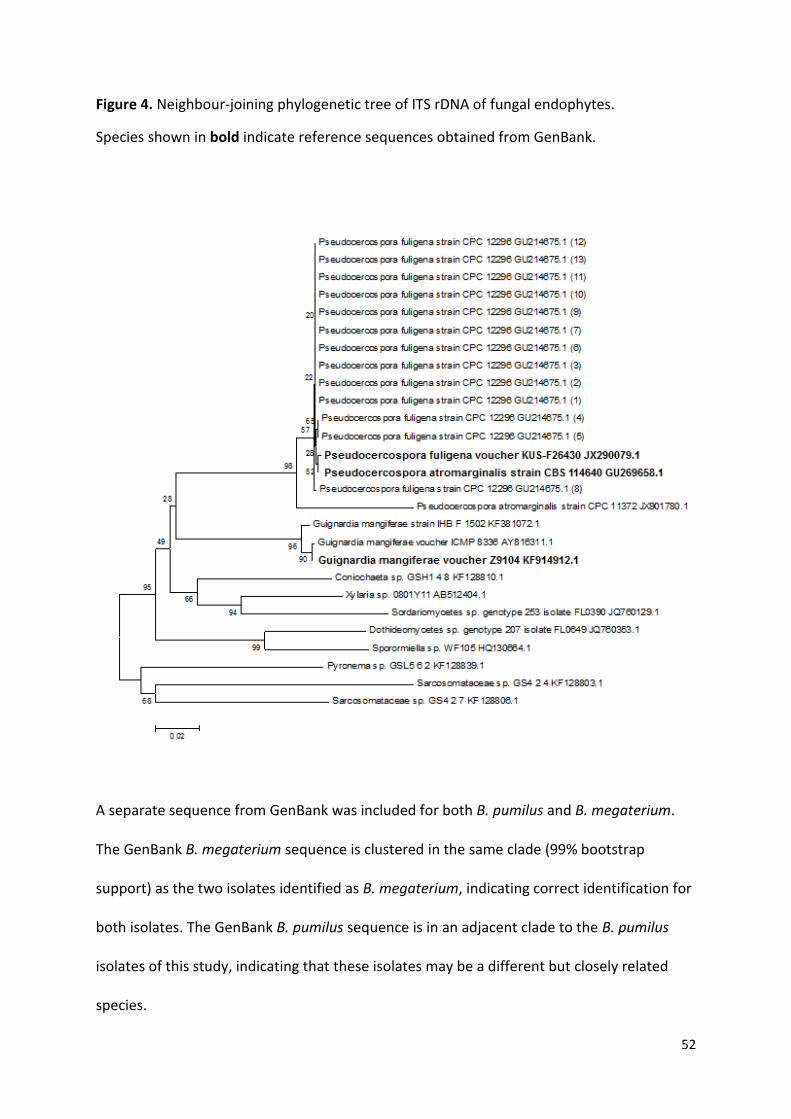
3.3 Phylogenetic Analysis
A neighbour joining tree was constructed using sequences which were identified to at least
a 97% match from BLAST searches in GenBank. P. fuligena, P. atromarginalis and G.
mangiferae were the only isolates included in the neighbour joining tree which were both
matched by BLAST to 97% or higher and identified to the species level. Reference sequences
representing each of these isolates were also obtained from GenBank and included in the
analysis. The reference sequence of P. fuligena was placed in a clade clustered among the
isolates of this study which were identified as P. fuligena. Some branching among these
isolates occurs, however, low bootstrap support (values of 20, 22, 28 etc.) decreases the
reliability that they should be in separate branches. The short horizontal distance of the
branches indicate that these isolates are genetically similar. P. atromarginalis is in an
adjacent clade to the P. fuligena isolates (98% bootstrap support) and its reference GenBank
sequence. This may indicate that the isolate is incorrectly identified and may be a different
but closely related species of Pseudocercospora. One G. mangiferae isolates is clustered
with the GenBank reference sequence with high bootstrap support (90%), however another
G. mangiferae isolate resides in an adjacent clade (96% bootstrap support).
52
Figure 4. Neighbour-joining phylogenetic tree of ITS rDNA of fungal endophytes.
Species shown in bold indicate reference sequences obtained from GenBank.
A separate sequence from GenBank was included for both B. pumilus and B. megaterium.
The GenBank B. megaterium sequence is clustered in the same clade (99% bootstrap
support) as the two isolates identified as B. megaterium, indicating correct identification for
both isolates. The GenBank B. pumilus sequence is in an adjacent clade to the B. pumilus
isolates of this study, indicating that these isolates may be a different but closely related
species.

53
Figure 5. Neighbour-joining phylogenetic tree of SSU rDNA of bacterial isolates.
Species shown in bold indicate sequences obtained from GenBank as a representative of
that species.

54
3.4 Host preference
Isolates which were successfully identified to the species level were graphed according to
the number of host plants they were isolated from (Figure 6). The bar graph shows that
three of the eight isolates were isolated from two plants (Pseudocercospora fuligena,
Guignardia mangiferae and Xylaria hypoxylon). The five other isolates were only found to
occur in one plant each.
Figure 6. Bar graph showing the amount of P. angustifolium plants that each species was isolated from.
Only those isolates which were identified to species level are included.

55
3.5 Primary Screening for Antimicrobial Activity
All but one isolate was screened for antimicrobial activity against the four test pathogens.
The one isolate that was not screened was unable to be subcultured onto Sensitest agar.
Four isolates appeared to show antimicrobial activity. Three of the four isolates were all
found to be the species Pseudocercospora fuligena. The fourth isolate (bacterial) that
displayed antimicrobial activity was unable to be identified and was therefore not chosen
for further investigation. The P. fuligena isolate displayed antimicrobial activity against the
test pathogen S. marcescens (Figure 7) and was therefore chosen for further investigation.
The three P. fuligena isolates were compared by the BLASTn tool from GenBank, which
revealed that they were each 99% similar to each other. This confirmed that the three
isolates were the same species.

56
Figure 7. Primary screening of endophytic isolates.
A) Primary screening of Pseudocercospora fuligena against test microbes 1, 2, 3 and 4 showing
reduced growth of 2. B) Primary screening against test microbes 1, 2, 3 and 4 showing no inhibition.
Test microbes: Staphylococcus aureus (1), Serratia marcescens (2), Escherichia coli (3) and Candida
albicans (4).
3.6 Bioassay-Guided Fractionation
Upon the completion of the ethyl acetate extraction, the total yield of the fungal crude
extract was 105mg. 25mg of this extract was retained and analysed later to identify whether
the compound/s responsible for the antibacterial activity was successfully extracted from P.
fuligena. Approximately 13mg of crude extract underwent an analytical HPLC run (Figure 8).
A range of peaks at 210nm spectra are observed from 1.5min to 4min over 500mAU and
one further peak at 15min over 500mAU. Another noticeable peak occurred at 12min with
various other smaller peaks occurring throughout.

57
Approximately 67mg of crude extract pre-adsorbed to C18-bonded silica then underwent
HPLC fractionation (Figure 9). Five fractions were collected.
Figure 8. Analytical HPLC chromatogram for P. fuligena crude extract showing fractions collected for antimicrobial screening.
Figure 9. HPLC fractionation chromatogram of P. fuligena crude extract showing fractions collected for antimicrobial screening.

58
3.7 HPLC fraction and crude extract analysis
3.7.1 Minimum Inhibitory Concentration
Dilutions were performed for each fraction to produce five samples of different
concentrations (Table 5). Dilutions were also performed in the same manner for the crude
extract (Table 6).
Table 5. Dilutions of fractions for use in MIC and MBC assays.
Table 6. Dilutions of crude extract for use in MIC and MBC assays.
The wells of the microdilution tray containing the positive (antibiotic) controls as well as the
contamination controls showed no visible growth while those containing the negative
controls and solvent controls did contain visible growth.
All wells containing S. aureus displayed visible growth. Fractions 1, 4 and 5 did not inhibit
the growth of S. marcescens for any concentration. Both fractions 2 and 3 inhibited the
Fraction Initial concentration (mg/ml) Dilution 1 (µg/ml)
Dilution 2 (µg/ml)
Dilution 3 (µg/ml)
Dilution 4 (µg/ml)
1 1 500 250 125 62.5
2 1 500 250 125 62.5
3 1 500 250 125 62.5
4 1 500 250 125 62.5
5 1 500 250 125 62.5
Sample Initial concentration (mg/ml)
Dilution 1 (mg/ml)
Dilution 2 (mg/ml)
Dilution 3 (mg/ml)
Dilution 4 (mg/ml)
Crude Extract 10 5 2.5 1.25 0.625

59
growth of S. marcescens at all concentrations except 62.5µg/ml where it showed visible
growth, therefore having a MIC of 125µg/ml (Table 7). The crude extract showed inhibition
of S. marcescens at all concentrations assayed with the lowest concentration being
625µg/ml (Table 7).
Table 7. MIC of test samples against S. marcescens.
3.7.2 Minimum Bactericidal Concentration
Fractions 2, 3 and the crude extract were tested for their MBC against S. marcescens. Both
fractions 2 and 3 were found to be bacteriostatic rather than bactericidal, as bacterial
growth was found to cover most of the STA plate used for testing. The crude extract was
found to be bactericidal with a MBC of 2.5mg/ml.
Sample MIC against S. marcescens
Fraction 2 125 µg/ml
Fraction 3 125 µg/ml
Crude extract 625 µg/ml

60
4.0 Discussion
4.1 Endophyte isolation
In this investigation, all plant samples of P. angustifolium were found to be host to at least
one endophyte. This finding supports Strobel & Daisy (2003) that each individual plant on
earth is host to one or more endophytes. Some of the plants sampled had higher numbers
of endophytes than others, which may be due to multiple possible reasons. The number of
endophytes able to be isolated may in part be determined during the leaf tissue surface
sterilization phase. The duration each leaf spent alight after passing through the Bunsen
burner flame may have affected the survival of some endophytes. Other types of
endophytes may also be unable to be isolated with the methods used in this investigation. It
is understood that culture dependent methods favour the fast-growing microbes and that
the unculturable or slow-growing microbes will not be isolated (Duong et al., 2006).
The time each leaf spent submerged in ethanol during the surface sterilization stage was
modified part way through the study to attempt to increase the number of endophytes
isolated. Time spent in ethanol was reduced from 70 sec to 60 sec from site D onwards. Site
D had an increase in endophytes (18) over preceding samples, however, subsequent
samples did not show any increases. As such, the time spent in ethanol may not have had a
major impact on the endophytes of the leaf tissue. The majority of isolates from site D were
of the same species (Pseudocercospora fuligena) which may explain the higher number of
isolates. With no previous research known to have been conducted on the endophytes of P.

61
angustifolium, this project gives the first indication for what endophytes reside within the
plant.
The number of endophytes isolated in this study varied between each plant sampled
ranging from two isolates (site C) to eighteen isolates (site D). These results may be due to
sampling plants from different locations as it has been indicated that the location a plant
resides in can strongly influence the abundance of endophytes (Hoffman and Arnold, 2008).
The plant’s locality may also affect endophyte diversity and species composition (Hoffman
and Arnold, 2008). Therefore, even though each plant sampled in this study was found in
similar habitats, there may be differences in each location that affects the endophytic
abundance within each plant as well as whether fungal or bacterial endophytes could
colonize the plants. This project found that most plants sampled harboured both fungal and
bacterial endophytes, however, only fungal endophytes were isolated from site F with only
bacterial endophytes isolated from site G. It may be that unseen environmental factors
differed between both locations. Further research could focus on such factors as the soil
composition, rainfall, mean temperature or weather patterns of each site at the time of
sampling to find if any of these affect the endophyte assemblages. Also, as sampling was
undertaken over several months, changes in climate over these months may have affected
the endophyte communities.
The sterilisation control plate for the site A samples displayed growth of a single endophyte
identified as Nigrospora oryzae. This led to discounting two isolates identified by DNA
sequencing of the same species due to the uncertainty that either isolate was a true
endophyte.

62
4.2 Identification of endophytic isolates
Endophytes have been traditionally identified based on their morphological characteristics
while in culture (Ko Ko et al., 2011). More recently endophytes have also been identified by
molecular methods involving DNA extraction, sequencing and comparison of obtained
sequences to known species via databases such as GenBank (Ko Ko et al., 2011).
Identification of endophytic isolates is an important part of ecological or bioprospecting
studies. Correct identification of isolates can afford researchers the ability to use the data as
a reference for future research. For example, when working with an unknown endophyte,
identification then allows the researcher to search for previous research on the same
endophytic species as well as allowing easier cataloguing of any results gained in their
research. Identification in bioprospecting studies allows other researchers to know what
species has produced what compound. It can also help to identify how isolated endophytes
are related to other endophytic species. For these reasons, and others, the ability to
successfully identify endophytes is an important for research in this field.
Mycological taxonomy has been developing for over 200 years and has settled on grouping
fungi into four major divisions, along with one ‘pseudo-division’. The four major divisions
are differentiated by their modes of sexual reproduction and consist of the Chytrids,
Zygomycetes, Ascomycetes and Basidiomycetes (Seifert, 2009). The pseudo-division consists
of the asexually reproducing fungi, though some have known sexual states, called the
Deuteromycetes or Fungi Imperfecti (Seifert, 2009). All of the fungal endophytes sequenced
in this study were found to be ascomycetes. As ascomycetes are the largest phylum of fungi
(approximately 64,000 species) (Schoch et al., 2009), it is not surprising that the endophytes
of this project belonged to this phylum.

63
Molecular methods were used in this study to attempt to identify each endophyte isolated.
Some isolates were ultimately unable to be identified due to either a failure to amplify the
DNA sequence with PCR or failure to be sequenced at AGRF. The majority of fungal isolates
were successfully amplified by PCR and as such suggests that the choice of primers for
fungal rDNA amplification was not at fault. It was suspected that the concentration of DNA
may have been too high in the PCR reactions. Some isolates DNA were able to be amplified
after reducing the volume of DNA extract in the PCR reaction. The isolates which were again
not amplified may have succeeded with further decreases to volume of DNA extract. The
failure of DNA sequencing of some isolates may have been due to the concentration of DNA
being too high and may have been overcome by decreasing the concentration. It is also
possible that the water used for the sequencing contained a sequencing inhibitor and that if
fresh pure water were used, would allow successful sequencing.
Of the isolates identified, P. fuligena, P. atromarginalis, G. mangiferae, B. pumilus and B.
megaterium all had ≥97% identity to their matched species by BLAST. 97% was used as the
threshold for correct identification. Pseudocercospora is a large genus of plant pathogenic
fungi which are commonly associated with leaf and fruit spots as well as blights on a wide
range of plant hosts (Crous et al., 2013). They are found in many areas including cool
temperate, sub-tropical and tropical regions (Crous et al., 2013). P. fuligena is known to be
the causal pathogen of black leaf mould, which is a major fungal disease of tomato in Asia
(Mersha et al., 2014). P. atromarginalis has been found to cause leaf spots on the plant
Lycianthes biflora (Phengsintham et al., 2013). Neither species appears to have been
identified as an endophyte in the literature, however, Crous et al. (2013) states that species
of Pseudocercospora are recognized as endophytes.

64
Guignardia is a genus of fungi which contains around 330 known species, with many of
these species being considered as endophytic fungi (Wickert et al., 2014). The species
Guignardia mangiferae has been previously identified as a ubiquitous endophyte and has
been confused with the citrus black spot pathogen Guignardia citricarpa (Romao et al.,
2011). Despite being endophytic in a wide range of hosts, it causes foliar spots in Mangifera
indica (mango) (Wickert et al., 2014).
Bacillus is a genus of gram positive bacteria and has members that are capable of producing
antibiotics, such as B. subtilis (Stein, 2005, Ouoba et al., 2007) and B. amyloliquefaciens
(Yuan et al., 2012). Members of the genus may also have potential agricultural uses due to
their ability to produce antimicrobial metabolites to control plant pathogens as well as to fix
nitrogen (Liu et al., 2006). Studies have shown that strains of the isolate B. pumilus, which
was isolated in this study, have displayed antimicrobial activity (Aunpad and Na-Bangchang,
2007, Ouoba et al., 2007). A strain of B. pumilus produced the compound pumilicin 4 which
was found to be active against two drug-resistant pathogenic bacteria, Methicillin-resistant
Staphylococcus aureus and vancomycin-resistant enterococci (Aunpad and Na-Bangchang,
2007). The second Bacillus species isolated in this study, B. megaterium, has demonstrated
nitrogen fixing abilities in some strains (Liu et al., 2006) and has also shown potential to
control the fungal caused disease septoria tritici blotch (Kildea et al., 2008). Both B. pumilus
and B. megaterium have previously been isolated as endophytes (Rai et al., 2007, Moore et
al., 2006).
Various isolates that were matched by BLAST had a closest match below 97%. This
introduces uncertainty to whether those endophytes are the same as their matched species.

65
As such, their species identities cannot be confirmed. It may be that these endophytes are
new species of known fungal genera. With the current knowledge on Australian endophytes
being sparse, it is likely that new species will be encountered during research into these
microbes.
Phylogenetic analysis of the fungal species (Figure 4) showed the P. fuligena isolates
grouped in adjacent clades near the GenBank P. fuligena sequence. While there is some
branching into separate clades among the isolates, the bootstrap values for these clades are
low which decreases the reliability of the branching. The horizontal distances of each branch
is minimal which suggests little genetic variation. The BLAST results for each P. fuligena
isolate also show low variation as each isolate was matched to P. fuligena with a % identity
of 99%. This suggests that the P. fuligena isolates have been correctly identified. The P.
atromarginalis isolate from this study resides in a clade adjacent to its GenBank reference
sequence with strong bootstrap support (98%). This suggests that the isolate was incorrectly
identified. The phylogenetic analysis suggests unreliability of the BLAST analysis as the
isolate matched with P. atromarginalis was matched with a % identity of 100% from BLAST.
This may suggest the isolate is a closely related species to P. atromarginalis. One of the G.
mangiferae isolates appears to be identified correctly as it was grouped together with the
GenBank reference sequence with a bootstrap support of 90%. The other isolate identified
by BLAST as G. mangiferae, however, is in an adjacent clade (96% bootstrap support) which
indicates it was incorrectly identified. It may be a closely related species to G. mangiferae.
The neighbour-joining phylogenetic tree of the bacterial species (Figure 5) indicates that the
B. pumilus isolates are closely related to the B. pumilus GenBank sequence, however the low

66
bootstrap support decreases the reliability of the branching. The isolates may be closely
related species rather than the same species. The B. megaterium isolates are clustered in
the same clade (99% bootstrap support), which suggests that both isolates have been
identified correctly. Arnold and Lutzoni (2007) note the limitations of using BLAST for
species identification which are that BLAST matches are based on non-evolutionary
matching criteria, are subject to error due to mis-identified sequences, can be difficult to
interpret when all top matches are unidentified isolates or environmental samples, and are
limited to those fungi present in GenBank.
The results of this study have shown that P. angustifolium is host to a variety of endophytes
and is a potential source for identifying new species.
4.3 Host preference
From the data gained in this study, it cannot be concluded that any host preferences or host
specific relationships exist with any of the endophytes isolated from P. angustifolium. Out of
the 54 endophyte isolates, only 8 were identified to the species level and of these only 3
isolates were found in multiple plants. The low frequency of occurrence (2 plants) observed
for the 3 isolates suggested no host preference.
There is the possibility that the endophyte community of a plant may vary from leaf to leaf.
As such, sampling of more leaves from each plant may have revealed endophytes which
occur frequently in different plants. Also, as mentioned previously, some endophytes may
not be able to be isolated with the methods used here and it may be these endophytes that
show a host preference.

67
The endophytic community of P. angustifolium may be affected by location more than host
preference. This appeared to be the case in a study by Arnold et al. (2001) which found that
when Heisteria concinna was sampled from two different sites, many of its endophytes
were found only at one of the sites, despite similar conditions. This corresponds to the
current study, where most endophyte species were found in only one plant. There may be
unknown environmental factors at each site that determine the ability of certain species to
colonize the plant, such as changes in soil nutrients, the amount of water available or the
temperature of each area.
The endophytic communities in tissue other than leaves may vary and potentially show host
preference. Studies have shown that endophytes are able to colonize plant tissue other than
the leaves, such as the roots and xylem (Macia-Vicente et al., 2008, Oses et al., 2008). While
previous research has indicated that there seems to be no host specificity among root
endophytes (Girlanda et al., 2002), a study has shown host preference of root endophytic
Fusarium spp. (Macia-Vicente et al., 2008). Host preference is similar to specificity, however,
the relationship is not exclusive to one species (Kernaghan and Patriquin, 2011). The
literature on root endophyte host preference or specificity is sparse so there is not enough
evidence to suggest whether host specific relationships exist. The possibility exists that
endophytes occupying the tissues of P. angustifolium other than the leaves, may show a
host preference.
The isolation of P. fuligena, G. mangiferae and X. hypoxylon from two different plants may
be an indication of host preference. If more P. angustifolium plants were sampled, the three
endophytes mentioned may be found to be commonly present. Also, if more leaves were

68
processed from the plants sampled in this project, it may be found that P. fuligena, G.
mangiferae, X. hypoxylon or others are found in each of the plants.
4.4 Primary Screening for Antimicrobial Activity
In this study, each endophyte isolated from P. angustifolium underwent a primary screening
for antimicrobial activity. Each of the four pathogens chosen to be screened against are
clinically significant pathogens. Within this study, two gram negative bacteria were chosen
to be screened against: Escherichia coli and Serratia marcescens. E. coli is one of the most
frequent and best studied bacterial organisms and is the most abundant facultative
anaerobic bacteria of the human intestinal flora (Dobrindt, 2005, Jaureguy et al., 2008). E.
coli has non-pathogenic commensal variants of the normal human gut flora, as well as
pathogenic variants which can cause intestinal or extraintestinal infection in humans
(Dobrindt, 2005). Extraintestinal E. coli infections typically include urinary tract infections
(UTI), meningitis (mostly in neonates and after neurosurgery), diverse intraabdominal
infections, pneumonia (particularly in hospitalized and institutionalized patients),
intravascular-device infections, osteomyelitis and soft-tissue infections (Russo and Johnson,
2000). E. coli is also a leading cause of bacteraemia and because of an increase in β-lactam
resistant strains, it can be difficult to treat (Courpon-Claudinon et al., 2010). Third-
generation cephalosporin (3GC) resistance has especially become problematic as 3GCs are a
common part of empirical antimicrobial chemotherapy in severe infections (Courpon-
Claudinon et al., 2010). S. marcescens is an opportunistic enteric pathogen and is
responsible for a significant proportion of hospital-acquired infections (Murdoch et al.,
2011). It was thought to be a nonpathogenic saprophytic water organism until late in the
20th century (Su et al., 2003). Many strains of S. marcescens are also resistant to multiple

69
antibiotics (Kurz et al., 2003). It is known to be an opportunistic pathogen which has caused
outbreaks of nosocomial infections of varying severity including urinary tract infections
(UTIs), respiratory tract infections, bacteraemia, conjunctivitis, endocarditis, meningitis, and
wound infections (Su et al., 2003).
Staphylococcus aureus is a gram positive bacterium which was chosen as one of the
pathogens to be screened against in this study. S. aureus is the most common cause of
nosocomial pneumonia and surgical site infections and the second most common cause of
bloodstream, cardiovascular, and eye, ear, nose, and throat infections in the United States
(Noskin et al., 2005). A major concern has been the emergence of methicillin resistant S.
aureus strains due to their resistance to nearly all β-lactam antibiotics (Arede et al., 2013).
The fourth pathogen screened against in this study is the fungus Candida albicans. C.
albicans is a commensal fungus which occurs in the gastrointestinal tract and the oral and
vaginal mucosa of many, if not all, healthy individuals but is also the most common human
fungal pathogen (Kim and Sudbery, 2011). C. albicans has the ability to grow either as a
unicellular budding yeast or in filamentous pseudohyphal and hyphal forms and it is the
hyphal form that, during mucosal infections, invades epithelial and endothelial cells and
causes damage (Sudbery, 2011). In immunocompromised individuals C. albicans can cause a
range of mucosal and systemic infections, including acute pseudomembranous oral
candidiasis (thrush), the most common opportunistic infection of HIV-infected patients
(Cheng et al., 2003). Immunocompromised individuals may also develop blood stream
infections called candidemia which can lead to colonization of internal organs, known as
disseminated candidiasis (Kim and Sudbery, 2011).

70
The aforementioned diseases caused by each of the test microbes chosen in this study along
with the increase in antibiotic resistance, gives finding novel antimicrobial compounds a
great importance. In this study, the endophyte P. fuligena inhibited the growth of S.
marcescens in the primary screening stage and therefore was chosen to undergo liquid
broth fermentation and extraction of any produced metabolites. Along with P. fuligena, a
bacterial endophyte was found to display inhibitory effects against the test microbes.
However, the endophyte was unable to be identified after multiple attempts and in order to
save time it was decided not to proceed with further experimentation with the isolate.
4.5 Bioassay-Guided Fractionation
In this study, the antimicrobial activity of P. fuligena was assessed using a bioassay-guided
fractionation approach. Previous studies have shown success with this method for isolating
fungal metabolites (Rosa et al., 2013, Zhao et al., 2012, Ratnaweera et al., 2014). Rosa et al.
(2013) reported the identification of the antifungal fatty acids caproic, caprylic, myristic,
palmitic, heptadecanoic, stearic, oleic, linoleic and stearic acids from a crude extract of the
endophytic fungus Coniochaeta ligniaria. Zhao et al. (2012) isolated two antimicrobial
compounds from the crude extract of the endophytic fungus Gliomastix murorum Ppf8 using
a bioassay-guided fractionation approach. They identified the compounds as ergosta-5,7,22-
trien-3-ol and 2,3-dihydro-5-hydroxy-α,α-dimethyl-2-benzofuranmethanol which displayed
antibacterial activity against five Gram negative and two Gram positive bacteria, and
antifungal activity against one fungus. Ratnaweera et al. (2014) also successfully isolated an
antibacterial compound with this approach. They isolated the antibacterial compound
helvolic acid from an endophytic Xylaria sp. which had activity against the Gram-positive
bacteria, Bacillus subtilis and methicillin-resistant Staphylococcus aureus. These studies

71
reinforce the worth of the bioassay-guided fractionation method as means to isolate
bioactive compounds.
In this study, extracts of P. fuligena afforded five fractions after HPLC fractionation. Testing
of each fraction found two fractions which had bacteriostatic activity against S. marcescens.
This confirms the finding from the primary screening that P. fuligena can inhibit the growth
of S. marcescens. Fraction 2 (Figure 9) which eluted between 6.5 to 10 minutes displayed
bacteriostatic inhibition against S. marcescens. Fraction 3 (Figure 9) which eluted between
10 to 13.5 minutes also displayed bacteriostatic inhibition against S. marcescens. Neither
fraction was bactericidal against the bacterium which coincides with the results from the
primary screening. As seen in Figure 7, the growth of S. marcescens was reduced rather than
prevented by the endophyte. This study appears to be the first report of the fungus P.
fuligena displaying antimicrobial activity.
The crude extract from the ethyl acetate extraction displayed bactericidal activity against S.
marcescens. The MBC for the crude extract was 2.5mg/ml which is 20-fold greater
concentration than the MIC for the two bacteriostatic fractions (125 µg/ml). This may
suggest that at higher concentrations, the two fractions may also be bactericidal. In a study
by Radhakrishnan et al. (2011) it was found that the MBC of the compound embelin was at a
higher concentration than the MIC was against all microbes tested. For example, embelin
had an MIC of 50µg/ml and an MBC of 400µg/ml against the bacteria Klebsiella pneumoniae
(Radhakrishnan et al., 2011). The compound responsible for the bactericidal activity of the
crude extract in this study most likely makes up only a small fraction of the mixture. As a

72
pure compound it may need less than the MBC concentration of the crude extract in order
to be bactericidal by itself.
P. fuligena did not display bactericidal activity during primary screening as no clear zone of
inhibition around the endophyte was observed. Instead, the growth of S. marcescens only
appeared to be reduced. It is possible that the endophyte did not produce sufficient
amounts of the bioactive compound that is needed to cause bactericidal activity. The
bioactivity displayed by P. fuligena supports the notion that endophytes are a promising
source of bioactive compounds and shows the potential of Australian plants as sources of
these endophytes. Further research of the bioactive compounds of P. fuligena could
potentially impact on future control of disease caused by S. marcescens.
4.6 Future Directions
This study could be continued in many directions. More P. angustifolium plants could be
sampled in order to gain access to a potentially greater variety of endophytic species which
would allow further investigation into the host-endophyte relationship as well as the
bioactive potential of isolated endophytes. All of the endophytes isolated in this project
could be identified morphologically as well as molecularly which would help to confirm the
species matches from BLAST. Other plant species could be sampled in the same area as the
P. angustifolium plants sampled in this study to observe whether the same endophytes are
present in both species. This would help to identify whether the endophytes identified in
this study are specific to the host plant rather than the habitat of the host. Further research
could be conducted on comparing the endophytes of other parts of the plant (such as the
roots or bark) with that of the leaves. Furthermore, roots may have a different variety of

73
endophytic species which may produce antimicrobial compounds. This would give a better
assessment of the overall endophyte species diversity of the plant.
P. fuligena could be regrown in bulk in order to achieve a greater yield of crude extract.
During HPLC a greater number of fractions could be generated within the time that fractions
2 and 3 from this study were eluted in order to isolate a pure compound. Any pure
compounds isolated could be identified via NMR spectroscopy or X-ray crystallography. Pure
compounds could be tested for MIC and MBC to identify the compound responsible for the
results found in this study.
The endophytes in this study could also be screened against other microbes such as MRSA
and Bacillus cereus. The fractions isolated from P. fuligena could also be tested against other
pathogens. Any antimicrobial pure compounds isolated from P. fuligena could be analysed
with cytotoxicity assays to gain a better idea of their potential use in medicine. The bacterial
endophyte displaying antimicrobial activity which was not further analysed could be further
attempted to be identified and its antimicrobial activity analysed. Research into P.
angustifolium endophytes could also aim to identify the endophytes anticancer activity.

74
5.0 Conclusion
Little is known about the endophyte diversity of Australian plants and even less is known
about their antimicrobial properties. As such, the results of this study have added to the
current pool of knowledge on Australian endophytes.
Many of the isolated endophytes were successfully sequenced in this study which resulted
in matches with sequences from GenBank. However, only eight of the 54 isolates were
identified down to the species level. This demonstrates the limitations of molecular
identification techniques. The isolates not matched to the species level may have been new
species of known genera. P. angustifolium was found to have more fungal endophytes than
bacterial, however, this may not be a good representation of the whole endophytic diversity
of the plant.
No host specific relationships were shown to exist between any of the isolated endophytes
and their host. However, this study only investigated the leaves of seven plants. Sampling of
more plants, leaves or plant tissue type may give different results than this study.
This study also adds to the growing evidence that endophytes are capable of producing
valuable antimicrobial compounds. A crude extract from one fungal endophyte (P. fuligena)
displayed bactericidal activity against the human pathogen S. marcescens while two
fractions of the crude action displayed bacteriostatic activity. The compounds causing the
antibacterial activity could be potential targets for further investigation to determine
whether they are of use in the medical arena.

75
This study demonstrates the potential of P. angustifolium as a source of undiscovered
endophytic species and antimicrobial compounds. The study may encourage further
research into the endophytes of P. angustifolium or other Australian plants as sources of
undiscovered species and novel antimicrobial compounds. Continued research into
endophytes may help to alleviate the problem of antibiotic resistant microbial pathogens.

76
6.0 References
ALY, A. H., DEBBAB, A., KJER, J. & PROKSCH, P. 2010. Fungal endophytes from higher plants:
a prolific source of phytochemicals and other bioactive natural products. Fungal
Diversity, 41, 1-16.
ALY, A. H., DEBBAB, A. & PROKSCH, P. 2011. Fungal endophytes: unique plant inhabitants
with great promises. Applied Microbiology and Biotechnology, 90, 1829-45.
AMNA, T., PURI, S. C., VERMA, V., SHARMA, J. P., KHAJURIA, R. K., MUSARRAT, J., SPITELLER,
M. & QAZI, G. N. 2006. Bioreactor studies on the endophytic fungus Entrophospora
infrequens for the production of an anticancer alkaloid camptothecin. Canadian
Journal of Microbiology, 52, 189-96.
ANDREOTE, F. D., DE ARAUJO, W. L., DE AZEVEDO, J. L., VAN ELSAS, J. D., DA ROCHA, U. N. &
VAN OVERBEEK, L. S. 2009. Endophytic colonization of potato (Solanum tuberosum
L.) by a novel competent bacterial endophyte, Pseudomonas putida strain P9, and its
effect on associated bacterial communities. Applied and Environmental Microbiology,
75, 3396-406.
AREDE, P., MINISTRO, J. & OLIVEIRA, D. C. 2013. Redefining the role of the beta-lactamase
locus in methicillin-resistant Staphylococcus aureus: beta-lactamase regulators
disrupt the MecI-mediated strong repression on mecA and optimize the phenotypic
expression of resistance in strains with constitutive mecA expression. Antimicrobial
Agents and Chemotherapy, 57, 3037-45.

77
ARNOLD, A. E. & LUTZONI, F. 2007. Diversity and Host Range of Foliar Fungal Endophytes:
Are Tropical Leaves Biodiversity Hotspots? Ecology, 88, 541-549.
ARNOLD, A. E., MAYNARD, Z. & GILBERT, G. S. 2001. Fungal endophytes in dicotyledonous
neotropical trees: patterns of abundance and diversity. Mycological Research, 105,
1502-1507.
ASRAFUL ISLAM, S. M., MATH, R. K., KIM, J. M., YUN, M. G., CHO, J. J., KIM, E. J., LEE, Y. H. &
YUN, H. D. 2010. Effect of plant age on endophytic bacterial diversity of balloon
flower (Platycodon grandiflorum) root and their antimicrobial activities. Current
Microbiology, 61, 346-56.
AUNPAD, R. & NA-BANGCHANG, K. 2007. Pumilicin 4, a novel bacteriocin with anti-MRSA
and anti-VRE activity produced by newly isolated bacteria Bacillus pumilus strain
WAPB4. Current Microbiology, 55, 308-13.
BÉRDY, J. 2005. Bioactive Microbial Metabolites. The Journal of Antibiotics, 58, 1-26.
BIHON, W., SLIPPERS, B., BURGESS, T., WINGFIELD, M. J. & WINGFIELD, B. D. 2011. Sources
of Diplodia pinea endophytic infections in Pinus patula and P. radiata seedlings in
South Africa. Forest Pathology, 41, 370-375.
BOK, J. W. & KELLER, N. P. 2004. LaeA, a Regulator of Secondary Metabolism in Aspergillus
spp. Eukaryotic Cell, 3, 527-535.

78
CALVO, A. M. 2008. The VeA regulatory system and its role in morphological and chemical
development in fungi. Fungal Genetics and Biology, 45, 1053-61.
CALVO, A. M., WILSON, R. A., BOK, J. W. & KELLER, N. P. 2002. Relationship between
Secondary Metabolism and Fungal Development. Microbiology and Molecular
Biology Reviews, 66, 447-459.
CANNON, P. & SIMMONS, C. 2002. Diversity and host preference of leaf endophytic fungi in
the Iwokrama Forest Reserve, Guyana. Mycologia, 94, 210-220.
CASTILLO, U. F., STROBEL, G. A., FORD, E. J., HESS, W. M., PORTER, H., JENSEN, J. B., ALBERT,
H., ROBISON, R., CONDRON, M. A. M., TEPLOW, D. B., STEVENS, D. & YAVER, D. 2002.
Munumbicins, wide-spectrum antibiotics produced by Streptomyces NRRL 30562,
endophytic on Kennedia nigriscans. Microbiology, 148, 2675-2685.
CAYZER, L. W., CRISP, M. D. & TELFORD, I. R. H. 2000. Revision of Pittosporum
(Pittosporaceae) in Australia. Australian Systematic Botany, 13, 845-902.
CHANDLER, G. T., PLUNKETT, G. M., PINNEY, S. M., CAYZER, L. W. & GEMMILL, C. E. C. 2007.
Molecular and morphological agreement in Pittosporaceae: phylogenetic analysis
with nuclear ITS and plastid trnL–trnF sequence data. Australian Systematic Botany,
20, 390.
CHEN, L., LUO, S., XIAO, X., GUO, H., CHEN, J., WAN, Y., LI, B., XU, T., XI, Q., RAO, C., LIU, C. &
ZENG, G. 2010. Application of plant growth-promoting endophytes (PGPE) isolated

79
from Solanum nigrum L. for phytoextraction of Cd-polluted soils. Applied Soil Ecology,
46, 383-389.
CHENG, S., CLANCY, C. J., CHECKLEY, M. A., HANDFIELD, M., HILLMAN, J. D., PROGULSKE-FOX,
A., LEWIN, A. S., FIDEL, P. L. & NGUYEN, M. H. 2003. Identification of Candida
albicans genes induced during thrush offers insight into pathogenesis. Molecular
Microbiology, 48, 1275-1288.
COURPON-CLAUDINON, A., LEFORT, A., PANHARD, X., CLERMONT, O., DORNIC, Q., FANTIN,
B., MENTRÉ, F., WOLFF, M., DENAMUR, E. & BRANGER, C. 2010. Bacteraemia caused
by third-generation cephalosporin-resistant Escherichia coli in France: prevalence,
molecular epidemiology and clinical features. Clinical Microbiology and Infection, 17,
557–565.
CROUS, P. W., BRAUN, U., HUNTER, G. C., WINGFIELD, M. J., VERKLEY, G. J., SHIN, H. D.,
NAKASHIMA, C. & GROENEWALD, J. Z. 2013. Phylogenetic lineages in
Pseudocercospora. Studies in Mycology, 75, 37-114.
DEBBAB, A., ALY, A. H. & PROKSCH, P. 2012. Endophytes and associated marine derived
fungi—ecological and chemical perspectives. Fungal Diversity, 57, 45-83.
DESHMUKH, S. K., MISHRA, P. D., KULKARNI-ALMEIDA, A., VEREKAR, S., SAHOO, M. R.,
PERIYASAMY, G., GOSWAMI, H., KHANNA, A., BALAKRISHNAN, A. & VISHWAKARMA,
R. 2009. Anti-Inflammatory and Anticancer Activity of Ergoflavin Isolated from an
Endophytic Fungus. Chemistry and Biodiversity, 6, 784-789.

80
DING, L., MUNCH, J., GOERLS, H., MAIER, A., FIEBIG, H. H., LIN, W. H. & HERTWECK, C. 2010.
Xiamycin, a pentacyclic indolosesquiterpene with selective anti-HIV activity from a
bacterial mangrove endophyte. Bioorganic & Medicinal Chemistry Letters, 20, 6685-7.
DOBRINDT, U. 2005. (Patho-)Genomics of Escherichia coli. International Journal of Medical
Microbiology, 295, 357-71.
DUONG, L. M., JEEWON, R., LUMYONG, S. & HYDE, K. D. 2006. DGGE coupled with ribosomal
DNA gene phylogenies reveal uncharacterized fungal phylotypes. Fungal Diversity, 23,
121-138.
EYBERGER, A. L., DONDAPATI, R. & PORTER, J. R. 2006. Endophyte fungal isolates from
Podophyllum peltatum produce podophyllotoxin. Natural Products, 69, 1121–1124.
FOX, E. M. & HOWLETT, B. J. 2008. Secondary metabolism: regulation and role in fungal
biology. Current Opinion in Microbiology, 11, 481-7.
GARDES, M. & BRUNS, T. 1993. ITS primers with enhanced specificity for basidiomycetes —
application to the identification of mycorrhizae and rusts. Molecular Ecology, 2, pp.
113–118.
GIRIDHARAN, P., VEREKAR, S. A., KHANNA, A., MISHRA, P. D. & DESHMUKH, S. K. 2012.
Anticancer activity of sclerotiorin, isolated from an endophytic fungus Cephalotheca
faveolata Yaguchi, Nishim. & Udagawa. Indian Journal of Experimental Biology, 50,
464-468.

81
GIRLANDA, M., GHIGNONE, S. & LUPPI, A. M. 2002. Diversity of sterile root-associated fungi
of two Mediterranean plants. New Phytologist, 155, 481-498.
GUNDEL, P. E., HELANDER, M., CASAS, C., HAMILTON, C. E., FAETH, S. H. & SAIKKONEN, K.
2012. Neotyphodium fungal endophyte in tall fescue (Schedonorus phoenix): a
comparison of three Northern European wild populations and the cultivar Kentucky-
31. Fungal Diversity, 60, 15-24.
GUO, B., DAI, J., NG, S., HUANG, Y., LEONG, C., ONG, W. & CARTE, B. K. 2000. Cytonic Acids A
and B: Novel Tridepside Inhibitors of hCMV Protease from the Endophytic Fungus
Cytonaema Species. Journal of Natural Products, 63, 602-604.
GUO, B., WANG, Y., SUN, X. & TANG, K. 2011. Bioactive natural products from endophytes: A
review. Applied Biochemistry and Microbiology, 44, 136-142.
HOFFMAN, M. T. & ARNOLD, A. E. 2008. Geographic locality and host identity shape fungal
endophyte communities in cupressaceous trees. Mycological Research, 112, 331-344.
HYDE, K. D. & SOYTONG, K. 2008. The fungal endophyte dillema. Fungal Diversity, 33, 163-
173.
JAUREGUY, F., LANDRAUD, L., PASSET, V., DIANCOURT, L., FRAPY, E., GUIGON, G.,
CARBONNELLE, E., LORTHOLARY, O., CLERMONT, O., DENAMUR, E., PICARD, B.,
NASSIF, X. & BRISSE, S. 2008. Phylogenetic and genomic diversity of human
bacteremic Escherichia coli strains. BMC Genomics, 9, 560.

82
JOSEPH, B. & PRIYA, R. M. 2011. Bioactive compounds from endophytes and their potential
in pharmaceutical effect: a review. American Journal of Biochemistry and Molecular
Biology, 1, 291-309.
KAUL, S., GUPTA, S., AHMED, M. & DHAR, M. K. 2012. Endophytic fungi from medicinal
plants: a treasure hunt for bioactive metabolites. Phytochemistry Reviews, 11, 487-
505.
KERNAGHAN, G. & PATRIQUIN, G. 2011. Host associations between fungal root endophytes
and boreal trees. Microbial Ecology, 62, 460-73.
KILDEA, S., RANSBOTYN, V., KHAN, M. R., FAGAN, B., LEONARD, G., MULLINS, E. & DOOHAN,
F. M. 2008. Bacillus megaterium shows potential for the biocontrol of septoria tritici
blotch of wheat. Biological Control, 47, 37-45.
KIM, J. & SUDBERY, P. 2011. Candida albicans, a major human fungal pathogen. The Journal
of Microbiology, 49, 171-7.
KJER, J., WRAY, V., EDRADA-EBEL, R. A., EBEL, R., PRETSCH, A., LIN, W. H. & PROKSCH, P.
2009. Xanalteric Acids I and II and Related Phenolic Compounds from an Endophytic
Alternaria sp. Isolated from the Mangrove Plant Sonneratia alba. Journal of Natural
Products, 72, 2053–2057.

83
KO KO, T. W., STEPHENSON, S. L., BAHKALI, A. H. & HYDE, K. D. 2011. From morphology to
molecular biology: can we use sequence data to identify fungal endophytes? Fungal
Diversity, 50, 113-120.
KURZ, C. L., CHAUVET, S., ANDRÈS, E., AUROUZE, M., VALLET, I., MICHEL, G. P. F., UH, M.,
CELLI, J., FILLOUX, A., DE BENTZMANN, S., STEINMETZ, I., HOFFMANN, J. A., FINLAY, B.
B., GORVEL, J.-P., FERRANDON, D. & EWBANK, J. J. 2003. Virulence factors of the
human opportunistic pathogen Serratia marcescens identified by in vivo screening.
The EMBO Journal, 22.
KUSARI, S., LAMSHÖFT, M. & SPITELLER, M. 2009a. Aspergillus fumigatus Fresenius, an
endophytic fungus from Juniperus communis L. Horstmann as a novel source of the
anticancer pro-drug deoxypodophyllotoxin. Journal of Applied Microbiology, 107,
1019-30.
KUSARI, S., ZÜHLKE, S. & SPITELLER, M. 2009b. An Endophytic Fungus from Camptotheca
acuminata That Produces Camptothecin and Analogues. Journal of Natural Products,
72, 2-7.
LI, J. Y., HARPERB, J. K., GRANT, D. M., TOMBEC, B. O., BASHYALD, B., HESS, W. M. &
STROBEL, G. 2001. Ambuic acid, a highly functionalized cyclohexenone with
antifungal activity from Pestalotiopsis spp. and Monochaetia sp. Phytochemistry, 56,
463-468.

84
LI, J. Y. & STROBEL, G. A. 2001. Jesterone and hydroxy-jesterone antioomycete
cyclohexenone epoxides from the endophytic fungus Pestalotiopsis jesteri.
Phytochemistry, 57, 261-265.
LIN, X., HEZARI, M., KOEPP, A. E., FLOSS, H. G. & CROTEAU, R. 1996. Mechanism of
Taxadiene Synthase, a Diterpene Cyclase That Catalyzes the First Step of Taxol
Biosynthesis in Pacific Yew. Biochemistry, 35, 2968-2977.
LINNEK, J., MITAINE-OFFER, A. C., PAULULAT, T. & LACAILLE-DUBOIS, M. A. 2012. Two new
triterpenoid saponins from Pittosporum senacia Putterlick (Pittosporaceae).
Magnetic Resonance in Chemistry, 50, 798-802.
LIU, A. R., CHEN, S. C., JIN, W. J., ZHAO, P. Y., JEEWON, R. & XU, T. 2012. Host specificity of
endophytic Pestalotiopsis populations in mangrove plant species of South China.
African Journal of Microbiology Research, 6, 6262-6269.
LIU, X., DONG, M., CHEN, X., JIANG, M., LV, X. & ZHOU, J. 2008. Antimicrobial activity of an
endophytic Xylaria sp.YX-28 and identification of its antimicrobial compound 7-
amino-4-methylcoumarin. Applied Microbiology and Biotechnology, 78, 241-7.
LIU, X., ZHAO, H. & CHEN, S. 2006. Colonization of maize and rice plants by strain Bacillus
megaterium C4. Current Microbiology, 52, 186-90.
MACIA-VICENTE, J. G., JANSSON, H. B., ABDULLAH, S. K., DESCALS, E., SALINAS, J. & LOPEZ-
LLORCA, L. V. 2008. Fungal root endophytes from natural vegetation in

85
Mediterranean environments with special reference to Fusarium spp. FEMS
Microbiology Ecology, 64, 90-105.
MAPPERSON, R. R., KOTIW, M., DAVIS, R. A. & DEARNALEY, J. D. 2014. The diversity and
antimicrobial activity of Preussia sp. endophytes isolated from Australian dry
rainforests. Current Microbiology, 68, 30-7.
MERSHA, Z., ZHANG, S. & HAU, B. 2014. Seasonal Dynamics of Black Leaf Mould
(Pseudocercospora fuligena) on Greenhouse-Grown Fresh Market Tomatoes. Journal
of Phytopathology, 162, 158-169.
MILLER, C. M., MILLER, R. V., GARTON-KENNY, D., REDGRAVE, B., SEARS, J., CONDRON, M.
M., TEPLOW, D. B. & STROBEL, G. 1998. Ecomycins, unique antimycotics from
Pseudomonas viridiflava. Journal of Applied Microbiology, 84, 937-944.
MILLER, K. I., INGREY, S. D., ALVIN, A., SZE, M. Y. D., ROUFOGALIS, B. D. & NEILAN, B. A. 2010.
Endophytes and the microbial genetics of traditional medicines. Microbiology
Australia, 60-63.
MILLER, K. I., QING, C., SZE, D. M., ROUFOGALIS, B. D. & NEILAN, B. A. 2012. Culturable
endophytes of medicinal plants and the genetic basis for their bioactivity. Microbial
Ecology, 64, 431-49.
MOORE, F. P., BARAC, T., BORREMANS, B., OEYEN, L., VANGRONSVELD, J., VAN DER LELIE, D.,
CAMPBELL, C. D. & MOORE, E. R. 2006. Endophytic bacterial diversity in poplar trees

86
growing on a BTEX-contaminated site: the characterisation of isolates with potential
to enhance phytoremediation. Systematic and Applied Microbiology, 29, 539-56.
MOUSA, W. K. & RAIZADA, M. N. 2013. The diversity of anti-microbial secondary
metabolites produced by fungal endophytes: an interdisciplinary perspective.
Frontiers in Microbiology, 4, 65.
MURDOCH, S. L., TRUNK, K., ENGLISH, G., FRITSCH, M. J., POURKARIMI, E. & COULTHURST, S.
J. 2011. The opportunistic pathogen Serratia marcescens utilizes type VI secretion to
target bacterial competitors. Journal of Bacteriology, 193, 6057-69.
NISSINEN, R. M., MÄNNISTÖ, M. K. & VAN ELSAS, J. 2012. Endophytic bacterial communities
in three arctic plants from low arctic fell tundra are cold-adapted and host-plant
specific. FEMS Microbiology Ecology, 82, 510-522.
NOSKIN, G. A., RUBIN, R. J., SCHENTAG, J. J., KLUYTMANS, J., HEDBLOM, E. C., SMULDERS, M.,
LAPETINA, E. & GEMMEN, E. 2005. The Burden of Staphylococcus aureus Infections
on Hospitals in the United States. JAMA Internal Medicine, 165, 1756-1761.
OSES, R., VALENZUELA, S., FREER, J., SANFUENTES, E. & RODRÍGUEZ, J. 2008. Fungal
endophytes in xylem of healthy Chilean trees and their possible role in early wood
decay Fungal Diversity, 33, 77-86.
OUOBA, L. I., DIAWARA, B., JESPERSEN, L. & JAKOBSEN, M. 2007. Antimicrobial activity of
Bacillus subtilis and Bacillus pumilus during the fermentation of African locust bean

87
(Parkia biglobosa) for Soumbala production. Journal of Applied Microbiology, 102,
963-70.
PHENGSINTHAM, P., BRAUN, U., MCKENZIE, E. H. C., CHUKEATIROTE, E., CAI, L. & HYDE, K. D.
2013. Monograph of Cercosporoid fungi from Thailand. Plant Pathology &
Quarantine, 3, 67-138.
PINRUAN, U., RUNGJINDAMAI, N., CHOEYKLIN, R., LUMYONG, S., HYDE, K. D. & JONES, E. B.
G. 2010. Occurrence and diversity of basidiomycetous endophytes from the oil palm,
Elaeis guineensis in Thailand. Fungal Diversity, 41, 71-88.
PURI, S. C., NAZIR, A., CHAWLA, R., ARORA, R., RIYAZ-UL-HASAN, S., AMNA, T., AHMED, B.,
VERMA, V., SINGH, S., SAGAR, R., SHARMA, A., KUMAR, R., SHARMA, R. K. & QAZI, G.
N. 2006. The endophytic fungus Trametes hirsuta as a novel alternative source of
podophyllotoxin and related aryl tetralin lignans. Journal of Biotechnology, 122, 494-
510.
RADHAKRISHNAN, N., GNANAMANI, A. & MANDAL, A. B. 2011. A potential antibacterial
agent Embelin, a natural benzoquinone extracted from Embelia ribes. Biology and
Medicine, 3, 1-7.
RAI, R., DASH, P. K., PRASANNA, B. M. & SINGH, A. 2007. Endophytic bacterial flora in the
stem tissue of a tropical maize (Zea mays L.) genotype: isolation, identification and
enumeration. World Journal of Microbiology and Biotechnology, 23, 853–858.

88
RASHID, S., CHARLES, T. C. & GLICK, B. R. 2012. Isolation and characterization of new plant
growth-promoting bacterial endophytes. Applied Soil Ecology, 61, 217-224.
RATNAWEERA, P. B., WILLIAMS, D. E., DE SILVA, E. D., WIJESUNDERA, R. L., DALISAY, D. S. &
ANDERSEN, R. J. 2014. Helvolic acid, an antibacterial nortriterpenoid from a fungal
endophyte, sp. of orchid endemic to Sri Lanka. Mycology, 5, 23-28.
REITER, B. & SESSITSCH, A. 2006. Bacterial endophytes of the wildflower Crocus albiflorus
analyzed by characterization of isolates and by a cultivation-independent approach.
Canadian Journal of Microbiology, 52, 140-9.
RODRIGUEZ, R. J., WHITE, J. F., JR., ARNOLD, A. E. & REDMAN, R. S. 2009. Fungal
endophytes: diversity and functional roles. New Phytologist, 182, 314-30.
ROMAO, A. S., SPOSITO, M. B., ANDREOTE, F. D., AZEVEDO, J. L. & ARAUJO, W. L. 2011.
Enzymatic differences between the endophyte Guignardia mangiferae
(Botryosphaeriaceae) and the citrus pathogen G. citricarpa. Genetics and Molecular
Research, 10, 243-52.
ROSA, L. H., QUEIROZ, S. C. N., MORAES, R. M., WANG, X., TECHEN, N., PAN, Z., CANTRELL, C.
L. & WEDGE, D. E. 2013. Coniochaeta ligniaria: antifungal activity of the cryptic
endophytic fungus associated with autotrophic tissue cultures of the medicinal plant
Smallanthus sonchifolius (Asteraceae). Symbiosis, 60, 133-142.

89
RUSSO, A. & JOHNSON, J. 2000. Proposal for a New Inclusive Designation for Extraintestinal
Pathogenic Isolates of Escherichia coli: ExPEC. The Journal of Infectious Diseases, 181,
1753-1754.
SADGROVE, N. J. & JONES, G. L. 2013. Chemical and biological characterisation of solvent
extracts and essential oils from leaves and fruit of two Australian species of
Pittosporum (Pittosporaceae) used in aboriginal medicinal practice. Journal of
Ethnopharmacology, 145, 813-21.
SANCHEZ-AZOFEIFA, A., OKI, Y., WILSON FERNANDES, G., BALL, R. A. & GAMON, J. 2011.
Relationships between endophyte diversity and leaf optical properties. Trees, 26,
291-299.
SANTOS, S. N., FERRARIS, F. K., SOUZA, A. O., HENRIQUES, M. D. G. & MELO, I. S. 2012.
Endophytic fungi from Combretum leprosum with potential anticancer and
antifungal activity. Symbiosis, 58, 109-117.
SCHOCH, C. L., SUNG, G. H., LOPEZ-GIRALDEZ, F., TOWNSEND, J. P., MIADLIKOWSKA, J.,
HOFSTETTER, V., ROBBERTSE, B., MATHENY, P. B., KAUFF, F., WANG, Z., GUEIDAN, C.,
ANDRIE, R. M., TRIPPE, K., CIUFETTI, L. M., WYNNS, A., FRAKER, E., HODKINSON, B. P.,
BONITO, G., GROENEWALD, J. Z., ARZANLOU, M., DE HOOG, G. S., CROUS, P. W.,
HEWITT, D., PFISTER, D. H., PETERSON, K., GRYZENHOUT, M., WINGFIELD, M. J.,
APTROOT, A., SUH, S. O., BLACKWELL, M., HILLIS, D. M., GRIFFITH, G. W.,
CASTLEBURY, L. A., ROSSMAN, A. Y., LUMBSCH, H. T., LUCKING, R., BUDEL, B.,
RAUHUT, A., DIEDERICH, P., ERTZ, D., GEISER, D. M., HOSAKA, K., INDERBITZIN, P.,
90
KOHLMEYER, J., VOLKMANN-KOHLMEYER, B., MOSTERT, L., O'DONNELL, K., SIPMAN,
H., ROGERS, J. D., SHOEMAKER, R. A., SUGIYAMA, J., SUMMERBELL, R. C.,
UNTEREINER, W., JOHNSTON, P. R., STENROOS, S., ZUCCARO, A., DYER, P. S.,
CRITTENDEN, P. D., COLE, M. S., HANSEN, K., TRAPPE, J. M., YAHR, R., LUTZONI, F. &
SPATAFORA, J. W. 2009. The Ascomycota tree of life: a phylum-wide phylogeny
clarifies the origin and evolution of fundamental reproductive and ecological traits.
Systematic Biology, 58, 224-39.
SEIFERT, K. A. 2009. Progress towards DNA barcoding of fungi. Molecular Ecology Resources,
9 Suppl s1, 83-9.
SHWETA, S., ZUEHLKE, S., RAMESHA, B. T., PRITI, V., MOHANA KUMAR, P., RAVIKANTH, G.,
SPITELLER, M., VASUDEVA, R. & UMA SHAANKER, R. 2010. Endophytic fungal strains
of Fusarium solani, from Apodytes dimidiata E. Mey. ex Arn (Icacinaceae) produce
camptothecin, 10-hydroxycamptothecin and 9-methoxycamptothecin.
Phytochemistry, 71, 117-22.
SIEBER, T. N. 2007. Endophytic fungi in forest trees: are they mutualists? Fungal Biology
Reviews, 21, 75-89.
SIEGEL, R., NAISHADHAM, D. & JEMAL, A. 2013. Cancer statistics, 2013. CA: A Cancer Journal
for Clinicians, 63, 11-30.
STEIN, T. 2005. Bacillus subtilis antibiotics: structures, syntheses and specific functions.
Molecular Microbiology, 56, 845-57.

91
STONE, M. & WILLIAMS, D. 2006. On the evolution of functional secondary metabolites
(natural products). Molecular Microbiology, 6, 29-34.
STROBEL, G. & DAISY, B. 2003. Bioprospecting for Microbial Endophytes and Their Natural
Products. Microbiology and Molecular Biology Reviews, 67, 491-502.
STROBEL, G., DAISY, B., CASTILLO, U. & HARPER, J. 2004. Natural Products from Endophytic
Microorganisms. Journal of Natural Products, 67, 257-268.
STROBEL, G., YANG, X., SEARS, J., KRAMER, R., SIDHU, R. & HESS, W. M. 1996. Taxol from
Pestalotiopsis microspora, an endophytic fungus of Taxus wallachiana. Microbiology,
142, 435-440.
STROBEL, G. A. 2003. Endophytes as sources of bioactive products. Microbes and Infection, 5,
535-544.
SU, L. H., OU, J. T., LEU, H. S., CHIANG, P. C., CHIU, Y. P., CHIA, J. H., KUO, A. J., CHIU, C. H.,
CHU, C., WU, T. L., SUN, C. F., RILEY, T. V. & CHANG, B. J. 2003. Extended Epidemic of
Nosocomial Urinary Tract Infections Caused by Serratia marcescens. Journal of
Clinical Microbiology, 41, 4726-4732.
SUDBERY, P. E. 2011. Growth of Candida albicans hyphae. Nature Reviews Microbiology, 9,
737-48.

92
SUN, X., DING, Q., HYDE, K. D. & GUO, L. D. 2012. Community structure and preference of
endophytic fungi of three woody plants in a mixed forest. Fungal Ecology, 5, 624-632.
TAECHOWISAN, T., LU, C., SHEN, Y. & LUMYONG, S. 2005. Secondary metabolites from
endophytic Streptomyces aureofaciens CMUAc130 and their antifungal activity.
Microbiology, 151, 1691-5.
TRAN, H. B. Q., MCRAE, J. M., LYNCH, F. & PALOMBO, E. A. 2010. Identification and bioactive
properties of endophytic fungi isolated from phyllodes of Acacia species. Current
Research, Technology and Education Topics in Applied Microbiology and Microbial
Biotechnology, 377-382.
VAISHNAV, P. & DEMAIN, A. L. 2011. Unexpected applications of secondary metabolites.
Biotechnology Advances, 29, 223-9.
WEBER, D., STERNER, O., ANKE, T., GORZALCZANCY, S., MARTINO, V. & ACEVEDO, C. 2004.
Phomol, a New Antiinflammatory Metabolite from an Endophyte of the Medicinal
Plant Erythrina crista-galli. The Journal of Antibiotics, 57, 559-563.
WELLENSIEK, B. P., RAMAKRISHNAN, R., BASHYAL, B. P., EASON, Y., GUNATILAKA, A. A. L. &
AHMAD, N. 2013. Inhibition of HIV-1 Replication by Secondary Metabolites From
Endophytic Fungi of Desert Plants. The Open Virology Journal, 7, 72-80.
WEST, E. R., COTHER, E. J., STEEL, C. C. & ASH, G. J. 2010. The characterization and diversity
of bacterial endophytes of grapevine. Canadian Journal of Microbiology, 56, 209-16.

93
WHITE, T. J., BRUNS, T. D., LEE, S. B. & TAYLOR, J. W. 1990. Amplification and direct
sequencing of fungal ribosomal RNA genes for phylogenetics. In: M.A. INNIS, D. H. G.,
J.J. SNINSKY, T.J. WHITE (ed.) PCR Protocols — a Guide to Methods and Applications.
San Diego, CA: Academic Press.
WICKERT, E., SOUZA, A. D., PEREIRA, R. M., KISHI, L. T., LEMOS, E. G. D. M. & GOES, A. D.
2014. Molecular and Pathogenic Study of Guignardia spp. Isolates Associated to
Different Hosts Advances in Microbiology, 04, 116-125.
WICKLOW, D. T., ROTH, S., DEYRUP, S. T. & GLOER, J. B. 2005. A protective endophyte of
maize: Acremonium zeae antibiotics inhibitory to Aspergillus flavus and Fusarium
verticillioides. Mycological Research, 109, 610-618.
WU, L., HAN, T., LI, W., JIA, M., XUE, L., RAHMAN, K. & QIN, L. 2013. Geographic and tissue
influences on endophytic fungal communities of Taxus chinensis var. mairei in China.
Current Microbiology, 66, 40-8.
YU, J., ZHOU, X. F., YANG, S. J., LIU, W. H. & HU, X. F. 2013. Design and application of specific
16S rDNA-targeted primers for assessing endophytic diversity in Dendrobium
officinale using nested PCR-DGGE. Applied Microbiology and Biotechnology, 97,
9825-36.
YU, J. H. & KELLER, N. 2005. Regulation of secondary metabolism in filamentous fungi.
Annual Review of Phytopathology, 43, 437-58.

94
YUAN, J., LI, B., ZHANG, N., WASEEM, R., SHEN, Q. & HUANG, Q. 2012. Production of
bacillomycin- and macrolactin-type antibiotics by Bacillus amyloliquefaciens NJN-6
for suppressing soilborne plant pathogens. Journal of Agricultural and Food
Chemistry, 60, 2976-81.
ZAFERANLOO, B., VIRKAR, A., MAHON, P. J. & PALOMBO, E. A. 2013. Endophytes from an
Australian native plant are a promising source of industrially useful enzymes. World
Journal of Microbiology and Biotechnology, 29, 335-45.
ZHAN, J., BURNS, A. M., LIU, M. X., FAETH, S. H. & GUNATILAKA, A. A. L. 2007. Search for Cell
Motility and Angiogenesis Inhibitors with Potential Anticancer Activity: Beauvericin
and Other Constituents of Two Endophytic Strains of Fusarium oxysporum. Journal of
Natural Products, 70, 227-232.
ZHANG, J. Y., TAO, L. Y., LIANG, Y. J., CHEN, L. M., MI, Y. J., ZHENG, L. S., WANG, F., SHE, Z. G.,
LIN, Y. C., TO, K. K. & FU, L. W. 2010. Anthracenedione derivatives as anticancer
agents isolated from secondary metabolites of the mangrove endophytic fungi.
Marine Drugs, 8, 1469-81.
ZHAO, J., LI, C., WANG, W., ZHAO, C., LUO, M., MU, F., FU, Y., ZU, Y. & YAO, M. 2013.
Hypocrea lixii, novel endophytic fungi producing anticancer agent cajanol, isolated
from pigeon pea (Cajanus cajan [L.] Millsp.). Journal of Applied Microbiology, 115,
102-13.

95
ZHAO, J., SUN, W., SHAN, T., MOU, Y., LOU, J., LI, Y., WANG, M. & ZHOU, L. 2012.
Antimicrobial metabolites from the endophytic fungus Gliomastix murorum Ppf8
associated with the medicinal plant Paris polyphylla var. yunnanensis. Journal of
Medicinal Plants Research, 6.
ZHOU, X., WANG, Z., JIANG, K., WEI, Y., LIN, J., SUN, X. & TANG, K. 2007. Screening of taxol-
producing endophytic fungi from Taxus chinensis var. mairei. Applied Biochemistry
and Microbiology, 43, 439-443.

96
Appendices
Appendix A
Media recipes for fungal and bacterial endophytes
Potato Dextrose Agar (PDA)
Dissolve 15.6g of PDA in 400ml of distilled water. Autoclave at 121oC for 15 minutes.
Sensitest Agar (STA)
Dissolve 12.8g of STA in 400ml of distilled water. Autoclave at 121oC for 15 minutes.
Malt Extract Broth (MEB)
Dissolve 20g of MEB powder in 1L of distilled water. Autoclave at 121oC for 15 minutes.
Mueller-Hinton Broth (MHB)
Dissolve 8.4g of MHB in 400ml of distilled water. Autoclave at 121oC for 15 minutes.

97
Appendix B
MIC data
The following tables represent the microdilution trays used for MIC assay. Five fractions
collected from HPLC were tested along with the crude extract from the ethyl acetate
extraction. Column 11/rows A-D contained the negative control. Column 11/rows E-H
contained the contamination control. Column 12/rows A-D contained the positive control.
Column 12/rows E-H contained the solvent control.
+ is shown to indicate growth of the bacteria.
- is shown to indicate no growth of the bacteria.
Microdilution tray 1
HPLC fractions
Row Column 1 2 3 4 5 6 7 8 9 10 11 12
Conc. µg/ml 1000 500 250 125 62.5 Controls
1 A (S. marcescens) + + + + + + + + + + + -
B (S. aureus) + + + + + + + + + + + -
2 C (S. marcescens) - - - - - - - - + + + -
D (S. aureus) + + + + + + + + + + + -
3 E (S. marcescens) - - - - - - - - + + - +
F (S. aureus) + + + + + + + + + + - +
4 G (S. marcescens) + + + + + + + + + + - +
H (S. aureus) + + + + + + + + + + - +

98
Microdilution tray 2
HPLC fractions
Row Column 1 2 3 4 5 6 7 8 9 10 11 12
Conc. µg/ml 1000 500 250 125 62.5 Controls
5 A (S. marcescens) + + + + + + + + + + + -
B (S. aureus) + + + + + + + + + + + -
Conc. mg/ml 10 5 2.5 1.25 0.625 Controls
Crude extract
C (S. marcescens) - - - - - - - - - - + -
D (S. aureus) + + + + + + + + + + + -
E - +
F - +
G - +
H - +