analysis of genes from inner ear developmental-stage cdna ...we recovered 33 different genes from...
TRANSCRIPT
www.elsevier.com/locate/ydbio
Developmental Biology 268 (2004) 7–23
Analysis of genes from inner ear developmental-stage cDNA
subtraction reveals molecular regionalization of the otic capsule
Michael Ficker,a,1 Nicola Powles,a,1 Nick Warr,a,1 Ulla Pirvola,b and Mark Maconochiea,c,*
aMammalian Genetics Unit, Medical Research Council, Harwell, Oxfordshire OX11 0RD, UKb Institute of Biotechnology, University of Helsinki, 00014 Helsinki, Finland
cSchool of Life Sciences, University of Sussex, Falmer, Brighton BN1 9QG, UK
Received for publication 21 November 2002, revised 10 November 2003, accepted 24 November 2003
Abstract
Although the gross embryology of inner ear development has been documented for several different vertebrate species at a descriptive
level, our understanding of the molecular mechanisms involved remains rudimentary. Therefore, we have used cDNA subtraction and
normalization procedures to define genes upregulated in the 13.5dpc mouse inner ear, a developmental stage where inner ear morphogenesis
and tissue remodeling is active and differentiation of future hair cells is being initiated. We recovered 33 different genes from this subtraction
and using gene-specific primers have confirmed the transcriptional upregulation of 26 of these in the 13.5dpc inner ear. Northern analyses
were used to investigate splicing differences between the inner ear and the whole embryo at 13.5dpc. Spatial localization of expression was
determined through whole-ear in situ hybridization analysis, and selected genes were analyzed in more detail through in situ hybridization of
tissue sections. These data illustrate that the genes isolated in this study are expressed in the developing otic capsule and/or neuroepithelium.
Furthermore, the expression patterns also reveal molecular heterogeneity in the developing capsule and indicate that for some genes, the
chondrogenic otic capsule is composed of distinct domains of gene expression.
D 2004 Published by Elsevier Inc.
Keywords: Mouse; Inner ear; Otic capsule; cDNA subtraction
Introduction
The adult inner ear is composed of specialized sensory
and nonsensory epithelia organized within an exquisitely
sculptured structure, with six different patches of sensory
epithelia in mammals differentiating in highly stereotypical
fashion at discrete locations. It is likely that many genes
interact to control and execute the developmental program
leading to the formation of such a complex structure.
Furthermore, this developmental program is required to
direct the coordinated development of epithelial, mesenchy-
mal, and neuronal components of the inner ear, and disrup-
tion of either the coordination of these processes or the
control and/or execution of individual programs leads to
aberrant inner ear development. Dysgenesis of inner ear
development has a significant impact on the human popu-
0012-1606/$ - see front matter D 2004 Published by Elsevier Inc.
doi:10.1016/j.ydbio.2003.11.023
* Corresponding author. School of Life Sciences, University of Sussex,
Falmer, Brighton BN1 9QG, UK.
E-mail address: [email protected] (M. Maconochie).1 Joint first authors.
lation: In one in 2000 children born deaf, the impairment
has an inherited basis (Steel and Brown, 1994). The com-
pletion of the human genome sequence (International Hu-
man Genome Sequencing Consortium, 2001), and the
mouse genome sequence following closely behind (Mouse
Genome Sequencing Consortium, 2002), presents unparal-
leled opportunities to dissect genetic pathways for disease
and development. A molecular understanding of the normal
developmental program is critical to provide future oppor-
tunities for gene therapy, which may be made more effective
by modification of the normal processes involved. However,
the initial challenge is to identify which genetic information
in the mammalian genome is relevant to a given system. We
have been identifying genes involved in inner ear develop-
ment in the mouse. In particular, we have been concerned
with the identification of molecules at two different embry-
ological stages in the developing mouse inner ear, at
10.5dpc and 13.5dpc.
The otic epithelium develops from a patch of specialized
surface neural ectoderm, the otic placode, located lateral to
rhombomeres 5 and 6 in the hindbrain, which, in mammals,

M. Ficker et al. / Developmental Biology 268 (2004) 7–238
invaginates and sinks below the surface ectoderm to generate
the otic vesicle. At 10.5dpc in mouse, the otic vesicle is a
simple ovoid epithelial sac with a dorsomedial evagination,
the anlage of the endolymphatic duct. During the following
72 h of development, large-scale morphological changes
occur to the vesicle to generate a structure clearly reminiscent
of the future inner ear, and the 13.5dpc inner ear is already
divided into auditory and vestibular components. Dorsally,
three semicircular canals are generated through dorsal evagi-
nations followed by central apposition of these evaginations
to leave behind three canals at right angles to one another
(Martin and Swanson, 1993). Ventrally, the cochlear duct is
still nascent but reaches the complete cochlear coil of 1.75
turns by birth. At 13.5dpc, the ampullary sensory patches
comprising early differentiating hair cells are present in the
base of each semicircular canal. Other vestibular sensory
patches, the maculae of the sacculus and utriculus, also show
signs of initial hair cell differentiation. The cochlear sensory
epithelium is still immature and terminal mitoses of hair cell
progenitors are ongoing. All sensory patches in the inner ear
originate from the otic epithelium, which also gives rise to
neurons of the vestibulo-acoustic (VIII) ganglion. In con-
trast, however, the bony capsule surrounding and protecting
these sensory and nonsensory epithelia has its embryonic
origin from the surrounding periotic mesenchyme.
Inner ear development proceeds through a series of
different tissue inductions throughout its embryology
(Jacobson, 1966). Otic placode induction requires signals
from the hindbrain and mesoderm (reviewed in Fritzsch et
al., 1998), in a series of independently regulated steps
(Groves and Bronner-Fraser, 2000). The hindbrain is also
required to signal to the overlying placode for otic vesicle
induction (Represa et al., 1991), and signaling from the
mesenchyme is required to direct cochlear coiling (Li et al.,
1978). The otic epithelium itself also acts as an inducer and
controls otic capsule chondrogenesis from the surrounding
periotic mesenchyme (Frenz and Van De Water, 1991).
Progress has been made on establishing the role of a
limited number of genes in inner ear development (reviewed
in (Fekete, 1999)). In addition, progress has also been made
in documenting examples of genes expressed in the devel-
oping inner ear (e.g., see ‘‘Table of gene expression in
the developing inner ear’’ online at www.ihr.mrc.ac.uk/
hereditary/genetable/index.shtml). However, given the com-
plexity of the different developmental events and that these
events need to be coordinated, the detailed functional role of
many more genes is required to build even a limited under-
standing of the molecular basis of inner ear development.
The generation and sequencing of normalized cDNA
libraries is a useful approach towards defining gene tran-
scription in a given tissue, but does not discriminate between
levels of expression of individual transcripts. cDNA sub-
traction or differential display on the other hand permits the
identification of transcripts predominant in one cDNA/
mRNA population. We reasoned that since embryologically
the 13.5dpc inner ear is fundamentally different to the
10.5dpc otic vesicle (see above), the 13.5dpc inner ear is
also likely to be fundamentally different at a transcriptional
level as well. These developmental-stage differences will in
part reflect the different developmental processes occurring
at 13.5dpc. Furthermore, cDNA subtractive approaches have
been improved by the incorporation of normalization/equal-
ization technology, and one variant of such a combined
method is suppression subtractive hybridization (Diatchenko
et al., 1996). In this report, we have used subtraction/
normalization technology to identify genes upregulated in
the 13.5dpc inner ear. Furthermore, we have performed
detailed expression studies to confirm their upregulation,
to investigate alternative splicing in the developing ear/
embryo, and have also defined their spatial pattern of
expression within the developing inner ear at 13.5dpc.
Materials and methods
Subtraction
10.5dpc otic vesicles and 13.5dpc inner ears were dis-
sected from mouse embryos generated from C57BL6/CBA
adult mice. Dissections were deliberately incomplete to leave
behind a small amount of adherent periotic mesenchyme at
10.5dpc and otic capsule at 13.5dpc, but in both cases,
excluding the vestibulo-acoustic ganglion from the dissec-
tions. Poly(A) RNA was prepared using the Micro mRNA
purification kit from Amersham-Pharmacia. In total, eight
hundred 10.5dpc otic vesicles were used to prepare 1.6 Ag ofpoly(A) RNA, and one hundred and seventy 13.5dpc inner
ears were used to prepare 3.5 Ag of poly(A) RNA, with OD
260:280 ratios of 1.8–2.0. cDNA was generated using
oligo(dT) primed first strand synthesis, Superscript RT II,
and E. coli enzymes DNA ligase, DNA Polymerase I, and
RNAse H (all from Invitrogen) according to manufacturers
instructions. T4 DNA polymerase was used to blunt the
double-stranded cDNA, purified through a Qiagen Qiaquick
PCR purification column, and precipitated. Adaptors were as
described in Diatchenko et al. (1996) and were ligated to
cDNA. cDNA subtraction was performed using the PCR
select kit (BDBiosciences) following the supplied protocol,
with two rounds of hybridization and subsequent use of
suppression PCR. Subtracted cDNA was subcloned to gen-
erate a subtracted library, and clones spotted onto mem-
branes in duplicate, and screened again using forward (13.5/
10.5dpc) and reverse (10.5/13.5dpc) subtracted cDNA
probes using the differential screening kit (BDBiosciences).
Clones that demonstrated greater than fivefold difference in
signal intensity, when hybridized with forward and reverse
subtracted probes, were grown up and analyzed in this study.
PCR analysis
Total RNA was prepared using RNAzol B (TEL-TEST
Inc) and regular cDNA prepared as described above. Gene-
M. Ficker et al. / Developmental Biology 268 (2004) 7–23 9
specific primers were designed that would give rise to a
product in the range of approximately 100–600 bp. Con-
trolled PCR reactions were conducted using 27 cycles, 0.2
AM Gapd primers, 1 AM each gene-specific primer and
inner ear template cDNA. The amount of template used was
first determined through serial dilution to define template
quantities that, following PCR with control Gapd primers,
give rise to products of equivalent intensity between sam-
ples. Individual experimental conditions were chosen where
Gapd amplification was equivalent and at a level <50 ng,
serving as both internal control and reference.
Northern analysis
RNA was fractionated through 1% denaturing formalde-
hyde gels, blotted onto nylon membranes (Hybond N+,
Amersham), and hybridized to a32P-dCTP labeled DNA
probes following standard protocols (Sambrook et al.,
1989). Following hybridization and autoradiography with
gene-specific probes, filters were subsequently hybridized
with Gapd to confirm integrity of RNA for individual filters.
In situ hybridization
Nonradioactive whole mount in situ hybridization to
dissected 13.5dpc inner ears (‘‘ear mounts’’), and to embryo
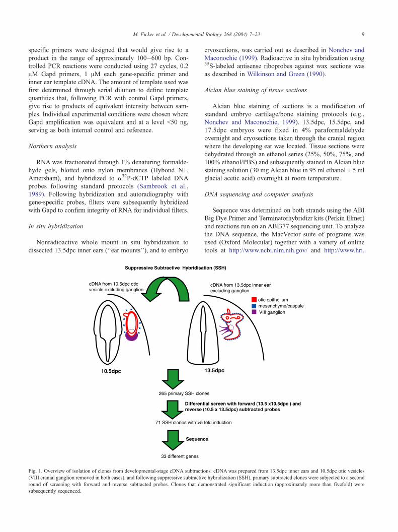
Fig. 1. Overview of isolation of clones from developmental-stage cDNA subtracti
(VIII cranial ganglion removed in both cases), and following suppressive subtractiv
round of screening with forward and reverse subtracted probes. Clones that de
subsequently sequenced.
cryosections, was carried out as described in Nonchev and
Maconochie (1999). Radioactive in situ hybridization using35S-labeled antisense riboprobes against wax sections was
as described in Wilkinson and Green (1990).
Alcian blue staining of tissue sections
Alcian blue staining of sections is a modification of
standard embryo cartilage/bone staining protocols (e.g.,
Nonchev and Maconochie, 1999). 13.5dpc, 15.5dpc, and
17.5dpc embryos were fixed in 4% paraformaldehyde
overnight and cryosections taken through the cranial region
where the developing ear was located. Tissue sections were
dehydrated through an ethanol series (25%, 50%, 75%, and
100% ethanol/PBS) and subsequently stained in Alcian blue
staining solution (30 mg Alcian blue in 95 ml ethanol + 5 ml
glacial acetic acid) overnight at room temperature.
DNA sequencing and computer analysis
Sequence was determined on both strands using the ABI
Big Dye Primer and Terminatorhybridizr kits (Perkin Elmer)
and reactions run on an ABI377 sequencing unit. To analyze
the DNA sequence, the MacVector suite of programs was
used (Oxford Molecular) together with a variety of online
tools at http://www.ncbi.nlm.nih.gov/ and http://www.hri.
ons. cDNA was prepared from 13.5dpc inner ears and 10.5dpc otic vesicles
e hybridization (SSH), primary subtracted clones were subjected to a second
monstrated significant induction (approximately more than fivefold) were

Table 1
List of genes recovered from 13.5dpc inner ear cDNA subtracted against 10.5dpc otic vesicle cDNA and subsequent analysis
A. Characterized
Gene description Gene Accession
number
e value Unigene MGI Class Capsule
ISH
Mouse map Human map
Activated leukocyte cell
adhesion molecule
(DM-GRASP)
Alcam L25274 e�139 Mm.28828 MGI:1313266 Signalling, CAM yes Ch16 3q13.1
ADP-ribosylation-like 1 Arl1 AK018138 e�95 Mm.291247 MGI:99436 Transport/signalling nd nm 3q13.1
Aggrecan Agc1 NM_007424 e�43 Mm.2759 MGI:99602 ECM yes Ch7 39.0cM 15q26.1
Cadherin 11/osteoblast
cadherin
Cdh11 AK012880 e�166 Mm.1571 MGI:99217 CAM yes Ch8 46.5cM 16q22.1
Clusterin Clu NM_013492 e�148 Mm.200608 MGI:88423 Glycoprotein nd Ch14 28.0cM 8p21-p12
Collagen II Col2a1 NM_031163 e�145 Mm.2423 MGI:88452 ECM/Struct yes Ch15 54.5cM 12q13.11-q13.2
Collagen IX, alpha1 Col9a1 AK017395 e�38 Mm.154662 MGI:88465 ECM/Struct nd Ch1 15.0cM 6q12-q14
Collagen XII, alpha1 Col12a1 NM_007730 e�74 Mm.3819 MGI:88448 ECM/Struct yes Ch9 43.0cM 6q12-q13
Eya3 Eya3 NM_010166 e�47 Mm.227733 MGI:109339 Transcription factor yes Ch4 64.6cM 1p36
Filamin B (human) ABP-280 (FLNB) XM_127565 e = 0.0 Hs.81008 Cytoskeleton nd nm 3p14.3
Follistatin-like induced kinase Fstl NM_008047 e�176 Mm.182434 MGI:102793 Signalling yes Ch16 27.3cM 3q13.33
High mobility group
nucleosomal binding
domain 2
Hmgn2 NM_016957 e�57 Mm.911 MGI:96136 Transcription factor nd Ch4 62.2cM 1p36.1
Lumican Lum BC005550 e = 0.0 Mm.18888 MGI:109347 ECM weak/absent Ch10 61.0cM 12q21.3-q22
Midkine Mdk NM_010784 e�62 Mm.906 MGI:96949 Growth factor yes Ch2 53.0cM 11p11.2
Moesin homologue S47577 e�136 none (ECM) yes nm
Osteoblast-specific
factor 2
(fasciclin I-like)(Runx2)
Osf2* NM_015784 e�83 Mm.4509 MGI:99829 Transcription factor yes nm 13q13.2
Otoraplin Otor NM_020595 e�150 Mm.157751 MGI:1888678 ECM weak/absent Ch2 20p12.1-p11.23
PCTAIRE2 (rat) protein kinase (PCTK2) AB005540 e = 0.0 none Signalling yes nm
Phosphodiesterase 4B Pde4b AF326556 e�136 Mm.20181 MGI:99557 Signalling yes Ch4 46.8cM 1p31
Ribosomal protein S4 Rps4x NM_009094 e�32 Mm.66 MGI:98158 Ribosome nd ChX 39.0cM Xq13.1
RNA binding motif protein 3 Rbm3 NM_016809 e�33 Mm.128512 MGI:1099460 Transcriptional regulation nd ChX 2.0cM Xp11.2
Tenascin C Tnc NM_011607 e = 0.0 Mm.980 MGI:101922 ECM yes Ch4 32.2cM 9q33
Transforming growth factor beta 2 Tgfb2 NM_009367 e�168 Mm.18213 MGI:98726 Growth factor yes Ch1 101.5cM 1q41
Translation initiation factor 3 Eif3s3 NM_080635 e�144 Mm.289800 MGI:1915385 Translation nd Ch15 8q24.11
Upstream transcription factor 2 Usf2 NM_011680 e�37 Mm.15781 MGI:99961 Transcription factor weak/absent Ch7 11.0cM 19q13
* pending
M.Ficker
etal./Develo
pmentalBiology268(2004)7–23
10

B. Novel/uncharacterized
Gene description Gene Accession
number
e value Unigene MGI Predicted domains Capsule IS Mouse map Human map
ssh04 FLJ20174 homologue BC006873 e = 0.0 Mm.156565 Tm domain x6; homology
to yeast S57180 membrane
protein (22% over 334aa)
weak/absent nm
ssh17, Band4.1 related XM_125565 e�27 none FERM
(F–ezrin– radixin–moesin)
domain, link cytoskeleton
to plasma membrane for
structural and regulatory roles
nd nm
ssh38, homologue to human
Zinc Finger protein U69274
AV381192 e�45 Mm.274729 Zn Fingers x6 and LIM
domain but entire ORF not yet
sequenced. Human gene has
12 zn fingers and BTB/POZ
domain
yes nm
ssh42, Zinc finger 142 AF332092 e�60 Mm.207298 Zn Fingers x36. Human gene
also has 36 Zn fingers
yes Ch1
ssh45, similar to
cytokine-like protein C17
XM_132070 e = 0.0 none No obvious domains.
Human protein includes 4
alpha helices and may be a
novel cytokine
yes nm
ssh48, RIKEN cDNA NM_145100 e�61 Mm.32727 Weak homology (30%/112aa)
to neurotensin receptor type I.
Single uPA-receptor-like domain
suggesting is GPI-linked cell
surface glycoprotein
nd Ch1
ssh57, not annotated BC031191 e�135 Mm.10160 Many ESTs from unannotated
region of genome. No human
matches to date, and no obvious
ORFs
nd nm
ssh70, ST7L/FLJ20284
homologue
XM_131106 e�33 Hs.146513 Moderately homologous to
tumor suppressor. N-terminal
TM domain in human and
mouse genes
weak/
absent
nm 1p12
Columns are as follows: (1) Gene description, gene name or clone ID. (2) Gene, gene symbol. (3) Accession number, accession number for best BLAST match. (4) e value, e value from BLAST score
corresponding to accession number. (5) Unigene, unigene identifier. (6) MGI, MGI reference identifier. (7) Class, type of molecule or associated process. (8) Capsule ISH, ear whole mount in situ hybridization
results summarized to reveal whether expression is present in capsule (yes) or not (absent); nd—not done. (9) Mouse map, map location in mouse genome; nm—not mapped. (10) Human map, map location in
human genome. (11) Predicted domains, in silico.
M.Ficker
etal./Develo
pmentalBiology268(2004)7–23
11

M. Ficker et al. / Developmental Biology 268 (2004) 7–2312
co.jp/atgpr/. Predictions concerning potential protein
domains were made using SMART analysis online at
http://smart.embl-heidelberg.de/ (Letunic et al., 2002).
Results
Isolation of genes from the 13.5dpc inner ear
To identify genes involved in the development of the
inner ear, we performed suppressive subtractive hybridiza-
tion with RNA from 13.5dpc inner ears against 10.5dpc otic
vesicle RNA (see Fig. 1 for overview of strategy). A total of
265 independent clones were recovered from the subtrac-
tion; however, following further differential screening with
forward and reverse subtracted probes, 71 clones remained
and formed the basis of this study. These clones were
sequenced and analyzed by BLAST analysis of the publicly
available DNA databases. The list of genes identified,
together with accession numbers and corresponding proba-
bility value, is presented in Table 1 and is subdivided into
(A) characterized genes and (B) novel/uncharacterized
genes. From the 71 clones sequenced, 33 different genes
were identified and included transcription factors, signaling
molecules, cell adhesion molecules, extracellular matrix
molecules, and genes involved in transcription/translation.
Relative abundance of transcripts at 13.5dpc and 10.5dpc
developmental stages
Both cDNA subtraction and differential screening proce-
dures utilized PCR amplified material, and thus we wished to
confirm that the genes identified were present in native
13.5dpc mouse inner ears. In addition, we also sought to
determine relative levels of transcription compared to the
10.5dpc otic vesicle. Gene-specific primers were synthesized
for each of the 33 genes, and each gene-specific primer pair
was used in combination with a control primer pair set from
the Gapd gene for PCR of regular (i.e., not normalized)
cDNA. In all cases, a specific band of the expected size was
detected in 13.5dpc inner ear cDNA (Fig. 2, right lanes). The
PCR reactions were also performed in parallel with the same
combination of primers using 10.5dpc otic vesicle cDNA
(Fig. 2, left lanes). In these experiments, equivalent amounts
of template cDNA were used in each reaction (empirically
determined, see PCR analysis section of Materials and
methods), PCR was kept sub-exponential, and for each gene,
reproduced on at least three separate occasions giving iden-
tical results. Thus, these experiments should reflect relative
differences in expression level between the 10.5dpc otic
vesicle and the 13.5dpc inner ear. From this analysis, 26/33
of the subtracted genes demonstrated clear comparative
transcriptional upregulation when examining regular cDNA
from 10.5dpc and 13.5dpc inner ears (Fig. 2A). Six genes did
not appear to be upregulated (Fig. 2B), and one gene
appeared to be downregulated (PCTAIRE, highlighted in
Fig. 2B). Approximate relative levels of induction are indi-
cated in brackets in Fig. 2 below individual gels.
Transcript analysis of subtracted clones
Alternative splicing mechanisms can give rise to func-
tionally different molecules by the incorporation or exclu-
sion of different functional domains within a protein. In
order to address the size of transcript generated in 13.5dpc
inner ears, and to investigate whether different major splice
variants are generated in the 13.5dpc inner ear, we per-
formed Northern analyses using total RNA from 13.5dpc
inner ears (we used total RNA since tissue size is a limiting
factor for efficient isolation of mRNA from ears at this
developmental stage). In addition, we wished to investigate
whether transcript(s) generated in the 13.5dpc inner ear
were identical in size to those transcripts generated in the
whole 13.5dpc embryo, or whether ear-specific isoforms are
generated. Therefore, we also used poly(A)RNA from
whole 13.5dpc embryos in the Northern analyses.
Known/previously characterized genes
Most of the genes tested gave a single mRNA species in
both inner ear and embryo (data not shown), although
multiple isoforms were detected in a few cases (Collagen
II, Eya3, follistatin-like induced kinase, tenascin, and
Tgfh2; Fig. 3). However, for the genes with multiple iso-
forms in the whole 13.5dpc embryo, one isoform predom-
inates in the inner ear for Tgfh2, follistatin-like induced
kinase (Flik), and Collagen II. In general, however, tran-
script size appeared identical for those genes where we were
able to detect transcripts in both 13.5dpc embryos and inner
ears (data not shown).
Novel/uncharacterized genes
Northern blot analysis using 13.5dpc inner ear total RNA
was attempted for all eight uncharacterized/novel clones.
mRNA species were detected in inner ear RNA using
hybridization probes against only one of the novel clones,
ssh45 (ssh-novel clone from Suppressive SubtractiveHybrid-
ization), identifying a single mRNA species of approximately
1.0 kb. The lack of hybridization to probes corresponding to
the remaining novel/uncharacterized genes suggests that
these are transcribed in the inner ear below the threshold
for detection using Northern analysis. In 13.5dpc embryo
RNA, single mRNA species of 4 and 7 kb were detected by
clones ssh04 and ssh42, respectively (data not shown).
Sequence analysis of uncharacterized/novel clones
Eight clones were identified in this study that were novel
or poorly characterized, and all eight are upregulated in the
13.5dpc inner ear (Fig. 2A). To understand the potential
function(s) of these genes, we performed a variety of se-
quence analyses. BLASTanalysis identified other ESTclones
and also annotation information from both human and mouse
Fig. 2. Relative transcript abundance of subtracted genes in 10.5dpc otic vesicle and 13.5dpc inner ear. Controlled PCR reactions were carried out using gene-
specific primers and Gapd control primers. Results for each gene are shown using 10.5dpc (left lane) and 13.5dpc (right lane) cDNA as template, with size of
expected product shown in bp (top band in each case—Gapd product 645 bp). Genes upregulated are detailed in (A), with upregulation at 13.5dpc estimated as
(+) slight, (++) moderate, and (+++) significant with Gapd serving as internal control. (B) Transcripts not demonstrating clear upregulation at 13.5dpc (�) and a
single example of a gene apparently downregulated at this stage (PCTAIRE; boxed in dotted lines) are illustrated.
M. Ficker et al. / Developmental Biology 268 (2004) 7–23 13
genome sequence drafts. When ORFs could be identified
from the recovered ESTor genome information, the predicted
peptides were analyzed for potential functional domains and
Fig. 3. Northern blot analysis of subtracted genes displaying major multiple isofor
ears (ear) and poly(A) RNA from 13.5dpc whole embryos (emb). Size of transcr
common protein motifs. Together, these analyses revealed
more information for six clones, suggesting two zinc-finger
containing transcription factors, two transmembrane domain
ms. Probes were hybridized against blots of total RNA from 13.5dpc inner
ipt(s) identified is indicated in kb.

M. Ficker et al. / Developmental Biology 268 (2004) 7–2314
containing genes, a gene encoding a FERM-domain (F for 4.1
protein, E for ezrin, R for radixin, and M for moesin and is a
domain believed to localize proteins to the plasma mem-
brane), and also a uPA-receptor domain containing gene (a
domain found in urokinase-type plasminogen activator re-
ceptor), suggesting that this protein may be a GPI-linked cell
surface glycoprotein (Table 1, section B).
Spatial localization of selected transcripts
Many different developmental processes are ongoing in
the 13.5dpc inner ear, and in this study, we provide a variety
of different candidates involved in either the control or
execution of these functions. However, to delimit which of
the different developmental processes each gene may be
associated with, an understanding of the precise spatial
localization of transcripts is required.
Using ‘‘whole ear mounts’’ (nonradioactive whole-mount
in situ hybridization to dissected 13.5dpc inner ears), we
demonstrated that many of the genes isolated through this
study are expressed generally throughout the developing
otic capsule at this stage (data not shown but summarized in
Table 1, column: Capsule ISH). Unfortunately, expression in
the otic capsule in whole ear mounts precludes the fine
details of expression to be observed within the capsule, and
also masks potential expression within the developing
epithelia. Therefore, for a selected set of these genes, we
also performed in situ hybridization on embryo sections
(Figs. 4 and 5; also refer to key at bottom of Fig. 5 for
explanation of plane of section).
Genes expressed in the otic capsule at 13.5dpc
Known/previously characterized genes
Phosphodiesterase expression is present throughout the
periotic mesenchyme and the developing otic capsule (Fig.
4A). However, expression is absent from the cochlear and
vestibular epithelium and the vestibulo-acoustic ganglion.
Expression of follistatin-like induced kinase (Flik) is
extensive throughout the periotic mesenchyme and otic
capsule (Fig. 4B). However, Flik expression is absent from
the vestibulo-acoustic ganglion and from both vestibular
and cochlear epithelia.
Cadherin 11 expression (Fig. 4C) was also present
throughout the periotic mesenchyme, but in a more discon-
tinuous manner than Flik and phosphodiesterase expression.
In addition, expression spreads into general mesenchyme
adjacent to the ear (e.g., see arrow below developing pinna
Fig. 4. In situ hybridization analysis of embryo sections for genes expressed
corresponding brightfield image is shown in the joined panel below the darkfield
phosphodiesterase (A); follistatin-like induced kinase (B); cadherin 11 (C); tenascin
M); and novel clone ssh04 (N, O). Planes of section through different levels of t
labeled are as indicated in key at bottom of Fig. 5 (which also schematically ill
labeling: mes—mesenchyme; ic—inner capsule domain; oc—outer capsule doma
mc—Meckel’s cartilage. Scale bar in each panel = 500 AM. Dorsal oriented top, pi
in panels C, J, K and should be ignored.
in Fig. 4C), but in common with the genes above, expres-
sion is absent from vestibular and cochlear epithelia and the
developing vestibulo-acoustic ganglion.
Tenascin expression was evident in dorsal aspects of the
forming capsule and periotic mesenchyme (Fig. 4D), but
was absent from the vestibulo-acoustic ganglion, vestibular,
and cochlear epithelia. In the adult chinchilla inner ear,
tenascin protein has previously been reported in osteocytes,
mesothelial cells and within the fibrous matrix of the basilar
membrane (Swartz and Santi, 1999).
Lumican expression is subtly different from the expres-
sion patterns noted above (Figs. 4E, F). Lumican expression
is evident in the otic capsule, but not throughout the capsule
and periotic mesenchyme, but instead, expression was
detected in a subpopulation of mesenchyme in apposition
to the inner ear epithelia (ic—‘‘inner capsule’’), as well as
strong expression just below the surface ectoderm (Fig. 4F).
In addition, expression was also evident in lateral aspects of
the cochlear duct and in restricted regions of the vestibular
epithelium (Fig. 4E, arrowheads).
This restricted otic capsule expression of lumican con-
trasts with Collagen II expression (Figs. 4G–I). Expression
of Collagen II is within the forming otic capsule, but in this
case is restricted to the outermost extent of the capsule (oc,
Figs. 4G–I). Expression of Collagen II was also observed in
the vertebral column and the cartilage primordia of the
malleus and incus and in Meckel’s cartilage.
In common with Collagen II expression, Tgfh2 expres-
sion was also observed in the outermost extent of the
developing otic capsule, largely overlapping the formers’
expression but absent from the mesenchyme directly sur-
rounding the developing vestibular and cochlear epithelia
(Figs. 4J, K). Tgfh2 expression was also observed in the
developing inner ear epithelium as well as the capsule, and
this expression is detailed below.
Radioactive in situ hybridization was also performed for
upstream transcription factor, moesin-like homologue, and
Eya3. Transcription factor Eya3 expression was observed
throughout the otic capsule. Upstream transcription factor 2
expression (Usf2-mouse homologue to human upstream
stimulatory factor 2/fos-interacting protein) and moesin
expression were both found to be at a low level generally
throughout the capsule and periotic mesenchyme (data not
shown).
Novel/uncharacterized genes
Some novel/uncharacterized genes also displayed vary-
ing expression patterns within the developing otic capsule.
in the developing otic capsule at 13.5dpc. For radioactive probes, the
view and highlightedV. Antisense riboprobes used are indicated in panels:
(D); lumican (E, F); collagen II (G– I); Tgfh2 (J, K); novel clone ssh45 (L,
he ear are denoted as dorsal (D), medial (M), or ventral (V) and structures
ustrates the relative location of individual inner ear structures). Additional
in; p—pinna; m+i—malleus and incus primordia; st—stapes primordium;
nna to the right in all panels. Artifacts of emulsion spots or dust are apparent

M. Ficker et al. / Developmental Biology 268 (2004) 7–23 15

Fig. 5. In situ hybridization analysis of embryo sections for genes expressed in the developing cochlear and vestibular epithelia at 13.5dpc. For the radioactive
clusterin probe (A, B), the corresponding brightfield image (AV, BV) is shown in the joined panel below the darkfield view; (C) expression of novel clone ssh38.
Plane of section is indicated in individual panels as medial (M) or ventral (V). Pinna oriented right. Below, key to labeling and planes of section taken for in situ
analyses presented in this figure and in Figs. 4–6, which indicates schematically the relative positions of inner ear structures and approximately which
structures are identified in individual sections. Scale bars as noted in individual panels are 500 AM. Additional labeling: mc—Meckel’s cartilage.
M. Ficker et al. / Developmental Biology 268 (2004) 7–2316
Expression of ssh45 was found to be restricted to the
outermost extent of the capsule (Figs. 4L, M), in a pattern
similar to that exhibited by Collagen II and Tgfh2 (Figs.
4G–K). Expression of ssh45 was also detected in cartilage
primordia of the developing middle ear ossicles (malleus,
incus, and stapes), as well as Meckel’s cartilage, but unlike
Tgfh2, expression was absent from both vestibular and
auditory sensory/nonsensory epithelia.
Expression of clone ssh04 was also detected within the
otic capsule (Figs. 4N, O), and predominantly localized to the
inner domains of the capsule. Capsule expression of ssh04
was unusual in appearing patchy or punctate, and from our
Fig. 6. Expression of selected genes at later stages of embryogenesis. In situ hybri
following probes: (A, B) lumican; (C–F) Tgfh2; (G–J) Collagen II; (K, L) novel cl
darkfield view for radioactive probes (AV–FV). Refer to key at bottom of Fig. 5 for e
highlights nonspecific staining of radiolabeled probe to brown cells either side of
(M), or Ventral (V). Labeling of individual structures is as detailed in Fig. 5, but in
studies, it appears that expression does not extend into the
outermost domain. However, double labeling in situ hybrid-
ization studies with genes such as ssh45, Collagen II, or
Tgfh2 together with ssh04 will be required to unequivocally
exclude ssh04 from the outermost region of the capsule.
Genes expressed in the developing sensory epithelium at
13.5dpc
Known/characterized genes
Tgfh2 expression in the outer capsule was noted above.
However, an additional major domain of Tgfh2 expression
dization on embryo sections of age as indicated in individual panels for the
one ssh45; (M, N) novel clone ssh42. Brightfield images shown joined below
xplanation of structures highlighted in individual sections. Asterisk (*) in (A)
crista. Plane of section is indicated in individual panels; Dorsal (D), Medial
addition, oc—outer capsule, p—pinna. Scale bars of 1000 AM as indicated.

M. Ficker et al. / Developmental Biology 268 (2004) 7–23 17

M. Ficker et al. / Developmental Biology 268 (2004) 7–2318
was clearly evident within the future sensory/supporting
cellular regions of both cochlear and vestibular epithelia
(Figs. 4J, K). Prominent Tgfh2 expression was seen in the
vestibular maculae and in the cristae in the base of each
semicircular canal (Figs. 4J, K), as well as in the ventral
wall of the cochlea that, for the most part, comprises the
future cochlear sensory/supporting epithelium. In these
regions, expression was detected throughout the extent of
the epithelium. Further detailed studies, such as double
labeling experiments with a variety of different molecular
markers for sensory and supporting cells, are required to
confirm whether all presumptive sensory and supporting
cells in each sensory patch are positive for Tgfh expression.
Of all the genes identified by subtractive hybridization,
clusterin expression was that most localized to the develop-
ing cochlear epithelium. Clusterin expression was restricted
to the ventral wall of the cochlear duct (Figs. 5A, B).
However, in contrast to Tgfh2 expression, clusterin expres-
sion was absent from vestibular epithelia (Fig. 5A). Clus-
terin expression was not detected in the otic capsule or
vestibulo-acoustic ganglion.
Novel/uncharacterized genes
Expression in the developing otic epithelia was noted for
novel clone ssh38. However, expression was evident in the
nonsensory potions of the vestibular epithelium lining the
Fig. 7. Cartilage formation in the developing otic capsule. Alcian blue staining of c
developmental age noted below panels: (A–C) 13.5dpc; (D–F) 15.5dpc; and (G
legends found at bottom of Fig. 5. Additional labeling: cap—cartilaginous capsule
scale bars as indicated below.
semicircular canals as well as in future sensory/supporting
epithelia in the saccule/utricular space (Fig. 5C).
Dynamics of capsule expression during later development
The spatial analysis of gene expression in the developing
capsule at 13.5dpc above presents one developmental win-
dow on the forming capsule. Given that at 13.5dpc, gene
expression patterns for some of the genes analyzed could be
detected in different capsule domains, we next sought to
address whether these domains were transitory or were
maintained throughout embryonic development. Thus, we
carried out in situ hybridization on sections of 15.5dpc and
17.5dpc embryos for five genes: lumican, Tgfh2, CollagenII, and novel clones ssh45 and ssh42 (Fig. 6).
At 17.5dpc, lumican expression was still detected in the
developing capsule (Figs. 6A, B). Whilst the developing
capsule is more acellular at this stage, lumican expression is
evident within cellular regions but does not appear to be
restricted to inner/outer capsule domains. In addition, the
limited epithelial expression noted at 13.5dpc has further
developed by 17.5dpc to be localized to cells lateral to the
organ of Corti in the developing outer sulcus (Fig. 6B, left
arrow). In the ventral–medial region of the cochlea, cells in
the greater epithelial ridge also were also strongly positive
for lumican expression (Fig. 6B, right arrow).
artilage (left panels) and H&E staining (right panels) of embryo sections of
– I) 17.5dpc. Plane of section as noted and as explained in key to figure
, m—macula, cr—crista, ce—cochlear epithelium. Ears oriented pinna left;

M. Ficker et al. / Developmental Biology 268 (2004) 7–23 19
At 15.5dpc, Tgfh2 expression was still evident in the
developing cristae (Fig. 6C) and in the cochlear epithelium
(Fig. 6D). At 17.5dpc, expression was maintained in the
patches of sensory/supporting cells of the cristae and
maculae (Fig. 6E), as well as throughout the cochlear
epithelium (Fig. 6F). Tgfh2 expression was still evident
in the developing capsule, although expression was clearly
not to the same levels as expression detected in the different
epithelia (Figs. 6E, F), and without the clear demarcation
between inner and outer capsule domains seen earlier
during development.
Collagen II expression was also found to be maintained at
both 15.5dpc and 17.5dpc (Figs. 6G–J). At both time points,
expression was restricted to a distinct domain (oc, Figs. 6G–
J) with non-hybridizing cellular material between the differ-
ent epithelia and the domain of collagen expression.
Similarly, expression of the two novel genes ssh45 and
ssh42 were examined at 17.5dpc, and expression also
localized to this outer capsule domain (Figs. 6K–N), again
separated from the different epithelia by non-hybridizing
cellular regions.
Dynamics of cartilage formation in the developing otic
capsule
In order to correlate the gene expression profiles iden-
tified in the forming otic capsule with the histological
development of the capsule, we performed a developmental
time course of Alcian blue staining of sections from
13.5dpc, 15.5dpc, and 17.5dpc embryos, and compared
these with H&E stained sections (Fig. 7). Alcian blue
staining highlights areas of cartilage formation, and at
13.5dpc, cartilage formation has been initiated in the
developing inner ear (Figs. 7A–C). However, cartilage
formation appears more advanced in dorsal regions com-
pared to ventral regions (compare levels of staining in A
with C; sections all cut same thickness and stained for
identical periods). By 15.5dpc, cartilage formation is well
underway throughout the entire capsule (Figs. 7D–F), and
by 17.5dpc, the layer of Alcian blue staining material is
expanding to match embryonic growth (Figs. 7G–I). This
progression of initiation of cartilage formation at 13.5dpc
to a clearly discernible capsule by 15.5dpc is mirrored by
the H&E stained sections; whilst at 13.5dpc the periotic
mesenchymal cells are in the process of being organized
(Figs. 7A–C), a distinct cellular layer corresponding to the
Alcian blue stained layer is evident at 15.5dpc and 17.5dpc
(Figs. 7D–I).
Discussion
The generation and use of cDNA libraries
As complete genome sequences are generated for differ-
ent vertebrate species, cDNA libraries are critical for efforts
to begin to interpret genome sequence data at a descriptive
level. Microarray analysis represents one powerful approach
toward analyzing the genome and transcriptome, but its use
in isolation is likely to lead to hidden biases (Powles et al.,
this issue). Therefore, we and others have used cDNA
subtraction as alternative and additional methods towards
identifying transcriptional changes under different experi-
mental conditions. Previously, human inner ear cDNA
libraries have been prepared and analyzed from the mem-
branous labyrinth of fetal cochlea (16–22 weeks develop-
mental age)(Robertson et al., 1994; Skvorak et al., 1999);
however, the absence of nonepithelial portions of the inner
ear in such libraries and at a later developmental time point
precludes the identification of those genes expressed in the
capsule that may be required for either the development of
the capsule or for development of the inner ear epithelium
through mesenchymal–epithelial signaling mechanisms.
Others have also illustrated the efficacy of subtractive
approaches for identifying genes upregulated on noise
exposure in chick cochlea (Gong et al., 1996), and a variant
subtractive hybridization approach was used to isolate
markers from late embryonic chick basilar papilla (Heller
et al., 1998). This study, to our knowledge, is the first
approach to use the principle of developmental-stage sub-
traction to begin to address the identity of molecules
involved in the developmental biology of the inner ear. In
an accompanying manuscript, we present detailed analysis
of genes from an earlier developmental stage, the 10.5dpc
otic vesicle.
Alternative transcription
Northern analyses presented in this study indicated that
very few genes displayed markedly different major splice
variants in the 13.5dpc embryo when compared to the inner
ear. Furthermore, of 21 genes that gave positive signals,
only five genes detected multiple major splice variants.
Northern analysis may not detect major isoforms present
at low levels or isoform differences that are subtle, involv-
ing small exons. However, we suggest that additional
mechanisms need to be invoked to generate the required
molecular complexity required for embryonic development,
and this is likely to be through posttranscriptional mecha-
nisms, either at a translational level or through differential
regulation of protein modification and localization.
Embryology of the otic capsule
The otic capsule develops from periotic mesenchyme
surrounding the developing inner ear epithelium. Brn4 is
expressed in this periotic mesenchyme, and analysis of Brn4
mutants demonstrates a requirement for this gene product in
otic capsule formation (Phippard et al., 1999). On the other
hand, mouse Dlx5 expression is restricted to neuroectoder-
mal components of the developing inner ear, and yet Dlx5
mouse mutants present with multiple defects including

M. Ficker et al. / Developmental Biology 268 (2004) 7–2320
dysgenesis of the otic capsule (Depew et al., 1999). This
suggests a requirement for epithelial signaling to the periotic
mesenchyme for capsule development, and this is corrobo-
rated by in vitro studies (McPhee and van de Water, 1986),
indicating that capsule development is not through a cell
autonomous program of the periotic mesenchyme. A num-
ber of molecules have been shown to effect chondrogenesis
of periotic mesenchyme in micromass culture, such as
retinoic acid, Fgf2, Fgf3, Bmp2a (Frenz and Liu, 1997;
Frenz and Liu, 1998; Frenz et al., 1996a,b), Tgfh1 (Frenz et
al., 1991), and Otoraplin/Fdp (Cohen-Salmon et al., 2000;
Robertson et al., 2000). Such experiments are suggestive for
some involvement in capsule development, although de-
tailed functional studies are required in vivo to confirm the
individual roles of each molecule. From the analysis pre-
sented in this study, we have identified several additional
molecules that also represent good candidates for capsule
development, and these similarly require functional analysis
in vivo to confirm and detail a developmental role.
Regionalization and heterogeneity of the otic capsule
In situ hybridization of tissue sections at 13.5dpc re-
vealed capsule expression as either throughout the otic
capsule, or restricted to an inner or outer zone, indicating
molecular heterogeneity of the forming capsule. Close
examination of histological sections at 13.5dpc also illus-
trates that cells in the outer capsule ‘‘layer’’ tend to be
smaller and spaced closer together than cells close to, or
between, the different developing inner ear epithelia, sug-
gesting that this molecular difference correlates with cellular
differences as well. H&E analysis at later developmental
stages clearly demonstrates different and distinct cellular
layers. Collagen II is expressed in chondrogenic cells in
advance of chondrocyte differentiation, suggesting that the
outer capsule layer is composed of proliferating chondro-
cytes. In a detailed study of Collagen II expression in later
human fetal cochlea, proliferative and resting chondrocytes
retain high levels of Collagen II expression, but as these
mature and ossification begins, expression is downregulated
(Khetarpal et al., 1994). The co-expression of Tgfh2 and
Collagen II supports the micromass culture experiments
mentioned above, which indicated that this molecule is able
to induce chondrogenesis in vitro, and together, these data
support the hypothesis that capsule chondrogenesis at
13.5dpc is active in the outermost capsule layer. That
chondrogenesis is active was confirmed by the Alcian blue
staining of 13.5dpc embryo sections. The developmental
time course of Alcian blue staining also suggests that
cartilage formation has been initiated at 13.5dpc in the
developing inner ear, with a distinct cartilaginous structure
present throughout the capsule some 2 days later. The
expression of novel/uncharacterized clone ssh45 is restricted
to this same outer capsule domain. Sequence analysis of
ssh45 indicated that this gene corresponds to a novel
cytokine-like protein, and perhaps this novel growth factor,
together with Tgfh2, is involved in the control of chondro-
genesis in the outer capsule.
Lumican is present in the inner capsule layer. Lumican is
a keratin sulfate proteoglycan that has been shown to
regulate collagen fibrillogenesis during development, and
mice deficient for lumican have opaque corneas with
abnormally thick collagen fibrils (Ezura et al., 2000). This
supports a negative regulatory role in collagen fibrillogen-
esis, and whereas the outer capsule is composed of prolif-
erating chondrocytes, the inner capsule may be undergoing
suppression of chondrocyte generation. In skin and the
cornea, lumican is required for the assembly and organiza-
tion of the extracellular matrix (Chakravarti et al., 1998),
and it is also tempting to speculate a similar but localized
role for lumican within the inner capsule. 13.5dpc otic
epithelium is able to suppress chondrogenesis in periotic
mesenchyme micromass cultures, and lumican may be one
of the genes involved in the initiation of this process, or at
the least, providing a molecular marker recording these
events. This suggests that chondrogenesis is suppressed in
the inner capsule domain in vivo, and this suppression may
be regulated by the otic epithelium. We suggest that sup-
pression of chondrogenesis in the otic capsule occurs in a
domain limited either by the extent of diffusion of a secreted
signaling molecule generated by the otic epithelium, or
signaling is limited to the extent of expression of the
receptor for this signaling molecule. The identification of
this epithelial-derived molecule and its receptor will be
required to establish which of these mechanisms operate
in the capsule. The biological effect of peri-epithelial
suppression of chondrogenesis is likely to permit the exten-
sive tissue remodeling and perilymphatic space develop-
ment in the 13.5dpc inner ear.
However, the suppression of chondrogenesis needs to be
limited to permit chondrogenesis to be carried out in the
outer capsule domain. In this study, we have demonstrated
compartmentalization or regionalization for the expression
of some genes in the developing otic capsule, and this is
summarized in Fig. 8. As mentioned above, this correlates
with cellular differences within outer/inner capsule domains,
and we suggest that those genes that are restricted to either
inner or outer capsule domains are involved in outer capsule
chondrogenesis and perilymphatic space development/
remodeling events. We suggest that these ‘‘compartments’’
or ‘‘domains’’ of gene expression are thus likely to correlate
with developmentally different regions of the capsule at this
embryonic stage.
Analysis of the expression of lumican and Tgfh2 at later
developmental time points suggests that these gene expres-
sion domains of inner/outer capsule might be transitory.
However, the maintenance of expression of Collagen II and
ssh45 suggests that expression domains remain for some
genes, but that these domains probably reflect a maturation of
the developing inner ear. Possibly then, those genes that are
only transiently expressed in a capsule domain might only be
involved in the initiation of cartilage formation in the capsule,
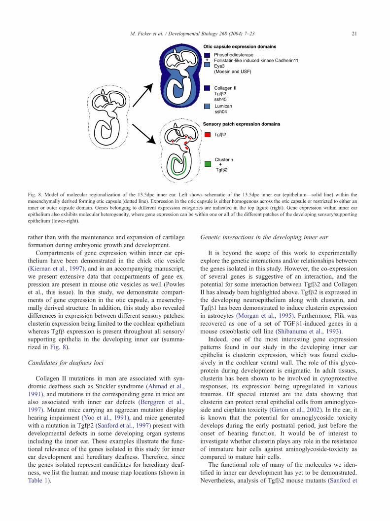
Fig. 8. Model of molecular regionalization of the 13.5dpc inner ear. Left shows schematic of the 13.5dpc inner ear (epithelium—solid line) within the
mesenchymally derived forming otic capsule (dotted line). Expression in the otic capsule is either homogenous across the otic capsule or restricted to either an
inner or outer capsule domain. Genes belonging to different expression categories are indicated in the top figure (right). Gene expression within inner ear
epithelium also exhibits molecular heterogeneity, where gene expression can be within one or all of the different patches of the developing sensory/supporting
epithelium (lower-right).
M. Ficker et al. / Developmental Biology 268 (2004) 7–23 21
rather than with the maintenance and expansion of cartilage
formation during embryonic growth and development.
Compartments of gene expression within inner ear epi-
thelium have been demonstrated in the chick otic vesicle
(Kiernan et al., 1997), and in an accompanying manuscript,
we present extensive data that compartments of gene ex-
pression are present in mouse otic vesicles as well (Powles
et al., this issue). In this study, we demonstrate compart-
ments of gene expression in the otic capsule, a mesenchy-
mally derived structure. In addition, this study also revealed
differences in expression between different sensory patches:
clusterin expression being limited to the cochlear epithelium
whereas Tgfh expression is present throughout all sensory/
supporting epithelia in the developing inner ear (summa-
rized in Fig. 8).
Candidates for deafness loci
Collagen II mutations in man are associated with syn-
dromic deafness such as Stickler syndrome (Ahmad et al.,
1991), and mutations in the corresponding gene in mice are
also associated with inner ear defects (Berggren et al.,
1997). Mutant mice carrying an aggrecan mutation display
hearing impairment (Yoo et al., 1991), and mice generated
with a mutation in Tgfh2 (Sanford et al., 1997) present with
developmental defects in some developing organ systems
including the inner ear. These examples illustrate the func-
tional relevance of the genes isolated in this study for inner
ear development and hereditary deafness. Therefore, since
the genes isolated represent candidates for hereditary deaf-
ness, we list the human and mouse map locations (shown in
Table 1).
Genetic interactions in the developing inner ear
It is beyond the scope of this work to experimentally
explore the genetic interactions and/or relationships between
the genes isolated in this study. However, the co-expression
of several genes is suggestive of an interaction, and the
potential for some interaction between Tgfh2 and Collagen
II has already been highlighted above. Tgfh2 is expressed inthe developing neuroepithelium along with clusterin, and
Tgfh1 has been demonstrated to induce clusterin expression
in astrocytes (Morgan et al., 1995). Furthermore, Flik was
recovered as one of a set of TGFh1-induced genes in a
mouse osteoblastic cell line (Shibanuma et al., 1993).
Indeed, one of the most interesting gene expression
patterns found in our study in the developing inner ear
epithelia is clusterin expression, which was found exclu-
sively in the cochlear ventral wall. The role of this glyco-
protein during development is enigmatic. In adult tissues,
clusterin has been shown to be involved in cytoprotective
responses, its expression being upregulated in various
traumas. Of special interest are the data showing that
clusterin can protect renal epithelial cells from aminoglyco-
side and cisplatin toxicity (Girton et al., 2002). In the ear, it
is known that the potential for aminoglycoside toxicity
develops during the early postnatal period, just before the
onset of hearing function. It would be of interest to
investigate whether clusterin plays any role in the resistance
of immature hair cells against aminoglycoside-toxicity as
compared to mature hair cells.
The functional role of many of the molecules we iden-
tified in inner ear development has yet to be demonstrated.
Nevertheless, analysis of Tgfh2 mouse mutants (Sanford et

M. Ficker et al. / Developmental Biology 268 (2004) 7–2322
al., 1997) revealed a role in the development of the spiral
limbus, a mesenchymally derived structure within the co-
chlea. However, given the additional areas of Tgfh2 ex-
pression in sensory/supporting cells in the vestibular
epithelium and the otic capsule as shown in the present
study, further detailed functional analyses would be appro-
priate in this and other mutant backgrounds to fully explore
the role of Tgfh signaling in the developing inner ear. Other
interactions may also exist between the genes isolated, and
the identification of different expression domains in the
forming capsule is of particular interest with respect to
understanding mesenchymal–epithelial interactions in the
developing inner ear. The next step is to test such inter-
actions through in vivo and in vitro approaches.
In summary, cDNA subtraction has identified several
genes potentially involved in different aspects of inner ear
development. Detailed expression analysis suggests molec-
ular heterogeneity in the developing otic capsule and indi-
cates the presence of compartments/domains of gene
expression within the capsule. Furthermore, co-incident
expression patterns suggest potential rudimentary pathways
that may be required for correct development of the otic
capsule and inner ear neuroepithelium.
Acknowledgments
We are very grateful to M. Lomax (Michigan) and T.
Schimmang (Hamburg) for critical reading and constructive
comments on the manuscript. MF, NSP, NW, and MKM are
supported by core funding from the Medical Research
Council. UHP acknowledges support from the Sigfrid
Juselius Foundation. In addition, we would like to thank
Angela and Wendy for excellent animal husbandry and
technology.
References
Ahmad, N.N., Ala-kokko, L., Knowlton, R.G., Jimenez, S.A., Weaver, E.J.,
Maguire, J.I., Tasman, W., Prockop, D.J., 1991. Stop codon in the
procollagen II gene (COL2A1) in a family with the Stickler syndrome
(arthro-opthalmopathy). Proc. Natl. Acad. Sci. U. S. A. 88, 6624–6627.
Berggren, D., Grenz, D., Galinovic-Schwarz, V., Van de Water, T., 1997.
Fine structure of extracellular matrix and basal laminae in two types of
abnormal collagen production: I—Proline analog treated otocyst cul-
tures and disproportionate micromelia (Dmn/Dmn) mutants. Hear. Res.
107, 125–135.
Chakravarti, S., Magnuson, T., Lass, J., Jepsen, K., LaMantia, C., Carroll,
H., 1998. Lumican regulates collagen fibril assembly: skin fragility and
corneal opacity in the absence of lumican. J. Cell Biol. 141, 1277–1286.
Cohen-Salmon, M., Frenz, D., Liu, W., Verpy, E., Voegeling, S., Petit, C.,
2000. Fdp, a new fibrocyte-derived protein related to MIA/CD-RAP,
has an in vitro effect on the early differentiation of the inner ear mes-
enchyme. J. Biol. Chem. 275, 40036–40041.
Depew, M., Liu, J., Long, J., Presley, R., Meneses, J., Pedersen, R., Ruben-
stein, L., 1999. Dlx5 regulates regional development of the branchial
arches and sensory capsules. Development 126, 3831–3846.
Diatchenko, L., Lau, Y.-F.C., Campbell, A.P., Chenchik, A., Moqadam, F.,
Huang, B., Lukyanov, K., Gurskaya, N., Sverdlov, E.D., Siebert, P.D.,
1996. Suppression subtractive hybridisation: a method for generating
differentially regulated or tissue-specific cDNA probes and libraries.
Proc. Natl. Acad. Sci. U. S. A. 93, 6025–6030.
Ezura, Y., Chakravarti, S., Oldberg, A., Chervoneva, I., Birk, D.E.,
2000. Differential expression of lumican and fibromodulin regulate
collagen fibrillogenesis in developing mouse tendons. J. Cell Biol.
151, 779–788.
Fekete, D.M., 1999. Development of the vertebrate ear: insights from
knockouts and mutants. Trends Neurosci. 22, 263–269.
Frenz, D., Liu, W., 1997. Effect of retinoic acid on otic capsule chondro-
genesis in high density culture suggests disruption of epithelial–mes-
enchymal interactions. Teratology 56, 233–240.
Frenz, D., Liu, W., 1998. Role of FGF3 in otic capsule chondrogenesis
in vitro: an antisense oligonucleotide approach. Growth Factors 15,
173–182.
Frenz, D.A., Van De Water, T., 1991. Epithelial control of periotic mesen-
chyme chondrogenesis. Dev. Biol. 144, 38–46.
Frenz, D., Van de Water, T., Galinovic-Schwartz, V., 1991. Transforming
growth factor beta: does it direct otic capsule formation? Ann. Otol.,
Rhinol., Laryngol. 100, 301–307.
Frenz, D., Liu, W., Capparelli, M., 1996a. Role of BMP-2a in otic capsule
chondrogenesis. Ann. N.Y. Acad. Sci. 785, 256–258.
Frenz, D., Liu, W., Galinovic-schwartz, V., Van de Water, T., 1996b. Ret-
inoic acid-induced embryopathy of the mouse inner ear. Teratology 53,
292–303.
Fritzsch, B., Barald, K.F., Lomax, M.I., 1998. Early embryology of the
vertebrate ear. In: Fay, R.R. (Ed.), Development of the Auditory Sys-
tem. Springer, New York, pp. 81–146.
Girton, R.A., Sundin, D.P., Rosenberg, M.E., 2002. Clusterin protects renal
tubular epithelial cells from gentamicin-mediated cytotoxicity. Am. J.
Renal: Physiol., Fluid Electrolyte Physiol. 282, 703–709.
Gong, T.-W.L., Hegeman, A.D., Shin, J.J., Adler, H.J., Raphael, Y., Lomax,
M.I., 1996. Identification of genes expressed after noise exposure in the
chick basilar papilla. Hear. Res. 96, 20–32.
Groves, A.K., Bronner-Fraser, M., 2000. Competence, specification and
commitment in otic placode induction. Development 127, 3489–3499.
Heller, S., Sheane, C., Javed, Z., Hudspeth, A., 1998. Molecular markers
for cell types of the inner ear and candidate genes for hearing disorders.
Proc. Natl. Acad. Sci. U. S. A. 95, 11400–11405.
International Human Genome Sequencing Consortium, 2001. Initial se-
quencing and analysis of the human genome. Nature 409, 860–921.
Jacobson, A.G., 1966. Inductive processes in embryonic development.
Science 157, 25–34.
Khetarpal, U., Robertson, N.G., Yoo, T.J., Morton, C.C., 1994. Expression
and localisation of COL2A1 mRNA and type II collagen in human
foetal cochlea. Hear. Res. 79, 59–73.
Kiernan, A.E., Nunes, F., Wu, D.K., Fekete, D.M., 1997. The expression
domain of two related homeobox genes defines a compartment in the
chicken inner ear that may be involved in semicircular canal formation.
Dev. Biol. 191, 215–229.
Letunic, I., Goodstadt, L., Dickens, N.J., Doerks, T., Schultz, J., Mott, R.,
Ciccarelli, F., Copley, R.R., Ponting, C.P., Bork, P., 2002. Recent
improvements to the SMART domain-based sequence annotation re-
source. Nucleic Acids Res. 30, 242–244.
Li, C.W., Van de Water, T.R., Ruben, R.J., 1978. The fate mapping of the
eleventh and twelfth day otocyst: an in vitro study of the sites of
origin of embryonic inner ear sensory structures. J. Morphol. 157,
249–268.
Martin, P., Swanson, G.J., 1993. Descriptive and experimental analysis of
the epithelial remodellings that control semicircular canal formation in
the developing mouse inner ear. Dev. Biol. 159, 549–558.
McPhee, J.R., van de Water, T., 1986. Epithelial –mesenchymal tissue
interactions guiding otic capsule formation: the role of the otocyst.
J. Embryol. Exp. Morphol. 97, 1–24.
Morgan, T.E., Laping, N.J., Rozovsky, I., Oda, T., Hogan, T.H., Finch,
C.E., Pasinetti, G.M., 1995. Clusterin expression by astrocytes is

M. Ficker et al. / Developmental Biology 268 (2004) 7–23 23
influenced by transforming growth factor beta-1 and heterotypic cell
interactions. J. Neuroimmunol. 58, 101–110.
Mouse Genome Sequencing Consortium, 2002. Initial sequencing and
comparative analysis of the mouse genome. Nature 420, 520–562.
Nonchev, S., Maconochie, M., 1999. Spatial analysis of gene expres-
sion. In: Jackson, I.J., Abbott, C.M. (Eds.), Mouse Genetics and
Transgenics: A Practical Approach. Oxford Univ. Press, New York,
pp. 61–86.
Phippard, D., Lu, L., Lee, D., Saunders, J., Crenshaw, E., 1999. Targeted
mutagenesis of the POU-domain gene Brn4/Pou3f4 causes develop-
mental defects in the inner ear. J. Neurosci. 19, 5980–5989.
Represa, J., Leon, Y., Miner, C., Giraldez, F., 1991. The int-2 proto-onco-
gene is responsible for inner ear induction. Nature 353, 561–563.
Robertson, N.G., Khetarpal, U., Gutierrez-Espeleta, G.A., Bieber, F.R.,
Morton, C.C., 1994. Isolation of novel and known genes from a human
fetal cochlear cDNA library using subtractive hybridisation and differ-
ential screening. Genomics 23, 42–50.
Robertson, N.G., Heller, S., Lin, J.S., Resendes, B.L., Weremowicz, S.,
Denis, C.S., Bell, A.M., Hudspeth, A.J., Morton, C.C., 2000. A novel
conserved cochlear gene, OTOR: identification, expression analysis and
chromosomal mapping. Genomics 66, 242–248.
Sambrook, J., Fritsch, E.F., Maniatis, T., 1989. Molecular Cloning: A
Laboratory Manual. Cold Spring Harbor Laboratory Press, Cold Spring
Harbor, New York.
Sanford, L.P., Ormsby, I., Gittenberger-de-Groot, A.C., Sariola, H., Fried-
man, R., Boivin, G.P., Cardell, E.L., Doetschman, T., 1997. TGFh2knockout mice have multiple developmental defects that are non-over-
lapping with other TGFh knockout phenotypes. Development 124,
2659–2670.
Shibanuma, M., Mashimo, J., Mita, A., Kuroki, T., Nose, K., 1993. Cloning
from a mouse osteoblastic cell line of a set of transforming-growth
factor b1 related genes, one of which seems to encode a follistatin-
related polypeptide. Eur. J. Biochem. 217, 13–19.
Skvorak, A., Weng, Z., Yee, A., Robertson, N., Morton, C., 1999. Human
cochlear expressed sequence tags provide insight into cochlear gene
expression and identify candidate genes for deafness. Hum. Mol. Genet.
8, 439–452.
Steel, K.P., Brown, S.D.M., 1994. Genes and deafness. Trends Genet. 10,
428–435.
Swartz, D., Santi, P., 1999. Immunolocalization of tenascin in the chinchilla
inner ear. Hear. Res. 130, 108–114.
Wilkinson, D., Green, J., 1990. In situ hybridisation and the three-dimen-
sional reconstruction of serial sections. In: Copp, A., Cockcroft, D.
(Eds.), Postimplantation Mammalian Embryos: A Practical Approach.
IRL Press, Oxford, pp. 151–171.
Yoo, T., Cho, H., Yamada, Y., 1991. Hearing impairment in mice with the
cmd/cmd (cartilage matrix deficiency) gene. Ann. N.Y. Acad. Sci. 630,
265–267.