analysis of smad nucleocytoplasmic shuttling in living...
TRANSCRIPT

IntroductionSignals from receptors for Transforming growth factor β (TGF-β) superfamily members are transduced to the nucleus by theSmads (Shi and Massagué, 2003). In the case of TGF-β itself,the prototype of the family, receptor activation leads tophosphorylation of the receptor-regulated Smads (R-Smads)Smad2 and Smad3 at two serines in an SSXS motif at theirextreme C termini. This results in activation of the R-Smadsthat then form complexes with the common mediator Smad,Smad4, which accumulate in the nucleus where they aredirectly involved in the regulation of transcription of targetgenes (Shi and Massagué, 2003). Recent work has suggestedthat both the strength and duration of signalling, reflected inthe levels of active nuclear Smads and their residence time inthe nucleus, are important for determining the biologicalresponse to a signal, and that mechanisms exist in the cell forcontinuously monitoring receptor activity and levels of activenuclear Smad (ten Dijke and Hill, 2004).
It is becoming clear that the distributions of Smads inuninduced or in TGF-β-induced cells are not static, but rather,the Smads appear to shuttle continuously between these twocompartments under both conditions (Inman et al., 2002b;Reguly and Wrana, 2003; Xu et al., 2003; Xu et al., 2002). Thefirst evidence for shuttling in uninduced cells came fromstudies of nucleocytoplasmic transport of Smad4. Smad4 wasshown to contain a leucine-rich nuclear export signal (NES)that is recognised by the nuclear exporter CRM1 (Pierreux et
al., 2000; Watanabe et al., 2000). In the absence of TGF-β,treatment of cells with an inhibitor of CRM1, leptomycin B(LMB), led to the rapid accumulation of Smad4 in the nucleus(Pierreux et al., 2000; Watanabe et al., 2000). This indicatedthat under basal conditions, Smad4 must be rapidly shuttlingbetween the cytoplasm and nucleus. Thus, if nuclear export isinhibited, Smad4 accumulates in the nucleus. This resultsuggested the presence of a constitutively active nuclearlocalisation signal (NLS) in Smad4, and such a signal wasindeed identified in the N-terminal so-called Mad homology(MH) 1 domain (Pierreux et al., 2000). Further characterisationrevealed that this sequence is a basic bipartite NLS that bindsimportin α (Xiao et al., 2003), which can then bind importinβ for nuclear import (Görlich and Kutay, 1999). Recently it hasbeen proposed, based on in vitro transport assays, that Smad4import is not driven by a transport receptor, but rather by directinteraction with the nucleoporins that are components of thenuclear pore (Xu et al., 2003).
Like Smad4, Smad2 and Smad3 also appear to shuttlebetween the cytoplasm and nucleus in the absence of TGF-β.This nuclear transport has also been proposed to be transportreceptor independent and mediated by direct contact betweenthe C-terminal (MH2) domains of the R-Smads andnucleoporins, in particular, CAN/Nup214 and Nup153 (Xu etal., 2003; Xu et al., 2002). However, an NLS has additionallybeen identified in the MH1 domain of Smad3, which is thoughtto bind directly to importin-β, and mutation of this NLS
4113
Transforming growth factor β (TGF-β) signalling leads tophosphorylation and activation of receptor-regulatedSmad2 and Smad3, which form complexes with Smad4 andaccumulate in the nucleus. The Smads, however, do notseem to reside statically in the cytoplasm in the absence ofsignalling or in the nucleus upon TGF-β stimulation, buthave been suggested to shuttle continuously between thesecellular compartments in both the absence and presenceof TGF-β. Here we investigate this nucleocytoplasmicshuttling in detail in living cells using fusions of Smad2 andSmad4 with enhanced GFP. We first establish that theGFPSmad fusions behave like wild-type Smads in a varietyof cellular assays. We go on to demonstrate directly, usingphotobleaching experiments, that Smad2 and Smad4shuttle between the cytoplasm and nucleus in both TGF-β-
induced cells and in uninduced cells. In uninduced cells,GFPSmad2 is less mobile in the cytoplasm than isGFPSmad4, suggesting that it may be tethered there. Inaddition, we show that both GFPSmad2 and GFPSmad4undergo a substantial decrease in mobility in the nucleusupon TGF-β stimulation, suggesting that active complexesof Smads are tethered in the nucleus, whereas unactivatedSmads are more freely diffusible. We propose thatregulated cytoplasmic and nuclear retention may play arole in determining the distribution of Smads between thecytoplasm and the nucleus in both uninduced cells andupon TGF-β induction.
Key words: TGF-β, Smad, Nucleocytoplasmic shuttling, GFP,Nuclear import, Nuclear export, Photobleaching
Summary
Analysis of Smad nucleocytoplasmic shuttling inliving cellsFrancisco J. Nicolás* ,§, Karolien De Bosscher ‡,§, Bernhard Schmierer and Caroline S. Hill ¶
Laboratory of Developmental Signalling, Cancer Research UK London Research Institute, 44 Lincoln’s Inn Fields, London, WC2A 3PX, UK*Present address: Centro Regional de Hemodonación, Ronda de Garay, s/n, 30003 Murcia, Spain‡Present address: Department of Molecular Biology, University of Gent, K.L. Ledeganckstraat 35, 9000 Gent, Belgium§Authors contributed equally¶Author for correspondence (e-mail: [email protected])
Accepted 22 April 2004Journal of Cell Science 117, 4113-4125 Published by The Company of Biologists 2004doi:10.1242/jcs.01289
Research Article

4114
prevents Smad3 accumulating in the nucleus upon TGF-βstimulation (Kurisaki et al., 2001; Xiao et al., 2000a; Xiao etal., 2000b). This NLS does not seem to be functional in Smad2although its sequence is conserved. Its function is thought tobe inhibited by the presence of adjacent residues encoded bySmad2 exon 3 (Kurisaki et al., 2001). Smad2 and 3 export isinsensitive to LMB treatment, indicating that CRM1 is notinvolved, but has been shown to be ATP-dependent, suggestingthat Smad2 and 3 are actively exported in a transport receptor-dependent manner (Inman et al., 2002b).
Recent work has suggested that the Smads also shuttleduring TGF-β signalling, and this acts as a mechanismwhereby the Smads continuously monitor receptor activity(Inman et al., 2002b). The data demonstrate that continuousTGF-β receptor activity is required for the R-Smads to remainphosphorylated, and for R-Smad/Smad4 complexes to persistin the nucleus. In addition, it has been shown that the R-Smadsexported from the nucleus are dephosphorylated (Inman et al.,2002b; Xu et al., 2002) and that the R-Smads and Smad4 areexported independently of each other (Inman et al., 2002b).The interpretation of this data is that in the nucleus the R-Smads are being continuously dephosphorylated and dissociatefrom complexes with Smad4. The monomeric inactivatedSmads are then exported to the cytoplasm, where the R-Smadsare rapidly rephosphorylated by the receptors, if they are stillactive. These activated R-Smads form complexes with Smad4and relocalise to the nucleus. If the receptors are no longeractive however, then the Smads accumulate back in thecytoplasm (Inman et al., 2002b). This continuousnucleocytoplasmic shuttling provides a mechanism wherebythe time that the Smad complexes remain in the nucleus willdirectly reflect the time that the receptors remain active.
Although the concept of Smad shuttling has been inferredfrom several studies, it has never been directly demonstrated.In addition, the constant Smad shuttling between the cytoplasmand the nucleus in both the absence and presence of TGF-βsignalling raises important questions as to what determines thedistribution of Smads between the cytoplasm and nucleus inuninduced cells and after TGF-β stimulation. In unstimulatedcells, the R-Smads are predominantly cytoplasmic and Smad4is distributed throughout the cytoplasm and nucleus. AfterTGF-β stimulation the Smads are all predominantly nuclear(Pierreux et al., 2000; Reguly and Wrana, 2003). Are thesedistributions dictated by the presence of cytoplasmic andnuclear retention factors that have different affinities formonomeric versus activated Smads? Alternatively, are theydictated by the relative rates of import and export ofmonomeric Smads versus activated complexed Smads?
To begin to answer these questions we have studied Smadnucleocytoplasmic shuttling in real time using fusions ofSmads with enhanced GFP. Focusing on Smad2 and Smad4,we have demonstrated that these GFP fusions faithfully mimicthe activity of wild-type Smads in both uninduced cells and inresponse to TGF-β. We have used Fluorescence Loss InPhotobleaching (FLIP) experiments (reviewed by Lippincott-Schwartz et al., 2003) to demonstrate nucleocytoplasmicshuttling and also to show that whereas Smad4 is relativelymobile in the cytoplasm in uninduced cells, Smad2 issignificantly less mobile, suggesting at least some degree ofcytoplasmic tethering. We have then used FluorescenceRecovery After Photobleaching (FRAP) experiments
(reviewed by Lippincott-Schwartz et al., 2003; Pederson, 2001)in combination with FLIP to investigate the mobility of theSmads in the nucleus. We demonstrate that both Smad2 andSmad4 undergo a significant decrease in their mobility afterTGF-β stimulation, suggesting that active Smad complexesmay be actively retained in the nucleus.
Materials and MethodsPlasmid and reagentsThe following plasmids have been previously described: ARE3-Luciferase, FLAG-XSmad2, XFoxH1a (formerly XFast-1) in an EF-FLAG expression vector (Germain et al., 2000), FLAG-hSmad3 andHA-hSmad4 (Inman and Hill, 2002), CAGA12-Luciferase (Dennler etal., 1998), EF-lacZ (Bardwell and Treisman, 1994) and plasmidsexpressing GFP, GFPNLS and GFPPKCα (Lillemeier et al., 2001).The plasmid expressing GFPRanBP1∆NES was a gift from PaulClarke and that expressing GFPβ-galactosidaseNLS was a gift fromRay Truant. The plasmids expressing enhanced GFPSmads weregenerated by amplifying the human Smad2, Smad3 and Smad4 codingsequences by PCR and subcloning them into pGFP-C1 (Clontech),such that the GFP was at the N-terminus in each case.
TGF-β1 (PeproTech) was dissolved in 4 mM HCl, 1 mg/ml BSAand used at a final concentration of 2 ng/ml. Leptomycin B (Sigma)in 70% methanol was used at a final concentration of 20 ng/ml. 12-O-tetradecanoyl-phorbol-13-acetate (TPA) in DMSO was used at afinal concentration of 400 nM. SB-431542 in DMSO was used at finalconcentrations as indicated in the Figure legends. Cycloheximidewas used at 20 µg/ml 20 minutes prior to TGF-β addition. Thisconcentration is sufficient to inhibit protein synthesis by >90%(Pierreux et al., 2000).
Cell culture, generation of cell lines, transient transfections,bandshift assays, western blotting and reporter assaysAll cell lines were grown in DMEM/10%FCS. HeLa thymidinekinase– (TK–) cells (Angel et al., 1987) were transfected withLipofectAMINE (Invitrogen), MDA-MB468 cells (Schutte et al.,1996) were transfected with Superfect Reagent (Qiagen) and HaCaTcells were transfected with FuGENE 6 (Roche), all according to themanufacturers’ instructions. The HaCaT cell lines stably expressingGFPSmads were generated by transiently transfecting HaCaT cellswith the appropriate plasmids, then selecting transfected cells using 1mg/ml G418. Pools of GFP-positive cells were selected by FACSsorting.
Whole-cell and nuclear extracts were prepared as described(Germain et al., 2000; Wong et al., 1999). Western blotting wasperformed using standard techniques. The following antibodies wereused: anti-Smad2/3 (BD Biosciences), anti-Smad4 (B8; Santa Cruz),anti-phosphorylated Smad2 (Faure et al., 2000), anti-phosphorylatedSmad3 (Wilkes et al., 2003) and anti-Smad3 (Zymed). Bandshiftassays using nuclear extracts and the probe corresponding to the Smadbinding region (SBR) from the c-jun 5′UTR were as described (Inmanand Hill, 2002), and those using whole-cell extracts and the probecorresponding to the activin responsive element (ARE) were asdescribed (Germain et al., 2000).
Luciferase assays were performed as described previously(Pierreux et al., 2000). β-galactosidase assays were performed usingGalactostar (Applied Biosystems) and analysed in a luminometer asfor luciferase.
Confocal microscopyFor transiently transfected HeLa TK– cells, 2 to 5 hours aftertransfection, cells were trypsinized and seeded in glass-bottommicrowell dishes (MatTek, Ashland, MA). Fourteen to eighteen hours
Journal of Cell Science 117 (18)
4115Smad nucleocytoplasmic shuttling
later cells were washed twice with PBS and incubated with DMEMcontaining 25 mM HEPES pH 7.4 and 10% FCS, low bicarbonate (2.2g/l NaHCO3), and no phenol red or fluorescent agents. An LSM 510confocal laser scanning microscope equipped with an argon laser(Zeiss, Germany) was used for analysis. GFP was detected at λ>505nm after excitation at λ=488 nm. For the HaCaT cell lines, cells wereseeded in the glass-bottom microwell dishes (MatTek), and analysed48 hours later. They were treated as for the HeLa TK– cells. All livecell imaging was performed at 37°C and for the experiments shownin Fig. 2, a humidified CO2 chamber was used.
FLIP experiments and analysisTo photobleach GFP-tagged proteins in living cells, a small region(20×20 pixels corresponding to an area of 4.8 µm2) of the cytoplasmwas scanned with maximum laser power for the times indicated in theFigure legends. Confocal sections of the cells were taken at the timesafter photobleaching indicated in the Figure legends. Fluorescencewas quantified at the bleaching point and at other areas of interest.The resulting intensities of fluorescence or relative fluorescence wereplotted against the accumulated time of bleaching.
FLIP/FRAP experiments and analysisBefore photobleaching, eight measurements of fluorescence weretaken over a period of 2 seconds. A region in the nucleus of 6×6 pixels(corresponding to an area of 2.5 µm2) was then photobleached for 11seconds using maximum laser power. A series of images of the samplewere taken every 250 milliseconds for up to 70 seconds. Thefluorescence was quantitated at the bleach point and at a reportingpoint in the nucleus diametrically opposite the bleach point.Fluorescence levels were normalised to the average levels offluorescence prior to photobleaching.
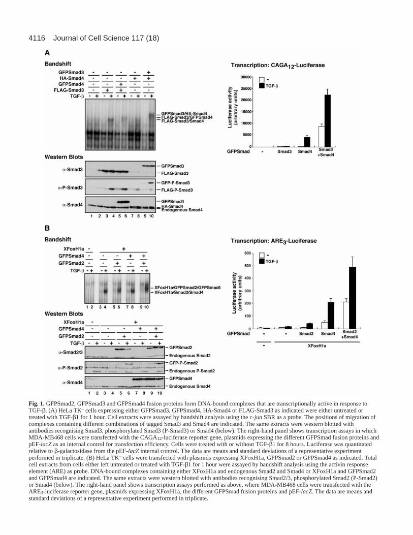
ResultsThe GFPSmads are activated in response to TGF-β andform transcriptionally active DNA-bound complexesWe constructed plasmids expressing enhanced GFP fusionsof human Smad2, Smad3 and Smad4, and then tested theiractivity in a variety of assays to ensure that they retained theproperties of wild-type Smads. A Smad3/Smad4-containingcomplex forms on the c-jun SBR upon TGF-β stimulation(Inman and Hill, 2002). In HeLa TK– cells, which containrelatively low levels of Smad3, this is readily detected bybandshift assay when Smad3 is overexpressed (Fig. 1A, lanes1-4). Both GFPSmad4 and GFPSmad3 were incorporated intothis complex, as demonstrated by expressing them in HeLaTK– cells and observing the change in mobility of the TGF-β-induced complex formed, as a result of the large size of theGFP. Thus, expression of GFPSmad4 and FLAG-Smad3resulted in a complex that migrated slightly more slowly thanthat resulting from expression of FLAG-Smad3 alone (Fig.1A, lanes 4, 6). Expression of HA-Smad4 and GFPSmad3resulted in a complex with a strikingly lower mobility thanthat observed with HA-Smad4 alone (Fig. 1A, lanes 8, 10).It is not clear why the presence of GFP on Smad3 has agreater effect than the presence of GFP on Smad4. Thewestern blots in Fig. 1A show that all tagged Smads wereequivalently expressed. In addition, GFPSmad3 wasphosphorylated efficiently in response to TGF-β, as wasFLAG-tagged Smad3.
Similarly, we demonstrated by bandshift assay thatGFPSmad2 and GFPSmad4 were incorporated into an Activin
responsive factor (ARF) complex on the ARE probe with thetranscription factor XFoxH1a in response to TGF-β (Fig. 1B).When XFoxH1a was expressed in HeLa TK– cells, a TGF-βinducible complex was detected (Fig. 1B, lanes 1-4) thatcontained XFoxH1a, Smad2 and Smad4 (data not shown)(Germain et al., 2000). The migration of this complex wasdecreased slightly when either GFPSmad2 or GFPSmad4 wasexpressed, and was markedly decreased when both wereexpressed, demonstrating that they were both incorporated intothe complex (Fig. 1B, lanes 4,6,8,10). The control westernblots demonstrate that all the Smads were well expressed, andthat the GFPSmad2 was phosphorylated in response to TGF-β, as was endogenous Smad2.
We also demonstrated that these GFPSmad-containingcomplexes were transcriptionally active by transfecting theGFPSmads into MDA-MB468 cells which lack endogenousSmad4, but contain R-Smads (Schutte et al., 1996), andmeasuring their activity on the Smad3/Smad4-dependentreporter CAGA12-luciferase (Dennler et al., 1998), or on theSmad2/Smad4-dependent reporter, ARE3-luciferase, togetherwith XFoxH1a (Germain et al., 2000). GFPSmad4 rescuedthe deletion of Smad4 in these cells, and this activity wasenhanced by addition of either GFPSmad3 (for the CAGA12-luciferase) or GFPSmad2 (for ARE3-luciferase) (Fig. 1A,B,right panels). Thus all three of these GFPSmads arefunctional in these assays, and behave similarly to wild-typeSmads.
TGF-β-induced nuclear translocation of GFPSmads isdependent upon continuous receptor signallingWe next investigated whether these GFPSmads translocated tothe nucleus in response to TGF-β, as has been shown forendogenous Smads (Pierreux et al., 2000). We made stable celllines of HaCaT cells expressing the GFPSmads, and analysedthe behaviour of the GFPSmads in the absence or presence ofTGF-β in pools of expressing cells (Fig. 2A). These cell linesexpress GFPSmads on average at levels comparable toendogenous Smads (data not shown). When performing theseexperiments, cells were pretreated with the protein synthesisinhibitor, cycloheximide. This was to ensure that during theexperiment, the same pool of GFPSmads was observed.In control experiments, the kinetics of GFPSmadnucleocytoplasmic shuttling were not affected by the presenceof cycloheximide (data not shown).
GFPSmad2 was predominantly cytoplasmic in uninducedcells, and predominantly nuclear after 60 minutes ofinduction with TGF-β (Fig. 2A, top panels, quantitated inFig. 2A left-hand graph). In the nucleus it was excluded fromthe nucleoli, as was endogenous Smad2 (Pierreux et al.,2000). If the ALK5 inhibitor, SB-431542 (Inman et al.,2002a; Laping et al., 2002) was added at the 60-minute time-point, GFPSmad2 accumulated back out in the cytoplasm bythe 220-minute time-point (Fig. 2A, top panels, quantitatedin Fig. 2A middle graph). If, however, no receptor inhibitorwas added, the GFPSmad2 remained nuclear during this time(data not shown). The GFPSmad2 protein behaved in anidentical fashion when transiently transfected into HeLa TK–
cells (Fig. 2B, upper panels). The same behaviour ofGFPSmad2 was observed over a wide range of expressionlevels. This activity and its kinetics faithfully mirrors that of
4116 Journal of Cell Science 117 (18)
Fig. 1.GFPSmad2, GFPSmad3 and GFPSmad4 fusion proteins form DNA-bound complexes that are transcriptionally active in response toTGF-β. (A) HeLa TK– cells expressing either GFPSmad3, GFPSmad4, HA-Smad4 or FLAG-Smad3 as indicated were either untreated ortreated with TGF-β1 for 1 hour. Cell extracts were assayed by bandshift analysis using the c-jun SBR as a probe. The positions of migration ofcomplexes containing different combinations of tagged Smad3 and Smad4 are indicated. The same extracts were western blotted withantibodies recognising Smad3, phosphorylated Smad3 (P-Smad3) or Smad4 (below). The right-hand panel shows transcription assays in whichMDA-MB468 cells were transfected with the CAGA12-luciferase reporter gene, plasmids expressing the different GFPSmad fusion proteins andpEF-lacZas an internal control for transfection efficiency. Cells were treated with or without TGF-β1 for 8 hours. Luciferase was quantitatedrelative to β-galactosidase from the pEF-lacZ internal control. The data are means and standard deviations of a representative experimentperformed in triplicate. (B) HeLa TK– cells were transfected with plasmids expressing XFoxH1a, GFPSmad2 or GFPSmad4 as indicated. Totalcell extracts from cells either left untreated or treated with TGF-β1 for 1 hour were assayed by bandshift analysis using the activin responseelement (ARE) as probe. DNA-bound complexes containing either XFoxH1a and endogenous Smad2 and Smad4 or XFoxH1a and GFPSmad2and GFPSmad4 are indicated. The same extracts were western blotted with antibodies recognising Smad2/3, phosphorylated Smad2 (P-Smad2)or Smad4 (below). The right-hand panel shows transcription assays performed as above, where MDA-MB468 cells were transfected with theARE3-luciferase reporter gene, plasmids expressing XFoxH1a, the different GFPSmad fusion proteins and pEF-lacZ. The data are means andstandard deviations of a representative experiment performed in triplicate.

4117Smad nucleocytoplasmic shuttling
endogenous Smad2 (Inman et al., 2002b). For endogenousSmad2 this observation has been interpreted as an indicationthat Smad2 is constantly shuttling between the nucleus andcytoplasm during active TGF-β signalling, undergoingcycles of phosphorylation by the receptors and
dephosphorylation by a phosphatase in the nucleus (Inmanet al., 2002b). If the TGF-β receptors are turned off by SB-431542, Smad2 is no longer activated in the cytoplasm andthus accumulates there.
Similarly, the behaviour of GFPSmad4 mimicked that of
Fig. 2.TGF-β-induced nucleartranslocation of GFPSmads isdependent upon continuous receptorsignalling. (A) HaCaT cell linesstably expressing GFPSmad2,GFPSmad3 or GFPSmad4 werepretreated with cycloheximide andwere then incubated with TGF-β1 for1 hour, followed by SB-431542 (7.5µM) for up to 160 minutes. In thecase of the GFPSmad4 cells,leptomycin B (LMB) was added 90minutes after SB-431542 addition(150-minute time-point).Fluorescence images are shown atdifferent time-points after initialTGF-β treatment. Arrows indicaterepresentative examples of cellsdemonstrating nucleocytoplasmicshuttling. For GFPSmad3 cells, theboxed region is shown magnifiedbelow to demonstrate that GFPSmad3is partially excluded from the nucleoliupon TGF-β treatment. Below aregraphs showing quantitation ofnuclear fluorescence, withfluorescence images collected every 3minutes. The left-hand graph showsthe average of the TGF-β-inducednuclear fluorescence of theGFPSmad2 and GFPSmad4 cellsmarked with an arrow. Means andstandard deviations are shown. Theright-hand graphs showquantifications of the nuclearfluorescence for GFPSmad2 andGFPSmad4 throughout the wholeexperiment for one of the indicatedcells in each case. (B) HeLa TK–
cells were transiently transfected withplasmids expressing GFPSmad2 orGFPSmad4 together with FLAG-Smad2 and treated withcycloheximide, TGF-β1, SB-431542and LMB as in A. Fluorescenceimages are shown at different time-points after initial TGF-β treatment.Arrows indicate representativeexamples of cells demonstratingnucleocytoplasmic shuttling. Thepunctate fluorescence observed in thecytoplasm of transiently transfectedHeLa TK– cells is not seen in thestable HaCaT cell lines and thusseems to be a consequence oftransient transfection. Theexperiments shown arerepresentatives from at least threeindependent experiments.
4118
endogenous Smad4. GFPSmad4 accumulated in the nucleusupon TGF-β signalling and then accumulated back in thecytoplasm after addition of SB-431542 (Fig. 2A, lowerpanels, quantitated Fig. 2A, graphs). In the nucleusGFPSmad4 was excluded from the nucleoli as wasendogenous Smad4 (Pierreux et al., 2000). It is striking thatthe TGF-β-induced nuclear accumulation of GFPSmad4 ismuch less complete than that of GFPSmad2, and alsoplateaus earlier. This is probably because of the fact that inresponse to TGF-β, nuclear accumulation of Smad4 requirescomplex formation with activated R-Smads, whereasaccumulation of activated homomeric complexes of Smad2can occur in the absence of Smad4 (De Bosscher et al., 2004;Nicolás and Hill, 2003). Expression of GFPSmad4 in the cellline at approximately endogenous levels results in excessSmad4 over endogenous R-Smads, and thus only aproportion of the GFPSmad4 can accumulate in the nucleusin response to TGF-β. The GFPSmad4 protein behaved in asimilar fashion when transiently transfected into HeLa TK–
cells (Fig. 2B, lower panels). In this case, to see anyaccumulation of GFPSmad4 in the nucleus we had to alsooverexpress FLAG-Smad2, possibly because of lower levelsof R-Smads in these cells. The same behaviour ofGFPSmad4 was observed over a wide range of expressionlevels. Smad4 export from the nucleus is mediated via thenuclear exporter CRM1 (Pierreux et al., 2000; Watanabe etal., 2000). When the inhibitor of CRM1 (LMB) was addedto the cells after prolonged incubation with SB-431542 (atthe 150-minute time-point), the GFPSmad4 rapidlyaccumulated in the nucleus (Fig. 2A, lower panels,quantitated in Fig. 2A, right-hand graph; Fig. 2B). Thisindicates that even in the absence of activated TGF-βreceptors, GFPSmad4 (presumably monomeric) isconstitutively imported into the nucleus and is exported byCRM1. If CRM1 activity is inhibited by LMB, GFPSmad4accumulates in the nucleus.
In contrast to GFPSmad2 and GFPSmad4, GFPSmad3did not behave as endogenous Smad3 in translocationexperiments, although in the biochemical assays it appearedto function normally (Fig. 1). In the absence of TGF-β,GFPSmad3 was predominantly nuclear, even though we haveshown that it is unphosphorylated (Fig. 1A, Fig. 2A middlepanels). It was also detected in the nucleoli, which was notthe case for endogenous Smad3 (Pierreux et al., 2000) (Fig.2A, middle panels). GFPSmad3 was, however, sensitive toTGF-β treatment, as this nuclear fluorescence intensifiedupon TGF-β stimulation, and some partial exclusion offluorescence from the nucleoli was detected, suggesting thatmonomeric GFPSmad3 is not excluded from the nucleoli, butcomplexed activated GFPSmad3 is (Fig. 2A, middle panels).The GFPSmad3 also responded to SB-431542 treatment,as upon addition of this receptor inhibitor, the nuclearfluorescence decreased again to the levels seen in uninducedcells. Because GFPSmad3 did not faithfully mimic thebehaviour of endogenous Smad3, we have not studied thisprotein further.
From the data presented in this section we conclude thatGFPSmad2 and GFPSmad4 closely mimic endogenous Smad2and Smad4, respectively, and can be used to investigate thenucleocytoplasmic shuttling behaviour of these Smads in bothHaCaT and HeLa TK– cells.
Shuttling of GFPSmad2 and GFPSmad4 between thecytoplasm and nucleus occurs in both unstimulated andTGF-β-induced cellsFLIP experiments can be used to investigate whether a proteinshuttles between two compartments of the cell, and also toindicate how mobile a protein is in a given compartment ofthe cell. A prolonged bleaching is applied to a defined area ofthe cell and the fluorescence at the bleaching point and at adistant reporting point is quantitated over time (reviewed byLippincott-Schwartz et al., 2003). If the GFP-labelledmolecules are shuttling between the bleaching and reportingpoints, then the fluorescence will decrease at both points.Relatively immobile proteins in contrast will be bleachedeffectively at the bleaching point, but not at the reportingpoint.
We first performed a series of controls to validate the FLIPexperiments. GFP is a small protein that diffuses throughoutthe cell. When HeLa TK– cells expressing GFP alone werebleached in the cytoplasm, the nuclear GFP also rapidlybleached (Fig. 3Ai), as expected for a protein freely diffusingthrough the nucleus and cytoplasm. GFPRanBP1∆NES is aGFP fusion of RanBP1 that is trapped in the nucleus becauseit is imported efficiently via its non-classical Ran-dependentNLS, but cannot be exported because its NES has been deleted(F.J.N. and P. R. Clarke, unpublished data) (Plafker andMacara, 2000). In this case, when the bleaching occurred inthe cytoplasm, virtually no nuclear bleaching was detected(Fig. 3Aii). This is the behaviour expected of a protein thatdoes not shuttle between the cytoplasm and nucleus. As acontrol for the use of this technique to determine the mobilityof a protein in a given compartment of the cell, we investigatedthe behaviour of a fusion protein of GFP with PKCα. Inuninduced cells this protein was distributed throughout thecytoplasm (Fig. 3Aiii) (Lillemeier et al., 2001; Ng et al., 1999).It was completely mobile in this compartment, as seen by therapid photobleaching at a point in the cytoplasm distant fromthe bleach point (Fig. 3Aiii) (Lillemeier et al., 2001). However,upon stimulation with the phorbol ester TPA for 10 minutes,GFPPKCα accumulated at the plasma membrane (Fig. 3B). Inthis compartment it was not very mobile, and hence virtuallyno photobleaching was detected at a reporting point on theplasma membrane distant from the bleach point (Fig. 3Aiv)(Lillemeier et al., 2001).
Having demonstrated that FLIP can be used to detectshuttling of GFP-tagged molecules between the cytoplasm andthe nucleus, we investigated the shuttling behaviour ofGFPSmad2 and GFPSmad4 in both the absence and presenceof TGF-β signalling, using the HaCaT cell lines stablyexpressing these fusion proteins. In these experiments, thecytoplasm was bleached. If nuclear bleaching is observedin the same cell, this indicates that the GFPSmad is indynamic equilibrium between these two compartments. Thusthis experiment provides a direct demonstration ofnucleocytoplasmic shuttling.
When GFPSmad2 was bleached in the cytoplasm of anuninduced cell, it also bleached in the nucleus of the samecell. In contrast, nuclei of adjacent cells did not bleach,indicating that this nuclear bleaching is specific (Fig. 4i).Similarly, when the same experiment was performed usingTGF-β-induced cells, cytoplasmic bleaching resulted innuclear bleaching in the same cell (Fig. 4ii). Note that in the
Journal of Cell Science 117 (18)

4119Smad nucleocytoplasmic shuttling
TGF-β-induced cell, the proportion of nuclear moleculesbleached was not as high as in an uninduced cell (comparegraphs in Fig. 4i and Fig. 4ii). This suggests that after TGF-β stimulation, the proportion of nuclear Smad that is mobile
is lower than in an uninduced cell (see below and Discussion).Exactly the same behaviour was observed for GFPSmad4(Fig. 4iii,iv). These data directly demonstrate that in bothuninduced cells and in TGF-β-induced cells, GFPSmad2 and
Fig. 3.Controls for FLIP analysis. (A) HeLa TK– cells were transiently transfected with plasmids expressing GFP, GFPRanBP1∆NES orGFPPKCα. Cells were unstimulated except for those expressing GFPPKCα which were stimulated with TPA. Cells were photobleached in thecytoplasm as indicated by the red diamond. The fluorescence images shown prior to photobleaching (time=0 seconds) and then after threeconsecutive 80-second bleaching periods. In all cases, the plane of focus was a cross-section through the cell, except for the TPA-induced cellsexpressing GFPPKCα where the plane of focus was in the plasma membrane. The right-hand graphs show the FLIP analysis. The fluorescencewas quantitated at the bleach point and at a reporting point indicated by the blue square. The intensity of fluorescence is represented in arbitraryunits. (B) Stimulation of cells with TPA induces the translocation of GFPPKCα to the membrane. HeLa TK– cells expressing GFPPKCα wereeither untreated or treated with TPA for up to 10 minutes. Fluorescence images are shown. Here, the plane of focus is again a cross-sectionthrough the cell. The experiments shown are representatives from at least three independent experiments.

4120
GFPSmad4 are constantly shuttling between the cytoplasmand the nucleus.
FLIP analysis reveals that GFPSmad4 is more mobile inthe cytoplasm than is GFPSmad2We noticed in the experiments shown in Fig. 4 that inuninduced cells GFPSmad2 bleached less readily incytoplasmic regions distant from the cytoplasmic bleach pointthan did GFPSmad4. This suggested that GFPSmad2 mightbe less mobile in the cytoplasm than is GFPSmad4. Weinvestigated this in more detail, using both transientlytransfected HeLa TK– cells and the stably transfected HaCaTcell lines. In both cell lines GFPSmad2 bleached more slowlyat the reporting point in the cytoplasm than does GFPSmad4(Fig. 5). The graphs demonstrate that the rate of bleaching at
the cytoplasmic reporting point for GFPSmad4 was verysimilar to the rate of bleaching at the bleach point. ForGFPSmad2 however, the rate of bleaching at the reportingpoint was significantly slower than the rate of bleaching at thebleach point itself. These data strongly suggest that GFPSmad2is substantially less mobile in the cytoplasm than isGFPSmad4.
FLIP/FRAP analysis reveals a TGF-β-dependent changein mobility of GFPSmad2 and GFPSmad4 in the nucleusFinally we investigated the mobility of GFPSmads in thenucleus in uninduced or TGF-β-induced cells. For this we usedFRAP in combination with FLIP. In these experiments aphotobleaching pulse (11 seconds) was applied to a particulararea of the cell, and then the recovery of fluorescence in that
Journal of Cell Science 117 (18)
Fig. 4.FLIP analysis of GFPSmad2 and GFPSmad4 proves nucleocytoplasmic shuttling. (A) HaCaT cell lines stably expressing GFPSmad2 orGFPSmad4 remained either unstimulated (i,iii) or were treated for 1 hour with TGF-β1 (ii,iv). The bleach region in the cytoplasm is indicated(red diamond). Each row shows the fluorescence image prior to bleaching (time=0 seconds) and after three consecutive 80-second bleachingperiods. The fluorescence was quantitated at the bleach point, at a reporting point in the nucleus of the same cell (blue square) and at a reportingpoint in the nucleus of an adjacent cell (white triangle). In each case the fluorescence was normalised to the initial fluorescence prior tophotobleaching, and the relative fluorescence was plotted. Note that in unstimulated cells expressing GFPSmad4, the cell to the right of thebleached cell also bleaches because the bleach point also contacts the cytoplasm of that cell. The data are representatives from at least threedifferent experiments.

4121Smad nucleocytoplasmic shuttling
area was monitored together with fluorescence loss at a distantpoint, also in the nucleus. Again we performed controls tovalidate the approach. For nuclear localised GFP (GFPNLS),it was evident that recovery at the bleach point was extremelyfast, as was the rate of bleaching at the reporting point (Fig.6i). When the same experiment was performed using a muchlarger nuclear GFP fusion protein (GFPβ-galactosidaseNLS)which has a native molecular weight as a tetramer of over 400kDa (Jacobson et al., 1994), the recovery after photobleachingwas slower, as was the rate of bleaching at the reporting point
(Fig. 6ii). This indicates that, as expected, the mobility of themuch larger GFPβ-galactosidaseNLS protein in the nucleus islower than that of GFP alone. We performed the sameexperiment on a protein that we have shown to be relativelyimmobile (GFPPKCα at the plasma membrane after TPAtreatment). In this case the recovery after photobleaching wasvery slow and no detectable bleaching was observed at thereporting point (Fig. 6iii).
Having demonstrated that the FLIP/FRAP experiment cangive an indication as to the mobility of a protein in a given
Fig. 5.FLIP analysis reveals that GFPSmad4 is more mobile in the cytoplasm than is GFPSmad2. FLIP analysis was performed on either HeLaTK– cells transiently transfected with plasmids expressing GFPSmad2 or GFPSmad4 and FLAG-Smad2 (A) or HaCaT cell lines stablyexpressing GFPSmad2 or GFPSmad4 (B). Each row shows the fluorescence image prior to bleaching (time=0 seconds) and then after threeconsecutive 80-second bleaching periods. The fluorescence was quantitated at the bleach point (red diamond) and at a distant reporting point inthe cytoplasm of the same cell (blue square) and at a reporting point in the cytoplasm of an adjacent cell (white triangle). In each case thefluorescence was normalised to the initial fluorescence prior to photobleaching, and the relative fluorescence was plotted. The data arerepresentatives from at least three different experiments.

4122
cellular compartment, we investigated the mobility of theGFPSmads in the nucleus. In uninduced cells expressingGFPSmad2 at relatively low levels the protein is predominantlycytoplasmic, but if highly expressed a substantial amount isnuclear, although it is not phosphorylated and thus likely to bemonomeric. Thus to investigate the behaviour of unactivatedGFPSmad2 in the nucleus, we used highly expressing cells.The behaviour of unactivated GFPSmad2 in the FLIP/FRAPassay was intermediate between that of GFPNLS and GFPβ-galactosidaseNLS (Fig. 6iv). The FLIP/FRAP kinetics werenot dependent on the level of unactivated monomericGFPSmad2 in the nucleus, because we could show that theFLIP/FRAP curves were the same for cells expressing verydifferent levels of nuclear GFPSmad2 (data not shown). UponTGF-β stimulation however, the behaviour of GFPSmad2 inthis assay changed to indicate that it became much less mobile(Fig. 6v). In this case the recovery at the bleach point wassubstantially slower, as was the rate of bleaching at thereporting point. This indicates that active complexes ofGFPSmad2 (probably a mixture of homomeric GFPSmad2complexes and heteromeric complexes with endogenousSmad4) are less mobile in the nucleus than unactivated
GFPSmad2. GFPSmad4 behaved in a similar manner. LMBtreatment was used to allow accumulation of unactivatedmonomeric Smad4 in the nucleus in the absence of TGF-β. Inthis case, the kinetics of FLIP/FRAP were similar to thoseobserved with GFPSmad2 in uninduced cells (Fig. 6iv,vi).Upon TGF-β stimulation, the FLIP/FRAP experimentindicated that, like GFPSmad2, GFPSmad4 becamesubstantially less mobile (Fig. 6vii). Again, the kinetics ofFLIP/FRAP were similar to those observed with GFPSmad2 inthe same conditions.
Thus we find a similar TGF-β-induced decrease in themobility of GFPSmad2 and GFPSmad4 in the nucleus. Wetherefore propose that activated Smad complexes are activelyretained in the nucleus through a tethering mechanism.
DiscussionHere we have investigated nucleocytoplasmic shuttling ofSmad2 and Smad4 using enhanced GFP fusions. We havedemonstrated that these Smad fusions behave as wild-typeSmads in terms of their activation in response to TGF-β andtheir formation of transcriptionally active Smad-transcription
Journal of Cell Science 117 (18)
Fig. 6.FLIP/FRAP analysis reveals a TGF-β-dependent change in mobility of GFPSmad2and GFPSmad4 in the nucleus. HeLa TK–
cells were transiently transfected withplasmids expressing GFPNLS, GFPβ-galactosidaseNLS, GFPPKCα, GFPSmad2 orGFPSmad4 with FLAG-Smad2. Cells wereeither unstimulated or pretreated for 1 hourwith LMB or TGF-β1 as indicated. The cellsexpressing GFPPKCα were pretreated for 10minutes with TPA. Prior to bleaching, eightmeasurements of fluorescence were taken atthe bleach point in the nucleus (red square)over a period of 2 seconds, then cells werebleached for 11 seconds. The recovery offluorescence in the bleached area andfluorescence loss at a distant point also in thenucleus (blue square) were measured at 250-millisecond intervals. The levels offluorescence were normalised to the mean offluorescence registered before thephotobleaching. In all cases, the plane of focusis a cross-section through the cell, except forthe TPA-induced cells expressing GFPPKCαwhere the plane of focus was in the plasmamembrane. The data are representatives fromthree different experiments.

4123Smad nucleocytoplasmic shuttling
factor complexes on TGF-β-responsive elements. We havethen used these Smad fusions to investigate Smadnucleocytoplasmic shuttling in detail in living cells. Twodifferent cell lines (HaCaTs and HeLa TK–) have been used andwe have also compared both stably transfected cell lines andtransiently transfected cells. The results obtained are similar inthe different systems, suggesting that neither the cell type northe particular levels of expression of GFPSmad are critical.
For Smad4, we demonstrate here in a simple experiment thateven though GFPSmad4 is predominantly localised in thecytoplasm in the absence of TGF-β signalling, it is actuallyrapidly shuttling between the cytoplasm and the nucleus. Wehave shown that after prolonged treatment of GFPSmad4-expressing cells with SB-431542, treatment of the cells withthe CRM1 inhibitor LMB leads to rapid accumulation ofGFPSmad4 in the nucleus. This is because GFPSmad4 isconstitutively imported into the nucleus and under theseconditions cannot be exported.
We have gone on to use photobleaching experiments (FLIPand FRAP) to study the Smad nucleocytoplasmic shuttling inmore detail. Indirect methods have previously suggested thatthe Smads undergo nucleocytoplasmic shuttling (Inman et al.,2002b; Pierreux et al., 2000; Watanabe et al., 2000; Xu et al.,2002), but here for the first time we have directly demonstratedthat both GFPSmad2 and GFPSmad4 shuttle between thecytoplasm and nucleus in both uninduced cells and in TGF-β-stimulated cells. There is a striking difference between theproportion of nuclear GFPSmad that bleaches during the 240seconds of cytoplasmic photobleaching in the uninduced cellsversus that in the TGF-β-induced cells, suggesting that theproportion of nuclear Smad that is mobile in TGF-β-inducedcells is substantially lower than that in unstimulated cells. Incontrast to uninduced cells, the nuclear GFPSmad2 andGFPSmad4 in TGF-β-induced cells are not completelybleached even after prolonged cytoplasmic photobleaching(data not shown). The FLIP/FRAP experiments confirm thatTGF-β induction results in a decrease in Smad mobility in thenucleus. The data obtained with endogenous Smads suggestthat only monomeric Smads are exported from the nucleus(Inman et al., 2002b). Thus in TGF-β-induced cells,dissociation of Smads from active Smad complexes afterdephosphorylation of the R-Smads will be a prerequisite fornuclear export. The rate of R-Smad dephosphorylation andcomplex dissociation will thus dictate the amount of nuclearSmad that is mobile, and can be exported to the cytoplasm.
We have also used photobleaching experiments toinvestigate the mobility of the GFPSmads in both thecytoplasm and the nucleus. This issue is important as itimpinges on the mechanism underlying the distribution of theSmads between the cytoplasm and nucleus in uninduced cellsand in TGF-β-induced cells. Two different models (notmutually exclusive) can be envisaged. One model proposes theexistence of cytoplasmic and nuclear retention factors (Regulyand Wrana, 2003; Xu et al., 2000; Xu et al., 2002). MonomericSmads would have a higher affinity for cytoplasmic retentionfactors and complexed Smads would have a higher affinity fornuclear retention factors. This could explain the cytoplasmiclocalisation of the Smads in unstimulated cells, and theiraccumulation in the nucleus upon TGF-β treatment. Analternative view is that the distribution of the Smads betweenthe cytoplasm and nucleus is determined by the relative
rates of import and export. Monomeric Smads would bepreferentially exported from the nucleus and complexedSmads, preferentially imported. Complex formation mighteither stimulate import or inhibit export (De Bosscher et al.,2004; Reguly and Wrana, 2003; Watanabe et al., 2000).
Our results demonstrate that in uninduced cells GFPSmad2is less mobile in the cytoplasm than is GFPSmad4. Thissuggests that monomeric inactivated GFPSmad2 is likely to beassociated with cytoplasmic retention factors. Two differentproteins have been suggested to act in such a way, and in bothcases monomeric Smad2 has been shown to preferentially bindthe factor compared with active complexed Smads. First, themicrotubule network may play this role. Smads 2, 3 and 4have been shown to bind tubulin and long-term (18 hour)treatment of cells with microtubule disrupting agents, suchas nocodazole, results in increased TGF-β-induced Smad2phosphorylation and increased TGF-β-induced transcription(Dong et al., 2000). We think, however, that the microtubulenetwork is unlikely to be the cytoplasmic retention factorpredicted by our experiments, because in HaCaT cells we findthat short-term treatment with nocodazole, which wassufficient to depolymerise the microtubules, had no effect onthe localisation of endogenous Smad2 (data not shown).Another candidate for the cytoplasmic retention factor forSmad2 is Smad Anchor for Receptor Activation (SARA) (Xuet al., 2000; Xu et al., 2002). In vitro, the purified Smad bindingregion of SARA inhibits nuclear import of Smad2 (Xu et al.,2000). However, SARA is unlikely to be the major endogenousSmad2 retention factor, because it is predominantly localisedon early endosomes (Di Guglielmo et al., 2003; Hayes et al.,2002; Itoh et al., 2002), whereas Smad2 is distributed equallythroughout the cytoplasm (Inman et al., 2002b; Pierreux et al.,2000). Moreover, there appears to be substantially more Smad2in a cell than SARA because overexpression of SARA has beenshown to relocalise Smad2 to the early endosomes (Tsukazakiet al., 1998). More work is obviously required to confirm theidentity of the putative cytoplasmic retention factor for Smad2.
In the nucleus we have used FRAP in combination withFLIP to investigate the mobility of GFPSmad2 and GFPSmad4in both uninduced cells and after 1 hour of TGF-β stimulation.GFPSmad2 and 4 behaved similarly in this assay, and in bothcases ligand-induced activation led to a substantial decrease inmobility. This strongly suggests that unactivated monomericSmads in the nucleus are not actively retained, but activatedSmad complexes are. Similar results have recently beendemonstrated for a subset of hormone receptors. In theseexperiments ligand binding has been shown to decreasethe mobility of GFP fusions of estrogen receptor andglucocorticoid receptor and to increase the fraction of receptorsunable to diffuse (Maruvada et al., 2003; Schaaf andCidlowski, 2003). What could be the nuclear retention factorsfor the active Smad complexes? Smads are known to associatewith transcription activators and repressors as well as co-activators and co-repressors in the nucleus, and these could actas nuclear retention factors (Shi and Massagué, 2003; Xu etal., 2002). If this were the case, the complexes would have tobe extremely large, because the active Smad complexes aresubstantially less mobile than GFPβ-galactosidaseNLS whichhas a native molecular weight as a tetramer of over 400 kDa.The other possibility is that DNA acts as a nuclear retentionfactor for the Smads. The Smads bind DNA both directly and
4124
indirectly through interactions with other transcription factors.We have demonstrated that here the GFPSmads, likeendogenous Smads, are located uniformly throughout theinterphase nucleus, excluded only from the nucleoli (see alsoPierreux et al., 2000). Thus it is unlikely that the associationwith DNA represents Smad complexes actively engaged intranscription at specific promoter sites, because these siteswould be relatively rare (Hager et al., 2002). It is most likelythat an association with DNA reflects Smad complexes weaklybound to DNA engaged in a ‘scanning process’ to locatespecific binding sites (Lillemeier et al., 2001; Pederson, 2001).These weakly bound Smads may be in dynamic equilibriumwith those bound to specific promoter elements, as has beenrecently demonstrated for hormone-bound glucocorticoidreceptor and its interacting protein GRIP-1 (Becker et al.,2002; Hager et al., 2002; McNally et al., 2000).
The work presented here provides evidence for cytoplasmicretention of Smad2 in uninduced cells and nuclear retention forboth Smad2 and Smad4 in TGF-β-induced cells. Because theSmads are constantly shuttling between the cytoplasm andnucleus in both uninduced and in TGF-β-induced cells, thebinding to the putative retention factors must be readilyreversible. Based on these findings, our previous work and thework of others, we propose the following model for regulationof Smad nucleocytoplasmic shuttling. UnphosphorylatedSmad2 may be retained in the cytoplasm through interactionwith retention factors, whereas Smad4 may be retained therebecause its export from the nucleus is dominant over import.Activation of Smad2 leads to dissociation of Smad2 fromcytoplasmic tethering, and to complex formation with Smad4.These complexes accumulate in the nucleus at least partlybecause they are actively retained there. In addition, it remainspossible that nuclear import of the Smad complexes dominatesover export, either because import is potentiated or export isinhibited by Smad complex formation (Kurisaki et al., 2001;Watanabe et al., 2000). The R-Smads in the nucleus arecontinuously being dephosphorylated, leading to dissociationof Smad complexes, release from nuclear tethering and exportof the Smads from the nucleus. For the duration of signalling,this cycle of phosphorylation and activation in the cytoplasmand dephosphorylation and deactivation in the nucleuscontinues. When the receptors are downregulated (Ebisawa etal., 2001; Kavsak et al., 2000), and signalling is terminated, theSmads accumulate back in the cytoplasm.
We thank Paul Clarke for GFPRanBP1∆NES, Björn Lillemeier andIan Kerr for GFPNLS, Scott Parkinson and Peter Parker forGFPPKCα and Ray Truant for GFPβ-galactosidaseNLS. We thank EdLeof for the anti-phosphorylated Smad3 antibody, Alastair Reith andNicholas Laping for SB-431542 and Peter ten Dijke for the anti-phosphorylated Smad2 antibody. We are very grateful to BjörnLillemeier, Daniel Zicha and Peter Jordan for help with thephotobleaching experiments and with confocal microscopy, and to theCR-UK FACS facility for FACS sorting the GFPSmad-expressingHaCaT cells. We thank Ian Kerr, Peter Parker and members of the Hilllab for useful discussions and helpful comments on the manuscript.The work was supported by Cancer Research UK, a long-term EMBOfellowship to K.D.B. and an M.R.C. training fellowship to F.J.N.
ReferencesAngel, P., Baumann, I., Stein, B., Delius, H., Rahmsdorf, H. J. and
Herrlich, P. (1987). 12-O-tetradecanoyl-phorbol-13-acetate induction of the
human collagenase gene is mediated by an inducible enhancer elementlocated in the 5′-flanking region. Mol. Cell. Biol. 7, 2256-2266.
Bardwell, V. J. and Treisman, R. (1994). The POZ domain: a conservedprotein-protein interaction motif. Genes Dev. 8, 1664-1677.
Becker, M., Baumann, C., John, S., Walker, D. A., Vigneron, M., McNally,J. G. and Hager, G. L. (2002). Dynamic behavior of transcription factorson a natural promoter in living cells. EMBO Rep. 3, 1188-1194.
De Bosscher, K., Hill, C. S. and Nicolás, F. J. (2004). Molecular andfunctional consequences of Smad4 C-terminal missense mutations incolorectal tumour cells. Biochem. J. 379, 209-216.
Dennler, S., Itoh, S., Vivien, D., ten Dijke, P., Huet, S. and Gauthier, J. M.(1998). Direct binding of Smad3 and Smad4 to critical TGF β-inducibleelements in the promoter of human plasminogen activator inhibitor-type 1gene. EMBO J. 17, 3091-3100.
Di Guglielmo, G. M., Le Roy, C., Goodfellow, A. F. and Wrana, J. L.(2003). Distinct endocytic pathways regulate TGF-β receptor signalling andturnover. Nat. Cell Biol. 5, 410-421.
Dong, C., Li, Z., Alvarez, R., Jr, Feng, X. H. and Goldschmidt-Clermont,P. J. (2000). Microtubule binding to Smads may regulate TGFβ activity.Mol. Cell 5, 27-34.
Ebisawa, T., Fukuchi, M., Murakami, G., Chiba, T., Tanaka, K., Imamura,T. and Miyazono, K. (2001). Smurf1 interacts with transforming growthfactor-β type I receptor through Smad7 and induces receptor degradation. J.Biol. Chem. 276, 12477-12480.
Faure, S., Lee, M. A., Keller, T., ten Dijke, P. and Whitman, M. (2000).Endogenous patterns of TGFβ superfamily signaling during early Xenopusdevelopment. Development127, 2917-2931.
Germain, S., Howell, M., Esslemont, G. M. and Hill, C. S. (2000).Homeodomain and winged-helix transcription factors recruit activatedSmads to distinct promoter elements via a common Smad interaction motif.Genes Dev. 14, 435-451.
Görlich, D. and Kutay, U. (1999). Transport between the cell nucleus and thecytoplasm. Ann. Rev. Cell Dev. Biol. 15, 607-660.
Hager, G. L., Elbi, C. and Becker, M. (2002). Protein dynamics in the nuclearcompartment. Curr. Opin. Genet. Dev. 12, 137-141.
Hayes, S., Chawla, A. and Corvera, S. (2002). TGF β receptor internalizationinto EEA1-enriched early endosomes: role in signaling to Smad2. J. CellBiol. 158, 1239-1249.
Inman, G. J. and Hill, C. S. (2002). Stoichiometry of active Smad-transcription factor complexes on DNA. J. Biol. Chem. 277, 51008-51016.
Inman, G. J., Nicolás, F. J., Callahan, J. F., Harling, J. D., Gaster, L. M.,Reith, A. D., Laping, N. J. and Hill, C. S. (2002a). SB-431542 is a potentand specific inhibitor of transforming growth factor-β superfamily type Iactivin receptor-like kinase receptors, ALK4, ALK5 and ALK7. Mol.Pharmacol. 62, 65-72.
Inman, G. J., Nicolás, F. J. and Hill, C. S. (2002b). Nucleocytoplasmicshuttling of Smads 2, 3 and 4 permits sensing of TGF-β receptor activity.Mol. Cell 10, 283-294.
Itoh, F., Divecha, N., Brocks, L., Oomen, L., Janssen, H., Calafat, J., Itoh,S. and Dijke ten, P. (2002). The FYVE domain in Smad anchor for receptoractivation (SARA) is sufficient for localization of SARA in early endosomesand regulates TGF-β/Smad signalling. Genes Cells7, 321-331.
Jacobson, R. H., Zhang, X. J., DuBose, R. F. and Matthews, B. W. (1994).Three-dimensional structure of β-galactosidase from E. coli. Nature 369,761-766.
Kavsak, P., Rasmussen, R. K., Causing, C. G., Bonni, S., Zhu, H.,Thomsen, G. H. and Wrana, J. L. (2000). Smad7 binds to Smurf2 to forman E3 ubiquitin ligase that targets the TGF β receptor for degradation. Mol.Cell 6, 1365-1375.
Kurisaki, A., Kose, S., Yoneda, Y., Heldin, C. H. and Moustakas, A. (2001).Transforming growth factor-β induces nuclear import of Smad3 in animportin-β1 and Ran-dependent manner. Mol. Biol. Cell12, 1079-1091.
Laping, N. J., Grygielko, E., Mathur, A., Butter, S., Bomberger, J., Tweed,C., Fornwald, J., Lehr, R., Harling, J. D., Gaster, L. M. et al. (2002).Inhibition of transforming growth factor (TGF)-β1-induced extracellularmatrix with a novel inhibitor of the TGF-β type I receptor kinase activity.Mol. Pharmacol. 62, 58-64.
Lillemeier, B. F., Koster, M. and Kerr, I. M. (2001). STAT1 from the cellmembrane to the DNA. EMBO J. 20, 2508-2517.
Lippincott-Schwartz, J., Altan-Bonnet, N. and Patterson, G. H. (2003).Photobleaching and photoactivation: following protein dynamics in livingcells. Nat. Cell Biol. Suppl, S7-S14.
Maruvada, P., Baumann, C. T., Hager, G. L. and Yen, P. M. (2003).
Journal of Cell Science 117 (18)

4125Smad nucleocytoplasmic shuttling
Dynamic shuttling and intranuclear mobility of nuclear hormone receptors.J. Biol. Chem. 278, 12425-12432.
McNally, J. G., Muller, W. G., Walker, D., Wolford, R. and Hager, G. L.(2000). The glucocorticoid receptor: rapid exchange with regulatory sites inliving cells. Science287, 1262-1265.
Ng, T., Squire, A., Hansra, G., Bornancin, F., Prevostel, C., Hanby, A.,Harris, W., Barnes, D., Schmidt, S., Mellor, H. et al. (1999). Imagingprotein kinase Cα activation in cells. Science283, 2085-2089.
Nicolás, F. J. and Hill, C. S. (2003). Attenuation of the TGF-β-Smad signalingpathway in pancreatic tumor cells confers resistance to TGF-β-inducedgrowth arrest. Oncogene22, 3698-3711.
Pederson, T. (2001). Protein mobility within the nucleus - what are the rightmoves? Cell 104, 635-638.
Pierreux, C. E., Nicolás, F. J. and Hill, C. S. (2000). Transforming growthfactor β-independent shuttling of Smad4 between the cytoplasm andnucleus. Mol. Cell. Biol. 20, 9041-9054.
Plafker, K. and Macara, I. G. (2000). Facilitated nucleocytoplasmic shuttlingof the Ran binding protein RanBP1. Mol. Cell. Biol. 20, 3510-3521.
Reguly, T. and Wrana, J. L. (2003). In or out? The dynamics of Smadnucleocytoplasmic shuttling. Trends Cell Biol.13, 216-220.
Schaaf, M. J. and Cidlowski, J. A. (2003). Molecular determinants ofglucocorticoid receptor mobility in living cells: the importance of ligandaffinity. Mol. Cell. Biol. 23, 1922-1934.
Schutte, M., Hruban, R. H., Hedrick, L., Cho, K. R., Nadasdy, G. M.,Weinstein, C. L., Bova, G. S., Isaacs, W. B., Cairns, P., Nawroz, H. etal. (1996). DPC4 gene in various tumor types. Cancer Res. 56, 2527-2530.
Shi, Y. and Massagué, J. (2003). Mechanisms of TGF-β signaling from cellmembrane to the nucleus. Cell 113, 685-700.
ten Dijke, P. and Hill, C. S. (2004). New insights into TGF-β-Smadsignalling. Trends Biochem. Sci. 29, 265-273.
Tsukazaki, T., Chiang, T. A., Davison, A. F., Attisano, L. and Wrana, J.L. (1998). SARA, a FYVE domain protein that recruits Smad2 to the TGFβreceptor. Cell 95, 779-791.
Watanabe, M., Masuyama, N., Fukuda, M. and Nishida, E. (2000).Regulation of intracellular dynamics of Smad4 by its leucine-rich nuclearexport signal. EMBO Rep. 1, 176-182.
Wilkes, M. C., Murphy, S. J., Garamszegi, N. and Leof, E. B. (2003). Cell-type-specific activation of PAK2 by transforming growth factor βindependent of Smad2 and Smad3. Mol. Cell. Biol. 23, 8878-8889.
Wong, C., Rougier-Chapman, E. M., Frederick, J. P., Datto, M. B.,Liberati, N. T., Li, J. M. and Wang, X. F. (1999). Smad3-Smad4 and AP-1 complexes synergize in transcriptional activation of the c-Jun promoter bytransforming growth factor β. Mol. Cell. Biol. 19, 1821-1830.
Xiao, Z., Latek, R. and Lodish, H. F. (2003). An extended bipartite nuclearlocalization signal in Smad4 is required for its nuclear import andtranscriptional activity. Oncogene22, 1057-1069.
Xiao, Z., Liu, X., Henis, Y. I. and Lodish, H. F. (2000a). A distinct nuclearlocalization signal in the N terminus of Smad 3 determines its ligand-induced nuclear translocation. Proc. Natl. Acad. Sci. USA 97, 7853-7858.
Xiao, Z., Liu, X. and Lodish, H. F. (2000b). Importin β mediates nucleartranslocation of Smad3. J. Biol. Chem. 275, 23425-23428.
Xu, L., Alarcon, C., Col, S. and Massagué, J. (2003). Distinct domainutilization by Smad3 and Smad4 for nucleoporin interaction and nuclearimport. J. Biol. Chem. 278, 42569-42577.
Xu, L., Chen, Y. G. and Massagué, J. (2000). The nuclear import functionof Smad2 is masked by SARA and unmasked by TGFβ-dependentphosphorylation. Nat. Cell Biol. 2, 559-562.
Xu, L., Kang, Y., Col, S. and Massagué, J. (2002). Smad2 nucleocytoplasmicshuttling by nucleoporins CAN/Nup214 and Nup153 feeds TGFβ signalingcomplexes in the cytoplasm and nucleus. Mol. Cell 10, 271-282.