analysis of the transgenerational iron deficiency stress ... · doi: 10.3389/fpls.2015.00745...
TRANSCRIPT

ORIGINAL RESEARCHpublished: 17 September 2015doi: 10.3389/fpls.2015.00745
Edited by:Wim Van den Ende,
KU Leuven, Belgium
Reviewed by:Ann Cuypers,
Hasselt University, BelgiumJean Molinier,
Centre National de la RechercheScientifique, France
*Correspondence:Irene Murgia,
Department of Biosciences,University of Milano, Via Celoria 26,
20133 Milano, [email protected]
Specialty section:This article was submitted to
Plant Physiology,a section of the journal
Frontiers in Plant Science
Received: 18 June 2015Accepted: 31 August 2015
Published: 17 September 2015
Citation:Murgia I, Giacometti S, Balestrazzi A,
Paparella S, Pagliano Cand Morandini P (2015) Analysis
of the transgenerationaliron deficiency stress memoryin Arabidopsis thaliana plants.
Front. Plant Sci. 6:745.doi: 10.3389/fpls.2015.00745
Analysis of the transgenerationaliron deficiency stress memoryin Arabidopsis thaliana plantsIrene Murgia1*, Sonia Giacometti1, Alma Balestrazzi2, Stefania Paparella2,Cristina Pagliano3 and Piero Morandini1
1 Department of Biosciences, University of Milano, Milano, Italy, 2 Department of Biology and Biotechnology ‘L. Spallanzani’,University of Pavia, Pavia, Italy, 3 Applied Science and Technology Department – BioSolar Lab, Polytechnic University ofTurin, Alessandria, Italy
We investigated the existence of the transgenerational memory of iron (Fe) deficiencystress, in Arabidopsis thaliana. Plants were grown under Fe deficiency/sufficiency, andso were their offspring. The frequency of somatic homologous recombination (SHR)events, of DNA strand breaks as well as the expression of the transcription elongationfactor TFIIS-like gene increase when plants are grown under Fe deficiency. However,SHR frequency, DNA strand break events, and TFIIS-like gene expression do notincrease further when plants are grown for more than one generation under the samestress, and furthermore, they decrease back to control values within two succeedinggenerations grown under control conditions, regardless of the Fe deficiency stresshistory of the mother plants. Seedlings produced from plants grown under Fe deficiencyevolve more oxygen than control seedlings, when grown under Fe sufficiency: however,this trait is not associated with any change in the protein profile of the photosyntheticapparatus and is not transmitted to more than one generation. Lastly, plants grown formultiple generations under Fe deficiency produce seeds with greater longevity: however,this trait is not inherited in offspring generations unexposed to stress. These findingssuggest the existence of multiple-step control of mechanisms to prevent a genuine andstable transgenerational transmission of Fe deficiency stress memory, with the tightestcontrol on DNA integrity.
Keywords: Arabidopsis thaliana, chlorophyll, DNA strand breaks, Fe deficiency, photosynthetic apparatus,transgenerational memory, somatic homologous recombination, seed longevity
Introduction
“Priming” in plants can be defined as the intensification of defense responses occurring againstpreviously encountered hostile factors, usually mediated by a complex network of priming targetsandmechanisms (Pastor et al., 2013a; Balmer et al., 2015). Well-known examples of priming are thesystemic acquired resistance (SAR; Fu and Dong, 2013) as well as the emission of volatile organiccompounds (VOCs) upon plant attack by herbivores or by exposure to abiotic stress, which cantrigger systemic defense responses even in neighboring plants (Aranega-Bou et al., 2014; Lee andSeo, 2014). Early responses induced by priming mechanisms include changes in redox homeostasisand production of specific reactive oxygen species (ROS) signals, interacting with various othersignaling molecules, such as hormones and reactive nitrogen species (RNS; Molassiotis et al., 2010;Mukherjee et al., 2010; Mittler et al., 2011; Pastor et al., 2013a,b). Priming can last long after
Frontiers in Plant Science | www.frontiersin.org 1 September 2015 | Volume 6 | Article 745

Murgia et al. Transgenerational iron deficiency stress memory
the first exposure to stress (Conrath et al., 2006) and long-lastingresponses may be mediated by chromatin remodeling, such ashistone modifications and DNA methylation (Jaskiewicz et al.,2011; Paszkowski and Grossniklaus, 2011; Pastor et al., 2013a;Vriet et al., 2015).
The inheritance of the primed state in the progenyis termed transgenerational memory and its occurrence,upon a challenge with abiotic stress (UV-C) or elicitors ofplants defenses (flagellin), has been demonstrated using SHR(somatic homologous recombination) trap reporter lines. SHRevents restore the marker gene β-glucuronidase (GUS), whoseenzymatic activity is easily detected by the well-known GUSstaining method (Tinland et al., 1994; Molinier et al., 2006;Puchta and Hohn, 2012). Various analyses of the stress memoryinheritance upon exposure to various abiotic (nutritional, heatstress) (Verhoeven and van Gurp, 2012; Bilichak et al., 2015)as well as biotic stresses (Luna et al., 2012; Pieterse, 2012;Rasmann et al., 2012; Slaughter et al., 2012) have been recentlyreported.
However, other research groups have not confirmed theexistence of a genuine stress memory in successive generations.The same Arabidopsis thaliana SHR-trap lines described byMolinier et al. (2006) have been exposed to an array ofdifferent abiotic stresses (e.g., salt, osmotic, freezing, oxidative,UV-B, UV-C), revealing only a sporadic transgenerationaltransmission of stress memory (Pecinka et al., 2009). The authorssuggest that the observed transgenerational stress effects on SHRmight be occurring in a stochastic manner, and might not belongto a general defense strategy against abiotic stresses (Pecinkaet al., 2009), as also suggested in Boyko et al. (2010).
Resetting pathways, responsible for the “erasure” of stressmemory and for protecting plants against negative effects of theaccumulation of epigenetic modifications, have been proposed(Hauser et al., 2011; Paszkowski and Grossniklaus, 2011). Indeed,the mechanism of “erasure” of the vernalized state in subsequentgenerations recently led to the identification of ELF6, possessingH3K27me3 demethylase activity (trimethylation of histone H3 onlysine 27) as the protein responsible for the erasure of chromatinmodifications regulating the floral repressor locus FLC (Crevillenet al., 2014).
Moreover, a screen for Arabidopsis mutants impaired in theerasure of epigenetic stress memory, identified the nucleosomeremodeller Morpheus’ Molecule 1 MOM1 as a chromatinregulator acting in the restoration of the Arabidopsis epigenometo a pre-stress condition, together with another chromatinregulator Decrease in DNA Methylation 1 DDM1 (Iwasaki andPaszkowski, 2014). The analysis of loci activated by heat stressand still transgenerationally active in ddm1mom1 double mutantsprogeny plants identified 340 genes, out of around a total 3000genes activated by same stress conditions (Tittel-Elmer et al.,2010), suggesting that DDM1 and MOM1 control only a fractionof the erasure of the transgenerational stress memory (Iwasakiand Paszkowski, 2014).
Up to now, no studies on plant transgenerational stressmemory occurring under Fe deficiency have been reported, andwith the present work we therefore intend to fill such a gap inknowledge.
Iron is an essential element for plants, since it is involved ina wide range of housekeeping functions; iron is also involved inthe response to biotic and abiotic stresses (Jeong and Guerinot,2009; Ravet et al., 2009; Kieu et al., 2012; Ravet and Pilon, 2013;Briat et al., 2015). Research on the mechanisms by which plantsacquire Fe from the soil through the roots and transport it to sinkorgans, without incurring the toxic effects of the free, redox-activeform of Fe, has led to quite a complex view of regulation of Fehomeostasis and trafficking, from subcellular organelle to wholeplant system (Kobayashi and Nishizawa, 2012; Vigani et al., 2013;Briat et al., 2015).
Plant growth and development are severely impaired whenFe uptake from the soil through the root apparatus is unableto satisfy the Fe demand of aerial parts and sink organs. Fedeficiency most frequently occurs when plants are grown inalkaline soils, where Fe solubility is reduced; such nutritionalstress represents a severe burden for agriculture, in term ofproductivity and crop quality and, in turn, for human nutrition(Murgia et al., 2012, 2013; Briat et al., 2015).
In the present work we investigated the existence of thetransgenerational Fe deficiency memory in A. thaliana plants.An SHR-trap line (Molinier et al., 2006; Puchta and Hohn,2012) and wt control line (ecotype Col) were grown in variousexperimental conditions of Fe deficiency. The Fe deficiencymemory was evaluated in subsequent generations, exposed ornot to stress, by following DNA damage, SHR events, expressionof transcription elongation factor II-S (TFIIS and TFIIS-like)genes as well as physiological parameters (chlorophyll content,O2 evolution, protein profile of the photosynthetic apparatus,seed longevity).
Materials and Methods
Arabidopsis thaliana GrowthArabidopsis thaliana wt Col and SHR-trap 1445 line (backgroundCol) were grown in control or alkaline soil, in a greenhouseat 21–25◦C, 100 μE m−2 s−1, in long day conditions(16 h/8 h light/dark) unless otherwise specified, and wateredwith deionized water. Control soil was Technic n.1 DueEmme(Netherlands; pH 6.6) whereas alkaline soil (pH 7.7 or pH 8.4)was prepared by supplementing Technic n.1 soil with CaO (6and 8 g CaO/kg soil, respectively). Soil was then moistenedwith water and thoroughly hand-mixed, its pH measured afterfew hours and adjusted with further supplements of CaO, asneeded. Soil was then mixed again and its pH measured severaltimes in the next 2 days and each time adjusted to the targetpH with CaO, before use. At the end of the experiments, pHof alkaline soils was measured again; the observed decrease inpH (even after prolonged growth, as in case of plants growntill maturity for seed collection) was never higher than 0.2 pHunits.
Sterilized A. thaliana seeds (45–60 seeds/plate) were placed onsquare Petri dishes (10 cm × 10 cm) containing AIS mediumwith either 50 μM final concentration Fe(III)-EDTA (indicatedas control, +Fe; Murgia et al., 1998), or without Fe addition(indicated as −Fe).
Frontiers in Plant Science | www.frontiersin.org 2 September 2015 | Volume 6 | Article 745

Murgia et al. Transgenerational iron deficiency stress memory
Seeds Collection, Maintenance, andSterilizationSeeds were collected from mature brownish siliques and, after1 week maintenance at room temperature, were either storedat −20◦C (preserved seeds) or stored at room temperaturefor various time-lengths. Seeds were surface sterilized byfumigation with chlorine fumes; for that, seeds were placedin microcentrifuge tubes and placed open, in a desiccator jar.Immediately prior to sealing the jar, 3 ml of concentrated HClwere added to a solution of 15% bleach and seeds were kept insidethe jar for 3–4 h.
O2 Evolution and Chlorophyll QuantificationOxygen evolution was measured with a Clark-type oxygenelectrode (Hansatech, Ltd., King’s Lynn, Norfolk, UK) at 25◦Cby using a leaf disk electrode chamber. Seedlings (11 days-old, 30–50 mg each independent sample) were mounted in asealed gas-tight chamber. Calibration was performed with 1 mLair at 25◦C, 101.32 kPa containing 8.58 μmol O2. Prior tomeasurements, 200 μl bicarbonate buffer (1 M Na2CO3: 1 MNaHCO3: 1:9) was added in the chamber. Net oxygen rates weremeasured at two light intensities (100 and 800 μE m−2 s−1)and analyzed with OxylabV1.15 software. Seedlings were thenremoved from the chamber and put in a vial containing 1–3 mldimethylformamide for chlorophyll extraction. Chlorophyllextraction and quantification was performed according toTarantino et al. (2005).
Comet AssayFor nuclei extraction, A. thaliana seedlings (14 days-old, 90seedlings per independent sample, roughly corresponding to100–200 mg) were frozen in liquid nitrogen, immediatelychopped with a sharp blade and transferred into 400 μl ofChopping solution (PBS 1X, 10 mM EDTA, pH 7.0). The nucleisuspension was then filtered with a 50 μm mesh funnel (SysmexPartec GmbH, Görlitz, Germany) and subsequently mixed with300 μl of 1% (w/v) low melting point agarose (Sigma-Aldrich,Milan, Italy) in phosphate-buffered saline (PBS) buffer at 37◦C.Two drops of the resulting suspension were then pipetted ontoagarose precoated slides and solidified on ice. To let the DNAunwind for the detection of single strand breaks (SSBs) anddouble strand breaks (DSBs), slides were incubated for 20 minin the dark, at 4◦C, in Alkaline Buffer (1 mM EDTA, 300 mMNaOH, pH 13.0). The slides were neutralized by washing in TBEbuffer (89 mM Tris base, 89 mM Boric acid, 2 mM EDTA), andthen electrophoresed in the same buffer for 5 min, 1 V cm−1,at 4◦C in the dark. After electrophoresis, slides were washed in70% (v/v) ethanol for 5 min, in ethanol 100% for 10 min andallowed to dry overnight at room temperature. Slides were stainedwith 20 μl of DAPI (4′,6-diamidino-2-phenylindole; 1 μg ml−1,Sigma-Aldrich; Menke et al., 2000). For each slide, 100 nucleoidswere scored using a fluorescence microscope (Leica DM4000 BLED, Leica Microsystems GmbH, Wetzlar, Germany) with anexcitation filter of 340–380 nm and a barrier filter of 400 nm.Nucleoids were classified and results were expressed in arbitraryunits (a.u.) according to Collins (2004). For each treatment,
three replicated samples were analyzed in two independentexperiments.
GUS Stainingβ-Glucuronidase staining solution [1 mM sodium phosphatebuffer pH 7.0, 10 mM EDTA, 0.3% Triton-X (v/v), 2 mMpotassium ferrocyanide, 2 mMpotassium ferricyanide, 0.5 mg/mlX-glucuronide] was infiltrated into submerged A. thaliana2–3 weeks-old leaves (20–120 mg weight range of each sampledleaf) or 11 days-old seedlings by vacuum, for about 2 h. Sampleswere then incubated overnight at 37◦C in the dark, rinsed indistilled water and destained 1 h in acetic acid/ethanol solution(3:1 v/v) and a final rinse in 70% (v/v) ethanol. SHR events,revealed as blue sectors, were evaluated and counted under astereomicroscope (Leica).
Quantitative Real-Time Polymerase ChainReaction (qRT-PCR)RNA was extracted from seedlings (after 14 days fromgermination) with TRIzol R© reagent (Life Technologies, Carlsbad,CA, USA), by following manufacturer’s instructions. qRT-PCRwas carried out using the iTaqTM universal SYBR R© Green one-step kit (Bio-Rad Laboratories, Hercules, CA, USA) using aRotor-Gene 6000 PCR apparatus (Corbett Robotics Pty Ltd.,Brisbane, QLD, Australia). Reaction conditions were as follows:reverse transcription at 50◦C for 10 min, denaturation at95◦C for 1 min, and 40 cycles of 95◦C 15 s and 60◦C30 s. After the amplification a melting reaction was carriedout. The expression profiles of A. thaliana TFIIS gene (TAIRannotation At2g38560) and TFIIS-like gene (TAIR annotationAt5g09850; Macovei et al., 2011) were obtained by usingβ-Tubulin (TAIR annotation At1g20010) and Ubiquitin4 (TAIRannotation At5g20620) as reference genes. Primers were designedwith the Primer Design online tool Primer31 and checked withan OligoAnalyzer tool (Integrated DNA Technologies2). For eacholigonucleotide set, a no-template water control was used. qRT-PCR data analysis was carried out according to Vandesompeleet al. (2002). Statistical analysis was performed with SigmaStat(Systat Software, Richmond, CA, USA).
Thylakoid IsolationArabidopsis thaliana seedlings (11 days-old, around 200–600 mgeach independent sample) were frozen in liquid nitrogen andground to obtain a fine powder, which was further homogenizedin 50 mM HEPES-NaOH, pH 7.2, 5 mM MgCl2, 10 mMNaCl, and 0.5 M sucrose. The homogenate was filtered throughsix layers of cotton cloth, then membranes were pelleted bycentrifugation for 10 min at 3,000 g at 4◦C. Pellets wereresuspended in previous buffer devoid of sucrose and spun downfor 10 min at 4,500 g. Finally, thylakoids were resuspended in50 mM HEPES-NaOH, pH 7.2, 5 mM MgCl2, 10 mM NaCl, and0.1 M sucrose. The chlorophyll concentration was determinedspectrophotometrically after extraction in 80% (v/v) acetoneaccording to Arnon (1949).
1http://primer3.ut.ee2https://eu.idtdna.com/calc/analyzer
Frontiers in Plant Science | www.frontiersin.org 3 September 2015 | Volume 6 | Article 745
Murgia et al. Transgenerational iron deficiency stress memory
Gel Electrophoresis and Western BlottingSodium dodecylsulfate polyacrylamide gel electrophoresis (SDS-PAGE) was performed on a 12.5% (w/v) polyacrylamide gelcontaining 5 M urea using Laemmli’s system (Laemmli, 1970);pre-stained protein markers (Precision plus, Bio-Rad) were usedfor the estimation of apparent molecular mass of the mainthylakoid proteins. After electrophoretic separation, proteinswere either detected by using a silver staining protocol, asdescribed in Shevchenko et al. (1996), or transferred ontoa nitro-cellulose membrane. Further immunodetection wasperformed with specific antisera against PsaA, CP43, PsbO, D1,PsbE (Agrisera codes AS06172, AS111787, AS05092, AS05084,AS06112, respectively) and LHCII polypeptides, by using thealkaline phosphatase conjugate method, with 5-bromo-4-chloro-3-indolyl phosphate/nitro blue tetrazolium as chromogenicsubstrates (Sigma-Aldrich).
Correlation and Statistical AnalysisCorrelation analysis was performed as described in Menges et al.(2008) and Berri et al. (2009), both in the linear and in the logspace. Statistical analysis was performed by using Student’s t-test.
Results
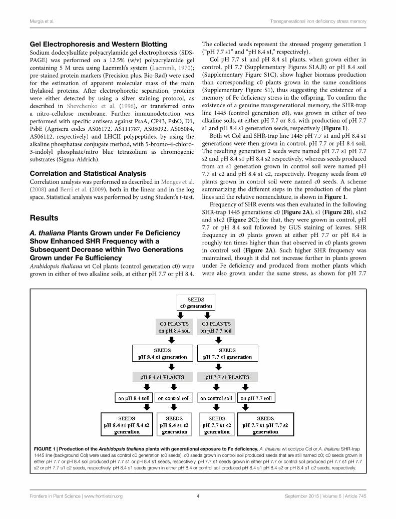
A. thaliana Plants Grown under Fe DeficiencyShow Enhanced SHR Frequency with aSubsequent Decrease within Two GenerationsGrown under Fe SufficiencyArabidopsis thaliana wt Col plants (control generation c0) weregrown in either of two alkaline soils, at either pH 7.7 or pH 8.4.
The collected seeds represent the stressed progeny generation 1(“pH 7.7 s1” and “pH 8.4 s1,” respectively).
Col pH 7.7 s1 and pH 8.4 s1 plants, when grown either incontrol, pH 7.7 (Supplementary Figures S1A,B) or pH 8.4 soil(Supplementary Figure S1C), show higher biomass productionthan corresponding c0 plants grown in the same conditions(Supplementary Figure S1), thus suggesting the existence of amemory of Fe deficiency stress in the offspring. To confirm theexistence of a genuine transgenerational memory, the SHR-trapline 1445 (control generation c0), was grown in either of twoalkaline soils, at either pH 7.7 or 8.4, with production of pH 7.7s1 and pH 8.4 s1 generation seeds, respectively (Figure 1).
Both wt Col and SHR-trap line 1445 pH 7.7 s1 and pH 8.4 s1generations were then grown in control, pH 7.7 or pH 8.4 soil.The resulting generation 2 seeds were named pH 7.7 s1 pH 7.7s2 and pH 8.4 s1 pH 8.4 s2 respectively, whereas seeds producedfrom an s1 generation grown in control soil were named pH7.7 s1 c2 and pH 8.4 s1 c2, respectively. Progeny seeds from c0plants grown in control soil were named c0 seeds. A schemesummarizing the different steps in the production of the plantlines and the relative nomenclature, is shown in Figure 1.
Frequency of SHR events was then evaluated in the followingSHR-trap 1445 generations: c0 (Figure 2A), s1 (Figure 2B), s1s2and s1c2 (Figure 2C); for that, they were grown in control, pH7.7 or pH 8.4 soil followed by GUS staining of leaves. SHRfrequency in c0 plants grown at either pH 7.7 or pH 8.4 isroughly ten times higher than that observed in c0 plants grownin control soil (Figure 2A). Such higher SHR frequency wasmaintained, though it did not increase further in plants grownunder Fe deficiency and produced from mother plants whichwere also grown under the same stress, as shown for pH 7.7
FIGURE 1 | Production of the Arabidopsis thaliana plants with generational exposure to Fe deficiency. A. thaliana wt ecotype Col or A. thaliana SHR-trap1445 line (background Col) were used as control c0 generation (c0 seeds). c0 seeds grown in control soil produced seeds that are still named c0; c0 seeds grown ineither pH 7.7 or pH 8.4 soil produced pH 7.7 s1 or pH 8.4 s1 seeds, respectively. pH 7.7 s1 seeds grown in either pH 7.7 or control soil produced pH 7.7 s1 pH 7.7s2 or pH 7.7 s1 c2 seeds, respectively. pH 8.4 s1 seeds grown in either pH 8.4 or control soil produced pH 8.4 s1 pH 8.4 s2 or pH 8.4 s1 c2 seeds, respectively.
Frontiers in Plant Science | www.frontiersin.org 4 September 2015 | Volume 6 | Article 745

Murgia et al. Transgenerational iron deficiency stress memory
FIGURE 2 | Somatic homologous recombination (SHR) in A. thalianaplants with generational exposure to Fe deficiency and grown underFe deficiency or sufficiency. (A) A. thaliana SHR-trap 1445 c0 plantsgrown in control, pH 7.7 or pH 8.4 soil. (B) s1 plants (pH 7.7 s1 and pH 8.4s1) grown in control, pH 7.7 or pH 8.4 soil. (C) s1s2 and s1c2 plants (pH 7.7s1 pH 7.7 s2, pH 8.4 s1 pH 8.4 s2, pH 7.7 s1 c2, pH 8.4 s1 c2) grown incontrol soil. For all experiments, plants were grown for 2–3 weeks and rosetteleaves were GUS-stained for detection of SHR events, counted as number ofindependent blue spots/mg fresh weight. Each value represents the meanspots number ± SE in five to twenty leaves. Significant differences (withrespect to control c0 value) are indicated with ∗∗(p < 0.01) or ∗(p < 0.05),according to Student’s t-test.
s1 grown in pH 7.7 soil and pH 8.4 s1 grown in pH 8.4 soil(Figure 2B). The lack of additive effect suggests a control of SHRfrequency, with a plateau effect. Notably, when either pH 7.7 s1or pH 8.4 s1 are grown in control soil, the SHR frequency wasdrastically reduced (Figure 2B) but was still higher than control,suggesting occurrence of a faint “stress memory.” Such memorywas, however, completely erased when plants were grown fortwo successive generations in control soil, as was evident in pH7.7 s1 c2 and pH 8.4 s1 c2 generations grown again in controlsoil (Figure 2C). It is also interesting to note that growth fortwo successive generations under Fe deficiency did not reinforce
FIGURE 3 | Arabidopsis thaliana seedlings with generational exposureto Fe deficiency and grown under Fe deficiency or sufficiency.(A) A. thaliana SHR-trap 1445 seedlings, from either control generation (c0) orfrom single (pH 7.7 s1) or multiple generational exposure to Fe deficiency (pH7.7 s1 pH 7.7 s2) germinated for 11 days in control AIS medium (+Fe), or AISmedium without Fe (−Fe). (B) Chlorophyll content, expressed as mgchlorophyll/g fresh weight of seedlings described in (A). (C) Weight (expressedas mg fresh weight/seedling) of seedlings described in (A). Bars representmean values ± SE, from at least three biological samples consisting of twentyseedlings each. Significant differences are indicated with letters (p < 0.05),according to Student’s t-test.
stressmemory in offspring unexposed to stress, as observed in pH7.7 s1 pH 7.7 s2 and pH 8.4 s1 pH 8.4 s2 lines, grown in controlsoil (Figure 2C).
Taken together, these results show that the number of SHRevents increased in plants grown under Fe deficiency, both inmild or in severe alkaline soil and that frequency of SHR eventsreturned to control values within two generations unexposed to
Frontiers in Plant Science | www.frontiersin.org 5 September 2015 | Volume 6 | Article 745

Murgia et al. Transgenerational iron deficiency stress memory
FIGURE 4 | Somatic homologous recombination events in A. thaliana seedlings with generational exposure to Fe deficiency and grown under Fedeficiency or sufficiency. (A) SHR events in A. thaliana SHR-trap 1445 seedlings, from control generation (c0), from single (pH 7.7 s1) or multiple generationalexposure to Fe deficiency (pH 7.7 s1 pH 7.7 s2) grown for 11 days in control AIS medium (+Fe), or in AIS medium without Fe (−Fe). Seedlings were stained for GUSexpression and SHR events evaluated as number of spots/seedling. Values are the mean spot number ± SE in ten seedlings. Significant differences (with respect tocontrol c0) are indicated with ∗ (p < 0.05), according to Student’s t-test. (B) Representative seedlings, described in (A), stained for detection of GUS activity.
stress. Moreover, growth of more than one generation in alkalinesoil was not associated with a higher frequency of SHR events.
To reduce any variability of results resulting from stuntedgrowth occurring in alkaline soil, SHR frequency was alsomeasured in c0, pH 7.7 s1 and pH 7.7 s1 pH 7.7 s2 seedlings,when germinated on control AIS medium (+Fe) or in AISmedium without iron supplement (−Fe) (Figure 3A). All testedseedlings growing under Fe deficiency were equally affected, interms of chlorophyll content (Figure 3B). Both pH 7.7 s1 andpH 7.7 s1 pH 7.7 s2 seedlings, grown in +Fe, showed insteada significantly higher chlorophyll content (but not significantlyhigher fresh weight), with respect to their control c0 seedlings inthe same condition (Figures 3B,C). As already observed for SHRfrequency (Figure 2), chlorophyll content and seedlings freshweight did not further increase upon exposure to Fe deficiencyfor multiple generations (Figures 3B,C). SHR frequency was
measured in seedlings treated as described in Figure 3; resultsconfirm that Fe deficiency strongly enhanced SHR frequency, asobserved for SHR-trap seedlings c0, pH 7.7 s1, pH 7.7 s1 pH 7.7s2 grown under Fe deficiency (Figure 4), thus confirming theobservations about plants grown on alkaline soil (Figure 2).
As also observed with plants grown in soil, SHR frequencywas not enhanced further in seedlings exposed for more thanone generation to Fe deficiency stress (Figure 4A), suggestingagain the plateau effect mentioned above. Vice-versa, unstressedoffspring seedlings of stress-treated plants, had an SHR frequencysimilar to c0, when grown in control soil (Figure 4A).
These results show that the number of SHR events increasedin seedlings grown under Fe deprivation and that the frequencyof SHR events returned to control values within one generationgrown under Fe sufficiency. Moreover, growth of more thanone generation under Fe deprivation was not associated with a
Frontiers in Plant Science | www.frontiersin.org 6 September 2015 | Volume 6 | Article 745

Murgia et al. Transgenerational iron deficiency stress memory
higher frequency of SHR events, thus confirming observationswith plants grown in alkaline soil.
DNA Damage Profiles and Expression of theTFIIS-like Gene are Enhanced in A. thalianaSeedlings Grown under Fe Deficiency butNeither in s1 nor in s1s2 Generations Grownunder Fe SufficiencyGenotoxic stress in plants is generally regarded as a mainfactor which affects genome stability, impairing productivity(Balestrazzi et al., 2011; Waterworth et al., 2011). Since thegenotoxic impact of Fe deficiency is still unexplored in plants, aninvestigation was carried out to assess the DNA damage profilesoccurring under Fe deficiency, using the Alkaline SCGE (singlecell gel electrophoresis, or Comet assay), which measures bothSSBs and DSBs.
DNA lesions were quantified in nuclei isolated from SHR-trap 1445 c0, pH 7.7 s1, pH 7.7 s1 pH 7.7 s2 seedlings,when germinated in control (+Fe) or in −Fe medium. Controlc0 seedlings grown in +Fe showed a DNA damage value of130.6 ± 41.7 a.u., which can be attributed to the experimentalmanipulations of nuclei; such a value is similar to those quantifiedin both pH 7.7 s1 seedlings (127.4 ± 17.5 a.u.) and pH 7.7 s1 pH7.7 s2 seedlings (123.6 ± 33.2 a.u.), when in +Fe (Figure 5A).Vice-versa, as already observed for the SHR events, the extentof DNA damage increased in c0 seedlings (208.3 ± 25.3 a.u.),in pH 7.7 s1 seedlings (198.8 ± 28.4 a.u.) and in pH 7.7 s1pH7.7 s2 seedlings (191.0 ± 22.1 a.u.), under Fe deficiency(−Fe; Figure 5A). Moreover, in accordance with the SHR events,growth for more than one generation under Fe deficiency did notfurther increase DNA damage, as such values measured in −FepH 7.7 s1 and −Fe pH 7.7 s1 pH 7.7 s2 seedlings were similar(Figure 5A).
These results show that DNA damage increased in seedlingsgrown under Fe deprivation and that it returned to control valueswithin one generation grown under Fe sufficiency. Moreover,growth of more than one generation under Fe deprivation wasnot associated with a more pronounced DNA damage, as alsoobserved for SHR events.
Mechanisms involved in genome protection includenucleotide excision repair (NER; Fousteri and Mullenders,2008), which operates through the TC (transcript-coupled)sub-pathway to remove DNA lesions and block RNA polymeraseII and restore transcription. One of the components of TC-NERpathway is the transcription elongation factor TFIIS, whichassociates with RNA polymerase II, possibly enabling repairunder UV-C exposure (Lagerwerf et al., 2011).
Plants possess the TFIIS gene (At2g38560/RDO2) which isinvolved in the regulation of seed dormancy and development,as described for Arabidopsis by Grasser et al. (2009) andMortensen and Grasser (2014). Also, TFIIS-like genes have beenidentified and characterized in Medicago truncatula (Macoveiet al., 2011) and rice (Oryza sativa L.) (Macovei et al., 2014);such genes encode proteins sharing some common featureswith the canonical TFIIS proteins. Both genes are responsiveto genotoxic stress induced by x-ray and salinity stress in
seedlings and mature plants (Macovei et al., 2014). A genewith 60% similarity to MtTFIIS-like, according to Phytozome(v10.23, Goodstein et al., 2012), is present in the A. thalianagenome, namely At5g09850 gene, which we therefore namedAtTFIIS-like. Alignment of protein sequences of AtTFIIS-likeand MtTFIIS-like (Medtr3g095380, according to Phytozome) isshown in Supplementary Figure S2 where the TFIIS domainis highlighted. Both TFIIS-like proteins share the LW motif,required to accomplish the nuclear import of proteins (Ling et al.,2006).
The expression profiles of bothAtTFIIS andAtTFIIS-like geneswere studied by qRT-PCR in SHR-trap 1445 c0, pH 7.7 s1, pH7.7s1 pH 7.7 s2 seedlings germinated in control (+Fe) or in −Fe. Asshown in Figure 5B, TFIIS expression was not dependent eitheron the Fe nutritional status, or on the stress pedigree of the testedlines, being similar in all tested seedlings, when grown in +Fe aswell as in −Fe (Figure 5B). On the other hand, the expression ofthe TFIIS-like gene significantly increased in all seedlings (c0, pH7.7 s1, pH 7.7 s1 pH 7.7 s2) grown in −Fe, compared with whatwas observed in +Fe (Figure 5B).
These results show that expression of the AtTFIIS-like geneincreased in seedlings grown under Fe deprivation and thatsuch expression returned to control values within one generationgrown under Fe sufficiency. Moreover, growth of more thanone generation under Fe deprivation was not associated with anincreased expression of the AtTFIIS-like gene.
Correlation analysis of gene expression has been successfullyapplied for identifying new candidate genes for a given biologicalprocess (Beekwilder et al., 2008; Murgia et al., 2011). Suchanalysis, was performed on At2g38560 (TFIIS/RDO2), At5g09850(TFIIS-like), At1g01210, At2g13640, At2g27780, At3g25940,At3g48060, At4g07950, At4g18720, At4g24200, At5g05140,At5g27310 and At5g51360, i.e., all the A. thaliana genes, whichare annotated as TFIIS or similar, according to the ArabidopsisInformation resource (TAIR4), and detected by the ATH1Affymetrix microarray gene chip. However, the analysis did notdetect any strong positive correlation between any of the testedgenes and the genes involved in the Fe-deficiency response (e.g.,FRO1, FIT1, IRT1, Popeye, etc.) with a threshold for the Pearson’scoefficient set to 0.6 (data not shown).
AtTFIIS-like has been identified as one of the putative proteinsforming subunit 26 of the Mediator complex, and namedAtMed26_3 (Mathur et al., 2011); interestingly, Mediator subunit16 gene (At4g04920), which is also part of the Mediator complexrepresenting a bridge between RNA polymerase and specifictranscriptional activators, functions in the regulation of ironhomeostasis (Yang et al., 2014; Zhang et al., 2014). Correlationanalysis of MED16 did not reveal a strong correlation with anyof the Fe deficiency response genes whose expression is MED16-dependent and reported in Zhang et al. (2014) (data not shown).Results of such correlation analyses, together with the evidencethat MED16 exerts a profound impact on the Fe deficiencyresponse without changing its expression under Fe deprivation(Zhang et al., 2014), suggests that the alteration of TFIIS-like gene
3http://phytozome.jgi.doe.gov/pz/portal.html4https://www.arabidopsis.org/
Frontiers in Plant Science | www.frontiersin.org 7 September 2015 | Volume 6 | Article 745

Murgia et al. Transgenerational iron deficiency stress memory
FIGURE 5 | DNA damage and expression of AtTFIIS and AtTFIIS-like genes, in A. thaliana seedlings with generational exposure to Fe deficiency andgrown in control AIS medium (+Fe) or AIS medium without Fe (−Fe). (A) Alkaline Comet assay on 14 days-old SHR-trap 1445 c0, pH 7.7 s1, pH 7.7 s1 pH7.7 s2 seedlings, when germinated in control (+Fe) or in −Fe AIS medium. One hundred cells were scored for each sample. Values are expressed as mean ± SD ofthree replicates from two independent experiments. (B) Expression profiles of AtTFIIS (in blue) and AtTFIIS-like genes (in red) in the samples described in (A), byqRT-PCR analysis. Values are the result of three independent experiments and have been normalized to the expression value in control c0 grown in +Fe. Statisticallysignificant differences (with respect to control c0) are indicated with ∗ (p < 0.05), according to Student’s t-test.
expression, under Fe deficiency, even if not striking, may be ofrelevance for the Fe deficiency response.
Photosynthetic Parameters in s1, s1s2, ands1c2 Generations: Oxygen Evolution,Chlorophyll Content, and Protein Compositionof the Photosynthetic ApparatusO2 evolution was higher in pH 7.7 s1 pH 7.7 s2 SHR-trapline seedlings than in control c0, when both were grown in+Fe, thus suggesting that the photosynthetic apparatus is moreefficient in seedlings whose parental plants experienced Fedeficiency (Figure 6A), in agreement with what was observed for
chlorophyll content. Interestingly, O2 evolution values were notsignificantly higher in pH 7.7 s1 c2 than in control c0 (Figure 6A),indicating a loss of this trait in the offspring unexposed to stress.When seedlings are illuminated with light intensities well abovegrowth light, i.e., at 800 μE m−2 s−1 (Figure 6A), the higherO2 evolution in pH 7.7 s1 pH 7.7 s2 is no longer statisticallysignificant (Figure 6A).
These results show that O2 evolution was higher in seedlingsgrown in control conditions and with parental plants which hadexperienced Fe deficiency, in agreement with what was observedfor chlorophyll content; however, this trait was lost within twogenerations unexposed to stress.
Frontiers in Plant Science | www.frontiersin.org 8 September 2015 | Volume 6 | Article 745

Murgia et al. Transgenerational iron deficiency stress memory
FIGURE 6 | O2 evolution and protein profile of photosynthetic apparatus in A. thaliana seedlings with generational exposure to Fe deficiency.(A) SHR-trap 1445 c0, pH 7.7 s1 pH 7.7 s2, pH 7.7 s1 c2 seedlings were grown for 11 days in control AIS medium and net O2 evolution (expressed as μmol O2
evolved min−1 mg chlorophyll−1) was measured under illumination at either 100 or 800 μE m−2 s−1. Values are mean ± SE of three biological replicas, eachconsisting of at least 15 seedlings. Letters represent statistical differences, according to Student’s t-test, with p < 0.05. (B) Western blot analysis with antibodiesagainst PsaA, CP43, PsbO, D1, LHCII, and PsbE of thylakoid membranes purified from 11 days-old SHR-trap 1445 c0 seedlings germinated in −Fe AIS medium orfrom 11 days-old SHR-trap 1445 c0, pH 7.7 s1, pH 7.7 s1 pH 7.7 s2, pH 7.7 s1 c2 seedlings germinated in control (+Fe) AIS medium. Samples corresponding to1 μg chlorophyll were loaded on each lane.
As a result of Fe deficiency, changes in the protein profilesof different plant parts and compartments, among which are thethylakoid membranes, have been reported (Andaluz et al., 2006;López-Millán et al., 2013).Within the thylakoid membranes, bothphotosystem I (PSI) and photosystem II (PSII) are affected by Fedeficiency; however, it can be expected that there will be a moresevere effect on PSI than on PSII, due to its higher number of Featoms per photosystem: 14 Fe atoms/PSI versus 2 Fe atoms/PSII(Yadavalli et al., 2012; Briat et al., 2015).
To further explore the impact of Fe deficiency stress andthe existence of the memory of such stress on the proteincomposition of the photosynthetic apparatus, the reaction centersubunits of PSI, PsaA, and of PSII, D1, together with other PSIIpolypeptides, among which the inner antenna protein CP43,the outer antenna protein LHCII, the extrinsic polypeptide ofthe Oxygen Evolving Complex PsbO and the PsbE protein (asubunit of cytochrome b559 binding an heme cofactor), wereanalyzed by SDS-PAGE and western blotting, in c0 seedlingsgerminated in −Fe, as well as in c0, pH 7.7 s1, pH 7.7 s1pH 7.7 s2, pH 7.7 s1 c2 seedlings when germinated in +Fe(Supplementary Figure S3 and Figure 6B). Upon equal loadingof chlorophyll in the protein gels for all analyzed samples,the amount of PsaA, as well as that of CP43 and D1, wasdramatically reduced in −Fe c0 seedlings, when compared with+Fe c0 seedlings (Figure 6B). Such results are in agreementwith proteomic profiles observed in thylakoids isolated from Fe-deficient Beta vulgaris leaves (Andaluz et al., 2006). However,it should be noted that such a decrease in PsaA has not beenobserved in Fe-deficient rice seedlings (Yadavalli et al., 2012).Fe deficiency dramatically affects the level of cytochrome b559 in−Fe c0 seedlings, indeed its a subunit, PsbE, could not be detectedin −Fe c0 seedlings (Figure 6B). Notably, under Fe deficiency,
no decrease was observed in the amount of the PsbO subunitin any of the tested samples, thus suggesting that, at least inA. thaliana, Fe deficiency does not equally affect all the proteincomponents of the two photosystems. Lastly and converselyto the behavior of the reaction center proteins of PSI (PsaA)and PSII (D1), and the inner antenna protein of PSII (CP43),the amount of the peripheral antenna proteins LHCII was notreduced in −Fe c0 seedlings when compared to any other +Fesamples.
A lack of substantial alterations in the protein profile ofphotosynthetic proteins in any of the generations grown in +Fe,i.e., pH 7.7 s1, pH 7.7 s1 pH 7.7 s2 and in pH 7.7 s1 c2, whencompared to +Fe c0 seedlings (Figure 6B), could be observed.
To further investigate chlorophyll as a hallmark of growthof the mother plant under Fe deficiency, the various c0, s1,s1s2, s1c2 generations of wt Col, described in Figure 1, werealso tested by growing seedlings in control (+Fe) or −Femedium. As already observed for SHR-trap line (Figure 3B),chlorophyll content was higher in pH 7.7 s1, pH 7.7 s1 pH 7.7s2, pH 8.4 s1 and pH 8.4 s1 pH 8.4 s2 Col seedlings, whencompared to c0 Col seedlings in same condition (SupplementaryFigure S4). Interestingly, chlorophyll concentration was higherin pH 7.7 s1 c2 and pH 8.4 s1 c2 offspring grown in controlconditions than c0 itself (Supplementary Figure S4), indicatingthat this trait can be maintained for more than one offspringgeneration unexposed to stress. Also, the plateau effect observedfor SHR events (Figures 2 and 4), DNA damage (Figure 5A),expression of TFIIS-like (Figure 5B) is confirmed once more,since chlorophyll content was statistically similar in pH 7.7 s1and pH 7.7 s1 pH 7.7 s2 seedlings, as well as in pH 8.4 s1 andpH 8.4 s1 pH 8.4 s2 seedlings, when grown in control conditions(Supplementary Figure S4).
Frontiers in Plant Science | www.frontiersin.org 9 September 2015 | Volume 6 | Article 745

Murgia et al. Transgenerational iron deficiency stress memory
FIGURE 7 | Germination of A. thaliana seeds, produced from plants with transgenerational exposure to Fe deficiency. (A) Germination of c0 or pH 8.4 s1Col seeds, after-ripened at room temperature for 3 months, or preserved at −20◦C for 26–30 months. (B) Germination of c0, pH 7.7 s1, pH 7.7 s1 pH 7.7 s2, pH7.7 s1 c2 Col seeds, after-ripened for 26–30 months at room temperature. As controls, c0 14.2 tAPX OE, 8.5 tAPX OE seeds after ripened for 26–30 months atroom temperature as well as c0 Col seeds after-ripened for 6 years at room temperature were also used. Values correspond to mean percent germination ± SE fromat least three plates containing 100 seeds each.
The various wt Col generations described above were alsogerminated in −Fe; as expected (see also Figure 3B) areduction in chlorophyll content was observed in −Fe seedlingswhen compared to control ones (Supplementary Figure S4).The tested s1, s1s2, and s1c2 seedlings showed chlorophyllcontent of variable intensity, with respect to c0, when in −Fe(Supplementary Figure S4). Such an effect was not observed inthe SHR-trap line (Figure 3), where all tested seedlings, when in−Fe, showed the same chlorophyll content, regardless of stresspedigree.
Seed Longevity Increases in s1s2 Seeds butnot in s1c2 OnesSeed longevity can be severely affected due to oxidativedamage occurring during prolonged storage and/or exposure tounfavorable environmental conditions (Donà et al., 2013). As aconsequence of aging, seed viability is compromised (Kranneret al., 2010). In order to assess the effects of Fe deficiencyon seed quality, germination of after-ripened (stored at room
temperature) or of dormancy-preserved (stored at −20◦C) Colc0, s1, s1s2, s1c2 generations was assessed; for that, seed batchesof various ages were used (3 months, 26–30 months, and 6 years-old seeds). As positive controls, after-ripened 26–30 months-oldc0 seeds of the transgenic A. thaliana lines tAPX OE 14.2 andtAPX OE 8.5 overexpressing thylakoidal ascorbate peroxidase,which show an enhanced resistance to oxidative stress (Murgiaet al., 2004) were also used; these lines allowed to investigate plantresponses to various abiotic and biotic oxidative stresses (Murgiaet al., 2004; Yao and Greenberg, 2006; Laloi et al., 2007; Skiryczet al., 2011). Nomenclature of all the tested seeds that keeps trackof stress history of the mother plants is according to Figure 1.
Germination curves at 25◦C in the light were assessed over a6-days time span, as a proxy for seed longevity; for that, seedswere first cold-stratified (4 days at 4◦C in the dark) to break anyresidual dormancy and to promote synchronous germination.
As expected, young (3 months-old) after-ripened c0 or pH8.4 s1 Col seeds all germinated within 2 days; full germinationwas also observed in older (26–30 months-old) c0 seeds, if stored
Frontiers in Plant Science | www.frontiersin.org 10 September 2015 | Volume 6 | Article 745

Murgia et al. Transgenerational iron deficiency stress memory
at −20◦C (Figure 7A). Next, germination of c0, pH 7.7 s1, pH7.7 s1 pH 7.7 s2, pH 7.7 s1 c2 after-ripened 26–30 months Colseeds was measured. Germination of c0 Col seeds was below 50%(Figure 7B) and therefore much lower than that of same-ageseeds stored at−20◦C (Figure 7A). Germination of pH 7.7 s1 pH7.7 s2 Col seeds was much higher, being above 80% (Figure 7B),whereas germination curves similar to control were observed inpH 7.7 s1 or pH 7.7 s1 c2 Col seeds (Figure 7B). Unfortunately,lack of 26–30 months after-ripened pH 7.7 s1 pH 7.7 s2 c3 Colseeds prevented us from further exploring longevity traits. Lastly,26–30 months after-ripened c0 14.2 and 8.5 tAPX OE seeds (usedas positive controls) showed 90–100% germination (Figure 7B),as high as that observed in young (3months-old) after-ripened wtCol seeds, whereas fairly old c0 Col seeds (6 years after ripening)did not germinate (Figure 7B).
These results showed that growth of more than one generationunder Fe deficiency has a positive impact on the longevity of seedsproduced.
Discussion
The analysis of stress-induced plant priming and relatedtransgenerational stress memory have been intensively debateddue to their great potential for crop improvement (Hauseret al., 2011; Vriet et al., 2015). Transgenerational memorycould be of advantage for subsequent generations exposed tonutritional stress: indeed, in the open, some environmentalabiotic stresses such as cold, drought, etc. might not even occurduring offspring growth, due to fluctuations of environmentalconditions; even pathogen attacks might be limited in time. Insuch cases, the advantage of “erasing” any memory of stressthat means all the burden of epigenetic modification, mightoutbalance the advantage of maintaining the memory of thatstress in the progeny. Vice-versa, nutritional deficiencies dueto soil composition are chronic stresses that hardly changewith time and therefore, in such a case, the maintenance ofthe burden of epigenetic modifications induced by nutritionalstresses might be of advantage for plants whose seeds often fall inclose proximity to the mother plant. Despite the increase in recentyears of reports on transgenerational stress memory, includingthe effects of nutrients limitations (Kou et al., 2011; Verhoevenand van Gurp, 2012), up to now, the stable inheritance of Fedeficiency stress memory in offspring generations has not beenanalyzed.
We investigated, for the first time, whether a genuinetransgenerational memory of Fe deficiency stress occurs in themodel plant A. thaliana; we demonstrated that Fe deficiencyalters DNA damage and repair and we analyzed such traitsin unstressed offspring (Vriet et al., 2015); moreover, we alsoanalyzed physiological effects of Fe deficiency occurring inunstressed offspring (Verhoeven and van Gurp, 2012), such asthe status of the photosynthetic apparatus (chlorophyll content,O2 evolution, protein profile) and also seed longevity; all thesetraits are indeed agronomically and economically relevant.
Most importantly, we analyzed such traits not only in thefirst generation but also in the second one, with and without
stress, to distinguish genuine transgenerational memory fromother possible mechanisms, such as transmission, from motherto progeny plant, of enzymes, toxins, hormones, etc. which canbe observed in progeny but do not represent genuine memory(Paszkowski and Grossniklaus, 2011; Verhoeven and van Gurp,2012).
We showed that frequency of DNA damage and repair,evaluated through events of SHR and DNA strand breaks,increased in plants grown under various conditions of Fedeficiency (mild or severe alkaline soil, depletion of Fe frommedium); however, their frequencies returned to control valueswithin one or two offspring generations, as was also observedfor SHR induced by various stresses and described in Pecinkaet al. (2009). It was interesting that SHR frequency, in s1 ors2 generations unexposed to stress, immediately declined tocontrol levels in experiments conducted in controlled medium,whereas it took one more generation in experiments conductedin soil; such lasting “stress memory” in soil, at least for oneunstressed generation, but not in medium, suggests that growthin alkaline soil might trigger a more complex response tostress than the Fe deficiency response obtained by simple Fedeprivation. Nevertheless, the need to obtain sufficient amountsof seeds from plants exposed to Fe deficiency stress andwith different stress pedigrees, together with the fact that soilalkalinisation is indeed the most common cause of Fe deficiencyencountered by plants in the open, prevented us from choosingto carry out in vitro experiments only. Anyway, the dataobtained from mild or severe alkaline pH (pH 7.7 or pH 8.4)confirmed the consistency of results obtained with soil-grownplants.
We also analyzed some photosynthetic parameters:chlorophyll content, O2 evolution, protein composition ofphotosynthetic apparatus. Chlorophyll content was still alteredin two offspring generations unexposed to stress; however,erasure of such alteration in further generations might besupposed. Indeed, results of increased chlorophyll content, inoffspring of plants grown under Fe deficiency and described inthe present work, were occasionally not reproducible, as alreadyreported for other transgenerational phenotypic effects: as anexample, apomictic dandelion (Taraxacum officinale, commondandelion) grown under nutrients limitation, shows increasedroot:shoot ratio in offspring produced from plants grownunder the same nutritional stress compared with the offspringfrom plants grown under control conditions: however, such aphenotype was variable and could not be always reproducedwithin the same lab and from the same authors (Verhoevenand van Gurp, 2012). As already pointed out by these authors,such lack of full consistency among various experiments, asfar as chlorophyll content is concerned, could be due to subtledifferences among experimental conditions which might overrideparental effects (Verhoeven and van Gurp, 2012).
Moreover, pH 7.7 s1 c2 seedlings, when germinated in controlconditions, evolved less oxygen than pH 7.7 s1 pH 7.7 s2 seedlingsin the same conditions. Provided that the chlorophyll contentcould be further tested in the offspring of the various generationsalready described in the present work, all the photosyntheticparameters measured so far do not support the existence of a
Frontiers in Plant Science | www.frontiersin.org 11 September 2015 | Volume 6 | Article 745

Murgia et al. Transgenerational iron deficiency stress memory
genuine and reproducible transgenerational transmission ofmemory. The same observations apply to the increased weight,not always observed in s1 progeny grown in control conditions,as reported in the present work; also in such a case, subtledifferences among experimental conditions which might overrideparental effects.
Arabidopsis thaliana AtTFIIS-like is one of the putativeproteins of subunit 26 of the Mediator complex, i.e., MED26_3(Mathur et al., 2011); although its expression is only slightlyinduced by Fe deficiency, nevertheless the expression of MED16,another subunit of Mediator which is undoubtedly involvedin the regulation of the Fe deficiency response (Yang et al.,2014; Zhang et al., 2014), does not change at all under Fedeficiency, making the observed change in AtTFIIS-like geneexpression, profoundly relevant. The reported data, highlightingthe involvement of the TFIIS-like gene in the plant responseto Fe-deficiency stress, provide for the first time evidence ofthe role played in this context by the TC-NER sub-pathway.The latter is critical for cell survival due to its anti-mutagenicproperties, which protect DNA against the long-term effectsof genotoxic stress (Fousteri and Mullenders, 2008). TC-NER, which specifically acts on the transcribed regions of thegenome, thus preserving gene expression, has been previouslydemonstrated to participate in the response to gamma irradiation(Macovei et al., 2014), as well as heavy metals and osmoticstresses (Macovei et al., 2011). The present work contributes tofurther expand the range of stresses that trigger TC-NER, but alsosignificantly contributes to the current scientific discussions onthe putative link between transgenerational memory and DNArepair response in plants.
Longevity of seeds represents a research focus of severalgroups, due to the tremendous ecological, economic andnutritional impact that such physiological processes have(Devaiah et al., 2007) and indeed one of the most importantquestions facing plant science research is what determines seedlongevity and dormancy (Grierson et al., 2011). The observedphenotype of increased longevity in offspring seeds of Fedeficiency stressed mother plants is new in the nutritional fieldand it also encourages further investigation in order to clarifythe link between seed longevity, Fe content, its localization andmobilization as well as levels of different ROS species, in seedsproduced from mother plants grown under Fe deficiency.
The next step in the elucidation of the Fe-deficiency associatedtraits described in the present work will be to understand ifand how their molecular regulation is associated with epigeneticmodifications.
Conclusion
We described for the first time profiles of SHR events andof DNA damage, occurring under Fe deficiency in A. thalianaand we also identified a new Fe-deficiency responsive gene, i.e.,AtTFIIS-like. We then analyzed SHR, DNA damage, AtTFIIS-like gene expression, as well as a set of physiological parametersin a multiple-generation cohort of plants with different stresspedigrees. The data obtained suggest the existence of multiple-step control of mechanisms orchestrating the prevention ofa genuine and stable transgenerational transmission of Fedeficiency stress memory, with the tightest control on DNAintegrity. The production of a wider cohort of s3 (andsuccessive) plant generations and the analysis of traits suchas chlorophyll content, O2 evolution and in particular seedlongevity in such extended cohort, when grown either inpresence or absence of stress, will allow to further test thishypothesis.
Author Contributions
IM conceived the work and wrote themanuscript; IM, SG, AB, SP,CP, and PM contributed the data; IM, SG, and PM contributed tothe analysis and the discussion of data.
Acknowledgments
We thank Prof. Holger Putchta and Dr. Alexander Knollfor providing the A. thaliana SHR-trap 1445 line and Prof.Roberto Barbato for providing antibodies against LHCII. SPwas financed by a Research Fellowship award from RegioneLombardia D.G. Istruzione, Formazione e Lavoro, StrutturaAsse V-Interregionalità e Transnazionalità POR FSE 2007–2013, Project ID 46547514 ‘Advanced Priming Techniques forthe Lombardy Agro-Seed Industry-PRIMTECH’ (Action 2).CP was financed by the Italian Ministry of Education,University and Research, “Futuro in Ricerca 2013” programRBFR1334SB.
Supplementary Material
The Supplementary Material for this article can be found onlineat: http://journal.frontiersin.org/article/10.3389/fpls.2015.00745
References
Andaluz, S., Lopez-Millan, A. F., De las Rivas, J., Aro, E. M., Abadia, J., andAbadia, A. (2006). Proteomic profiles of thylakoid membranes and changes inresponse to iron deficiency. Photosynth. Res. 89, 141–155. doi: 10.1007/s11120-006-9092-6
Aranega-Bou, P., de la O Leyva, M., Finiti, I., Garcia-Augustin, P., andGonzalez-Bosch, C. (2014). Priming of plant resistance by natural compounds.Hexanoic acid as a model. Front. Plant Sci. 5:488. doi: 10.3389/fpls.2014.00488
Arnon, D. J. (1949). Copper enzymes in isolated chloroplasts.Polyphenoloxidase in Beta vulgaris. Plant Physiol. 24, 1–14. doi: 10.1104/pp.24.1.1
Balestrazzi, A., Confalonieri, M., Macovei, A., Donà, M., and Carbonera, D. (2011).Genotoxic stress and DNA repair in plants: emerging functions and tools forimproving crop productivity. Plant Cell Rep. 30, 287–295. doi: 10.1007/s00299-010-0975-9
Balmer, A., Pastor, V., Gamir, J., Flors, V., and Mauch-Mani, B. (2015). The ‘prime-ome’: towards a holistic approach to priming.Trends Plant Sci. 20, 443–452. doi:10.1016/j.tplants.2015.04.002
Frontiers in Plant Science | www.frontiersin.org 12 September 2015 | Volume 6 | Article 745

Murgia et al. Transgenerational iron deficiency stress memory
Beekwilder, J., van Leeuwen, W., van Dam, N. M., Bertossi, M., Grandi, V.,Mizzi, L., et al. (2008). The impact of the absence of aliphaticglucosinolates on insect herbivory in Arabidopsis. PLoS ONE 3:e2068.doi: 10.1371/journal.pone.0002068
Berri, S., Abbruscato, P., Faivre-Rampant, O., Brasileiro, A. C., Fumasoni, I.,Satoh, K., et al. (2009). Characterization of WRKY co-regulatory networksin rice and Arabidopsis. BMC Plant Biol. 9:120. doi: 10.1186/1471-2229-9-120
Bilichak, A., Ilnytskyy, Y., Woycicki, R., Kepeshchuk, N., Fogen, D., andKovalchuk, I. (2015). The elucidation of stress memory inheritance in Brassicarapa plants. Front. Plant Sci. 6:5. doi: 10.3389/fpls.2015.00005
Boyko, A., Blevins, T., Yao, Y., Golubov, A., Bilichak, A., Ilnytskyy, Y., et al.(2010). Transgenerational adaptation of Arabidopsis to stress requires DNAmethylation and the function of DICER-like proteins. PLoS ONE 5:e9514. doi:10.1371/journal.pone.0009514
Briat, J. F., Dubos, C., and Gaymard, F. (2015). Iron nutrition, biomassproduction, and plant product quality. Trends Plant Sci. 20, 33–40. doi:10.1016/j.tplants.2014.07.005
Collins, A. R. (2004). The comet assay for DNA damage and repair.Mol. Biotechnol.26, 249–261. doi: 10.1385/MB:26:3:249
Conrath, U., Beckers, G. J. M., Flors, V., García-Agustín, P., Jakab, G., Mauch, F.,et al. (2006). Priming: getting ready for battle. Mol. Plant Microbe Interact. 19,1062–1071. doi: 10.1094/MPMI-19-1062
Crevillen, P., Yang, H., Cui, X., Greeff, C., Trick, M., Qiu, Q., et al. (2014).Epigenetic reprogramming that prevents transgenerational inheritance of thevernalized state. Nature 515, 587–590. doi: 10.1038/nature13722
Devaiah, S. P., Pan, X., Hong, Y., Roth, M., Welti, R., and Xuemin Wang, X.(2007). Enhancing seed quality and viability by suppressing phospholipase Din Arabidopsis. Plant J. 50, 950–957. doi: 10.1111/j.1365-313X.2007.03103.x
Donà, M., Balestrazzi, A., Mondoni, A., Rossi, G., Ventura, L., Buttafava, A.,et al. (2013). DNA profiling, telomere analysis and antioxidant propertiesas tools for monitoring ex situ seed longevity. Ann. Bot. 111, 987–998. doi:10.1093/aob/mct058
Fousteri, M., and Mullenders, L. (2008). Transcription-coupled repair nucleotideexcision repair in mammalian cells: biological mechanisms and moleculareffects. Cell Res. 18, 73–84. doi: 10.1038/cr.2008.6
Fu, Z. Q., and Dong, Z. (2013). Systemic acquired resistance: turning local infectioninto global defence. Annu. Rev. Plant Biol. 64, 839–863. doi: 10.1146/annurev-arplant-042811-105606
Goodstein, D., Shu, S., Howson, R., Neupane, R., Hayes, R. D., Fazo, J., et al. (2012).Phytozome, a comparative platform for green plant genomics. Nucleic AcidsRes. 40, D1178–D1186. doi: 10.1093/nar/gkr944
Grasser, M., Kane, C. M., Merkle, T., Melzer, M., Emmersen, J., and Grasser,K. D. (2009). Transcript elongation factor TFIIS is involved in Arabidopsis seeddormancy. J. Mol. Biol. 386, 598–611. doi: 10.1016/j.jmb.2008.12.066
Grierson, C. S., Barnes, S. R., Chase, M.W., Clarke, M., Grierson, D., and Edwards,K. J. et al. (2011). One hundred important questions facing plant scienceresearch. New Phytol. 192, 6–12. doi: 10.1111/j.1469-8137.2011.03859.x
Hauser, M. T., Aufsatz, W., Jonak, C., and Luschnig, C. (2011). Transgenerationalepigenetic inheritance in plants. Biochim. Biophys. Acta 1809, 459–468. doi:10.1016/j.bbagrm.2011.03.007
Iwasaki, M., and Paszkowski, J. (2014). Identification of genes preventingtransgenerational transmission of stress-induced epigenetic states. Proc. Natl.Acad. Sci. U.S.A. 111, 8547–8552. doi: 10.1073/pnas.1402275111
Jaskiewicz, M., Conrath, U., and Peterhänsel, C. (2011). Chromatin modificationacts as a memory for systemic acquired resistance in the plant stress response.EMBO Rep. 12, 50–55. doi: 10.1038/embor.2010.186
Jeong, J., and Guerinot, M. L. (2009). Homing in on iron homeostasis in plants.Trends Plant Sci. 14, 280–285. doi: 10.1016/j.tplants.2009.02.006
Kieu, N. P., Aznar, A., Segond, D., Rigault, M., Simond-Cote, E., Kunz, C., et al.(2012). Iron deficiency affects plant defence responses and confers resistanceto Dickeya dadantii and Botrytis cinerea. Mol. Plant Pathol. 13, 816–827. doi:10.1111/j.1364-3703.2012.00790.x
Kobayashi, T., and Nishizawa, N. K. (2012). Iron uptake, translocation andregulation in higher plants. Annu. Rev. Plant Biol. 63, 131–152. doi:10.1146/annurev-arplant-042811-105522
Kou, H. P., Li, Y., Song, X. X., Ou, X. F., Xing, S. C., Ma, J., et al. (2011).Heritable alteration in DNA methylation induced by nitrogen-deficiency stress
accompanies enhanced tolerance by progenies to the stress in rice. J. PlantPhysiol. 68, 1685–1693. doi: 10.1016/j.jplph.2011.03.017
Kranner, I., Minibayeva, F. V., Beckett, R. P., and Seal, C. E. (2010). What isstress? Concepts, definitions and applications in seed science. New Phytol. 188,655–673. doi: 10.1111/j.1469-8137.2010.03461.x
Laemmli, U. K. (1970). Cleavage of structural proteins during the assembly of thehead of bacteriophage T4. Nature 227, 680–685. doi: 10.1038/227680a0
Lagerwerf, S., Vrouwe, M. G., Overmeer, R. M., Fousteri, M. I., and Mullenders,L. H. (2011). DNA damage response and transcription. DNA Repair 10, 743–750. doi: 10.1016/j.dnarep.2011.04.024
Laloi, C., Stachowiak, M., Pers-Kamczyc, E., Warzych, E., Murgia, I., and Apel, K.(2007). Cross-talk between singlet oxygen- and hydrogen peroxide-dependentsignaling of stress response in Arabidopsis thaliana. Proc. Natl. Acad. Sci. U.S.A.104, 672–677. doi: 10.1073/pnas.0609063103
Lee, K., and Seo, P. (2014). Airborne signals from salt-stressed Arabidopsis plantstrigger salinity tolerance in neighboring plants. Plant Signal. Behav. 9:e28392.doi: 10.4161/psb.28392
Ling, Y., Smith, A. J., and Morgan, G. T. (2006). A sequence motif conservedin diverse nuclear proteins identifies a protein interaction domain utilisedfor nuclear targeting by human TFIIS. Nucleic Acids Res. 34, 2219–2229. doi:10.1093/nar/gkl239
López-Millán, A. F., Grusak, M. A., Abadía, A., and Abadía, J. (2013). Irondeficiency in plants: an insight from proteomic approaches. Front. Plant Sci.4:254. doi: 10.3389/fpls.2013.00254
Luna, E., Bruce, T. J. A., Roberts, M. R., Flors, V., and Ton, J. (2012). Next-generation systemic acquired resistance. Plant Physiol. 58, 844–853. doi:10.1104/pp.111.187468
Macovei, A., Balestrazzi, A., Confalonieri, M., Buttafava, A., and Carbonera, D.(2011). The TFIIS and TFIIS-like genes from Medicago truncatula are involvedin oxidative stress response. Gene 470, 20–30. doi: 10.1016/j.gene.2010.09.004
Macovei, A., Garg, B., Raikwar, S., Balestrazzi, A., Carbonera, D., Buttafava, A.,et al. (2014). Synergistic exposure of rice seeds to different doses of γ-Ray andsalinity stress resulted in increased antioxidant enzyme activities and gene-specificmodulation of TC-NER pathway. Biomed. Res. Intern. 2014:676934. doi:10.1155/2014/676934
Mathur, S., Vyas, S., Kapoor, S., and Tyagi, A. K. (2011). The Mediator complexin plants: structure, phylogeny, and expression profiling of representative genesin a dicot (Arabidopsis) and a monocot (rice) during reproduction and abioticstress. Plant Physiol. 157, 1609–1627. doi: 10.1104/pp.111.188300
Menges, M., Dóczi, R., Ökrész, L., Morandini, P., Mizzi, L., Soloviev, M.,et al. (2008). Comprehensive gene expression atlas for the Arabidopsis MAPkinase signalling pathways. New Phytol. 179, 643–662. doi: 10.1111/j.1469-8137.2008.02552.x
Menke, M., Meister, A., and Schubert, I. (2000). N-Methyl-N-nitrosourea-inducedDNA damage detected by the comet assay in Vicia faba nuclei during allinterphase stages is not restricted to chromatid aberration hot spots. Mutagen15, 503–506. doi: 10.1093/mutage/15.6.503
Mittler, R., Vanderauwera, S., Suzuki, N., Miller, G., Tognetti, V. B.,Vanderpoele, K., et al. (2011). ROS signaling: the new wave? Trends PlantSci. 16, 300–309. doi: 10.1016/j.tplants.2011.03.007
Molassiotis, A., Tanou, G., and Diamantidis, G. (2010). NO says more than ‘YES’ tosalt tolerance. Salt priming and systemic nitric oxide signaling in plants. PlantSignal. Behav. 5, 209–212. doi: 10.4161/psb.5.3.10738
Molinier, J., Ries, G., Zipfel, C., and Hohn, B. (2006). Transgeneration memory ofstress in plants. Nature 442, 1046–1049. doi: 10.1038/nature05022
Mortensen, S. A., and Grasser, K. D. (2014). The seed dormancy defect ofArabidopsis mutants lacking the transcript elongation factor TFIIS is causedby reduced expression of the DOG1 gene. FEBS Lett. 588, 47–51. doi:10.1016/j.febslet.2013.10.047
Mukherjee, M., Larrimore, K. E., Ahemd, N. J., Bedick, T. S., Barghouthi, N. T.,Traw, M. B., et al. (2010). Ascorbic acid deficiency in Arabidopsis inducesconstitutive priming that is dependent on hydrogen peroxide, salicylic acid andthe NPR1 gene. Mol. Plant Microbe Interact. 23, 340–351. doi: 10.1094/MPMI-23-3-0340
Murgia, I., Arosio, P., Tarantino, D., and Soave, C. (2012). Biofortificationfor combating “hidden hunger” for iron. Trends Plant Sci. 17, 47–55. doi:10.1016/j.tplants.2011.10.003
Frontiers in Plant Science | www.frontiersin.org 13 September 2015 | Volume 6 | Article 745

Murgia et al. Transgenerational iron deficiency stress memory
Murgia, I., De Gara, L., and Grusak, M. (2013). Biofortification: how can we exploitplant science to reduce micronutrient deficiencies? Front. Plant Sci. 4:429. doi:10.3389/fpls.2013.00429
Murgia, I., Morandini, P., Moroni, A., and Soave, C. (1998). A non-destructiveselection method for resistance to fusicoccin in Arabidopsis thaliana. Plant CellRep. 18, 255–259. doi: 10.1007/s002990050567
Murgia, I., Tarantino, D., Soave, C., and Morandini, P. (2011). ArabidopsisCYP82C4 expression is dependent on Fe availability and circadian rhythm,and correlates with genes involved in the early Fe deficiency response. J. PlantPhysiol. 168, 894–902. doi: 10.1016/j.jplph.2010.11.020
Murgia, I., Tarantino, D., Vannini, C., Bracale, M., Carravieri, S., and Soave, C.(2004). Arabidopsis thaliana plants overexpressing thylakoidal ascorbateperoxidase show increased resistance to Paraquat-induced photooxidativestress and to nitric oxide-induced cell death. Plant J. 38, 940–953. doi:10.1111/j.1365-313X.2004.02092.x
Pastor, V., Luna, E., Mauch–Mani, B., Ton, J., and Flors, V. (2013a).Primed plants do not forget. Environ. Exp. Bot. 94, 46–56. doi:10.1016/j.envexpbot.2012.02.013
Pastor, V., Luna, E., Ton, J., Cerezo, M., García-Agustín, P., and Flors, V. (2013b).Fine tuning of reactive oxygen species homeostasis regulates primed immuneresponses in Arabidopsis. Mol. Plant Microbe Interact. 26, 1334–1344. doi:10.1094/MPMI-04-13-0117-R
Paszkowski, J., and Grossniklaus, U. (2011). Selected aspects of transgenerationalepigenetic inheritance and resetting in plants. Curr. Opin. Plant Biol. 14,195–203. doi: 10.1016/j.pbi.2011.01.002
Pecinka, A., Rosa, M., Schikora, A., Berlinger, M., Hirt, H., Luschnig, C.,et al. (2009). Transgenerational stress memory is not a general response inArabidopsis. PLoS ONE 4:e5202. doi: 10.1371/journal.pone.0005202
Pieterse, C. M. J. (2012). Prime time for transgenerational defense. Plant Physiol.58, 545. doi: 10.1104/pp.112.900430
Puchta, H., and Hohn, B. (2012). In planta somatic homologous recombinationassay revisited: a successful and versatile, but delicate tool. Plant Cell 24,4324–4331. doi: 10.1105/tpc.112.101824
Rasmann, S., De Vos, M., Casteel, C. L., Tian, D., Halitschke, R., Sun, J. Y., et al.(2012). Herbivory in the previous generation primes plants for enhanced insectresistance. Plant Physiol. 58, 854–863. doi: 10.1104/pp.111.187831
Ravet, K., and Pilon, M. (2013). Copper and iron homeostasis in plants:the challenges of oxidative stress. Antiox. Redox Signall. 19, 919–932. doi:10.1089/ars.2012.5084
Ravet, K., Touraine, B., Boucherez, J., Briat, J. F., Gaymard, F., and Cellier, F. (2009).Ferritins control interaction between iron homeostasis and oxidative stress inArabidopsis. Plant J. 57, 400–412. doi: 10.1111/j.1365-313X.2008.03698.x
Shevchenko, A., Wilm, M., Vorm, O., and Mann, M. (1996). Mass spectrometricsequencing of proteins silver-stained polyacrylamide gels. Anal. Chem. 68,850–858. doi: 10.1021/ac950914h
Skirycz, A., Vandenbroucke, K., Clauw, P., Maleux, K., De Meyer, B., Dhondt, S.,et al. (2011). Survival and growth of Arabidopsis plants given limited water arenot equal. Nat. Biotechnol. 29, 211–214. doi: 10.1038/nbt.1800
Slaughter, A., Daniel, X., Flors, V., Luna, E., Hohn, B., and Mauch-Mani, B. (2012).Descendants of primed Arabidopsis plants exhibit resistance to biotic stress.Plant Physiol. 58, 835–843. doi: 10.1104/pp.111.191593
Tarantino, D., Vannini, C., Bracale, M., Campa, M., Soave, C., and Murgia, I.(2005). Antisense reduction of thylakoidal ascorbate peroxidase in Arabidopsisenhances Paraquat-induced photooxidative stress and nitric oxide-induced celldeath. Planta 221, 757–765. doi: 10.1007/s00425-005-1485-9
Tinland, B., Hohn, B., and Puchta, H. (1994). Agrobacterium tumefaciens transferssingle-stranded transferred DNA (T-DNA) into the plant cell nucleus. Proc.Natl. Acad. Sci. U.S.A. 91, 8000–8004. doi: 10.1073/pnas.91.17.8000
Tittel-Elmer, M., Bucher, E., Broger, L., Matheu, O., Paszkowski, J., and Vaillant, I.(2010). Stress-induced activation of heterochromatin transcription. PloS Genet.6:e1001175. doi: 10.1371/journal.pgen.1001175
Vandesompele, J., De Preter, K., Pattyn, F., Poppe, B., Van Roy, N., De Paepe, A.,et al. (2002). Accurate normalization of real-time quantitative RT-PCR databy geometric averaging of multiple internal control genes. Genome Biol.3:RESEARCH0034.
Verhoeven, K. J. F., and van Gurp, T. P. (2012). Transgenerational effects of stressexposure on offspring phenotypes in apomictic dandelion. PLoS ONE 7:e38605.doi: 10.1371/journal.pone.0038605
Vigani, G., Zocchi, G., Bashir, K., Philippar, K., and Briat, J. F. (2013). Signals forchloroplast andmitochondria for iron homeostasis regulation. Trends Plant Sci.18, 305–311. doi: 10.1016/j.tplants.2013.01.006
Vriet, C., Hennig, L., and Laloi, C. (2015). Stress-induced chromatin changes inplants: of memories, metabolites and crop improvement. Cell. Mol. Life Sci. 72,1261–1273. doi: 10.1007/s00018-014-1792-z
Waterworth, W. M., Drury, G. E., Bray, C. M., and West, C. E. (2011). Repairingbreaks in the plant genome: the importance of keeping it together. New Phytol.192, 805–822. doi: 10.1111/j.1469-8137.2011.03926.x
Yadavalli, V., Neelam, S., Rao, A. S. V. C., Reddy, A. R., and Subramanyam, R.(2012). Differential degradation of photosystem I subunit under iron deficiencyin rice. J. Plant Physiol. 169, 753–759. doi: 10.1016/j.jplph.2012.02.008
Yang,Y., Ou, B., Zhang, J., Si, W., Gu, H., Qin, G., et al. (2014). TheArabidopsismediator subunit MED16 regulates iron homeostasis by associatingwith EIN3/EIL1 through subunit MED25. Plant J. 77, 838–851. doi:10.1111/tpj.12440
Yao, N., and Greenberg, J. T. (2006). Arabidopsis ACCELERATED CELLDEATH2 modulates programmed cell death. Plant Cell 18, 397–411. doi:10.1105/tpc.105.036251
Zhang, Y., Wu, H., Wang, N., Fan, H., Chen, C., Cui, Y., et al. (2014). Mediatorsubunit 16 functions in the regulation of iron uptake gene expression inArabidopsis. New Phytol. 203, 770–783. doi: 10.1111/nph.12860
Conflict of Interest Statement: The authors declare that the research wasconducted in the absence of any commercial or financial relationships that couldbe construed as a potential conflict of interest.
Copyright © 2015 Murgia, Giacometti, Balestrazzi, Paparella, Pagliano andMorandini. This is an open-access article distributed under the terms of the CreativeCommons Attribution License (CC BY). The use, distribution or reproduction inother forums is permitted, provided the original author(s) or licensor are creditedand that the original publication in this journal is cited, in accordance with acceptedacademic practice. No use, distribution or reproduction is permitted which does notcomply with these terms.
Frontiers in Plant Science | www.frontiersin.org 14 September 2015 | Volume 6 | Article 745