analysisoftheexpressionof repetitivednaelementsin osteosarcoma
TRANSCRIPT
ORIGINAL RESEARCHpublished: 30 November 2017
doi: 10.3389/fgene.2017.00193
Frontiers in Genetics | www.frontiersin.org 1 November 2017 | Volume 8 | Article 193
Edited by:
Sven Bilke,
National Cancer Institute (NIH),
United States
Reviewed by:
Parvin Mehdipour,
Tehran University of Medical Sciences,
Iran
Howard Donninger,
University of Louisville, United States
*Correspondence:
Sulev Kõks
Specialty section:
This article was submitted to
Cancer Genetics,
a section of the journal
Frontiers in Genetics
Received: 18 September 2017
Accepted: 15 November 2017
Published: 30 November 2017
Citation:
Ho XD, Nguyen HG, Trinh LH,
Reimann E, Prans E, Kõks G,
Maasalu K, Le VQ, Nguyen VH,
Le NTN, Phung P, Märtson A,
Lattekivi F and Kõks S (2017) Analysis
of the Expression of Repetitive DNA
Elements in Osteosarcoma.
Front. Genet. 8:193.
doi: 10.3389/fgene.2017.00193
Analysis of the Expression ofRepetitive DNA Elements inOsteosarcomaXuan D. Ho 1, 2, Hoang G. Nguyen 3, Le H. Trinh 3, Ene Reimann 2, 4, Ele Prans 2, Gea Kõks 2,
Katre Maasalu 5, 6, Van Q. Le 3, Van H. Nguyen 3, Nghi T. N. Le 7, Phuong Phung 1,
Aare Märtson 5, 6, Freddy Lattekivi 2 and Sulev Kõks 2, 4*
1Department of Oncology, College of Medicine and Pharmacy, Hue University, Hue, Vietnam, 2Department of
Pathophysiology, University of Tartu, Tartu, Estonia, 3Department of Oncology, Hanoi Medical University, Hanoi, Vietnam,4Department of Reproductive Biology, Estonian University of Life Sciences, Tartu, Estonia, 5Department of Traumatology and
Orthopedics, University of Tartu, Tartu, Estonia, 6Clinic of Traumatology and Orthopaedics of Tartu University Hospital, Tartu,
Estonia, 7Department of Orthopedics, College of Medicine and Pharmacy, Hue University, Hue, Vietnam
Osteosarcoma (OS) is a rare malignant bone tumor. It affects mostly young persons and
has poor outcome with the present treatment. No improvement was observed since the
introduction of chemotherapy. The better understanding of osteosarcoma development
could indicate better management strategy. Repetitive DNA elements were found to
play a role in cancer mechanism especially in epithelial tumors but not yet analyzed in
osteosarcoma. We conducted the study to analyse the expression profile of repetitive
elements (RE) in osteosarcoma.
Methods: Fresh bone paired (tumor and normal bone) samples were obtained from
excised parts of tumors of 18 patients with osteosarcoma. We performed sequencing
of RNA extracted from 36 samples (18 tumor tissues and 18 normal bone for controls),
mapped raw reads to the human genome and identified the REs. EdgeR package was
used to analyse the difference in expression of REs between osteosarcoma and normal
bone.
Results: 82 REs were found differentially expressed (FDR < 0.05) between
osteosarcoma and normal bone. Out of all significantly changed REs, 35 were
upregulated and 47 were downregulated. HERVs (THE1C-int, LTR5, MER57F and
MER87B) and satellite elements (HSATII, ALR-alpha) were the most significantly
differential expressed elements between osteosarcoma and normal tissues. These results
suggest significant impact of REs in the osteosarcoma. The role of REs should be further
studied to understand the mechanism they have in the genesis of osteosarcoma.
Keywords: osteosarcoma, human endogenous retrovirus, sequence, satellite, repetitive elements, transposable
elements
IMPACT STATEMENT
Osteosarcoma is a very aggressive bone cancer. It is rare but it touches dominantly adolescence.No changes in outcome of the present treatment for this poor prognosis disease makes additionalresearch necessary. We sequenced total RNA from 36 fresh-frozen paired samples (18 tumor and18 non-tumor) from osteosarcoma patients.We found 82 repetitive DNA elements (REs) expresseddifferently between osteosarcoma and normal bone. We believe that the results would contributeto the understanding of OS and provide new approach for further studies in this area.
Ho et al. Repetitive Elements in Osteosarcoma
INTRODUCTION
Osteosarcoma is a rare cancer which affects 0.2–0.35/100 000across countries with a slightly higher frequency in male thanfemale (Mirabello et al., 2009a; Valery et al., 2015). It is themost common malignant bone tumor (Mirabello et al., 2009b).The highest peaked incidence was observed in women at 10–14years and in men at age 15–19 years worldwide. There is alower second peak in elderly (Mirabello et al., 2009a; Hung et al.,2014). The combination of surgery and chemotherapy remainsstandard of care. Introduction of effective chemotherapy during1960s−1980s increased the cure rate from <10% to 60–75% butno more gain later on (Jaffe et al., 2013). Many studies werecarried out for understanding more about the mechanism ofosteosarcoma and to look for more new effective treatment.
The initial sequencing of the human genome in 2001showed that repetitive DNA sequences accounted for at least50% of the genome (Lander et al., 2001). But more recentcomputational approaches stated a higher proportion as two-thirds of repetitive elements in the human genome (de Koninget al., 2011). They were classified into five classes: transposon-derived repeats, simple sequence repeats; segmental duplications;blocks of tandemly repeated sequences and ribosomal geneclusters (Lander et al., 2001). They can also be classified intotwo broad classes: tandem repeats and transposable elements(Padeken et al., 2015). Tandem repeats included satellite DNA,minisatellite and microsatellite (Padeken et al., 2015). Whiletransposable elements were classified into retrotransposon (classI) and DNA transposons (class II). About 45% of genome belongsto transposon-derived repeats. Class I retrotransposons includeslong terminal repeat (LTR)/human endogenous retroviruses(HERVs) and non-LTR retrotransposons such as LINEs (LongInterspersed Nuclear Elements) and SINEs (Short InterspersedNuclear Elements) (Lander et al., 2001; Rebollo et al., 2012).Human endogenous retroviruses (HERVs) are a family of virusesintegrated in our genome which have similarities with thenowadays exogenous retroviruses (Nelson et al., 2003). HERVsaccount for about 8% of human DNA (Lander et al., 2001;Cegolon et al., 2013). They are typically composed of gag, pol andenv regions sandwiched between the two LTRs (long terminalrepeats) (Nelson et al., 2003; Bannert and Kurth, 2004; Mager andStoye, 2015).
Repetitive elements are found to be associated to someepithelial cancers. Overexpression of CT (centromeric) and PCT(pericentric) sequences was found in cancer tissues comparedto normal tissues of the same patient who got testicular, liver,ovarian, and lung cancers (Eymery et al., 2009). Digital geneexpression analysis of 15 pancreatic ductal carcinomas showeda median 21-fold increased expression of total amount of allsatellite transcripts compared with normal pancreas. And thepericentromeric human satellite II (HSATII) was the biggestdifferentially expressed satellite subfamily which was not ableto be detected in normal human pancreas and was expressedminimally in other normal tissues. Overexpression of HSATIIwas also observed in other human cancers including lung (2of 2), kidney (2 of 2), ovarian (2 of 2), and prostate (3 of 3)(Ting et al., 2011; Bersani et al., 2015). The presence of a level
of GSATII, TAR1, and/or SST1 satellite transcripts below thereference level indicates that subject has a tumor (Schiavettiet al., 2002; Ting et al., 2012; Bersani et al., 2015). Alpha humansatellite DNA was overexpressed (43 times) in pancreatic cancercompared to normal pancreas (Ting et al., 2011). Transcriptsfrom HERV-K HML-2 have been found to be associated withmany cancers such as melanoma (Schiavetti et al., 2002) leukemiaand lymphoma (Contreras-Galindo et al., 2008) as well as tumorsof the breast (Pichon et al., 2006; Wang-Johanning et al., 2007)testis (Pichon et al., 2006) and ovary (Wang-Johanning et al.,2007). The HERV-E family has been found to be correlated withprostate, kidney, ovarian and uterine cancers (Wang-Johanninget al., 2003; Gimenez et al., 2010). HERV-H sequences was foundto be overexpressed in colorectal carcinogenesis (Pérot et al.,2015).
For non-LTR retrotransposons, LINE-1 was found to beoverexpressed in tumor samples of pancreatic and prostatecancers (Contreras-Galindo et al., 2008; Criscione et al., 2014).De novo L1 insertions were found in colorectal cancer (Solyomet al., 2012). L1-mediated retrotransposition was suggested apotentially crucial source of mutations that can decrease thetumor suppression of somatic cells in hepatocellular carcinoma(Shukla et al., 2013). Few SINE subfamily were expresseddifferently in prostate cancer (Criscione et al., 2014).
Interesting findings about repetitive elements especially onepithelial cancers led to approval of patent for using some kindsof REs as biomarker in detection, prognosis and follow-up ofseveral carcinomas. It raised the question about REs expression insarcoma, a different kinds of malignant diseases. These repeatedDNA sequences are not yet analyzed in case of osteosarcomaand it would be necessary to analyse their impact on the disease.Hence, we conducted this study with aim to reveal the differentialexpression of repeated DNA elements in osteosarcoma.
MATERIALS AND METHODS
PatientsThis study was carried out in accordance with therecommendations of The Helsinki declaration. The protocolsand informed consent forms used in the study were approved bythe Ethics Review Committee on Biomedical Research of HueUniversity of medicine and pharmacy. All the participants orrepresentative of patients signed the informed consents afterbeing explained about the study.
Eighteen Vietnamese patients who had histologicallyconfirmed osteosarcoma and who were indicated for surgery(limb sparing or amputation) were involved. The normal boneand cancerous bone were sampled from the removal part rightafter the operation. All of these samples were transported withdry ice and were stored at−80◦C until RNA extraction.
RNA Extraction from Bone TissueForty to fifty milligrams of bone sample was grinded withnitrogen by pestle and mortar into powder and pre-treatedwith trizol. We used RNeasy Fibrous Tissue Mini Kit (Qiagen,Valencia CA, USA) to extract total RNA from bone tissueaccording to the manufacturer’s protocol. Extracted RNA was
Frontiers in Genetics | www.frontiersin.org 2 November 2017 | Volume 8 | Article 193
Ho et al. Repetitive Elements in Osteosarcoma
dissolved in RNase free water and stored at −80◦C. Agilent 2100Bioanalyzer and the RNA 6000 Nano Kit (Agilent TechnologiesInc., CA, USA) were used to measure the quality of total RNA.
By applying Ovation RNA-Seq System V2 (NuGen,Emeryville, CA, USA), Fifty nanograms of total RNA wasamplified. The collected cDNAswere pooled in equal amount andwe used the pool to prepare the DNA fragment library by usingSOLiD System chemistry (Life Technologies Corp, Carlsbad, CA,USA). SOLiD 5500W platform and DNA sequencing chemistry(Life Technologies Corp., Carlsbad, CA, USA) were applied forsequencing. By using Maxmapper algorithm implemented in theLifescope software (Life Technologies, Ltd), Raw reads (75 bp)were color-space mapped to the human genome hg19 referenceusing. It was permitted to map multiple locations. The mappingconfidence was more than 90 because the quality threshold wasset to 10. Reads were filtered out if the score was <10. Mappingquality was at an average of 30. The RepEnrich pipeline withRepeatMasker hg19 library as reference was used for obtainingread counts based on reads aligning to repeated element loci.
Statistical AnalysisFor statistical analysis, edgeR (empirical analysis of DGE-digitalgene expression in R) package for R was used (Robinsonet al., 2010). EdgeR is a bioconductor package in R used toanalyse the differential expression of digital gene expressiondata. It was developed for RNA-seq or other counts data. Itprovides methods to analyse the differential expression by usingthe negative binomial distribution and a shrinkage estimatorfor the distribution’s variance. The Package performs samplecomparison and it can adjust the P-value to overcome problemsof multiple testing (Li et al., 2012; McCarthy et al., 2012;Anders et al., 2013). EdgeR package uses Benjamini-Hochbergprocedure to control the false discovery rate (FDR) (Benjaminiand Hochberg, 1995).
RESULTS
General Characteristics of OsteosarcomaPatients in the StudyWe collected samples after surgical removal of affected bone from18 Vietnamese osteosarcoma patients. Each surgically removedsample contained paired tumor and normal part. Pathologistconfirmed the diagnosis after histological analysis. Among 18patients, there were 06 (33.33%) females and 12 (66.67%) males.The mean age was 18.11, which ranged from 7 to 52 years.Affected sites were at femur 55.56%, tibia (33.33%) and humerus(11.11%). The Table 1 shows an overview of the involved OSpatients.
Repetitive Elements/Repeated Sequence(Repeats) Were Found DifferentlyExpressed between Osteosarcoma andNormal Bone TissueWe analyzed 1116 different repeated elements from the Repbase.Benjamini-Hochberg (BH) adjustment which is implemented inedgeR was used to eliminate the false positives. These values,
TABLE 1 | Characteristics of osteosarcoma patients in the present study.
Patient
code
Age at
diagnosis
Gender Site of
tumor
Metastasis
at diagnosis
Chemotherapy
OSVN001 16 Female Femur No Yes
OSVN003 13 Male Femur No Yes
OSVN004 16 Female Femur No Yes
OSVN005 18 Male Femur No Yes
OSVN006 18 Male Femur No Yes
OSHN008 24 Female Tibia No yes
OSVN008 52 Male Femur Yes No
OSHN009 16 Male Femur No Yes
OSHN010 20 Female Femur No Yes
OSHN011 07 Male Tibia No Yes
OSHN012 11 Male Humerus No No
OSHN013 17 Male Femur No No
OSHN014 16 Female Tibia No Yes
OSVN015 15 Male Tibia No Yes
OSHN015 8 Female Tibia No Yes
OSHN016 20 Male Femur No Yes
OSHN017 16 Male Humerus No Yes
OSDN001 23 Male Tibia Yes Yes
called the BH-adjusted p-values (FDR), were demonstrated in thecolumn FDR. Hence, if we consider that 10 percent is a acceptablefraction for false positives, we can consider all repetitive elementswith an adjusted p-value <10% = 0.1 as significant ones. Herewe accepted the level of 5% false positives by using FDR <0.05.Consequently, 82 repetitive elements were found differentiallyexpressed between the normal and osteosarcoma tissues. Ofwhich, 35 elements were upregulated and 47were downregulated.
Ranking by FDR, we have the list of elements with thelowest FDR known as the most significant elements differentiallyexpressed in OS samples compared to normal ones. Ten elementswith the lowest FDR were shown in the Table 2. The highestsignificant ones were THE1C-int, LTR5,MER57F,MER87Bwhichare all belong to HERVs, a part of repetitive elements. We willdiscuss more about these elements in the discussion part. We alsohave two satellites HSATII and ALR_Alpha in the list.
Ranking by logFC, we got a list of the most upregulatedelements (Table 3) which have the highest logFC (1.12–2.05).The most upregulated elements were SAR, HSATII, _CATTC_n,MER136, ALR_Alpha, _GAATG_n. The most downregulatedelements were mostly HERVs (Table 4).
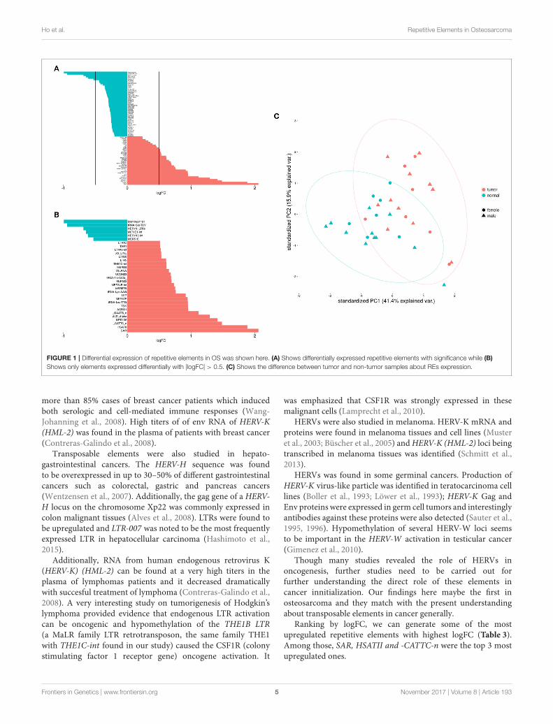
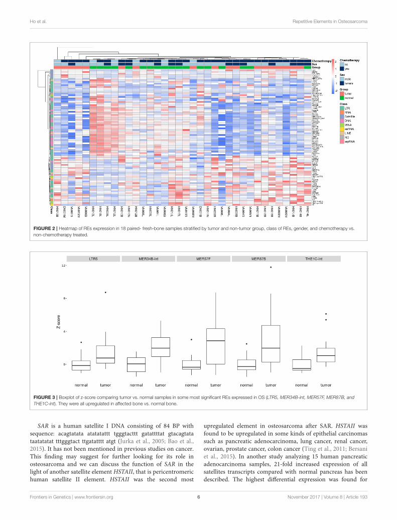
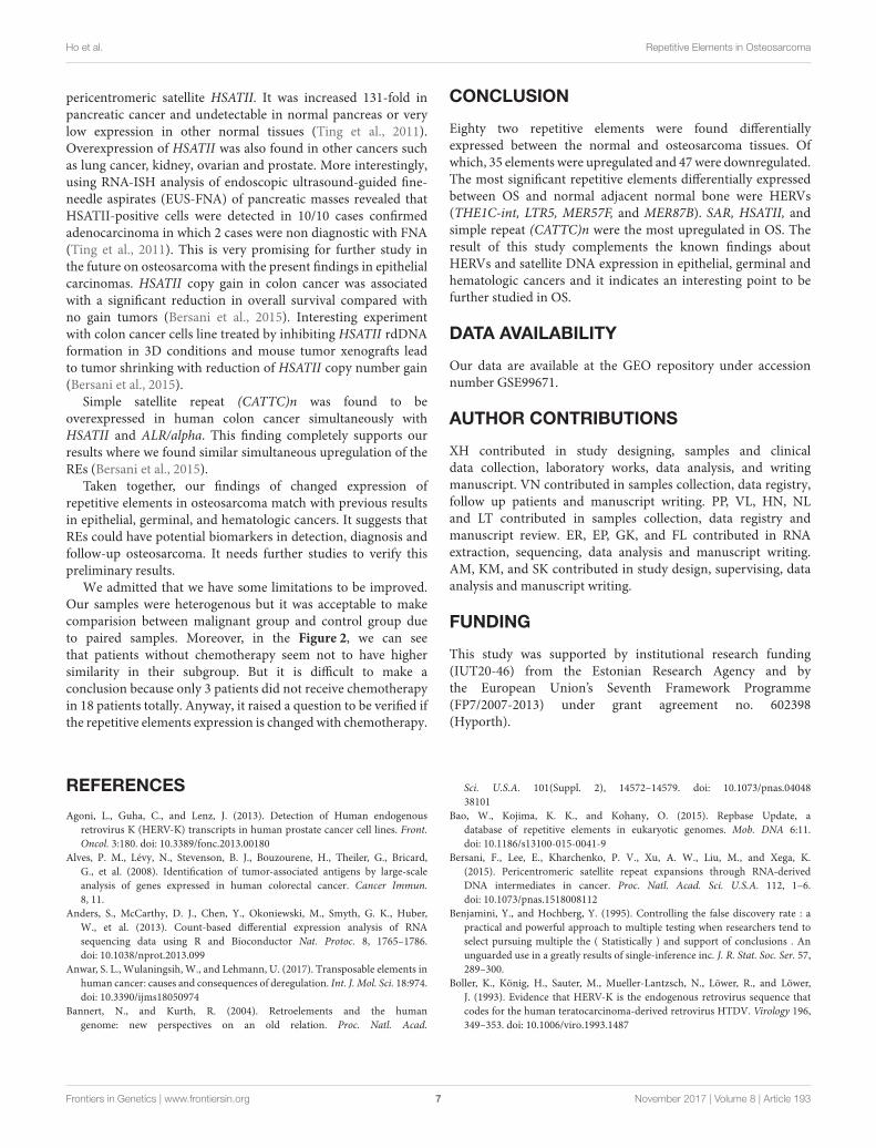
The Figure 1 shows clearly the upregulated anddownregulated repetitive elements in OS where we can seethe most upregulated and the most downregulated ones with therange of logFC (Figures 1A,B). PCA shows nicely the differencebetween OS and normal bone in repetitive elements expression(Figure 1C). Figure 2 is a heatmap of differentially expressedrepetitive elements at FDR ≤ 0.05 in OS vs. normal bone tissue.We can see clear pattern that differentiate between tumor andnormal samples. Z-scores of the top five differentially expressedrepetitive elements with lowest FDR values representes asboxplots in the Figure 3.
Frontiers in Genetics | www.frontiersin.org 3 November 2017 | Volume 8 | Article 193

Ho et al. Repetitive Elements in Osteosarcoma
TABLE 2 | Ranking from the lowest FDR, ten most significant REs expressed
differentially between osteosarcoma and normal control, FDR < 0.001.
Class Family Element logFC p-value FDR
LTR ERVL-MaLR THE1C-int 0.612785 9.83E-10 1.07E-06
LTR ERVK LTR5 0.606272 4.58E-08 2.49E-05
LTR ERV1 MER57F 0.943969 1.09E-07 2.98E-05
LTR ERV1 MER87B 0.748537 1.01E-07 2.98E-05
RNA RNA 7SK 0.9486 3.17E-07 6.91E-05
LTR ERV1 MER34B-int 0.72914 4.78E-07 8.68E-05
LTR Gypsy MamGypLTR3 −0.41918 7.13E-07 0.000111
Satellite centr ALR_Alpha 1.291039 1.08E-06 0.000147
DNA DNA MER136 1.412429 1.27E-06 0.000154
Satellite Satellite HSATII 1.878344 3.66E-06 0.000366
REs with the lowest FDR were THE1C-int, LTR5, MER57F, MER87B. These elements
belong to long terminal repeats (LTR) class and they are all upregulated in OS (logFC
> 0.5). ALR_Alpha and HSATII were the most significant satellites that expressed
differentially between OS and normal control samples.
TABLE 3 | Satellites were found to be the most upregulated elements in OS.
Class Family Element logFC p-value FDR
Satellite Satellite SAR 2.052918 5.08E−05 0.003461
Satellite Satellite HSATII 1.878344 3.66E−06 0.000366
Satellite Satellite _CATTC_n 1.481369 3.7E−06 0.000366
DNA DNA MER136 1.412429 1.27E−06 0.000154
Satellite centr ALR_Alpha 1.291039 1.08E−06 0.000147
Satellite Satellite _GAATG_n 1.115843 0.000789 0.01829
By ranking logFC, the highest ones compose of SAR, HSATII, and -CATTC-n with high
significance (FDR < 0.01).
TABLE 4 | Ranking by the lowest logFC, the table includes the most
downregulated repetitive elements in osteosarcoma.
Class Family Element logFC p-value FDR
DNA TcMar MamRep1161 −0.99091 6.38E−05 0.003862
tRNA tRNA tRNA-Ser-TCY −0.94009 0.001438 0.026562
LTR ERV1 HERV1_LTRe −0.68333 0.000166 0.007247
LTR ERVL-MaLR MLT1E1-int −0.64431 0.00306 0.043679
LTR ERV1 HERV15-int −0.61309 0.000353 0.010703
LTR ERV1 MER51E −0.5277 0.000964 0.02144
LTR ERV1 MER83B −0.43948 0.000292 0.010112
LTR ERVL LTR47B4 −0.43666 7.71E−05 0.0042
LTR Gypsy MamGypLTR3 −0.41918 7.13E−07 0.000111
LTR ERV1 MER72B −0.37858 0.000402 0.011198
LTR Gypsy LTR81 −0.35087 0.003015 0.043679
LTR ERV1 LTR31 −0.34699 0.000763 0.01808
They are mostly HERVs.
DISCUSSION
To our present knowledge, no similar study was carried outin osteosarcoma. We found herein 82 repetitive elements
which were expressed differentially with significance FDR<0.05 between osteosarcoma and adjacent normal bone inpaired samples. Of which, 35 were upregulated and 47 weredownregulated with FDR <0.05.
The top most significant differentially expressed repetitiveelements (with lowest FDR) were THE1C-int, LTR5, MER57F,and MER87B. Interestingly, they all belong to LTR elementsor HERVs with DNA transposons made up of transposableelements (TEs). THE1C-int is a ERV3, a retrovirus-like MaLRelement, consisting of 375 bp. LTR5 is ERV2, clone of HERV-K18 consisting of 969 bp. MER57F is a ERV1 with 435 bpand MER87B is also a ERV1 of 509 bp (Jurka et al., 2005;Bao et al., 2015). To our knowledge, THE1C-int, MER57F,and MER87B were not yet mentioned to be correlated withdiseases in previous literature. While LTR5 hypomethylationwas found to be correlated to systemic lupus erythematosusmechanism (Nakkuntod et al., 2013). Many studies andobservations suggested an important role of transposableelements in genomic instability, transcriptional control, non-coding RNA regulation, oncogenic activation and chromosomalrearrangements, Anwar et al. (2017). HERVs was mentioned a lotrecently as a TE that may have a role in carcinogenesis in manydifferent cancers that we got some evidences based on presentliterature.
In prostate cancer, HERV-E (and/or ERV3) env genes wasexpressed only in prostate tumor cells that suggested as targets forimmunotherapy for the disease (Wang-Johanning et al., 2003).HERV-K was also identified to be involved to prostate cancer. Itwas found that forward and reverse transcripts of severalHERV-Kloci were detected in prostate cancer cell lines (Agoni et al., 2013)and overexpression of HERV-K (22q11.23) was associated withhypomethylation of the HERV-K locus in the disease (Goeringet al., 2011). Interestingly, in a study of 14 patients with differentgrades of prostate cancer in paired samples (prostate tumor andnormal tissue), 475 retrotransposon subfamilies were detected tobe significantly differential expression in tumor tissue with FDR< 0.05. Among thoses elements, LTR was the most prevalent andmost of them were endogenous retroviruses with ERV1 being themost represented (Ren et al., 2012; Criscione et al., 2014).
In ovarian cancer, HERV-K env protein was expressed inepithelial ovarian cancer with high frequency (90%) withoutexpression in normal and benign ovarian surface epithelialtissue. Interestingly, ERV3 and HERV-E were found to expressedsimultaneously in the same ovarian cancer tissues and antibodiesto HERVs were present in the sera of ovarian cancer. Authorssuggested that HERVs should be further studied to provide anew ovarian cancer screening tool and potentially serve as anew target for detection, diagnosis and treatment for this cancer(Wang-Johanning et al., 2007). Methylation levels of HERV-Kand HERV-E were decreased in advanced ovarian cancer. HERV-K hypomethylation was found to be correlated to ovarian canceraggressiveness and poor response to treatment (Iramaneeratet al., 2011).
In breast cancer, HERV-K env was suggested to be a tumormarker as it was expressed only in breast cancer tissues and celllines analyzed but not detected in normal breast tissues (Wang-Johanning et al., 2001). HERV-K Env proteins were expressed in
Frontiers in Genetics | www.frontiersin.org 4 November 2017 | Volume 8 | Article 193
Ho et al. Repetitive Elements in Osteosarcoma
FIGURE 1 | Differential expression of repetitive elements in OS was shown here. (A) Shows differentially expressed repetitive elements with significance while (B)
Shows only elements expressed differentially with |logFC| > 0.5. (C) Shows the difference between tumor and non-tumor samples about REs expression.
more than 85% cases of breast cancer patients which inducedboth serologic and cell-mediated immune responses (Wang-Johanning et al., 2008). High titers of of env RNA of HERV-K(HML-2) was found in the plasma of patients with breast cancer(Contreras-Galindo et al., 2008).
Transposable elements were also studied in hepato-gastrointestinal cancers. The HERV-H sequence was foundto be overexpressed in up to 30–50% of different gastrointestinalcancers such as colorectal, gastric and pancreas cancers(Wentzensen et al., 2007). Additionally, the gag gene of a HERV-H locus on the chromosome Xp22 was commonly expressed incolon malignant tissues (Alves et al., 2008). LTRs were found tobe upregulated and LTR-007 was noted to be the most frequentlyexpressed LTR in hepatocellular carcinoma (Hashimoto et al.,2015).
Additionally, RNA from human endogenous retrovirus K(HERV-K) (HML-2) can be found at a very high titers in theplasma of lymphomas patients and it decreased dramaticallywith succesful treatment of lymphoma (Contreras-Galindo et al.,2008). A very interesting study on tumorigenesis of Hodgkin’slymphoma provided evidence that endogenous LTR activationcan be oncogenic and hypomethylation of the THE1B LTR(a MaLR family LTR retrotransposon, the same family THE1with THE1C-int found in our study) caused the CSF1R (colonystimulating factor 1 receptor gene) oncogene activation. It
was emphasized that CSF1R was strongly expressed in thesemalignant cells (Lamprecht et al., 2010).
HERVs were also studied in melanoma. HERV-K mRNA andproteins were found in melanoma tissues and cell lines (Musteret al., 2003; Büscher et al., 2005) andHERV-K (HML-2) loci beingtranscribed in melanoma tissues was identified (Schmitt et al.,2013).
HERVs was found in some germinal cancers. Production ofHERV-K virus-like particle was identified in teratocarcinoma celllines (Boller et al., 1993; Löwer et al., 1993); HERV-K Gag andEnv proteins were expressed in germ cell tumors and interestinglyantibodies against these proteins were also detected (Sauter et al.,1995, 1996). Hypomethylation of several HERV-W loci seemsto be important in the HERV-W activation in testicular cancer(Gimenez et al., 2010).
Though many studies revealed the role of HERVs inoncogenesis, further studies need to be carried out forfurther understanding the direct role of these elements incancer innitialization. Our findings here maybe the first inosteosarcoma and they match with the present understandingabout transposable elements in cancer generally.
Ranking by logFC, we can generate some of the mostupregulated repetitive elements with highest logFC (Table 3).Among those, SAR, HSATII and -CATTC-n were the top 3 mostupregulated ones.
Frontiers in Genetics | www.frontiersin.org 5 November 2017 | Volume 8 | Article 193
Ho et al. Repetitive Elements in Osteosarcoma
FIGURE 2 | Heatmap of REs expression in 18 paired- fresh-bone samples stratified by tumor and non-tumor group, class of REs, gender, and chemotherapy vs.
non-chemotherapy treated.
FIGURE 3 | Boxplot of z-score comparing tumor vs. normal samples in some most significant REs expressed in OS (LTR5, MER34B-int, MER57F, MER87B, and
THE1C-int). They were all upregulated in affected bone vs. normal bone.
SAR is a human satellite I DNA consisting of 84 BP withsequence: acagtatata atatatattt tgggtacttt gatattttat gtacagtatataatatatat tttgggtact ttgatatttt atgt (Jurka et al., 2005; Bao et al.,2015). It has not been mentioned in previous studies on cancer.This finding may suggest for further looking for its role inosteosarcoma and we can discuss the function of SAR in thelight of another satellite element HSTAII, that is pericentromerichuman satellite II element. HSTAII was the second most
upregulated element in osteosarcoma after SAR. HSTAII wasfound to be upregulated in some kinds of epithelial carcinomassuch as pancreatic adenocarcinoma, lung cancer, renal cancer,ovarian, prostate cancer, colon cancer (Ting et al., 2011; Bersaniet al., 2015). In another study analyzing 15 human pancreaticadenocarcinoma samples, 21-fold increased expression of allsatellites transcripts compared with normal pancreas has beendescribed. The highest differential expression was found for
Frontiers in Genetics | www.frontiersin.org 6 November 2017 | Volume 8 | Article 193
Ho et al. Repetitive Elements in Osteosarcoma
pericentromeric satellite HSATII. It was increased 131-fold inpancreatic cancer and undetectable in normal pancreas or verylow expression in other normal tissues (Ting et al., 2011).Overexpression of HSATII was also found in other cancers suchas lung cancer, kidney, ovarian and prostate. More interestingly,using RNA-ISH analysis of endoscopic ultrasound-guided fine-needle aspirates (EUS-FNA) of pancreatic masses revealed thatHSATII-positive cells were detected in 10/10 cases confirmedadenocarcinoma in which 2 cases were non diagnostic with FNA(Ting et al., 2011). This is very promising for further study inthe future on osteosarcoma with the present findings in epithelialcarcinomas. HSATII copy gain in colon cancer was associatedwith a significant reduction in overall survival compared withno gain tumors (Bersani et al., 2015). Interesting experimentwith colon cancer cells line treated by inhibiting HSATII rdDNAformation in 3D conditions and mouse tumor xenografts leadto tumor shrinking with reduction of HSATII copy number gain(Bersani et al., 2015).
Simple satellite repeat (CATTC)n was found to beoverexpressed in human colon cancer simultaneously withHSATII and ALR/alpha. This finding completely supports ourresults where we found similar simultaneous upregulation of theREs (Bersani et al., 2015).
Taken together, our findings of changed expression ofrepetitive elements in osteosarcoma match with previous resultsin epithelial, germinal, and hematologic cancers. It suggests thatREs could have potential biomarkers in detection, diagnosis andfollow-up osteosarcoma. It needs further studies to verify thispreliminary results.
We admitted that we have some limitations to be improved.Our samples were heterogenous but it was acceptable to makecomparision between malignant group and control group dueto paired samples. Moreover, in the Figure 2, we can seethat patients without chemotherapy seem not to have highersimilarity in their subgroup. But it is difficult to make aconclusion because only 3 patients did not receive chemotherapyin 18 patients totally. Anyway, it raised a question to be verified ifthe repetitive elements expression is changed with chemotherapy.
CONCLUSION
Eighty two repetitive elements were found differentiallyexpressed between the normal and osteosarcoma tissues. Ofwhich, 35 elements were upregulated and 47were downregulated.The most significant repetitive elements differentially expressedbetween OS and normal adjacent normal bone were HERVs(THE1C-int, LTR5, MER57F, and MER87B). SAR, HSATII, andsimple repeat (CATTC)n were the most upregulated in OS. Theresult of this study complements the known findings aboutHERVs and satellite DNA expression in epithelial, germinal andhematologic cancers and it indicates an interesting point to befurther studied in OS.
DATA AVAILABILITY
Our data are available at the GEO repository under accessionnumber GSE99671.
AUTHOR CONTRIBUTIONS
XH contributed in study designing, samples and clinicaldata collection, laboratory works, data analysis, and writingmanuscript. VN contributed in samples collection, data registry,follow up patients and manuscript writing. PP, VL, HN, NLand LT contributed in samples collection, data registry andmanuscript review. ER, EP, GK, and FL contributed in RNAextraction, sequencing, data analysis and manuscript writing.AM, KM, and SK contributed in study design, supervising, dataanalysis and manuscript writing.
FUNDING
This study was supported by institutional research funding(IUT20-46) from the Estonian Research Agency and bythe European Union’s Seventh Framework Programme(FP7/2007-2013) under grant agreement no. 602398(Hyporth).
REFERENCES
Agoni, L., Guha, C., and Lenz, J. (2013). Detection of Human endogenous
retrovirus K (HERV-K) transcripts in human prostate cancer cell lines. Front.
Oncol. 3:180. doi: 10.3389/fonc.2013.00180
Alves, P. M., Lévy, N., Stevenson, B. J., Bouzourene, H., Theiler, G., Bricard,
G., et al. (2008). Identification of tumor-associated antigens by large-scale
analysis of genes expressed in human colorectal cancer. Cancer Immun.
8, 11.
Anders, S., McCarthy, D. J., Chen, Y., Okoniewski, M., Smyth, G. K., Huber,
W., et al. (2013). Count-based differential expression analysis of RNA
sequencing data using R and Bioconductor Nat. Protoc. 8, 1765–1786.
doi: 10.1038/nprot.2013.099
Anwar, S. L., Wulaningsih, W., and Lehmann, U. (2017). Transposable elements in
human cancer: causes and consequences of deregulation. Int. J. Mol. Sci. 18:974.
doi: 10.3390/ijms18050974
Bannert, N., and Kurth, R. (2004). Retroelements and the human
genome: new perspectives on an old relation. Proc. Natl. Acad.
Sci. U.S.A. 101(Suppl. 2), 14572–14579. doi: 10.1073/pnas.04048
38101
Bao, W., Kojima, K. K., and Kohany, O. (2015). Repbase Update, a
database of repetitive elements in eukaryotic genomes. Mob. DNA 6:11.
doi: 10.1186/s13100-015-0041-9
Bersani, F., Lee, E., Kharchenko, P. V., Xu, A. W., Liu, M., and Xega, K.
(2015). Pericentromeric satellite repeat expansions through RNA-derived
DNA intermediates in cancer. Proc. Natl. Acad. Sci. U.S.A. 112, 1–6.
doi: 10.1073/pnas.1518008112
Benjamini, Y., and Hochberg, Y. (1995). Controlling the false discovery rate : a
practical and powerful approach to multiple testing when researchers tend to
select pursuing multiple the ( Statistically ) and support of conclusions . An
unguarded use in a greatly results of single-inference inc. J. R. Stat. Soc. Ser. 57,
289–300.
Boller, K., König, H., Sauter, M., Mueller-Lantzsch, N., Löwer, R., and Löwer,
J. (1993). Evidence that HERV-K is the endogenous retrovirus sequence that
codes for the human teratocarcinoma-derived retrovirus HTDV. Virology 196,
349–353. doi: 10.1006/viro.1993.1487
Frontiers in Genetics | www.frontiersin.org 7 November 2017 | Volume 8 | Article 193

Ho et al. Repetitive Elements in Osteosarcoma
Büscher, K., Trefzer, U., Hofmann, M., Sterry, W., Kurth, R., and Denner, J. (2005).
Expression of human endogenous retrovirus K in melanomas and melanoma
cell lines. Cancer Res. 65, 4172–4180. doi: 10.1158/0008-5472.CAN-04-2983
Cegolon, L., Salata, C., Weiderpass, E., Vineis, P., Palù, G., and Mastrangelo, G.
(2013). Human endogenous retroviruses and cancer prevention: evidence and
prospects. BMC Cancer 13:4. doi: 10.1186/1471-2407-13-4
Contreras-Galindo, R., Kaplan,M. H., Leissner, P., Verjat, T., Ferlenghi, I., Bagnoli,
F., et al. (2008). Human endogenous retrovirus K ( HML-2 ) elements in the
plasma of people with lymphoma and breast cancer ?†. J. Virol. 82, 9329–36.
doi: 10.1128/JVI.00646-08
Criscione, S. W., Zhang, Y., Thompson, W., Sedivy, J. M., and Neretti, N. (2014).
Transcriptional landscape of repetitive elements in normal and cancer human
cells. BMC Genomics 15:583. doi: 10.1186/1471-2164-15-583
de Koning, A. P., Gu, W., Castoe, T. A., Batzer, M. A., and Pollock, D. D. (2011).
Repetitive elements may comprise over two-thirds of the human genome. PLoS
Genet. 7:e1002384. doi: 10.1371/journal.pgen.1002384.
Eymery, A., Horard, B., El Atifi-Borel, M., Fourel, G., Berger, F., Vitte, A. L.,
et al. (2009). A transcriptomic analysis of human centromeric and pericentric
sequences in normal and tumor cells. Nucleic Acids Res. 37, 6340–6354.
doi: 10.1093/nar/gkp639
Gimenez, J., Montgiraud, C., Pichon, J. P., Bonnaud, B., Arsac, M., and
Ruel, K. et al. (2010). Custom human endogenous retroviruses dedicated
microarray identifies self-induced HERV-W family elements reactivated in
testicular cancer upon methylation control. Nucleic Acids Res. 38, 2229–2246.
doi: 10.1093/nar/gkp1214
Goering, W., Ribarska, T., and Schulz, W. A. (2011). Selective changes
of retroelement expression in human prostate cancer. Carcinogenesis 32,
1484–1492. doi: 10.1093/carcin/bgr181
Hashimoto, K., Suzuki, A.M., Dos Santos, A., Desterke, C., Collino, A., Ghisletti, S.,
Braun, E., et al. (2015). CAGE profiling of ncRNAs in hepatocellular carcinoma
reveals widespread activation of retroviral LTR promoters in virus-induced
tumors. Genome Res. 25, 1812–1824. doi: 10.1101/gr.191031.115
Hung, G. Y., Horng, J. L., Yen, H. J., Yen, C. C., Chen, W. M., and
Chen, P. C. (2014). Incidence patterns of primary bone cancer in taiwan
(2003–2010): a population-based study. Ann. Surg. Oncol. 21, 2490–2498.
doi: 10.1245/s10434-014-3697-3
Iramaneerat, K., Rattanatunyong, P., Khemapech, N., Triratanachat, S., and
Mutirangura, A. (2011). HERV-K Hypomethylation in ovarian clear cell
carcinoma is associated with a poor prognosis and platinum resistance. Int. J.
Gynecol. Cancer 21, 51–57. doi: 10.1097/IGC.0b013e3182021c1a
Jaffe, N., Puri, A., and Gelderblom, H. (2013). Osteosarcoma: evolution of
treatment paradigms. Sarcoma 2013:203531. doi: 10.1155/2013/203531
Pichon, J. P., Bonnaud, B., and Cleuziat, P. (2006). Multiplex degenerate PCR
coupled with an oligo sorbent array for human endogenous retrovirus
expression profiling. Nucleic Acids Res. 34, 1–10. doi: 10.1093/nar/gkl086
Jurka, J., Kapitonov, V. V., Pavlicek, A., Klonowski, P., Kohany, O., and
Walichiewicz, J. (2005). Repbase update, a database of eukaryotic repetitive
elements. Cytogenet. Genome Res. 110, 462–467. doi: 10.1159/000084979
Lamprecht, B., Walter, K., Kreher, S., Kumar, R., Hummel, M., Lenze, D.,
et al. (2010). Derepression of an endogenous long terminal repeat activates
the CSF1R proto-oncogene in human lymphoma. Nat. Med. 16, 571–579.
doi: 10.1038/nm.2129
Lander, E. S., Linton, L. M., Birren, B., Nusbaum, C., Zody, M. C., Baldwin, J.,
et al. (2001). Initial sequencing and analysis of the human genome. Nature 409,
860–921. doi: 10.1038/35057062
Li, J., Witten, D. M., Johnstone, I. M., and Tibshirani, R. (2012). Normalization,
testing, and false discovery rate estimation for RNA-sequencing data.
Biostatistics 13, 523–538. doi: 10.1093/biostatistics/kxr031
Löwer, R., Boller, K., Hasenmaier, B., Korbmacher, C., Müller-Lantzsch, N.,
and Löwer, J. (1993). Identification of human endogenous retroviruses with
complex mRNA expression and particle formation. Proc. Natl. Acad. Sci. U.S.A.
90, 4480–4484.
Mager, D. L., and Stoye, J. P. (2015). Mammalian endogenous retroviruses.
Microbiol. Spectr. 3:MDNA3-0009-2014. doi: 10.1128/microbiolspec.MDNA3-
0009-2014
McCarthy, D. J., Chen, Y., and Smyth, G. K. (2012). Differential expression analysis
of multifactor RNA-seq experiments with respect to biological variation.
Nucleic Acids Res. 40, 4288–4297. doi: 10.1093/nar/gks042
Mirabello, L., Troisi, R. J., and Savage, S. A. (2009a). Osteosarcoma
incidence and survival improvement. Cancer 115, 1531–1543.
doi: 10.1002/cncr.24121.Osteosarcoma
Mirabello, L., Troisi, R. J., and Savage, S. A. (2009b). International osteosarcoma
incidence patterns in children and adolescents, middle ages and elderly persons.
Int. J. Cancer. 125, 229–234. doi: 10.1002/ijc.24320
Muster, T., Waltenberger, A., Grassauer, A., Hirschlm, S., Caucig, P., Romirer, I.,
et al. (2003). An endogenous retrovirus derived from human melanoma cells
an endogenous retrovirus derived from humanmelanoma cells. Cancer Res. 63,
8735–8741.
Nakkuntod, J., Sukkapan, P., Avihingsanon, Y., Mutirangura, A., and Hirankarn,
N. (2013). DNA methylation of human endogenous retrovirus in systemic
lupus erythematosus. J. Hum. Genet. 58, 241–249. doi: 10.1038/jhg.2013.6
Nelson, P. N., Carnegie, P. R., Martin, J., Davari Ejtehadi, H., Hooley, P., Roden,
D., et al. (2003). Demystified. Human endogenous retroviruses.Mol. Pathol. 56,
11–18. doi: 10.1136/mp.56.1.11
Padeken, J., Zeller P., and Gasser, S. M. (2015). Repeat DNA in genome
organization and stability. Curr. Opin. Genet. Develop. 31, 12–19.
doi: 10.1016/j.gde.2015.03.009
Pérot, P., Mullins, C. S., Naville, M., Bressan, C., Hühns, M., and Gock, M.
(2015). Expression of young HERV-H loci in the course of colorectal carcinoma
and correlation with molecular subtypes. Oncotarget 6, 40095–40111.
doi: 10.18632/oncotarget.5539
Rebollo, R., Romanish, M. T., and Mager, D. L. (2012). Transposable elements : an
abundant and natural source of regulatory sequences for host genes. Ann. Rev.
Genet. 46, 21–42. doi: 10.1146/annurev-genet-110711-155621
Ren, S., Peng, Z., Mao, J. H., Yu, Y., Yin, C., Gao, X., et al. (2012). RNA-seq analysis
of prostate cancer in the chinese population identifies recurrent gene fusions,
cancer-associated long noncoding RNAs and aberrant alternative splicings. Cell
Res. 22, 806–821. doi: 10.1038/cr.2012.30
Robinson, M. D., McCarthy, D. J., and Smyth, G. K. (2010). edgeR: a
bioconductor package for differential expression analysis of digital gene
expression data. Bioinformatics 26, 139–140. doi: 10.1093/bioinformatics/
btp616
Sauter, M., Roemer, K., Best, B., Afting, M., Schommer, S., and Seitz, G.
(1996). Specificity of antibodies directed against env protein of human
endogenous retroviruses in patients with germ cell tumors. Cancer Res. 56,
4362–4365.
Sauter, M., Schommer, S., Kremmer, E., Remberger, K., Dölken, G., and
Lemm, I. (1995). Human endogenous retrovirus K10: expression of gag
protein and detection of antibodies in patients with seminomas. J. Virol. 69,
414–421.
Schiavetti, F., Thonnard, J., Colau, D., Boon, T., and Coulie, P. G. (2002). A human
endogenous retroviral sequence encoding an antigen recognized on melanoma
by cytolytic T lymphocytes 1. Cancer Res. 62, 5510–5516.
Schmitt, K., Reichrath, J., Roesch, A., Meese, E., and Mayer, J. (2013).
Transcriptional profiling of human endogenous retrovirus group
HERV-K(HML-2) loci in melanoma. Genome Biol. Evol. 5, 307–28.
doi: 10.1093/gbe/evt010
Shukla, R., Upton, K. R., Muñoz-Lopez, M., Gerhardt, D. J., Fisher, M. E., Nguyen,
T., et al. (2013). Endogenous retrotransposition activates oncogenic pathways
in hepatocellular carcinoma. Cell 153, 101–111. doi: 10.1016/j.cell.2013.02.032
Solyom, S., Ewing, A. D., Rahrmann, E. P., Doucet, T., Nelson, H. H., Burns, M.
B., et al. (2012). Extensive somatic L1 retrotransposition in colorectal tumors.
Genome Res. 2328–2338. doi: 10.1101/gr.145235.112.2328
Ting, D. T., Haber, D. A., Maheswaran, S., and Lipson, D. (2012). Biomarkers
of Cancer. Patent WO 2012048113 A2. Boston, MA: The General Hospital
Corporation.
Ting, D. T., Lipson, D., Paul, S., Brannigan, B. W., Akhavanfard, S., Coffman, E. J.,
et al. (2011). Aberrant overexpression of satellite repeats in pancreatic and other
epithelial cancers. Science 331, 593–596. doi: 10.1126/science.1200801.Aberrant
Valery, P. C., Laversanne, M., and Bray, F. (2015). Bone cancer incidence
by morphological subtype: a global assessment. Cancer Causes Control 26,
1127–1139. doi: 10.1007/s10552-015-0607-3
Wang-Johanning, F., Frost, A. R., Jian, B., Azerou, R., Lu, D. W., and Chen,
D. T. (2003). Detecting the expression of human endogenous retrovirus E
envelope transcripts in human prostate adenocarcinoma. Cancer 98, 187–197.
doi: 10.1002/cncr.11451
Frontiers in Genetics | www.frontiersin.org 8 November 2017 | Volume 8 | Article 193
Ho et al. Repetitive Elements in Osteosarcoma
Wang-Johanning, F., Frost, A. R., Johanning, G. L., Khazaeli, M. B., LoBuglio,
A. F., and Shaw, D. R. (2001). Expression of human endogenous retrovirus
K envelope transcripts in human breast cancer. Clin. Cancer Res. 7,
1553–1560.
Wang-Johanning, F., Liu, J., Rycaj, K., Huang, M., Tsai, K., and Rosen, D.
G. (2007). Expression of multiple human endogenous retrovirus surface
envelope proteins in ovarian cancer. Int. J. Cancer 120, 81–90. doi: 10.1002/ijc.
22256
Wang-Johanning, F., Radvanyi, L., Rycaj, K., Plummer, J. B., Yan, P., and
Sastry, K. J. (2008). Human endogenous retrovirus K triggers an antigen-
specific immune response in breast cancer patients. Cancer Res. 68, 5869–77.
doi: 10.1158/0008-5472.CAN-07-6838
Wentzensen, N., Coy, J. F., Knaebel, H. P., Linnebacher, M., Wilz, B., and
Gebert, J. (2007). Expression of an endogenous retroviral sequence from
the HERV-H group in gastrointestinal cancers. Int. J. Cancer 121, 1417–23.
doi: 10.1002/ijc.22826
Conflict of Interest Statement: The authors declare that the research was
conducted in the absence of any commercial or financial relationships that could
be construed as a potential conflict of interest.
Copyright © 2017 Ho, Nguyen, Trinh, Reimann, Prans, Kõks, Maasalu, Le, Nguyen,
Le, Phung, Märtson, Lattekivi and Kõks. This is an open-access article distributed
under the terms of the Creative Commons Attribution License (CC BY). The use,
distribution or reproduction in other forums is permitted, provided the original
author(s) or licensor are credited and that the original publication in this journal
is cited, in accordance with accepted academic practice. No use, distribution or
reproduction is permitted which does not comply with these terms.
Frontiers in Genetics | www.frontiersin.org 9 November 2017 | Volume 8 | Article 193