anatomical basis of differences in locomotor - bioone
TRANSCRIPT

Bull. Mus. Comp. Zool., 159(4): 213–238, December, 2008 213
ANATOMICAL BASIS OF DIFFERENCES IN LOCOMOTOR BEHAVIORIN ANOLIS LIZARDS: A COMPARISON BETWEEN TWOECOMORPHS
ANTHONY HERREL,1 BIEKE VANHOOYDONCK,2 JOANNE PORCK,3 AND DUNCAN J. IRSCHICK4
ABSTRACT. Anolis lizards have become model or-ganisms for the study of adaptive radiation, as on eachof the larger islands in the Caribbean, animals withsimilar morphologies have independently radiatedinto similar ecological niches. Central in the study ofthese animals have been the investigations correlatingdifferences in limb dimensions to substrate charac-teristics and locomotor performance. However, littleis known about differences in the musculoskeletalsystem that could underlie the observed differencesin performance or locomotor style (i.e., gait charac-teristics). Here, we provide data on the morphologyof the appendicular skeleton and musculature in twospecies of Anolis that differ greatly in habitat use andlocomotor performance: A. sagrei and A. valencienni.The first and principal objective was to provide a de-tailed description of the appendicular morphologythat could serve as a basis for further study. Our sec-ond objective was to test for quantitative differencesin muscle mass and muscle mass distribution betweenthe two species. Finally, we explore how the observeddifferences in the musculoskeletal system might becorrelated with locomotor performance and locomo-tor style by analyzing data on the spatiotemporal gaitcharacteristics in these two species while they weremoving on substrates of different diameters. Our datashow distinct differences in the morphology, musclemass, and muscle mass distribution and illustrate howthese may result in greater step and stride lengths inA. sagrei, allowing it to achieve higher sprint speeds.Anolis valencienni has less robust muscles that mightconstrain step and stride length, which in turn couldprovide it with greater stability on narrow substrates.
1 Department of Organismic and Evolutionary Bi-ology, Harvard University, 26 Oxford Street, Cam-bridge, Massachusetts 02138. Author for correspon-dence ([email protected]).
2 Department of Biology, University of Antwerp,Universiteitsplein 1, B-2610 Antwerpen, Belgium.
3 Lopsenstraat 22, 2312 ZZ Leiden, The Nether-lands.
4 Department of Biology, 221 Morrill Science Cen-ter, University of Massachusetts at Amherst, Amherst,Massachusetts 01003.
INTRODUCTION
Anolis lizards have become model or-ganisms for the study of adaptive radiation,in that on each of the larger islands in theCaribbean, species with similar morphol-ogies have independently adapted to sim-ilar ecological niches (Losos, 1990a,b; Lo-sos et al., 1998; Schluter, 2000; Williams,1983). Central to the study of these ani-mals have been the investigations correlat-ing differences in limb dimensions to sub-strate characteristics and locomotor per-formance (Irschick and Losos, 1998, 1999;Losos and Irschick, 1996; Losos andSinervo, 1989; Losos et al., 1997; Van-hooydonck et al., 2006a,b). These and oth-er studies have demonstrated how the in-teraction of limb morphology with sub-strate characteristics is crucial in settinglimits on locomotor performance (Irschickand Losos, 1998; Losos and Sinervo, 1989;Vanhooydonck et al., 2005). For example,lizards with long limbs can achieve highervelocities on broad substrates (Irschickand Losos, 1998; Losos and Sinervo, 1989;Sinervo and Losos, 1991) and have greateracceleration capacities on both broad andnarrow substrates (Vanhooydonck et al.,2006b) but could face a decrease in sta-bility on narrow substrates, causing themto stumble and fall more often (Losos andSinervo, 1989). Moreover, the trade-offbetween sprint speed and surefootednesshas been suggested to be an importantcomponent of habitat choice in arboreallizards (Irschick and Losos, 1999).
Despite the importance of limb mor-
214 Bulletin Museum of Comparative Zoology, Vol. 159, No. 4
phology in shaping locomotor perfor-mance and habitat use, little is knownabout differences in the musculoskeletalsystem that are responsible for the ob-served differences in performance and lo-comotor style among species and eco-morphs (but see Vanhooydonck et al.,2006a). Yet differences in muscle mass,muscle architecture, and muscle positioncould be crucially important in allowinganimals to achieve greater performance inspecific ecological settings. For example,Zaaf and co-workers (1999, 2001) dem-onstrated how differences in the mass andposition of the fore and hindlimb musclesmight provide a performance advantage toclimbing geckos moving on vertical sub-strates. Consequently, one would expectthat Anolis lizards that spend more time inarboreal habitats would also show special-izations in the forelimb muscles, allowingthem to generate greater forces to moveagainst gravity, as has been demonstratedfor geckos (Autumn et al., 2006). Specifi-cally, we predict that arboreal species willallocate more of the total forelimb musclemass to humerus retractors, which arethought to be important in generatingpulling forces with the forelimbs (Zaaf etal., 1999). Conversely, fast terrestrial orsemi-arboreal species can be expected tohave more robust hindlimb extensors thatallow them to achieve greater velocitiesand accelerations (see also Vanhooydoncket al., 2006a). Given the trade-off previ-ously noted between sprint speed andsurefootedness (Losos and Sinervo, 1989),we predict that animals adapted to movingon narrow substrates will have shorter stepand stride lengths and will move theirlimbs at lower frequency, which would al-low them to maintain stability on narrowsubstrates (Spezzano and Jayne, 2004).
Here, we explore differences in themorphology of the appendicular system intwo species of Anolis lizards, A. sagrei andA. valencienni, that differ markedly inoverall body and limb shape (Losos,1990a,b; Fig. 1), maximal locomotor speed(Losos, 1990b), acceleration capacity (Van-
hooydonck et al., 2006b), and habitat use(Losos, 1990b) but are relatively closelyrelated to each other (both belong to theNorops clade of Anolis; see Nicholson etal., 2005). Whereas A. sagrei is a typicaltrunk-ground anole that often occurs onthe ground and on broad substrates, A.valencienni is a twig anole that spendsmost of its time moving on narrow sub-strates. The first and principal goal of thispaper is to give a complete and detaileddescription of the anatomy of the fore- andhindlimb muscles. Our second objective isto test for quantitative differences in mus-cle mass and muscle mass distribution be-tween species. Our final objective is to ex-plore whether the observed differences inthe morphology of the appendicular skel-eton can be linked to differences in loco-motor style by analyzing the spatiotempo-ral gait characteristics of both species mov-ing on two substrates of different diame-ters.
MATERIALS AND METHODS
Animals
Between November 2001 and February2002, we captured 15 male A. sagrei Coc-teau (snout–vent length [SVL] � 59.22 �0.36 mm; mean �77 SD) and 10 male A.valencienni Dumeril and Bibron (SVL �67.9 � 1.4 mm) by hand or noose. The A.sagrei individuals were captured on themainland United States (Miami, Florida).Anolis valencienni individuals were caughtaround the Discovery Bay Marine Labo-ratory in Jamaica. All the animals weretransported back to the laboratory at Tu-lane University (New Orleans, Louisiana).Upon arrival in the lab, the lizards werehoused in pairs in 40-liter terraria linedwith leaf litter and containing a dowel.Terraria were placed in a temperature-controlled room (29 � 2� C) with a 12:12hour light:dark photoperiod. We fed theanimals live crickets dusted with calciumand vitamin supplements three times aweek; lizards were sprayed with water dai-ly.

LIMB MUSCLES IN ANOLIS • Herrel et al. 215
Figure 1. Drawing of (A) Anolis sagrei and (B) Anolis valencienni in ventral, dorsal, and lateral view illustrating differences inoverall body and limb proportions. Note how A. sagrei has relatively longer distal hindlimb segments and a longer tail but shorterbody and head compared with A. valencienni. Differences in distal hindlimb segments (tibia, metatarsus, longest toe) and thelength of the longest toe of the forelimb are significantly different between species. Scale bar: 1 cm.
Morphology
A different set of preserved specimensof both A. sagrei (N � 6; SVL � 55.79 �1.88 mm) and A. valencienni (N � 6; SVL� 66.26 � 2.52 mm) were dissected, andall muscles were taken out and sorted byfunction into the following groups: femurprotractors, femur retractors, femur ad-ductors, femur abductors, knee flexors,knee extensors, ankle flexors, ankle exten-sors, a miscellaneous group (containingthe lower hindlimb pronators and rota-tors), humerus protractors, humerus re-tractors, humerus adductors, humerus ab-ductors, elbow flexors, elbow extensors,wrist flexors, wrist extensors, and a groupcontaining the lower forelimb pronatorsand rotators. The assignment of muscles tofunctional groups is based on their posi-tion and on manipulation of dissectedspecimens. Note, however, that the assign-ment of muscles to functional groups
needs to be confirmed by in vivo studiesof muscle function and that our assign-ment need not correspond to descriptionsfor other species because of variation inmuscle attachment sites. Muscles werestored by group in vials with 70% ethanol,blotted dry, and weighed per functionalgroup on a Mettler MT5 electronic bal-ance (�0.1 mg). Note that, given the smallsize of these animals, muscle groups oftenweigh less than 10 mg and thus require theuse of a precision balance.
Running Trials
We induced lizards to run up a plasticdowel covered with metal wire mesh(mesh width 1 mm) by clapping our handsor tapping the lizards slightly on the baseof their tail. All lizards were tested on botha broad and a narrow dowel (diameters of8 and 1 cm, respectively). Both dowelswere 2 m long and placed against the wall
216 Bulletin Museum of Comparative Zoology, Vol. 159, No. 4
at an angle of 45� (see Vanhooydonck etal., 2006a,b). Lizards were filmed in lateralview over a distance of 1 m with a high-speed video camera (Redlake MotionscopePCI camera) set at 250 frames/s. We per-formed between five and 10 trials per in-dividual on each dowel. Trials were con-ducted on several nonconsecutive days,with trials on the broad and narrow dowelalternated among days. Before experimen-tation and between trials, the lizards wereplaced in an incubator set at 32� C for atleast 1 hour to allow the lizards to attainbody temperatures similar to their pre-ferred field body temperatures (see alsoToro et al., 2003).
After filming, we digitized the tip of thesnout at 250 frames/s with the use of PeakPerformance MOTUS software from themoment the lizard started running until itran out of view. Of the same sequences,we obtained footfall patterns by recordingthe frames at which the right hindfoottouched the substrate (i.e., foot contact)and the frames at which the right hindfootlost contact with the substrate (i.e., footrelease). On the basis of displacement ofthe snout tip and the footfall patterns, wesubsequently calculated stride length (thedistance traveled by the center of mass ofan animal in a complete cycle of limbmovements), stride frequency (the num-ber of cycles per second), step length (thedistance the body moves forward duringthe stance of a particular leg), and meanspeed per stride for successive strides dur-ing steady state locomotion.
Statistical Analyses
Morphometric and muscle mass datawere log transformed before analysis toconform to assumptions of homoscedascityand normality required for parametricanalyses. Analyses of variance were used totest for differences between species inlimb dimensions, in total hindlimb andfront limb muscle mass, and in the massof the different functional groups.
Because we were mainly interested insteady state locomotion, we only used
data on gait characteristics of the third,fourth, and fifth stride in a sequence (i.e.,after the initial acceleration phase, andwhen locomotion was largely steady, as in-dicated by the absence of fluctuations inthe velocity profile; see Vanhooydonck etal., 2006b). Before statistical analyses,stride length, step length, and stride fre-quency were expressed in units of hind-limb length, because hindlimb length dif-fers significantly between A. valencienniand A. sagrei and because gait character-istics are determined on the basis of hind-limb footfall patterns. Total hindlimblength was determined on the basis of thesum of the individual limb segments. Allgait characteristics were log transformedbefore analyses. A two-way multivariateanalysis of covariance, with stride length,stride frequency, and step length as de-pendent variables; stride speed as covar-iate; and species and dowel as factors, wasperformed. Nonsignificant interaction ef-fects were removed from the final model.All analyses were performed using SPSSV.13.0.
RESULTS
Anatomy
Here, we first provide a brief qualitativedescription of the differences in the pec-toral and pelvic girdle of the two species.Next, we give a brief description of theligaments of the pelvic girdle and a de-scription of the fore- and hindlimb mus-cles. The descriptions are principally basedon A. sagrei except where important dif-ferences between species were observed.We follow the terminology adopted byZaaf et al. (1999), Moro and Abdala(2004), and Abdala and Moro (2006) inour descriptions of the muscles. In addi-tion, we used papers by Landsmeer (1984,1990) and Russell (1988) as a basis for ourdescriptions of the limb muscles.
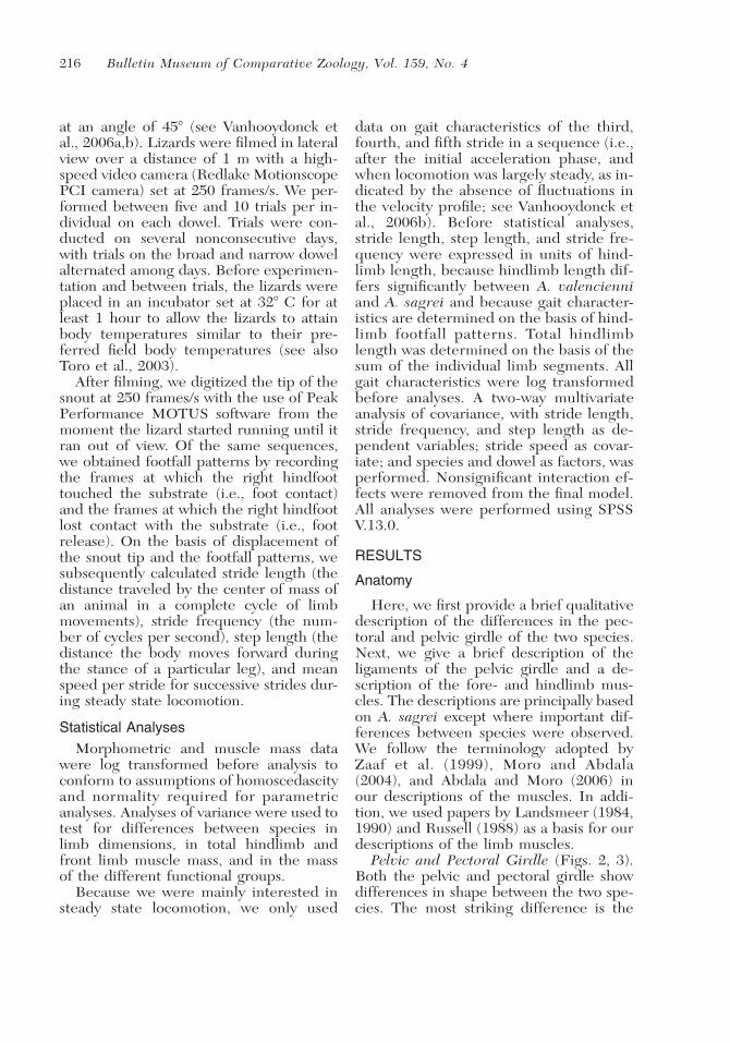
Pelvic and Pectoral Girdle (Figs. 2, 3).Both the pelvic and pectoral girdle showdifferences in shape between the two spe-cies. The most striking difference is the

LIMB MUSCLES IN ANOLIS • Herrel et al. 217
Figure 2. Schematic drawing illustrating the pectoral girdle in (A) A. sagrei and (B) A. valencienni. Shown are a ventral, adorsal, and a lateral view (from top to bottom) for each species. The position of the girdle in the body, as well as a blow-updetailing the girdle itself, is shown for each view. Note the more elongated and narrower pectoral girdle with elongated sternumin A. valencienni. I, Ligamentum sternoclaviculare; II, Ligamentum sternocoracoideum; A, sternum; B, coracoid; C, epicoracoid;D, glenoid fossa; E, interclavicula; F, clavicula; G, scapula; H, suprascapula.
more elongated and narrower appearanceof the girdles in A. valencienni comparedwith A. sagrei (see also Beuttell and Losos,1999). The pectoral girdle in A. valencien-ni is further characterized by an elongatedsternum, an expanded suprascapula and amore perpendicularly positioned scapulaand clavicula. The pelvic girdle in A. sagrei
is generally broader and has an ilium thatis directed more dorsally compared withthat of A. valencienni (Fig. 3). The anteriorpart of the ischium is, however, more elon-gated in A. valencienni.
Ligaments (Figs. 2, 3). The followingdescription of the ligaments applies toboth species.
218 Bulletin Museum of Comparative Zoology, Vol. 159, No. 4
Figure 3. Schematic drawing illustrating the pelvic girdle in (A) A. sagrei and (B) A. valencienni. Shown are a ventral, a dorsaland, a lateral view (from top to bottom) for each species. The position of the girdle in the body, as well as a blow up detailingthe girdle itself, is shown for each view. Note the more elongate and narrower pelvic girdle with relatively shorter ilium in A.valencienni. III, ligamentum puboischiadum pars lateralis; IV, ligamentum puboischiadum pars medialis; V, ligamentum iliois-chiadum; O, pubis; P, epipubis; Q, ischium; R, hypoischium; S, acetabulum; T, pectineal tubercle; U, ilium.
→
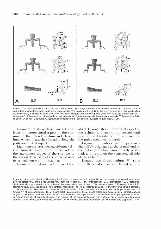
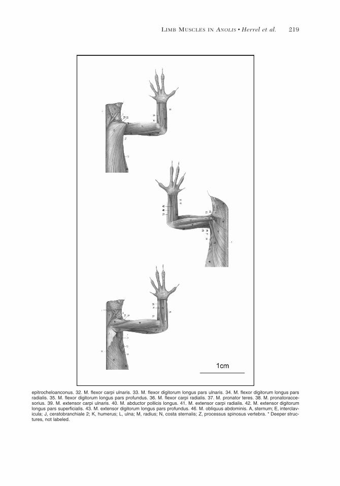
Figure 4. Anatomical drawings illustrating the forelimb musculature in A. sagrei. Shown are a superficial ventral view, a su-perficial dorsal view, and a deep ventral view (from top to bottom). 1. M. constrictor colli. 2. M. pterygoideus. 3. M. episterno-cleidomastoideus pars anterior. 4. M. episternocleidomastoideus pars posterior. 5. M. levator scapula. 6. M. omohyoideus. 7. M.sternohyoideus. 8. M. trapezius. 9. M. depressor mandibulae. 10. M. cervicomandibularis. 11. M. transverso spinalis complex.12. M. serratus. 13. Mm. levatores costae. 14. M. intercostalis. 15. M. pectoralis pars superficialis. 16. M. pectoralis pars pro-fundus. 17. M. coracohumeralis. 18. M. biceps brachii pars ventralis. 19. M. biceps brachii pars dorsalis. 20. M. coracobrachialislongus. 21. M. coracobrachialis brevis. 22. M. clavodeltoideus pars superficialis. 23. M. clavodeltoideus pars profundus. 24. M.latissimus dorsi. 25. M. scapulodeltoideus pars anterior. 26. M. scapuladeltoideus pars posterior. 27. M. triceps pars humeralisanterior. 28. M. triceps pars humeralis posterior. 29. M. triceps pars scapulohumeralis. 30. M. triceps pars scapularis. 31. M.
Ligamentum sternoclaviculare (I): runsfrom the lateroventral aspect of the ster-num to the interclaviculum and clavicu-lum, where it attaches broadly along theposterior ventral aspect.
Ligamentum sternocoracoideum (II):runs from its origin on the dorsal side atthe lateralmost aspect of the sternum tothe lateral dorsal side of the coracoid nearits articulation with the scapula.
Ligamentum puboischiadum pars later-
alis (III): originates at the ventral aspect ofthe ischium and runs to the ventrolateralside of the lateralmost protruberance ofthe pubis (pectineal tubercle).
Ligamentum puboischiadum pars me-dialis (IV): originates at the cranial end ofthe pubis (epipubis), runs directly poste-riad, and inserts on the ventrocranial sideof the ischium.
Ligamentum ilioischiadum (V): runsfrom the caudodorsal and lateral side of
LIMB MUSCLES IN ANOLIS • Herrel et al. 219
epitrocheloanconus. 32. M. flexor carpi ulnaris. 33. M. flexor digitorum longus pars ulnaris. 34. M. flexor digitorum longus parsradialis. 35. M. flexor digitorum longus pars profundus. 36. M. flexor carpi radialis. 37. M. pronator teres. 38. M. pronatoracce-sorius. 39. M. extensor carpi ulnaris. 40. M. abductor pollicis longus. 41. M. extensor carpi radialis. 42. M. extensor digitorumlongus pars superficialis. 43. M. extensor digitorum longus pars profundus. 46. M. obliquus abdominis. A, sternum; E, interclav-icula; J, ceratobranchiale 2; K, humerus; L, ulna; M, radius; N, costa sternalis; Z, processus spinosus vertebra. * Deeper struc-tures, not labeled.

220 Bulletin Museum of Comparative Zoology, Vol. 159, No. 4
the ischium to the posterolateral side ofthe ilium.
Forelimb Musculature (Figs. 4, 5). Mus-culus pectoralis (15, 16): consists of twodistinct parts:
1) M. pectoralis pars superficialis (15):This is the largest part of the M. pectoralisand originates at the lateral edge of thesternum. In A. valencienni, the M. pecto-ralis pars superficialis originates at the an-terior aspect of the last sternal rib, as wellas the anterior aspects of the abdominalribs. In both species, the muscle inserts bymeans of a short, robust tendon at the an-terior proximal aspect of the humerus onthe humeral tubercle. Proposed function:humeral retraction.
2) M. pectoralis pars profundus (16):The pars profundus is partly hidden underthe pars superficialis in superficial view.The fibers originate at the ventromedialaspect of the sternum and the interclavic-ula. The fibers converge near their inser-tion and insert proximal to the humeral tu-bercle. Proposed function: humeral adduc-tion.
M. coracohumeralis anterior (17): orig-inates at the cranioventral surface of thecoracoid and inserts at the medial side ofthe humeral tubercle. Proposed function:humeral protraction.
M. biceps (18, 19): consists of two parts.The first part (19) originates by means ofa long narrow tendon on the medioventralaspect of the coracoid. The second part(18) originates fleshy along the entire cran-ioventral side of the humerus, anterior tothe humeral tubercle. The fibers of bothparts merge and insert partly fleshy andpartly by means of a short aponeurosis atthe proximal aspect of both the ulna andradius. Proposed function: elbow flexion.
M. coracobrachialis longus (20): origi-nates by means of a short tendon at theposterior aspect of the coracoid and insertsalong the ventral aspect of the humerus,near the elbow joint. Proposed function:humeral adduction.
M. coracobrachialis brevis (21): origi-nates along the posterior half of the ventral
aspect of the coracoid and inserts ventrallyalong the proximal 30% of the humerus.In A. valencienni the M. coracobrachialisinserts along the proximal 60% of the hu-merus. Proposed function: humeral adduc-tion. Note that this muscle functions to re-tract the humerus in Varanus (Jenkins andGoslow, 1983). In the Anolis species stud-ied here, this muscle could also induce hu-meral retraction following a full protrac-tion of the arm. This needs, however, tobe corroborated by in vivo studies.
M. clavodeltoideus superficialis (22):originates at the ventral aspect of the in-terclavicula and the posteroventral aspectof the clavicula. The fibers run obliquelyposterolaterad and insert on the cranial as-pect of the humerus proximal to the del-topectoral tubercle. Proposed function:humeral protraction.
M. clavodeltoideus profundus (23): orig-inates at the ventral side of the intercla-vicula, runs anteriad, curves around the in-terclavicula, runs posteriad in between theclavicula and scapula, and inserts proxi-mally on the dorsocranial side of the hu-merus. Proposed function: humeral abduc-tion.
M. latissimus dorsi (24): originates atthe mid-dorsal cervical connective tissueraphe and the neural spines of the thoracicvertebrae. The fibers run anteroventradand insert by means of a short and thicktendon along the proximal dorsocaudal as-pect of the humerus. Proposed function:humeral retraction.
M. scapulodeltoideus anterior (25): orig-inates at the junction of the scapula andsuprascapula as well as on the medioven-tral side of the scapula. The fibers insertby means of a short but clear tendon atthe dorsal side of the humerus, anterior tothe insertion of the M. scapulodeltoideusposterior. Proposed function: humeral ab-duction.
M. scapulodeltoideus posterior (26):originates at the external side of the su-prascapula and inserts proximally on thedorsal side of the humerus at the level of

LIMB MUSCLES IN ANOLIS • Herrel et al. 221
the humeral tubercle. Proposed function:humeral abduction.
M. triceps brachii (27–30): consists ofthree bundles: The medial bundle origi-nates by means of a thin tendon at thelateral side of the base of the scapula(pars scapularis, 30). The caudal bundleoriginates by means of a long, thin tendonfrom the scapulocoracoid ligament. Somefibers coming from the caudal side of thehumerus join the bundle (pars scapulo-humeralis, 29). The cranial bundle origi-nates at the cranial aspect of the humerus(pars humeralis anterior, 27). A secondslip originating at the caudal aspect of thehumerus (pars humeralis posterior, 28)joins this bundle about midway. Both slipsare separated from one another by the in-sertion of the M. latissmus dorsi. All partsmerge near the elbow and insert onto acommon thick tendon that curves aroundthe elbow and inserts at the proximal sideof the ulna. Proposed function: elbow ex-tension.
M. epitrocleoanconus (31): originates bymeans of a short tendon on the ventralside of the distal aspect of the humerusand runs alongside the ulna to insert alongthe first quarter of the ventral side of theulna. Proposed function: radio-ulnar rota-tion.
M. flexor carpi ulnaris (32): originatesby means of a short tendon at the ventralside of the distalmost aspect of the hu-merus. The muscle consists of two parts: alateral part that inserts onto the ulnare bymeans of a short tendon and a medial partthat inserts along the distal aspect of theulna. Proposed function: the lateral part,wrist flexor; the medial part, elbow flexor.
M. flexor digitorum longus (33–35):consists of three parts. The pars radialis(34) lies at the radial side, originates at thedistal tubercle of the humerus, and runsbetween the radius and ulna. It inserts bymeans of a tendon which splits to inserton the distal phalanges of toes three andfour. The M. flexor digitorum longus parsulnaris (33) is composed of two bellies thatboth originate at the distal aspect of the
humerus by means of a short tendon. Thetwo bellies unite about halfway down andconverge into a thick tendon that ulti-mately splits and inserts on the distal pha-langes of toes 2, 3, and 4. A small groupof fibers coming from the ulna joins thismuscle along its course. The M. flexor dig-itorum longus pars profundus (35) is thedeepest of the three parts. It originates onthe ventral side of the ulna and inserts bymeans of a clear tendon that trifurcates atthe level of the hand. The first tendon in-serts on the distal phalanx of toe 1, thesecond on the distal phalanx of toe 2, andthe third on the distal phalanx of toe 3.Proposed function: wrist and digit flexion.
M. flexor carpi radialis (36): originatesat the dorsolateral surface of the distal tu-bercle of the humerus. The muscle runsalongside the radius and inserts on the dis-tal half thereof. Proposed function: elbowflexion.
M. pronator teres (37): originates bymeans of a short tendon at the ventral sideof the distal aspect of the humerus andinserts fleshy on the proximal fourth of theradius. In A. valencienni, this muscle in-serts on the distal fourth of the radius.Proposed function: radio-ulnar rotation.
M. pronator accesorius (38): originatesalong the proximal two thirds of the ulnaand inserts on the middle third of the ra-dius. Proposed function: radio-ulnar rota-tion.
M. extensor carpi radialis (39): origi-nates by means of a short tendon at thedistal aspect of the humerus. The muscleruns alongside the radius and inserts alongits entire dorsal side. Proposed function:elbow extension.
M. abductor longis pollici (40): origi-nates broadly along the distal third of theulna. The muscle narrows toward its in-sertion and inserts tendinously at the dis-tal, dorsal aspect of the first metacarpal ofdigit 1. Proposed function: wrist extension.
M. extensor carpi ulnaris (41): originatesat the distal head of the humerus by meansof a short tendon. The muscle runs along-side the ulna and the fibers insert partly

222 Bulletin Museum of Comparative Zoology, Vol. 159, No. 4
Figure 5. Anatomical drawings illustrating the forelimb musculature in A. valencienni. Shown are a superficial ventral view, asuperficial dorsal view, and a deep ventral view (from top to bottom). Note the more robust musculature in A. sagrei depictedin Figure 4. 1. M. constrictor colli. 2. M. pterygoideus. 3. M. episternocleidomastoideus pars anterior. 4. M. episternocleidomas-toideus pars posterior. 5. M. levator scapula. 6. M. omohyoideus. 7. M. sternohyoideus. 8. M. trapezius. 9. M. depressor man-dibulae. 10. M. cervicomandibularis. 11. M. transverso spinalis complex. 12. M. serratus. 13. Mm. levatores costae. 14. M.intercostalis. 15. M. pectoralis pars superficialis. 16. M. pectoralis pars profundus. 17. M. coracohumeralis. 18. M. biceps brachiipars ventralis. 19. M. biceps brachii pars dorsalis. 20. M. coracobrachialis longus. 21. M. coracobrachialis brevis. 22. M. cla-

LIMB MUSCLES IN ANOLIS • Herrel et al. 223
←
vodeltoideus pars superficialis. 23. M. clavodeltoideus pars profundus. 24. M. latissimus dorsi. 25. M. scapulodeltoideus parsanterior. 26. M. scapuladeltoideus pars posterior. 27. M. triceps pars humeralis anterior. 28. M. triceps pars humeralis posterior.29. M. triceps pars scapulohumeralis. 30. M. triceps pars scapularis. 31. M. epitrocheloanconus. 32. M. flexor carpi ulnaris. 33.M. flexor digitorum longus pars ulnaris. 34. M. flexor digitorum longus pars radialis. 35. M. flexor digitorum longus pars profundus.36. M. flexor carpi radialis. 37. M. pronator teres. 38. M. pronator accesorius. 39. M. extensor carpi ulnaris. 40. M. abductorpollicis longus. 41. M. extensor carpi radialis. 42. M. extensor digitorum longus pars superficialis. 43. M. extensor digitorumlongus pars profundus. 46. M. obliquus abdominis. A, sternum; E, interclavicula; J, ceratobranchiale 2; K, humerus; L, ulna; M,radius; N, costa sternalis; Z, processus spinosus vertebra. * Deeper structures, not labeled.
fleshy, partly through a joined tendon withthe M. flexor carpi ulnaris at the distal partof the ulna. A tendon coming from thismuscle also runs to the lateral aspect ofthe fifth metacarpal. Proposed function:elbow and wrist extension.
M. extensor digitorum longus pars su-perficialis (42): originates by means of ashort tendon at the distal aspect of the hu-merus together with the M. extensor carpiradialis. Both muscles run adjacent to oneanother for the first third of their length.The M. extensor digitorum longus pars su-perficialis inserts at the dorsal aspect of thefifth metacarpal. Proposed function: wristextension.
M. extensor digitorum longus pars pro-fundus (43): runs alongside the M. exten-sor carpi radialis and inserts at the dorsalside of metacarpals 2 and 3. Proposedfunction: wrist extension.
M. scapulohumeralis superficialis (notdrawn): originates at the cranial aspect ofthe ventral part of the suprascapula andthe dorsal part of the scapula. The muscleinserts proximally on the caudal aspect ofthe humerus. Proposed function: humeralabduction.
M. scapulohumeralis profundus (notdrawn): originates at the caudal aspect ofthe scapula and inserts at the proximaldorsal side of the humerus. Proposedfunction: humeral abduction.
M. coracohumeralis posterior (notdrawn): originates at the ventral surface ofthe coracoid, posterior to the coracoidalfenestra, and inserts proximally at the ven-tral aspect of the humerus, caudal to thehumeral tubercle. Proposed function: hu-meral adduction.
M. supracoracoideus (not drawn): orig-
inates at the anterior dorsal side of the cor-acoid and inserts at the proximodorsal as-pect of the humerus. Proposed function:humeral retraction and shoulder stabiliza-tion (see also Jenkins and Goslow, 1983).
Mm. extensores digitorum breves (notdrawn): is a set of short muscles that orig-inate at the dorsal side of the ulnare andinsert on metacarpals 2 to 4. The last oneruns to the base of the first phalanx of thefifth toe. Proposed function: wrist extension.
M. pronator profundus (not drawn):originates on the distal two thirds of theulna and inserts on the distal two thirds ofthe radius. Proposed function: radio-ulnarrotation.
Hindlimb Musculature (Figs. 6–9).M. puboischiotibilais (50): is the super-
ficialmost muscle in ventral view. It origi-nates at the ventral side of the lateral pu-boischiadic ligament (the cranialmost fi-bers), the ventrolateral side of the ischium,and the ilioischiadic ligament. The fibersconverge toward their insertion on the cra-nial, ventromedial side of the tibia. Theinsertion is partly fleshy, partly by a sharedtendon with the M. flexor tibialis internus.Proposed function: knee flexion and fem-oral adduction.
M. pubofibularis (51): originates on theaponeurosis communis. The muscle cross-es the M. adductor femoris and inserts to-gether with the M. ilioischiofibularis bymeans of a short tendon at the cranial as-pect of the fibula. Proposed function: fem-oral adduction.
M. tensor aponeurosis communis (52):is a short and small muscle that originatesat the aponeurosis communis and insertsat the cranioventral side of the femoral

224 Bulletin Museum of Comparative Zoology, Vol. 159, No. 4
Figure 6. Anatomical drawings illustrating the superficial hindlimb musculature in A. sagrei. Shown are a ventral and dorsalview (top and bottom, respectively). 15. M. pectoralis pars superficialis. 44. M. rectus abdominis pars superficialis. 45. M. rectusabdominis pars profundus. 46. M. obliquus abdominis. 47. M. transversus abdominis complex. 48. M. transversus perinei. 49.M. cocygeus inferior. 50. M. puboischiotibialis. 51. M. pubofibularis. 56. M. flexor tibialis externus. 57. M. flexor tibialis internus.58. M. caudofemoralis longus; 58a. tendino m. caudofemoralis longus. 59. M. caudofemoralis brevis. 60. M. adductor femoris.61. M. ilioischiofibularis. 62. M. ambiens pars ventralis. 63. M. ambiens pars dorsalis. 64. M. femorotibialis pars ventralis. 65.M. femorotibialis pars dorsalis. 66. M. pubofemoralis pars dorsalis externus. 67. M. pubofemoralis pars dorsalis internus. 68. M.

LIMB MUSCLES IN ANOLIS • Herrel et al. 225
←
ischiofemoralis dorsalis pars anterior. 69. M. iliofibularis. 70. M. iliofemoralis. 71. M. ilioischiotibialis. 72. M. tibialis anterior. 73.M. extensor digitorum longus. 74. M. gastrocnemius pars major. 75. M. gastrocnemius pars minor. 77. M. flexor digitorumcommunis. 78. M. extensor ossi metatarsi hallucis. 79. M. peroneus brevis. 80. M. popliteus. 81. M. peroneus longus. 82. M.longissimus. 83. M. spinalis. 84. M. iliocostalis. 85. M. longus cauda. 86. M. iliocaudalis. 87. M. ischiocaudalis. V, ligamentumilioischiadum; T, pectineal tubercle; U, ilium; W, tibia; X, fibula; Y, astragalocalcaneum; Z, processus spinosus vertebra. * Deeperstructures, not labeled.
head. Proposed function: femoral protrac-tion/moment arm changes.
M. ischiofemoralis posterior (53): origi-nates at the posterolateral ventral side ofthe ischium. A number of fibers originateat the anterolateral part of the ischium, su-perficial to the fibers of the M. ischiofe-moralis anterior. The muscle inserts at thedorsocaudal part of the femoral head. Pro-posed function: femoral retraction.
M. pubofemoralis pars ventralis (54):originates at the entire ventral surface ofthe pubis and partially also from the me-dial puboischiadic ligament. The muscleinserts ventrally on the proximal aspect ofthe trochanter. Proposed function: femoraladduction.
M. ischiofemoralis anterior (55): origi-nates at the cartilaginous anterolateral as-pect of the ischium, the medial pubois-chiadic ligament and the medioventraledge of the pubis. The muscle inserts atthe ventral aspect of the base of the tro-chanter. Proposed function: femoral ad-duction.
M. flexor tibialis externus (56): origi-nates at the ventral side of the ilioischiad-ic ligament and runs from its origin to-ward the tibia, where the muscle insertson the ventral side of the tibial head bymeans of a short aponeurosis. This is themost superficially positioned muscle orig-inating from the ilioischiadic ligament.Proposed function: femoral adduction.Note that this muscle is typically consid-ered a knee flexor (Higham and Jayne,2004; Snyder, 1954). However, given itsattachment at the knee joint, this muscledoes not appear to result in knee flexionin the species studied here.
M. flexor tibialis internus (57): origi-nates at the ilioischiadic ligament, but
deep, dorsal, and caudal to the M. flexortibialis externus. This is the most caudallypositioned of the four muscles originatingin this area. The fibers run slightly out-ward and insert by means of a tendon atthe ventral, cranial side of the tibia, distalto the insertion of the M. flexor tibialis ex-ternus. Near its origin it runs adjacent tothe M. iliotibialis. Proposed function: kneeflexion and femoral adduction.
M. caudofemoralis longus (58): origi-nates at the ventral processi, the ventralside of the vertebral body, and the ventralside of the transverse processi of caudalvertebrae 2–8 (2–9 in A. valencienni). Themuscle runs dorsal to the ilioischiadic lig-ament and inserts by means of a short andthick tendon on the cranial face of the fe-mur, just distal to the trochanter. An ac-cessory tendon splits off from the maintendon and runs toward the tibia, where itinserts just distal to the knee joint. In A.valencienni, the insertion is shifted moredistally. Proposed function: femoral retrac-tion.
M. caudofemoralis brevis (59): origi-nates at the ventral side of the vertebralbody and at the transverse processi of cau-dal vertebrae 1–4. The muscle lies externalto the M. caudofemoralis longus and in-serts on the ilioischiadic ligament. Pro-posed function: ilioischiadic ligament ten-sion/changing moment arm of the M. cau-dofemoralis longus.
M. adductor femoris (60): the proximalfibers originate at the caudal aspect of thelateral puboischiadic ligament, the inter-mediate fibers at the ventral aspect of theischium, and the more caudal fibers lat-erally at the caudal side of the ischium. Asmall group of fibers originating on the il-ioischiadic ligament also join the rest of
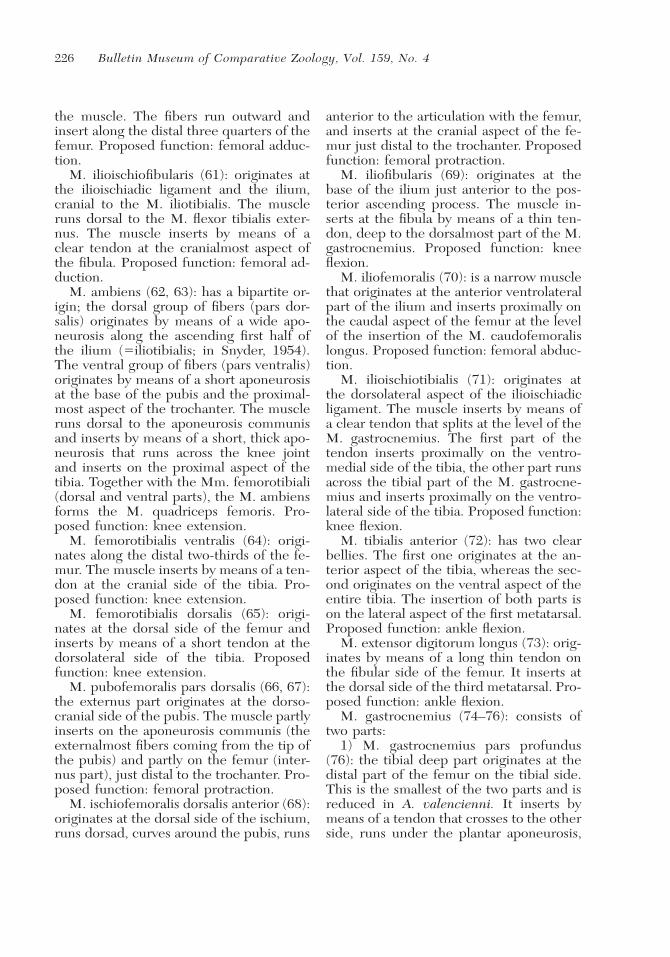
226 Bulletin Museum of Comparative Zoology, Vol. 159, No. 4
the muscle. The fibers run outward andinsert along the distal three quarters of thefemur. Proposed function: femoral adduc-tion.
M. ilioischiofibularis (61): originates atthe ilioischiadic ligament and the ilium,cranial to the M. iliotibialis. The muscleruns dorsal to the M. flexor tibialis exter-nus. The muscle inserts by means of aclear tendon at the cranialmost aspect ofthe fibula. Proposed function: femoral ad-duction.
M. ambiens (62, 63): has a bipartite or-igin; the dorsal group of fibers (pars dor-salis) originates by means of a wide apo-neurosis along the ascending first half ofthe ilium (�iliotibialis; in Snyder, 1954).The ventral group of fibers (pars ventralis)originates by means of a short aponeurosisat the base of the pubis and the proximal-most aspect of the trochanter. The muscleruns dorsal to the aponeurosis communisand inserts by means of a short, thick apo-neurosis that runs across the knee jointand inserts on the proximal aspect of thetibia. Together with the Mm. femorotibiali(dorsal and ventral parts), the M. ambiensforms the M. quadriceps femoris. Pro-posed function: knee extension.
M. femorotibialis ventralis (64): origi-nates along the distal two-thirds of the fe-mur. The muscle inserts by means of a ten-don at the cranial side of the tibia. Pro-posed function: knee extension.
M. femorotibialis dorsalis (65): origi-nates at the dorsal side of the femur andinserts by means of a short tendon at thedorsolateral side of the tibia. Proposedfunction: knee extension.
M. pubofemoralis pars dorsalis (66, 67):the externus part originates at the dorso-cranial side of the pubis. The muscle partlyinserts on the aponeurosis communis (theexternalmost fibers coming from the tip ofthe pubis) and partly on the femur (inter-nus part), just distal to the trochanter. Pro-posed function: femoral protraction.
M. ischiofemoralis dorsalis anterior (68):originates at the dorsal side of the ischium,runs dorsad, curves around the pubis, runs
anterior to the articulation with the femur,and inserts at the cranial aspect of the fe-mur just distal to the trochanter. Proposedfunction: femoral protraction.
M. iliofibularis (69): originates at thebase of the ilium just anterior to the pos-terior ascending process. The muscle in-serts at the fibula by means of a thin ten-don, deep to the dorsalmost part of the M.gastrocnemius. Proposed function: kneeflexion.
M. iliofemoralis (70): is a narrow musclethat originates at the anterior ventrolateralpart of the ilium and inserts proximally onthe caudal aspect of the femur at the levelof the insertion of the M. caudofemoralislongus. Proposed function: femoral abduc-tion.
M. ilioischiotibialis (71): originates atthe dorsolateral aspect of the ilioischiadicligament. The muscle inserts by means ofa clear tendon that splits at the level of theM. gastrocnemius. The first part of thetendon inserts proximally on the ventro-medial side of the tibia, the other part runsacross the tibial part of the M. gastrocne-mius and inserts proximally on the ventro-lateral side of the tibia. Proposed function:knee flexion.
M. tibialis anterior (72): has two clearbellies. The first one originates at the an-terior aspect of the tibia, whereas the sec-ond originates on the ventral aspect of theentire tibia. The insertion of both parts ison the lateral aspect of the first metatarsal.Proposed function: ankle flexion.
M. extensor digitorum longus (73): orig-inates by means of a long thin tendon onthe fibular side of the femur. It inserts atthe dorsal side of the third metatarsal. Pro-posed function: ankle flexion.
M. gastrocnemius (74–76): consists oftwo parts:
1) M. gastrocnemius pars profundus(76): the tibial deep part originates at thedistal part of the femur on the tibial side.This is the smallest of the two parts and isreduced in A. valencienni. It inserts bymeans of a tendon that crosses to the otherside, runs under the plantar aponeurosis,
LIMB MUSCLES IN ANOLIS • Herrel et al. 227
and inserts medially at the level of the fifthmetatarsal.
2) M. gastrocnemius pars fibularis: Thisis the most prominent part of the M. gas-trocnemius and is positioned on the fibularside. It originates on the dorsal tubercle ofthe femur on the fibular side by means ofa thick tendon. This muscle belly can besplit into two parts. The pars major (74)inserts onto the first phalanx of the fourthand fifth toe; the pars minor (75) insertson the first phalanx of the fourth toe. Pro-posed function: ankle extension.
M. flexor digitorum communis (77):consists of two parts. The tibial part orig-inates fleshy at the proximal third alongthe inner aspect of both tibia and fibula.The fibular part originates at the proximalthird along the inner part of the fibula.The two bellies converge onto a tendonthat wraps around the ankle. The tibialpart inserts by means of a long narrow ten-don on the distal phalanges of toes 1–4.The fibular part inserts by means of a longthin tendon at the distal phalanx of thefifth toe. Proposed function: ankle exten-sion and toe flexion.
M. extensor ossi metatarsi hallucis (78):originates at the ventral side of the distaltwo thirds of the fibula and inserts bymeans of a short tendon on the dorsal as-pect on the tibial side of the astragalocal-caneum. Proposed function: ankle exten-sion and rotation.
M. peroneus brevis (79): originates fromthe distal two thirds of the cranial edge ofthe fibula and inserts at the posterodorsalside of the fifth metatarsal. Proposed func-tion: ankle extension.
M. popliteus (80): originates at the me-sial side of the most proximal part of thefibula. The muscle runs obliquely ventradto insert on the mesial side of the proximalfifth of the tibia. Proposed function: tibio-fibular rotation.
M. peroneus longus (81): originates bymeans of a long, narrow tendon on the fib-ular side of the femur. The muscle wrapsaround the ankle and inserts at the ventral
side of the fifth metatarsal. Proposed func-tion: ankle extension.
M. iliofemoralis posterior (not drawn):originates from the posterior part of theilium (at the level of the attachment of theilioischiadic ligament) and from the ventralaspect of the first caudal vertebra. Themuscle inserts onto the femur, proximal tothe tendon of the M. caudofemoralis lon-gus. Proposed function: femoral abduc-tion.
M. ischiofemoralis dorsalis posterior(not drawn): originates at the dorsocaudalside of the ischium and inserts at the dor-socaudal side of the femur. Proposed func-tion: femoral abduction.
M. pronator profundus (not drawn):originates fleshy at the distal quarter onthe mesial aspect of the fibula. The muscleruns obliquely ventrad and inserts on themesial side of the distal fifth of the tibia.Proposed function: tibio-fibular rotation.
Morphometrics
The A. valencienni in our sample weresignificantly larger than the A. sagrei (SVL:F1,16 � 68.35, P � 0.001). However, for itsbody size, A. sagrei had significantly longertibia (analyses of covariance with SVL ascovariate; slope: F1,6 � 1.28, P � 0.30; in-tercept: F1,7 � 15.32, P � 0.006), metatarsi(slope: F1,6 � 0.23, P � 0.65; intercept: F1,7
� 6.59, P � 0.037) and toes on both frontlimbs (slope: F1,6 � 1.16, P � 0.32; inter-cept: F1,7 � 16.89, P � 0.005) and hind-limbs (slope: F1,6 � 0.35, P � 0.58; inter-cept: F1,7 � 7.48, P � 0.029) comparedwith A. valencienni. These results are con-sistent with previous analyses of limb di-mensions in these species (Beuttell andLosos, 1999; Higham et al., 2001).
Muscle Mass and Muscle Massdistribution (Table 1)
The two species differed significantly intotal hindlimb (ANOVA: F1,8 � 13.79, P �0.004) and total forelimb (F1,8 � 25.67, P� 0.001) muscle mass, with A. sagrei hav-ing heavier fore- and hindlimb musclesthan A. valencienni despite its smaller
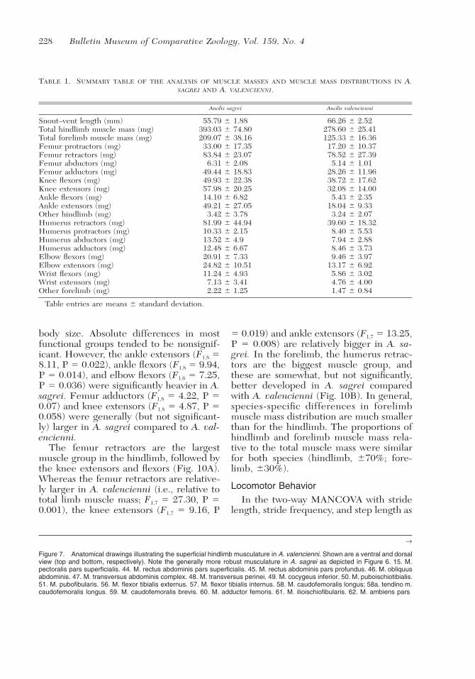
228 Bulletin Museum of Comparative Zoology, Vol. 159, No. 4
TABLE 1. SUMMARY TABLE OF THE ANALYSIS OF MUSCLE MASSES AND MUSCLE MASS DISTRIBUTIONS IN A.SAGREI AND A. VALENCIENNI.
Anolis sagrei Anolis valencienni
Snout–vent length (mm) 55.79 � 1.88 66.26 � 2.52Total hindlimb muscle mass (mg) 393.03 � 74.80 278.60 � 25.41Total forelimb muscle mass (mg) 209.07 � 38.16 125.33 � 16.36Femur protractors (mg) 33.00 � 17.35 17.20 � 10.37Femur retractors (mg) 83.84 � 23.07 78.52 � 27.39Femur abductors (mg) 6.31 � 2.08 5.14 � 1.01Femur adductors (mg) 49.44 � 18.83 28.26 � 11.96Knee flexors (mg) 49.93 � 22.38 38.72 � 17.62Knee extensors (mg) 57.98 � 20.25 32.08 � 14.00Ankle flexors (mg) 14.10 � 6.82 5.43 � 2.35Ankle extensors (mg) 49.21 � 27.05 18.04 � 9.33Other hindlimb (mg) 3.42 � 3.78 3.24 � 2.07Humerus retractors (mg) 81.99 � 44.94 39.60 � 18.32Humerus protractors (mg) 10.33 � 2.15 8.40 � 5.53Humerus abductors (mg) 13.52 � 4.9 7.94 � 2.88Humerus adductors (mg) 12.48 � 6.67 8.46 � 3.73Elbow flexors (mg) 20.91 � 7.33 9.46 � 3.97Elbow extensors (mg) 24.82 � 10.51 13.17 � 6.92Wrist flexors (mg) 11.24 � 4.93 5.86 � 3.02Wrist extensors (mg) 7.13 � 3.41 4.76 � 4.00Other forelimb (mg) 2.22 � 1.25 1.47 � 0.84
Table entries are means � standard deviation.
→
Figure 7. Anatomical drawings illustrating the superficial hindlimb musculature in A. valencienni. Shown are a ventral and dorsalview (top and bottom, respectively). Note the generally more robust musculature in A. sagrei as depicted in Figure 6. 15. M.pectoralis pars superficialis. 44. M. rectus abdominis pars superficialis. 45. M. rectus abdominis pars profundus. 46. M. obliquusabdominis. 47. M. transversus abdominis complex. 48. M. transversus perinei. 49. M. cocygeus inferior. 50. M. puboischiotibialis.51. M. pubofibularis. 56. M. flexor tibialis externus. 57. M. flexor tibialis internus. 58. M. caudofemoralis longus; 58a. tendino m.caudofemoralis longus. 59. M. caudofemoralis brevis. 60. M. adductor femoris. 61. M. ilioischiofibularis. 62. M. ambiens pars
body size. Absolute differences in mostfunctional groups tended to be nonsignif-icant. However, the ankle extensors (F1,8 �8.11, P � 0.022), ankle flexors (F1,8 � 9.94,P � 0.014), and elbow flexors (F1,6 � 7.25,P � 0.036) were significantly heavier in A.sagrei. Femur adductors (F1,8 � 4.22, P �0.07) and knee extensors (F1,8 � 4.87, P �0.058) were generally (but not significant-ly) larger in A. sagrei compared to A. val-encienni.
The femur retractors are the largestmuscle group in the hindlimb, followed bythe knee extensors and flexors (Fig. 10A).Whereas the femur retractors are relative-ly larger in A. valencienni (i.e., relative tototal limb muscle mass; F1,7 � 27.30, P �0.001), the knee extensors (F1,7 � 9.16, P
� 0.019) and ankle extensors (F1,7 � 13.25,P � 0.008) are relatively bigger in A. sa-grei. In the forelimb, the humerus retrac-tors are the biggest muscle group, andthese are somewhat, but not significantly,better developed in A. sagrei comparedwith A. valencienni (Fig. 10B). In general,species-specific differences in forelimbmuscle mass distribution are much smallerthan for the hindlimb. The proportions ofhindlimb and forelimb muscle mass rela-tive to the total muscle mass were similarfor both species (hindlimb, �70%; fore-limb, �30%).
Locomotor Behavior
In the two-way MANCOVA with stridelength, stride frequency, and step length as

LIMB MUSCLES IN ANOLIS • Herrel et al. 229
ventralis. 63. M. ambiens pars dorsalis. 64. M. femorotibialis pars ventralis. 65. M. femorotibialis pars dorsalis. 66. M. pubofe-moralis pars dorsalis externus. 67. M. pubofemoralis pars dorsalis internus. 68. M. ischiofemoralis dorsalis pars anterior. 69. M.iliofibularis. 70. M. iliofemoralis. 71. M. ilioischiotibialis. 72. M. tibialis anterior. 73. M. extensor digitorum longus. 74. M. gastroc-nemius pars major. 75. M. gastrocnemius pars minor. 77. M. flexor digitorum communis. 78. M. extensor ossi metatarsi hallu-cis.79. M. peroneus brevis. 80. M. popliteus. 81. M. peroneus longus. 82. M. longissimus. 83. M. spinalis. 84. M. iliocostalis. 85.M. longus cauda. 86. M. iliocaudalis. 87. M. ischiocaudalis. V, ligamentum ilioischiadum; T, pectineal tubercle. U, ilium. W, tibia.X, fibula. Y, astragalocalcaneum. Z, processus spinosus vertebra. * Deeper structures, not labeled.
230 Bulletin Museum of Comparative Zoology, Vol. 159, No. 4
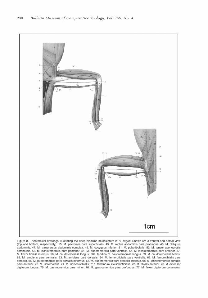
Figure 8. Anatomical drawings illustrating the deep hindlimb musculature in A. sagrei. Shown are a ventral and dorsal view(top and bottom, respectively). 15. M. pectoralis pars superficialis. 45. M. rectus abdominis pars profundus. 46. M. obliquusabdominis. 47. M. transversus abdominis complex. 49. M. cocygeus inferior. 51. M. pubofibularis. 52. M. tensor aponeurosiscommunis. 53. M. ischiofemoralis pars posterior. 54. M. pubofemoralis pars ventralis. 55. M. ischiofemoralis pars anterior. 57.M. flexor tibialis internus. 58. M. caudofemoralis longus; 58a. tendino m. caudofemoralis longus. 59. M. caudofemoralis brevis.62. M. ambiens pars ventralis. 63. M. ambiens pars dorsalis. 64. M. femorotibialis pars ventralis. 65. M. femorotibialis parsdorsalis. 66. M. pubofemoralis pars dorsalis externus. 67. M. pubofemoralis pars dorsalis internus. 68. M. ischiofemoralis dorsalispars anterior. 70. M. iliofemoralis. 71. M. ilioischiotibialis; 71a. tendino m. ilioischiotibialis. 72. M. tibialis anterior. 73. M. extensordigitorum longus. 75. M. gastrocnemius pars minor. 76. M. gastrocnemius pars profundus. 77. M. flexor digitorum communis.
LIMB MUSCLES IN ANOLIS • Herrel et al. 231
←
78. M. extensor ossi metatarsi hallucis. 79. M. peroneus brevis. 80. M. popliteus. 82. M. longissimus. 83. M. spinalis. 84. M.iliocostalis. 85. M. longus cauda. 86. M. iliocaudalis. 87. M. ischiocaudalis. III, ligamentum puboischiadum pars lateralis; V,ligamentum ilioischiadum; VI, aponeurosis communis; Q, ischium; R, hypoischium; T, pectineal tubercle; U, ilium; V, femur; W,tibia; X, fibula; Y, astragalocalcaneum; Z, processus spinosus vertebra. * Deeper structures, not labeled.
dependent variables, stride speed as co-variate and species and dowel as factor,none of the interaction effects were sig-nificant (all P � 0.07). Average velocityover a stride had a significant effect on thespatiotemporal gait characteristics (Wilks’� � 0.044, F3,6 � 42.99, P � 0.0001). Inaddition, gait characteristics differed sig-nificantly between species (Wilks’ � �0.064, F3,6 � 29.02, P � 0.001; Fig. 11).For a given speed, A. sagrei takes longerstrides and longer steps at lower frequen-cies compared with A. valencienni. Differ-ences between substrates were also signif-icant (Wilks’ � � 0.086, F3,6 � 21.36, P �0.001; Fig. 11). Locomotion on broaddowels was associated with higher step andstride lengths, but lower stride frequenciescompared with narrow dowels. UnivariateF tests showed that species and substrateeffects were highly significant for all pa-rameters tested (Table 2).
DISCUSSION
A number of distinct and striking dif-ferences in the morphology of the pectoralgirdle and associated appendicular mus-culoskeletal system were observed whencomparing two distinct Anolis species. Thepectoral girdle itself, for example, is rela-tively narrower and longer in A. valencien-ni compared with A. sagrei and is reflectedin the more gracile overall body shape inthe former species (see also Beuttell andLosos, 1999). The difference in bodyshape itself can be related to selection forincreased stability on narrow substrates inA. valencienni (Losos and Irschick, 1996;Losos and Sinervo, 1989). Additionally, itallows this species and other twig anoleswith a similar body shape to remain crypticagainst its preferred substrate of narrowbranches and twigs (Huyghe et al., 2007;
Irschick and Losos, 1996; Vanhooydoncket al., 2007).
Our results on muscle mass show thattotal forelimb muscle mass is considerablygreater in A. sagrei despite its smallerbody size (Table 1). Although not differentin absolute size, the relative contributionof the humerus retractors to the total fore-limb muscle mass is greater (but not sig-nificantly so) in this species as well. Thiswas unexpected given the subordinate roleof the forelimbs during locomotion on hor-izontal substrates, more characteristic ofthe habitat use of A. sagrei, and the im-portance of humerus retraction duringclimbing (Zaaf et al., 1999, 2001), which isexpected to be associated with the morearboreal lifestyle of A. valencienni. How-ever, twig anoles like A. valencienni, de-spite being highly arboreal, do spend a sig-nificant proportion of their time on hori-zontal or inclined substrates (Irschick andLosos, 1996; Mattingly and Jayne, 2004,2005). Thus, humerus retraction might beless important for these species than ini-tially expected. Anolis sagrei, on the otherhand, although spending a considerableamount of time on the ground, does runup the vertical bases of tree trunks (i.e.,trunk-ground anoles use steeper surfacesthan other ecomorphs; Mattingly and Jayne,2004) which might explain the importanceof humerus retraction in this species. Thesignificantly greater absolute mass of theelbow flexors (also important duringclimbing) in A. sagrei corroborates thisfinding. Additional data on other trunk-ground versus twig anoles are needed,however, to test the generality of thesefindings.
As predicted a priori, the anatomy of thehindlimb muscles also differs between thetwo species, with a larger absolute overall

232 Bulletin Museum of Comparative Zoology, Vol. 159, No. 4
Figure 9. Anatomical drawings illustrating the deep hindlimb musculature in A. valencienni. Shown are a ventral and dorsalview (top and bottom, respectively). Note the subtle differences in the insertion of the m. caudofemoralis and its accessorytendon in A. valencienni compared with A. sagrei, as depicted in Figure 8. 15. M. pectoralis pars superficialis. 45. M. rectusabdominis pars profundus. 46. M. obliquus abdominis. 47. M. transversus abdominis complex. 49. M. cocygeus inferior. 51. M.pubofibularis. 52. M. tensor aponeurosis communis. 53. M. ischiofemoralis pars posterior. 54. M. pubofemoralis pars ventralis.55. M. ischiofemoralis pars anterior. 57. M. flexor tibialis internus. 58. M. caudofemoralis longus; 58a. tendino m. caudofemoralis

LIMB MUSCLES IN ANOLIS • Herrel et al. 233
TABLE 2. SUMMARY OF UNIVARIATE ANALYSIS ON SIZE-CORRECTED GAIT CHARACTERISTICS FOR A. SAGREI ANDA. VALENCIENNI MOVING ON TWO DIFFERENT SUBSTRATES.
df F P
DowelStride length 1, 8 24.41 0.001Stride frequency 1, 8 40.28 �0.0001Step length 1, 8 14.02 0.006
SpeciesStride length 1, 8 55.25 �0.0001Stride frequency 1, 8 11.98 0.009Step length 1, 8 8.48 0.02
Stride speedStride length 1, 8 0.26 0.62Stride frequency 1, 8 22.63 0.001Step length 1, 8 3.68 0.09
Note that all kinematic variables were expressed relative to hindlimb length. df, degrees of freedom.
←
longus. 59. M. caudofemoralis brevis. 62. M. ambiens pars ventralis. 63. M. ambiens pars dorsalis. 64. M. femorotibialis parsventralis. 65. M. femorotibialis pars dorsalis. 66. M. pubofemoralis pars dorsalis externus. 67. M. pubofemoralis pars dorsalisinternus. 68. M. ischiofemoralis dorsalis pars anterior. 70. M. iliofemoralis. 71. M. ilioischiotibialis; 71a. tendino m. ilioischiotibialis.72. M. tibialis anterior. 73. M. extensor digitorum longus. 75. M. gastrocnemius pars minor. 76. M. gastrocnemius pars profundus.77. M. flexor digitorum communis. 78. M. extensor ossi metatarsi hallucis. 79. M. peroneus brevis. 80. M. popliteus. 82. M.longissimus. 83. M. spinalis. 84. M. iliocostalis. 85. M. longus cauda. 86. M. iliocaudalis. 87. M. ischiocaudalis. III, ligamentumpuboischiadum pars lateralis; V, ligamentum ilioischiadum; VI, aponeurosis communis; Q, ischium; R, hypoischium; T, pectinealtubercle; U, ilium; V, femur; W, tibia; X, fibula; Y, astragalocalcaneum; Z, processus spinosus vertebra. * Deeper structures, notlabeled.
hindlimb muscle mass as well as signifi-cantly larger ankle extensors and ankleflexors observed in A. sagrei (Table 1).Muscle mass allocation also differed sig-nificantly between species, with A. sagreihaving relatively larger knee and ankle ex-tensors and A. valencienni having relative-ly larger femur retractors. Also, the shapeof the pelvic girdle shows remarkable dif-ferences in the two species. Whereas thepelvis is longer and narrower in A. valen-cienni, the ilium is elongated and inclinedmore dorsally in A. sagrei (see also Beut-tell and Losos, 1999). Whereas the overallshape differences in the pelvis can be re-lated to overall differences in body shape(longer and narrower in A. valencienni),the differences in ilial structure can be di-rectly related to differences in locomotormode. The ilium is the principal area ofinsertion for the knee extensors, and theknee extensors are the principal determi-nants of variation in sprint speed, jump,
and acceleration capacity in Anolis lizards(James et al., 2007; Vanhooydonck et al.,2006a). The longer the ilium, the morespace is available for the knee extensors.Indeed, A. sagrei has more massive kneeextensors in both absolute and relativeterms compared with A. valencienni. An-olis valencienni, on the other hand, ap-pears to rely more on femoral retraction,as suggested by the relatively greater al-location of muscle mass to femur retrac-tors and the distal shift of the insertion ofthe M. caudofemoralis, thus providing fora greater moment arm and, consequently,moment around the hip joint.
Given the importance of keeping thecenter of mass close to the middle of thesubstrate when running on narrow branch-es, relying on knee extension for propul-sion could effectively be less optimal be-cause of the induced lateral displacementaway from the center of the branch, atleast when the foot is positioned at the lev-

234 Bulletin Museum of Comparative Zoology, Vol. 159, No. 4

LIMB MUSCLES IN ANOLIS • Herrel et al. 235
←
Figure 10. Graphs illustrating the relative muscle mass distribution expressed as the proportion of the total hindlimb (A) orforelimb (B) muscle mass. Data are represented as means � standard deviation for the two Anolis species included in our study.Whereas differences in muscle mass allocation are strikingly different for the hindlimb muscles, differences for the forelimb areless conspicuous. Solid circles, A. sagrei; white circles, A. valencienni. Also plotted are literature data on hindlimb muscle massdistribution taken from Snyder (1954). Note, however, that Snyder did not consider femur abductors.
el of or in front of the pelvis (Spezzanoand Jayne, 2004). This appears to be re-flected in the morphology of the hindlimbmuscles and locomotor patterns in the twospecies. Indeed, an additional importantdifference between the two species in lo-comotor style is the reduced protraction ofthe limb during the swing phase in A. val-encienni, causing it to take smaller stepsand strides and resulting in a placement ofthe foot behind the pelvic girdle. Again,this can be coupled to differences in hind-limb morphology between the two species.The insertion of the accessory tendon ofthe M. caudofemoralis is displaced moredistally from the knee joint in A. valen-cienni, thus effectively preventing knee ex-tension when the femur is protracted. Themorphology is however different in A. sa-grei, with the insertion being at the levelof the joint, thus allowing knee extensionand femur protraction at the same time.This results in greater step and stridelengths and ultimately also sprint speeds.The cost of the greater protraction and de-pendence on knee extension is a potentialdecrease in stability because of the greaterlateral displacement of the center of massaway from the center of the branch.
A comparison of our data on musclemass distribution with previously pub-lished data for several species of iguanid(sensu lato) and agamid lizards indicatesthat the differences between the two spe-cies of anole included here are nearly asgreat as between a dedicated terrestrial liz-ard (Crotaphytus collaris) and an exclu-sively arboreal species (Iguana iguana)(see Fig. 10A). Moreover, differences ob-served between the two anole ecomorphstend to mimic those (although being dif-ferent in absolute terms) observed be-tween the terrestrial and the arboreal spe-
cies, with A. sagrei being more similar toC. collaris and A. valencienni being moresimilar to I. iguana. Among iguanid liz-ards, Sceloporus undulatus, being semi-ar-boreal, resembles A. valencienni and I.iguana in its muscle mass distribution.Basiliscus basiliscus, being ecologicallymore similar to A. sagrei in that it usesboth arboreal and terrestrial substrates, isgenerally intermediate between the morededicated ground dweller C. collaris andthe arboreal I. iguana. Among the agamidlizards, Amphibolurus cristatus (nowplaced in the genus Ctenophorus) is un-usual in that it resembles more arboreallizards like I. iguana and A. valencienni insome features of the hindlimb musclemass distribution, despite being a largelyterrestrial lizard. Like A. valencienni andI. iguana, A. cristatus appears to rely pre-dominantly on femur retraction ratherthan knee and ankle extension for gener-ating propulsion. Clearly, data for addi-tional agamid and iguanid lizards are needto investigate the generality of these pat-terns and to quantitatively test for associ-ations between muscle morphology andhabitat use in a comparative context.
Our data show how an understanding ofthe morphology of the locomotor appara-tus might help explain the correlated evo-lution of morphology, performance, loco-motor style (i.e., gait characteristics) andhabitat use in Anolis lizards. In providingthis detailed morphological account, wehope to provide a basis for future studiesinvestigating the morphology of the mus-culoskeletal system and its role in the evo-lution of locomotor performance and hab-itat use in Anolis lizards and to show thatmorphological adaptations to habitat usego beyond mere external differences inlimb size and shape.

236 Bulletin Museum of Comparative Zoology, Vol. 159, No. 4
Figure 11. Graphs illustrating the differences in spatiotemporal gait characteristics in A. sagrei and A. valencienni while movingacross two different substrates. Data are represented as means � standard deviation. Illustrated are the stride length (A), stridefrequency (B), and step length (C) expressed in hindlimb lengths. Thus, for a given hindlimb length, A. sagrei takes larger stepsand strides on both substrates. Broad substrates are associated with greater step and stride lengths and lower stride frequenciesin both species. hl, hindlimb.

LIMB MUSCLES IN ANOLIS • Herrel et al. 237
ACKNOWLEDGEMENTS
B.V. is a postdoctoral researcher of theFund for Scientific Research, Flanders(FWO-Vl). D.J.I. was supported by IOB0421917. All figures drawn by J.P.
LITERATURE CITEDABDALA, V., AND S. MORO. 2006. Comparative my-
ology of the forelimb of Liolaemus sand lizards(Liolaemidae). Acta Zoologica, 87: 1–12.
AUTUMN, K., S. T. HSIEH, D. M. DUDEK, J. CHEN,C. CHITAPHAN, AND R. J. FULL. 2006. Dynamicsof geckos running vertically. Journal of Experi-mental Biology, 209: 260–272.
BEUTTELL, K., AND J. B. LOSOS. 1999. Ecologicalmorphology of Caribbean anoles. HerpetologicalMonographs, 13: 1–28.
HIGHAM, T. E., M. S. DAVENPORT, AND B. C. JAYNE.2001. Maneuvering in an arboreal habitat: theeffects of turning angle on the locomotion ofthree sympatric ecomorphs of Anolis lizards.Journal of Experimental Biology, 204: 4141–4155.
, AND B. C. JAYNE. 2004. In vivo muscle ac-tivity in the hindlimb of the arboreal lizard, Cha-maeleo calyptratus: general patterns and the ef-fects of incline. Journal of Experimental Biology,207: 249–261.
HUYGHE, K., A. HERREL, B. VANHOOYDONCK, J. J.MEYERS, AND D. J. IRSCHICK. 2007. Microhabi-tat use, diet, and performance data on the His-paniolan twig anole, Anolis sheplani. Zoology,110: 2–8.
IRSCHICK, D. J., AND J. B. LOSOS. 1996. Morphology,ecology, and behavior of the twig anole, Anolisangusticeps, pp. 291–301. In R. Powell and R.W. Henderson (eds.), Contributions to West In-dian Herpetology: A Tribute to Albert Schwartz.Ithaca, New York: Society for the Study of Am-phibians and Reptiles.
, AND . 1998. A comparative analysis ofthe ecological significance of maximal locomotorperformance in Caribbean Anolis lizards. Evo-lution, 52: 219–226.
, AND . 1999. Do lizards avoid habitatsin which performance is submaximal? The rela-tionship between sprinting capabilities and struc-tural habitat use in Caribbean anoles. AmericanNaturalist, 154: 293–305.
JAMES, R. S., C. A. NAVAS, AND A. HERREL. 2007.How important are skeletal muscle mechanics insetting limits on jumping performance? Journalof Experimental Biology, 210: 923–933.
JENKINS, F. A., AND G. E. GOSLOW. 1983. The func-tional anatomy of the shoulder of the savannahmonitor lizard (Varanus exanthematicus). Journalof Morphology, 175: 195–216.
LANDSMEER, J. M. F. 1984. Morphology of the an-terior limb in relation to sprawling gait in Var-
anus. Symposia Zoological Society London, 52:27–45.
. 1990. Functional morphology of the hind-limb in some lacertilia. European Journal ofMorphology, 28: 3–34.
LOSOS, J. B. 1990a. The evolution of form and func-tion: morphology and locomotor performanceability in West Indian Anolis lizards. Evolution,44: 1189–1203.
. 1990b. Ecomorphology, performance capa-bility, and scaling of West Indian Anolis lizards:an evolutionary analysis. Ecological Monographs,60: 369–388.
, AND D. J. IRSCHICK. 1996. The effect ofperch diameter on the escape behavior of Anolislizards: laboratory-based predictions and fieldtests. Animal Behaviour, 51: 593–602.
, T. R. JACKMAN, A. LARSON, K. DE QUEIROZ,AND L. RODRIGUEZ-SCHETTINO. 1998. Historicalcontingency and determinism in replicated adap-tive radiations of island lizards. Science, 279:2115–2118.
, AND B. SINERVO. 1989. The effects of mor-phology and perch diameter on sprint perfor-mance of Anolis lizards. Journal of ExperimentalBiology, 145: 23–30.
, K. I. WARHEIT, AND T. W. SCHOENER. 1997.Adaptive differentiation following experimentalisland colonization in Anolis lizards. Nature, 387:70–73.
MATTINGLY, W. B., AND B. C. JAYNE. 2004. Resourceuse in arboreal habitats: structure affects loco-motion of four ecomorphs of Anolis lizards. Ecol-ogy, 85: 1111–1124.
, AND . 2005. The choice of arborealescape paths and its consequences for the loco-motor behaviour of four species of Anolis lizards.Animal Behaviour, 70: 1239–1250.
MORO, S., AND V. ABDALA. 2004. Analisis descriptivode la miologıa flexora y extensora del miembroanterior de Polychrus acutirostris (Squamata,Polychrotidae). Papeis Avulsos de Zoologia, 44:81–90.
NICHOLSON, K. E., R. E. GLOR, J. J. KOLBE, A. LAR-SON, S. B. HEDGES, AND J. B. LOSOS. 2005.Mainland colonization by island lizards. Journalof Biogeography, 32: 929–938.
RUSSELL, A. P. 1988. Limb muscles in relation to liz-ard systematics: a reappraisal, pp. 119–218. In R.Estes and G. Pregill (eds.), Phylogenetic Rela-tionships of Lizard Families: Essays Commem-orating Charles L. Camp. Palo Alto, California:Stanford University Press.
SCHLUTER, D. 2000. The Ecology of Adaptive Radi-ation. Oxford, United Kingdom: Oxford Univer-sity Press.
SINERVO, B., AND J. B. LOSOS. 1991. Walking thetight rope: arboreal sprint performance amongSceloporus occidentalis lizard populations. Ecol-ogy, 72: 1225–1233.
SNYDER, R. C. 1954. The anatomy and function of

238 Bulletin Museum of Comparative Zoology, Vol. 159, No. 4
the pelvic girdle and hindlimb in lizard locomo-tion. American Journal of Anatomy, 95: 1–45.
SPEZZANO, L. C., AND B. C. JAYNE. 2004. The effectsof surface diameter and incline on the hindlimbkinematics of an arboreal lizard (Anolis sagrei).Journal of Experimental Biology, 207: 2115–2131.
TORO, E., A. HERREL, B. VANHOOYDONCK, AND D.J. IRSCHICK. 2003. A biomechanical analysis ofintra- and interspecific scaling of jumping bio-mechanics and morphology in Caribbean Anolislizards. Journal of Experimental Biology, 206:2641–2652.
VANHOOYDONCK, B., A. ANDRONESCU, A. HERREL,AND D. J. IRSCHICK. 2005. Effects of substratestructure on speed and acceleration capacity inclimbing geckos. Biological Journal of the Lin-nean Society London, 85: 385–393.
, A. HERREL, R. VAN DAMME, AND D. J. IR-SCHICK. 2006a. The quick and the fast: the evo-lution of acceleration capacity in Anolis lizards.Evolution, 60: 2137–2147.
, , AND D. J. IRSCHICK. 2006b. Out on
a limb: the differential effect of substrate diam-eter on acceleration capacity in Anolis lizards.Journal of Experimental Biology, 209: 4515–4523.
, , AND . 2007. Determinants ofsexual differences in escape behavior in Anolislizards: a comparative approach. Integrative andComparative Biology, 47: 200–210.
WILLIAMS, E. E. 1983. Ecomorphs, faunas, islandsize, and diverse end points in island radiationsof Anolis, pp. 326–370. In R. B. Huey, E. R.Pianka, and T. W. Schoener (eds.), Lizard Ecol-ogy: Studies of a Model Organism. Cambridge,Massachusetts: Harvard University Press.
ZAAF, A., A. HERREL, P. AERTS, AND F. DE VREE.1999. Morphology and morphometrics of the ap-pendicular musculature in geckoes with differentlocomotor habits (Lepidosauria). Zoomorpholo-gy, 119: 9–22.
, R. VAN DAMME, A. HERREL, AND P. AERTS.2001. Spatio temporal gait characteristics of leveland vertical locomotion in a level-running and aclimbing gecko. Journal of Experimental Biology,204: 1233–1246.