anatomically preserved strobili and leaves from the...
TRANSCRIPT
Anatomically preserved "strobili" and leaves fromthe Permian of China (Dorsalistachyaceae, fam.nov.) broaden knowledge of Noeggerathiales andconstrain their possible taxonomic affinitiesWang, Shi-Jun; Bateman, Richard M.; Spencer, Alan R.t.; Wang, Jun; Shao, Longyi; Hilton,JasonDOI:10.3732/ajb.1600371
License:Creative Commons: Attribution-NonCommercial (CC BY-NC)
Document VersionPublisher's PDF, also known as Version of record
Citation for published version (Harvard):Wang, S-J, Bateman, RM, Spencer, AR, Wang, J, Shao, L & Hilton, J 2017, 'Anatomically preserved "strobili"and leaves from the Permian of China (Dorsalistachyaceae, fam. nov.) broaden knowledge of Noeggerathialesand constrain their possible taxonomic affinities' American Journal of Botany, vol 104, no. 1, pp. 127-149. DOI:10.3732/ajb.1600371
Link to publication on Research at Birmingham portal
General rightsUnless a licence is specified above, all rights (including copyright and moral rights) in this document are retained by the authors and/or thecopyright holders. The express permission of the copyright holder must be obtained for any use of this material other than for purposespermitted by law.
•Users may freely distribute the URL that is used to identify this publication.•Users may download and/or print one copy of the publication from the University of Birmingham research portal for the purpose of privatestudy or non-commercial research.•User may use extracts from the document in line with the concept of ‘fair dealing’ under the Copyright, Designs and Patents Act 1988 (?)•Users may not further distribute the material nor use it for the purposes of commercial gain.
Where a licence is displayed above, please note the terms and conditions of the licence govern your use of this document.
When citing, please reference the published version.
Take down policyWhile the University of Birmingham exercises care and attention in making items available there are rare occasions when an item has beenuploaded in error or has been deemed to be commercially or otherwise sensitive.
If you believe that this is the case for this document, please contact [email protected] providing details and we will remove access tothe work immediately and investigate.
Download date: 23. Jul. 2018
A M E R I C A N J O U R N A L O F B OTA NY 104 (1): 127 – 149 , 2017; http://www.amjbot.org/ © 2017 Wang et al. Published by the Botanical Society of America. This work is licensed
under a Creative Commons Attribution License (CC-BY-NC). • 127
A M E R I C A N J O U R N A L O F B O T A N Y
R E S E A R C H A R T I C L E
1 Manuscript received 18 October 2016; revision accepted 22 November 2016.
2 State Key Laboratory of Systematic and Evolutionary Botany, Institute of Botany, Chinese
Academy of Sciences, Xiangshan, Beijing 100093, P. R. China;
3 Royal Botanic Gardens Kew, Richmond, Surrey, TW9 3DS, UK;
4 Department of Earth Sciences and Engineering, Imperial College London, London, SW7
2BP, UK;
5 State Key Laboratory of Palaeobiology and Stratigraphy, Nanjing Institute of Geology and
Palaeontology, Chinese Academy of Sciences, Nanjing 210008, P. R. China;
6 State Key Laboratory of Coal Resources and Safe Mining, School of Geosciences and Surveying
Engineering, China University of Mining and Technology, Beijing 100083, P. R. China; and
7 Birmingham Institute of Forestry Research & School of Geography, Earth and Environmental
Sciences, University of Birmingham, Edgbaston, Birmingham, B15 2TT, UK
8 Author for correspondence (e-mail: [email protected])
doi:10.3732/ajb.1600371
Anatomically preserved “strobili” and leaves from the Permian of China (Dorsalistachyaceae, fam. nov.) broaden knowledge of Noeggerathiales and constrain their possible taxonomic affi nities 1 Shi-Jun Wang 2 , Richard M. Bateman 3 , Alan R. T. Spencer 4 , Jun Wang 5 , Longyi Shao 6 , and Jason Hilton 7,8
PREMISE OF THE STUDY: Noeggerathiales are an extinct group of heterosporous shrubs and trees that were widespread and diverse during the Pennsylvanian–
Permian Epochs (323–252 Ma) but are of controversial taxonomic affi nity. Groups proposed as close relatives include leptosporangiate ferns, sphenopsids,
progymnosperms, or the extant eusporangiate fern Tmesipteris . Previously identifi ed noeggerathialeans lacked anatomical preservation, limiting taxo-
nomic comparisons to their external morphology and spore structure. We here document from the upper Permian of China the fi rst anatomically pre-
served noeggerathialeans, which enhance the perceived distinctiveness of the group and better indicate its systematic affi nity.
METHODS: We describe in detail the newly discovered, anatomically preserved heterosporous strobilus Dorsalistachya quadrisegmentorum , gen. et sp. nov.,
and redescribe its suspected foliar correlate, the pinnate leaf Plagiozamites oblongifolius .
KEY RESULTS: Plagiozamites possesses an omega ( Ω )-shaped vascular trace and prominent cortical secretory cavities—a distinctive anatomical organiza-
tion that is echoed in the newly discovered strobili. Dorsalistachya strobili bear highly dissected sporophylls alternately in two vertical rows, suggesting
that they are homologs of leaf pinnae. If so, the “strobilus” is strictly a pseudostrobilus and consists of sporangium-bearing units that are one hierarchical
level below true sporophylls. The “sporophylls” bear four microsporangia on the lower (abaxial) surface, occasionally interspersed with short longitudi-
nal rows of megasporangia. A single functional megaspore develops within each winged megasporangium, suggesting adaptation for dispersal as a
single unit.
CONCLUSIONS: Dorsalistachya presents a unique combination of reproductive features that amply justifi es establishment of a new family, Dorsalistachyaceae. Noeg-
gerathiales represent a distinct taxonomic Order of free-sporing plants that most resembles early-divergent eusporangiate ferns and the more derived among the
extinct progymnosperms. By the early Permian, noeggerathialeans had attained levels of reproductive sophistication similar to the most derived among the Pa-
leozoic sphenophytes and lycophytes, but their heterosporous life history may have contributed to their extinction during the Triassic climatic aridifi cation.
KEY WORDS 3D reconstruction; Dorsalistachya ; Dorsalistachyaceae; eusporangiate fern; heterospory; Noeggerathiales; paleobotany; Permian; phylogeny;
progymnosperm; Xuanwei Formation
Approximately half of the major groups of land-plants recognized today have left no close living relatives (e.g., Hilton and Bateman, 2006 ). However, gradually characterizing such groups until they can take their place in rigorous evolutionary studies requires pains-taking paleobotanical research, building conceptual whole-plants from exceptionally preserved examples of their component organs ( Bateman and Hilton, 2009 ). Our knowledge of land-plant phylog-eny would remain rudimentary even today if paleobotanists had not recognized and circumscribed fossil groups that provide phylo-genetic bridges between extant groups (e.g., Taylor et al., 2009 ): rhynio-phytes linking bryophytes with lycophytes, progymnosperms linking ferns with seed-plants, and—within seed-plants—pteridosperms

128 • A M E R I C A N J O U R N A L O F B OTA NY
linking several disparate groups of extant gymnosperms and the angiosperms. One major group that, until now, has evaded such confi dent phylogenetic placement is the Noeggerathiales sensu Němejc, 1931 (= Noeggerathiopsida sensu Boureau, 1964 ); a group for which we suggest the colloquial name 'bottle-brush ferns'.
Extending from the Mississippian to the Triassic, noeggerathia-lean fossils are a particularly important component of peat-forming plant communities in the Permian fossil fl oras of China ( Wang et al., 2009 , 2012 ). Recent research on in situ fossil forests has reconstructed members of the Noeggerathiales as fern-like trees that produced a dense crown of fern-/cycad-like leaves and elongate strobili of crowded sporophylls ( Wang et al., 2009 ; Wang et al., 2012 ). Although these investigations provided important information on the growth archi-tecture of members of the Noeggerathiales, the absence of anatomically preserved fossils precluded detailed comparison with other phyloge-netically important plant groups. Consequently, noeggerathialeans have long been a ‘phylogenetic football’; they have been linked with an extraordinarily wide range of major groups, including leptospo-rangiate ferns ( Němejc, 1931 ), sphenopsids ( Browne, 1933 ), pro-gymnosperms ( Beck, 1976 , 1981 ; Meyen, 1987 ; Taylor et al., 2009 ), or ranked alongside these clades as a distinct group in its own right ( Boureau, 1964 ); comparison has even been made with the primitive extant eusporangiate fern Tmesipteris Bernh. ( Bierhorst, 1971 ).
Here, we document for the fi rst time anatomically preserved noeggerathialean leaves, rachises, and strobili, whose spatial juxta-position and anatomical similarity together suggest that they repre-sent diff erent parts of the same whole-plant species ( Bateman and Hilton, 2009 ). Th e diffi culty that we have experienced in placing taxonomically these morphologically disparate organs prompts a broader review of the classifi cation and potential evolutionary sig-nifi cance of Noeggerathiales.
MATERIALS AND METHODS
Geological context — Th e plant fossils described here were found in Guizhou Province, southwestern China ( Wang et al., 2011 ; Fig. 1 ), occurring within the Xuanwei Formation, which extends through western Guizhou and eastern Yunnan provinces. Th ese deposits date to the Wuchiapingian to Changhsingian stages of the Lopin-gian Epoch (late Permian Period), ca 260–252 Ma ( Seyfullah et al., 2010 ; Neregato et al., 2016 ). They consist of gray sandstones, gray siltstones, dark gray mudstones, and abundant coal seams, with some pebbly conglomerates in the lower part of the sequence ( Wang et al., 2011 ). Th e formation represents a series of deposi-tional environments that formed along the eastern side of the Kangdian Oldland landmass, and includes terrestrial, fl uvial, peat-forming mire, and deltaic facies with periodic marginal marine in-fl uences, as well as pebble-conglomerate tuff aceous horizons that contain anatomically preserved plants ( Seyfullah et al., 2010 ; Wang et al., 2011 ; Neregato et al., 2016 ). Th e origin of the tuff aceous sedi-ments is unknown, as they have only been found ex situ, but it is most likely related to the later phases of the volcanism that em-placed the underlying Emeishan Basalt ( Bond et al., 2010 ).
Exceptionally preserved noeggerathialean cones plus several iso-lated rachises were collected at Huopu coal mine (25 ° 39 ′ 31 ′ ′ N 104 ° 25 ′ 04 ′ ′ E; Fig. 1 ). Th ey are here compared with leaves of Plagio-zamites oblongifolius Halle ( Guo et al., 1990 ) that were obtained from nearby compression/impression fl oras within the same for-mation. Th is particular leaf-species was fi rst described as a cycad,
even though the leaf-genus Plagiozamites is more commonly inter-preted as being of noeggerathialean affi nity ( Wang, 2008 ).
Plant organs preserved as two-dimensional compression–impression fossils are abundant in the Xuanwei Formation ( Zhao et al., 1980 ), but anatomical preservation is rare. Such preservation was facilitated by dissolution and redeposition of calcium carbonate within tuff aceous sediments; permineralization occurred rapidly af-ter deposition, thus limiting plant decay ( Neregato et al., 2016 ).
Specimens — Th e present specimens were preserved in a large block of volcaniclastic tuff (numbered as HP2007-1). Th e block was fi rst cut perpendicular to bedding into six parallel slabs to provide ten cut sur-faces (labeled A, B/Top, B/Bot, C/Top, C/Bot, D/Top, D/Bot, E/Top, E/Bot, and F); these exposed several plant fossils, including the noeggerathialean strobili.
Th e most complete strobilus extends for 115 mm from slab B to slab F, and is well preserved but apically and distally incomplete.
FIGURE 1 Location of Huopu coal mine. (A) Outline map of China show-
ing provincial boundaries; boxed area enlarged in (B) . (B) The location of
Huopu coal mine in Guizhou Province indicated by a white hammer-and-
pick symbols in a black circle. Major towns are shown as fi lled circles,
provincial boundaries as dashed lines, major roads as solid gray lines,
and other major coal mines by black hammer-and-pick symbols in gray
circles. Map modifi ed after Wang et al. (2006) .

J A N UA RY 2017 , V O LU M E 104 • WA N G E T A L . — A N ATO M I C A L LY P R E S E R V E D P E R M I A N N O E G G E R AT H I A L E A N S • 129
Numbered HP2007-1-A, it is here designated as the holotype of a new species, which also serves as the type of a new genus and fam-ily. Th e block also contains two partially preserved strobili, one re-vealed in transverse section (HP2007-1-B) and the other in oblique section (HP2007-1-C). In addition, the block contains an isolated strobilar axis that is exposed in a longitudinal section (HP2007-1-D) but bears only basal portions of the sporophylls. Th e cut sur-faces also expose isolated microsporangia and megasporangia of the present species, plus isolated vascular bundles of vegetative ra-chises (numbered HP2007-1-E, F, and G, respectively).
Information on the morphology and anatomy of the new strobi-lus species was gathered primarily from slab E of HP2007-1-A (ho-lotype). Serial acetate peeling (e.g., Galtier and Phillips, 1999 ) of the upper surface (numbered as HP2007-1 E/Top) of slab E yielded 41 transverse sections, revealing the arrangement and orientation of the sporophylls on the axis. Serial peeling of a tangential section parallel to a sporophyll, extending from the surface of the cone to near the axis, was conducted on the remaining portion of slab E, followed by serial peeling perpendicular to the sporophyll from the surface of the cone to the center of the axis.
Slab C of the strobilus was trimmed with a rock saw, then mounted on a Buehler Isomet low-speed saw (Buehler, Lake Bluff , Illinois, USA) and cut into serial wafers, each 0.7 mm thick, using a 0.8 mm-wide diamond blade. Sections were mounted on glass slides under coverslips using Eukitt (Sigma-Aldrich Corp., St. Louis, Missouri, USA), studied primarily in refl ected light, and photographed using a Zeiss Tessovar (ZEISS United States, Dublin, California, USA) with a Canon EOS D40 digital SLR camera. Type and fi gured specimens have been deposited at the Institute of Bot-any of the Chinese Academy of Sciences in Beijing, China.
Tomographic reconstruction — Th ree-dimensional reconstructions were based on tomographic datasets created from the optical photog-raphy of the mounted slides; subsequent manual alignment used SPIERSAlign ( Sutton et al., 2012 ). Th e datasets were imported into SPIERSEdit ( Sutton et al., 2012 ), where separation of the fossil from the surrounding matrix was achieved through a combination of thresholding and masking. Individual ‘masks’ were assigned to distinct anatomical structures that were rendered as separate isosur-faces. Th is process allowed the production of accurate false-color models that emphasized the spatial relationships among the anatom-ical structures, which could then be virtually manipulated within three-dimensional-space using SPIERSView ( Sutton et al., 2012 ; Ap-pendix S1, see Supplemental Data of this article). Blender was subse-quently used to produce rendered raytraced images and animations ( Garwood and Dunlop, 2014 ; Appendix S2).
RESULTS
Brief description of Plagiozamites oblongifolius — Th ese mega-phyllous leaves consist of a linear rachis that bears in two opposing rows numerous alternating pinnae, each elliptic-oblong to oblong and multiveined ( Guo et al., 1990 ; Fig. 2A ). Th e specimen fi gured by Guo et al. (1990) is 71 mm long and incomplete both basally and apically. Th e comparatively small (5 mm diameter) rachis supplies the pinnae via traces emitted from the margins of the distinctive in-verted, omega ( Ω )-shaped vascular bundle ( Guo et al., 1990 ; Fig. 2B ). Large secretory cavities are abundant within the cortex of the rachis ( Guo et al., 1990 ) and the vascular bundle consists of metaxylem
tracheids showing scalariform pitting. Th e three-dimensional ra-chises found by us at Huopu mine are slightly smaller (ca 4 mm in diameter) than those of the compression leaves originally assigned to P. oblongifolius , but are anatomically near-identical, united by inverted Ω -shaped vascular bundles that are 6–7 tracheids thick ( Fig. 2C ).
Detailed description of Dorsalistachya quadrisegmentorum —
Introductory note— Th roughout most of this paper we have used the terms strobilus/cone and sporophyll in the manner that they have traditionally been applied to noeggerathialean reproductive organs. However, in the fi rst section of the Discussion, we reinter-pret the homologies of those organs with regard to Dorsalistachya . We argue that the “strobilus” is more likely a highly complex, strongly three-dimensional sporophyll and its similarly complex lateral ap-pendages (i.e., traditional “sporophylls”) are therefore homologous with vegetative pinnae. Most of the information in the following description is derived from the holotype, with additional insight from the three-dimensional model (Appendix S1 and S2).
Gross morphology— Th e partial strobilus selected by us as the holo-type has a length of 115 mm, constituting a minimum length for the strobilus as neither the pedicel nor the strobilar apex has been pre-served. When viewed in transverse section, the cone is almost iso-diametric distally (ca 8 mm: Fig. 3A ), but more proximally it appears moderately elliptical, ca 12 × 8 mm ( Fig. 3B ), the shorter (vertical) diameter probably being a result of postburial compression.
Axis— Th e strobilus axis is 2–3 mm in diameter ( Fig. 3A, B ) with a centrally located vascular bundle that in transverse section is un-equivocally Ω -shaped ( Fig. 3C–E ) and wider than high, dimin-ishing from 0.8–1.0 mm × ca 0.8 mm proximally ( Fig. 3D, E ) to 0.6 mm × 0.4 mm distally ( Fig. 3C ). Th e xylem strand is 5–10 tra-cheids high. Protoxylem is located at the two lateral margins of the vascular bundle and is mesarch ( Fig. 3D, E ). Metaxylem tracheids are nearly isodiametric or slightly radially elongated, 15–25 μm in diameter, apparently exhibiting scalariform thickenings ( Fig. 4B ). Secondary xylem is absent.
Th e cortex can be divided into two zones. Th e outer zone con-sists of parenchymatous cells 30–70 μm in diameter that are nearly isodiametric or slightly tangentially elongated. Two cortical strands composed of smaller sclerenchymatous cells 25–45 μm in diameter occur on opposite sides of the axis, occupying the plane of bilateral symmetry defi ned by the vascular trace ( Fig. 3C–E ). Th e inner zone of the cortex is also composed of parenchymatous cells but they are on average slightly smaller than those of the outer zone. All cortical cells are elongated parallel to the axis ( Fig. 4A ). Large secretory bodies are common and apparently randomly distributed through-out this zone. They appear circular or more often elliptical in shape in transverse section, varying considerably in size between 60 × 80 μm and 180 × 300 μm ( Figs. 3D, E, 4A ); longitudinal sec-tions show that they vary in length from 350 μm to more than 2 mm in length ( Fig. 4A ). Th e surface of the axis expands outwards to form many stellate protuberances ( Figs. 3B, 4C ). Th ese protuber-ances have a maximum length of 0.5 mm. In transverse section, epidermal cells appear rectangular, square, or columnar in outline, 15–20 × 20–25 μm; some have thickened anticlinal walls ( Fig. 3C–E ).
Sporophyll traces diverge from the two margins of the vascular bundle, either as a pair or a single trace that rapidly divides to form a pair ( Fig. 3C , arrows). Traces extend distally through the cortex in

130 • A M E R I C A N J O U R N A L O F B OTA NY
vascular bundle sheath (vbs) in the rachis of P. oblongifolius from
Huopu coal mine, Guizhou Province. Slide WP2-0212. Scale bars =
1 mm. Figs. 2A , B modifi ed after Guo et al. (1990) .
FIGURE 2 Structure of the leaf and rachis of Plagiozamites oblongifolius.
(A) Outline drawing of leaves with central rachis and bearing alternate,
oblong pinnules in two opposing rows. Scale bar = 20 mm. (B) Rachis
anatomy of P. oblongifolius showing an inverted Ω -shaped vascular
bundle (vb), opposite leaf traces (lt), xylem (x), phloem (p), cortex (c),
epidermis (e), and position of an exemplar secretory cavities (s). Scale
bar = 1 mm. (C) Isolated Ω -shaped vascular bundle (vb) and sclerotic
a nearly vertical course for one internode (i.e., for a distance of ca 3.0–3.5 mm) before entering the sporophyll at the succeeding node (see Fig. 7B ).
Sporophyll— Sporophylls are half-disciform and are attached to the axis distichously and alternately ( Figs. 3A, B, 4A, 5A–G, 6A, B ). Each sporophyll is deeply divided into four segments ( Figs. 3B, 6A, B ). Each segment extends perpendicular to the cone axis for 2.5–3.0 mm as a proximal lamina before bifurcating to form two distal laminae that immediately and abruptly curve distally to parallel the cone axis ( Figs. 3A, B, 4A, 5A–G, 6A, B, 7A, B ). Viewed laterally, the distal lamina is 3–3.5 mm long, of which 0.5–1.0 mm is a proxi-mally oriented heel ( Figs. 4A, 5A, B, 6C, D, 7A ).
Th e proximal lamina (“segment”) expands distally from 1.2–1.5 mm to ca 2 mm in width ( Figs. 7A, 8A ). It extends adaxially into two conspicuously raised lateral margins separated by a shallowly concave central region ( Figs. 7A, 8A ). Narrow abaxial ridges 0.8–1.0 mm in height ( Fig. 7A ) extend radially near the margins of the proximal lamina, reaching the base of the distal lamina ( Fig. 8B ).
Hypodermal tissue is well developed on the adaxial surface of the proximal lamina. Along its margins the hypodermis consists of several layers of small sclerenchymatous cells 20–40 μm in di-ameter, but when the hypodermis is traced inward the cells be-come larger and more elongated; they are 40–50 μm high, 80 μm in tangential direction, and up to 200 μm in radial direction, with thick but loosely constructed walls ( Fig. 7A, C ). Th e mesophyll consists of comparatively light-colored, tangentially isodiametric, and radially elongated parenchymatous cells ( Fig. 7D ). Typically, elliptical idioblasts 30–40 μm in diameter are dispersed through-out the mesophyll ( Fig. 7D ). Cells of the lower epidermis are rect-angular in transverse section and rectangular, polygonal, or irregular in periclinal section. Th e anticlinal walls of some cells are thickened and/or sinuous ( Fig. 7E ). No unequivocal stomata have been documented, though one possible stoma was observed on the abaxial surface of a sporophyll adjacent to a microsporangium ( Fig. 9E ).
Th e four proximal laminae of each segment are united only close to the junction with the cone axis ( Figs. 3A, B, 6A, B, 7A, 8A, B ), where a longitudinally elongated body of cells resembling those in the center of the adaxial hypodermis extends for ca 500–600 × 350–370 μm ( Fig. 7A ).
Th e distal lamina is ca 2 mm wide at its base, and is parallel-sided for approximately one-third of its length before tapering distally to a pointed apex ( Fig. 7F ). The thickness of the lamina is only 0.2–0.3 mm at its base, but increases to 0.8–1.5 mm midway along its length ( Fig. 7G–J ). Transverse sections of the basal portion of the distal lamina are complex in shape with margins that vary consider-ably in orientation and acuteness ( Figs. 3A, B, 6A–D, 7A, I, J, 8A, B ), whereas more distal portions of the lamina tend to have a more con-sistent transverse section that is either subtriangular or shallowly concave when viewed from the exterior of the cone ( Figs. 3A, B, 6A, C, D, 7A, F–H ). Th e mesophyll is less well-preserved in the basal portion of the distal lamina than in the apical portion, where it clearly consists of a mixture of parenchymatous and occasional sclerenchymatous cells ( Fig. 7G–J ). Parenchymatous cells are large

J A N UA RY 2017 , V O LU M E 104 • WA N G E T A L . — A N ATO M I C A L LY P R E S E R V E D P E R M I A N N O E G G E R AT H I A L E A N S • 131
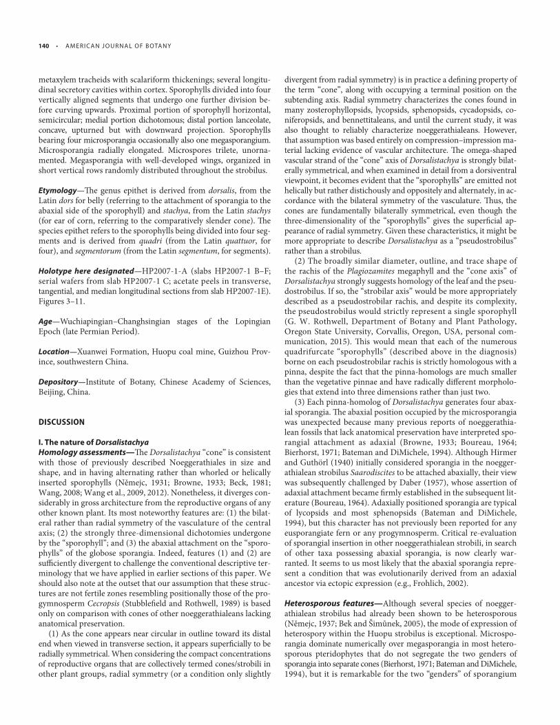
FIGURE 3 Anatomy and organization of Dorsalistachya quadrisegmentorum. (A) Transverse section through upper part of strobilus showing the axis
(ax) with an Ω -shaped vascular bundle (vb) that has split post-mortem, and sporophylls at diff erent levels with microsporangia (mi). Each sporo-
phyll is divided into four segments (sp1.1–4; dashed lines), each of which divides into two distal laminae (dl) that curve upwards. Slide WP2-0208.
Scale bar = 1 mm. (B) Transverse section through lower part of strobilus showing the axis (ax) with an Ω -shaped vascular bundle, and two sporo-
phylls at diff erent levels. Each sporophyll is divided into four segments (sp2.1–4 and sp3.1–4), each of which divides into two distal laminae (dl) that
curve upwards. Each segment bears a single microsporangium (mi) while sporophyll 2 (sp2) also bears a single winged megasporangium (me)
between adjacent microsporangia. Slide WP2-0271 E/Top (37). Scale bar = 1 mm. (C–E) Series of transverse peels showing the changes in axial
anatomy and morphology from the apex toward the base. All scale bars = 200 μ m. (C) Enlargement of Fig. 3A showing the Ω -shaped vascular
bundle (vb) within the axis; arrows indicate sporophyll traces diverging from the periphery of the Ω bundle. Slide WP2-0208. (D) Enlargement of
Fig. 3B showing the Ω -shaped vascular bundle (vb) surrounded by secretory channels (sc). Note increase in vascular bundle size compared with
Fig. 3D . Slide WP2-0271 E/Top (37). (E) Transverse section through basal part of the strobilus showing the Ω -shaped vascular bundle (vb) sur-
rounded by secretory channels (sc); note the slight increase in vascular bundle width compared with Fig. 3D . Slide WP2-0277.
132 • A M E R I C A N J O U R N A L O F B OTA NY
and typically isodiametric with a diameter of 40–80 μm, though in some places they are somewhat radially elongated ( Fig. 7G, H ). In longitudinal section parenchymatous cells appear slightly vertically elongated, rectangular and polygonal in shape ( Fig. 7F ). One or two layers of mesophyll beneath the abaxial epidermis consist of small and mostly isodiametric cells. Secretory cavities and/or idioblasts are oft en present in the basal portion of the distal lamina, but are absent from the remainder ( Fig. 7F–H ). Th e small, terete vascular bundle is generally located in the center of the lamina, though in some sections it appears closer to the adaxial surface ( Fig. 7F–J ).
Microsporangia and microspores— In all sections of the strobilus, microsporangia are typically seen close to, or in some cases in or-ganic attachment with, the abaxial surface of the proximal lamina close to its boundary with the strobilus axis ( Figs. 4A, 7B, 9A, D, E ). In most cases a gap separates the microsporangium from the upper surface of the underlying (proximal) sporophyll segment ( Figs. 4A, 5A–E ). Each segment bears a single microsporangium on its proxi-mal lamina, such that in total four microsporangia are present on each sporophyll. Because they are positioned equidistant from the cone axis, the four sporangia are seen to be arranged in a shallow arc when viewed in transverse section ( Figs. 3A, B, 6A, B ).
Individual microsporangia are radially elongated; they reach ca 3 mm long, ca 1.5 mm wide, and up to 2 mm high ( Figs. 3A, B, 4A, 5A–G, 6A, B, 9A–C, 10A–E ). Th e proximal area of the microspo-rangium is subglobose ( Figs. 9C, 10A, D, E ), whereas the distal area is narrower and more prismatic ( Figs. 9C, 10B, D, E ). Th e adaxial surface is rounded, whereas the abaxial surface and lower regions of the lateral surfaces are expanded to form three radially aligned ridges—one median and two lateral ( Figs. 9B, 10B–D ). Approxi-mately 1.5 mm from the apex of the microsporangium, the proxi-mal abaxial surface is truncated at ca 28 degrees relative to the base of the microsporangium to form an aperture 0.5 × 0.7 mm ( Figs. 9A, 10A, C, D ). We were unable to detect vascular tissue supplying indi-vidual sporangia.
Th e sporangium wall consists of two zones ( Fig. 9F ). Th e outer zone is a single layer of large columnar cells 60–200 μm in radial diameter that are characterized by extremely thick and dark-col-ored walls ( Fig. 9F, G ). Overall thickness increases from the proxi-mal end to the distal end and from the upper surface to the lower surface ( Fig. 9A–C ), where it is thinnest along the central ridge. Viewed in periclinal section, cells of the outer wall appear elongate, their long axis paralleling the long axis of microsporangium; the wall appears to be perforated by micropores ( Fig. 9G ). Th e inner zone of the sporangial wall consists of 2–5 layers of cells. Individual cells are 15‒30 μm in diameter; in transverse section they appear isodiametric at the ridge but elongated elsewhere on the sporan-gium ( Fig. 9C, F ), whereas in longitudinal section all cells appear elongated ( Fig. 9A, B ).
Dehiscence probably took place along the proximal lower sur-face of the microsporangium through the open aperture ( Figs. 9A,
FIGURE 4 Anatomy and organization of Dorsalistachya quadrisegmento-
rum . (A) Longitudinal section through strobilus showing strobilus axis
(ax) and vascular bundle (vb), vertically elongated secretory channels
(sc), and alternate rows of sporophylls on opposite sides of the strobilus.
Sporophylls extend from the axis forming a 2.5–3.0 mm subhorizontal
lamina (hl), to which microsporangia (mi) are attached on the abaxial
(lower) surface, before abruptly turning upwards to form a 3.0–3.5 mm
distal lamiae (dl); the base of the lamina at this point extends downward
for a length of 0.5–1.0 mm. Slide WP2-0275 (X2 53). Scale bar = 1 mm.
(B) Higher magnifi cation of the Ω -shaped vascular bundle in longitudinal
section showing tracheid walls bearing spiral and scalariform thicken-
ings. Slide WP2-0275 (X2 53). Scale bar = 50 μ m. (C) Transverse section of
the strobilus axis showing stellate-shaped protuberances (arrowed),
Ω -shaped vascular bundle ( Ω vb), cortex (cx) and microsporangia (mi).
Slide WP2-0203. Scale bar = 200 μm.

J A N UA RY 2017 , V O LU M E 104 • WA N G E T A L . — A N ATO M I C A L LY P R E S E R V E D P E R M I A N N O E G G E R AT H I A L E A N S • 133

134 • A M E R I C A N J O U R N A L O F B OTA NY
FIGURE 6 Raytraced three-dimensional reconstructions of Dorsalistachya quadrisegmentorum . (A) Oblique apex projection showing distribution of
microsporangia (mi) and megasporangia (me) within the sporophylls (sp0–sp4) that surround the central axis (ax). Also note the alternating vertical
arrangement of the sporophylls, which terminate in distal laminae (dl). (B) Transverse projection of model viewed from the apex showing the distribu-
tion of microsporangia (mi) and megasporangia (me) across the sporophylls. Within the axis four secretory channels (sc) surrounding the Ω -shaped
vascular bundle (vb) are shown. The most prominent sporophylls (sp1, sp2) best illustrate the division into four segments (sp1.1–4, sp2.1–4), the distal
part of each segment further dividing into two distal laminae. (C) Projection showing the vertical face of the strobili showing the alternating vertical
arrangement of the half-disciform sporophylls. (D) Projection showing the vertical face of the strobili tangentially from the axis showing arrangement
of the half-disciform sporophylls. All scale bars = 1 mm. Also see Appendix S1 and S2 (see Supplemental Data with the online version of this article).
10A, C, D ). Although numerous sporangia are split along a thin-ning of their wall adaxially, toward the midlongitudinal region and along the ridges, this is believed to represent a post-mortem taphonomic phenomenon (e.g., Figs. 3A, B, 5, 6B, 9B ). Most of the presumed microsporangia no longer contain microspores, whereas in most of the remainder the remains of the microspores are ta-phonomically fragmented ( Figs. 9C, 11A ). Th e best-preserved mi-crospores are nearly spherical, circular, or rounded-triangular in
outline in polar view, and ca 30 μm in diameter. Th ey possess a comparatively small trilete suture and apparently lack any other surface ornamentation ( Figs. 9H, 11A ).
According to Permo-Triassic miospore specialist Liu Feng (Nanjing Institute of Geology and Palaeontology, Chinese Academy of Sciences, Nanjing, China, personal communication, 2016), the closest (though imperfect) named comparators among dispersed spore-species are Patellisporites meishanensis Ouyang (1962) and
FIGURE 5 Anatomy and organization of Dorsalistachya quadrisegmentorum . (A–G) Sequential serial peels, showing tangential longitudinal sections of
three rows of sporophylls, through their subhorizontal laminae (hl) to their distal laminae (dl). Four vertical rows of segments at the point where they
separate (arrowed in B and C) are shown, together with microsporangia (mi) and a short vertical row of megasporangia (me). Note that microsporan-
gia (mi) are attached on the abaxial surface, leaving a gap to the upper surface of the lower sporophyll subhorizontal part. Slides A–G: WP2-0248 (u/24),
WP2-0247 (u/22), WP2-0245 (u/19), WP2-0243 (u/15), WP2-0241 (u/11), WP2-0240 (u/8), WP2-0239 (u/7). All scale bars = 1 mm.
←

J A N UA RY 2017 , V O LU M E 104 • WA N G E T A L . — A N ATO M I C A L LY P R E S E R V E D P E R M I A N N O E G G E R AT H I A L E A N S • 135
Densosporites paranulatus Ouyang (1986) ; the zona is equatorial in the latter but displaced toward the proximal pole in the former, making Patellisporites a better match with the microspores observed in Dorsalistachya. Patellisporites meishanensis is common throughout
the Wuchiapingian (upper Permian) of China, extending sporadi-cally into the lower Triassic, and should be sought in other noeg-gerathialean fossils. Patellisporites has not previously been recorded in other noeggerathialean cones.
FIGURE 7 Anatomy and organization of Dorsalistachya quadrisegmentorum sporophylls. (A) Transverse section showing the anatomy of one quarter of a
sporophyll from the proximal attachment to the axis (ax) and the bifurcation into two subhorizontal laminae (hl) at the concave central region (ccr),
which encloses a single microsporangium (mi). Each horizontal lamina (hl) divides to produce a pair of distal laminae (dl). Slide WP2-0271 E/Top (37).
Scale bar = 1 mm. (B) Longitudinal section through sporophylls showing the anatomy from the axial Ω -shaped vascular bundle ( Ω vb) with its secretory
channels (sc), through the subhorizontal lamina (hl) to its distal lamina (dl). Vascular bundles (vb) for the sporophyll separate from the axial vascular
bundle ( Ω vb) one internode (arrow) below the point of entry at the next internode, and then travel along the center of the subhorizontal lamina (hl).
Slide WP2-0275 (53). Scale bar = 1 mm. (C) High magnifi cation image showing the hypodermal cells of the subhorizontal lamina in transverse section.
At the lateral edge the hypodermis consists of several layers of small sclerenchymatous cells. The cells of the central zone are larger and tangentially
elongated. Slide WP2-0271 E/Top (37). Scale bar = 200 μ m. (D) High magnifi cation image showing the mesophyll in transverse section. Consisting of
parenchymatous cells that are tangentially isodiametric and radially elongated and isolated; round-rectangular idioblasts are dispersed throughout the
mesophyll (arrows). Slide WP2-0248. Scale bar = 100 μ m. (E) High magnifi cation image showing lower epidermis cells in transverse section. Arrows in-
dicate idioblasts. Slide WP2-0233. Scale bar = 100 μ m. (F) Longitudinal section through distal lamina showing the hypodermis (upper dermis = ud),
mesophyll (mp), and abaxial epidermis (lower dermis = ld), with putative vascular bundle (vb). Slide WP2-0275 (53). Scale bar = 200 μ m. (G–J) Transverse
sections through the distal lamina showing cellular anatomy from the apex to the base. The hypodermis (upper dermis = ud), mesophyll (mp), and ab-
axial epidermis (lower dermis = ld) surround a single vascular bundle (vb) in the center of the lamina. All scale bars = 200 μ m. (G) Point near apex. Slide
WP2-0208. (H) Midpoint between apex and the subhorizontal lamina. Slide WP2-0271 E/Top (37). (I) Point above the subhorizontal lamina. Slide WP2-
0271 E/Top (37). (J) Point below the subhorizontal lamina showing the convoluted shape of the basal protrusion. Slide WP2-0271 E/Top (37).

136 • A M E R I C A N J O U R N A L O F B OTA NY
FIGURE 8 Raytraced three-dimensional reconstructions of Dorsalistachya quadrisegmentorum
showing sporophyll morphology with digital removal of the microsporangia and megasporangia.
(A) Slightly oblique projection of model viewed from the apex showing the upper surface mor-
phology, subhorizontal (hl) and distal laminae (dl) of two sporophylls (sp1, sp2) surrounding the
axis (ax), which contains four secretory channels (sc). The sporophylls show the division into four
segments (sp1.1–4, sp2.1–4), the distal part of each segment further dividing into two distal lami-
nae. (B) Slightly oblique projection of model viewed from the base showing the lower surface
morphology of the two sporophylls (sp1, sp2) seen in A. All scale bars = 1 mm. Also see Appendix
S1 and S2 (see Supplemental Data with this article).
Megasporangia and megaspores— Th e majority of sporophylls bear only microsporangia, but the remainder produce one megaspo-rangium alongside four microsporangia ( Figs. 3B, 5B, D ). Where present, the megasporangia occupy the space between the hori-zontal lamina and distal lamina of the sporophyll holding the third and fourth microsporangia ( Figs. 3B, 5B–D, 6A, B ). When the strobilus is viewed laterally, megasporangia are seen to be dis-tributed in short, vertical rows of three or four megasporangia,
the rows apparently being distributed ran-domly throughout the strobilus.
Th e megasporangium is peripterous, the wing being moderately well developed and 0.7–0.9 mm wide ( Figs. 10F, G, 11A–C ). Th e dimensions of the megasporangium resem-ble those of the microsporangium, though the cavea (void) surrounding the former is much smaller than that surrounding the lat-ter. Th e cavea of the megasporangium ap-proximates 1.5 mm in radial direction, 1 mm in tangential direction, and up to 2 mm in vertical direction ( Figs. 10F, G, 11A, B ); this allows the megasporangium to become verti-cally elongated, whereas the microsporan-gium is radially elongated. Unfortunately, we were unable to discern details of the attach-ment to the sporophyll.
The megasporangium wall is bilayered and 50–150 μ m thick. Th e dark-colored co-lumnar cells that form the outer wall have a radial diameter of 8–20 μ m, whereas the inner wall consists of approximately six lay-ers of cells each 2–10 μ m in radial diameter ( Fig. 11C ). Putative megaspores within the megasporangia are typically poorly pre-served and fragmentary (e.g., Fig. 11A ); it is the approximate alignment of the fragments ( Fig. 11D ) to describe a crude circle within some megasporangia that implies the pres-ence of only one functional megaspore per megasporangium.
SYSTEMATICS
Phylum Tracheophyta Sinnott (1935) ex Cavalier-Smith (1998) .
Subphylum Euphyllophytina sensu Crane & Kenrick (1997) .
?Class (paraphylon) Progymnospermopsida C.B. Beck (1960b) .
(arguably incertae sedis at Class level; see Discussion).
Order Noeggerathiales sensu Němejc (1931) .
Emended diagnosis — Heterosporous plants with compact, pedunculate, bisporangiate
strobili. Sporophylls usually emitted linearly rather than heli-cally, bipartite. Sporangia sessile, attached to adaxial (or rarely abaxial) surface of sporophyll. Strobilar axis (where known) with bilateral vascular bundle. Foliage (where known) pinnate and multiveined.
Family Discinitaceae W. Remy & R. Remy (1977) , emend. S.J. Wang & A.R.T. Spencer.

J A N UA RY 2017 , V O LU M E 104 • WA N G E T A L . — A N ATO M I C A L LY P R E S E R V E D P E R M I A N N O E G G E R AT H I A L E A N S • 137
FIGURE 9 Anatomy and morphology of the microsporangium of Dorsalistachya quadrisegmentorum . (A) Longitudinal section through a single
microsporangium (mi), showing the attachment to the horizontal lamina (hl) above, the proximal aperture (ap) facing the axis (ax), and the in-
ner (iw) and outer (ow) microsporangial walls. Slide WP2-0271 E/Top (53). Scale bar = 0.5 mm. (B) Tangential section through a single microspo-
rangium (mi) containing microspores, showing the attachment to the horizontal lamina (hl) above, the inner (iw) and outer (ow) microsporangial
walls, and the three ribs (arrows). Slide WP2 0247 (u/18). Scale bar = 0.5 mm. (C) Transverse section through a single microsporangium (mi),
showing the inner (iw) and outer (ow) microsporangial walls, and the remaining microspores (ms) distally. Slide WP2-0271 E/Top (37). Scale bar = 0.5
mm. (D) Obliquely longitudinal section of a sporophyll showing the horizontal lamina (hl) to distal lamina (dl), with a microsporangium (mi)
attached to the horizontal lamina lower surface. A single megasporangium (me) is also present. White boxed area is enlarged in Fig. 9E . Slide
WP2-0246. Scale bar = 0.5 mm. (E) Enlargement of boxed area in Fig. 9D showing the organic connection of the microsporangium (mi) to the
horizontal lamina (hl) of a segment. A single possible stoma possesses putative paired guard cells (gc) and subsidiary cells (s). Slide WP2-0246.
Scale bar = 50 μ m. (F) Enlargement of a part of transverse section of the microsporangium showing the detail of the sporangial wall, divisible
into outer wall (ow) and inner wall (iw). Slide WP2-0264. Scale bar = 100 μ m. (G) Paradermal section through the sporangium wall showing
longitudinally elongate cells and micropores (p) through wall. Slide WP2-0248. Scale bar = 100 μ m. (H) High magnification image of a well-
preserved microspore showing the trilete mark. See Fig. 11A for location of spore. Slide WP2 0271 E/Top (36). Scale bar = 10 μ m.

138 • A M E R I C A N J O U R N A L O F B OTA NY
FIGURE 10 Raytraced three-dimensional reconstructions of Dorsalistachya quadrisegmentorum showing morphology of the microsporangia and
megasporangia. (A–E) Microsporangia. (A) View of proximal surface, showing the basal aperture (ap). (B) View of distal surface; arrows and dashed lines
indicate the three ridges. (C) Lateral view with the proximal (P) surface to the left and distal (D) surface to the right; the basal aperture (ap) truncates
the proximal surface at an angle of ca 28 ° . The arrow and dashed line indicate a single ridge that circumscribes the microsporangium. (D) Basal view
showing the abaxial surface from proximal (P) to distal (D) with the midline ridge (arrow and dashed line) and the basal aperture (ap). (E) Adaxial view
showing the top surface from proximal (P) to distal (D). All scale bars = 500 μ m. (F, G). Megasporangia. (F) Lower portion of a single megasporangium
showing the wings (w) in both proximal (P) and distal (D) directions. The dashed line indicates the position on the internal cavity. (G) View of the lower
portion seen in Fig. 10F from the apex showing the cross-sectional shape. Both scale bars = 0.5 mm. Also see Appendix S1 and S2 (see Supplemental
Data with this article).
Emended diagnosis — Sporophylls whorled; proximal part of sporo-phyll strongly discoid, distal part of sporophyll upturned and dis-sected into small laminae. Sporangia attached to the adaxial surface of the proximal part, numerous, arranged in several rings.
Genera — Discinites Feistmantel (1880) , Saarodiscites Hirmer (1940) .
Satellite genera — Yuania Sze (1953) , Palaeopteridium Kidston (1923) , Saaropteris Hirmer (1940) .
Note — Specimens of this family are all preserved as compression–impressions so their anatomy is unknown. Russellites Mamay (1968) , obtained from the Cisuralian (lower Permian) of Texas and previously considered to belong to the family, is now recognized as being a junior synonym of Yuania ( Wang and Chaney, 2010 ). Wang and Wang (1986) described Discinites sunjiagouensis Z.-Q. Wang & L.-X. Wang from the latest Permian Sunjiagou Formation in North China. However, as the sporophylls of this species are half-discs, they more likely belong within the genus Lacoea C.B. Read of the family Noeggerathiaestrobaceae.
Family Tingiostachyaceae Z. Gao & B.A. Th omas (1987 ), emend. S.J. Wang & A.R.T. Spencer.
Emended diagnosis — Sporophylls spiral or whorled; proximal part of sporophyll vaguely disc-shaped or dissected into segments, distal part of sporophyll upturned and dissected into small laminae. Spo-rangia attached to the adaxial surface of the proximal part, few, ar-ranged in a single ring.
Genera — Tingiostachya Kon’no (1929) , Paratingiostachya Sun et al. (1999) , strobili of the whole-plant Paratingia wudensis J. Wang et al. (2009) + Tingia unita J. Wang (2006) + Plagiozamites oblongifolius Halle (1927) .
Satellite genera — Tingia Halle (1954) , Paratingia Zhang (1987) , Plagiozamites Zeiller (1894) .
Note — Specimens of this family are all preserved as compression–impressions so their anatomy is unknown. Th e spiral arrangement attributed to the sporophylls is contentious. On the basis of a large collection of strobili of Paratingia , Wang et al. (2012) documented whorled sporophylls that were partially fused proximally but dis-tally dissected into small laminae. Similarly, a large proportion of known specimens of the strobili of Tingia demonstrate that the spo-rophylls are whorled rather than spiraled. Th is topic will be the sub-ject of a subsequent paper produced by some of the present authors (J. Wang et al., unpublished manuscript).

J A N UA RY 2017 , V O LU M E 104 • WA N G E T A L . — A N ATO M I C A L LY P R E S E R V E D P E R M I A N N O E G G E R AT H I A L E A N S • 139
FIGURE 11 Anatomy and morphology of the megasporangia of Dorsalistachya quadrisegmentorum . (A) Transverse section through a single megaspo-
rangium (me), positioned between two microsporangia (mi). The megasporangium displays two wings (w) in the proximal and distal direction and
contains the remains of a single megaspore (mg). Slide WP2-0271 E/Top (36). Scale bar = 0.5 mm. (B) Tangential section through a single megasporan-
gium, showing the elongate shape and possible bifurcating wings at the base (w). Slide WP2 0247 (u/18). Scale bar = 0.5 mm. (C) Enlargement of a part
of the megasporangium showing the detail of the sporangial wall and wing, diff erentiating inner wall zone (iw) and outer wall zone (ow). Slide WP2-
0265. Scale bar = 100 μ m. (D) Enlargement of a piece of fragmented megasporangium shown in Fig. 11A . Slide WP2-0271 E/Top (36). Scale bar = 50 μ m.
Family Noeggerathiaestrobaceae (= Noeggerathiaceae) Němejc (1963) , emend. S.J. Wang & A.R.T. Spencer.
Emended diagnosis — Sporophylls distichous; proximal part of spo-rophyll semicircular, distal part upturned and dissected into small laminae. Sporangia attached to adaxial surface of proximal part, nu-merous, arranged in several rings.
Genera — Noeggerathiaestrobus O. Feistmantel (1872) ; Archaeonoeg-gerathia W. Remy and R. Remy (1986) ; Lacoea C.B. Read (1946) .
Satellite genera — Noeggerathia Sternberg (1821) ; Palaeopteridium Kidston (1923) ; Conchophyllum Schenk (1883) .
Note — Specimens of this family are all preserved as compression–impressions so their anatomy is unknown. As mentioned above, Discinites sunjiagouensis sensu Z.-Q. Wang and L.-X. Wang (1986) belongs within Lacoea C.B. Read.
Family Dorsalistachyaceae S.J. Wang & A.R.T. Spencer, fam. nov.
Diagnosis — Sporophylls distichous, alternate; proximal part of spo-rophyll semicircular, dichotomous, distal part upturned. Sporangia abaxial, arranged in a single crescent. Microsporangia with proxi-mal lower surface dehiscence. Vascular bundle of strobilar axis (and petiole and rachis of correlated foliage) bilaterally symmetrical, sporophyll traces diverging from the two opposed margins. Secre-tory cavities within cortex of both strobilar axis and sporophyll.
Type genus — Dorsalistachya S.J. Wang & A.R.T. Spencer, gen. nov.
Satellite genus — Plagiozamites Halle (1927) .
Note — Th is family presently consists only of anatomically preserved disarticulated organs that are clearly attributable to Noeggerathiales.
Dorsalistachya quadrisegmentorum S.J. Wang & A.R.T. Spencer, gen. et sp. nov.
Diagnosis — Strobilus slender, cylindrical, ca 10 mm in diameter. Vascular bundle of axis inverted Ω -shaped in transverse section;
140 • A M E R I C A N J O U R N A L O F B OTA NY
metaxylem tracheids with scalariform thickenings; several longitu-dinal secretory cavities within cortex. Sporophylls divided into four vertically aligned segments that undergo one further division be-fore curving upwards. Proximal portion of sporophyll horizontal, semicircular; medial portion dichotomous; distal portion lanceolate, concave, upturned but with downward projection. Sporophylls bearing four microsporangia occasionally also one megasporangium. Microsporangia radially elongated. Microspores trilete, unorna-mented. Megasporangia with well-developed wings, organized in short vertical rows randomly distributed throughout the strobilus.
Etymology — Th e genus epithet is derived from dorsalis , from the Latin dors for belly (referring to the attachment of sporangia to the abaxial side of the sporophyll) and stachya , from the Latin stachys (for ear of corn, referring to the comparatively slender cone). Th e species epithet refers to the sporophylls being divided into four seg-ments and is derived from quadri (from the Latin quattuor , for four), and segmentorum (from the Latin segmentum , for segments).
Holotype here designated — HP2007-1-A (slabs HP2007-1 B–F; serial wafers from slab HP2007-1 C; acetate peels in transverse, tangential, and median longitudinal sections from slab HP2007-1E). Figures 3–11 .
Age — Wuchiapingian–Changhsingian stages of the Lopingian Epoch (late Permian Period).
Location — Xuanwei Formation, Huopu coal mine, Guizhou Prov-ince, southwestern China.
Depository — Institute of Botany, Chinese Academy of Sciences, Beijing, China.
DISCUSSION
I. The nature of Dorsalistachya Homology assessments— Th e Dorsalistachya “cone” is consistent with those of previously described Noeggerathiales in size and shape, and in having alternating rather than whorled or helically inserted sporophylls ( Němejc, 1931 ; Browne, 1933 ; Beck, 1981 ; Wang, 2008 ; Wang et al., 2009 , 2012 ). Nonetheless, it diverges con-siderably in gross architecture from the reproductive organs of any other known plant. Its most noteworthy features are: (1) the bilat-eral rather than radial symmetry of the vasculature of the central axis; (2) the strongly three-dimensional dichotomies undergone by the “sporophyll”; and (3) the abaxial attachment on the “sporo-phylls” of the globose sporangia. Indeed, features (1) and (2) are suffi ciently divergent to challenge the conventional descriptive ter-minology that we have applied in earlier sections of this paper. We should also note at the outset that our assumption that these struc-tures are not fertile zones resembling positionally those of the pro-gymnosperm Cecropsis ( Stubblefi eld and Rothwell, 1989 ) is based only on comparison with cones of other noeggerathialeans lacking anatomical preservation.
(1) As the cone appears near circular in outline toward its distal end when viewed in transverse section, it appears superfi cially to be radially symmetrical. When considering the compact concentrations of reproductive organs that are collectively termed cones/strobili in other plant groups, radial symmetry (or a condition only slightly
divergent from radial symmetry) is in practice a defi ning property of the term “cone”, along with occupying a terminal position on the subtending axis. Radial symmetry characterizes the cones found in many zosterophyllopsids, lycopsids, sphenopsids, cycadopsids, co-niferopsids, and bennettitaleans, and until the current study, it was also thought to reliably characterize noeggerathialeans. However, that assumption was based entirely on compression–impression ma-terial lacking evidence of vascular architecture. Th e omega-shaped vascular strand of the “cone” axis of Dorsalistachya is strongly bilat-erally symmetrical, and when examined in detail from a dorsiventral viewpoint, it becomes evident that the “sporophylls” are emitted not helically but rather distichously and oppositely and alternately, in ac-cordance with the bilateral symmetry of the vasculature. Th us, the cones are fundamentally bilaterally symmetrical, even though the three-dimensionality of the “sporophylls” gives the superfi cial ap-pearance of radial symmetry. Given these characteristics, it might be more appropriate to describe Dorsalistachya as a “pseudostrobilus” rather than a strobilus.
(2) The broadly similar diameter, outline, and trace shape of the rachis of the Plagiozamites megaphyll and the “cone axis” of Dorsalistachya strongly suggests homology of the leaf and the pseu-dostrobilus. If so, the “strobilar axis” would be more appropriately described as a pseudostrobilar rachis, and despite its complexity, the pseudostrobilus would strictly represent a single sporophyll (G. W. Rothwell, Department of Botany and Plant Pathology, Oregon State University, Corvallis, Oregon, USA, personal com-munication, 2015). Th is would mean that each of the numerous quadrifurcate “sporophylls” (described above in the diagnosis) borne on each pseudostrobilar rachis is strictly homologous with a pinna, despite the fact that the pinna-homologs are much smaller than the vegetative pinnae and have radically diff erent morpholo-gies that extend into three dimensions rather than just two.
(3) Each pinna-homolog of Dorsalistachya generates four abax-ial sporangia. Th e abaxial position occupied by the microsporangia was unexpected because many previous reports of noeggerathia-lean fossils that lack anatomical preservation have interpreted spo-rangial attachment as adaxial ( Browne, 1933 ; Boureau, 1964 ; Bierhorst, 1971 ; Bateman and DiMichele, 1994 ). Although Hirmer and Guthörl (1940) initially considered sporangia in the noegger-athialean strobilus Saarodiscites to be attached abaxially, their view was subsequently challenged by Daber (1957) , whose assertion of adaxial attachment became fi rmly established in the subsequent lit-erature ( Boureau, 1964 ). Adaxially positioned sporangia are typical of lycopsids and most sphenopsids ( Bateman and DiMichele, 1994 ), but this character has not previously been reported for any eusporangiate fern or any progymnosperm. Critical re-evaluation of sporangial insertion in other noeggerathialean strobili, in search of other taxa possessing abaxial sporangia, is now clearly war-ranted. It seems to us most likely that the abaxial sporangia repre-sent a condition that was evolutionarily derived from an adaxial ancestor via ectopic expression (e.g., Frohlich, 2002 ).
Heterosporous features— Although several species of noegger-athialean strobilus had already been shown to be heterosporous ( Němejc, 1937 ; Bek and Šimůnek, 2005 ), the mode of expression of heterospory within the Huopu strobilus is exceptional. Microspo-rangia dominate numerically over megasporangia in most hetero-sporous pteridophytes that do not segregate the two genders of sporangia into separate cones ( Bierhorst, 1971 ; Bateman and DiMichele, 1994 ), but it is remarkable for the two “genders” of sporangium

J A N UA RY 2017 , V O LU M E 104 • WA N G E T A L . — A N ATO M I C A L LY P R E S E R V E D P E R M I A N N O E G G E R AT H I A L E A N S • 141
to diff er as radically in morphology and anatomy as they do in Dor-salistachya ( Bateman and DiMichele, 1994 ).
Indeed, the vertical rows of megasporangia within the strobilus appear to have been added as a developmental afterthought, squeezed in between a symmetrical array of microsporangial rows; this pattern is particularly evident when the cone is viewed in trans-verse section ( Figs. 3B, 5A–D, 6A, B ). It is tempting to view this peculiar architecture as a teratological phenomenon ( Bateman and DiMichele, 2002 ; Rudall et al., 2011 ), but this interpretation is con-tradicted by the unequivocal presence of the megaspore within. Also, expression of megasporangia in vertical rows within bispo-rangiate cones has occasionally been observed in the extant lycop-sid Selaginella P. Beauv. , some authors arguing that it refl ects gravitational pooling of auxin within horizontally oriented cones (e.g., Horner and Arnott, 1963 ). Although we do not know the ori-entation of the strobili or the structure of the Dorsalistachya plant, as the fossils were found fragmented, reconstructions of noegger-athialeans based on articulated plants preserved in situ by volcanic ash from the Cisuralian (lower Permian) of China demonstrate that their cones were held vertically and positioned in the center of a previously vegetative rosette of branches ( Wang et al., 2009 , 2012 ).
Bateman and DiMichele (1994) estimated that the monomega-sporous level of heterospory had been achieved independently by fi ve diff erent land-plant lineages: (1) selaginellalean plus isoetalean lycopsids; (2) some extinct equisetalean sphenopsids; (3) leptospo-rangiate hydropterid water-ferns: (4) derived progymnosperms plus seed-plants; and (5) possibly the noeggerathialean compres-sion/impression genus Discinites ( Němejc, 1937 ; Bek and Šimůnek, 2005 ), which Bateman and DiMichele tentatively ascribed to the pro-gymnosperms. Monomegaspory within the strobili of Dorsalistachya clearly demonstrates that during the Permian the Noeggerathiales had reached a level of reproductive sophistication similar to that achieved by lycopsids and sphenopsids in the preceding Carbonif-erous Period. In particular, the Mississippian sphenopsid Protocal-amostachys farringtonii R.M. Bateman was able to develop both megasporangia and microsporangia in diff erent sporangia borne on the same quadripartite sporangiophore ( Bateman, 1991 ). Also, by the Pennsylvanian, the sphenopsid Calamocarpon insignis Baxter had achieved monomegaspory ( Bateman and DiMichele, 1994 ), albeit in cones that were more strongly and more conven-tionally developmentally canalized; its megasporangia are typically concentrated toward the base of most bisporangiate cones and matured earliest, whereas we infer simultaneous maturation in the Dorsalistachya cone.
Functional morphology— A winged sporangium of this nature is unique to the cone of the Dorsalistachya plant. Th e monomega-spory and wing cause the megasporangium to superfi cially resem-ble a seed, though this appearance is misleading; it possesses neither megasporangial modifi cation for reception and retention of the male gametes prior to fertilization nor an enclosing integument ( Bateman and DiMichele, 1994 ; Hilton and Bateman, 2006 ). Th e closest analog among pteridophytes is found in some of the more derived isoetalean clubmosses, but here the wing extends horizon-tally rather than vertically and originates from the tissue of the spo-rophyll rather than the sporangium.
We suggest that the winged megasporangium of Dorsalistachya assisted wind dispersal in a similar way to bona fide winged seeds ( Greene and Johnson, 1989 ) such as those of contemporaneous cordaitaleans ( Wang et al., 2003 )—a conclusion supported by the
apparent absence of dehiscence structures in the megasporangia that would have allowed release of the megaspore alone. However, it would have been necessary to fi rst shed the sporophylls that tightly envelop the winged megasporangia ( Figs. 3B, 5A–C, 6A, B ) before the megasporangia could disperse. We have observed within the Huopu mine succession isolated sporangia and sporo-phylls, together with cone axes that have retained only the basal parts of the sporophyll lamina, though we cannot be certain that the disassembly of these cones was the result of routine abscission rather than post-mortem taphonomic processes ( Neregato et al., 2016 ).
Although the apparent demise of the last noeggerathialeans in the Triassic ( Boureau, 1964 ; Sun et al., 1995 ; Taylor et al., 2009 ) may have been the result of competition with ascendant groups such as conifers, it is possible that it can be at least partly attributed directly to their heterosporous life history. Heterospory is largely a feature of hydrophilic plants ( Bateman and DiMichele, 1994 ), which are comparatively vulnerable to restricted availability of free water. Although the Noeggerathiales survived the end-Permian ex-tinction, they had passed their Permian acme and were already de-clining in species numbers ( Greene and Johnson, 1989 ; Bond et al., 2010 ). Th ey were primarily restricted to low-latitude wetlands in southern Cathaysia that suff ered aridifi cation during the Triassic ( Zhao et al., 1980 ; Sun et al., 1995 ), the drying climate being the most likely key to the extinction of these wetland plants.
II. Comparisons of Dorsalistachya with previously described taxa —
When attempting to assign a species to higher taxa it is important to distinguish between the circumscriptions of those taxa, the evo-lutionary relationships among them, and the respective formal ranks to which they are assigned. All three properties are linked and require a comparative approach, but they should not be confl ated. When attempting to place the genus Dorsalistachya , it is important to note that an entirely passive approach, simply placing the genus in a pre-existing taxonomic pigeonhole, may be deemed inappro-priate with respect to limited current understanding of the most likely pigeonhole, Noeggerathiales.
Noeggerathialeans have been viewed by previous authors vari-ously as sphenopsids, eusporangiate ferns, leptosporangiate ferns, progymnosperms, or none of the above (i.e., constituting a Class-level taxon in their own right). Th us, it is not surprising that the rank attributed to the group has also varied wildly, fl uctuating be-tween Phylum/Division ( Zimmermann, 1959 ; Boureau, 1964 ), Class, and more commonly Order. It is therefore essential that we ask whether Dorsalistachya is indeed most appropriately viewed as a noeggerathialean, and if so, to what degree circumscription of Noeggerathiales should be modifi ed to accommodate the new cone and what taxonomic rank is most appropriately occupied by the recircumscribed higher taxon.
Comparison with relevant Classes of vascular plants — Classes read-ily dismissed as being of close affi nity— Th e strobilus described here as Dorsalistachya is both heterosporous and heterosporangiate (viable spores are bimodal in size, the two modes being produced in separate sporangia). Th e holotype cone produced both microsporangia and megasporangia, and thus deviated from the reproductive organiza-tion that characterizes coeval gymnospermous seed-plant groups such as early-diverging pteridosperms, cycads, ginkgos, and coni-fers—all plants that combine microsporangia and megasporangia only in rare teratological individuals (e.g., Rudall et al., 2011 ).
142 • A M E R I C A N J O U R N A L O F B OTA NY
Moving on to consider pteridophytic groups, those capable of emulating Dorsalistachya by producing compact strobili in-clude the more derived portion of the lycopsid clade, all sphe-nopsids, and (by definition) all noeggerathialeans. Strobilar axes of both lycopsids and sphenopsids have radially symmetri-cal steles that differ substantially from the bilateral inverted Ω -shaped vascular bundle observed in the axis of Dorsalis-tachya . Lycopsids are further differentiated from Dorsalistachya by their helically inserted sporophylls that bear sporangia on the adaxial surface of the sporophyll, rather than being disti-chous with abaxial sporangia. Sphenopsids are further differen-tiated from Dorsalistachya by possessing sporangiophores that are whorled and produce pedicellate sporangia on the adaxial surface, in contrast with the distichous, alternate sporophylls of Dorsalistachya .
Eliminating these higher taxa leaves us to consider only ferns and progymnosperms. Progymnosperms are an exclusively Pa-leozoic group that is most commonly treated taxonomically as a Phylum/Class but is also widely viewed as being paraphyletic and containing the likely ancestor of all seed-plants (e.g., Beck and Wight, 1988 ; Rothwell and Serbet, 1994 ). Morphological cladistic studies strongly suggest that the group lacks close living relatives (e.g., Doyle and Donoghue, 1986 ; Hilton and Bateman, 2006 ), which leaves its classifi cation largely a matter for debate among paleobotanists. By contrast, 'ferns' sensu Kenrick and Crane (1997) (i.e., nonsphenopsid monilophytes) retain a relatively good taxonomic representation in the extant fl ora that permits construction of molecular phylogenies ( Pryer et al., 2004 ; Rai and Graham, 2010 ; Lehtonen, 2011 ; Grewe et al., 2013 ; Knie et al., 2015 ). Admittedly, several morphologically divergent higher taxa of Paleozoic ferns sensu lato—a much broader and less cohesive phylogenetic grouping than nonsphenopsid monilo-phytes studied by neobotanists—also lack close living relatives (e.g., Iridopteridales, Cladoxylales, Zygopteridales, Stauropteri-dales), but none of these groups bears a close resemblance to Dorsalistachya .
Ferns sensu lato— Sporangium morphology alone is suffi cient to re-ject a close relationship between Dorsalistachya and leptosporangi-ate ferns, thereby restricting comparison to the morphologically and taxonomically diverse paraphyletic group informally termed “eusporangiate ferns”. Driven primarily by molecularly determined phylogenies, most recent classifi cations based on extant ferns have circumscribed four groups of equal rank, three of them eusporangi-ate—Equisetopsida (representing “Sphenopsida” as discussed in the preceding paragraphs), Psilotopsida (Psilotales plus Ophioglos-sales), and Marattiopsida—and one leptosporangiate—Polypodi-opsida. Circumscription into these four major groups has become widely accepted, the main outstanding diff erence of taxonomic opinion being whether these four extant monophyletic taxa are bet-ter treated as Subclasses (e.g., Christenhusz and Chase, 2014 ) or Classes (e.g., Smith et al., 2006 ). However, the molecular phyloge-netic relationships among the four groups actually remain equivo-cal, other than reliably agreeing that Marattiopsida are sister to Polypodiopsida (cf. Pryer et al., 2004 ; Rai and Graham, 2010 ; Lehtonen, 2011 ; Grewe et al., 2013 ; Knie et al., 2015 ). Confi dence in inferred relationships among higher taxa of ferns deteriorates fur-ther when considering the few more radical morphological phylog-enies that have incorporated extinct fern groups (e.g., Rothwell and Nixon, 2006 ).
Many morphological diff erences separate Marattiopsida from Dorsalistachya , not least the absence of any feature that even vaguely resembles a reproductively specialized cone; Marattiopsida simply bear the (reliably homosporous) eusporangia on the abaxial surface of otherwise normal ultimate pinnae of their large, highly pinnate fronds. All Psilotopsida are small, herbaceous, rhizoma-tous plants that are reliably homosporous. Although unequivocal fossil evidence of the group does not extend further back in time than the earliest Cenozoic ( Rothwell and Stockey, 1989 ), molecular clock methods suggest that they diverged from other pteridophytes at some point between the late Devonian and late Pennsylvanian ( Rothfels et al., 2015 ). Psilotales lack any structure that could legiti-mately be termed a cone, instead bearing sporangia at the junction of the fertile pinna and stem, either singly or in clusters of three. Comparison with Ophioglossales appears more encouraging, as the eusporangia of Ophioglossum L., Helminthostachys Kaulf., Mankyua B.Y. Sun, M.H. Kim & C.H. Kim, and Botrychium Sw. appear to be borne on at least distally naked axes—they form compact, bilater-ally symmetrical structures that resemble cones and are generally termed “fertile pinnae”, “fertile pinnules” or, less specifi cally, “fer-tile spikes” (though the term “cone” could not legitimately be ap-plied if the sporangia are actually borne on very narrow laminae: G. W. Rothwell, personal communication, 2016). In addition, Botrychium is also famously the only genus of extant ferns to show putative secondary cambial activity ( Stevenson, 1980 ), though its “secondary xylem” is viewed by some workers merely as aligned metaxylem representing exceptionally slow but nonetheless deter-minate growth ( Rothwell and Karrfalt, 2008 ). Until the advent of molecular phylogenetics, these observations led to occasional sug-gestions that Ophioglossales were direct descendants of Paleozoic progymnosperms ( Kato, 1988 ). Overall, Dorsalistachya could cred-ibly be viewed as an early-divergent ophioglossalean.
Nonetheless, there are suffi cient morphological diff erences evi-dent between Dorsalistachya and any of the four Classes/Subclasses of extant ferns that we remain uncomfortable assigning Dorsalis-tachya (and other noeggerathialeans) to any of the four, implying that if we chose to regard noeggerathialeans as ferns, they are argu-ably best treated as a separate higher taxon at a rank equivalent to those occupied by the four extant groups. In the light of the range of characters documented in Dorsalistachya , we were indeed ini-tially inclined to treat the noeggerathialeans as a Class of compara-tively complex and specialized eusporangiate ferns. Only later did we reconsider, with greater care, the range of morphologies shown by the various previously described progymnosperm groups.
Progymnosperms— Th e progymnosperms are most commonly de-scribed as the Class Progymnospermopsida, encompassing three Orders: (1) the mid–upper Devonian Aneurophytales; (2) mid–upper Devonian Archaeopteridales ( Beck, 1960a , 1960b , 1962 ); and (3) comparatively poorly known Mississippian Protopityales ( Walton, 1958 ; Beck, 1960b ; Smith, 1962 ) (reviewed by Bonamo, 1975 ; Beck, 1976 ; Beck and Wight, 1988 ; Stubblefi eld and Rothwell, 1989 ). Some authors recognize a fourth Order, Cecropsidales, es-tablished comparatively recently on the basis of a single anatomically preserved upper Pennsylvanian reproductive foliate axis described by Stubblefield and Rothwell (1989) . The Aneurophytales are widely regarded as the most primitive group of progymnosperms, possessing lobed protosteles rather than sympodial protosteles or eusteles, and a sparganum-type rather than a dictyoxylon-type cor-tex. More importantly in the present context, their sporangia are

J A N UA RY 2017 , V O LU M E 104 • WA N G E T A L . — A N ATO M I C A L LY P R E S E R V E D P E R M I A N N O E G G E R AT H I A L E A N S • 143
terminal on naked axes, dehisce along only one side, and are reli-ably homosporous. Th ey thus diverge greatly from Dorsalistachya ; moreover, the Plagiozamites fronds associated with Dorsalistachya bear little resemblance to the small, three-dimensional “pre-fronds” that characterize aneurophytaleans.
Unsurprisingly, the Devonian Aneurophytales contrast most strongly with the more recent (Pennsylvanian) Cecropsidales ( Stubblefi eld and Rothwell, 1989 ). Both taxa have globose sporan-gia, though those of Cecropsis have an apical dehiscence resembling that of the early divergent lineage leading to the “fern” Botrychium . Both taxa also share the characteristic of having reached the mono-megasporangiate grade of heterospory, but this grade has been achieved by a minimum of six diff erent lineages of vascular land plant ( Bateman and DiMichele, 1994 ). Moreover, the small clusters of sporangia borne on the sporophylls of Cecropsis are evenly dis-tributed on the adaxial rather than abaxial surface, and the highly invaginated sporophylls are borne among—and otherwise identical to—the sterile leaves; both kinds of determinate organs are sub-tended by a slender but nonetheless woody branch. Th e Cecropsis plant unquestionably lacked bona fi de strobili.
Intermediate in age, and arguably also in morphology, to the Aneurophytales and Cecropsidales are the two remaining progym-nosperm orders. Th e Archaeopteridales diff er in only few charac-ters from Cecropsidales but are less similar to Dorsalistachya in several reproductive characters. Protopityales have achieved at best a substantially lower grade of heterospory and bear fusiform spo-rangia in similar fashion to aneurophytaleans ( Walton, 1958 ; Smith, 1962 ; Bateman and DiMichele, 1994 ). Th eir branches do share with Dorsalistachya and Plagiozamites the feature of having vascular systems that are bilaterally organized, though the two taxa diff er considerably in the details of their vascular architecture. Cru-cially, none of the four recognized Orders of progymnosperms bore cones. Th us, if noeggerathialeans (not least Dorsalistachya ) were to be viewed as progymnosperms, they would most logically consti-tute a separate Order within the Class—an Order that may prove to be delimited by possession of fertile organs that consist entirely of a single leaf homolog.
Comparison with other noeggerathialean genera— All strobili previously assigned to the Noeggerathiales ( Noeggerathiaestrobus , Tetraphyllostrobus Gao & Zodrow, Archaeonoeggerathiostrobus , Tin-giostachya , Discinites , Lacoea , Saarodiscites , and Paratingiostachya ) were preserved as compression–impressions and therefore lacked anatomical preservation. Th ey resemble Dorsalistachya in being compact and heterosporangiate, with sporophylls (oft en distichous) bearing sessile sporangia. Although each of these cone-genera clearly shares some similarities with the present strobilus, each also appears distinct from it ( Table 1 ).
Sporophylls are spiral or whorled in Tingiostachya and Paratin-giostachya ( Gao and Th omas, 1987 ; Sun et al., 1999 ; Wang, 2006 ; Wang et al., 2009 ), or decurrent–disks in Saarodiscites and Dis-cinites ( Hirmer, 1940 ; Boureau, 1964 ; Bek and Šimůnek, 2005 ), con-trasting with the distichous arrangement of Dorsalistachya . Th e sporophylls of Tetraphyllostrobus are decussate and each bears up to 50 sporangia ( Gao and Zodrow, 1990 ), whereas the present strobi-lus consists of distichous sporophylls, each bearing only four micro-sporangia. Although Lacoea and Noeggerathiaestrobus bear distichous semicircular sporophylls comparable with those of Dorsalistachya , each sporophyll bears a much greater number of sporangia, and they are arranged in several rings on the adaxial surface ( Read, 1946 ;
Leary, 1973 , 1980 ; Leary and Pfeff erkorn, 1977 ; Šimůnek and Bek, 2003 ) rather than in a single ring on the abaxial surface. In Saarod-iscites the distal margin of the sporophyll is reputedly turned down-ward ( Hirmer, 1940 ; Daber, 1957 ; Šimůnek and Bek, 2003 ); if correctly interpreted, this is a unique feature within Noeggerathiales. Lastly, none of the previously recognized cones produced megasporangia that are winged, and none conclusively bore sporangia abaxially.
We conclude that the present strobilus is a bona fi de member of the Noeggerathiales, but deviates considerably from the circum-scriptions of all the previously described genera, prompting us to establish the new genus Dorsalistachya .
Comparison with other noeggerathialean families— Three families (or Orders according to Boureau, 1964 ) have previously been recognized within Noeggerathiales. Members of the Discinit-aceae have whorled sporophylls. Each sporophyll consists of a near-horizontal disc-shaped proximal part and an upturned (at least partially dissected) distal part; the sporophyll bears numerous spo-rangia that are arranged in several rings ( Remy and Remy, 1977 ; Gao and Th omas, 1994 ). Tingiostachyaceae are characterized by helical or whorled sporophylls. Th e proximal part of the sporophyll is disc-shaped or dissected into segments, whereas the distal part is upturned and dissected into smaller laminae; sporangia are ar-ranged in a single ring ( Gao and Th omas, 1987 ; Wang, 2006 ; Wang et al., 2009 ). By contrast, Noeggerathiaestrobaceae bear sporophylls distichously; the proximal part of the sporophyll is semicircular, the distal part upturned and dissected into small laminae; the sporangia are arranged in several rings ( Němejc, 1963 ). Th e pri-mary justifi cation for recognizing the new family, Dorsalistachya-ceae, is that all three pre-existing families have adaxial sporangia, whereas insertion is abaxial in Dorsalistachya quadrisegmentorum .
CONCLUSIONS
We have shown that noeggerathialeans were capable of achieving a level of reproductive sophistication that is comparable with those of the most ‘advanced’ late Paleozoic lycopsids and sphenopsids, and greater than that achieved by any other group of eusporangiate ferns. In parallel with several groups of seed plants, they appear to have evolved wind-dispersed propagules. And in parallel with ferns of the eusporangiate Marattiopsida and leptosporangiate Cy-atheaceae, they evolved the tree-fern growth habit that allowed them to reach at least 10 m in height ( Wang et al., 2009 , 2012 ). But like so many groups that rise to ecological (co)dominance, their success was short-lived when viewed on a geological timescale.
We can only off er a less concrete answer to the key question 'Just what are Noeggerathiales?' Th e bilateral organization of their shoots alone is suffi cient to distinguish them from the radial sym-metry that characterizes the shoots of all sphenopsids ( Browne, 1933 ; Boureau, 1964 ; Taylor et al., 2009 ) and most progymno-sperms ( Beck, 1981 )—indeed, it also challenges our very concepts of strobilus and sporophyll. Based on the present (limited) evi-dence, the most credible comparisons are with the psilopsid ferns (especially the extant ophioglossalean genera Botrychium and Helminthostachys ) on the one hand and the more derived cecropsi-dalean and protopityalean progymnosperms on the other. Studies that require comparison of a potentially crucial fossil with mem-bers of not one but two taxonomic Classes may at fi rst glance ap-pear to be the epitome of ignorance, but in truth, the phylogenetic

144 • A M E R I C A N J O U R N A L O F B OTA NY
TAB
LE
1.
Co
mp
ari
son
of
no
eg
ge
rath
iale
an
str
ob
ili.
Ge
nu
s S
aa
rod
isci
tes
Dis
cin
ites
T
ing
iost
ach
ya
Arc
ha
eo
-
no
eg
ge
rath
io-
stro
bu
s La
coe
a
Tetr
ap
hyl
lost
rob
us
Pa
rati
ng
ia
wu
den
sis
1
Tin
gia
un
ita
1
No
eg
ge
rath
io-
stro
bu
s
Pa
rati
ng
io-
sta
chya
D
ors
ali
sta
chya
Fam
ily
D
isci
nit
ace
aeD
isci
nit
ace
aeTi
ng
iast
ach
y-
ace
ae
?No
eg
ge
rath
iae
-
stro
bac
eae
No
eg
ge
rath
iae
-
stro
bac
eae
?No
egg
erat
hia
e-
stro
bac
eae
Tin
gia
stac
hy-
ace
ae
Tin
gia
stac
hy-
ace
ae
No
eg
ge
rath
iae
-
stro
bac
eae
Tin
gia
stac
hy-
ace
ae
Do
rsal
ista
chy-
ace
ae
Ep
och
/Ag
e
Car
bo
nife
rou
s
(Mid
dle
Pen
nsy
lvan
ian
)
Car
bo
nife
rou
s–
Perm
ian
Car
bo
nife
rou
s–
Perm
ian
Car
bo
nife
rou
s
(Mis
siss
ipp
ian
–
Low
er
Pen
nsy
lvan
ian
)
Car
bo
nife
rou
s
(Pen
nsy
lvan
ian
)
Car
bo
nife
rou
s
(Up
pe
r Pe
nn
sy-
lvan
ian
)
Earl
y Pe
rmia
nEa
rly
Perm
ian
Pen
nsy
lvan
ian
–
Perm
ian
(Cis
ura
lian
)
Perm
ian
(Cis
ura
lian
)
Perm
ian
(Lo
pin
gia
n)
Str
ob
ilu
s sh
ap
e
Cyl
ind
rica
l,
tap
eri
ng
to
p
and
bo
tto
m
Cyl
ind
rica
lC
ylin
dri
cal?
?C
ylin
dri
cal
Cyl
ind
rica
lC
ylin
dri
cal
Cyl
ind
rica
lC
ylin
dri
cal
Cyl
ind
rica
lC
ylin
dri
cal
Pe
du
ncl
e
??
Be
ars
pin
nae
;
fork
ed
, eac
h
bra
nch
be
arin
g
a st
rob
ilus
??
?La
cks
pin
nae
Lack
s p
inn
aeB
ear
s N
oeg
ger
ath
ia
pin
nae
??
Str
ob
ilu
s le
ng
th
(mm
)
50
–2
00
6
>2
10
ca 1
30
??
ca 3
0–
35
>1
20
>2
00
10
0–
20
0
(inco
mp
lete
)
80
11
5 (
inco
mp
lete
)
Str
ob
ilu
s d
iam
ete
r
(mm
)
45
–5
01
5–
70
80
–1
20
?<
20
8–
10
15
20
–2
52
8–
38
10
–1
4ca
10
Ax
is d
iam
ete
r
(mm
)
?2
–1
0Sl
en
de
r?
?0
.7?
?2
.0–
2.6
2.0
1.5
Sp
oro
ph
yll
arr
an
ge
me
nt
De
curr
en
t
dis
k?
De
curr
en
t d
isk
He
lical
or
vert
icill
ate
(un
fuse
d)
?D
isti
cho
us,
op
po
site
De
cuss
ate
(w
ho
rls
ove
rlap
pin
g
slig
htl
y)
De
curr
en
t d
isk
(so
me
wh
at
ove
rlap
pin
g)
Ve
rtic
illat
eW
eak
ly
dis
ti ch
ou
s,
alte
rnat
e
He
lical
or
vert
icill
ate,
per
pen
dic
ula
r
Dis
tich
ou
s,
alte
rnat
e
Sp
oro
ph
yll
spa
cin
g (
mm
)
5.0
–6
.04
.0–
7.0
??
1.6
2.0
–3
.0?
?4
.5–
8.0
?1
.0
Sp
oro
ph
yll
ori
en
tati
on
an
d s
ha
pe
Pro
xim
al p
art
dis
coid
al, fl
at,
?dis
tal m
arg
in
do
wn
turn
ed
up
to
8 c
m
Pro
xim
al p
art
dis
coid
al, fl
at;
dis
tal m
arg
in
up
turn
ed
to
par
alle
l axi
s,
de
nta
te o
r
en
tire
Perp
en
dic
ula
r
to a
xis
Re
lati
vely
lon
g
and
nar
row
,
fl ab
elli
form
,
rad
ially
corr
ug
ate
an
d/
or
rib
be
d; d
ista
l
mar
gin
sh
arp
ly
up
turn
ed
,
bas
ally
co
nn
ate,
mar
gin
sin
uo
us
and
/or
de
nta
te
Sem
icir
cula
r,
frin
gin
g
laci
nia
te
bo
rde
r
Incu
rve
d,
fl ab
elli
form
;
4–
5 v
ein
s
term
inat
ing
in a
n u
nd
ula
te
dis
tal m
arg
in
Dis
tal m
arg
in
up
turn
ed
to
par
alle
l
axis
, de
ep
ly
de
nta
te
Perp
en
dic
ula
r
to a
xis
Sem
icir
cula
r,
mar
gin
de
nta
te
Lin
ear
to
line
ar-
lan
ceo
late
,
dis
tal m
arg
in
up
turn
ed
to
par
alle
l axi
s
Sem
icir
cula
r in
ou
tlin
e, d
ivid
ing
to p
rod
uce
fou
r
do
rsiv
en
tral
ly
fl at
ten
ed
seg
me
nts
th
at
un
de
rgo
on
e
furt
he
r d
ivis
ion
be
fore
cu
rvin
g
up
war
ds,
bu
t
wit
h d
ista
l he
els
that
pro
ject
abax
ially
; lat
era
l
abax
ial r
idg
es
fl an
k m
icro
-
spo
ran
gia
Bis
po
ran
gia
te
Yes?
Yes
Yes?
?Ye
s?
Yes
Yes
Yes
?Ye
s
No
. of
mic
rosp
ora
ng
ia
pe
r sp
oro
ph
yll
?3
0–
14
00
? 7
1?
ca 3
0–
65
10
+ca
45
–6
51
0–
14
8–
10
sp
ora
ng
ia
and
spo
rop
hyl
ls
on
a w
ho
rl
??
4 (
sin
gle
spo
ran
giu
m
pe
r sp
oro
ph
yll)
con
tin
ued

J A N UA RY 2017 , V O LU M E 104 • WA N G E T A L . — A N ATO M I C A L LY P R E S E R V E D P E R M I A N N O E G G E R AT H I A L E A N S • 145
Ge
nu
s S
aa
rod
isci
tes
Dis
cin
ites
T
ing
iost
ach
ya
Arc
ha
eo
-
no
eg
ge
rath
io-
stro
bu
s La
coe
a
Tetr
ap
hyl
lost
rob
us
Pa
rati
ng
ia
wu
den
sis
1
Tin
gia
un
ita
1
No
eg
ge
rath
io-
stro
bu
s
Pa
rati
ng
io-
sta
chya
D
ors
ali
sta
chya
Mic
rosp
ora
ng
ium
sha
pe
in
ou
tlin
e
?O
val/
circ
ula
rC
ircu
lar
Ova
lEl
lipti
cal
Cir
cula
r-
ren
iform
4
Ellip
tica
l (ap
ex
and
bas
e
slig
htl
y
con
trac
ted
)
Ellip
tica
lO
val
Cir
cula
r/e
llip
tica
lO
val-
elli
pso
idal
Mic
rosp
ora
ng
ium
size
2.5
mm
lon
g,
1.5
mm
wid
e
?1
.0–
1.5
mm
dia
me
ter
1.0
–1
.2 m
m
lon
g
1.5
mm
dia
me
ter,
4.0
mm
lon
g
0.6
–0
.8 m
m
lon
g
2.0
–3
.0 m
m
dia
me
ter,
5.0
–7
.0 m
m
lon
g
2.0
–4
.0 m
m
dia
me
ter,
5.0
–7
.0 m
m
lon
g
??
1.5
mm
dia
me
ter,
3.0
mm
lon
g
Mic
rosp
ora
ng
ium
de
his
cen
ce
?Lo
ng
itu
din
al?
??
?Lo
ng
itu
din
alLo
ng
itu
din
al?
?P
roxi
mal
low
er
surf
ace
Mic
rosp
ora
ng
ium
att
ach
me
nt
?Sh
ort
, ro
bu
st
“sp
ora
ng
iop
ho
re”
Sess
ileSe
ssile
??
Sess
ileSe
ssile
Sess
ile?
Sess
ile
Mic
rosp
ora
ng
ia
arr
an
ge
me
nt
on
sp
oro
ph
yll
?R
adia
l, ad
axia
l
atta
chm
en
t
to p
roxi
mal
po
rtio
n o
f
spo
rop
hyl
l
Ad
axia
l
atta
chm
en
t
to p
roxi
mal
po
rtio
n o
f
spo
rop
hyl
l
Ad
axia
l
atta
chm
en
t
to in
fl e
xed
mar
gin
Spir
al3
–4
ro
ws,
15
pe
r ro
w,
low
er
spo
ran
gia
ove
rlap
pin
g
up
pe
r
Sin
gle
rin
g,
adax
ial
Ad
axia
lA
dax
ial,
in
3–
5 r
ow
s
?So
litar
y ro
w,
abax
ial
Mic
rosp
ore
typ
e(s
)
? C
ala
mo
spo
ra -t
ype,
Pter
ore
tis
pri
mu
m ,
Vest
isp
ora
laev
iga
ta ,
Ca
lam
osp
ora
laev
iga
ta
??
Ca
lam
osp
ora
cf. l
iqu
ida
,
Pun
cta
tisp
orit
es
Laev
iga
tosp
or-
ites
min
or
Ca
lam
osp
ora
,
Pun
ctat
ispo
rites
Pun
ctat
ispo
rites
Ve
rru
cosi
spo
rite
s
mic
rotu
ber
osu
s ,
Ca
lam
osp
ora
-
typ
e, C
yclo
-
gra
nis
po
rite
s ,
Pun
cta
tisp
ori
tes
??
Me
ga
spo
rag
ium
sha
pe
??
??
??
Cir
cula
r/su
bci
rc-
ula
r, w
ith
eq
uat
ori
al
exp
ansi
on
??
?B
ilate
rally
fl at
ten
ed
, wit
h
do
rsiv
en
tral
win
gs
Me
ga
spo
rag
ium
size
??
??
??
1.0
–1
.3 m
m
dia
me
ter
??
?2
.0 x
1.0
x
1.5
mm
No
. of
me
ga
spo
ran
gia
pe
r sp
oro
ph
yll
16
?ca
16
(w
he
re
pre
sen
t)
??
??
? 5
??
?1
(w
he
re p
rese
nt,
arra
ng
ed
vert
ical
ly)
Me
ga
spo
re
typ
e(s
)
? C
ala
mo
spo
ra -t
ype
??
Ca
lam
osp
ora
-
typ
e
?Eq
uat
ori
al b
ulg
eC
ircu
lar/
elli
pti
cal,
>1
.0 m
m in
dia
me
ter
Ca
lam
osp
ora
?
? 3
Pre
serv
ati
on
C
om
pre
ssio
n/
imp
ress
ion
Co
mp
ress
ion
/
imp
ress
ion
Co
mp
ress
ion
/
imp
ress
ion
Co
mp
ress
ion
/
imp
ress
ion
Co
mp
ress
ion
/
imp
ress
ion
Co
mp
ress
ion
/
imp
ress
ion
(co
alifi
ed
)
Co
mp
ress
ion
/
imp
ress
ion
(in t
uff
)
Co
mp
ress
ion
Co
mp
ress
ion
Co
mp
ress
ion
/
imp
ress
ion
An
ato
mic
al
pre
serv
atio
n
via
carb
on
ate
pe
rmin
era
lizat
ion
Kn
ow
n
loca
liti
es
Ge
rman
y,
Cze
ch
Re
pu
blic
Be
lgiu
m, C
hin
a,
Cze
ch R
ep
ub
lic,
Fran
ce,
Ge
rman
y, S
aud
i
Ara
bia
, USA
Ch
ina,
Ko
rea,
Om
an
Be
lgiu
m, C
zech
Re
pu
blic
,
Ge
rman
y
USA
Can
ada
Ch
ina
Ch
ina
Cze
ch R
ep
ub
lic,
Ge
rman
y
Ch
ina
Ch
ina
TA
BL
E 1
, co
nti
nu
ed
con
tin
ued

146 • A M E R I C A N J O U R N A L O F B OTA NY
Ge
nu
s S
aa
rod
isci
tes
Dis
cin
ites
T
ing
iost
ach
ya
Arc
ha
eo
-
no
eg
ge
rath
io-
stro
bu
s La
coe
a
Tetr
ap
hyl
lost
rob
us
Pa
rati
ng
ia
wu
den
sis
1
Tin
gia
un
ita
1
No
eg
ge
rath
io-
stro
bu
s
Pa
rati
ng
io-
sta
chya
D
ors
ali
sta
chya
Ass
ign
ed
spe
cie
s
S. g
uth
oer
lii
D. b
oh
enic
us ,
D. d
elec
tus ,
D. d
enti
lon
gu
s ,
D. e
gre
gio
us ,
D. ?
fi m
bri
ata
,
D. h
liza
e ,
D. j
on
gm
an
si ,
D. n
emej
cii ,
D. m
ajo
r ,
D. o
rien
talis
,
D. r
aco
nic
ensi
s ,
D. s
inen
sis ,
D. s
un
jiaqu
ouen
sis
T. te
tra
locu
lari
s ,
T. m
ing
sha
nen
is ,
T. s
an
tia
nen
sis
A. s
cha
tzla
ren
sis ,
A. g
oth
an
ii
L. s
eria
ta
T. b
rog
an
ensi
s P.
wu
den
sis 1
T.
un
ita
1
N. b
oh
emic
us ,
N. v
icin
alis
,
stro
bilu
s o
f
No
egg
era
thia
zam
ito
ides
2
P. c
ath
ays
ian
a
D. t
etra
seg
men
tus
Foli
ag
e a
sso
cia
ted
wit
h
fru
ctifi
ca
tio
n
Saa
rop
teri
s
gu
thö
rlii?
Pala
eop
teri
diu
m ,
Yua
nia
,
Ru
ssel
lites
?
Tin
gia
??
Pala
eop
ter i
diu
m ?
? Pa
rati
ng
ia
Tin
gia
N
oeg
ger
ath
ia
Para
tin
gia
Pl
ag
ioza
mit
es
ob
lon
gifo
lius
Re
fere
nce
s H
irm
er,
19
40
;
Dab
er,
19
57
;
Ně
me
jc, 1
96
3 ;
Bo
ure
au, 1
96
4
Gra
nd
’Eu
ry, 1
87
7 ;
Ně
me
jc, 1
93
7 ,
19
41
; Hir
me
r,
19
40
; Arn
old
,
19
44
, 19
49
;
Mam
ay, 1
95
4 ;
Re
my
and
Re
my,
19
56
; Sto
ckm
ans
and
Mat
hie
u,
19
57
; Bo
ure
au,
19
64
; Po
ton
ié,
19
65
; Bo
hlin
,
19
71
; Gu
and
Zh
i, 1
97
4 ;
Liu
an
d S
he
n,
19
78
; Zh
ao e
t al
.,
19
80
; Gao
an
d
Tho
mas
, 19
94
;
Me
i et
al.,
19
96
;
Wan
g, 2
00
0 ;
Wan
g a
nd
Sh
en
,
2000
; Wan
g e
t al
.,
20
04
; Be
k an
d
Šim
ůn
ek,
20
05
Ko
n’n
o, 1
92
9 ;
Bro
wn
e, 1
93
3 ;
Sto
ckm
ans
and
Mat
hie
u, 1
93
9 ,
19
57
; Bo
ure
au,
19
64
; Gao
an
d
Tho
mas
, 19
87
;
He
et
al.,
19
96
;
Me
i et
al.,
19
96
;
Be
rth
elin
et
al.,
20
03
Re
my
and
Re
my,
19
86
; Le
ary,
19
88
; Šim
ůn
ek
and
Be
k, 2
00
3
Re
ad, 1
94
6 ; L
ear
y,
19
73
, 19
80
Gao
an
d
Zo
dro
w, 1
99
0
Wan
g e
t al
., 2
00
9 ,
20
12
Wan
g, 2
00
6
Ge
init
z, 1
86
5 ;
O. F
eis
tman
tel,
18
72
, 18
79
;
We
iss,
18
79
;
Ste
rze
l, 1
91
8 ;
Ně
me
jc, 1
92
8 ;
Hir
me
r, 1
94
0 ;
Hal
le, 1
95
4 ;
Re
my
and
Re
my,
19
56
;
Bo
ure
au, 1
96
4 ;
Šim
ůn
ek
and
Be
k, 2
00
3
Sun
et
al.,
19
99
Pre
sen
t p
ape
r
No
tes:
? =
Un
kno
wn
/un
cert
ain
sta
te. 1
No
eg
ge
rath
iale
an p
lan
t w
ith
leav
es
and
un
nam
ed
str
ob
ili a
ttac
he
d; 2
Stro
bilu
s m
en
tio
ne
d b
y St
erz
el (
19
18
) b
ut
no
t fo
rmal
ly n
ame
d; 3
Sin
gle
fu
nct
ion
al m
eg
asp
ore
of
un
kno
wn
typ
e; 4
Bas
ed
on
Pl.
I, 5
an
d F
ig. 4
of
Gao
an
d Z
od
row
(1
99
0) ;
5 M
eg
asp
ore
s w
ere
fo
un
d s
catt
ere
d o
n t
he
su
rfac
e o
f th
e s
tro
bili
an
d in
sit
u w
ith
in m
eg
asp
ora
ng
ium
by
Wan
g e
t al
. (2
00
9 , fi
gs.
37
–4
1);
6 M
axim
um
len
gth
est
imat
ion
by
Bo
ure
au (
19
64
) . 7 U
pp
er
ran
ge
va
lue
re
po
rte
d in
Dis
cin
ites
cf.
raco
nic
ensi
s b
y B
ek
and
Šim
ůn
ek
(20
05
) .
TA
BL
E 1
, co
nti
nu
ed

J A N UA RY 2017 , V O LU M E 104 • WA N G E T A L . — A N ATO M I C A L LY P R E S E R V E D P E R M I A N N O E G G E R AT H I A L E A N S • 147
distance separating progymnosperms from the early divergent eus-porangiate ferns is not great. Moreover, relationships among the basally divergent fern groups remain highly contentious, especially when (a) extinct groups are taken into account, and (b) some phylogeneticists have formally treated the recognized Orders of progymnosperms as plesions, together forming a (most likely para-phyletic) group located phylogenetically between ferns sensu latissimo and seed-plants, but of uncertain relationships among themselves (e.g., Kenrick and Crane, 1997 ).
We also note that the circumscription of Progymnospermopsida per se has been much-debated throughout the last half-century (cf. Bonamo, 1975 ; Beck, 1976 ; Beck and Wight, 1988 ; Stubblefi eld and Rothwell, 1989 ), a fact that is hardly surprising when one considers that this group is defi ned by a supposedly unique combination of two character suites that are most defi nitely not unique to the group, spe-cifi cally a bifacial vascular cambium (generating 'gymnospermous wood') plus free-sporing reproduction. We also acknowledge that we presently lack any evidence that the Dorsalistachya plant produced wood. It is not surprising that the Dorsalistachya cone and Plagioza-mites frond both lack wood, as we regard these two organs as likely occupying the same hierarchical level within the growth architecture of the source plant (i.e., exhibiting positional homology); it is unlikely that wood would be present in one of these organs but absent from the other. Nonetheless, in regarding this source plant as potentially progymnospermous we would, by defi nition, be assuming that the stem and branches underwent secondary thickening—a hypothesis that can be tested only by fi nding these correlated organs in the fossil record ( Bateman and Hilton, 2009 ).
We are now confi dent that noeggerathialeans (including Dor-salistachya ) constitute a major group of vascular land-plants that lie within the general plexus of land plants encompassing eusporangi-ate ferns plus progymnosperms. Whether they lie within one of the “fern Classes”, “progymnosperm Orders”, or represent a separate Class outside either of these “grab-bag” taxa remains a matter for ongoing debate. To be constructive, that debate will need to be fu-eled by (a) new, more informative fossils, and (b) a morphological cladistic analysis that combines the most informative among the conceptually reconstructed fossils with relevant extant taxa. Such work is now underway.
ACKNOWLEDGEMENTS
The authors thank Li Tong-Shen and colleagues at the Ministry of Coal Mining, Guizhou Province for assistance during fieldwork, Paul Kenrick, Peta Hayes, and the library/archival team at the Natural History Museum (London) for help acquir-ing reference literature, Paula Rudall, Guillaume Chomicki, Gar Rothwell (twice), and Bill DiMichele for commenting on earlier versions of the manuscript, and Liu Feng for advice on miospore taxonomy. This research was funded by the National Natural Science Foundation of China (Awards 41162001, 41172014, 41372011, and 41530101), the State Key Laboratory of Palaeobiology and Stratigraphy (Nanjing Institute of Geology and Palaeontology, Chinese Academy of Sciences, Awards 133116 and 123103), Chinese Academy of Science Project KZCX2-EW-120, National Basic Research Program of China (973 Program, 2012CB821901). Fieldwork was also supported by the Natural Environment Research Council (NE/E004369/1). Th is work consti-tuted part of a doctoral investigation by A.R.T.S. at Imperial College London.
LITERATURE CITED
Arnold , C. A. 1944 . A heterosporous species of Bowmanites from the Michigan
Coal Basin. American Journal of Botany 31 : 466 – 470 .
Arnold , C. A. 1949 . Fossil fl ora of the Michigan Coal Basin. In Fossil fl ora of
the Michigan Coal Basin. Contributions from the Museum of Paleontology,
University of Michigan, 131–269. University of Michigan Press, Ann Arbor,
Michigan, USA.
Bateman , R. M. 1991 . Palaeobiological and phylogenetic implications of
anatomically-preserved archaeocalamites from the Dinantian of Oxroad
Bay and Loch Humphrey Burn, southern Scotland . Palaeontographica,
Abteilung B, Paläeophytologie 223 : 1 – 59 , 8 plates.
Bateman , R. M. , and W. A. DiMichele . 1994 . Heterospory: Th e most iterative
key innovation in the evolutionary history of the plant kingdom. Biological
Reviews of the Cambridge Philosophical Society 69 : 345 – 417 .
Bateman , R. M. , and W. A. DiMichele . 2002 . Generating and fi ltering major
phenotypic novelties: neoGoldschmidtian saltation revisited. In Q. C. B.
Cronk, R. M. Bateman, and J. A. Hawkins [eds.], Developmental genetics
and plant evolution, 109–159. Taylor & Francis, London, UK.
Bateman , R. M. , and J. Hilton . 2009 . Palaeobotanical systematics for the phylo-
genetic age: Applying organ-species, form-species and phylogenetic species
concepts in a framework of reconstructed fossil and extant whole-plants.
Taxon 58 : 1254 – 1280 .
Beck , C. B. 1960a . Connection between Archaeopteris and Callixylon. Science
131 : 1524 – 1525 .
Beck , C. B. 1960b . Th e identity of Archaeopteris and Callixylon. Brittonia 12 :
351 – 368 .
Beck , C. B. 1962 . Reconstructions of Archaeopteris , and further considerations
of its phylogenetic position . American Journal of Botany 49 : 373 – 382 .
Beck , C. B. 1976 . Current status of the Progymnospermopsida. Review of
Palaeobotany and Palynology 21 : 5 – 23 .
Beck , C. B. 1981 . Archaeopteris and its role in vascular plant evolution . In K.
J. Niklas [ed.], Paleobotany, paleoecology, and evolution, vol. 1, 193–230.
Praeger, New York, New, York, USA.
Beck , C. B. , and D. C. Wight . 1988 . Progymnosperms . In C. B. Beck [ed.],
Origin and evolution of gymnosperms, 1–84. Columbia University Press,
New York, New York, USA.
Bek , J. , and Z. Šimůnek . 2005 . Revision of the cone genus Discinites from
the Carboniferous continental basins of Bohemia. Palaeontology 48 :
1377 – 1397 .
Berthelin , M. , J. Broutin , H. Kerp , S. Crasquin-Soleau , J.-P. Platel , and J. Roger . 2003 .
Th e Oman Gharif mixed paleofl ora: A useful tool for testing Permian Pangea
reconstructions. Palaeogeography, Palaeoclimatology, Palaeoecology 196 : 85 – 98 .
Bierhorst , D. W. 1971 . Morphology of vascular plants. Macmillan, New York,
New York, USA.
Bohlin , B. 1971 . Late Palaeozoic plants from Yuerhhung, Kansu, China . Sven
Hedin Foundation, Stockholm, Sweden.
Bonamo , P. M. 1975 . Th e Progymnospermopsida: Building a concept. Taxon
24 : 569 – 579 .
Bond , D. P. G. , J. Hilton , P. B. Wignall , L. G. Stevens , J. R. Ali , Y. Sun , and X. L.
Lai . 2010 . Th e Middle Permian (Capitanian) mass extinction on land and
in the oceans. Earth-Science Reviews 102 : 100 – 116 .
Boureau , É. 1964 . Traité de paléobotanique, III. Sphenophyta, Noeggerathiophyta.
Masson et Cie, Paris, France.
Browne , I. 1933 . Th e Noeggerathiae and Tingiae. Th e eff ects of their recogni-
tion upon the classifi cation of the Pteridophyta: An essay and a review. New
Phytologist 32 : 344 – 358 .
Cavalier-Smith , T. 1998 . A revised six-kingdom system of life. Biological
Reviews of the Cambridge Philosophical Society 73 : 203 – 266 .
Christenhusz , M. J. M. , and M. W. Chase . 2014 . Trends and concepts in fern
classifi cation. Annals of Botany 113 : 571 – 594 .
Crane , P. R. , and P. Kenrick . 1997 [ 1996]. Problems in cladistic classifi cation:
Higher-level relationships in land plants. Aliso 15 : 87 – 104 .
Daber , R. 1957 . Parallelisierung der Flöze des Zwickauer und des Lugau-
Oelznitzer Steinkohlenreviers auf Grund paläobotanischer Untersuchungen.
Geologie 19 : 1 – 76 .

148 • A M E R I C A N J O U R N A L O F B OTA NY
Doyle , J. A. , and M. J. Donoghue . 1986 . Seed plant phylogeny and the origin of an-
giosperms: An experimental cladistic approach. Botanical Review 52 : 321 – 431 .
Feistmantel , C. 1879 . Über die Noeggerathien und deren Verbreitung in
der Böhmischen Steinkohlenformation. Sitzungsberichte der königlich
Böhmischen Gesellschaft der Wissenschaft en in Prag 1879 : 75 – 88 .
Feistmantel , O. 1872 . Fruchtstände fossiler Pfl anzen aus der böhmischen
Steinkohlenformation. 1. Hälft e: Equisetaceae und Filices . Abhandlungen
der königlichen Böhmischen Gesellschaft der Wissenschaft en in Prag , series 6,
5 : 1 – 51 , plates 1–6.
Feistmantel , O. 1880–1881 . Fossil fl ora of the Gondwana System, II, Th e fl ora
of the Damuda and Panchet Divisions . Memoirs of the Geological Survey of
India, Palaeontologia Indica , series 12, 3 : 1 – 77 , 1880 ; 79 – 149 , 1881 .
Frohlich , M. W. 2002 . Th e Mostly Male theory of fl ower origins: Summary and
update regarding the Jurassic pteridosperm Pteroma . In Q. C. B. Cronk, R.
M. Bateman, and J. A. Hawkins [eds.], Developmental genetics and plant
evolution, 85–108. Taylor & Francis, London, UK.
Galtier , J. , and T. L. Phillips . 1999 . Th e acetate peel technique. In T. P. Jones
and N. P. Rowe [eds.]. Fossil plants and spores: Modern techniques, 67–71.
Geological Society of London, London, UK.
Gao , Z. , and B. A. Th omas . 1987 . A re-evaluation of the plants Tingia and
Tingiostchya from the Permian of Taiyuan, China. Palaeontology 30 : 815 – 828 .
Gao , Z. , and B. A. Th omas . 1994 . A new species of Discinites from the Lower
Permian of China. Review of Palaeobotany and Palynology 81 : 185 – 192 .
Gao , Z. , and E. L. Zodrow . 1990 . A new strobilus Tetraphyllostrobus broganen-
sis gen. et sp. nov. from the Upper Carboniferous, Sydney Coalfi eld, Nova
Scotia, Canada. Review of Palaeobotany and Palynology 66 : 3 – 11 .
Garwood , R. J. , and J. A. Dunlop . 2014 . Th e walking dead: Blender as a tool
for palaeontologists with a case study of extinct arachnids. Journal of
Paleontology 88 : 735 – 746 .
Geinitz , H. B. 1865 . Über einige seltene Versteinerungen aus der unteren Dyas
und der Steinkohlen-Formation. Neues Jahrbuch für Mineralogie . Geologie
und Paläeontologie 1865 : 385 – 394 .
Grand’Eury , M. 1877 . Flore carbonifère du départment de Loire et du centre
de la France. Mémoires présentés par divers savants à l’Académie des Sciences
24 : 1 – 624 , plates 1-34.
Greene , D. F. , and E. A. Johnson . 1989 . A model of wind dispersal of winged or
plumed seeds. Ecology 70 : 339 – 347 .
Grewe , F. , W. Guo , E. A. Gubbels , A. K. Hansen , and J. P. Mower . 2013 . Complete
plastid genomes from Ophioglossum californicum, Psilotum nudum , and
Equisetum hyemale reveal an ancestral land plant genome structure and re-
solve the position of Equisetales among monilophytes. BMC Evolutionary
Biology 13 : 8 .
Gu , Z. and L. Zhi [eds.]. 1974 . Palaeozoic plants from China. Fossil plants of
China, vol. 1. Science Press, Beijing, China [in Chinese].
Guo , Y. T. , B. L. Tian , and D. X. Han . 1990 . Th e anatomical study of
Plagiozamites oblongifolius and the systematic position of Plagiozamites.
Acta Botanica Sinica 30 : 799 – 804 [in Chinese with English abstract] .
Halle , T. G. 1927 . Palaeozoic plants from central Shansi . Palaeontologica Sinica
Series A , 2 ( i ). Geological Survey of China, Peking.
Halle , T. G. 1954 . Notes on Noeggerathiineae. Svensk Botanisk Tidskrift 48 :
368 – 380 .
He , X. L. , D. S. Liang , and S. Z. Shen . 1996 . Research on the Permian Flora
from Jiangxi Province, China . China University of Mining and Technology
Press, Xuzhou, China. [in Chinese with English summary]
Hilton , J. , and R. M. Bateman . 2006 . Pteridosperms are the backbone of seed-
plant phylogeny. Journal of the Torrey Botanical Society 133 : 119 – 168 .
Hirmer , M. 1940 . Noeggerathiineae . In M. Hirmer and P. Guthörl, Abt. 3,
Filicales und Verwandte. In Hirmer, M, and W. Gothan [eds.], Die Karbon-
Flora des Saargebietes. Palaeontographica, Supplement 9: 1-60.
Hirmer , M. , and P. Guthörl . 1940 . Abt. 3, Filicales und Verwandte . In
Hirmer, M, and W. Gothan [eds.], Die Karbon-Flora des Saargebietes.
Palaeontographica, Supplement 9, Lieferung 1: 1–44.
Horner , H. T. , and H. J. Arnott . 1963 . Sporangial arrangement in North American
species of Selaginella. Botanical Gazette (Chicago, Illinois, USA) 124 : 371 – 383 .
Kato , M. 1988 . Th e phylogenetic relationship of Ophioglossaceae. Taxon 37 :
381 – 386 .
Kenrick , P. , and P. R. Crane . 1997 . Th e origin and early diversifi cation of land
plants. Smithsonian Institution Press, Washington, District of Columbia, USA.
Kidston , R. 1923 . Fossil plants of the Carboniferous rocks of Great Britain.
Great Britain Geological Survey Memoirs 2 ( 1–3 ): 1 – 276 , plates 1-68.
Knie , N. , S. Fischer , F. Grewe , M. Polsackiewicz , and V. Knoop . 2015 . Horsetails
are the sister group to all other monilophytes and Marattiales are sister to
leptosporangiate ferns. Molecular Phylogenetics and Evolution 90 : 140 – 149 .
Kon’no , E. 1929 . On genera Tingia and Tingiostachya from the Lower Permian
and Permo-Triassic beds in northern Korea. Japanese Journal of Geology
and Geography 6 : 113 – 147 .
Leary , R. L. 1973 . Lacoea , a Lower Pennsylvanian noeggerathialian cone from
Illinois. Review of Palaeobotany and Palynology 15 : 43 – 50 .
Leary , R. L. 1980 . Lacoea with sporangia and Calamospora spores from Rock
Island, Illinois. Review of Palaeobotany and Palynology 29 : 23 – 28 .
Leary , R. L. 1988 . Additional specimen of Archaeonoeggerathia gothani Remy
and Remy 1986 from the Namurian of Belgium. Bulletin de l’Institut Royal
des Sciences Naturelles de Belgique . Sciences de la Terre 58 : 5 – 176 .
Leary , R. L. , and H. W. Pfeff erkorn . 1977 . An Early Pennsylvanian fl ora with
Megalopteris and noeggerathiales from west-central Illinois . Illinois State
Geological Survey Circular 500 : 1 – 77 .
Lehtonen , S. 2011 . Towards resolving the complete fern tree of life. PLoS One
6 : e24851 .
Liu , H. , and G. Shen . 1978 . Carboniferous and Permian fl oras of the Longshou
Mountains in Gansu. Lanzhou University, Lanzhou, China [in Chinese].
Mamay , S. H. 1954 . A Permian Discinites cone. Journal of the Washington
Academy of Sciences 44 : 7 – 11 .
Mamay , S. H. 1968 . Russellites , new genus, a problematical plant from the Lower
Permian of Texas. U.S. Geological Survey Professional Paper 593-I : 1 – 15 .
Mei , M. , Q. C. Huang , M. Du , and D. L. Dilcher . 1996 . Th e Xu-Huai-Yu
Subprovince of the Cathaysian Floral Province. Review of Palaeobotany and
Palynology 90 : 63 – 77 .
Meyen , S. V. 1987 . Fundamentals of palaeobotany. Chapman & Hall, London, UK.
Němejc , F. 1928 . A revision of the Carboniferous and Permian fl ora of the
coal-districts in central Bohemia. Palaeontographica Bohemiae 12 : 1 – 82 [in
Czech with English summary] .
Němejc , F. 1931 . Th e morphology and the systematic relations of the
Carboniferous Noeggerathiae with regard to the “genera” Tingia and
Plagiozamites of eastern Asia. Preslia 10 : 111 – 114 .
Němejc , F. 1937 . On Discinites K. Feistm . Bulletin International de l’Academie
des Sciences de Boheme 38 : 3 – 10 .
Němejc , F. 1941 . Weitere Fructifi kationem vom Typus Discinites nebst einigen
Bemerkungen über die Archaeopteriden der Mittelböhmischen Kohlenbecken.
Mitteilungen der Tschechischen Akademie der Wissenschaft en 19 : 1 – 13 .
Němejc , F. 1963 . Subclass Noeggerathiidae. Paleobotanika 2 : 242 – 250 , plates
XXI–XXV.
Neregato , R. , C. D’Apolito , I. J. Glasspool , S.-J. Wang , L. Feng , P. Windslow ,
J. Lu , et al. 2016 . Palynological constraints on the provenance and strati-
graphic range of a Lopingian (Late Permian) inter-extinction fl oral lager-
stätte from the Xuanwei Formation, Guizhou Province, China. International
Journal of Coal Geology 162 : 139 – 150 .
Ouyang , S. 1962 . Th e microspore assemblage from the Lungtan Series of
Changhsing, Chekiang . Acta Palaeontologica Sinica 10 : 76 – 119 [Chinese
with English summary].
Ouyang , S. 1986 . Palynology of Upper Permian and Lower Triassic Strata of
Fuyuan District, Eastern Yunnan. Science Press, Beijing, China [Chinese
with English abstract].
Potonié , R. 1965 . Fossile Sporae in situ: Vergleich mit der Sporae dispersae
Nachtrag zur Synopsis der Sporae in situ . Forschungsberichte des Landes
Nordrhein-Westfalen 1483 : 1 – 71
Pryer , K. M. , E. Schuettpelz , P. G. Wolf , H. Schneider , A. R. Smith , and R.
Cranfi ll . 2004 . Phylogeny and evolution of ferns (Monilophytes) with a fo-
cus on early leptosporangiate divergences. American Journal of Botany 91 :
1582 – 1598 .
Rai , H. S. , and S. W. Graham . 2010 . Utility of a large, multigene plastid dataset
in inferring higher-order relationships in ferns and relatives (Monilophytes).
American Journal of Botany 97 : 1444 – 1456 .

J A N UA RY 2017 , V O LU M E 104 • WA N G E T A L . — A N ATO M I C A L LY P R E S E R V E D P E R M I A N N O E G G E R AT H I A L E A N S • 149
Read , C. B. 1946 . A Pennsylvanian fl orule from the Forkston Coal in the Dutch
Mountain outlier, northeastern Pennsylvania, U.S.A. U.S. Geological Survey
Professional Paper 210-B: 17–27.
Remy , W. , and R. Remy . 1956 . Noeggerathiostrobus vicinalis E. Weiss und
Bemerkungen zu ähnlichen Fruktifi kationen. Abhandlungen der Deutschen
Akademie der Wissenschaft en zu Berlin, Klasse für Chemie, Geologie und
Biologie 2 : 1 – 11 .
Remy , W. , and R. Remy . 1977 . Die Floren des Erdaltertums. Verlag Glückauf
GMBH, Essen, Germany.
Remy , W. , and R. Remy . 1986 . Archaeonoeggerathia gothani nov. gen., nov.
spec., eine Noeggerathiale aus dem Namur A. Argumenta Palaeobotanica 7 :
109 – 121 [in German] .
Rothfels , C. J. , F.-W. Li , E. M. Sigel , L. Hulet , A. Larsson , D. O. Burge , M.
Rusham , et al. 2015 . Th e evolutionary history of ferns inferred from 25
low-copy nuclear genes. American Journal of Botany 102 : 1089 – 1107 .
Rothwell , G. W. , and E. E. Karrfalt . 2008 . Growth, development, and systemat-
ics of ferns: Does Botrychium s.l. (Ophioglossales) really produce secondary
xylem? American Journal of Botany 95 : 414 – 423 .
Rothwell , G. W. , and K. C. Nixon . 2006 . How does the inclusion of fossil data
change our conclusions about the phylogenetic history of euphyllophytes?
International Journal of Plant Sciences 167 : 737 – 749 .
Rothwell , G. W. , and R. Serbet . 1994 . Lignophyte phylogeny and the origin of sper-
matophytes: A numerical cladistic analysis. Systematic Botany 19 : 443 – 482 .
Rothwell , G. W. , and R. A. Stockey . 1989 . Fossil Ophioglossales in the early
Paleocene of western North America. American Journal of Botany 76 : 637 – 644 .
Rudall , P. J. , J. Hilton , F. Vergara-Silva , and R. M. Bateman . 2011 . Recurrent
abnormalities in conifer cones and the evolutionary origins of fl ower-like
structures. Trends in Plant Science 16 : 151 – 159 .
Schenk , A. 1883 . Pfl anzen aus der steinkohlen formation . Richthofen F.V.
China 4 : 221 – 244 .
Seyfullah , L. J. , J. Hilton , M. M. Liang , and S. J. Wang . 2010 . Resolving the
systematic and phylogenetic position of isolated ovules: A case study from
a new genus from the Upper Permian of China. Botanical Journal of the
Linnean Society. Linnean Society of London 164 : 84 – 108 .
Šimůnek , Z. , and J. Bek . 2003 . Noeggerathiaceae from the Carboniferous basins
of the Bohemian Massif. Review of Palaeobotany and Palynology 125 : 249 – 284 .
Sinnott , E. W. 1935 . Botany: Principles and problems. 3rd ed . McGraw-Hill,
New York, New York, U.S.A.
Smith , A. R. , K. M. Pryer , E. Schuettpelz , P. Korall , H. Schneider , and P. G. Wolf .
2006 . A classifi cation for extant ferns. Taxon 55 : 705 – 731 .
Smith , D. L. 1962 . Th ree fructifi cations from the Scottish Lower Carboniferous.
Palaeontology 5 : 225 – 237 .
Sternberg , K. M. 1821 . Noeggerathia . In Versuch einer geognostisch-bota-
nischen Darstellung der Flora der Vorwelt [1820-1838], 33, plate 20. F.
Fleischer, Leipzig, Germany.
Sterzel , J. T. 1918 . Die organischen Reste des Kulms und Rotliegenden der
Gegend von Chemnitz . Abhandlungen der mathematisch-physischen Classe
der königlich\Ssächsischen Gesellschaft der Wissenschaft en 35 : 205 – 315 .
Stevenson , D. W. 1980 . Ontogeny of the vascular system of Botrychium mul-
tifi dum (S. G. Gmelin) Rupr. (Ophioglossaceae) and its bearing on stelar
theories. Botanical Journal of the Linnean Society. Linnean Society of London
80 : 41 – 52 .
Stockmans , F. , and F.-F. Mathieu . 1939 . La fl ore Paléozoïque du bassin
houiller de Kaiping (Chine), 49-165. Patrimoine du Musée Royal d’Histoire
Naturelle de Belgique, Brussels, Belgium.
Stockmans , F. , and F.-F. Mathieu . 1957 . La fl ore Paléozoïque du bassin houiller de
Kaiping (Chine) (Deuxième partie). Association pour l’Étude de la Paléontologie
et de la Stratigraphie Houillères, Bruxelles, Belgium, publication 32, 1-89.
Stubblefi eld , S. P. , and G. W. Rothwell . 1989 . Cecropsis luculentum gen. et
spec. nov.: Evidence for heterosporous progymnosperms in the Upper
Pennsylvanian of North America. American Journal of Botany 76 : 1415 – 1428 .
Sun , G. , F. S. Meng , L. J. Qian , and S. Ouyang . 1995 . Triassic fl oras. In X. X.
Li [ed.], Fossil fl oras of China through the geological ages, 305–342. Science
and Technology Press, Guangdong, China.
Sun , K.-Q. , S. Deng , J.-Z. Cui , and P. Shan . 1999 . Discovery of Paratingia and
Paratingiostachya from the Shanxi Formation of the Early Permian in the
Wuda area of Inner Mongolia, China. Acta Botanica Sinica 41 : 1024 – 1026
[largely in Chinese] .
Sutton , M. D. , R. J. Garwood , D. J. Siveter and D.J. Siveter . 2012 . SPIERS and
VAXML: a soft ware toolkit for tomographic visualisation and a format for
virtual specimen interchange. Palaeontologia Electronica 15 : 2.4T.
Sze , C. 1953 . Notes on some fossil remains from the Shihchienfeng Series in
northwestern Shensi . Acta Palaeontologica Sinica 1 : 11 – 22 .
Taylor , T. N. , E. L. Taylor , and M. Krings . 2009 . Th e biology and evolution of
fossil plants (2 nd ed.). Elsevier, Amsterdam, Netherlands.
Walton , J. 1958 . XV.-On Protopitys(Göppert): Wwith a description of a fertile
specimen Protopitys scotica sp. nov. from the Calciferous Sandstone Series
of Dunbartonshire. Earth and Environmental Science Transactions of the
Royal Society of Edinburgh 63 : 333 – 340 .
Wang , H. , L. Shao , L. M. Hao , P. F. Zhang , I. J. Glasspool , J. R. Wheeley , P.
B. Wignall , et al . 2011 . Sedimentology and sequence stratigraphy of
the Lopingian (Late Permian) coal measures in southwestern China.
International Journal of Coal Geology 85 : 168 – 183 .
Wang , J. 2000 . Discovery of a petrifi ed noeggerathialean strobilus, Discinites
sinensis sp. nov. from the Permian of Shizuishan, Ningxia, China. Chinese
Science Bulletin 45 : 560 – 566 .
Wang , J. 2006 . Tinga unita sp. nov. (Noeggerathiales) with strobilus from the
Lower Permian of Wuda, Inner Mongolia, China. Chinese Science Bulletin
51 : 2624 – 2633 .
Wang , J. 2008 . A note on fructifi cation of Plagiozamites Zeiller (Noeggerathiales)
based on association. Global Geology 11 : 115 – 124 .
Wang , J. , and D. Chaney . 2010 . A re-examination of the type specimens of
Yuania H.C. Sze 1953 and its junior synonym Russellites Mamay 1968
(Noeggerathiales). Taxon 59 : 517 – 524 .
Wang , J. , H. W. Pfeff erkorn , and J. Bek . 2009 . Paratingia wudensis sp. nov., a
whole noeggerathialean plant preserved in an earliest Permian air-fall tuff in
Inner Mongolia, China. American Journal of Botany 96 : 1676 – 1689 .
Wang , J. , H. W. Pfeff erkorn , Z. Feng , and G. L. Shen . 2004 . A new species of
Discinites (Noeggerathiales) associated with a new species of Yuania from
the Lower Permian of Inner Mongolia, China. International Journal of Plant
Sciences 165 : 1107 – 1119 .
Wang , J. , H. W. Pfeff erkorn , Z. Zhang , and Z. Feng . 2012 . Permian vegeta-
tional Pompeii from Inner Mongolia and its implications for landscape
paleoecology and paleobiogeography of China. Proceedings of the National
Academy of Sciences, USA 109 : 4927 – 4932 .
Wang , J. , and G. Shen . 2000 . A new species of Discinites (Noeggerathiales)
from the Upper Permian of Weibei Coalfi eld, North China. Review of
Palaeobotany and Palynology 110 : 175 – 190 .
Wang , S.-J. , J. Hilton , M.-M. Liang , and L. G. Stevens . 2006 . Permineralized
seed plants from the Upper Permian of southern China: A new species of
Cardiocarpus. International Journal of Plant Sciences 167 : 1247 – 1257 .
Wang , S.-J. , J. Hilton , B. Tian , and J. Galtier . 2003 . Cordaitean seed plants from
the Early Permian of North China. 1. Delimitation and reconstruction of the
Shanxioxylon sinense plant. International Journal of Plant Sciences 164 : 89 – 112 .
Wang , Z.-Q. , and L.-X. Wang . 1986 . Late Permian fossil plants from the Lower
part of the Shihchienfeng Group in North China. Bulletin of the Tianjin
Institute of Geology and Mineral Resources . Chinese Academy of Geological
Sciences 15 : 1 – 80 [Chinese, with English abstract] .
Weiss , C. E. 1879 . Bemerkungen zur Fructifi kation von Noeggerathia. Zeitschrift
der Deutschen Geologischen Gesellschaft 31 : 111 – 116 .
Zeiller , R. 1894 . Notes sur le fl ore des couches permiennes de Trienbach
(Alsace). Bulletin de la Société Géologique de France 4 : 163 – 182 .
Zhang , H. 1987 . Palaeobotany . In Institute of Geology Exploration, CCMRI
Coal Ministry and Provincial Coalfi elds Exploration Corporation of Shanxi,
Sedimentary Environment of the Coal-Bearing Strata in Pinglu-Shuoxian Mining
Area, China, 195–204. Shaanxi Peoples Education Publishing House, Xi’an, China.
Zhao , X. H. , Z. G. Mo , S. Z. Zhang , and Z. Q. Yao . 1980 . Late Permian fl ora
from western Guizhou and eastern Yunnan. In Nanjing Institute of Geology
and Palaeontology, Academia Sinica [eds.], Stratigraphy and palaentology of
the Upper Permian Coal Measures of western Guizhou and eastern Yunnan,
70–122. Science Press, Beijing, China [in Chinese].
Zimmermann , W. 1959 . Die Phylogenie der Pfl anzen. Fischer, Stuttgart, Germany.