anatomy and physiology of the skin
DESCRIPTION
Anatomy and Physiology of the SkinTRANSCRIPT

ANATOMY AND PHYSIOLOGY OF THE SKIN
04/18/23

INTRODUCTION
Skin :
- Body organ : site outer
- a complex organ that protect its host from environment, at the same time allowing interaction w/ the environment
04/18/23

Skin : very complex, elastic & sensitive.
Variation according : climate, age, sex,
race & depend to location of the body.
The skin character’s : smooth, hard,
reguler/not thin, thick, strong attach with
the body nothing similarity.
04/18/23

Skin can move & stretch depend
on:
Thick and the fold, elastisitasity
The skin attach with underlying
tissue
Skin elastic and loose : palpebra,
lip and preputium.
04/18/23

Skin thick and not stretched
hand and feet
Skin thin face, that smooth
neck and the body, hard form
head Dermatochalasis
palmar and plantar
04/18/23

Embriology of the skinEmbriology of the skin :
1. Ectoderm : epidermis
2. Mesoderm :
a. Dermis – corium – cutis
b. Subcutis
04/18/23

Skin Appendage : Gland on the skin Apocrin Ecrin Sebacea Hair Muscle Nerve, blood vessel, lymphoid vessel
04/18/23

Epidermis contain = ecrin gland, apocrin gland, sebaceus gland, hair and nail
Ecrin gland : 2-5 million , the most in palmar areaApocrin gland : location in armpit, anogenital,
areola mammaeSebaceus gland : all over the body, except
palmar, plantar and dorsum pedis. Lots of in scalp, face, forehead and chin
Hair: all over the body, except palmar, plantar, dorsal of distal phalangs fingers & toes, penis, labia minora, lips
04/18/23

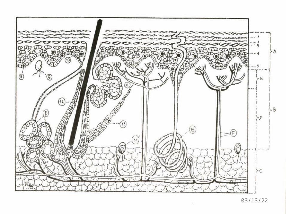
Anatomi based histopathology
The skin as large line, consist of 3 layer :
I. Epidermis layer / cutikel
II. Dermis layer / corium, cutis vera, true skin
III. Subcutis layer (hipodermis)
04/18/23

04/18/23

Dermis and subcutis
characterized by connective tissue
(nothing big line) and present cell
and fat tissue
04/18/23

1. Epidermis layer
Epidermis consist of 5 layer :
1.Stratum corneum (SC) ; complete differentiation of granular cells results in stacked layers of a nucleate, flattened cornified cells that form SC
Mechanical protection to the skin & a barrier to water loss & permeation of soluble substances from the environment
04/18/23

2. Stratum Lucidum
1 layers. flattened cornified cells , no
nucleous
Protoplasma change protein eleidin
On the palmar and plantar pedis
04/18/23

3. Stratum granulosum (Ganulosum layer)Basophilic keratohyalin granules that are
prominent within cellsKeratohyalin granules are compound primarily of
profilaggrin, keratin filaments, & loricrin4. Stratum spinosum (Spinosum layer)Spine-like appearanceSuprabasal spinous cell : polyhedral in shape
w/a rounded nucleusSpinous cells also contain large bundle of keratin
filaments
04/18/23

5. Stratum Basale (BL) = (stratum germinativum)
Palisade form (columnar shaped Kc) Basal cell mitotically active Layers consist of 2 :1. Columnar cell2. Melanocytes
are neural crest-derived pigment-synthesizing dendritic cells that reside primarily in the BL
04/18/23

2. Dermis Layer
Elastic dan fibrous layer with cellular element and hair follicle 2 part :
a. Stratum papillare to epidermis b. Stratum reticulare to subcutis, more
thick & connective tissue. Containing: nerve, blood vessel, hair, sweat gland n sebaceous gland
04/18/23

3. Subcutis Layer
Adipocytes form the bulk of of the cells in the hypodermis.
organized into lobules defined by septa of fibrous
connective tissue
Nerves, vessels and lymphatics are located within the septa
& supply the region.
Two regions (Dermis & Subcutis) = structurally &
functionally integrated through networks of nerves &
vessels & the continuity of the epidermal appendages
04/18/23

Phisiology of The Skin
Main function :
1. Protection from infectious agent
2. Ultraviolet protection
3. Perception
4. Regulatory body temperature (thermoregulation)
5. Respiration
04/18/23

5. Excretion
6. Expression
7. Wound repair/regeneration
8. Forming precussor vitamin D
9. Physical appearence
10.Recently immunologic agent
04/18/23
04/18/23

04/18/23

Transit time for a basal cell from the time it loses contact with the basal layer to the time it enters stratum corneum , is at least 14 days
Transit through the SC and subsequent desquamation require another 14 days
Thickness of epid from 0.4to 1.5 mm as compared with the 1.5 mm to 4.0 mm full thickness skin
04/18/23

04/18/23