anatomy of the lower...
TRANSCRIPT

Joints of the lower limb
By Dr. Amjad Shatarat
Dr. Shatarat, M.D, PhD

Hip joint
Dr. Shatarat, M.D, PhD

Synovial multi-
axial
ball-and-socketjoint.
1-Type:
Dr. Shatarat, M.D, PhD
shatarat

a. head of femur
b. lunate surface of acetabulum
2-Articular surfaces:
3-Nerve
Supply: Femoral
nerve Obturator
nerve Sciatic nerve
Which is deepened by the
fibrocartilaginous labrum acetabulare
Dr. Shatarat, M.D, PhD
shatarat

4-The capsule of the hip is attached
proximally to the margins of the acetabulum
Distally, it is
attached along
the
intertrochanteric
line, the bases of
the greater and
lesser trochanters
Dr. Shatarat, M.D, PhD
shatarat

posteriorly,
to the femoral
neck about 0.5 in
(12mm) from the
intertrochanteric
crest.
Part of the neck posteriorly
is extra -capsular
Dr. Shatarat, M.D, PhD
shatarat

1-Medial and lateral
circumflex femoral
arteries
The main blood supply
is from
the retinacular arteries
arising as branches from
the circumflex femoral
arteries (especially the
medial circumflex femoral artery).
2-Artery to the
head of femur, a
branch of the
obturator artery
that traverses
the ligament of
the head.
5- Blood supply of the head of the
femur
Dr. Shatarat, M.D, PhD
shatarat

Obturator artery
Femoral artery
Dr. Shatarat, M.D, PhD
shatarat

1-The Trochanteric Anastomosis
The trochanteric anastomosis :
Branches pierce the capsule to give the main
blood supply to THE HEAD OF
THE FEMUR
The following arteries take part in the
anastomosis:
A) The superior gluteal artery, the
inferior gluteal artery and the
obturator artery (from the internal
iliac artery)
B) The medial femoral circumflex artery,
and the lateral femoral circumflex artery (from the femoral artery)
Dr. Shatarat, M.D, PhD
shatarat

The Cruciate
Anastomosis
The cruciate anastomosis
is situated at the level of
the lesser trochanter of
the femur and, together
with the trochanteric
anastomosis, provides a
connection between the
internal iliac and the
femoral arteries
2-The Cruciate Anastomosis
Dr. Shatarat, M.D, PhD
shatarat

Dr. Shatarat, M.D, PhD

b-Pubofemoral:
Limits extension and abduction
A-Iliofemoral: is a strong, inverted Y-
shaped
ligamentPrevents
hyperextension of hip joint during standing
Dr. Shatarat, M.D, PhD
shatarat

c-Ischiofemoral:
limits extension
Dr. Shatarat, M.D, PhD
shatarat

D-The ligament of head of femur (ligamentum teres)
primarily a synovial fold
conducting a blood vessel
is weak and of little importance in strengthening the hip joint
Dr. Shatarat, M.D, PhD
shatarat

The
non-articular lower part of the
acetabulum,
the acetabular notch, is closed off
below by thetransverse acetabular ligament
Dr. Shatarat, M.D, PhD
shatarat

Flexion is performed by the iliopsoas,rectus
femoris, and sartoriusExtension is performed by the gluteusmaximusand the hamstring muscles.
Abduction is performed by the gluteus medius
and minimus, assisted by the sartorius, tensor fasciae latae,
and piriformis.
Adduction is performed by the adductor longus and
brevis and the adductor fibers of the adductor magnus. These muscles are assisted by
the pectineus
and the gracilis.
Lateral rotation is performed by the short lateralrotator muscles and assisted by the gluteus maximus.
Medial rotation is performed by the anterior fibers of the gluteus
medius and gluteus minimus and the tensor fasciae latae.
Flexion is limited by
the hamstring
muscle group.
Extension is limited
by the ligamentous
thickening of the
capsule; abduction,
by the adductor
group of muscles;
adduction, by the
tensor muscle and
fascia of the
abductor
muscles; and
rotation, by the
fibrous capsular
Self-Study
Dr. Shatarat, M.D, PhD

8- ANGLE OF INCLINATION
Approx. 125o
it is the angle between the neck and shaft of the femur
typically ranges from 115 to 140 degrees
is about 160 ° in the young child and
about 125° in the adult
Dr. Shatarat, M.D, PhD
shatarat

Ankle joint
Dr. Shatarat, M.D, PhD
Ankle Joint
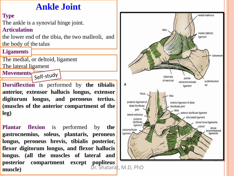
Dorsiflextion is performed by the tibialis
anterior, extensor hallucis longus, extensor
digitorum longus, and peroneus tertius.
(muscles of the anterior compartment of the
leg)
Plantar flexion is performed by the
gastrocnemius, soleus, plantaris, peroneus
longus, peroneus brevis, tibialis posterior,
flexor digitorum longus, and flexor hallucis
longus. (all the muscles of lateral and
posterior compartment except popliteus
muscle)
Type
The ankle is a synovial hinge joint.
Articulation
the lower end of the tibia, the two malleoli, and
the body of the talus
Ligaments
The medial, or deltoid, ligament
The lateral ligament
Movements
Dr. Shatarat, M.D, PhD
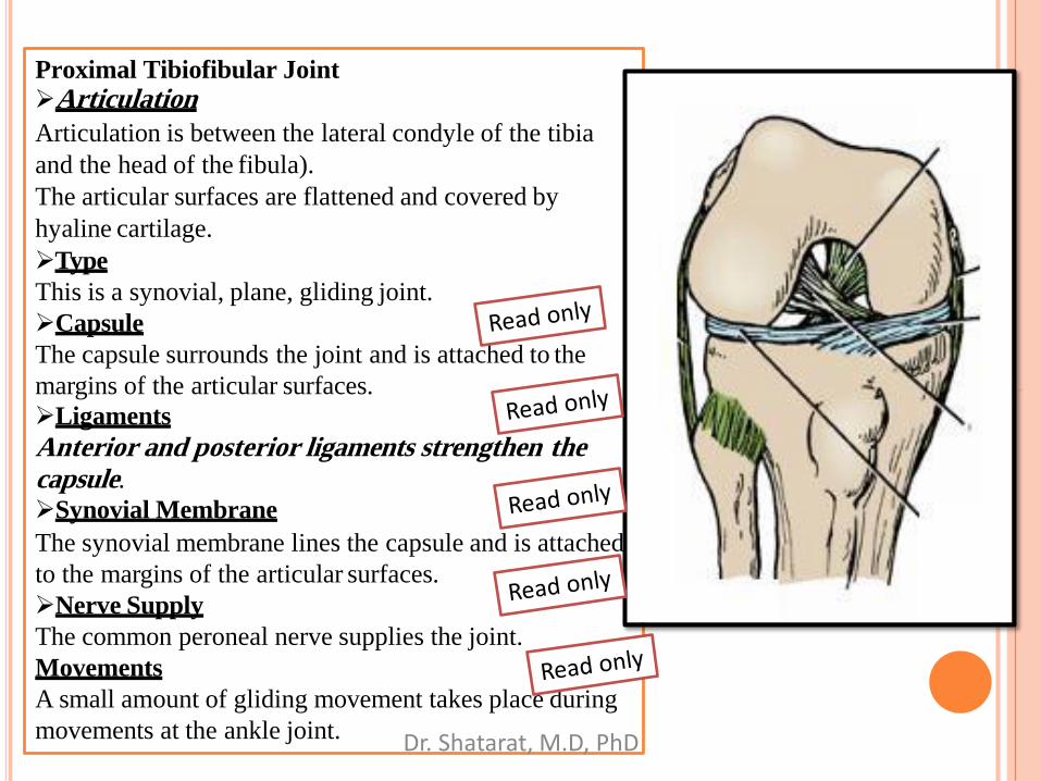
Proximal Tibiofibular Joint
Articulation
Articulation is between the lateral condyle of the tibia
and the head of the fibula).
The articular surfaces are flattened and covered by
hyaline cartilage.
Type
This is a synovial, plane, gliding joint.
Capsule
The capsule surrounds the joint and is attached to the
margins of the articular surfaces.
Ligaments
Anterior and posterior ligaments strengthen thecapsule.
Synovial Membrane
The synovial membrane lines the capsule and is attached
to the margins of the articular surfaces.
Nerve Supply
The common peroneal nerve supplies the joint.
Movements
A small amount of gliding movement takes place during
movements at the ankle joint. Dr. Shatarat, M.D, PhD

Distal Tibiofibular Joint
Articulation
Articulation is between the fibular notch at the
lower end of the tibia and the lower end of the
fibula
Type
The distal tibiofibular joint is
a fibrous jointCapsule
There is no capsule.
Ligaments
1The interosseous ligament is a strong, thick
band of fibrous tissue that binds the two bones
together.
2The anterior and posterior ligaments are
flat bands of fibrous tissue connecting the two
bones together in front and behind the
interosseous ligament
3 The inferior transverse ligament
Dr. Shatarat, M.D, PhD

K n e e J o i n t
Dr. Shatarat, M.D, PhD

K n e e J o i n tIs the most complicated joint in the
body!!!!
1-Consists of two condylar joints between:
A-The medial and lateral condyles of the femur
and The condyles of the tibia
and
Dr. Shatarat, M.D, PhD
shatarat

B- a gliding jointbetween the patella and the patellar surface
of the femur
Note that the fibula is not directly involved in
the joint.
Patellar surfaceOF THE FEMUR
Dr. Shatarat, M.D, PhD
shatarat
shatarat

2-Type OF JOINT
The joint between the
patella and femur is a
synovial joint of the plane gliding variety.
The joint between the femur and tibia is a
synovial joint of the hinge variety, but some
degree of rotatory movement ispossible.
MEDIAL AND LATERAL ROTATION
Dr. Shatarat, M.D, PhD
shatarat

The capsule is attached to the margins of thearticular surfaces
surrounds the sides and posterior aspect of the joint.
On the front of the joint, the capsuleis absent permitting the synovial
membrane to pouch upward beneath the
quadriceps tendon, forming the
suprapatellar bursa
3 - C a p s u l e
Dr. Shatarat, M.D, PhD
shatarat

4-MenisciMedial and lateral menisci are C-
shaped sheets of fibrocartilage.
(composed of fibrous connective
tissue and NOT of cartilage.
Their function is to deepenthe articular surfaces of the
tibial condyles to receive the
convex femoral condyles;
They also serve as cushionsbetween the two bones
Each meniscus is attached to
the upper surface of the tibia
by anterior and posterior
horns.
Anterior
Posterior
Dr. Shatarat, M.D, PhD
shatarat

5-Li ga me n ts o f the
k n e e jo in tThe ligaments may be
divided into
A-Extracapsular
Ligaments
The ligamentum
patellae
is attached
above to the lower border
of the patella and below to
the tuberosity of the tibia.
Dr. Shatarat, M.D, PhD
shatarat

The lateral
collateral ligament
is cordlike and is attached
above to the lateral condyle of
the femur and below to the
head of the fibula.
What does this mean?Dr. Shatarat, M.D, PhD
shatarat

The medial collateral ligament
is a flat band and is attached above to the medial condyle of the femur and below to
the medial surface of the shaft of the tibia.It is firmly attached to the edge of the
medial meniscus ?!
Dr. Shatarat, M.D, PhD
shatarat
shatarat

poThe oblique popliteal ligament
Is a tendinous expansion derived from the semimembranosus muscle.
It strengthens the posterior aspect of the capsule
Dr. Shatarat, M.D, PhD

B-
I n t r a c a p s u l a r
L i g a m e n t s
The cruciate
ligaments
They are named
anterior and posterior,
according to their
tibial attachments
The anterior and posterior cruciate
ligaments are the main bond
between the femur and the tibia
during the joint's range of
movement. They prevent posterior
and anterior displacement,
respectively
Dr. Shatarat, M.D, PhD
shatarat

6.Locking mechanism
When standing, the knee joint is
'locked' which reduces the amount of
muscle work needed to
maintain the standing position
Another feature that keeps the
knee extended when standing
is that the body's center of
gravity is positioned along a
vertical line that passes
anterior to the knee joint.
The locking mechanism is achieved
by medial rotation of the
femur on the tibia
during extension. Medial
rotation and full extension tighten all
the associated ligaments
Tight ligaments during standing
Locked joint
The extended knee is said to be inthe locked positionDr. Shatarat, M.D, PhD
shatarat
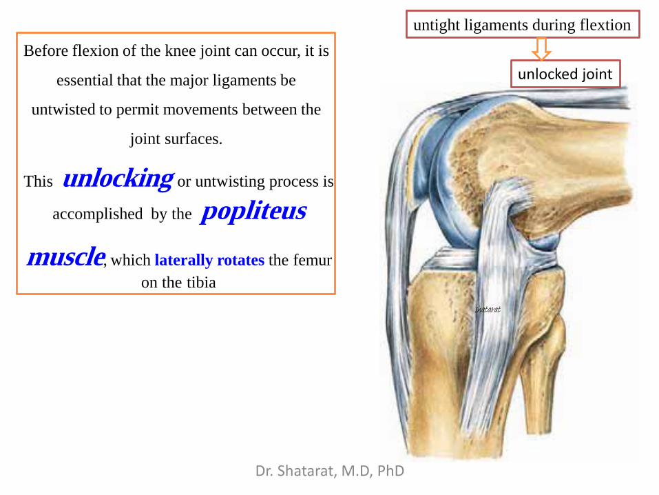
Before flexion of the knee joint can occur, it is
essential that the major ligaments be
untwisted to permit movements between the
joint surfaces.
This unlocking or untwisting process is
accomplished by the popliteus
muscle, which laterally rotates the femur
on the tibia
untight ligaments during flextion
unlocked joint
Dr. Shatarat, M.D, PhD
shatarat

The muscle arises
within the capsule of
the knee joint
•its tendon separates
the lateral meniscus from the
lateral ligament of the joint.
It emerges through
the lower part of the
posterior
surface of the capsule
of the joint to pass to
its
insertion.
Dr. Shatarat, M.D, PhD

Prepatellar bursitis (“housemaid's knee”)
is usually a friction bursitis caused by friction between the skin and the patella
7-Bursae Related to the Knee Joint
Dr. Shatarat, M.D, PhD

9-movements of the knee joint
Flexion
The biceps femoris, semitendinosus, and semimembranosus muscles,
assisted by the gracilis, and sartorius, produce flexion.
Flexion is limited by the contact of the back of the leg with the thigh.
Extension
The quadriceps femoris.
Extension is limited by the tension of all the major ligaments of the
joint.
Medial Rotation
The sartorius, gracilis, and semitendinosus
Lateral Rotation
The biceps femoris
Note:
The stability of the knee joint depends on the tone of the strong muscles
acting on the joint and the strength of the ligaments.
Dr. Shatarat, M.D, PhD

10- blood supply
Branch of
the
femoral
artery in
the
adductor
canal
Fro
m t
he
popli
teal
art
ery
Fro
m th
e popliteal artery
Dr. Shatarat, M.D, PhD