anatomy, physiology, & disease - somerset canyons...2014/10/04 · anatomy, physiology, &...
TRANSCRIPT

Copyright ©2011 by Pearson Education, Inc. Upper Saddle River, New Jersey 07458
All rights reserved.
Anatomy, Physiology, & Disease, Revised First Edition Bruce J. Colbert, Jeff E. Ankney, and Karen T. Lee
Anatomy, Physiology, & Disease
Revised First Edition
Chapter 4 Tissues and Systems:
The Inside Story
Copyright ©2011 by Pearson Education, Inc. Upper Saddle River, New Jersey 07458
All rights reserved.
Anatomy, Physiology, & Disease, Revised First Edition Bruce J. Colbert, Jeff E. Ankney, and Karen T. Lee
Multimedia Directory
Slide 49 !Skin Cancer Video!Slide 78 !Body Systems Animation!Slide 79 !Histotechnology Video!
Copyright ©2011 by Pearson Education, Inc. Upper Saddle River, New Jersey 07458
All rights reserved.
Anatomy, Physiology, & Disease, Revised First Edition Bruce J. Colbert, Jeff E. Ankney, and Karen T. Lee
Introduction
• Cells are basic building blocks of our bodies • Similar cells are organized into tissues that
perform similar functions • A collection of tissues designed to perform
similar or several functions is called an organ • Organs that work together to perform major
specific activities, often with help of accessory structures, form systems

Copyright ©2011 by Pearson Education, Inc. Upper Saddle River, New Jersey 07458
All rights reserved.
Anatomy, Physiology, & Disease, Revised First Edition Bruce J. Colbert, Jeff E. Ankney, and Karen T. Lee
Learning Objectives
• Explain the relationship between cells, tissues, organs, and systems • List and describe the four main types of
tissue • Identify and describe the various body
membranes • Differentiate the three main types of
muscle tissues
Copyright ©2011 by Pearson Education, Inc. Upper Saddle River, New Jersey 07458
All rights reserved.
Anatomy, Physiology, & Disease, Revised First Edition Bruce J. Colbert, Jeff E. Ankney, and Karen T. Lee
Learning Objectives (cont’d)
• Describe the main components of nerve tissues • List and describe the main functions of the
body systems • Provide general examples of how
pathologic conditions can impact on cells, tissues, organs, and body systems
Copyright ©2011 by Pearson Education, Inc. Upper Saddle River, New Jersey 07458
All rights reserved.
Anatomy, Physiology, & Disease, Revised First Edition Bruce J. Colbert, Jeff E. Ankney, and Karen T. Lee
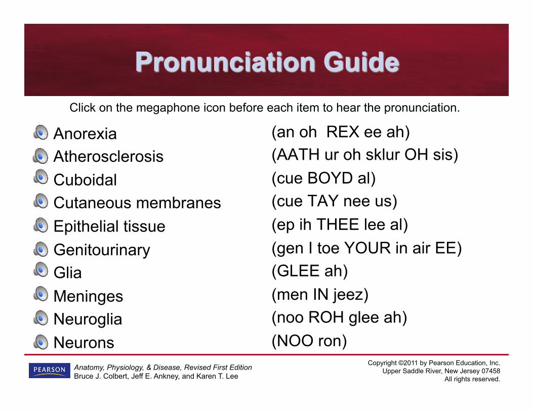
Pronunciation Guide
Anorexia Atherosclerosis Cuboidal Cutaneous membranes Epithelial tissue Genitourinary Glia Meninges Neuroglia Neurons
(an oh REX ee ah) (AATH ur oh sklur OH sis) (cue BOYD al) (cue TAY nee us) (ep ih THEE lee al) (gen I toe YOUR in air EE) (GLEE ah) (men IN jeez) (noo ROH glee ah) (NOO ron)
Click on the megaphone icon before each item to hear the pronunciation.

Copyright ©2011 by Pearson Education, Inc. Upper Saddle River, New Jersey 07458
All rights reserved.
Anatomy, Physiology, & Disease, Revised First Edition Bruce J. Colbert, Jeff E. Ankney, and Karen T. Lee
Pronunciation Guide (cont’d)
Parietal Serous membrane Skeletal muscle Squamous Stratified Striated muscle Synovial membrane Transitional Visceral
(pah RYE eh tal) (SEER us) (SKELL eh tal) (SKWAY muss) (STRAT ih fied) (STRY ate ed) (sin OH vee al) (tran ZISH ion al) (VISS er al)
Click on the megaphone icon before each item to hear the pronunciation.

Copyright ©2011 by Pearson Education, Inc. Upper Saddle River, New Jersey 07458
All rights reserved.
Anatomy, Physiology, & Disease, Revised First Edition Bruce J. Colbert, Jeff E. Ankney, and Karen T. Lee
Tissues
• Formed when there is collection of similar cells that act together to perform function • Imagine cells as bricks, placed in specific pattern
to create functional walls, or tissues, of building • Four main types:
– Epithelial – Connective – Muscle – Nervous
Copyright ©2011 by Pearson Education, Inc. Upper Saddle River, New Jersey 07458
All rights reserved.
Anatomy, Physiology, & Disease, Revised First Edition Bruce J. Colbert, Jeff E. Ankney, and Karen T. Lee
Epithelial Tissue
• Covers and lines much of body and also covers many of parts found in body • Cells are packed tightly together, forming a
sheet that usually has no blood vessels in it • Further classified by shape, as well as
arrangement – Flat or scale-like cells: squamous – Cube shaped: cuboidal – Column-like: columnar

Copyright ©2011 by Pearson Education, Inc. Upper Saddle River, New Jersey 07458
All rights reserved.
Anatomy, Physiology, & Disease, Revised First Edition Bruce J. Colbert, Jeff E. Ankney, and Karen T. Lee
Epithelial Tissue (cont’d)
• If cells are arranged in single layer and are all same type of cell, classified as simple • If several layers deep, they are stratified
and will be named by type of cell on outer layer • Exception is pseudostratified epithelium,
which is single layer of cells that looks stratified

Copyright ©2011 by Pearson Education, Inc. Upper Saddle River, New Jersey 07458
All rights reserved.
Anatomy, Physiology, & Disease, Revised First Edition Bruce J. Colbert, Jeff E. Ankney, and Karen T. Lee
Figure 4-1 Types and locations of epithelial tissues.

Copyright ©2011 by Pearson Education, Inc. Upper Saddle River, New Jersey 07458
All rights reserved.
Anatomy, Physiology, & Disease, Revised First Edition Bruce J. Colbert, Jeff E. Ankney, and Karen T. Lee
Membranes
• Sheet-like structures found throughout body that perform special functions • Can be classified as organs • Those classified as epithelial membranes
possess layer of epithelial tissue and bottom layer of specialized connective tissue
Copyright ©2011 by Pearson Education, Inc. Upper Saddle River, New Jersey 07458
All rights reserved.
Anatomy, Physiology, & Disease, Revised First Edition Bruce J. Colbert, Jeff E. Ankney, and Karen T. Lee
Membranes (cont’d)
• Epithelial membranes can be: – Cutaneous – Serous – Mucous

Copyright ©2011 by Pearson Education, Inc. Upper Saddle River, New Jersey 07458
All rights reserved.
Anatomy, Physiology, & Disease, Revised First Edition Bruce J. Colbert, Jeff E. Ankney, and Karen T. Lee
Table 4-1 Types of Epithelial Membranes !

Copyright ©2011 by Pearson Education, Inc. Upper Saddle River, New Jersey 07458
All rights reserved.
Anatomy, Physiology, & Disease, Revised First Edition Bruce J. Colbert, Jeff E. Ankney, and Karen T. Lee
Figure 4-2 Location of serous and mucous membranes.
Copyright ©2011 by Pearson Education, Inc. Upper Saddle River, New Jersey 07458
All rights reserved.
Anatomy, Physiology, & Disease, Revised First Edition Bruce J. Colbert, Jeff E. Ankney, and Karen T. Lee
Connective Tissue
• Most common of tissues, and is found scattered throughout body
• Found in organs, bones, nerves, muscles, membranes, and skin
• Holds things together and provides structure and support
• Can form fine, delicate webs or strong cord-like structures similar to wire cables
• Included in composition of bones and cartilage, as well as adipose tissue (fat)
Copyright ©2011 by Pearson Education, Inc. Upper Saddle River, New Jersey 07458
All rights reserved.
Anatomy, Physiology, & Disease, Revised First Edition Bruce J. Colbert, Jeff E. Ankney, and Karen T. Lee
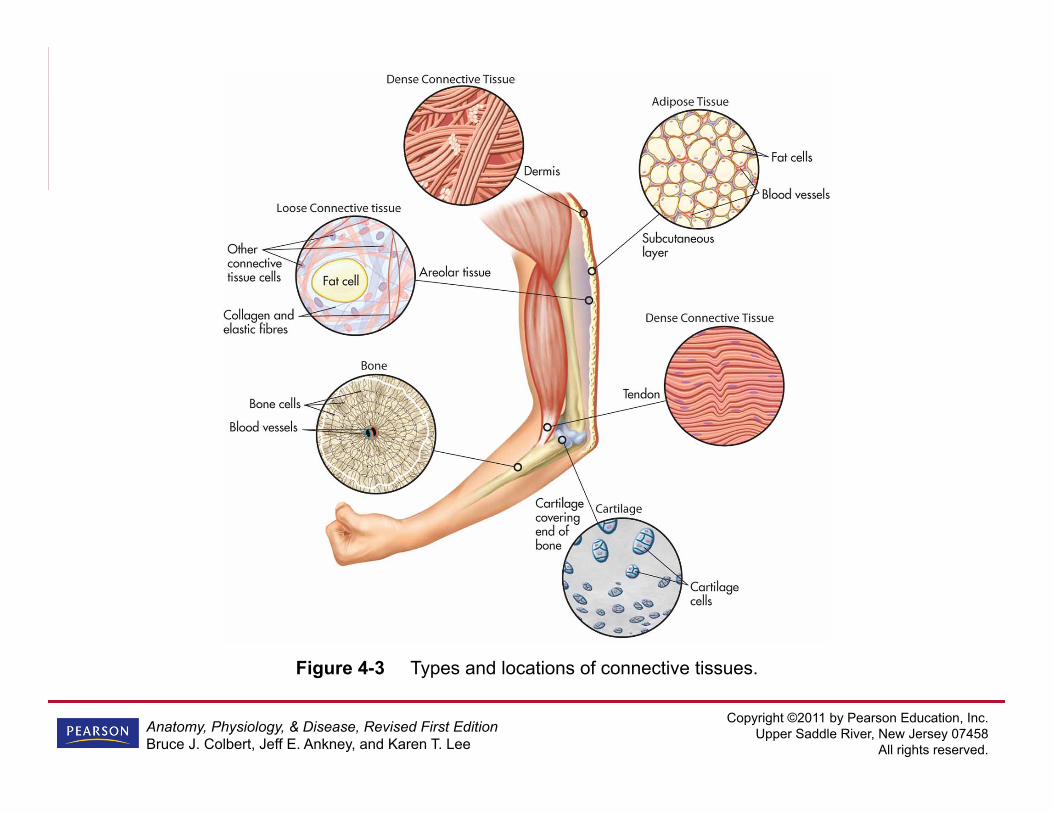
Figure 4-3 Types and locations of connective tissues.
Copyright ©2011 by Pearson Education, Inc. Upper Saddle River, New Jersey 07458
All rights reserved.
Anatomy, Physiology, & Disease, Revised First Edition Bruce J. Colbert, Jeff E. Ankney, and Karen T. Lee
Synovial Membrane
• Membrane type associated with connective tissue • Important membrane found in space
between bone joints and produces slippery substance called synovial fluid • This special fluid greatly reduces friction
when joints move
Copyright ©2011 by Pearson Education, Inc. Upper Saddle River, New Jersey 07458
All rights reserved.
Anatomy, Physiology, & Disease, Revised First Edition Bruce J. Colbert, Jeff E. Ankney, and Karen T. Lee
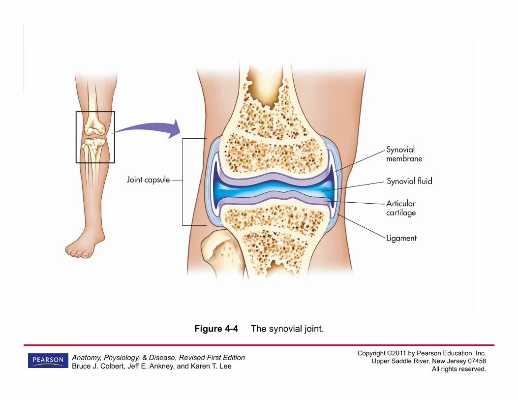
Figure 4-4 The synovial joint.

Copyright ©2011 by Pearson Education, Inc. Upper Saddle River, New Jersey 07458
All rights reserved.
Anatomy, Physiology, & Disease, Revised First Edition Bruce J. Colbert, Jeff E. Ankney, and Karen T. Lee
Muscle Tissue
• Provides means for movement, by and in our bodies • This form of tissue has ability to shorten
itself (contractility) • Three types:
– Skeletal – Cardiac – Smooth

Copyright ©2011 by Pearson Education, Inc. Upper Saddle River, New Jersey 07458
All rights reserved.
Anatomy, Physiology, & Disease, Revised First Edition Bruce J. Colbert, Jeff E. Ankney, and Karen T. Lee
Skeletal Muscle
• Called striated because of striped appearance • Attached to bones; causes movement by
contracting and relaxing • Often found around openings in body, such
as mouth, where it controls size of opening • Long, fiber-like cells with many nuclei in
each cell

Copyright ©2011 by Pearson Education, Inc. Upper Saddle River, New Jersey 07458
All rights reserved.
Anatomy, Physiology, & Disease, Revised First Edition Bruce J. Colbert, Jeff E. Ankney, and Karen T. Lee
Skeletal Muscle (cont’d)
• Brain controls muscle contraction and relaxation; because these muscles are controlled consciously, they are called voluntary muscles

Copyright ©2011 by Pearson Education, Inc. Upper Saddle River, New Jersey 07458
All rights reserved.
Anatomy, Physiology, & Disease, Revised First Edition Bruce J. Colbert, Jeff E. Ankney, and Karen T. Lee
Cardiac Muscle
• Found in walls of heart • Heart beats without us having to think
about it, so muscle type considered involuntary muscle • Cells within tissue interlock with each
other; makes for more efficient contraction

Copyright ©2011 by Pearson Education, Inc. Upper Saddle River, New Jersey 07458
All rights reserved.
Anatomy, Physiology, & Disease, Revised First Edition Bruce J. Colbert, Jeff E. Ankney, and Karen T. Lee
Smooth Muscles
• Forms walls of hollow organs such as in digestive system (often called visceral tissue) and blood vessels • We don’t control these muscles with
thoughts, so they are also involuntary muscles • Cells within tissue not as long and fibrous as
skeletal muscles and each has only one nucleus

Copyright ©2011 by Pearson Education, Inc. Upper Saddle River, New Jersey 07458
All rights reserved.
Anatomy, Physiology, & Disease, Revised First Edition Bruce J. Colbert, Jeff E. Ankney, and Karen T. Lee
Figure 4-5 Labeled diagram and flowchart of the three muscle tissue types.
Copyright ©2011 by Pearson Education, Inc. Upper Saddle River, New Jersey 07458
All rights reserved.
Anatomy, Physiology, & Disease, Revised First Edition Bruce J. Colbert, Jeff E. Ankney, and Karen T. Lee
Nervous Tissue
• Acts as rapid messenger service for body; messages can cause actions to occur; two types: – Neurons: conduction of information § Dendrites: branch-like formations on neurons that
receive sensory information § Axon: trunk-shaped structure that transports
information away from cell body – Glia (or neuroglia): support and connection cells

Copyright ©2011 by Pearson Education, Inc. Upper Saddle River, New Jersey 07458
All rights reserved.
Anatomy, Physiology, & Disease, Revised First Edition Bruce J. Colbert, Jeff E. Ankney, and Karen T. Lee
Nervous Tissue (cont’d)
• Membranes that cover brain and spinal cord called meninges • Many nerves have insulating layer called
myelin sheath

Copyright ©2011 by Pearson Education, Inc. Upper Saddle River, New Jersey 07458
All rights reserved.
Anatomy, Physiology, & Disease, Revised First Edition Bruce J. Colbert, Jeff E. Ankney, and Karen T. Lee
Figure 4-6 The two main types of nerve cells. !

Copyright ©2011 by Pearson Education, Inc. Upper Saddle River, New Jersey 07458
All rights reserved.
Anatomy, Physiology, & Disease, Revised First Edition Bruce J. Colbert, Jeff E. Ankney, and Karen T. Lee
Meningitis
• Inflammation of meninges (membranes that cover brain and spinal cord); caused by bacteria or virus • Bacterial form can spread via droplets
from sneezing or coughing; can also spread through contact with saliva of infected person; college students and military personnel in crowded situations at higher risk

Copyright ©2011 by Pearson Education, Inc. Upper Saddle River, New Jersey 07458
All rights reserved.
Anatomy, Physiology, & Disease, Revised First Edition Bruce J. Colbert, Jeff E. Ankney, and Karen T. Lee
Meningitis (cont’d)
• Once infected, you become carrier of disease – Only some people who become carriers will
develop disease – In others, immune system actually destroys
and removes pathogen before illness develops

Copyright ©2011 by Pearson Education, Inc. Upper Saddle River, New Jersey 07458
All rights reserved.
Anatomy, Physiology, & Disease, Revised First Edition Bruce J. Colbert, Jeff E. Ankney, and Karen T. Lee
Meningitis (cont’d)
• Symptoms – Non-specific headaches – Fever – Nausea – Neck stiffness – Skin rash – Hearing loss – Neurologic/ brain damage – Kidney failure
Copyright ©2011 by Pearson Education, Inc. Upper Saddle River, New Jersey 07458
All rights reserved.
Anatomy, Physiology, & Disease, Revised First Edition Bruce J. Colbert, Jeff E. Ankney, and Karen T. Lee
Meningitis (cont’d)
• Bacterial form has approximately 10% fatality rate • Vaccine available for prevention
– Does not protect against all pathogens that can cause meningitis
– Has been associated with adverse reactions, including headaches, dizziness, vomiting, convulsions, and even death
Copyright ©2011 by Pearson Education, Inc. Upper Saddle River, New Jersey 07458
All rights reserved.
Anatomy, Physiology, & Disease, Revised First Edition Bruce J. Colbert, Jeff E. Ankney, and Karen T. Lee
Meningitis (cont’d)
• Right now, vaccine not mandatory for high-risk groups; individuals should weigh risks and benefits in deciding if they would like to be vaccinated
Copyright ©2011 by Pearson Education, Inc. Upper Saddle River, New Jersey 07458
All rights reserved.
Anatomy, Physiology, & Disease, Revised First Edition Bruce J. Colbert, Jeff E. Ankney, and Karen T. Lee
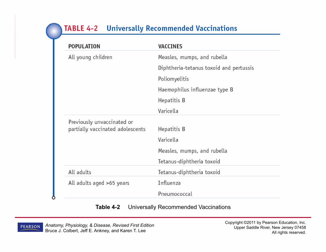
Table 4-2 Universally Recommended Vaccinations

Copyright ©2011 by Pearson Education, Inc. Upper Saddle River, New Jersey 07458
All rights reserved.
Anatomy, Physiology, & Disease, Revised First Edition Bruce J. Colbert, Jeff E. Ankney, and Karen T. Lee
Pathology Connection: Blood Sugar and Tissue Damage
• Diabetes (and associated high blood sugar) can cause damage to body tissues – Since glucose cannot be moved inside body
cells, cells must burn fats and proteins in body for energy § As body uses up protein, tissues start to break
down, becomes difficult to produce more tissue – With impaired tissue production, wounds
become more difficult to heal, and infections become harder to fight

Copyright ©2011 by Pearson Education, Inc. Upper Saddle River, New Jersey 07458
All rights reserved.
Anatomy, Physiology, & Disease, Revised First Edition Bruce J. Colbert, Jeff E. Ankney, and Karen T. Lee
Pathology Connection: Blood Sugar and Tissue Damage (cont’d)
• Lipids released from fat so that cell can burn lipids for energy – Lipids can deposit around inside walls of blood
vessels (atherosclerosis develops) – Deposits cause impaired blood flow to tissues
• Together, tissue break down, deficient blood flow, and impaired wound healing mean that diabetics prone to tissue death and gangrene; can lead to loss of toes, feet, and even legs

Copyright ©2011 by Pearson Education, Inc. Upper Saddle River, New Jersey 07458
All rights reserved.
Anatomy, Physiology, & Disease, Revised First Edition Bruce J. Colbert, Jeff E. Ankney, and Karen T. Lee
Organs
• Result of two or more types of tissues organizing in such a way as to accomplish something that tissues cannot do on their own • Some occur singly and some in pairs • Vital ones are those you can’t live without • Others, like spleen, appendix, or gallbladder,
can be removed without causing problems • Work as part of a system
Copyright ©2011 by Pearson Education, Inc. Upper Saddle River, New Jersey 07458
All rights reserved.
Anatomy, Physiology, & Disease, Revised First Edition Bruce J. Colbert, Jeff E. Ankney, and Karen T. Lee
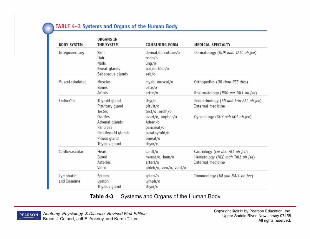
Table 4-3 Systems and Organs of the Human Body !
Copyright ©2011 by Pearson Education, Inc. Upper Saddle River, New Jersey 07458
All rights reserved.
Anatomy, Physiology, & Disease, Revised First Edition Bruce J. Colbert, Jeff E. Ankney, and Karen T. Lee
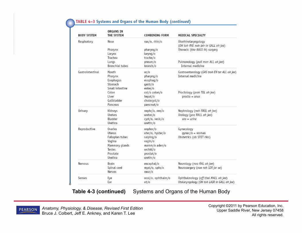
Table 4-3 (continued) Systems and Organs of the Human Body !

Copyright ©2011 by Pearson Education, Inc. Upper Saddle River, New Jersey 07458
All rights reserved.
Anatomy, Physiology, & Disease, Revised First Edition Bruce J. Colbert, Jeff E. Ankney, and Karen T. Lee
Systems
• Formed by organs that work together to accomplish something more complex than what single organ can do on its own • Each is interrelated, often depending on
each other for proper functioning of body
Copyright ©2011 by Pearson Education, Inc. Upper Saddle River, New Jersey 07458
All rights reserved.
Anatomy, Physiology, & Disease, Revised First Edition Bruce J. Colbert, Jeff E. Ankney, and Karen T. Lee
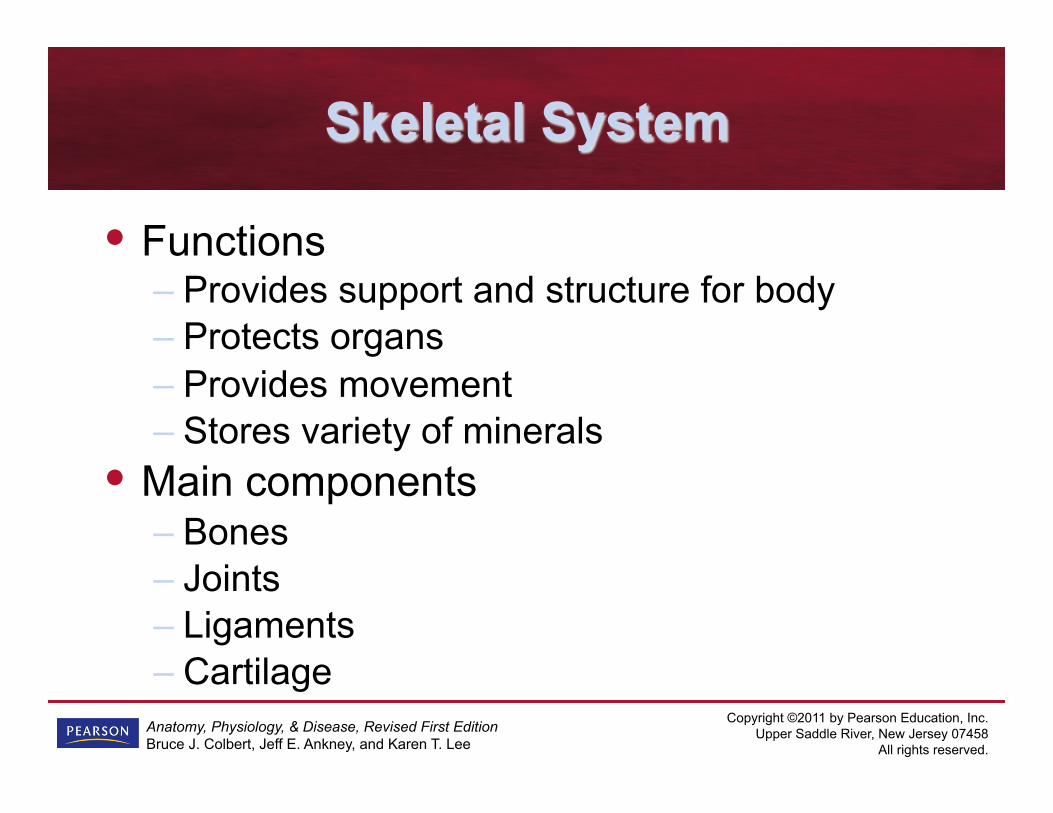
Skeletal System
• Functions – Provides support and structure for body – Protects organs – Provides movement – Stores variety of minerals
• Main components – Bones – Joints – Ligaments – Cartilage

Copyright ©2011 by Pearson Education, Inc. Upper Saddle River, New Jersey 07458
All rights reserved.
Anatomy, Physiology, & Disease, Revised First Edition Bruce J. Colbert, Jeff E. Ankney, and Karen T. Lee
Figure 4-7 The skeletal system. !

Copyright ©2011 by Pearson Education, Inc. Upper Saddle River, New Jersey 07458
All rights reserved.
Anatomy, Physiology, & Disease, Revised First Edition Bruce J. Colbert, Jeff E. Ankney, and Karen T. Lee
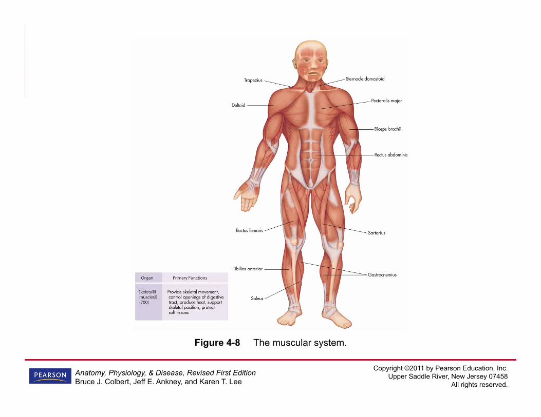
Muscular System
• Voluntary muscles – Movement created by conscious thought, like
scratching your nose – Skeletal muscles attached to bones
• Involuntary muscles – Perform without conscious thought – Classified as smooth or cardiac muscle – Found in blood vessels, airways, and organs
Copyright ©2011 by Pearson Education, Inc. Upper Saddle River, New Jersey 07458
All rights reserved.
Anatomy, Physiology, & Disease, Revised First Edition Bruce J. Colbert, Jeff E. Ankney, and Karen T. Lee
Muscular System (cont’d)
• External muscles attached to bones and help with movement; internal muscles found inside body, like those that allow you to swallow
Copyright ©2011 by Pearson Education, Inc. Upper Saddle River, New Jersey 07458
All rights reserved.
Anatomy, Physiology, & Disease, Revised First Edition Bruce J. Colbert, Jeff E. Ankney, and Karen T. Lee
Figure 4-8 The muscular system. !

Copyright ©2011 by Pearson Education, Inc. Upper Saddle River, New Jersey 07458
All rights reserved.
Anatomy, Physiology, & Disease, Revised First Edition Bruce J. Colbert, Jeff E. Ankney, and Karen T. Lee
Integumentary System
• Includes skin (body’s first line of protection) • Regulates temperature through sweating,
shivering, and changes in diameter of blood vessels in skin • Sensory information received from outside
world (heat, cold, pain, pressure, etc.) comes from sensors in skin

Copyright ©2011 by Pearson Education, Inc. Upper Saddle River, New Jersey 07458
All rights reserved.
Anatomy, Physiology, & Disease, Revised First Edition Bruce J. Colbert, Jeff E. Ankney, and Karen T. Lee
Integumentary System (cont’d)
• Glands in skin help lubricate and waterproof skin, and inhibit growth of unwanted bacteria • Main components of system: skin, hair,
sweat glands, sebaceous glands, and nails

Copyright ©2011 by Pearson Education, Inc. Upper Saddle River, New Jersey 07458
All rights reserved.
Anatomy, Physiology, & Disease, Revised First Edition Bruce J. Colbert, Jeff E. Ankney, and Karen T. Lee
Figure 4-9 The integumentary system.
Copyright ©2011 by Pearson Education, Inc. Upper Saddle River, New Jersey 07458
All rights reserved.
Anatomy, Physiology, & Disease, Revised First Edition Bruce J. Colbert, Jeff E. Ankney, and Karen T. Lee
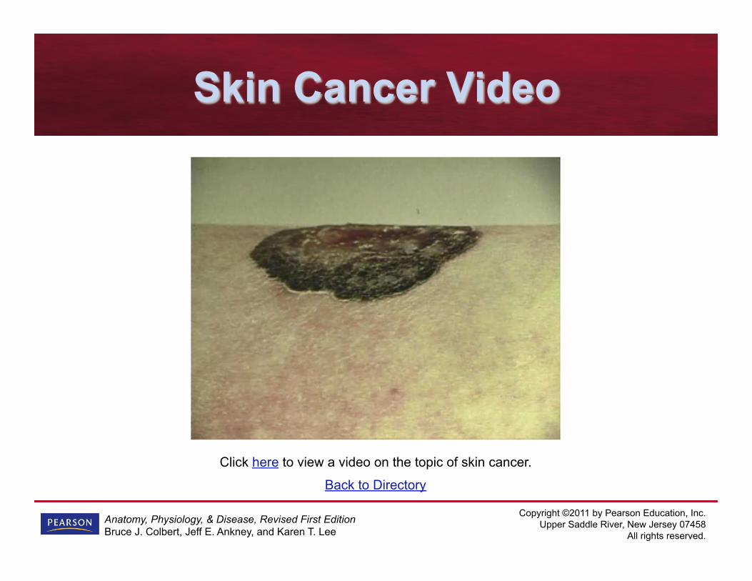
Back to Directory
Click here to view a video on the topic of skin cancer.
Skin Cancer Video

Copyright ©2011 by Pearson Education, Inc. Upper Saddle River, New Jersey 07458
All rights reserved.
Anatomy, Physiology, & Disease, Revised First Edition Bruce J. Colbert, Jeff E. Ankney, and Karen T. Lee
Nervous System
• Sends and receives messages, stimulated by body’s internal and external environments, affecting how we perceive world and protecting us from harm • Conscious sensations occur as result of
stimulation of our sensory receptors

Copyright ©2011 by Pearson Education, Inc. Upper Saddle River, New Jersey 07458
All rights reserved.
Anatomy, Physiology, & Disease, Revised First Edition Bruce J. Colbert, Jeff E. Ankney, and Karen T. Lee
Nervous System
• Main parts of system: spinal cord, brain, peripheral nerves, nerve cells, and spinal fluid; special sensory organs: eyes, ears, nose, tongue, and skin • Three main functions:
– Sensory messages – Processing and interpreting messages – Sending messages

Copyright ©2011 by Pearson Education, Inc. Upper Saddle River, New Jersey 07458
All rights reserved.
Anatomy, Physiology, & Disease, Revised First Edition Bruce J. Colbert, Jeff E. Ankney, and Karen T. Lee
Figure 4-10 The nervous system. !
Copyright ©2011 by Pearson Education, Inc. Upper Saddle River, New Jersey 07458
All rights reserved.
Anatomy, Physiology, & Disease, Revised First Edition Bruce J. Colbert, Jeff E. Ankney, and Karen T. Lee
Endocrine System
• Acts as control center for virtually all of body’s organs • Endocrine glands release chemicals called
hormones that are circulated via cardiovascular system, regulating metabolic processes and utilizing metabolites for growth and reproduction
Copyright ©2011 by Pearson Education, Inc. Upper Saddle River, New Jersey 07458
All rights reserved.
Anatomy, Physiology, & Disease, Revised First Edition Bruce J. Colbert, Jeff E. Ankney, and Karen T. Lee
Endocrine System
• Helps regulate fluid and electrolyte balance; helps cope with stresses produced by infection and trauma • Main components of system: hypothalamus,
pineal, pituitary, thyroid, parathyroid, thymus, and adrenal glands, pancreas, and gonads, plus large variety of hormones

Copyright ©2011 by Pearson Education, Inc. Upper Saddle River, New Jersey 07458
All rights reserved.
Anatomy, Physiology, & Disease, Revised First Edition Bruce J. Colbert, Jeff E. Ankney, and Karen T. Lee
Figure 4-11 The endocrine system. !

Copyright ©2011 by Pearson Education, Inc. Upper Saddle River, New Jersey 07458
All rights reserved.
Anatomy, Physiology, & Disease, Revised First Edition Bruce J. Colbert, Jeff E. Ankney, and Karen T. Lee
Cardiovascular System
• Referred to as circulatory system; main transportation system to each cell of body • Water, oxygen, and variety of nutrients
and substances required for life transported to cells, while waste products removed from cells • Main components of system: heart,
arteries, veins, capillaries, and blood

Copyright ©2011 by Pearson Education, Inc. Upper Saddle River, New Jersey 07458
All rights reserved.
Anatomy, Physiology, & Disease, Revised First Edition Bruce J. Colbert, Jeff E. Ankney, and Karen T. Lee
Figure 4-12 The cardiovascular system.
Copyright ©2011 by Pearson Education, Inc. Upper Saddle River, New Jersey 07458
All rights reserved.
Anatomy, Physiology, & Disease, Revised First Edition Bruce J. Colbert, Jeff E. Ankney, and Karen T. Lee
Pathology Connection: Septicemia
• Also called sepsis or blood poisoning; condition in which pathogen is present in blood • Because blood needed by all body
systems, sepsis can lead to multi-system infection – Blood can spread bacteria to organs – Once bacteria get in organs, they may
continue to grow, adversely affecting organ function
Copyright ©2011 by Pearson Education, Inc. Upper Saddle River, New Jersey 07458
All rights reserved.
Anatomy, Physiology, & Disease, Revised First Edition Bruce J. Colbert, Jeff E. Ankney, and Karen T. Lee
Pathology Connection: Septicemia (cont’d)
• Sepsis syndrome: infection causes decrease in blood perfusion to organs along with other systemic signs • Septic shock: decreased perfusion to
organs causes drop in blood pressure

Copyright ©2011 by Pearson Education, Inc. Upper Saddle River, New Jersey 07458
All rights reserved.
Anatomy, Physiology, & Disease, Revised First Edition Bruce J. Colbert, Jeff E. Ankney, and Karen T. Lee
Pathology Connection: Septicemia (cont’d)
• Multiple Organ Dysfunction Syndrome (MODS or multi-system failure) can develop if septic shock not quickly and effectively treated – As number of involved organ systems
increases, mortality rate rises; can approach 100% if continued for more than 4 hospital days, depending on patient

Copyright ©2011 by Pearson Education, Inc. Upper Saddle River, New Jersey 07458
All rights reserved.
Anatomy, Physiology, & Disease, Revised First Edition Bruce J. Colbert, Jeff E. Ankney, and Karen T. Lee
Pathology Connection: Septicemia (cont’d)
• Signs and symptoms related to sepsis syndrome – Fever – Chills – Tachypnea – Tachycardia – Skin lesions
Copyright ©2011 by Pearson Education, Inc. Upper Saddle River, New Jersey 07458
All rights reserved.
Anatomy, Physiology, & Disease, Revised First Edition Bruce J. Colbert, Jeff E. Ankney, and Karen T. Lee
Pathology Connection: Septicemia (cont’d)
• Signs and symptoms related to sepsis syndrome – Redness of skin, either in selected areas
(erythema), or over widespread areas of skin (erythroderma)
– Hypoxemia – Changes in mental status

Copyright ©2011 by Pearson Education, Inc. Upper Saddle River, New Jersey 07458
All rights reserved.
Anatomy, Physiology, & Disease, Revised First Edition Bruce J. Colbert, Jeff E. Ankney, and Karen T. Lee
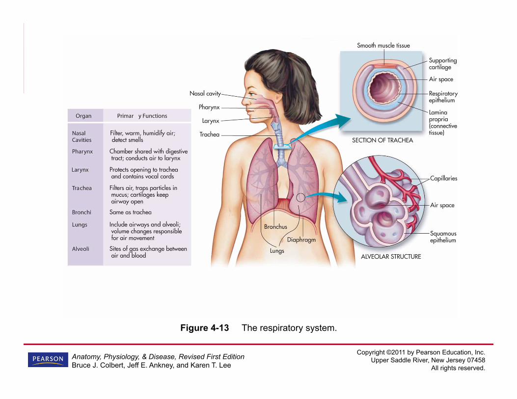
Respiratory System
• Supplies cells with oxygen and removes carbon dioxide without conscious effort • Filters, warms, and moistens air we
breathe • Mucous lining of airway helps trap foreign
particles and germs

Copyright ©2011 by Pearson Education, Inc. Upper Saddle River, New Jersey 07458
All rights reserved.
Anatomy, Physiology, & Disease, Revised First Edition Bruce J. Colbert, Jeff E. Ankney, and Karen T. Lee
Respiratory System (cont’d)
• System also helps maintain proper acid-base balance • Main parts of system: nose, nasal cavity,
trachea, larynx, pharynx, bronchial tubes, and lungs
Copyright ©2011 by Pearson Education, Inc. Upper Saddle River, New Jersey 07458
All rights reserved.
Anatomy, Physiology, & Disease, Revised First Edition Bruce J. Colbert, Jeff E. Ankney, and Karen T. Lee
Figure 4-13 The respiratory system.
Copyright ©2011 by Pearson Education, Inc. Upper Saddle River, New Jersey 07458
All rights reserved.
Anatomy, Physiology, & Disease, Revised First Edition Bruce J. Colbert, Jeff E. Ankney, and Karen T. Lee
Lymphatic System
• Responsible for helping to maintain proper fluid balance and protect us from infection • Special structures, called lymph nodes, act
as filters to capture unwanted infectious agents
Copyright ©2011 by Pearson Education, Inc. Upper Saddle River, New Jersey 07458
All rights reserved.
Anatomy, Physiology, & Disease, Revised First Edition Bruce J. Colbert, Jeff E. Ankney, and Karen T. Lee
Lymphatic System (cont’d)
• Produces special white cells, called T-lymphocytes, to fight infection • Major parts of system: lymph vessels,
lymph ducts, lymph nodes, thymus gland, tonsils, and spleen

Copyright ©2011 by Pearson Education, Inc. Upper Saddle River, New Jersey 07458
All rights reserved.
Anatomy, Physiology, & Disease, Revised First Edition Bruce J. Colbert, Jeff E. Ankney, and Karen T. Lee
Figure 4-14 The lymphatic system. !

Copyright ©2011 by Pearson Education, Inc. Upper Saddle River, New Jersey 07458
All rights reserved.
Anatomy, Physiology, & Disease, Revised First Edition Bruce J. Colbert, Jeff E. Ankney, and Karen T. Lee
Gastrointestinal (Digestive) System
• Often called GI system; takes raw material (food) and breaks it down both mechanically and chemically into usable substances, then absorbs substances for transportation to cells • Transports waste (produced by unused
materials) out of body • Main parts of system: mouth, pharynx,
esophagus, stomach, intestines, accessory organs, bowel, and anal canal

Copyright ©2011 by Pearson Education, Inc. Upper Saddle River, New Jersey 07458
All rights reserved.
Anatomy, Physiology, & Disease, Revised First Edition Bruce J. Colbert, Jeff E. Ankney, and Karen T. Lee
Figure 4-15 The digestive system. !

Copyright ©2011 by Pearson Education, Inc. Upper Saddle River, New Jersey 07458
All rights reserved.
Anatomy, Physiology, & Disease, Revised First Edition Bruce J. Colbert, Jeff E. Ankney, and Karen T. Lee
Pathology Connection: Body Image
• Obesity – US population becoming increasingly
overweight every year; current generation of adolescents may potentially may have shorter life span than their parents
• Anorexia nervosa: condition in which there is progressive and severe weight loss – Patients avoid eating or eat too little food to
sustain healthy weight – Patients deny problem

Copyright ©2011 by Pearson Education, Inc. Upper Saddle River, New Jersey 07458
All rights reserved.
Anatomy, Physiology, & Disease, Revised First Edition Bruce J. Colbert, Jeff E. Ankney, and Karen T. Lee
Pathology Connection: Body Image (cont’d)
• Bulimia: individual has eating binges, overeats, and then attempts to get rid of food by vomiting or using laxatives to avoid weight gain
Copyright ©2011 by Pearson Education, Inc. Upper Saddle River, New Jersey 07458
All rights reserved.
Anatomy, Physiology, & Disease, Revised First Edition Bruce J. Colbert, Jeff E. Ankney, and Karen T. Lee
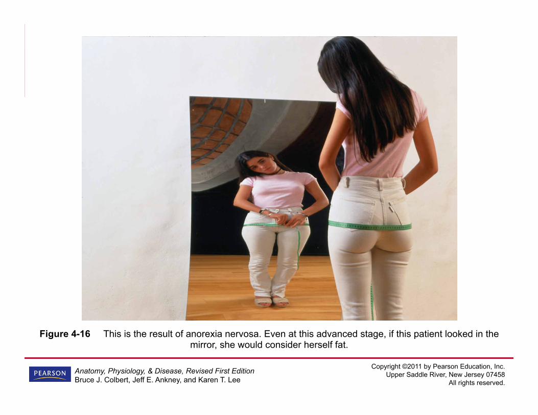
Figure 4-16 This is the result of anorexia nervosa. Even at this advanced stage, if this patient looked in the mirror, she would consider herself fat.
Copyright ©2011 by Pearson Education, Inc. Upper Saddle River, New Jersey 07458
All rights reserved.
Anatomy, Physiology, & Disease, Revised First Edition Bruce J. Colbert, Jeff E. Ankney, and Karen T. Lee
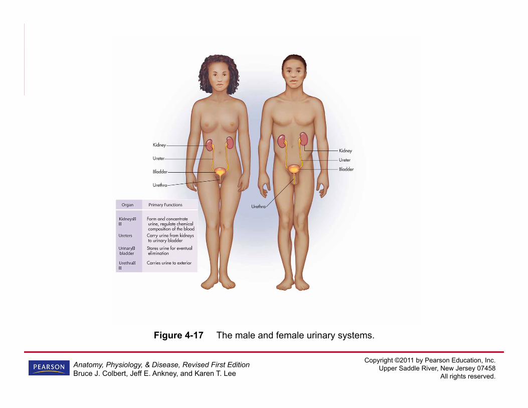
Urinary System
• Plays important role in elimination of waste products, electrolytes, drugs, and other toxins, as well as excessive water • Functions: water regulation, blood pressure
regulation, regulation of red blood cells, electrolyte balance, and pH balance • Main parts of system: kidneys, ureters,
urinary bladder, and urethra
Copyright ©2011 by Pearson Education, Inc. Upper Saddle River, New Jersey 07458
All rights reserved.
Anatomy, Physiology, & Disease, Revised First Edition Bruce J. Colbert, Jeff E. Ankney, and Karen T. Lee
Figure 4-17 The male and female urinary systems.
Copyright ©2011 by Pearson Education, Inc. Upper Saddle River, New Jersey 07458
All rights reserved.
Anatomy, Physiology, & Disease, Revised First Edition Bruce J. Colbert, Jeff E. Ankney, and Karen T. Lee
Reproductive System
• Often combined with urinary system to make genitourinary system, or GU system • Purpose is to make new humans • Main female parts of system: ovaries,
uterus, fallopian tubes, eggs, and vagina • Main male parts of system: testes, sperm,
and penis

Copyright ©2011 by Pearson Education, Inc. Upper Saddle River, New Jersey 07458
All rights reserved.
Anatomy, Physiology, & Disease, Revised First Edition Bruce J. Colbert, Jeff E. Ankney, and Karen T. Lee
Figure 4-18 The male and female reproductive systems.

Copyright ©2011 by Pearson Education, Inc. Upper Saddle River, New Jersey 07458
All rights reserved.
Anatomy, Physiology, & Disease, Revised First Edition Bruce J. Colbert, Jeff E. Ankney, and Karen T. Lee
Click here to view an animation showing the body systems.
Back to Directory
Body Systems Animation

Copyright ©2011 by Pearson Education, Inc. Upper Saddle River, New Jersey 07458
All rights reserved.
Anatomy, Physiology, & Disease, Revised First Edition Bruce J. Colbert, Jeff E. Ankney, and Karen T. Lee
Click here to view a video on the topic of histotechnology. Back to Directory
Histotechnology Video

Copyright ©2011 by Pearson Education, Inc. Upper Saddle River, New Jersey 07458
All rights reserved.
Anatomy, Physiology, & Disease, Revised First Edition Bruce J. Colbert, Jeff E. Ankney, and Karen T. Lee
Snapshots from the Journey
• Cells are basic building blocks of our bodies • Tissue is collection of similar cells acting
together to perform function • Four types of tissue: epithelial, connective,
muscle, and nervous
Copyright ©2011 by Pearson Education, Inc. Upper Saddle River, New Jersey 07458
All rights reserved.
Anatomy, Physiology, & Disease, Revised First Edition Bruce J. Colbert, Jeff E. Ankney, and Karen T. Lee
Snapshots from the Journey (cont’d)
• Membranes are sheet-like structures found throughout body that perform specific functions • Four major membrane types: cutaneous,
serous, mucous, and synovial • Tissues that combine to perform specific
function or functions called organ
Copyright ©2011 by Pearson Education, Inc. Upper Saddle River, New Jersey 07458
All rights reserved.
Anatomy, Physiology, & Disease, Revised First Edition Bruce J. Colbert, Jeff E. Ankney, and Karen T. Lee
Snapshots from the Journey (cont’d)
• Organs that work together, often with help of accessory structures, to perform specific activities create system • 11 major body systems: skeletal, muscular,
integumentary, nervous, endocrine, cardiovascular, respiratory, lymphatic/immune, gastrointestinal, urinary, and reproductive

Copyright ©2011 by Pearson Education, Inc. Upper Saddle River, New Jersey 07458
All rights reserved.
Anatomy, Physiology, & Disease, Revised First Edition Bruce J. Colbert, Jeff E. Ankney, and Karen T. Lee
Snapshots from the Journey (cont’d)
• While each system has distinct functions, they are interrelated and function of one affects functioning of other systems • Disease process can affect more than one
body system at a time often making it difficult to treat; septicemia for example, if left unchecked, can lead to multiple organ dysfunction syndrome (MODS)

Copyright ©2011 by Pearson Education, Inc. Upper Saddle River, New Jersey 07458
All rights reserved.
Anatomy, Physiology, & Disease, Revised First Edition Bruce J. Colbert, Jeff E. Ankney, and Karen T. Lee
Case Study: Group Project
• A 73-year-old male presents to an emergency department of a local hospital. Initial assessment reveals the following: – Afebrile – Mild tachypnea with mild shortness of breath – Acrocyanosis – Mild tachycardia – History of smoking – History of diabetes – Moderately overweight
Copyright ©2011 by Pearson Education, Inc. Upper Saddle River, New Jersey 07458
All rights reserved.
Anatomy, Physiology, & Disease, Revised First Edition Bruce J. Colbert, Jeff E. Ankney, and Karen T. Lee
Case Study Questions
• Based on the information given, have members of the group identify which system or systems of the body they would want to further investigate to determine why this individual has come to the emergency department. You have had some medical terminology already but may need additional help from the text, medical dictionary, or Web site. Compile a list of specialists or health care professionals to whom you might refer this patient, and explain why
Copyright ©2011 by Pearson Education, Inc. Upper Saddle River, New Jersey 07458
All rights reserved.
Anatomy, Physiology, & Disease, Revised First Edition Bruce J. Colbert, Jeff E. Ankney, and Karen T. Lee
Case Study: Ray’s Story
• If you think back to our patient, Ray, you will recall that he is unable to move on his own due to his spinal cord injury. This condition presents many problems for the health care professional. Most tissues can repair themselves over time, but what about nervous tissue? Can nerve cells regenerate?

Copyright ©2011 by Pearson Education, Inc. Upper Saddle River, New Jersey 07458
All rights reserved.
Anatomy, Physiology, & Disease, Revised First Edition Bruce J. Colbert, Jeff E. Ankney, and Karen T. Lee
Case Study: Ray’s Story (cont’d)
• Take the time to research new theories and ways to promote the repair and growth of nerve cells • List the results of your research • What are some other areas that relate to
tissue care that we must consider for an individual who cannot move unassisted and will be bedridden for most of his or her life?