ancestral notch-mediated segmentation revealed in the ... et al pnas 2008.pdf · in the cockroach...
TRANSCRIPT

Ancestral Notch-mediated segmentation revealedin the cockroach Periplaneta americanaJ. I. Pueyo*, R. Lanfear*†, and J. P. Couso‡
School of Life Sciences, University of Sussex, Falmer, Brighton BN1 9QG, United Kingdom
Edited by Sean B. Carroll, University of Wisconsin, Madison, WI, and approved August 27, 2008 (received for review April 30, 2008)
Through division into segments, animal bodies can reach higherdegrees of complexity and functionality during development andevolution. The segmentation mechanisms of insects and vertebrateshave been seen as fundamentally different at the anatomical andmolecular levels, and consequently, independently evolved. How-ever, this conclusion was mostly based on observations of derivedinsects such as Drosophila. We have cloned the Delta, Notch, and hairygenes in the cockroach Periplaneta americana, a basal insect withshort germ-band development, and carried out functional assays ofNotch activity during its segmentation. Our results show that, in morebasal insects, segmentation involves a similar developmental mech-anism to that in vertebrates, including induction of segment forma-tion by cyclic segmental stripes of hairy and Delta expression. Thisresult indicates that Notch-mediated segmentation is the ancestralsegmentation mechanism of insects, and together with previousresults in the literature [Stollewerk A, Schoppmeier M, Damen WGM(2003) Nature 423:863–865], of arthropods as well. The similarity withvertebrate segmentation might suggest that Notch-mediated seg-mentation is an ancient developmental mechanism inherited from acommon ancestor of insects and vertebrates.
arthropod � hairy gene � insect � segmental clock � evolution
Segmentation is a basic developmental process that divides thebody into units which can then follow independent develop-
mental programs. In this way, animals can repeatedly use theirdevelopmental genes to extend and diversify their bodies. Thesegmentation of vertebrates and insects, giving rise to somites andsegments, respectively, has been regarded as different based onobservations of embryonic morphology and developmental genet-ics. Vertebrate somites are formed by wavefronts of Notch signalingactivity emanating from the posterior end of the embryo andleading to the formation of somite borders within a proliferatinganlage (1–3) [supporting information (SI) Fig. S1A]. Notch signal-ing proceeds by binding of the transmembrane protein Delta to thereceptor Notch and activation of downstream genes of the hairy/HES family (2, 4). A similar mechanism has been found in anarthropod, the spider Cupiennus salei (5). However, it is not knownwhether this similarity is the result of shared ancestry or indepen-dent parallel evolution in this spider, as functional evidence sup-porting a role for Notch during segmentation has not been foundin any other arthropod and, in particular, seems absent in Drosoph-ila and Tribolium species (6, 7). However, these two insect modelsystems have derived modes of development, including a fullmetamorphosis, that do not reflect the ancestral insect conditionand may not be representative of insects as a whole. Particularly,Drosophila has a long germ-band development in which the embryoforms all its segments at the blastoderm stage through a mechanisminvolving a cascade of diffusible transcription factors (i.e., gap-segmentation) (8) (Fig. S1 B and C). More primitive insects have ashort germ-band development in which most segments are formedby cell proliferation and bud off from a growth zone at the posteriorend of the animal (Fig. S1 D–F). Typically, head segments and thefirst thoracic segment (T1) form in the blastoderm, whereas the restof the thoracic and abdominal segments are added sequentially atthe growth zone (9, 10), a process that resembles somite addition atthe posterior end of vertebrate embryos. After segment border
formation, the mesoderm segregates from the ectoderm and formsseparated cell blocks with an internal cavity, called ‘‘somites’’because of their similarity to vertebrate somites (9, 10) (Fig. S1G).In addition, RNAi works better in short-germ band insect classessuch as Periplaneta and Blatella species (Dictyoptera) (12, 17) andOncopeltus (Hemiptera) (6) than in Drosophila (Diptera), in whichco-expression of Dicer is often required. These facts led us to studythe role of Notch in the segmentation of Periplaneta americana.
Results and DiscussionNotch Pathway Expression in Segmental Stripes. We have clonedseveral members of the Notch signaling pathway from Peripla-neta: the ligand Delta (Pa-Dl), the receptor Notch (Pa-N), and thesegmentation transcriptional target hairy (Pa-h; see Methods).As in other insects, a single copy of these genes seems to existin Periplaneta, containing protein domains similar to othermetazoan homologues. Amino acid sequence analyses confirmthat they are homologous to the Notch, Delta, and hairy genes inother metazoan phyla. The P. americana sequences are recov-ered in the expected phylogenetic positions, and the overallstructure of the trees is broadly consistent with the expectedphylogenetic trees (SI Text and Fig. S2).
Expression of the engrailed gene (Pa-en), a segmental marker(11, 12), in Periplaneta is found in stripes at the posterior side ofthe segments as in other insects, and precedes the appearance ofthe segmental furrow, which appears abutting the posterior edgeof the Pa-en stripe (Fig. 1 A, E, and I). The Pa-en stripes appearsequentially at regular intervals (�6 h at 29°C) at the posteriorend of the embryo, just anterior to the growth zone, and remainuntil the end of development (Fig. 3E) (12).
Interestingly, we also find expression of Pa-Dl in stripes at thegrowth zone where segments form (Fig. 1 B, F, and J). These stripesform just earlier than, and anterior to, the stripes of Pa-en expres-sion in the developing segments. Thus, the expression of Pa-Dlappears several hours before the segregation of the mesoderm andthe formation of segmental furrows. Expression of Pa-N is seentransitorily in a single segmental stripe, which appears at the sametime as the earliest Pa-Dl stripe and disappears after Pa-en expres-sion is established (Fig. 1 C, G, and K). Thus, in older, more anteriorsegments, the striped expression of Pa-Dl and Pa-N fades and newpatterns form in mesodermal and neurogenic regions (Fig. 1F), as
Author contributions: J.I.P., R.L., and J.P.C. designed research; J.I.P., R.L., and J.P.C. per-formed research; J.I.P., R.L., and J.P.C. analyzed data; and J.P.C. wrote the paper.
The authors declare no conflict of interest.
This article is a PNAS Direct Submission.
Data deposition: The sequence reported in this paper has been deposited in the GenBankdatabase (Pa-Dl accession number FJ222590; Pa-N accession number FJ222591; Pa-H acces-sion number FJ222592).
Freely available online through the PNAS open access option.
*J.I.P. and R.L. contributed equally to this work.
†Current address: Centre for Macroevolution and Macroecology, School of Botany andZoology, Australian National University, Canberra ACT 0200, Australia.
‡To whom correspondence should be addressed. E-mail: [email protected].
This article contains supporting information online at www.pnas.org/cgi/content/full/0804093105/DCSupplemental.
© 2008 by The National Academy of Sciences of the USA
16614–16619 � PNAS � October 28, 2008 � vol. 105 � no. 43 www.pnas.org�cgi�doi�10.1073�pnas.0804093105
in other insects (7). The transitory segmental striped expression ofPa-Dl and Pa-N is not present in Drosophila, but is found inchelicerates and vertebrates, in which it underlies the role of Notchsignaling in segmentation. Finally, we also observe expression ofPa-h in a pattern of stripes in the growth zone of the roach embryo.This expression has single-segmental periodicity, with one stripe ofhairy per segment (Fig. 1 D, H, and L), as opposed to thedouble-segmental periodicity in Drosophila (13). The stripes of Pa-hemerge from the posterior growth zone at the same time as thoseof Pa-Dl and Pa-N, and fade similarly to those of Pa-Dl aftersegmental furrows start to form (Fig. 1). The timing of Pa-Dl, Pa-N,and Pa-h stripes resemble those found during vertebrate segmen-tation (1, 3) in that they precede both the morphological andmolecular signs of segmentation and are thus compatible with a rolefor Notch signaling in cockroach segmentation.
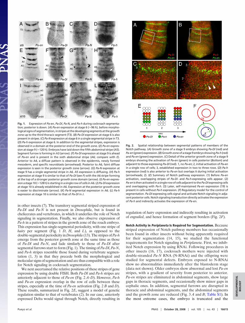
We next ascertained the relative positions of these stripes of geneexpression by using double FISH. Both Pa-Dl and Pa-h stripes areanteriorly adjacent to those of Pa-en (Fig. 2 A–D). However, Pa-hand Pa-en expression overlap in the row of cells between thesestripes, especially at the time of Pa-en activation (Fig. 2 B and D).These results, summarized in Fig. 2E, suggest a model of geneticregulation similar to that of vertebrates (2). In our case, anteriorlyexpressed Delta would signal through Notch, directly resulting in
regulation of hairy expression and indirectly resulting in activationof engrailed, and hence formation of segment borders (Fig. 2F).
Notch Activity in Periplaneta Is Required for Segmentation. Becausestriped expression of Notch pathway members has occasionallybeen found in other insects without being apparently requiredfor their segmentation (14, 15), we studied the functionalrequirements for Notch signaling in Periplaneta. First, we inhib-ited Notch expression by using RNAi. Following procedures inother insects (16, 17), cockroach mothers were injected withdouble-stranded Pa-N RNA (N-RNAi) and the offspring werestudied for segmental defects. Embryos exposed to N-RNAishow no abnormalities immediately after the blastoderm stage(data not shown). Older embryos show abnormal and lost Pa-enstripes, with a gradient of severity from posterior to anterior.Pa-en stripes are eliminated in abdominal segments, show largegaps in thoracic segments, and occasionally show minor gaps incephalic ones. In addition, segmental furrows are disrupted inthoracic and abdominal segments, and the abdominal segmentsand the growth zone are reduced (Fig. 3 A and B; Table S1). Inthe most extreme cases, the embryo is truncated and the
A B C D
E F G H
I J K L
Fig. 1. Expression of Pa-en, Pa-Dl, Pa-N, and Pa-h during cockroach segmenta-tion; posterior is down. (A) Pa-en expression at stage 6 (�96 h), before morpho-logical signsofsegmentation, instripesatthedevelopingsegmentsatthegrowthzone up to the third thoracic segment (T3). (B) Pa-Dl expression at stage 6 is alsopresent in stripes. (C) Pa-N expression at stage 6 in a single segmental stripe in T3.(D) Pa-h expression at stage 6. In addition to the segmental stripes, expression isobserved in a domain at the posterior end of the growth zone. (E) Pa-en expres-sion at stage 9 (�120 h). Embryos have laid down the fifth abdominal stripe (A5).Segment furrow is forming in A3 (arrow). (F) Pa-Dl expression at stage 9 is aheadof Pa-en and is present in the sixth abdominal stripe (A6; compare with E).Anterior to A4, a diffuse pattern is observed in the epidermis, newly formedmesoderm, and specific neuroblasts (arrowhead). Posterior to A6, faint diffuseexpression is seen in the posterior growth zone (arrow). (G) Pa-N expression atstage 9 has a single segmental stripe in A6. A5 expression is diffusing. (H) Pa-hexpression at stage 9 is similar to that of Pa-Dl (see F) with the A6 stripe formingat the top of a stronger posterior growth zone domain (arrow). (I) Pa-en expres-sionastage10 (�128h) is starting inasingle rowofcells inA6. (J)Pa-Dlexpressionat stage 10 is already established in A6. Expression at the posterior growth zoneis easier to discriminate (arrow). (K) Pa-N segmental expression in A6. (L) Pa-hexpression at stage 10 is similar to that of Pa-Dl in J.
Fig. 2. Spatial relationship between segmental patterns of members of theNotch pathway. (A) Growth zone of a stage 9 embryo showing Pa-Dl (red) andPa-en (green)expression. (B)Growthzoneofastage9embryoshowingPa-h (red)and Pa-en (green) expression. (C) Detail of the anterior growth zone of a stage 9embryo showing the activation of Pa-en (green) in cells posterior (Bottom) andadjacent to those expressing Pa-Dl (red). 1, no Pa-en; 2, initial activation of Pa-enin a single row of cells; 3, established expression in two to three rows. (D) Pa-hexpression (red) is also anterior to Pa-en but overlaps it during initial activation(arrowheads; 2). (E) Summary of Notch pathway expression. (1) Before Pa-enactivation, overlapping stripes of Pa-Dl- and Pa-h-expressing cells appear. (2)Pa-en is thenactivated inasinglerowofcellsadjacent tothePa-Dlexpressingcellsand overlapping with Pa-h. (3) Later, self-maintained Pa-en expression (19) ispresent in cells without Pa-h expression. (F) Regulatory model for the control ofsegmentation. Pa-Dl-expressing cells signal and activate Notch signaling in adja-cent posterior cells. Notch signaling transduction directly activates the expressionof Pa-h and indirectly activates the expression of Pa-en.
Pueyo et al. PNAS � October 28, 2008 � vol. 105 � no. 43 � 16615
DEV
ELO
PMEN
TAL
BIO
LOG
Y

posterior abdominal segments are missing. Remarkably, leavingN-RNAi-treated embryos to develop longer produces a moredramatic phenotype, as the addition of new posterior segmentsis abolished while more anterior segments continue developing(Fig. 3 E and F).
Three independent lines of evidence indicate that these N-RNAi phenotypes result from specific effects of the RNAitreatment rather than nonspecific effects. First, we observed lossof Pa-N RNA expression in N-RNAi embryos (data not shown);second, injection of double-stranded RNA for other genes doesnot produce this phenotype (A. Popadic, J.I.P., and J.P.C.,unpublished work); and third, no alterations were observed inthe expression of genes not directly involved in segmentation(Fig. 3 C, D, G, and H). Distal-less (Dll) gene expressionprefigures the development of appendages in a variety of or-ganisms including arthropods and insects (18). Expression of theHox genes Ultrabithorax (Ubx) and abdominalA (AbdA) is re-quired in insects to specify the correct identity of T3 andabdominal segments A1 to A8, but not for development ofsegmental primordia or segmental furrows (8, 19). Dll, Ubx, andAbd-A expression are still present in N-RNAi-treated embryos,even in segments showing clear morphological disruptions andloss of Pa-en (Fig. 3 C, D, G, and H). Finally, N-RNAi-treatedembryos show enlargement of the cephalic nervous system (datanot shown), which is similar to the neurogenic phenotype inDrosophila Notch mutant embryos (7).
To ascertain the developmental basis of segment loss in N-RNAiembryos, we studied changes in cell proliferation and death pat-terns. Cell proliferation is found in many cells in WT embryos,specifically in two bands running the length of the segmented partof the embryo and the growth zone (Fig. 3I). In N-RNAi embryos,the density of mitosis is lower within the very much reduced growthzone, and the mitotic pattern in the segmented region is alsodisorganized (Fig. 3J). Cell death is prominent in the cephalicregions of WT embryos (data not shown) but is rare toward theirposterior region (Fig. 3K). In N-RNAi embryos, there is an increasein cell death in the posterior region, but mostly after segmentationhas occurred (Fig. 3L). Thus, cell death is unlikely to account on itsown for the phenotypes of segment loss observed, and the mostlikely cause involves a reduction of growth that is either a directrequirement for Pa-N in cell proliferation or an indirect conse-quence of the reduction in the number and size of segmentprimordia.
We conclude that the N-RNAi results highlight a specificrequirement for Notch signaling in Pa-en expression, and insegments posterior to T1, establishment of segmental borders,segment primordia, and segment growth.
Periodic Generation of Segments and Stripes of Gene Expression byNotch. We have carried out two further independent tests of Notchsignaling function during segmentation. First, we adapted a previ-ously existing method to culture roach embryos (see Methods) toadminister DAPT (2,5-bis[4-dimethylaminophenyl]-1,3,4-thiadia-zole), a chemical inhibitor that interferes with Notch signaling (20).Embryos collected from a single ootheca at stage 8 are laying downthe A3 stripe of Pa-en at the start of the culture (Fig. 4 A and F).Control embryos add two or three stripes of Pa-en expression (A4to A6) at their growth zone during a 26-h culture period (Fig. 4 Band G). However, their sibling embryos cultured with DAPT do notcomplete any further proper Pa-en stripes (Fig. 4 C and H).Presumptive Pa-en stripes under DAPT culture consist of brokenstretches at best, or at worst do not form at all. Previously laid downPa-en stripes, such as A2 or A3, are not affected, suggesting thatPa-N is required during the activation, but not the maintenance, ofPa-en expression.
Second, we assessed the requirement for Pa-h by h-RNAi andobserved similar defects to those produced by DAPT. h-RNAiembryos display lost and abnormal Pa-en stripes and abnormalsegmental furrows leading to segment fusions (Fig. 4 D, E, I, andJ). Altogether, these results pinpoint the requirement for Notchsignaling to the unsegmented growth zone during Pa-en activa-tion and generation of segment primordia.
A B C D
E F G H
I J K L
Fig. 3. N-RNAi precludes segmentation specifically. (A) Control embryos atstage 11 (about 144 h.) have laid down the seventh abdominal stripe of Pa-en(A7). Limbs buds, somites and segment borders in more anterior segments (e.g.,T1) are apparent. (B) N-RNAi-treated embryos by stage 12 develop with gaps inPa-en stripes posterior to T1 (arrow), and the last vestigial stripe is that of A4. Thegrowth zone is reduced and more posterior segments are absent. (C) Controlembryo showing wild-type expression of Dll (red), AbdA and Ubx (green). (D)N-RNAi does not eliminate the expression of Dll (red), AbdA or Ubx (green). (E) Bystage 15 control embryos have finished segmentation and contain eleven ab-dominal segments (A11) with stripes of Pa-en expression. (F) Stage 15 N-RNAiembryo showing normal development of the anterior segments although theembryo is truncated, and the segments posterior to A4 are absent (arrow). (G)Stage 15 control embryo showing the Dll, and AbdA and Ubx patterns of expres-sion. The tail of the embryo is folded and the tip (the cerci) are highlighted by anarrow. (H) N-RNAi stage 15 embryo stained as in C. The posterior end of thetruncated abdomen is denoted by an arrow. Dll (red), AbdA and Ubx (green) arestill expressed. (I) Posterior part of a stage 11 embryo showing cells undergoingmitosis (red). The posterior growth zone contains a high number of mitotic cells(arrow). Proliferation continues in the newly formed segments. (J) Posterior partof a stage 11 N-RNAi embryo stained as in I. The posterior growth zone is muchreduced but there are still cells undergoing mitosis with a lower density than theWT. (K) Posterior part of a stage 11 control embryo labeled as in L. Only a few cellsscattered throughout segments show apoptosis. (L) Cell death in the posteriorpart of a N-RNAi stage 11 embryo. Cells undergoing apoptosis are detected by ananti-Caspase 3 antibody (red). Segments are labeled by anti-engrailed antibody(green). Cells undergoing apoptosis appear in clusters mostly after Pa-En expres-sion, and thus segmentation, is established.
16616 � www.pnas.org�cgi�doi�10.1073�pnas.0804093105 Pueyo et al.

We observed the expression of Pa-Dl (Fig. 5 A–C) and Pa-h (Fig.5 D–F) in detail in the growth zone during the formation of asegment. Roach embryos are laid sequentially in clutches inside anootheca (9). Older embryos from one end of the ootheca do notdiffer in age by more than a whole segment from younger embryosfrom the opposite end (J.I.P., R.L., and J.P.C., unpublished data).Comparison of single-ootheca siblings thus illustrates a time seriesshowing how segmental stripes of Pa-Dl (Fig. 5 A-C) and Pa-h (Fig.5 D–F) are generated from the growth zone in a cyclical manner.Stripes emerge at the edge of the more diffuse expression domainat the posterior growth zone. Expression then decays behind thenascent stripe, which moves to a more anterior position. Thisprocess (i.e., decay behind the stripe and forward movement of thestripe) continues until Pa-en expression starts (Fig. 5 C and F). Pa-Nexpression in the growth zone also shows similar dynamic patterns,albeit fainter (data not shown). To test whether Notch signaling isrequired for Pa-h expression, we used N-RNAi (Fig. 5 G-I). Weobserve that Pa-h loses its cyclical, striped pattern and only adisorganized background remains. Similarly, in embryos culturedwith DAPT cyclic Pa-h expression is also disrupted (Fig. 5 J–L).Only a few new stretches of Pa-h expression are formed, and cyclingof Pa-h expression in the posterior growth zone stops.
In summary, our analyses of expression patterns and DAPT andRNAi treatments reveal a requirement for Notch signaling duringsegmentation of Periplaneta americana. Stripes of Pa-Dl, Pa-h, andPa-N expression appear shortly before Pa-en stripes and severalhours before the morphological onset of segmentation in the regionwhere presumptive segments form. The stripes of Pa-Dl appearanterior to the positions where Pa-en stripes will form, whereas Pa-hstripes overlap both domains. These expression patterns are com-patible with the hypothesis that Pa-Dl signals through Pa-N toregulate Pa-h and Pa-en expression, and this hypothesis is corrob-orated by the DAPT and N-RNAi functional experiments. Thesefunctional results also confirm that the timing of Notch signaling is
coupled to the sequential timing of segment development, a resultreminiscent of the interplay between Notch signaling and thevertebrate segmentation clock (2–4). N-RNAi embryos at stage 15show that, after an initial window during segment formation, Notchsignaling has no major requirement in segmentation. Similarly,when we administer DAPT, we obtain defects only in the specificsegments that are forming at the time. Finally, the results regardingthe role of hairy in P. americana and vertebrates are also compa-rable. hairy expression appears in stripes that originate at theunsegmented zone in a cyclical manner, although the length of theregion of the embryo where such oscillation takes place is smallerin the roach than in vertebrates. Blocking Notch signaling in bothsystems does not completely eliminate hairy expression, but abol-ishes its periodic pattern, and altering hairy expression leads tosegmentation defects similar to those observed when Notch func-tion is perturbed (1–3).
Ancestral Notch-Mediated Segmentation. Our results supportNotch-mediated segmentation as a basal developmental mech-anism for insects (Fig. S3A). More derived insects such asDrosophila and Tribolium seem to have lost Notch-mediatedsegmentation and replaced it with gap segmentation (8, 16) incorrelation with the evolution of specialized ovaries to supportit (21). A gradual mechanism has been suggested for thisevolutionary shift (22), and in this view, it would be possible fordifferent insect species to have all, some, or none of theirsegments develop under the control of Notch, according to theinsect’s position in the phylogenetic tree, with closer relatives ofDrosophila more likely to lack a role for Notch in segmentation.In Periplaneta N-RNAi embryos, we occasionally observed mi-nor Pa-en expression defects in cephalic segments anterior to T1,but total loss of Pa-en and truncation of growth occur only inmore posterior segments. This suggests that the requirement forNotch is less strong anterior to T1, and fits with the observation
A B C D E
F G H I J
Fig. 4. Interfering with N signaling via chemical inhibition by DAPT or via hairy RNAi disrupts segmental engrailed expression. (A) Pa-en expression in a late stage8 embryo showing the third abdominal stripe of Pa-en (A3) being laid down before embryo culture. (B) Control embryo after 26 h of in vitro culture, new segmentsA4andA5haveformedposteriorly (also seeG). (C)NonewPa-en stripesareobserved inaDAPT-treatedembryoafter26hofculture.Pa-en stripes thathadbeenalreadyformed before culture were not affected in these embryos (compare with B; details in G and H). (D) Pa-en expression in a stage 11 control embryo. The last stripe formedis A6. (E) Stage 11 h-RNAi embryo shows Pa-en expression. Pa-en is disrupted in the last abdominal segments (arrows, see detail in J). Segmentation irregularities arealsoobserved inthethirdthoracic segment (T3)as thesegmental furrowis truncatedandagap inPa-enexpressionappears (arrowhead). (F)Posteriorendof theembryoshown in A. (G) Detail of the posterior part of the embryo shown in B. (H) Detail of the DAPT-treated embryo shown in C. (I) Detail of the posterior part of the embryoshowninD. (J)Magnificationof theposteriorpartof theh-RNAiembryoshowninE.Pa-en ispresentonly inhemi-segmentsonthe left. Inaddition, theposteriorgrowthzone appears abnormal (arrow; compare with I). This phenotype is similar to that produced by DAPT in H.
Pueyo et al. PNAS � October 28, 2008 � vol. 105 � no. 43 � 16617
DEV
ELO
PMEN
TAL
BIO
LOG
Y

that these are the segments formed in the blastoderm (J.I.P.,R.L., and J.P.C., unpublished data and refs. 9, 10) and thuspresumably under the control of gap segmentation as in othershort-germ band insects (6, 22). In this view, the segmentalstripes of expression of Serrate in Drosophila and fringe ingrasshoppers, which do not seem to have a role in segmentation(14, 15), might be seen as vestiges of an ancestral Notchsegmentation mechanism. In Drosophila at least, the Notchsegmentation mechanism seems to remain in a derived formhaving been co-opted for the formation of leg segments (23–25).The independent role of Notch in neurogenesis seems conservedas well (7).
The ancestral role of Notch segmentation is reinforced by resultsin the chelicerate Cupiennus salei. Notch, Delta, and other pathwaymembers are expressed and required during segmentation in thisspider (5), and homology with the Notch-mediated vertebratesegmentation clock has been debated, but not proven, as a func-tional requirement for Notch during segmentation had not beenshown in any other arthropod (6, 16, 22). However, the strikingsimilarity between Periplaneta and Cupiennus in the Pa-Dl, Pa-N,and Pa-h striped expression patterns in the developing segmentsand growth zone—and in the Notch RNAi effects on morphologyand engrailed and hairy expression—suggests, as the most parsimo-nious explanation, that Notch segmentation is homologous in bothlineages, insects and chelicerates, and therefore ancestral not onlyto insects but to arthropods as well (Fig. S3A). Such ancestralNotch-mediated segmentation would also explain the expression instripes of Notch-related genes in another arthropod lineage, myri-apods, represented by the centipede Strigamia maritima (26).Conservation of Notch segmentation in arthropods could be quitecommon.
Our findings re-ignite the question of whether Notch signalinghas been independently recruited for segmentation in vertebratesand arthropods, or whether they have both inherited it from acommon ancestor (Fig. S3A). Notch signaling activity is present inCnidarians (27) and thus Notch signaling could have been recruitedindependently during the evolution of segmentation in separatevertebrate and arthropod ancestors. It would remain to be ex-plained what constraints limited evolution’s choice to the samesignaling pathway given the ancestral presence of other pathwayscapable of similar developmental functions, such as Wnt and Hh(19, 27). Similarly, a putative conservation of Notch segmentationfrom a common ancestor begs the question of the constraints andmechanisms that have conserved this role of Notch and adapted itto changing embryonic architectures. Such a conservation wouldalso fuel the debate about whether the last common ancestor ofarthropods and vertebrates was a simple or complex animal (28, 29),and would raise the question of why, when, and how segmentationwas lost in unsegmented species. In this regard, the recent findingsin hemichordates supporting a common origin of dorsal-ventralpatterning in vertebrates and insects are interesting (30). Theconservation of Notch segmentation could also extend to lopho-trochozoans. In annelids, expression of engrailed at segment borders(11) and dynamic expression of Notch pathway members duringsegmentation (31) has been reported. In primitive mollusks withmetameric organization, recent results also reveal the presence ofengrailed stripes (32).
An intermediate possibility is segmentation may have evolvedindependently from a simpler Notch-mediated metameric orga-
is observed (arrow). (K) Control embryo after 26 h in culture has laid downthree Pa-h stripes, similar to that observed with Pa-en expression (Fig. 4 A andF and B and G). (L) DAPT-treated embryo cultured for 26 h shows a few abortivesegmental stretches of Pa-h expression (bracket) posterior to A3. No furtherposterior stripes are formed, and a longer unstained and unsegmented areadevelops anterior to the posterior growth zone domain, which appears irreg-ular (arrow).
A B C
D E F
G H I
J K L
Fig. 5. Cyclic N signaling at the posterior growth zone controls roach segmen-tation. (A-C) Pa-Dl expression at the growth zone of WT embryos during theformation of the sixth abdominal stripe (arrow). Blue boxes denote Pa-Dl reced-ing expression at the posterior growth zone. (A) The first sign of the formation ofA6 starts with the up-regulation of Pa-Dl at the anterior edge of the posteriorgrowth zone domain (arrow). Note that Pa-Dl expression covers the wholeposterior growth zone (blue box). (B) Slightly later, Pa-Dl expression behind thenascent A6 stripes regresses (blue box) whereas the A6 stripe becomes stronger.(C) Finally, posterior expression of Pa-Dl remains only at the end of the growthzone,andtheA6stripe is fully formed.Notethat,duringthisprocess, thedistancebetween the A6 stripe and the posterior growth zone increases (compare with AandB). (D-F)DynamicexpressionofPa-hatthegrowthzoneduringtheformationof the sixth abdominal stripe (A6; arrow) in WT embryos. (D) The presumptivesixth abdominal stripe appears by the down-regulation of Pa-h behind theanterior edge of the posterior growth zone. (E) As the A6 stripe is forming, thePa-h pattern in the posterior growth zone regresses. (F) Finally, the Pa-h A6 stripedetaches from the posterior growth zone domain. (G-I) N-RNAi disrupts Pa-hcyclic expression. (G) Pa-h pattern of expression in a stage 10 control embryo. (H)N-RNAi stage 10 embryo shows Pa-h expression reduced posterior to T1 butremaining in more anterior segments in the somites, brain, and midline. (I) DetailoftheposteriorpartoftheN-RNAiembryoinE.NoPa-h segmental stripesappear;instead, a diffuse and irregular pattern is observed in the growth zone (brackets;compare with D-F). (J-L) DAPT treatment disrupts cyclic Pa-h expression. (J) Pa-hexpression pattern in a stage 8 embryo before culture. The A3 stripe has just beenlaid down. In addition, dynamic Pa-h expression at the posterior growth zone
16618 � www.pnas.org�cgi�doi�10.1073�pnas.0804093105 Pueyo et al.
nization involving repeated organs or body boundaries in acommon ancestor of protostomes and deuterostomes. Notchsignaling is a good candidate to drive this hypothetical transitionfrom metamerism to a more advanced segmental organization,as it is involved in extant animals in the segregation of repeatedpattern elements from equivalence groups, in the establishmentof boundaries or patterning lines, and in the segregation ofspecific cell lineages (2, 4, 7, 23–25). It is not difficult to imaginea Notch-mediated evolutionary transition from the developmentof repeated pattern elements along the body axis to theirassociation with patterning boundaries, culminating in the as-sociation of these patterning boundaries with a lineage and faterestriction—the essence of segmentation in chordates, arthro-pods, and lophotrochozoans (Fig. S3B). Further studies shouldclarify which of these elements of segmental organization werepresent in the common ancestor, which evolved independently ineach segmented lineage, and which have been lost in apparentlynon-metameric species.
MethodsPeriplaneta americana Rearing. Adult cockroaches were kept at 29°C. Freshlylaid oothecae were collected and placed in a humidified chamber at 29°C.Embryos were staged according to laying date and morphology (9).
Cloning Pa-Dl Pa-N and Pa-h Genes. We designed degenerate primers based onconserved amino acid domains in arthropod Delta exons 4 and 6. Periplanetagenomic DNA was used for PCR amplification. Low-specificity PCR reactions withouter primers (Dl3F, Dl3R; see SI Text) were followed by nested PCR reactions withinner primers (Dl3F, Dl4R). Expected-size PCR fragments were sequenced. Gene-specific primers were generated to isolate overlapping fragments of Pa-Dl cDNAusing a �ZAP II cDNA library [gift from B. Marie and J. Bacon (University of Sussex,Brighton, UK)] as template. PCR fragments were assembled into a full-lengthPa-Dl cDNA of 3.2 kb. Degenerated primers used for Pa-N cloning are describedby Stollewerk et al (5). Specific primers were designed to isolate Pa-N cDNA byRACE (Ambion). A 1.5-kb fragment of the 5� end of the Pa-N transcript wasisolated. For Pa-h degenerate primers using the second-exon, conserved se-quences from other arthropods were used initially. Subsequently specific outerprimers and nested primers were generated for 5� and 3� PCR reactions by usinga �ZAP II cDNA library as a template, and fragments were assembled into afull-length 1.7-kb Pa-h cDNA.
ISH and FISH. We followed the protocol described by Marie and Bacon (12).digoxigenin antisense RNA probes were made using a 1.4-kb Pa-Dl cDNA
fragment, a 1.5-kb Pa-N cDNA fragment, a 1.4-kb Pa-h cDNA fragment, and a1.3-kb Pa-en2 cDNA fragment. Probes were hydrolyzed into smaller fragmentsas described by Moens (Division of Basic Sciences, Fred Hutchinson CancerResearch Center, Seattle, WA). For FISH, we used TSA system protocols (Perkin–Elmer).
Immunocytochemistry. We dissected oothecae in cold PBS solution and fixedembryos in cold 4% paraformaldehyde for 20 min. Afterward, we followedstandard Drosophila protocols. The primary antibodies used recognize homolo-gous proteins in a range of arthropod species. The mouse monoclonal antibodiesanddilutionswere:anti-Dll (18)at1:200;FP6.87antibodythatrecognizesUbxandAbd-A proteins [Rob White (University of Cambridge, Cambridge)] at 1:10; andanti-En 4F11 at 1:5 (11). We used a rabbit anti-cleaved caspase 3 from CellSignaling Technology at 1:50 to detect apoptosis and a rabbit anti-phosphory-lated Histone 3 (Upstate) to label cells in mitosis at 1:1,000. Secondary antibodiesconjugated with biotin or fluorophores were from Jackson Immunochemicals.
Embryo Culture and DAPT Treatment. Embryos from stage 8 onward werecultured according to the method described by Wang and Denburg (33),which allows normal development for up to 48 h, with some modifications.Embryos were dissected in sterile culture medium at 29°C and transferredrapidly into a sterile low-adhesion tissue culture plate with prewarmed culturemedium. Culture plates were placed in a humidified chamber at 29°C. Culturemedium was replaced every 12 h. For DAPT treatment, a 10-mM stock in DMSOwas diluted to 100 �M in culture medium. Control embryos were treated withthe same amount of DMSO. Treatment with 50 �M of DAPT produced slightlyweaker phenotypes but allowed longer culture.
RNAi. Forward- and reverse-specific primers containing T7 polymerase pro-moter sequences were used to amplify the Pa-N RACE and Pa-h 3� cDNAtemplates, giving rise to 700-bp and 850-bp fragments respectively. In vitrotranscription of these fragments was carried out with the T7 RiboMAX kit(Promega). For Pa-N, dsRNA for injection was 0.5 �g/�l, whereas for Pa-h,dsRNA for injection was 1.5 �g/�l. Maternal injection was done as describedby Cruz et al (17) by loading 4 �l dsRNA into a Hamilton 75N syringe andinjecting newly molted adult virgin females in the sternites between thefourth and fifth abdominal segments. After 2 h, females were injected againto counteract loss of dsRNA resulting from wound leakage. Injected femaleswere reared with males at 29°C, and oothecae were collected daily andcultured as described earlier. No segmental defects were observed in controlembryos from mothers injected with dH20.
ACKNOWLEDGMENTS. We thank B. Marie for techniques and materials; R.Phillips and S. A. Bishop for technical help; and A. Popadic, J. Baguna, R. Ray,and C. Alonso for manuscript comments. This work was funded by a WellcomeTrust Senior Fellowship reference no. 057730 (J.P.C.) and a Biotechnology andBiological Sciences Research Council studentship (to R.L.).
1. Palmeirim I, Henrique D, IshHorowicz D, Pourquie O (1997) Avian hairy gene expressionidentifies a molecular clock linked to vertebrate segmentation and somitogenesis. Cell91:639–648.
2. Pourquie O (2003) The segmentation clock: Converting embryonic time into spatialpattern. Science 301:328–330.
3. Jiang YJ, et al. (2000) Notch signalling and the synchronization of the somite segmen-tation clock. Nature 408:475–479.
4. Bray SJ (2006) Notch signalling: a simple pathway becomes complex. Nat Rev Mol CellBiol 7:678–689.
5. Stollewerk A, Schoppmeier M, Damen WGM (2003) Involvement of Notch and Deltagenes in spider segmentation. Nature 423:863–865.
6. Tautz D (2004) Segmentation. Dev Cell 7:301–312.7. Campos-Ortega JA, Knust E (1990) Genetics of early neurogenesis in Drosophila
melanogaster. Ann Rev Gen 24:387–407.8. Akam M (1987) The molecular basis for metameric pattern in the Drosophila embryo.
Development (Cambridge, U.K.) 101:1–22.9. Lenoir-Rousseaux JJ, Lender T (1970) Table de developpement embryonnaire de
Periplaneta americana. Bull Soc Zool France 95:737–751.10. Anderson DT (1972) in Developmental Systems: Insects, eds Counce SJ, Waddington CH
(Acad. Press, London), Vol. 1, pp 95–163.11. Patel NH, et al. (1989) Expression of engrailed proteins in arthropods, annelids, and
chordates. Cell 58:955–968.12. Marie B, Bacon JP (2000) Two engrailed-related genes in the cockroach: cloning, phylo-
genetic analysis, expression and isolation of splice variants. Dev Genes Evol 210:436–448.13. Ingham PW, Howard KR, Ish-Horowicz D (1985) Transcription pattern of the Drosophila
segmentation gene hairy. Nature 318:439–445.14. Dearden P, Akam M (2000) A role for Fringe in segment morphogenesis but not
segment formation in the grasshopper, Schistocerca gregaria. Dev Genes Evol210:329–336.
15. Bachmann A, Knust E (1998) Dissection of cis-regulatory elements of the Drosophilagene Serrate. Dev Genes Evol 208:346–351.
16. Choe CP, Miller SC, Brown SJ (2006) A pair-rule gene circuit defines segments sequentiallyin the short-germ insect Tribolium castaneum. Proc Natl Acad Sci USA 103:6560–6564.
17. Cruz J, Mane-Padros D, Belles X, Martin D (2006) Functions of the ecdysone receptorisoform-A in the hemimetabolous insect Blattella germanica revealed by systemic RNAiin vivo. Dev Biol 297:158–171.
18. Panganiban G, Sebring A, Nagy L, Carroll S (1995) The development of crustacean limbsand the evolution of arthropods. Science 270:1363–1366.
19. Martinez-Arias A (1993) In The Development of Drosophila melanogaster, eds BateCM, Martinez-Arias A (Cold Spring Harbor Laboratory, Cold Spring Harbor, NY), pp517–608.
20. Geling A, Steiner H, Willem M, Bally-Cuif L, Haass C (2002) A gamma-secretase inhibitorblocks Notch signaling in vivo and causes a severe neurogenic phenotype in zebrafish.EMBO Rep 3:688–694.
21. Patel NH, Condron BG, Zinn K (1994) Pair-rule expression patterns of even-skipped arefound in both short- and long-germ beetles. Nature 367:429–434.
22. Peel A, Akam M (2003) Evolution of segmentation: Rolling back the clock. Curr Biol13:708–710.
23. de Celis JF, Tyler DM, de Celis J, Bray SJ (1998) Notch signalling mediates segmentationof the Drosophila leg. Development (Cambridge, U.K.) 125:4617–4626.
24. Bishop SA, Klein T, Arias AM, Couso JP (1999) Composite signalling from Serrate andDelta establishes leg segments in Drosophila through Notch. Development (Cam-bridge, U.K.) 126:2993–3003.
25. Rauskolb C, Irvine KD (1999) Notch-mediated segmentation and growth control of theDrosophila leg. Dev Biol 210:339–350.
26. Chipman AD, Arthur W, Akam M (2004) A double seament periodicity underliessegment generation in centipede development. Curr Biol 14:1250–1255.
27. Technau U, et al. (2005) Maintenance of ancestral complexity and non-metazoan genesin two basal cnidarians. Trends Gen 21:633–639.
28. De Robertis EM (2008) Evo-devo: variations on ancestral themes. Cell 132:185–195.29. Erwin DH, Davidson EH (2002) The last common bilaterian ancestor. Development
(Cambridge, U.K.) 129:3021–3032.30. Lowe CJ, et al. (2006) Dorsoventral patterning in hemichordates: Insights into early
chordate evolution. PLOS Biol 4:1603–1619.31. Thamm K, Seaver EC (2008) Notch signaling during larval and juvenile development in
the polychaete annelid Capitella sp. Dev Biol 320:304–318.32. Jacobs DK, et al. (2000) Molluscan engrailed expression, serial organization, and shell
evolution. Evol Dev 2:340–347.33. Wang L, Denburg JL (1992) A role for proteoglycans in the guidance of a subset of
pioneer axons in cultured embryos of the cockroach. Neuron 8:701–714.
Pueyo et al. PNAS � October 28, 2008 � vol. 105 � no. 43 � 16619
DEV
ELO
PMEN
TAL
BIO
LOG
Y