and resilontm root canal filling material: a comparative ... · with the resilon/epiphany...
TRANSCRIPT
Microbial Leakage of Gutta-perchaand ResilonTM Root Canal FillingMaterial: A Comparative Study
Using a new Homogeneous Assayfor Sequence Detection
DAMIANO PASQUALINI,1,*, NICOLA SCOTTI,1 LIVIO MOLLO1,
ELIO BERUTTI1, EMMA ANGELINI
2, GIUSEPPE MIGLIARETTI,3
ANNAMARIA CUFFINI3
AND DANIEL ADLERSTEIN4
1Department of Endodontics, School of Dentistry, University of Turin
Turin, Italy; Corso A.M. Dogliotti 14 - 10126 Torino, Italy2Department of Material Science and Chemical Engineering
Polytechnical School of Turin, Turin, Italy3Department of Public Health and Microbiology
University of Turin, Turin, Italy4Diasorin SpA, via Crescentino snc
13040 Saluggia (VC), Italy
ABSTRACT: The sealing ability of gutta-percha/sealer root canal filling wascompared to a new thermoplastic synthetic polymer-based obturation material(ResilonTM), using a microleakage model and a new sequence detection assayOne Cut Event Amplification (OCEANTM). Eighty-eight extracted human teeth,shaped with K-Files and the ProTaper Technique, were randomly assigned tofour groups (n¼ 22) and obturated in the apical 5 mm. Group R were obturatedwith the Resilon/Epiphany technique; group GP were obturated with gutta-percha and Zinc oxide eugenoe sealer; group RCH and GPCH received calciumhydroxide intracanal medication before being obturated. Sterilized specimenswere inoculated with Enterococcus faecalis and incubated in sterile medium for47 days. DNA extracted from the specimens was amplified by PCR and thenidentified by the OCEAN technique. Samples obturated with Resilon root canalfilling material showed a greater number of microleakage events than the other
*Author to whom correspondence should be addressed.E-mail: [email protected] 1 appears in color online:http://jba.sagepub.com
JOURNAL OF BIOMATERIALS APPLICATIONS Volume 00 — 2007 1
0885-3282/07/00 0001–16 $10.00/0 DOI: 10.1177/088532820?077411� 2007 SAGE Publications
+ [Ver: 8.07r /W] [31.3.2007–5:37pm] [1–16] [Page No. 1] REVISED PROOFS {SAGE_REV}Jba/JBA 077411.3d (JBA) Paper: JBA 077411 Keyword

groups ( p¼ 0.036). Calcium hydroxide medication did not have a relevantimpact on the quality of the apical seal ( p¼ 0.044).
KEY WORDS: root canal filling, resilon, gutta-percha, microleakage, PCR,Enterococcus faecalis, materials science.
INTRODUCTION
Successful endodontic treatment depends upon three-dimen-sional obturation of the root canal system to the end of the canal [1].
Since it is virtually impossible to completely eliminate bacteria from thecanal system, the obturation must provide a hermetic seal at the apex inorder to prevent reinfection of periapical tissue [2]. Thus, the mainobjective of root canal filling is the entombment of most survivingbacteria and the creation of a barrier to stop periapical tissue fluids fromreaching them [3].
Gutta-percha associated with a sealer has, from its introduction intoclinical practice in 1848 until the present time, been the most widelyused root canal obturation material; still today it is considered the‘standard of care’ in endodontic therapy. A recent review of clinicalstudies has shown the favorable long-term prognosis of root canaltherapy performed with gutta-percha and a sealer [4]. Teeth withoutpreoperative apical periodontitis can remain free from disease afterinitial or orthograde treatment in 92–98% of cases; teeth with apicalperiodontitis can completely heal after the root canal treatment orretreatment in 74–86% of cases, being however functional in 91–97%.However, the seal of root canal filling in gutta-percha is not always fullyhermetic, revealing microleakage, in different study conditions, bothapically and coronally [5–8]. The long-term clinical implication may bean increased risk of failure of the endodontic therapy.
With the aim of rectifying this possible unfavorable aspect of thegutta-percha seal in some clinical situations, a new thermoplasticsynthetic polyester polymers-based root canal filling material(ResilonTM) has recently been proposed [9–12]. The system comprisesmaster cones or pellets for backfill with thermoplastic characteristics,similar to those of gutta-percha, used in combination with a dual curabledental resin composite sealer. Initial studies showed significantly lessmicroleakage in Resilon sealed groups than in gutta-percha sealedgroups [9] and higher mean fracture loads in the former ones [10].An in vivo model in dogs has also determined greater resistance tocoronal bacterial infiltration in subjects obturated with Resilon, withconsequently weaker periapical inflammatory response [12]. All these
2 D. PASQUALINI ET AL.
+ [Ver: 8.07r /W] [31.3.2007–5:37pm] [1–16] [Page No. 2] REVISED PROOFS {SAGE_REV}Jba/JBA 077411.3d (JBA) Paper: JBA 077411 Keyword

advantages appear to derive from the so-called ‘Resilon MonoblockSystem.’ The dual curable composite sealer forms a bond with the dentinstructure on one side and the synthetic polymer of the Resilon on theother side, creating an intimate contact between the material and thecanal walls. Thus a more hermetic seal and improved mechanicalproperties of the endodontically treated tooth should be established.However, some other studies demonstrate that both gutta-percha andResilon seem to be unable to reinforce root-filled teeth [13,14]. Calciumhydroxide is widely used in endodontics as intermediatedressing between two sessions of root canal therapy, with the aim ofimproving the disinfection of the canal system before obturation [15,16].The hypothesis has been advanced that calcium hydroxide may havea negative influence on the quality of apical seal of the gutta-percharoot canal filling [17–19], due to residues present on the canal walls[19,20], especially when a zinc oxide eugenol (ZOE) sealer isused [20,21].
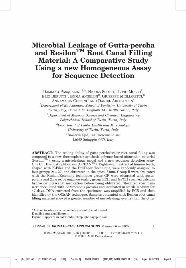
The primary objective of this in vitro study was to evaluate theoccurrence of Enterococcus faecalis microleakage events through gutta-percha and sealer root canal filling (active controls), compared to Resilonobturation (test groups) using a new homogeneous method for sequenceidentification known as One Cut Event AmplificatioN (OCEANTM).(Figure 1). Its reaction exploits the action of a restriction enzyme and isbased on interaction of the target sequence with two probes: an anchorprobe, complementary to the target for most of its extension, and aprobe marked with a fluorophore (F) known as the amplifier. Theduplex target-anchor is stable at the reaction temperature of 65�C andfavors formation of a ternary complex with the amplifier. This complexcontains the recognition site of a restriction enzyme present in thesolution. Reaction of this enzyme causes fragmentation of the amplifierinto two segments that dissociate from the complex, while the anchor-target again becomes available for bonding with a new amplifier,promoting a linear amplification reaction.
The secondary objective was to analyze the influence of calciumhydroxide root canal dressing on the apical seal with both testedmaterials.
MATERIALS AND METHODS
Specimen Preparation
Extracted human single-root teeth with fully formed apex (uppercentral incisors and canines with substantially equal canal curvature
Gutta-percha versus Resilon Leakage 3
+ [Ver: 8.07r /W] [31.3.2007–5:37pm] [1–16] [Page No. 3] REVISED PROOFS {SAGE_REV}Jba/JBA 077411.3d (JBA) Paper: JBA 077411 Keyword
and morphology) that had not undergone prior endodontic treatmentwere utilized. After debriding the root surface, specimens wereimmersed in a 5% solution of sodium hypochlorite (Niclor 5, OGNA,Muggio, Italy) for 1 h and then stored in 0.9% sodium chloride solutionuntil preparation. Each specimen was then sectioned so as to obtain aresidual root of length 15 mm. Each root canal was preflared usingK-Flexofiles (Dentsply Maillefer, Ballaigues, Switzerland) up to #20 andthen shaped using ProTaper S1-S2-F1-F2-F3 (Dentsply Maillefer,Ballaigues, Switzerland) at the working length. The working lengthwas established under microscopic vision (Pro Magis, Carl Zeiss,Oberkochen, Germany) at 10� magnification, when the tip of theinstrument was visible at the apical foramen. Irrigation was performedwith a 22 gauge needle syringe: 33 mL of 5% sodium hypochlorite at50�C (Niclor 5, OGNA, Muggio, Italy), alternating with 2 mL of 10%EDTA (Tubuliclean, OGNA, Muggio, Italy), total irrigation time 10 minper specimen. After drying with paper points, the roots were inspected
Figure 1. The OCEAN reaction. Step 1: a stabilizing probe (anchor) hybridizes to the
target DNA, complementary of most of its extension. Step 2: the duplex target–anchor is
stable at the reaction temperature of 65�C and a fluorescently labeled (F) probe(amplifier), specific for the E. faecalis primer pair, hybridizes to the target–anchor
complex. The ternary structure forms the recognition site for a restriction endonuclease
(RE) present in the solution. Step 3: fragmentation by the enzyme produces a cut amplifier
which dissociates into two segments, while the anchor–target again becomes available forbonding with a new amplifier, promoting a linear amplification reaction. Production of the
specific signal for E. faecalis was visualized by resolving the fragment of amplifier marked
with the fluorescence generated by the reaction on polyacrylamide gel.
4 D. PASQUALINI ET AL.
+ [Ver: 8.07r /W] [31.3.2007–5:37pm] [1–16] [Page No. 4] REVISED PROOFS {SAGE_REV}Jba/JBA 077411.3d (JBA) Paper: JBA 077411 Keyword
under the microscope at 10� magnification to check the integrity andshape of the apical foramen, the absence of cracks, and the canalcleanliness. The diameter of the apical foramen was checked with a K-File Nitiflex (Dentsply Maillefer, Ballaigues, Switzerland) fitting at theapex at the working length. Only specimens with an apical diameter of#30 and #35 entered the study.
One hundred and fifty-two specimens were selected. Fifty-six speci-mens were used for training on the use of the new EpiphanyTM SoftResin Endodontic Obturation System (Pentron Clinical Technologies,Wallingford, CT, USA) and were not included in the study. Fourspecimens were used as positive controls and four as negative controls.The remaining 88 specimens were randomly subdivided by a blindexaminer into four groups (GP, GPCH, R, RCH) of 22 each using arandom numbers table.
Before canal obturation specimens in groups RCH and GPCH, weremedicated with calcium hydroxide (DT Temporary Dressing, DentalTherapeutics AB, Sweden), delivered with a lentulo, sealed coronallywith a temporary filling (CavitTM W, 3M ESPE, Seefeld, Germany) andthen stored in 100% relative humidity. After 7 days, the root canaldressing was removed as follows: flushing with 5 mL 5% NaOCl at40–50�C; K-Flexofile 20 ISO (Dentsply Maillefer, Ballaigues,Switzerland) 1 mm short of the working length (WL); flushing with5 mL 10% EDTA; flushing with 5 mL 5% NaOCl at 40–50�C; K-Flexofile20 ISO 1 mm short of the WL; flushing with 5 mL 10% EDTA;K-Flexofile 20 ISO 1 mm short of the WL; final flush with 5 mL 10%EDTA for 2 min.
Specimens in groups GP and GPCH were obturated in the apical 5 mmwith nonstandard medium gutta-percha cones and the continuouswave of condensation technique with System B and mediumplugger (Analytic Technologies, Redmond, WA, USA) at 220�C andPulp Canal Sealer EWT (Kerr, Orange, CA, USA). Before obturation,a final flush with 96� ethyl alcohol was applied followed by drying withpaper points.
Specimens in groups R and RCH were obturated in the apical 5 mmwith nonstandardized ResilonTM cones and the dual curable dental resincomposite sealer (Pentron Clinical Technologies, Wallingford, CT, USA),following the manufacturer’s instructions. Before obturation, a finalflush with 17% EDTA was applied followed by drying with paper points.A paper point soaked in primer was used to transfer the liquid into thecanal; excess was removed with a dry paper point. The resin-basedcement was then placed in the apical third of the root canal with alentulo. A nonstandardized medium ResilonTM cone (Pentron Clinical
Gutta-percha versus Resilon Leakage 5
+ [Ver: 8.07r /W] [31.3.2007–5:37pm] [1–16] [Page No. 5] REVISED PROOFS {SAGE_REV}Jba/JBA 077411.3d (JBA) Paper: JBA 077411 Keyword

Technologies, Wallingford, CT, USA) adapted to the apical diameterwas inserted into the root canal and the specimen obturated with thecontinuous wave of condensation technique with System B and mediumplugger (Analytic Technologies, Redmond, WA, USA) at 150�C.Lastly the material was polymerized for 40 s using a photopolymerizinglamp XL 3000 (3M ESPE Dental Products, St Paul, MN, USA). Negativecontrols (n¼ 4) were obturated (two with gutta-percha/ZOE sealerand two with ResilonTM/composite sealer) and completely sealed on theirentire surface with a layer of varnish and then covered with sticky wax,also covering the apex; positive controls (n¼ 4) were not obturatedand were sealed externally with varnish and sticky wax to 3 mm fromthe apex.
All obturated specimens in all groups were then checked radio-graphically to verify the quality of the canal obturation, sealed withtemporary filling (CavitTM W, 3M ESPE, Seefeld, Germany) andstored in 100% relative humidity. After 15 days, all specimens wereprepared for microleakage testing by a double chamber model as hasbeen previously described [22]. A pipette tip was placed in each canaland cemented to the dentine with cyanoacrylic cement. Surfaces weresealed with varnish and sticky wax to cover 5 mm of the pipette tipadjacent to the root, and the root walls from the pipette tip to 3 mmfrom the apical foramen. Each specimen was fixed with cyanoacryliccement onto a steel wire support (diameter 0.80 mm) which wasplaced in a stable, vertical position in a 50 mL Falcon tube withscrew stopper, such that the canal apex corresponded to the 25 mLnotch on the tube. The tubes were placed in envelopes and sterilizedin ethylene oxide, which does not alter the structure of the materialswhen it comes into contact with and does not produce a temperatureincrease. It is a volatile gas that, at the end of the sterilization cycle,does not leave any residue, even inside the dentin tubules, notinfluencing the growth of vitality of bacterial inoculated subsequently[23]. The procedure was as follows: 6 h at 40�C, 3 h humidifying at70–75% humidity, 6 h application of 10% ethylene oxide, and totalremoval of the gas from the envelope by repeated replacement of theair content.
Inoculation
Under a laminar flow hood (CLANLAF - VFR 1206, USA) eachtube was filled with 30 mL of sterile culture medium (yeast extract1%, glucose 1%), completely immersing the root apex in medium
6 D. PASQUALINI ET AL.
+ [Ver: 8.07r /W] [31.3.2007–5:37pm] [1–16] [Page No. 6] REVISED PROOFS {SAGE_REV}Jba/JBA 077411.3d (JBA) Paper: JBA 077411 Keyword
(lower chamber). The upper chamber was inoculated with 150 mL ofovernight culture of E. faecalis ATCC 29212 and incubated at 37�C.The culture medium of each specimen was inspected for contamina-tion by the same examiner for 47 days of incubation via opticalexamination. Specimens showing turbidity were stored in a refrig-erator at 4�C and sent for analysis of the target DNA with PCRfollowed by One Cut Event AmplificatioN (OCEAN, Diasorin,Crescentino, Italy) assay [24], performed by another examiner. Bothexaminers were blind to experimental groups. At the end of theexperiment session all the remaining specimens were checked forsterility by PCR and OCEAN.
DNA Extraction and PCR Amplification
DNA was extracted from 1 mL culture with NucleoSpin� (Macherey-Nagel, Germany) spin columns and was eluted in a 100 mL volume. Tenmicroliter DNA were amplified by PCR with PCR primers, specific for E.faecalis 16S/23S rDNA: EF2fwd 50-CAA-GGC-ATC-CAC-CGT -30 (for-ward primer) and EF2rv 50-GAA-GTC-GTA-ACA-AGG -30 (reverseprimer). Fifty microliter PCR reaction medium contained: DMSO 10%,EF2fwd and EF2rv 1mM, 1� Taq polymerase reaction buffer (Sigma–Aldrich, USA), dNTPs 0.8 nM, Taq polymerase 0.05 U/mL (Taqpolymerase SuperPak (D5938) Sigma–Aldrich, USA). The PCR cyclingconditions applied were: an initial denaturation step at 95�C for 2 min,40 cycles of 95�C for 1 min (denaturation), 55�C for 1 min (primerannealing), 72�C for 1 min (extension), and a final extension at 72�C for5 min. PCR cycling was carried out in a DNA thermocycler (PCR Sprint,Hybaid, USA). The reaction produced two amplicons of 314 and 414 bp,which were resolved on 1% agarose gel containing ethidium bromide andvisualized under UV.
One Cut Event AmplificatioN (OCEAN) Assay
The identity of the PCR product was then confirmed through theOCEAN reaction (Figure 1). Production of the specific signal for E.faecalis was visualized by resolving the fragment of amplifier markedwith the fluorescence generated by the reaction on polyacrylamide gel.
A total reaction volume of 19 mL containing 2 mL di 10�BSA, 2mL10�NEB2 buffer (New England Biolabs, Ipswitch, MA), 11mL di-steriledeionized water, 2mL EF9F amplifier (2mM) marked with FAMfluorophore, 1 mL EF11 anchor (10 nM), and 1 mL of amplified DNA
Gutta-percha versus Resilon Leakage 7
+ [Ver: 8.07r /W] [31.3.2007–5:37pm] [1–16] [Page No. 7] REVISED PROOFS {SAGE_REV}Jba/JBA 077411.3d (JBA) Paper: JBA 077411 Keyword
was incubated for 5 min at 95�C to denature the amplified DNA and theprobes, then incubated for 10 min at 65�C to enable formation ofthe OCEAN reaction complex (amplifierþ anchorþ target DNA).One microliter of the enzyme BsoBI (New England Biolabs, Ipswitch,MA USA) was then added to the reaction mixture and maintained for 1 hat 65�C. The reaction was blocked by adding a loading buffer2� (0.0892 M Tris, 0.0889 M boric acid, 0.052 EDTA, 7 M urea, 120 g/LFicoll (R) 400, 0.1 g/L bromophenol blue), 8mL of this mixture wereloaded onto a 15% denatured polyacrilamide gel [15% acrylamide (acryl/bis 19 : 1)], 7 M urea, 0.0892 N Tris(hydroxymethyl)amminomethane,0.0889 M boric acid, 0.002 M EDTA, 3.6 mM ammonium persulphate,6.67 mM TEMED) and resolved by vertical electrophoresis. The gel wasthen visualized by fluorimetry with wavelengths specific for the FAMfluorophore (excitation wavelength 492 nm, emission wavelength517 nm). Two negative controls were inserted to check that nocontamination had occurred: 1 mL of 1�BSA was placed in one, and inthe other 1 mL of sterile deionized water instead of the amplified DNA.
Statistical Analysis
The number of events (microleakage) for each group was describedwith observed and expected values by cross-tabulation analysis. Theanalysis was performed first for the four groups separately and thencalcium hydroxide medicated (GPCH and RCH) Versus nonmedicated(GP and R). The differences among groups were investigated with �2
test. The null hypothesis tested was that there would be no significantdifference (�¼ 0.05) between the groups considered in terms ofmicroleakage events under standard experimental conditions. Allstatistical analyses were performed using the SPSS for Windows 12.0package (SPSS, Inc., Chicago, IL USA).
RESULTS
Positive controls showed turbidity after 24 h and the PCR-OCEANanalysis confirmed the presence of the target DNA, whereas negativecontrols did not exhibit any infection of the culture medium until theend of the experiment, thus validating the experimental model.
Table 1 shows the infiltration number separately for the four groups.All the other remaining samples, checked for sterility by PCR andOCEAN at the end of the experiment, resulted noncontaminated.Analysis of the four groups separately (Table 2) showed a greaternumber of events in group R (�2
¼ 8.57; df¼ 3; p¼ 0.036). When the
8 D. PASQUALINI ET AL.
+ [Ver: 8.07r /W] [31.3.2007–5:37pm] [1–16] [Page No. 8] REVISED PROOFS {SAGE_REV}Jba/JBA 077411.3d (JBA) Paper: JBA 077411 Keyword

groups were reclassified as (GPCHþRCH) Versus (GPþR) thedifferences found were at the margin of the statistical significance(�2¼ 4.06; df¼ 1; p¼ 0.044).
DISCUSSION
In light of the results of this microleakage study, the null hypothesisthat there is no difference between ResilonTM and gutta-percha has to berejected. The ResilonTM groups showed a greater number of microleak-age events than the gutta-percha/sealer groups. This seems to be incontrast with the results of another study evaluating apical microleak-age with the same two materials [9], where the gutta-percha groups,associated with AH26 and Epiphany sealers, leaked significantly more
Table 1. Specimens affected by microleakage in the four groups as detectedby OCEAN analysis.
Days
Group GP(gutta-percha/ZOE
sealer)
Group GPCH(gutta-percha/ZOE
sealer)*
Group R(ResilonTM/resin
sealer)
Group RCH(ResilonTM/resin
sealer)*
10 3 115 120 1 130 1 140 147 2 0 6 2
*Calcium hydroxide medication performed before obturation.
Table 2. Cross-tabulation analysis of the number of events (microleakage)for each group.
Event
Yes No Total
Groups GP N 2 20 22Expected value 2.5 19.5 22
GPCH N 0 22 22Expected value 2.5 19.5 22
R N 6 16 22Expected value 2.5 19.5 22
RCH N 2 20 22Expected value 2.5 19.5 22
Total N 10 78 88Expected value 10 78 88
Gutta-percha versus Resilon Leakage 9
+ [Ver: 8.07r /W] [31.3.2007–5:37pm] [1–16] [Page No. 9] REVISED PROOFS {SAGE_REV}Jba/JBA 077411.3d (JBA) Paper: JBA 077411 Keyword

than Resilon/Epiphany groups. These results were confirmed in anothercomparative study based on a fluid filtration model [25]. A greatertendency to microleakage has been reported with AH26 cement thanwith ZOE sealer [26,27] or glass-ionomer cement [28]. when used withthermoplasticized gutta-percha techniques. These studies hypothesizedthat the thermophysical properties of an epoxy resin based sealer couldbe negatively influenced by the increased temperature due tothe thermoplasticization of gutta-percha during warm vertical con-densation. Wiener & Schilder [29] found that AH26 expanded at1 week, followed by a shrinkage from 1 week to 30 days. In our study thegutta-percha groups were associated with a ZOE sealer (Pulp CanalSealer EWT, Kerr, USA) and this could explain their better perfor-mance. Furthermore, the lower rate of microleakage in gutta-percha/sealer groups may be supposed to be due to the antimicrobial effect ofeugenol and antiseptics in the sealer. Another study, based on a dyepenetration model, found that ResilonTM resulted in less microleakagethan gutta-percha [30]. However, dye penetration studies sufferfrom several limitations and their relevance is actually consideredquestionable [31–33]. Bacterial leakage studies, although they do notdetermine the ability of filling materials to prevent leakage of bacterialmetabolites and their by-products, are recommended to test the sealingproperties of materials in endodontics [34].
The present bacterial leakage model used E. faecalis to test the sealingproperties of the obturation materials under comparison. Enterococcusfaecalis is not particularly demanding from the nutritional standpoint.This resistant microorganism can survive extreme challenges and isfrequently isolated in cases of endodontic failure [35,36], since it canpenetrate the dentine tubules and escape chemomechanical treatment ofthe root canal system [37].
In this study, the use of calcium hydroxide as intracanal dressingappears not to have drastically influenced the degree of microleakageof Resilon and gutta-percha, compared to specimens in which anintermediate medication was not used. These findings are inagreement with another study on ResilonTM [38]. On the contrary,Porkaew et al. [39] and Holland et al. [40] reported a reducedtendency to apical leakage in specimens treated with calciumhydroxide versus untreated specimens. It has been hypothesizedthat calcium hydroxide may influence apical seal, due to residuespresent on the canal walls in the apical zone that are not removed byirrigant solutions [17–20]. Actually, there is agreement that the bestprotocol for the removal of calcium hydroxide consists in alternatingirrigation with 5% sodium hypochlorite and EDTA in concentrations
10 D. PASQUALINI ET AL.
+ [Ver: 8.07r /W] [31.3.2007–5:37pm] [1–16] [Page No. 10] REVISED PROOFS {SAGE_REV}Jba/JBA 077411.3d (JBA) Paper: JBA 077411 Keyword

between 10% and 17% with the use of manual files, as was done inthis study.
Bacterial leakage was evaluated through PCR followed by a newhomogenous assay for sequence detection denominated OCEAN(OCEANTM – Diasorin SpA, Crescentino, Italy) [24]. It has beendemonstrated that molecular methods are superior in terms ofsensitivity and specificity if compared to cultivation procedures [41],which can be limited by the difficulty of cultivation and identification[42]. Molecular genetic methods offer several advantages in microbialidentification and, in consequence, in microleakage studies. They enablecultivable and uncultivable microbial species or strains to be detected,directly from samples, with no need for cultivation, and do not requirecarefully controlled anaerobic conditions during sampling and transpor-tation of anaerobic bacteria or other fragile microorganisms; they arefaster and less time-consuming and they offer a rapid diagnosis [42]. Inparticular, a protocol for PCR detection of E. faecalis and E. faeciumfrom the root canal has shown high sensitivity and specificity indetecting microbiota from root canal specimens [43]. These techniquesare not quantitative and do not require a viable organism in order togenerate a PCR product. Nevertheless, the objective of the present studywas not to assess the viability of E. faecalis, but to determine theeffectiveness of a barrier (the tested material) against bacterialinfiltration (the event considered). However, PCR may be inherentlyprone to generate false positives due to the extreme performance of thereaction, which may result in nonspecific amplification of even traces ofcontaminants. Thus, the identity of a PCR product needs to be furtherassessed by some post-PCR technique. Commonly used techniques suchas reverse dot blot rely on specific capture of the PCR product byhybridization to a complementary DNA strand immobilized on a solidsurface. The hybrid double strand is then labeled by differenttechniques. This approach results quite labor intensive and timeconsuming. The OCEAN reaction provides an innovative way to verifythe identity of a PCR product. The architecture of this assay, relying onthe formation of a triplex (target–amplifier–anchor) gives to the systema very high specificity. Furthermore, the assay involves a recycling of thelabeled amplifier on the target DNA resulting in a sequence-specificsignal amplification. The OCEAN assay produces an easily detectableamount of cleaved labeled amplifier in <1 h. Being a fast, homogenousassay, OCEAN can be used for rapid detection and typing of bacteria,without the need for running gels. As a future development, a quickmultiplex OCEAN assay could be employed for rapid diagnosis toidentify different bacteria in a point of care format.
Gutta-percha versus Resilon Leakage 11
+ [Ver: 8.07r /W] [31.3.2007–5:37pm] [1–16] [Page No. 11] REVISED PROOFS {SAGE_REV}Jba/JBA 077411.3d (JBA) Paper: JBA 077411 Keyword
In our study, all the specimen were obturated in the apical 5 mm, withthe aim of analyzing microleakage in the most critical portion of the rootcanal seal. In this situation, which is very frequent in clinical practice,when a post space is prepared for optimal preprosthetic reconstruction,the gutta-percha/sealer groups showed better apical seal than thoseobturated with ResilonTM. Moreover, adhesion to root canal walls is stillconsidered an unfavorable clinical situation, compared to indirectintracoronal restorations [44]. A weak link may exist in ResilonTM-filled root canals along the sealer–dentin interface [45–47]: the gapswere hypothesized to have been created by the inability of the materialto counteract the rapid polymerization contraction of the methacrylate-based resin sealer. This may be due to high concavity configurationfactors that might have contributed to polymerization stresses along theroot canal walls [48]. Furthermore, manipulation of partially polymer-ized sealer during condensation could also negatively affect the bondsdeveloping between the self-etching primer and dentin surface. Anotherquestion has been considered about the biodegradability of ResilonTM ifa hermetic seal is not established: whereas gutta-percha is a relativelyinert material, polycaprolactone, which gives thermoplasticity to thepolymer of which ResilonTM is constituted, seems to be susceptible toalkaline hydrolisis [49] and biodegradation under microbial attack [50],due to enzymatic hydrolysis [51] by bacterial lipases that can cleave theester bonds [52,53].
CONCLUSIONS
Within the limitations of the study, it may be concluded that thetraditional root canal filling technique using thermoplasticized gutta-percha and a ZOE sealer appears to provide a better apical seal thanthe ResilonTM filling technique, which has been introduced as analternative method to obturate root canals, challenging the use ofgutta-percha. There is a need for further investigation and long-termclinical studies to increase our knowledge of an item of current andfuture great interest, i.e., the use of obturation systems that exploitadhesive techniques within the canal system. These will enableimmediately postendodontic reconstruction to be applied, reducing therisk of bacterial infiltration by providing an immediate coronal seal[54] and thus improving the long-term prognosis of prostheticrestorations [55].
12 D. PASQUALINI ET AL.
+ [Ver: 8.07r /W] [31.3.2007–5:37pm] [1–16] [Page No. 12] REVISED PROOFS {SAGE_REV}Jba/JBA 077411.3d (JBA) Paper: JBA 077411 Keyword
ACKNOWLEDGEMENTS
The authors gratefully thank Prof. Alessandra Angeretti andDr Narcisa Mandras (Department of Public Health and Microbiology,University of Turin) for valuable support and Dr Annalisa Castella(Department of Public Health and Microbiology, University of Turin) forthe cooperation in statistical analysis.
A special thank to Dr. Alessandra Bosia who participated in the designof the OCEAN assay used in this study.
Dr. Daniel Adlerstein is leading the Molecular Diagnostics team atDiaSorin SpA.
REFERENCES
1. Schilder, H. (1967). Filling Root Canals in Three Dimensions, Dent. Clin.North. Am., 11(2): 723–744.
2. Peters, L.B., Wesselink, P.R. and Moorer, W.R. (1995). The Fate and Role ofBacteria Left in Root Dentinal Tubules, Int. Endod. J., 28(2): 95–99.
3. Sundqvist, G., Figdor, D., Persson, S. and Sjogren, U. (1998) MicrobiologicalAnalysis of Teeth with Failed Endodontic Treatment and the Outcome ofConservative Retreatment, Oral. Surg. Oral. Med. Oral. Pathol. Oral. Rad.Endodon., 85(1): 86–93.
4. Friedman, S. and Mor, C. (2004). The Success of Endodontic Therapy –Healing and Functionality, CDA. Journal., 32(6): 493–503.
5. Swanson, K. and Madison, S. (1987). An Evaluation of CoronalMicroleakage in Endodontically Treated Teeth. Part 1. Time periods,J. Endodon., 13(2): 56–59.
6. Khayat, A., Lee, S.J. and Torabinejad, M. (1993). Human Saliva Penetrationof Coronally Unsealed Obturated Root Canals, J. Endodon., 19(9): 458–461.
7. Trope, M., Chow, E. and Nissan, R. (1995). In Vitro EndotoxinPenetration of Coronally Unsealed Endodontically Treated Teeth, EndodDent Traumatol, 11(2): 90–94.
8. Friedman, S., Torneck, C.D., Komorowsky, R., Ouzounian, Z., Syrtash, P.and Kaufman, A. (1997). A Model for In Vivo Assessment of Root CanalLeakage, J. Endodon., 23(9): 557–561.
9. Shipper, G., ørstavik, D., Teixeira, F.B. and Trope, M. (2004). An Evaluationof Microbial Leakage in Roots Filled with a Thermoplastic Synthetic Polymer-Based Root Canal Filling Material (Resilon), J. Endodon., 30(5): 342–347.
10. Teixeira, F.B., Teixeira, E.C., Thompson, J.Y. and Trope, M. (2004).Fracture Resistance of Roots Endodontically Treated with a New ResinFilling Material, J. Am. Dent. Ass., 135(5): 646–652.
11. Teixeira, F.B., Teixeira, E.C., Thompson, J.Y., Leinfelder, K.F. andTrope, M. (2004). Dentinal Bonding Reaches the Root Canal System,J. Esthet. Rest. Dent., 16(6): 348–354.
Gutta-percha versus Resilon Leakage 13
+ [Ver: 8.07r /W] [31.3.2007–5:37pm] [1–16] [Page No. 13] REVISED PROOFS {SAGE_REV}Jba/JBA 077411.3d (JBA) Paper: JBA 077411 Keyword
12. Shipper, G., Teixeira, F.B., Arnold, R.R. and Trope, M. (2005). PeriapicalInflammation after Coronal Microbial Inoculation of Dog Roots Filled withGutta-Percha or Resilon, J. Endodon., 31(2): 91–96.
13. Williams, C., Loushine, R.J., Weller, R.N., Pashley, D.H. and Tay, F.R.(2006). A Comparison of Cohesive Strength and Stiffness of Resilon andGutta-Percha, J. Endodon., 32(6): 553–555.
14. Gesi, A., Raffaelli, O., Goracci, C., Pashley, D.H., Tay, F.R. and Ferrari, M.(2005). Interfacial Strength of Resilon and Gutta-Percha to IntraradicularDentin, J. Endodon., 31(11): 809–813.
15. Bystrom, A., Claesson, R. and Sundqvist, G. (1985). The Antibacterial Effectof Camphorated Paramonochlorophenol, Camphorated Phenol and CalciumHydroxide in the Treatment of Infected Root Canals, Endod. Dent.Traumatol., 1(5): 170–175.
16. Sjogren, U., Figdor, D., Spangberg, L. and Sundqvist, J.F. (1991). TheAntimicrobial Effect of Calcium Hydroxide as a Short Term IntracanalDressing, Int. Endod. J., 24(3): 119–125.
17. Guignes, P., Brunel, F. and Maurette, A. (1991). Removal of Two CalciumHydroxide Preparations: SEM Study, Rev. French. Endod., 10(4): 29–35.
18. Lambrianidis, T., Margelos, J. and Beltes, P. (1999). Removal Efficiency ofCalcium Hydroxide Dressing from the Root Canal, J. Endodon., 25(2): 85–88.
19. Lambrianidis, T., Kosti, E., Boutsioukis, C. and Mazinis, M. (2006). RemovalEfficacy of Various Calcium Hydroxide/Clorexidine Medicaments from theRoot Canal, Int. Endod. J., 39(1): 55–61.
20. Margelos, J., Eliades, G., Verdelis, C. and Palaghias, G. (1997). Interactionof Calcium Hydroxide with Zinc Oxide Eugenol Type Sealers: A PotentialClinical Problem, J. Endodon., 23(1): 43–48.
21. Kim, S.K. and Kim, Y.O. (2002). Influence of Calcium Hydroxide IntracanalMedication on Apical Seal, Int. Endod. J., 35(7): 623–628.
22. Leiss de Leimburg, M., Angeretti, A., Ceruti, P., Lendini, M., Pasqualini, D.and Berutti, E. (2004). MTA Obturation of Pulpless Teeth with OpenApices: Bacterial Leakage as Detected by Polymerase Chain Reaction Assay,J. Endodon., 30(12): 883–886.
23. Berutti, E., Marini, R. and Angeretti, A. (1997). Penetration Ability ofDifferent Irrigants Into Dentinal Tubules, J. Endodon., 23(12): 725–727.
24. Alajem, S., Reinhartz, A. and Waksman, M. (2001). PCT Patent ApplicationWO 01/38570 A1.
25. Stratton, R.K., Apicella, M.J. and Mines, P. (2006). A Fluid FiltrationComparison of Gutta-Percha Versus Resilon, A New Soft Resin EndodnticObturation System, J. Endodon., 32(7): 642–645.
26. Yared, G.M. and Bou Dagher, F. (1996). Sealing Ability of the VerticalCondensation with Different Root Canal Sealers, J. Endodon., 22(1): 6–8.
27. Fan, B., Wu, M.K. and Wesselink, P.R. (2000). Leakage along Warm Gutta-Percha Fillings in the Apical Canals of Curved Roots, Endod. Dent.Traumatol., 16(1): 29–33.
28. Wu, M.K., De Gee, A.J. and Wesselink, P.R. (1997). Leakage of AH26and Ketac-Endo Used with Injected Warm Gutta-percha, J. Endodon.,23(5): 331–334.
14 D. PASQUALINI ET AL.
+ [Ver: 8.07r /W] [31.3.2007–5:37pm] [1–16] [Page No. 14] REVISED PROOFS {SAGE_REV}Jba/JBA 077411.3d (JBA) Paper: JBA 077411 Keyword

29. Wiener, H.B. and Schilder, H. (1971). A Comparative Study of ImportantPhysical Properties of Various Root Canal Sealers. II. Evaluation ofdimensional changes, Oral. Surg., 32(2): 928–937.
30. Aptekar, A. and Ginnan, K. (2006). Comparative Analysis of Microleakageand Seal for 2 Obturation Materials: Resilon/Epiphany and Gutta-Percha,J. Can. Dent. Assoc., 72(3): 245.
31. Wu, M.K. and Wesselink, P.R. (1993). Endodontic Leakage StudiesReconsidered. Part I. Methodology, Application and Relevance, Int.Endod. J., 26(1): 37–43.
32. Pommel, L., Jacquot, B. and Camps, J. (2001). Lack of Correlation amongThree Methods for Evaluation of Apical Leakage, J. Endodon., 27(5):347–350.
33. Camps, J. and Pashley, D. (2003). Reliability of the Dye Penetration Studies,J. Endodon., 29(9): 592–594.
34. Torabinejad, M. and Pitt Ford, T.R. (1996). Root End Filling Materials:A Review, Endod. Dent. Traumatol., 12(4): 161–178.
35. Siren, E.K., Haapasalo, M.P., Ranta, K., Salmi, P. and Kerosuo, E.N. (1997).Microbiological Findings and Clinical Treatment Procedures in EndodonticCases Selected for Microbiological Investigation, Int. Endod. J., 30(2):91–95.
36. Kayaoglu, G. and Ørstavik, D. (2004). Virulence Factors of EnterococcusFaecalis: Relationship to Endodontic Disease, Crit. Rev. Oral. Biol. Med.,15(3): 308–320.
37. Waltimo, T.M.T., Ørstavik, D., Siren, E.K. and Haapasalo, M.P.P. (2000).In Vitro Yeast Infection of Human Dentine, J. Endodon., 26(4): 207–209.
38. Wang, C.S., Debelian, G.J. and Teixeira, F.B. (2006). Effect of IntracanalMedicament on the Sealing Ability of Root Canals Filled with Resilon,J. Endodon., 32(6): 532–536.
39. Porkaew, P., Retief, D.H., Barfield, R.D. and Lacefield, W.R. (1990). Effectsof Calcium Hydroxide Paste as an Intracanal Medicament on Apical Seal,J. Endodon., 16(8): 369–374.
40. Holland, R., Alexandre, A.C., Murata, S.S., Dos Santos, C.A. and Junior,E.D. (1995). Apical Leakage Following Root Canal Dressing with CalciumHydroxide, Endod. Dent. Traumatol., 11(6): 261–263.
41. Molander, A., Reit, C., Dahlen, G. and Krist, T. (1998). MicrobiologicExamination of Root Filled Teeth with Apical Periodontitis, Int. Endod. J.,31(1): 1–7.
42. Siqueira, J.F. Jr. and Rocas, I.N. (2005). Exploiting Molecular Methods toExplore Endodontic Infections: Part 1 – Current Molecular Technologies forMicrobiological Diagnosis, J. Endodon., 31(6): 411–423.
43. Molander, A., Lundquist, P., Papapanou, P.N., Dahlen, G. and Reit, C.A.(2002). Protocol for Polymerase Chain Reaction Detection of EnterococcusFaecalis and Enterococcus Faecium from the Root Canal, Int. Endod. J.,35(1): 1–6.
44. Tay, F.R., Loushine, R.J., Lambrechts, P., Weller, R.N. and Pashley, D.H.(2005). Geometric Factors Affecting Dentin Bonding in Root Canals: ATheoretical Modeling Approach, J. Endodon., 31(8): 584–589.
Gutta-percha versus Resilon Leakage 15
+ [Ver: 8.07r /W] [31.3.2007–5:37pm] [1–16] [Page No. 15] REVISED PROOFS {SAGE_REV}Jba/JBA 077411.3d (JBA) Paper: JBA 077411 Keyword
45. Tay, F.R., Loushine, R.J., Weller, R.N., Kimbrough, W.F., Pashley, D.H.,Mak, Y.F., Lai, C.S., Raina, R. and Williams, M.C. (2005). UltrastructuralEvaluation of the Apical Seal in Roots Filled with Polycaprolactone-BasedRoot Canal Filling Material, J. Endodon., 31(7): 514–519.
46. Hiraishi, N., Papacchini, F., Loushine, R.J., Weller, R.N., Ferrari, M. andPashley, D.H. (2005). Shear Bond Strength of Resilon to a Methacrylate-Based Root Canal Sealer, Int. Endod. J., 38(10): 753–763.
47. Tay, F.R., Hiraishi, N., Pashley, D.H., Loushine, R.J., Weller, R.N.,Gillespie, W.T. and Doyle, M.D. (2006). Bondability of Resilon toMethacrylate-Based Root Cala Sealer, J. Endodon., 32(2): 133–137.
48. Bouillaguet, S., Troesch, S., Wataha, J.C., Krejci, I., Meyer, J.M. andPashley, D.H. (2003). Microtensile Bond Strength Between AdhesiveCements and Root Canal Dentin, Dent. Mat., 19(3): 199–205.
49. Tay, F.R., Pashley, D.H., Williams, M.C., Raina, R., Loushine, R.J., Weller,R.N., Kimbrough, W.F. and King, N.M. (2005). Susceptibility of aPolycaprolactone-Based Root Canal Filling Material to Degradation. I.Alkaline Hydrolysis, J. Endodon., 31(8): 593–598.
50. Mochizuki, M. and Hirami, M. (1997). Structural Effects on theBiodegradation of Aliphatic Polyester, Polym. Adv. Technol., 8(3): 203–209.
51. Tay, F.R., Pashley, D.H., Yiu, C.K.Y., Yau, J.Y.Y., Yiu-fai, M., Loushine,R.J., Weller, R.N., Kimbrough, F. and King, N.M. (2005). Susceptibility of aPolycaprolactone-Based Root Canal Filling Material to Degradation. II.Gravimetric Evaluation of Enzymatic Hydrolysis, J. Endodon., 31(10):737–741.
52. Gan, Z.H., Liang, Q., Zhang, J. and Jing, X. (1997). Enzymatic Degradationof Poly(2-caprolactone) Film in Phosphate Buffer Solution ContainingLipases, Polym. Degrad. Stabil., 56(4): 209–213.
53. Lefevre, C., Tidjani, A., Vander Wauven, C. and David, C. (2002). TheInteraction Mechanism Between Microorganisms and Substrate in theBiodegradation of Polycaprolactone, J. Appl. Polym. Sci., 83(10):1334–1340.
54. Fan, B., Wu, M.K. and Wesselink, P.R. (1999). Microleakage along ApicalRoot Fillings After Immediate and Delayed Post Space Preparation, Endod.Dent. Traumatol., 15(3): 124–126.
55. Heling, I., Gorfil, C., Slutzky, H., Kopolovic, K. and Zalkind, M. (2002).Endodontic Failure Caused by Inadequate Restorative Procedures: Reviewand Treatment Recommendations, J. Prosthet. Dent., 87(6): 674–678.
16 D. PASQUALINI ET AL.
+ [Ver: 8.07r /W] [31.3.2007–5:37pm] [1–16] [Page No. 16] REVISED PROOFS {SAGE_REV}Jba/JBA 077411.3d (JBA) Paper: JBA 077411 Keyword