angiomyomatous hamartoma in the inguinal lymph · pdf filethis report describes a case of...
TRANSCRIPT

ABSTRACT
Angiomyomatous hamartoma is a rare tumour of lymphnodes. This report describes a case of angiomyomatoushamartoma in the inguinal lymph node. The patient wasa 33-year-old woman who underwent surgery becauseof a right inguinal mass. The excised specimen consistedof a grossly enlarged lymph node covered with fatty tis-sue, measuring 4.5 cm in diameter. On microscopic exa-mination, the lymph node parenchyma was replaced byhaphazardly dispersed thick-walled vessels and smoothmuscle cells in a fibrous background. This process ex-tended to the cortex from the hilum, and there was athin cortical lymphoid tissue. Immunohistochemical ac-tin staining indicated smooth muscle cells dispersing in-to the fibrous background. Although angiomyomatoushamartoma of lymph nodes is very rare, its recognitionis important for differential diagnosis from angiomato-us malignant tumors of lymph nodes.
Key words: Angiomyomatous hamartoma, inguinal,lymph node
ÖZET
Anjiomyomatöz hamartom lenf dü¤ümlerinin nadir gö-rülen bir tümörüdür. Bu çal›flmada, inguinal lenf dü¤ü-münde anjiomyomatöz hamartom saptanan bir olgu su-nulmaktad›r. Otuz üç yafl›nda kad›n hastada eksizyonelbiyopsi ile al›nan sa¤ inguinal kitlenin, makroskopikolarak 4.5 cm çap›nda, ya¤ dokusu ile kapl› bir lenf dü-¤ümü oldu¤u saptand›. Mikroskopik incelemede, fibrözzeminde gelifligüzel yerleflmifl kal›n duvarl› vasküler ya-p›lar ve arada da¤›lm›fl düz kas hücrelerinin lenf dü¤ü-mü parankiminin yerini alarak hilustan kortekse do¤ruuzand›¤› izlendi. Kortekste ince bir kortikal lenfoid do-ku mevcuttu. Zeminde da¤›lm›fl düz kas hücreleri yap›-lan immünhistokimyasal çal›flmada aktin ile pozitif bo-yand›. Lenf dü¤ümlerinin anjiomiyomatöz hamartomuçok nadir görülmekle birlikte, lenf nodlar›n›n anjioma-töz malign tümörlerinin ay›r›c› tan›s›nda önemlidir.
Anahtar sözcükler: Anjiomyomatöz hamartom, ingui-nal, lenf dü¤ümü
INTRODUCTION
Primary vascular tumors other than Kaposisarcoma are rare in lymph nodes (1). Angiomyo-matous hamartoma is a benign vascular diseaseof lymph nodes with unknown etiology. This ra-re disease particularly involves inguinal lymphnodes. Few cases of femoral or cervical lymphnode involvement have been reported (1-6).
We report here a case of angiomyomatoushamartoma in the inguinal lymph node in a 33-year-old woman.
CASE REPORT
A 33-year-old woman complaining ofswelling that had persisted for 10 years in herright inguinal region was admitted in our clinics.On examination, a hard, mobile mass with a di-ameter of 3 cm was found in the right inguinalregion, and the mass was excised with a clinicaldiagnosis of soft tissue tumor. Gross examinati-on of the excised material demonstrated a mass
Angiomyomatous hamartoma in the inguinallymph node: A case report
‹nguinal lenf dü¤ümünde anjiyomatöz hamartom: Olgu sunumu
Yurdanur SÜLLÜ1, Seda GÜN1, Nevzat DABAK2, Filiz KARAGÖZ1
Department of Pathology1 and Department of Orthopedics2, Ondokuz Mayis University, Faculty of Medicine
This study was presented as a poster presentation in the XXIII.World Congress of Pathology and Laboratory Medicine Meeting(May 26-30, 2005, Istanbul).Corresponding Author: Dr. Yurdanur Sullu, Department ofPathology Ondokuz Mayis University Faculty of Medicine,55139, Samsun, Turkey
42
Turkish Journal of Pathology 2006;22(1):42-44
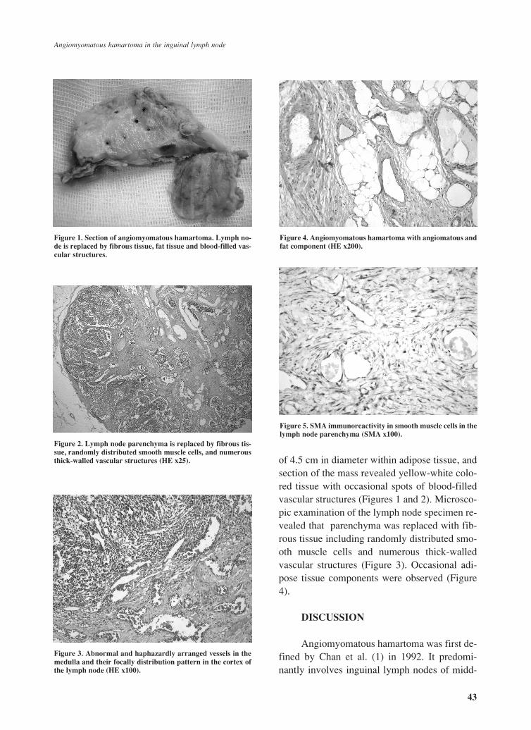
of 4.5 cm in diameter within adipose tissue, andsection of the mass revealed yellow-white colo-red tissue with occasional spots of blood-filledvascular structures (Figures 1 and 2). Microsco-pic examination of the lymph node specimen re-vealed that parenchyma was replaced with fib-rous tissue including randomly distributed smo-oth muscle cells and numerous thick-walledvascular structures (Figure 3). Occasional adi-pose tissue components were observed (Figure4).
DISCUSSION
Angiomyomatous hamartoma was first de-fined by Chan et al. (1) in 1992. It predomi-nantly involves inguinal lymph nodes of midd-
Figure 1. Section of angiomyomatous hamartoma. Lymph no-de is replaced by fibrous tissue, fat tissue and blood-filled vas-cular structures.
Figure 2. Lymph node parenchyma is replaced by fibrous tis-sue, randomly distributed smooth muscle cells, and numerousthick-walled vascular structures (HE x25).
Figure 3. Abnormal and haphazardly arranged vessels in themedulla and their focally distribution pattern in the cortex ofthe lymph node (HE x100).
Figure 4. Angiomyomatous hamartoma with angiomatous andfat component (HE x200).
Figure 5. SMA immunoreactivity in smooth muscle cells in thelymph node parenchyma (SMA x100).
43
Angiomyomatous hamartoma in the inguinal lymph node

le-aged patients, but it has been reported in thefemoral and cervical lymph nodes (1-6). Chan etal. reported 12 patients with ages ranging from10 to 80 years (median 41.5 years); 10 of the 12cases were males.
Angiomyomatous hamartomas were des-cribed as lesions that extended from the hilus tothe cortex and comprised thick-walled vascularstructures distributed within a collagenous stro-ma, and smooth muscle cells that were ran-domly distributed in or in close proximity tovascular structures, but not arranged in a fasci-cular fashion (1). In some cases, angiomyoma-tous hamartoma included adipose tissue (2,4).Thus, it should be differentiated from lymph no-de involvement of angiomyolipoma. The smo-oth muscle cells of angiomyolipoma had a pro-minent perivascular arrangement and expressedmelanoma-associated antigen HMB-45 (7). Ourcase had an adipose tissue component, but thesmooth muscle cells did not show HMB-45 im-munoreactivity.
Angiomyomatous hamartoma should bedifferentiated from lymphangiomatosis, whichusually involves intrathoracic and intraabdomi-nal lymph nodes, with smooth muscle cells ar-ranged in bundles and groups around the ectaticvascular structures (1). Nodal leiomyomatosistypically involves intraabdominal lymph nodes.It is characterized by proliferation of smoothmuscle cells and lacks prominent vascular proli-feration particularly resembling uterine leiom-yoma.
The pathogenesis of angiomyomatous ha-martoma has not yet been explained. Two pos-
sible mechanisms have been suggested. Accor-ding to these hypotheses hamartomatous lesionis either acquired or it represents a reparative re-action against previous nodal inflammation (1).The present case had no history of surgery orinflammation. Recurrences and metastases ofangiomyomatous hamartomas have not been re-ported (1-4). However, a secondary lesion aftertumor resection may develop due to impairedlymphatic transport (5).
Recognizing angiomyomatous hamartomaas a rare and benign vascular tumor of lymphnodes is important in discriminating it from ot-her benign and malignant vascular lesions oflymph nodes.
REFERENCES
1. Chan JKC, Frizzera G, Fletcher CDM, Rosai J. Pri-mary vascular tumors of lymph nodes other than Kapo-si's sarcoma. Am J Surg Pathol 1992;16:335-350.
2. Allen PW, Hoffman GJ. Fat in angiomyomatous ha-martoma of lymph node. Am J Surg Pathol1993;17:748-749.
3. Laeng RH, Hotz MA, Borisch B. Angiomyomatous ha-martoma of a cervical lymph node combined with hae-mangiomatoids and vascular transformation of sinuses.Histopathology 1996;29:80-84.
4. Magro G, Grasso S. Angiomyomatous hamartoma ofthe lymph node: Case report with adipose tissue com-ponent. Gen Diagn Pathol 1997;143:247-249.
5. Sakurai Y, Shoji M, Matsubara T, Imazu H, HasegawaS, Ochiai M, et al. Angiomyomatous hamartoma andassociated stromal lesions in the right inguinal lymphnode: A case report. Pathol Int 2000;50:655-659.
6. Dargent JL, Lespagnard L, Verdebout JM, BourgeoisP, Munck D. Glomeruloid microvascular proliferationin angiomyomatous hamartoma of the lymph node.Virchows Arch 2004;445:320-322.
7. Weiss SW, Goldblum JR. Enzinger and Weiss's SoftTissue Tumors, 4th ed., Mosby Inc., St Louis, 2001.p. 605-609.
44
Turkish Journal of Pathology 2006;22(1):42-44