angle-stabile distal radius plate - athrodax · angle-stabile distal radius plate ... flexion...
TRANSCRIPT

Königsee Implantatewww.koenigsee-implantate.de
Titanium
Variable angle-stable
Distributed in the UK by
Angle-stabile
Distal Radius PlateMini fragment 2,7 mm
2
كسور عظام الساعد الناتجة عن املد غري الثابتة داخل )23-A3/C1/C2/C3( وخارج املفصل
)23-B3( كسور عظام الساعد الناتجة عن اإلنثناء
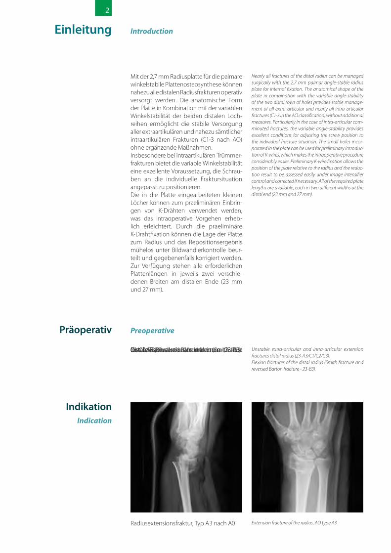
Unstable extra-articular and intra-articular extension fractures distal radius (23-A3/C1/C2/C3).Flexion fractures of the distal radius (Smith fracture and reversed Barton fracture - 23-B3).
الساعد عظام كسور جميع عالج تقريبا املمكن من التي ملم 2.7 الساعد عظام لوحة بواسطة جراحيا تستخدم للتثبيت العظمي مع استعمال لوحات راحية )من راحة اليد( ثابتة الزاوية. وتمكن اللوحة املطابقة القابلة الزاوية ثبات مع سوية الترشيحي للشكل لجميع املتني العالج الثقوب ملمجموعتي للتغيري داخل الكسور لكل وتقريبا املفصل خارج الكسور اجراءات اىل الحاجة دون )AO حتى 3-C1( املفصل
إضافية.ثبات يتيح املفصل داخل املفتتة للكسور باألخص براغي لضبط متقن وضع للتغيري القابلة الزاوية مالئمة ألوضاع الكسور املختلفة. ومن املمكن استخدام الثقوب الصغرية التي قد تم ثقبها يف اللوحة إلدراج أويل ألسالك ك )K (، الىشء الذي ييرس اإلجراء الداخيل للعملية )K( بشكل ملحوظ. وبواسطة التثبيت األويل ألسالك كلعظام بالنسبة اللوحة وضع تقييم بسهولة فيمكن الساعد وبالنسبة لنتيجة اإلرجاع اىل الوضع الطبيعي والتعديل عن طريق التحكم بشدة التصوير الشعاعي جميع تتوفر الغرض لهذا األمر. لزم اذا والتصحيح مختلفني عرضني يف الرضوري بالطول اللوحات
)23 ملم و 27 ملم(.
Nearly all fractures of the distal radius can be managed surgically with the 2.7 mm palmar angle-stable radius plate for internal fixation. The anatomical shape of the plate in combination with the variable angle-stability of the two distal rows of holes provides stable manage-ment of all extra-articular and nearly all intra-articular fractures (C1-3 in the AO classification) without additional measures. Particularly in the case of intra-articular com-minuted fractures, the variable angle-stability provides excellent conditions for adjusting the screw position to the individual fracture situation. The small holes incor-porated in the plate can be used for preliminary introduc-tion of K-wires, which makes the intraoperative procedure considerably easier. Preliminary K-wire fixation allows the position of the plate relative to the radius and the reduc-tion result to be assessed easily under image intensifier control and corrected if necessary. All of the required plate lengths are available, each in two different widths at the distal end (23 mm and 27 mm).
Preoperative قبل القيام بالعملية الجراحية
AO حتى A3 كرسلعظام الساعد الناتج عن املد، نوع Extension fracture of the radius, AO type A3
Introduction مقدمة
تعليماتIndication
Instabila extraartikulära och intraartikulära distala radiusextensionsfrakturer (23-A3/C1/C2/C3).Distala radiusflexionsfrakturer (Smith fracture respektive Reversed Barton fracture - 23-B3).
Unstable extra-articular and intra-articular extension fractures distal radius (23-A3/C1/C2/C3).Flexion fractures of the distal radius (Smith fracture and reversed Barton fracture - 23-B3).
IndikationIndication
Med en 2,7 mm radiusplatta för en palmar vinkelstabil platosteosyntes kan man operativt behandla nästan alla distala radiusfrakturer. Plattans anatomiska form i kombination med den variabla vinkelstabiliteten i de båda distala hålraderna möjliggör en stabil behand-ling av alla extraartikulära och nästan alla intraartikulära frakturer (C1-3 enligt AO) utan kompletterande åtgärder.I synnerhet vid intraartikulära pulveriserade frakturer utgör den variabla vinkelstabilite-ten en utmärkt förutsättning för att kunna placera skruvarna med en anpassning till den individuella fraktursituationen. De små hål som är infogade i plattan kan användas till att preliminärt placera K-trådar, vilket underlättar den intraoperativa proces-sen betydligt. Genom den preliminära fixeringen av K-tråden kan man utan pro-blem bedömma och vid behov genom en bildomvandlingskontroll både korrigera plattans läge i förhållande till radius och repositionsresultatet. Alla nödvändiga plattlängder med vardera två olika bred-der vid den distala ändan (23 mm och 27 mm) står till förfogande.
Nearly all fractures of the distal radius can be managed surgically with the 2.7 mm palmar angle-stable radius plate for internal fixation. The anatomical shape of the plate in combination with the variable angle-stability of the two distal rows of holes provides stable manage-ment of all extra-articular and nearly all intra-articular fractures (C1-3 in the AO classification) without additional measures. Particularly in the case of intra-articular com-minuted fractures, the variable angle-stability provides excellent conditions for adjusting the screw position to the individual fracture situation. The small holes incor-porated in the plate can be used for preliminary introduc-tion of K-wires, which makes the intraoperative procedure considerably easier. Preliminary K-wire fixation allows the position of the plate relative to the radius and the reduc-tion result to be assessed easily under image intensifier control and corrected if necessary. All of the required plate lengths are available, each in two different widths at the distal end (23 mm and 27 mm).
Inledning Introduction
Preoperativt Preoperative
Radiusextensionsfraktur, typ A3 enligt A0 Extension fracture of the radius, AO type A3
IndicationIndication
Introduction Introduction
Fractures distales instables, extraarticulaires et intraarticulaires dues à une extension du radius (23-A3/C1/C2/C3).Fractures distales dues à une extension du radius (Smith fracture bzw. Reversed Barton fracture - 23-B3).
Unstable extra-articular and intra-articular extension fractures distal radius (23-A3/C1/C2/C3).Flexion fractures of the distal radius (Smith fracture and reversed Barton fracture - 23-B3).
La plaque radiale de 2,7 mm pour l‘ostéo-synthèse à angle stable des plaques per-met de traiter chirurgicalement quasiment toutes les fractures distales. La forme anato-mique de la plaque, combinée à la stabilité variable de l‘angle des deux rangées distales de trous, permet un traitement stable de toutes les fractures extra articu-laires, ainsi que de presque toutes les frac-tures intraarticulaires (C1 à 3 selon AO), sans devoir procéder à des mesures complé-mentaires.C‘est surtout pour les fractures comminu-tives intraarticulaires que la stabilité variable de l‘angle offre les conditions idéales pour adapter la position des vis individuellement à chaque fracture. Les petits trous prévus dans la plaque peuvent être utilisés pour la pose préliminaire de fils métalliques K, ce qui facilite considérablement la procédure intraopératoire. Cette fixation préliminaire des fils métalliques K permet d‘évaluer sans difficulté la position de la plaque par rapport au radius, ainsi que le résultat du repositionnement, tout en effectuant en même temps un contrôle par convertis-seur d‘images, et de la repositionner si nécessaire. Toutes les longueurs de plaque nécessaires sont disponibles en deux largeurs à l‘extrémité distale (23 mm et 27 mm).
Nearly all fractures of the distal radius can be managed surgically with the 2.7 mm palmar angle-stable radius plate for internal fixation. The anatomical shape of the plate in combination with the variable angle-stability of the two distal rows of holes provides stable manage-ment of all extra-articular and nearly all intra-articular fractures (C1-3 in the AO classification) without additional measures. Particularly in the case of intra-articular com-minuted fractures, the variable angle-stability provides excellent conditions for adjusting the screw position to the individual fracture situation. The small holes incor-porated in the plate can be used for preliminary introduc-tion of K-wires, which makes the intraoperative procedure considerably easier. Preliminary K-wire fixation allows the position of the plate relative to the radius and the reduc-tion result to be assessed easily under image intensifier control and corrected if necessary. All of the required plate lengths are available, each in two different widths at the distal end (23 mm and 27 mm).
Pré-opératoire Preoperative
Fracture due à une extension du radius, type A3, selon A0
Extension fracture of the radius, AO type A3
Instabile extraartikuläre und intraartikuläre distale Radiusextensionsfrakturen (23-A3/C1/C2/C3).Distale Radiusflexionsfrakturen (Smith frac-ture bzw. Reversed Barton fracture - 23-B3).
Unstable extra-articular and intra-articular extension fractures distal radius (23-A3/C1/C2/C3).Flexion fractures of the distal radius (Smith fracture and reversed Barton fracture - 23-B3).
IndikationIndication
Mit der 2,7 mm Radiusplatte für die palmare winkelstabile Plattenosteosynthese können nahezu alle distalen Radiusfrakturen operativ versorgt werden. Die anatomische Form der Platte in Kombination mit der variablen Winkelstabilität der beiden distalen Loch-reihen ermöglicht die stabile Versorgung aller extraartikulären und nahezu sämtlicher intraartikulären Frakturen (C1-3 nach AO) ohne ergänzende Maßnahmen.Insbesondere bei intraartikulären Trümmer-frakturen bietet die variable Winkelstabilität eine exzellente Voraussetzung, die Schrau-ben an die individuelle Fraktursituation angepasst zu positionieren.Die in die Platte eingearbeiteten kleinen Löcher können zum praeliminären Einbrin-gen von K-Drähten verwendet werden, was das intraoperative Vorgehen erheb-lich erleichtert. Durch die praeliminäre K-Drahtfixation können die Lage der Platte zum Radius und das Repositionsergebnis mühelos unter Bildwandlerkontrolle beur-teilt und gegebenenfalls korrigiert werden. Zur Verfügung stehen alle erforderlichen Plattenlängen in jeweils zwei verschie- denen Breiten am distalen Ende (23 mm und 27 mm).
Nearly all fractures of the distal radius can be managed surgically with the 2.7 mm palmar angle-stable radius plate for internal fixation. The anatomical shape of the plate in combination with the variable angle-stability of the two distal rows of holes provides stable manage-ment of all extra-articular and nearly all intra-articular fractures (C1-3 in the AO classification) without additional measures. Particularly in the case of intra-articular com-minuted fractures, the variable angle-stability provides excellent conditions for adjusting the screw position to the individual fracture situation. The small holes incor-porated in the plate can be used for preliminary introduc-tion of K-wires, which makes the intraoperative procedure considerably easier. Preliminary K-wire fixation allows the position of the plate relative to the radius and the reduc-tion result to be assessed easily under image intensifier control and corrected if necessary. All of the required plate lengths are available, each in two different widths at the distal end (23 mm and 27 mm).
Einleitung Introduction
Präoperativ Preoperative
Radiusextensionsfraktur, Typ A3 nach A0 Extension fracture of the radius, AO type A3
IndicaçãoIndication
Introdução Introduction
Fraturas distais extra-articulares e intra-articulares instáveis da extensão do rádio (23-A3/C1/C2/C3). Fraturas distais de flexão do rádio (Smith fracture ou Reversed Barton fracture - 23-B3).
Unstable extra-articular and intra-articular extension fractures distal radius (23-A3/C1/C2/C3).Flexion fractures of the distal radius (Smith fracture and reversed Barton fracture - 23-B3).
Com a placa radial de 2,7 mm para a osteo-ssíntese palmar com estabilidade angular, é possível tratar cirurgicamente quase todas as fraturas do rádio. A forma anatômica da placa, em combinação com a estabilidade angular variável das duas primeiras filas de furos permite o tratamento estabilizador de todas as fraturas extra-articulares e quase todas as infra-articulares (C1-3 segundo a classificação AO), sem medidas suplemen-tares.Principalmente em fraturas intra-articulares cominutivas, a estabilidade angular variá-vel oferece um excelente pressuposto para posicionar os parafusos adequadamente a cada situação concreta. Os pequenos furos incorporados à placa podem ser usados para a introdução preliminar de arames K, o que facilita sobremaneira os procedimen-tos intra-operativos. Através da fixação pre-liminar por arames K, a posição da placa em relação ao rádio e o resultado da redução podem ser facilmente avaliados e, se for o caso, corrigidos mediante observação em intensificador de imagem. Encontram-se à disposição todos os comprimentos de placas necessários, cada qual em duas larguras diferentes na ponta distal (23 mm e 27 mm).
Nearly all fractures of the distal radius can be managed surgically with the 2.7 mm palmar angle-stable radius plate for internal fixation. The anatomical shape of the plate in combination with the variable angle-stability of the two distal rows of holes provides stable manage-ment of all extra-articular and nearly all intra-articular fractures (C1-3 in the AO classification) without additional measures. Particularly in the case of intra-articular com-minuted fractures, the variable angle-stability provides excellent conditions for adjusting the screw position to the individual fracture situation. The small holes incor-porated in the plate can be used for preliminary introduc-tion of K-wires, which makes the intraoperative procedure considerably easier. Preliminary K-wire fixation allows the position of the plate relative to the radius and the reduc-tion result to be assessed easily under image intensifier control and corrected if necessary. All of the required plate lengths are available, each in two different widths at the distal end (23 mm and 27 mm).
Pré-operatório Preoperative
Fratura de extensão do rádio, tipo A3 con-forme AO
Extension fracture of the radius, AO type A3
ПоказаниеIndication
Введение Introduction
Нестабильные внесуставные и внутрису-ставные разгибательные переломы дис-тальной части лучевой кости (23-A3/C1/C2/C3). Флексионные переломы дисталь-ной части лучевой кости (перелом Смита или обратный перелом Бартона - 23-B3).
Unstable extra-articular and intra-articular extension fractures distal radius (23-A3/C1/C2/C3).Flexion fractures of the distal radius (Smith fracture and reversed Barton fracture - 23-B3).
При помощи лучевой пластины 2,7 мм для ладонного остеосинтеза с угло-вой стабильностью можно оперативно лечить почти все дистальные переломы лучевой кости. Анатомическая форма пластины в сочетании с изменяемой угло-вой стабильностью обоих дистальных рядов отверстий обеспечивают стабиль-ное лечение внесуставных и почти всех внутрисуставных переломов (C1-3 по AO) без дополнительных мер.Особенно при внутрисуставных осколь-чатых переломах изменяемая угловая стабильность пластин дает прекрас-ную возможность размещения винтов с учетом индивидуальных особенностей перелома. Имеющиеся в пластине малень-кие отверстия могут использоваться для предварительной установки К-проволоки, что делает интраоперационный процесс значительно легче. Предварительная фиксация К-проволокой позволяет без труда оценивать, а при необходимости и корректировать, положение пластины относительно лучевой кости и результаты репозиции под контролем рентгеноско-пии. В наличии имеются пластины любой необходимой длины, при этом для каждой длины имеются две различные ширины дистального конца (23 мм и 27 мм).
Nearly all fractures of the distal radius can be managed surgically with the 2.7 mm palmar angle-stable radius plate for internal fixation. The anatomical shape of the plate in combination with the variable angle-stability of the two distal rows of holes provides stable manage-ment of all extra-articular and nearly all intra-articular fractures (C1-3 in the AO classification) without additional measures. Particularly in the case of intra-articular com-minuted fractures, the variable angle-stability provides excellent conditions for adjusting the screw position to the individual fracture situation. The small holes incor-porated in the plate can be used for preliminary introduc-tion of K-wires, which makes the intraoperative procedure considerably easier. Preliminary K-wire fixation allows the position of the plate relative to the radius and the reduc-tion result to be assessed easily under image intensifier control and corrected if necessary. All of the required plate lengths are available, each in two different widths at the distal end (23 mm and 27 mm).
До операции Preoperative
Разгибательный перелом лучевой кости, тип A3 по A0
Extension fracture of the radius, AO type A3
Introducción Introduction
Fracturas en extensión distales extraarti-culares e intraarticulares inestables del radio (23-A3/C1/C2/C3).Fracturas de flexión distales del radio (Smith fracture o bien Reversed Barton fracture - 23-B3).
Unstable extra-articular and intra-articular extension fractures distal radius (23-A3/C1/C2/C3).Flexion fractures of the distal radius (Smith fracture and reversed Barton fracture - 23-B3).
IndicaciónIndication
Mediante la placa radial de 2,7 mm usada para la osteosíntesis palmar con placas de ángulo estable es posible tratar quirúrgica-mente casi todas las fracturas distales del radio. La forma anatómica de la placa en combinación con la estabilidad de ángulo variable de ambas filas de orificios distales permite un tratamiento estable de todas las fracturas extraarticulares y de casi todas las fracturas intraarticulares (C1-3 según la clasi-ficación AO), sin ser necesarias más medidas complementarias.Especialmente tratándose de fracturas astilladas, la estabilidad de ángulo variable ofrece una excelente base para ajustar la posición de los tornillos a la situación indi-vidual de la fractura. Los pequeños orificios incorporados en la placa pueden utilizarse para la introducción preliminar de alam-bres de Kirschner, lo cual facilita conside-rablemente la intervención intraoperativa. La fijación preliminar mediante alambres de Kirschner permite fácilmente apreciar y, si fuera necesario, corregir tanto la posi-ción de la placa respecto al radio como el resultado de la reposición, utilizándose para este efecto el intensificador de imágenes. Cada una de todas las longitudes de placa requeridas está disponible en dos anchuras diferentes en el extremo distal (23 mm y 27 mm).
Nearly all fractures of the distal radius can be managed surgically with the 2.7 mm palmar angle-stable radius plate for internal fixation. The anatomical shape of the plate in combination with the variable angle-stability of the two distal rows of holes provides stable manage-ment of all extra-articular and nearly all intra-articular fractures (C1-3 in the AO classification) without additional measures. Particularly in the case of intra-articular com-minuted fractures, the variable angle-stability provides excellent conditions for adjusting the screw position to the individual fracture situation. The small holes incor-porated in the plate can be used for preliminary introduc-tion of K-wires, which makes the intraoperative procedure considerably easier. Preliminary K-wire fixation allows the position of the plate relative to the radius and the reduc-tion result to be assessed easily under image intensifier control and corrected if necessary. All of the required plate lengths are available, each in two different widths at the distal end (23 mm and 27 mm).
Preoperativa Preoperative
Frauctura en extensión del radio, tipo A3 según clasificación A0
Extension fracture of the radius, AO type A3
IndicazioniIndication
Introduzione Introduction
Fratture per estensione radiali distali extra-articolari e intra-articolari instabili (23-A3/C1/C2/C3).Fratture di flessione radiali distali (Smith fracture e Reversed Barton fracture - 23-B3).
Unstable extra-articular and intra-articular extension fractures distal radius (23-A3/C1/C2/C3).Flexion fractures of the distal radius (Smith fracture and reversed Barton fracture - 23-B3).
Con la piastra radiale da 2,7 mm per l‘osteo-sintesi con piastra palmare ed angolo stabile è possibile trattare pressoché tutte le fratture radiali distali in sede operatoria. La forma anatomica della piastra rende possi-bile, unitamente alla stabilità angolare delle due file di fori distali, il trattamento stabile di tutte le fratture extra-articolari e di quasi tutte le fratture intra-articolari (C1-3 secondo la classificazione AO) senza dover ricorrere a misure di natura integrativa.In particolare nel caso delle fratture comminute intra-articolari, la stabilità angolare variabile rappresenta condizio-ni eccellenti ai fini di un posizionamento delle viti in modo adatto alle specifiche caratteristiche della frattura in oggetto. I fori praticati nella piastra possono essere utilizzati per l‘applicazione preliminare di fili K, con la conseguente considerevole agevolazione della procedura intraopera-toria. Attraverso il fissaggio preliminare dei fili K è possibile valutare e, se necessario, correggere senza difficoltà la posizione della piastra rispetto al radio nonché il risultato del riposizionamento mediante il controllo con convertitore di immagine. Sono in tal senso disponibili tutte le misure di lunghezza per la piastra, ognuna delle quali in due diverse larghezze all‘estremità distale (23 mm e 27 mm).
Nearly all fractures of the distal radius can be managed surgically with the 2.7 mm palmar angle-stable radius plate for internal fixation. The anatomical shape of the plate in combination with the variable angle-stability of the two distal rows of holes provides stable manage-ment of all extra-articular and nearly all intra-articular fractures (C1-3 in the AO classification) without additional measures. Particularly in the case of intra-articular com-minuted fractures, the variable angle-stability provides excellent conditions for adjusting the screw position to the individual fracture situation. The small holes incor-porated in the plate can be used for preliminary introduc-tion of K-wires, which makes the intraoperative procedure considerably easier. Preliminary K-wire fixation allows the position of the plate relative to the radius and the reduc-tion result to be assessed easily under image intensifier control and corrected if necessary. All of the required plate lengths are available, each in two different widths at the distal end (23 mm and 27 mm).
Sede preoperatoria Preoperative
Frattura per estensione, tipo A3 ai sensi della classificazione A0
Extension fracture of the radius, AO type A3
3
العالجTreatment
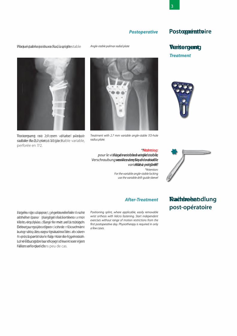
لوحة عظام الساعد الراحية )راحة اليد( ثابتة الزاوية Angle-stable palmar radial plate
بعد القيام بالعملية الجراحية Postoperative
اورتوبيدية قطعة واستعمال )السناد( جبرية وضع للمعصم قابلة لإلزالة بسهولة مع سدادة الفيلكرو اذا الحركة بتمارين البدء ثم ومن . لذلك الرضورة دعت اليوم األول الذاتية دون تقييد مدى الحركه وهذا بعد للمعالجة حاجة توجد وال الجراحية. العملية من
بالتمارين الرياضية اال يف قليل من الحاالت.
Positioning splint, where applicable, easily removable wrist orthesis with Velcro fastening. Start independent exercises without range of motion restrictions from the first postoperative day. Physiotherapy is required in only a few cases.
العالج بلوحة عظام الساعد ثابتة الزاوية والقابلة للتغيري 2.7 ملم وذات 7/2 ثقوب.
Treatment with 2.7 mm variable angle-stable 7/2-hole radius plate.
After-Treatment متابعة العالج
*تنبيه: استخدم للرباغي املتغرية ثابتة الزاوية
جلبة مرنة ذات مقبض!*Attention:
For the variable angle-stable locking use the variable drill-guide sleeve!
BehandlingTreatment
Vinkelstabil palmar radiusplatta Angle-stable palmar radial plate
Postoperative Postoperativt
Positionsskena, vid behov lätt avtagbar handledsortesis med kardborrelås. Påbörja oberoende rörelseövningar utan inskränk-ning av rörelseomfattningen fr.o.m. posto-perativ dag 1. En övningsbehandling med sjukgymnastik kräve endast i få fall.
Positioning splint, where applicable, easily removable wrist orthesis with Velcro fastening. Start independent exercises without range of motion restrictions from the first postoperative day. Physiotherapy is required in only a few cases.
Behandling med 2,7 mm variabel vinkel-stabil radiusplatta 7/2 hål.
Treatment with 2.7 mm variable angle-stable 7/2-hole radius plate.
After-Treatment Efterbehandling
*Achtung: Für dir variabel winkelstabile
Verschraubung verwenden Sie die variable Hülse mit Griff!
*Attention:For the variable angle-stable locking
use the variable drill-guide sleeve!
TraitementTreatment
Plaque palmaire du radius, à angle stable Angle-stable palmar radial plate
Postoperative Post-opératoire
Attelle de support, éventuellement une orthèse pour poignet facilement amo-vible, équipée d’une fermeture à scratch. Début au moyen d’exercices de mouvement automatiques, sans limitation des mouve-ments à partir du lendemain de l’opération. La rééducation au moyen d’exercices n’est nécessaire que dans peu de cas.
Positioning splint, where applicable, easily removable wrist orthesis with Velcro fastening. Start independent exercises without range of motion restrictions from the first postoperative day. Physiotherapy is required in only a few cases.
Traitement au moyen d’une plaque radiale de 2,7 mm, à angle stable variable, perforée en 7/2.
Treatment with 2.7 mm variable angle-stable 7/2-hole radius plate.
After-Treatment Traitement post-opératoire
*Attention:pour le vissage variable à angle stable,
veuillez employer la douille variable à poignée!
*Attention:For the variable angle-stable locking
use the variable drill-guide sleeve!
VersorgungTreatment
Winkelstabile palmare Radiusplatte Angle-stable palmar radial plate
Postoperative Postoperativ
Lagerungsschiene, gegebenenfalls leicht abnehmbare Handgelenksorthese mit Klettverschluss. Beginn mit selbsttätigen Bewegungsübungen ohne Einschrän-kung des Bewegungsausmaßes ab dem 1. postoperativen Tag. Krankengymnasti-sche Übungsbehandlung ist nur in wenigen Fällen erforderlich.
Positioning splint, where applicable, easily removable wrist orthesis with Velcro fastening. Start independent exercises without range of motion restrictions from the first postoperative day. Physiotherapy is required in only a few cases.
Versorgung mit 2,7 mm variabel winkel-stabiler Radiusplatte 7/2 Loch.
Treatment with 2.7 mm variable angle-stable 7/2-hole radius plate.
After-Treatment Nachbehandlung
*Achtung: Für dir variabel winkelstabile
Verschraubung verwenden Sie die variable Hülse mit Griff!
*Attention:For the variable angle-stable locking
use the variable drill-guide sleeve!
TratamentoTreatment
Placa radial palmar com estabilidade angular
Angle-stable palmar radial plate
Postoperative Pós-operativo
Tala de apoio e, se for o caso, órtese de punho facilmente removível com fecho de velcro. Início com exercícios de movimen-tos espontâneos sem restrição de amplitu-de de movimento, a partir do primeiro dia pós-operatório. O tratamento com exercí-cios fisioterapêuticos é necessário apenas em poucos casos.
Positioning splint, where applicable, easily removable wrist orthesis with Velcro fastening. Start independent exercises without range of motion restrictions from the first postoperative day. Physiotherapy is required in only a few cases.
Tratamento com placa radial de 2,7 mm de estabilidade angular variável, furo 7/2.
Treatment with 2.7 mm variable angle-stable 7/2-hole radius plate.
After-Treatment Tratamento posterior
*Atenção: Para o aparafusamento com estabilidade
angular variável, utilize a bucha variável com alça!
*Attention:For the variable angle-stable locking
use the variable drill-guide sleeve!
ЛечениеTreatment
Ладонная лучевая пластина с угловой стабильностью
Angle-stable palmar radial plate
Postoperative После операции
Лестничная проволочная шина, при необходимости легкосъемный ортез для лучезапястного сустава с застежкой-липучкой. Начало самостоятельных дви-гательных упражнений без ограничения амплитуды движения, начиная с перво-го дня после операции. Физиотерапия необходима лишь в редких случаях.
Positioning splint, where applicable, easily removable wrist orthesis with Velcro fastening. Start independent exercises without range of motion restrictions from the first postoperative day. Physiotherapy is required in only a few cases.
Лечение при помощи лучевой пластины с изменяемой угловой стабильностью, количество отверстий 7/2 под винты 2,7 мм
Treatment with 2.7 mm variable angle-stable 7/2-hole radius plate.
*Внимане:Для изменяемого резьбового
соединения с угловой стабильностью используйте изменяемую втулку
с ручкой!*Attention:
For the variable angle-stable locking use the variable drill-guide sleeve!
After-Treatment Послеоперационное лечение
TratamientoTreatment
Placa radial palmar de ángulo estable Angle-stable palmar radial plate
Postoperative Postoperativa
Férula de postura o en caso dado ortosis radiocarpiana fácilmente desmontable, con fijación Velcro. Comienzo con ejercicios independientes sin restringir la dimensión de los movimientos, a partir del 1er día postoperativo. Un tratamiento fisioterapéu-tico es necesario sólo en pocos casos.
Positioning splint, where applicable, easily removable wrist orthesis with Velcro fastening. Start independent exercises without range of motion restrictions from the first postoperative day. Physiotherapy is required in only a few cases.
Tratamiento con placa radial de 2,7 mm de ángulo estable variable de 7/2 orificios.
Treatment with 2.7 mm variable angle-stable 7/2-hole radius plate.
After-Treatment Tratamiento posterior
*Atención:¡Para la atornilla dura variable de ángulo
estable sírvase usar el casquillo variable con mango!
*Attention:For the variable angle-stable locking
use the variable drill-guide sleeve!
TrattamentoTreatment
Piastra radiale palmare ad angolo stabile Angle-stable palmar radial plate
Postoperative Sede postoperatoria
Stecca di posizionamento, se necessario con un‘ortesi da polso facilmente rimovibi-le con sistema di chiusura a strappo. Inizio con esercizi motori autonomi senza limita-zione dell‘entità del movimento a partire dal 1° giorno postoperatorio. Il trattamento con esercizi di ginnastica medica risulta neces-sario soltanto in pochi casi.
Positioning splint, where applicable, easily removable wrist orthesis with Velcro fastening. Start independent exercises without range of motion restrictions from the first postoperative day. Physiotherapy is required in only a few cases.
Trattamento con piastra radiale da 2,7 mm variabile ad angolo stabile, foro 7/2.
Treatment with 2.7 mm variable angle-stable 7/2-hole radius plate.
After-Treatment Posttrattamento
*Attenzione:Per il fissaggio ad angolo stabile variabile
si consiglia di utilizzare il manicotto variabile con impugnatura!
*Attention:For the variable angle-stable locking
use the variable drill-guide sleeve!
4
يتم تنفيذ العملية بوضع اإلستلقاء ويتم وضع الذراع او الساعد عىل املنضدة املختصة لذلك. بعدها يتم القيام اوال باإلرجاع للوضع العادي/الطبيعي والتحكم بشدة
التصوير الشعاعي اذا دعت الرضورة لذلك. يتم 2C A3 وايضا غالبية كسور يف كسور من نوع التمكن يف حالة اإلحتفاظ بالتقنية املستخدمة يف جراحة العظام من اإلرجاع اىل الشكل الترشيحي اىل حد كبري.
The operation is performed in supine position. The arm is placed on an arm table. Closed reduction is carried out first, if necessary under image intensifier control.In A3 fractures and the majority of C2 fractures, anatomical reduction is largely achieved when ligamentotaxis is maintained.
يتم تنفيذ العملية دون وقف النزف ويتبع توقف نزيف )بطول بسيط وقصري قطبني. شق ذو تخثر مع الدم لوتر التعرض املعصم. سطوح عىل تقريبا( سم 3-5
عظام الساعد مثني الرسغ امللتوي قليال اتجاه الزند.
The operation is performed without a tourniquet and meticulous haemostasis is obtained with bipolar coagulation. Short (approx. 3-5 cm) incision over the flexor surface of the wrist. Exposure of the flexor carpi radialis tendon, which is retracted slightly toward the ulnar side.
أسفل من مبارشة للساعد الضام النسيج قطع ويتم عظام وتر ويبقى للوتر الساعد عظام جانب من او محتفظ اوتار من تحته وما الرسغ ااملثني الساعد العصب ذاتي(. حامل بمساعدة األمر لزم )اذا جانبيا )بإستثناء يتأثر ال الرسغي والنفق يظهر ال املتوسط ويتم قبل(. من املوجودة الرسغي النفق متالزمة باإلنفاذ العالج بواسطة املربعة الكابة )عضلة( قطع العضلة الساعد ودفع بطن الحراري من جانب عظام
بعيدا من الناحية املنحنية لعظام الساعد.
The forearm fascia is divided immediately below or just to the radial side of the tendon.The flexor carpi radialis tendon and the finger flexor tendons / muscle bellies beneath it are held aside (using a self-retaining retractor if necessary). The median nerve is not exposed and the carpal tunnel is not touched (exception: pre-existing CTS).Division of pronator quadratus with diathermy on the radial side, and the muscle belly is pushed away from the flexor surface of the radius.
عن بوضوح املنحني الجانب من الكرس خط يظهر طريق تجنب إزالة السمحاق )غشاء العظم( وهذا يتبع
اإلرجاع النهائي للشكل الترشيحي.
The fracture line on the flexor surface is exposed cleanly by sparing removal of periosteum.This is followed by definitive anatomical reduction.
اختيار اللوحة املناسبة )عادة تكفي اللوحة ذات ثقبني 27 ملم وهذا او 23 ملم العظم ذات عرض يف محور
وفقا لعرض عظام الساعد(.
A suitable plate is selected (the two-hole plate is usually sufficient, 23 mm or 27 mm wide, depending on the width of the radius).
تعليمات حول العملية الجراحيةOP-Instruction
Ingreppet genomförs med patienten i ryggläge. Armen placeras på ett extra arm-bord. Först genomförs en sluten reposition respektive vid behov en bildomvandlings-kontroll.Vid A 3 frakturer och till övervägande del även vid C 2 frakturer uppnår man vid en bibehållen ligamentotaxis i stort sett en anatomisk reposition.
The operation is performed in supine position. The arm is placed on an arm table. Closed reduction is carried out first, if necessary under image intensifier control.In A3 fractures and the majority of C2 fractures, anatomical reduction is largely achieved when ligamentotaxis is maintained.
Operations- OP-Instruction
instruktion
Ingreppet genomförs utan kompressor, en subtil haemostasis uppnås med bipolär koagulering. En kort (ca. 3 – 5 cm lång) incision över handledens böjmuskel.Exponering av flexor carpi radialis senan, som dras något tillbaka i ulnar riktning.
The operation is performed without a tourniquet and meticulous haemostasis is obtained with bipolar coagulation. Short (approx. 3-5 cm) incision over the flexor surface of the wrist. Exposure of the flexor carpi radialis tendon, which is retracted slightly toward the ulnar side.
Underarmsfascia skiljs av omedelbart under respektive direkt vid radialsidan av senan. Flexor carpi radialis senan och finger flexor senorna / muskelbukarna, som ligger under dessa, hålls åt sidan (vid behov med hjälp av en automatisk retractor). Nervus media-nus exponeras inte,carpaltunneln tangeras inte (undantag: vid redan existerande CTS).Pronator quadratus delas med diatermi på radialsidan. Muskelbuken skjuts åt sidan från radius flexorsidan.
The forearm fascia is divided immediately below or just to the radial side of the tendon.The flexor carpi radialis tendon and the finger flexor tendons / muscle bellies beneath it are held aside (using a self-retaining retractor if necessary). The median nerve is not exposed and the carpal tunnel is not touched (exception: pre-existing CTS).Division of pronator quadratus with diathermy on the radial side, and the muscle belly is pushed away from the flexor surface of the radius.
Frakturlinjen på flexorsidan exponeras tydligt genom att man sparsamt avlägsnar periosteum.Därefter utförs den slutgiltiga anatomiska repositionen.
The fracture line on the flexor surface is exposed cleanly by sparing removal of periosteum.This is followed by definitive anatomical reduction.
Välj en lämplig platta med 2 skafthål, bredd 23 mm eller 27 mm, beroende av radius bredd).
A suitable plate is selected (the two-hole plate is usually sufficient, 23mm or 27mm wide, depending on the width of the radius).
Instructions OP-Instruction
opératoires
L’intervention s’effectue en position sur le dos. Le bras est posé sur une table spéciale. Dans un premier temps est effectué le repositionnement fermé, éventuellement au moyen d’un contrôle par convertisseur d’images. En cas de fractures A3, et pour la plupart des fractures C2, si l’otaxis des ligaments a été conservée, un repositionne-ment anatomique est effectué.
The operation is performed in supine position. The arm is placed on an arm table. Closed reduction is carried out first, if necessary under image intensifier control.In A3 fractures and the majority of C2 fractures, anatomical reduction is largely achieved when ligamentotaxis is maintained.
L’intervention est réalisée sans exsanguina-tion, une hémostase subtile a lieu au moyen d’une coagulation bipolaire. Incision sur le côté le plus court (3 à 5 cm de long), du côté de la flexion, sur le poignet.Représentation du tendon du flexor carpi raidalis, légèrement rétracté vers l’ulnaire.
The operation is performed without a tourniquet and meticulous haemostasis is obtained with bipolar coagulation. Short (approx. 3-5 cm) incision over the flexor surface of the wrist. Exposure of the flexor carpi radialis tendon, which is retracted slightly toward the ulnar side.
Le fascia de l’avant-bras est coupé directe-ment sous le tendon, ou alors du coté du radius. Le tendon flexor carpi radialis, ainsi que les tendons / les muscles fléchisseurs des doigts situés en-dessous sont mainte-nus de côté (éventuellement à l’aide d’un autorétracteur). Le nerf médian n’est pas représenté, le tunnel carpal n’est pas touché (exception: CTS pré-existant).Section du pronator quadratus au moen de la diathermie, du coté du radius, déca-lage du muscle depuis le côté de flexion du radius.
The forearm fascia is divided immediately below or just to the radial side of the tendon.The flexor carpi radialis tendon and the finger flexor tendons / muscle bellies beneath it are held aside (using a self-retaining retractor if necessary). The median nerve is not exposed and the carpal tunnel is not touched (exception: pre-existing CTS).Division of pronator quadratus with diathermy on the radial side, and the muscle belly is pushed away from the flexor surface of the radius.
La ligne de fracture du côté de la flexion est représentée proprement, en retirant soigneusement le périostéum.Le repositionnement anatomique définitif est effectué.
The fracture line on the flexor surface is exposed cleanly by sparing removal of periosteum.This is followed by definitive anatomical reduction.
Sélection d’une plaque adéquate (généra-lement, la plaque à 2 trous suffit) Largeur 23 mm ou 27 mm, selon la largeur du radius).
A suitable plate is selected (the two-hole plate is usually sufficient, 23 mm or 27 mm wide, depending on the width of the radius).
Der Eingriff erfolgt in Rückenlage. Der Arm wird auf einem Armtisch ausgelagert.Zunächst erfolgt die geschlossene Reposi-tion, ggf. Bildwandlerkontrolle.Bei A 3 Frakturen und überwiegend auch bei C 2 Frakturen gelingt bei erhaltener Ligamentotaxis eine weitgehend anatomi-sche Reposition.
The operation is performed in supine position. The arm is placed on an arm table. Closed reduction is carried out first, if necessary under image intensifier control.In A3 fractures and the majority of C2 fractures, anatomical reduction is largely achieved when ligamentotaxis is maintained.
OP-Anleitung OP-Instruction
Der Eingriff wird ohne Blutsperre durch-geführt, es erfolgt eine subtile Blutstillung mit bipolarer Koagulation. Kurzstreckige (ca. 3-5 cm lange ) Inzision beugeseitig über dem Handgelenk.Darstellen der Sehne des Flexor carpi radialis, die leicht nach ulnar verzogen wird.
The operation is performed without a tourniquet and meticulous haemostasis is obtained with bipolar coagulation. Short (approx. 3-5 cm) incision over the flexor surface of the wrist. Exposure of the flexor carpi radialis tendon, which is retracted slightly toward the ulnar side.
Die Unterarmfascie wird unmittelbar unter bzw. knapp radialseitig der Sehne durch-trennt. Die Flexor carpi radialis Sehne und die darunter liegenden Sehnen / Muskel-bäuche der Fingerbeuger werden beiseite gehalten (ggf. unter Zuhilfenahme eines Selbsthalters). Der Nervus medianus wird nicht dargestellt, der Carpaltunnel nicht tangiert (Ausnahme: vorbestehendes CTS).Durchtrennen des Pronator quadratus mit der Diathermie radialseitig, Abschieben des Muskelbauches von der Beugeseite des Radius.
The forearm fascia is divided immediately below or just to the radial side of the tendon.The flexor carpi radialis tendon and the finger flexor tendons / muscle bellies beneath it are held aside (using a self-retaining retractor if necessary). The median nerve is not exposed and the carpal tunnel is not touched (exception: pre-existing CTS).Division of pronator quadratus with diathermy on the radial side, and the muscle belly is pushed away from the flexor surface of the radius.
Die beugeseitige Frakturlinie wird durch sparsames Deperiostieren sauber darge-stellt. Es erfolgt die endgültige anatomische Reposition.
The fracture line on the flexor surface is exposed cleanly by sparing removal of periosteum.This is followed by definitive anatomical reduction.
Auswählen einer geeigneten Platte (im Regelfall reicht die Platte mit 2 Schaft- löchern aus. Breite 23 mm oder 27 mm, je nach Breite des Radius).
A suitable plate is selected (the two-hole plate is usually sufficient, 23 mm or 27 mm wide, depending on the width of the radius).
Instruções OP-Instruction
cirúrgicas
A intervenção é feita com decúbito dorsal. O braço é apoiado em uma mesa para braço. Inicialmente é feita a redução fechada e, se for o caso, controle por imagem.Em fraturas A3 e na maioria das fraturas C2 e com ligamentotaxia conservada, atinge-se considerável redução anatômica.
The operation is performed in supine position. The arm is placed on an arm table. Closed reduction is carried out first, if necessary under image intensifier control.IIn A3 fractures and the majority of C2 fractures, anatomical reduction is largely achieved when ligamentotaxis is maintained.
A intervenção é realizada sem torniquete, realizando-se estancamento sutil, com coagulação bipolar. Incisão pequena (cerca de 3 a 5 cm) do lado flexor, no punho.Exposição do tendão carpi radialis, que é deslocado levemente em sentido ulnar.
The operation is performed without a tourniquet and meticulous haemostasis is obtained with bipolar coagulation. Short (approx. 3-5 cm) incision over the flexor surface of the wrist. Exposure of the flexor carpi radialis tendon, which is retracted slightly toward the ulnar side.
A fascie do antebraço é seccionada logo abaixo ou no lado radial do tendão.O tendão flexor carpi radialis e os tendões / músculos tensores dos dedos são manti-dos de lado (se necessário com o auxílio de um refrator automático). O nervo mediano não é exposto e o túnel carpal não é tocado (exceção: CTS pré-existente).Divisão do pronator quadratus com a diatermia do lado radial, empurrando a barriga do músculo do lado flexor do rádio.
The forearm fascia is divided immediately below or just to the radial side of the tendon.The flexor carpi radialis tendon and the finger flexor tendons / muscle bellies beneath it are held aside (using a self-retaining retractor if necessary). The median nerve is not exposed and the carpal tunnel is not touched (exception: pre-existing CTS).Division of pronator quadratus with diathermy on the radial side, and the muscle belly is pushed away from the flexor surface of the radius.
A linha da fratura do lado flexor é exposta com perfeição através de econômica remoção do periósteo. Faz-se a redução anatômica definitiva.
The fracture line on the flexor surface is exposed cleanly by sparing removal of periosteum.This is followed by definitive anatomical reduction.
Seleção da placa adequada (via de regra basta a placa com 2 furos. Largura 23 mm ou 27 mm, de acordo com a largura do rádio).
A suitable plate is selected (the two-hole plate is usually sufficient, 23 mm or 27 mm wide, depending on the width of the radius).
Операционная OP-Instruction
методика
Операцию осуществляют в положении на спине. Руку кладут на специальный столик для руки. Затем осуществляют за-крытую репозицию, при необходимости под контролем рентгеноскопии.Для переломов A 3 и для большинства переломов C 2 удается добиться анато-мической репозиции при сохранении лигаментотаксиса.
The operation is performed in supine position. The arm is placed on an arm table. Closed reduction is carried out first, if necessary under image intensifier control.In A3 fractures and the majority of C2 fractures, anatomical reduction is largely achieved when ligamentotaxis is maintained.
Операцию проводят без кровооста-навливающего жгута, полный гемостаз достигают при помощи биполярной коагуляции. Небольшие (длиной около 3-5 см) надрезы со стороны сгибатель-ной поверхности запястья. Обнажение сухожилия лучевого сгибателя запястья, которое слегка отведено в сторону лок-тевой кости.
The operation is performed without a tourniquet and meticulous haemostasis is obtained with bipolar coagulation. Short (approx. 3-5 cm) incision over the flexor surface of the wrist. Exposure of the flexor carpi radialis tendon, which is retracted slightly toward the ulnar side.
Фасцию предплечья рассекают непо-средственно под или почти с лучевой стороны сухожилия. Сухожилие лучево-го сгибателя запястья и расположенные под ним сухожилия / мышечные брюшка сгибателей пальцев удерживают в стороне (воспользовавшись при необходимости самоудерживающимся ранорасшири-телем). Срединный нерв не оголяется, канал запястья не затрагивается (исклю-чение: имеющийся CTS).Рассечение квадратного пронатора с лучевой стороны путем диатермии,
отодвигание мышечного брюшка от сгибательной поверхности лучевой кости.
The forearm fascia is divided immediately below or just to the radial side of the tendon.The flexor carpi radialis tendon and the finger flexor tendons / muscle bellies beneath it are held aside (using a self-retaining retractor if necessary). The median nerve is not exposed and the carpal tunnel is not touched (exception: pre-existing CTS).Division of pronator quadratus with diathermy on the radial side, and the muscle belly is pushed away from the flexor surface of the radius.
Путем аккуратного отделения надкост-ницы оголяют линию перелома на сгиба-тельной поверхности. После этого про-водят анатомическую репозицию.
The fracture line on the flexor surface is exposed cleanly by sparing removal of periosteum.This is followed by definitive anatomical reduction.
Осуществляют выбор подходящей пла-стины (как правило, в теле пластины достаточно 2 отверстий, ширина 23 мм или 27 мм, в зависимости от ширины лучевой кости).
A suitable plate is selected (the two-hole plate is usually sufficient, 23 mm or 27 mm wide, depending on the width of the radius).
Instrucciones OP-Instruction
para la operación
La intervención tiene lugar en decúbito supino. El brazo se posiciona sobre una mesa de soporte para el brazo. En primer lugar se lleva a cabo la reposición cerrada, en caso dado, controlando con un intensifica-dor de imágenes. Tratándose de las fracturas A 3 y también en la mayoría de las fracturas C 2, se logra una extensa reposición anató-mica, si se mantiene la ligamentotaxis.
The operation is performed in supine position. The arm is placed on an arm table. Closed reduction is carried out first, if necessary under image intensifier control.In A3 fractures and the majority of C2 fractures, anatomical reduction is largely achieved when ligamentotaxis is maintained.
La intervención tiene lugar sin bloqueo circulatorio llevándose a cabo una minuciosa hemostasia con coagulación bipolar. Incisión de corto alcance (longitud de 3-5 cm aproxi-madamente) más arriba de la muñeca en la superficie flexora. Visualización del tendón del flexor carpi radialis, que se retrae fácil-mente hacia el ulnar.
The operation is performed without a tourniquet and meticulous haemostasis is obtained with bipolar coagulation. Short (approx. 3-5 cm) incision over the flexor surface of the wrist. Exposure of the flexor carpi radialis tendon, which is retracted slightly toward the ulnar side.
La fascia del antebrazo se secciona direc-tamente bajo el tendón o justo en su la-do radial. El tendón del flexor carpi radialis y los tendones de los dedos así como los cuerpos de sus músculos flexores que se encuentran debajo se desplazan hacia los lados (si fuera necesario, utilizando un re-tractor autoretenedor). El nervus medianus no se visualiza y el túnel carpiano no se toca (excepción: CTS pre-existente).Seccionar el pronator quadratus con diater-mia en el lado radial, desplazar el cuerpo muscular de la cara de flexión del radio.
The forearm fascia is divided immediately below or just to the radial side of the tendon.The flexor carpi radialis tendon and the finger flexor tendons / muscle bellies beneath it are held aside (using a self-retaining retractor if necessary). The median nerve is not exposed and the carpal tunnel is not touched (exception: pre-existing CTS).Division of pronator quadratus with diathermy on the radial side, and the muscle belly is pushed away from the flexor surface of the radius.
La línea de la fractura en la cara de flexión puede visualizarse claramente después de una leve reducción del periostio. A conti-nuación tiene lugar la reposición anatómica definitiva.
The fracture line on the flexor surface is exposed cleanly by sparing removal of periosteum.This is followed by definitive anatomical reduction.
Seleccionar una placa adecuada (gene-ralmente es suficiente la placa con dos orificios en el vástago. Anchura 23 mm ó 27 mm, según la anchura del radio).
A suitable plate is selected (the two-hole plate is usually sufficient, 23 mm or 27 mm wide, depending on the width of the radius).
Istruzioni OP-Instruction
operatorie
L‘intervento ha luogo in posizione supina. Il braccio viene disposto su un apposito tavolo. In primo luogo si procede alla riduzione chiusa e, se necessario, al controllo mediante convertitore di immagine.Nel caso specifico di fratture di tipo A 3 e, in gran parte, anche per le fratture di tipo C 2, può aver luogo con successo una riduzione anatomica con legamentotassi trattenuta.
The operation is performed in supine position. The arm is placed on an arm table. Closed reduction is carried out first, if necessary under image intensifier control.In A3 fractures and the majority of C2 fractures, anatomical reduction is largely achieved when ligamentotaxis is maintained.
L‘intervento viene effettuato senza trequarti, e ha luogo una meticolosa emostasi con coagulazione bipolare. Breve incisione (con una lunghezza di ca. 3-5 cm) al di sopra del polso sul lato di flessione.Esposizione del tendine flessore radiale del carpo, ritrato lievemente verso il lato ulnare.
The operation is performed without a tourniquet and meticulous haemostasis is obtained with bipolar coagulation. Short (approx. 3-5 cm) incision over the flexor surface of the wrist. Exposure of the flexor carpi radialis tendon, which is retracted slightly toward the ulnar side.
La fascia dell‘avambraccio viene recisa immediatamente al di sotto o direttamente sul lato radiale dle tendine. Il tendine fles-sore radiale del carpo e i tendini/ventri muscolari sottostanti sono tenuti lateralmente (se necessario con l‘ausilio di un divaricatore autostatico). Il nervo mediano non viene esposto e non si ha contatto alcuno con il tunnel carpale (eccezione: CTS pre-esistente).Recisione del pronatore quadrato con diatermia sul lato radiale. Scostamento del ventre muscolare dal lato di flessione del radio.
The forearm fascia is divided immediately below or just to the radial side of the tendon.The flexor carpi radialis tendon and the finger flexor tendons / muscle bellies beneath it are held aside (using a self-retaining retractor if necessary). The median nerve is not exposed and the carpal tunnel is not touched (exception: pre-existing CTS).Division of pronator quadratus with diathermy on the radial side, and the muscle belly is pushed away from the flexor surface of the radius.
La linea di frattura sul lato di flessione è esposta in modo chiaro attraverso un risparmio in termini di asportazione del periostio. Segue quindi la riduzione anato-mica definitiva.
The fracture line on the flexor surface is exposed cleanly by sparing removal of periosteum.This is followed by definitive anatomical reduction.
Scelta di una piastra adeguata (in genere è sufficiente una piastra dotata di 2 fori, con una larghezza di 23 mm o 27 mm in funzio-ne della larghezza del radio).
A suitable plate is selected (the two-hole plate is usually sufficient, 23 mm or 27 mm wide, depending on the width of the radius).

5
بسلك العظم ومحور املعصم من لكل اللوحة تثبيت او اكثر من اسالك كريشنر بواسطة الثقوب )الفتحات( املعدة لذلك يف اللوحة. وبهذا يتم كل من ضبط اللوحة اإلرجاع نتيجة ضبط وكذلك الساعد عظام عىل التحضريي واألويل )التحكم بشدة التصوير الشعاعي(. اللوحة وضع بتصحيح القيام فيتم األمر لزم واذا بواسطة تثبيت اويل ومن جديد لسلك كريشنر وبالتايل
التحكم بشدة التصوير الشعاعي.
The plate is fixed to the articular fragment and to the shaft with one or more Kirschner wires through the holes provided in the plate. This achieves preliminaryfixation of the plate to the radius and of thereduction (image intensifier control).The plate position is corrected if necessary with further preliminary Kirschner wirefixation and subsequent image intensifiercontrol.
مع بحذر القرشة نقب/ثقب يتم الحفر عملية اثناء لعظام الظهري الجانب عىل االصبع بوضع التحكم الساعد ويتم قياس الطول بأداة التثقيب ثابتة الزاوية او باستعمال اداة التثقيب ثابتة الزاوية القابلة للتغيري من القياس جهاز راس تجربة املمكن من انه بحيث وبهذا العظام تحت الساعد لعظام الخلفي الجانب
التمكن من التحديد بدقة لطول الرباغي.
During drilling, the opposite cortex is deliberately drilled cautiously with a finger on the dorsal side of the radius. The length is measured through the angle-stable drill bushing or after using the variable angle-stable drill bushing with corresponding distance sleeve on the length gauge, palpating the tip of the gauge on the dorsal side of the radius beneath the skin so that the length of the screws can be established exactly.
يف املوجودة الرباغي تالمس يتم ان املفروض من اآلن العظم مع القرشة برشط ان ال يتم دفع شظايا القرشة الصغرية! لهذا فمن املفضل كما ذكر من قبل اوال نقب/يف يتم انه حيث بحذر الرقيقة الظهرية القرشة ثقب يف براغي 4 او 3 عن يقل ال ما إدراج التقنية هذه احصار املفصل وبهذا يجب تباعد برغي عظام الساعد لعظام األبري النتوء من للميل وفقا األخص عىل تجنب يجب داخلية مفصلية كسور حالة يف الساعد.
وضع الرباغي يف شق الكرس.
The screws introduced distally should just reach the dorsal cortex but not push away smaller cortical frag-ments (which are common when there is a dorsal zone of comminution). It is therefore beneficial, as mentioned above, to drill the thin dorsal cortex carefully first. In this technique, at least 3 or 4 screws are introduced in suc-cession into the articular block and the radial screw in particular should diverge into the radial styloid process depending on the inclination. In the case of intra-articu-lar fractures, a screw position in the fracture gap should be avoided.
مهم: عند إدخال الرباغي ثابتة الزاوية فيجب الضغط باالصبع من الخلف باإلتجاه العكيس لكي يتم ضغط استخدام اىل يؤدي الذي الىشء اللوحة، ضد العظام الترشيحي إلرجاع دقيق للشكل املطابق اللوحة شكل
للوضع العادي.
Important: when tightening the distal angle-stable screws, counterpressure should be exerted with a finger from the dorsal side in order to press the bone against the plate. That way, the anatomical shape of the plate is used for exact reduction.
بالتايل يتم التحكم بشدة التصوير الشعاعي وإلتقاط الوثائق/الصور من املستويني.
Final image intensifier control and documentationin two planes.
بعد اشعة رونتجن والشطف، استعمال أداة إلمتصاص ب السوائل ومن ثم إغالق الجلد. ترس
After X-ray documentation and irrigation, insertion of a vacuum drain and skin closure.
Plattan fixeras vid ledfragmentet och vid skaftet med vardera en eller flera Kirsch-nertrådar genom de därför avsedda hålen i plattan. Därigenom fixeras både plattan vid radius och resultatet av repositionen preli-minärt. Korrigera vid behov plattans posi-tion med en förnyad preliminär fixering av Kirschnertråden och med en efterfölande bildomvandlarkontroll.
The plate is fixed to the articular fragment and to the shaft with one or more Kirschner wires through the holes provided in the plate. This achieves preliminaryfixation of the plate to the radius and of thereduction (image intensifier control).The plate position is corrected if necessary with further preliminary Kirschner wirefixation and subsequent image intensifiercontrol.
Under borrningen borrar man avsiktligt under kontroll med fingret på den dorsala sidan av radius försiktigt igenom motsatt cortikalis. Längden mäts via den vinkelsta-bila borrhylsan eller efter användning av den variabelt vinkelstabila borrhylsan med en motsvarande distanshylsa på mätin-strumentet för längdmätning. Därvid kan man känna spetsen på mätinstrumentet på radius dorsala sida under huden och kan därmed exakt bestämma skruvarnas längd.
During drilling, the opposite cortex is deliberately drilled cautiously with a finger on the dorsal side of the radius. The length is measured through the angle-stable drill bushing or after using the variable angle-stable drill bushing with corresponding distance sleeve on the length gauge, palpating the tip of the gauge on the dorsal side of the radius beneath the skin so that the length of the screws can be established exactly.
De distalt fastsatta skruvarna skall precis nå fram till den dorsala cortikalis, mindre cortikala fragment förekommer ofta vid en dorsal pulverisering, men dessa skall dock inte skjutas undan! Därtör är det fördelak-tigt, vilket har nämnts ovan, att först för-siktigt borra igenom den dorsala tunna cortikalis. Med den här tekniken för man efter varandra in minst 3 eller 4 skruvar i ledblocket. Därvid bör i synnerhet den radiala skruven i överensstämmelse med lutningen divergera in i proc. styloideus radii. Vid intraartikulära frakturer måste men
undvika att skruven befinner sig i fraktur-springan.
The screws introduced distally should just reach the dorsal cortex but not push away smaller cortical frag-ments (which are common when there is a dorsal zone of comminution). It is therefore beneficial, as mentioned above, to drill the thin dorsal cortex carefully first. In this technique, at least 3 or 4 screws are introduced in suc-cession into the articular block and the radial screw in particular should diverge into the radial styloid process depending on the inclination. In the case of intra-articu-lar fractures, a screw position in the fracture gap should be avoided.
OBS! När man vrider fast de distala vinkel-stabila skruvarna bör man från den dorsala sidan utöva ett mottryck med fingret, så att benet pressas mot plattan. Därigenom använder man sig av plattans anatomiska form för att uppnå en exakt reposition.
Important: when tightening the distal angle-stable screws, counterpressure should be exerted with a finger from the dorsal side in order to press the bone against the plate. That way, the anatomical shape of the plate is used for exact reduction.
Slutligen görs en bildomvandlarkontroll samt en dokumentation i två nivåer.
Final image intensifier control and documentationin two planes.
Efter röntgendokumentation och skölj-ning lägger man i ett Redom dränage och tillsluter huden.
After X-ray documentation and irrigation, insertion of a vacuum drain and skin closure.
Fixation de la plaque sur le fragment de l’articulation et sur la tige au moyen d’un ou plusieurs fils de Kirschner, en les faisant passer par les trous prévus à cet effet dans la plaque. Cela permet de fixer de manière préliminaire la plaque sur le radius, ainsi que le résultat du repositionnement (contrôle par convertisseur d’images). Si nécessaire, correc-tion de la position des plaques en procédant à une nouvelle fixation préliminaire du fil de
Kirschner puis au moyen d’un contrôle par convertisseur d’images.
The plate is fixed to the articular fragment and to the shaft with one or more Kirschner wires through the holes provided in the plate.This achieves preliminary fixation of the plate to the radius and of the reduction (image intensifier control). The plate position is corrected if necessary with further preliminary Kirschner wire fixation and subsequent image intensifier control.
Lors du perçage, le cortex opposé est percé prudemment sur le côté dorsal du radius, au doigt. Mesure de la longueur au moyen du guide de perçage fixe à angle stable, ou après l’utilisation du guide de perçage à angle variable stable, avec une douille d’écart correspondante sur l’appareil de mesure de la longueur. La pointe de l’appareil de mesure tate du côté dorsal du radius, sous la peau, ce qui permet de déterminer précisément la longueur des vis.
During drilling, the opposite cortex is deliberately drilled cautiously with a finger on the dorsal side of the radius. The length is measured through the angle-stable drill bushing or after using the variable angle-stable drill bushing with corresponding distance sleeve on the length gauge, palpating the tip of the gauge on the dorsal side of the radius beneath the skin so that the length of the screws can be established exactly.
Les vis introduites à l’extrémité distale doivent juste atteindre le cortex dorsal, mais sans repousser les petits fragments corticaux (fréquents dans les commotions dorsales). C’est pourquoi il est donc prudent de percer dans un premier temps le cortex dorsal fin. Cette technique consiste à intro-duire dans le bloc articulatoire trois à quatre vis, l’une après l’autre. Il est alors nécessaire que la vis radiale diverge en fonction de l’in-clinaison dans les proc styloideus radii. En cas de fracture intra-articulaire, il faut éviter de placer la vis dans l’entaille de la fracture.
The screws introduced distally should just reach the dorsal cortex but not push away smaller cortical frag-ments (which are common when there is a dorsal zone of comminution). It is therefore beneficial, as mentioned above, to drill the thin dorsal cortex carefully first. In this technique, at least 3 or 4 screws are introduced in suc-cession into the articular block and the radial screw in particular should diverge into the radial styloid process depending on the inclination. In the case of intra-articu-lar fractures, a screw position in the fracture gap should be avoided.
Important : Lors du vissage des vis distales à angle stable, il faut, avec les doigts, exer-cer une pression opposée depuis le côté dorsal, afin d’appuyer l’os contre la plaque. Cela permet d’utiliser la forme anatomique de la plaque afin d’obtenir un reposition-nement exact.
Important: when tightening the distal angle-stable screws, counterpressure should be exerted with a finger from the dorsal side in order to press the bone against the plate. That way, the anatomical shape of the plate is used for exact reduction.
Enfin, le contrôle par convertisseur d’images, ainsi que la documentation, sont réalisés en deux niveaux.
Final image intensifier control and documentationin two planes.
Après la documentation radiographique et le rinçage, placer un drainage Redon y suturer la peau.
After X-ray documentation and irrigation, insertion of a vacuum drain and skin closure.
Fixieren der Platte am Gelenkfragment und am Schaft mit jeweils einem oder meh-reren Kirschnerdrähten durch die dafür vorgesehenen Löcher in der Platte. Dadurch wird sowohl die Platte am Radius als auch das Repositionsergebnis praeliminär fixiert (Bildwandlerkontrolle). Ggf. Korrigieren der Plattenposition mit erneuter praeliminärer Kirschnerdrahtfixation und nachfolgender Bildwandlerkontrolle.
The plate is fixed to the articular fragment and to the shaft with one or more Kirschner wires through the holes provided in the plate. This achieves preliminaryfixation of the plate to the radius and of thereduction (image intensifier control).The plate position is corrected if necessary with further preliminary Kirschner wirefixation and subsequent image intensifiercontrol.
Beim Bohrvorgang wird bewusst unter Kon-trolle mit dem Finger auf der Dorsalseite des Radius die Gegenkorticalis vorsichtig durch-bohrt. Längenmessung über die winkelsta-bile Bohrbuchse oder nach Verwendung der variabel winkelstabilen Bohrbuchse mit entsprechender Distanzhülse am Längen-messgerät, wobei die Spitze des Messge-rätes auf der Dorsalseite des Radius unter der Haut ertastet und somit die Länge der Schrauben exakt festgelegt werden kann.
During drilling, the opposite cortex is deliberately drilled cautiously with a finger on the dorsal side of the radius. The length is measured through the angle-stable drill bushing or after using the variable angle-stable drill bushing with corresponding distance sleeve on the length gauge, palpating the tip of the gauge on the dorsal side of the radius beneath the skin so that the length of the screws can be established exactly.
Die distal eingebrachten Schrauben sollen die dorsale Kortikalis gerade eben errei-chen, kleinere Kortikalisfragmente (häufig bei dorsaler Trümmerzone) aber nicht weg-drücken! Es ist daher günstig, wie bereits erwähnt, die dorsale dünne Kortikalis primär vorsichtig zu durchbohren. In dieser Technik werden nacheinander mindestens 3 bzw. 4 Schrauben in den Gelenkblock eingebracht. Dabei sollte insbesondere die radiale Schraube entsprechend der Neigung in den Proc. styloideus radii diver-gieren.
Bei intraartikulären Frakturen muss die Schraubenlage im Frakturspalt vermieden werden.
The screws introduced distally should just reach the dorsal cortex but not push away smaller cortical frag-ments (which are common when there is a dorsal zone of comminution). It is therefore beneficial, as mentioned above, to drill the thin dorsal cortex carefully first. In this technique, at least 3 or 4 screws are introduced in suc-cession into the articular block and the radial screw in particular should diverge into the radial styloid process depending on the inclination. In the case of intra-articu-lar fractures, a screw position in the fracture gap should be avoided.
Wichtig: Beim Eindrehen der distalen winkel- stabilen Schrauben sollte von dorsal mit dem Finger ein Gegendruck erzeugt wer-den, um den Knochen gegen die Platte zu pressen. Dadurch wird die anatomische Form der Platte für eine exakte Reposition genutzt.
Important: when tightening the distal angle-stable screws, counterpressure should be exerted with a finger from the dorsal side in order to press the bone against the plate. That way, the anatomical shape of the plate is used for exact reduction.
Abschließend erfolgt die Bildwandler- kontrolle und Dokumentation in zwei Ebenen.
Final image intensifier control and documentationin two planes.
Nach Röntgendokumentation und Spülung einlegen einer Redon Drainage und Haut-verschluss.
After X-ray documentation and irrigation, insertion of a vacuum drain and skin closure.
Fixação da placa no fragmento articular e no corpo do osso com um ou mais arames Kirschner, usando-se os furos específicos da placa. Isto permite a fixação preliminar da placa no rádio e também o resultado da redução (controle por imagem). Se for o caso, efetuar a correção da posição da placa com nova fixação Kirschner preliminar e subse-quente controle por imagem.
The plate is fixed to the articular fragment and to the shaft with one or more Kirschner wires through the holes provided in the plate. This achieves preliminaryfixation of the plate to the radius and of thereduction (image intensifier control).The plate position is corrected if necessary with further preliminary Kirschner wirefixation and subsequent image intensifiercontrol.
No processo de furação, o cortex oposto é deliberadamente per furado com cuidado, com controle com o dedo no lado dorsal do rádio. Medição de comprimento através da bucha de furação de estabilidade angular ou após a utilização da bucha de estabilidade angular variável com luva distanciadora no medidor de distância, sendo que a ponta do medidor é localizada sob a pele com o tato no lado dorsal do
rádio, de modo a permitir a determinação exata do comprimento dos parafusos.
During drilling, the opposite cortex is deliberately drilled cautiously with a finger on the dorsal side of the radius. The length is measured through the angle-stable drill bushing or after using the variable angle-stable drill bushing with corresponding distance sleeve on the length gauge, palpating the tip of the gauge on the dorsal side of the radius beneath the skin so that the length of the screws can be established exactly.
Os parafusos fixados distalmente deverão tocar o cortex dorsal, porém sem deslocar fragmentos corticais menores (frequentes em zona de esmigalhamento dorsal). Por isto é vantajoso, como já citamos, perfu-rar com cuidado o fino cortex dorsal. Com esta técnica são fixados, em sequência, pelo menos 3 ou 4 parafusos no bloco articular. Neste sentido, principalmente o parafu-so radial deverá divergir de acordo com a inclinação, para o proc. styloideus radii. No caso de fraturas intra-articulares, deve ser
evitado o posicionamento de parafusos na fenda da fratura.
The screws introduced distally should just reach the dorsal cortex but not push away smaller cortical frag-ments (which are common when there is a dorsal zone of comminution). It is therefore beneficial, as mentioned above, to drill the thin dorsal cortex carefully first. In this technique, at least 3 or 4 screws are introduced in suc-cession into the articular block and the radial screw in particular should diverge into the radial styloid process depending on the inclination. In the case of intra-articu-lar fractures, a screw position in the fracture gap should be avoided.
Importante: Durante o aparafusamento dos parafusos de estabilidade angular, deve-se exercer, com o dedo, uma contrapressão para fixar o osso contra a placa. Assim a forma anatômica da placa é utilizada para uma redução exata.
Important: when tightening the distal angle-stable screws, counterpressure should be exerted with a finger from the dorsal side in order to press the bone against the plate. That way, the anatomical shape of the plate is used for exact reduction.
Concluindo, efectua-se o controle por intensificador de imagem e a documenta-ção em dois planos.
Final image intensifier control and documentationin two planes.
Após documentação radiológica e limpe-za, instalar drenagem Redon e fechamento da pele.
After X-ray documentation and irrigation, insertion of a vacuum drain and skin closure.
Пластину крепят к фрагменту сустава и телу пластины при помощи одной или нескольких проволок Киршнера через предназначенные для этого отверстия в пластине. В результате достигается пред-варительная фиксация пластины на луче-вой кости и репозиция фрагмента (кон-троль рентгеноскопией). При необходи-мости положение пластины корректируют путем дополнительной предварительной
фиксации проволкой Киршнера и после-дующего контроля рентгеноскопией.
The plate is fixed to the articular fragment andto the shaft with one or more Kirschner wiresthrough the holes provided in the plate.This achieves preliminary fixation of theplate to the radius and of the reduction(image intensifier control). The plate position is corrected if necessary with further preliminaryKirschner wire fixation and subsequent imageintensifier control.
Во время сверления аккуратно просвер-ливают противоположный кортикальный слой, контролируя процесс пальцем с тыльной стороны лучевой кости. Измере-ние длины осуществляют через направ-ляющую втулку с угловой стабильностью или после использования направляющей втулки с изменяемой угловой стабиль-ностью при помощи соответствующей распорной втулки прибора для измере-ния длины, при этом острие измеритель-
ного инструмента нащупывает тыльную сторону лучевой кости под кожей, что позволяет точно определить длину винтов.
During drilling, the opposite cortex is deliberately drilled cautiously with a finger on the dorsal side of the radius. The length is measured through the angle-stable drill bushing or after using the variable angle-stable drill bushing with corresponding distance sleeve on the length gauge, palpating the tip of the gauge on the dorsal side of the radius beneath the skin so that the length of the screws can be established exactly.
Установленные дистально винты должны лишь доходить до кортикального слоя и не выталкивать мелкие фрагменты кор-тикального слоя (которые часто имеют место в тыльной осколочной зоне)! Поэ-тому, как уже говорилось, лучше сначала аккуратно просверлить тонкий тыльный кортикальный слой. При использовании этого метода в суставный блок последо-вательно устанавливают не менее 3 или 4 винтов. При этом в особенности ради-альный винт должен войти в шиловидный отросток лучевой кости под соответству-
ющим наклоном. В случае внутрисустав-ных переломов необходимо избегать размещения винтов в щели перелома.
The screws introduced distally should just reach the dorsal cortex but not push away smaller cortical frag-ments (which are common when there is a dorsal zone of comminution). It is therefore beneficial, as mentioned above, to drill the thin dorsal cortex carefully first. In this technique, at least 3 or 4 screws are introduced in suc-cession into the articular block and the radial screw in particular should diverge into the radial styloid process depending on the inclination. In the case of intra-articu-lar fractures, a screw position in the fracture gap should be avoided.
Важно: При вкручивании дистальных вин-тов с угловой стабильностью необходимо надавливать пальцем с тыльной стороны, чтобы прижать кость к пластине. Таким образом, анатомическая форма пласти-ны используется для точной репозиции фрагмента.
Important: when tightening the distal angle-stable screws, counterpressure should be exerted with a finger from the dorsal side in order to press the bone against the plate. That way, the anatomical shape of the plate is used for exact reduction.
В заключение осуществляется контроль путем рентгеноскопии и делаюunitedтся документальные снимки в двух плоско-стях.
Final image intensifier control and documentationin two planes.
После получения рентгеновских снимков и промывки вводят дренаж по методу Редона и зашивают кожу.
After X-ray documentation and irrigation, insertion of a vacuum drain and skin closure.
Fijar la placa al fragmento articular y a la diáfisis pasando uno o más alambres de Kirschner respectivamente por los orificios de la placa previstos para este efecto. De es-ta manera se fija preliminarmente la placa al radio y también el resultado de la reposición (control con intensificador de imágenes). En caso dado, corregir la posición de la placa mediante una nueva fijación preliminar con alambres de Kirschner y controlando luego el
resultado con el intensificador de imágenes.
The plate is fixed to the articular fragment andto the shaft with one or more Kirschner wires through the holes provided in the plate. This achieves preliminary fixation of the plate to the radius and of the reduction (image intensifier control). The plateposition is corrected if necessary with further preliminary Kirschner wire fixation and subsequent image intensifier control.
A continuación, con el dedo se perfora cui-dadosamente y controlando con delibera-ción la corteza cortical opuesta en el lado dorsal del radio. La longitud se mide con el aparato de medición de longitud por la guía de perforación de ángulo estable o bien después de utilizar la guía de perfo-ración de ángulo estable variable con un casquillo espaciador adecuado; la punta del aparato de medición palpa bajo la piel en el lado dorsal del radio permitiendo así
determinar exactamente la longitud de los tornillos.
During drilling, the opposite cortex is deliberately drilled cautiously with a finger on the dorsal side of the radius. The length is measured through the angle-stable drill bushing or after using the variable angle-stable drill bushing with corresponding distance sleeve on the length gauge, palpating the tip of the gauge on the dorsal side of the radius beneath the skin so that the length of the screws can be established exactly.
Los tornillos introducidos distalmente deben llegar precisamente a la corteza cortical dorsal, sin embargo no deben ejercer presión desplazando los pequeños fragmentos corticales (frecuentes en las zonas astilladas dorsales). Por esta razón es conveniente, como ya hemos mencionado, perforar primero cuidadosamente la corteza cortical delgada. Mediante esta técnica se introducen en el bloque articular uno tras del otro por lo menos 3 ó 4 tornillos. Especialmente el tornillo radial debe colocarse conforme a la inclinación en posición divergente en el processus styloi-
deus radii. Tratándose de fracturas intraarticu-lares es necesario evitar un posicionamiento de los tornillos en la fisura de la fractura.
The screws introduced distally should just reach the dorsal cortex but not push away smaller cortical frag-ments (which are common when there is a dorsal zone of comminution). It is therefore beneficial, as mentioned above, to drill the thin dorsal cortex carefully first. In this technique, at least 3 or 4 screws are introduced in suc-cession into the articular block and the radial screw in particular should diverge into the radial styloid process depending on the inclination. In the case of intra-articu-lar fractures, a screw position in the fracture gap should be avoided.
Importante: Al introducirse los tornillos distales de ángulo estable debe ejercerse una contrapresión en el lado dorsal con el dedo, a fin de presionar el hueso contra la placa. De esta manera se aprovecha la forma anatómica de la placa para obtener una reposición exacta.
Important: when tightening the distal angle-stable screws, counterpressure should be exerted with a finger from the dorsal side in order to press the bone against the plate. That way, the anatomical shape of the plate is used for exact reduction.
Finalmente tiene lugar el control con el intensificador de imágenes y la documenta-ción en dos planos.
Final image intensifier control and documentationin two planes.
Después de la documentación radiográfica y del lavado tiene lugar la inserción de un drenaje de vacío y la clausura la piel.
After X-ray documentation and irrigation, insertion of a vacuum drain and skin closure.
La piastra viene fissata al frammento artico-lare e al gambo con l‘ausilio di uno o più fili di Kirschner attraverso i fori della piastra appositamente previsti allo scopo. Questo rende possibile il fissaggio preliminare della piastra al Radius nonché della riduzione (controllo mediante convertitore di im-magine). Se necessario, la posizione della piastra viene corretta con un ulteriore fissaggio preliminare del filo di Kirschner
e con il successivo controllo mediante convertitore di immagine.
The plate is fixed to the articular fragment andto the shaft with one or more Kirschner wires through the holes provided in the plate. This achieves preliminary fixation of the plate to the radius and of the reduction (image intensifier control).The plate position is corrected if necessary with further preliminary Kirschner wire fixation and subsequent image intensifier control.
Durante la procedura di perforazione, si perfora intenzionalmente e con cautela la corteccia opposta, controllando al tempo stesso con il dito sul lato dorsale del radio. La lunghezza viene misurata attraverso la bussola per foratura ad angolo stabile oppure in seguito all‘utilizzo della bussola per foratura ad angolo stabile con relativo manicotto distanziale sull‘apparecchio di misurazione della lunghezza, tastando con la punta dell‘appareccho di misurazione sul
lato dorsale del radio al di sotto della pelle, in modo tale per cui la lunghezza della viti può essere definita con precisione.
During drilling, the opposite cortex is deliberately drilled cautiously with a finger on the dorsal side of the radius. The length is measured through the angle-stable drill bushing or after using the variable angle-stable drill bushing with corresponding distance sleeve on the length gauge, palpating the tip of the gauge on the dorsal side of the radius beneath the skin so that the length of the screws can be established exactly.
Le viti applicate ditstalmente devono rag-giungere appena la corteccia dorsale, senza tuttavia scostare i frammenti corticali più piccoli (spesso consueti laddove sussiste una zona a frattura comminuta dorsale)!Come già menzionato, è vantaggioso perforare dapprima con cautela la sottile corteccia dorsale. Attraverso questa tecnica vengono applicate almeno 3 o 4 viti in successione nel blocco articolare, laddove in particolare, la vite radiale deve divergere nel processo stiloideo radio conforme-mente all‘inclinazione in oggetto. Nel caso
delle fratture intra-articolari, è necessario evitare il posizionamento della vite nella fessura della frattura.
The screws introduced distally should just reach the dorsal cortex but not push away smaller cortical frag-ments (which are common when there is a dorsal zone of comminution). It is therefore beneficial, as mentioned above, to drill the thin dorsal cortex carefully first. In this technique, at least 3 or 4 screws are introduced in suc-cession into the articular block and the radial screw in particular should diverge into the radial styloid process depending on the inclination. In the case of intra-articu-lar fractures, a screw position in the fracture gap should be avoided.
Importante: durante il serraggio delle viti distali ad angolo stabile, si deve esercitare una contropressione con il dito dal lato dorsale, al fine di premere l‘osso contro la piastra. In questo modo si utilizza la forma anatomica della piastra per ottenere una riduzione precisa.
Important: when tightening the distal angle-stable screws, counterpressure should be exerted with a finger from the dorsal side in order to press the bone against the plate. That way, the anatomical shape of the plate is used for exact reduction.
Infine, ha luogo il controllo mediante convertitore di immagine, nonché la documentazione su due livelli.
Final image intensifier control and documentationin two planes.
In seguito alla documentazione radiologica e il lavaggio, si procede all‘inserimento di un sistema di drenaggio Redon e alla sutura cutanea.
After X-ray documentation and irrigation, insertion of a vacuum drain and skin closure.

6
Treatment العالج
مثال رقم 1Example1
مثال رقم 2Example2
كرس العظام الناتج عن املد من A3 نوع
Extension fracture type A3
العالج بلوحة عظام الساعد ثابتة الزاوية والقابلة للتغيري ذات 5/2 ثقوب
Treated with 5/2-hole variable angle-stable radius plate
كرس العظام الناتج عن املد مع C3 انبعاج سطح املفصل، نوع
Extension fracture withimpression of the articularsurface, type C3
العالج بلوحة عظام الساعد ثابتة الزاوية والقابلة للتغيريذات 2/7 ثقوب
Treated with 7/2-hole variable angle-stable radius plate
Behandling Treatment
Exempel 1Example1
Exempel 2Example2
Extensionsfraktur typ A3
Extension fracture type A3
Behandling med 5/2 hål variabelt vinkelstabil radiusplatta
Treated with 5/2-hole variable angle-stable radius plate
Extensionsfraktur medImpression av ledytan, typ C3
Extension fracture withimpression of the articularsurface, type C3
Behandling med 7/2 hål variabelt vinkelstabil radiusplatta
Treated with 7/2-hole variable angle-stable radius plate
Traitement Treatment
Exemple 1 Example1
Exemple 2 Example2
Fracture d’extension de type A3Extension fracture type A3
Traitement avec un trou 5/2 Plaque radiale à angle stable variable
Treated with 5/2-hole variable angle-stable radius plate
Fracture d’extension avecImpression de la surface de l’articulation, type C3
Extension fracture withimpression of the articularsurface, type C3
Traitement avec un trou 7/2 Plaque radiale à angle stable variable
Treated with 7/2-hole variable angle-stable radius plate
Versorgung Treatment
Beispiel 1Example1
Beispiel 2Example2
Extensionsfraktur Typ A3
Extension fracture type A3
Versorgung mit 5/2 Loch variabel winkelstabiler Radiusplatte
Treated with 5/2-hole variable angle-stable radius plate
Extensionsfraktur mitImpression der Gelenk-fläche, Typ C3
Extension fracture withimpression of the articularsurface, type C3
Versorgung mit 7/2 Loch variabel winkelstabiler Radiusplatte
Treated with 7/2-hole variable angle-stable radius plate
Tratamento Treatment
Exemplo 1 Example1
Exemplo 2Example2
Fratura extensiva tipo A3
Extension fracture type A3
Tratamento com furo 5/2 Placa radial de estabilida-de angular variável
Treated with 5/2-hole variable angle-stable radius plate
Fratura extensiva comimpressão da área articu-lar, tipo C3
Extension fracture withimpression of the articularsurface, type C3
Tratamento com furo 7/2 Placa radial de estabilida-de angular variável
Treated with 7/2-hole variable angle-stable radius plate
Лечение Treatment
Пример 1Example1
Пример 2 Example2
Разгибательный перелом типа А3
Extension fracture type A3
Лечение при помощи лучевой пластины с изменяемой угловой стабильностью, количество отверстий 5/2
Treated with 5/2-hole variable angle-stable radius plate
Разгибательный перелом с вдавлением суставной поверхности, тип C3
Extension fracture withimpression of the articularsurface, type C3
Лечение при помощи лучевой пластины с изменяемой угловой стабильностью, коли-чество отверстий 7/2
Treated with 7/2-hole variable angle-stable radius plate
Tratamiento Treatment
Ejemplo 1 Example1
Ejemplo 2 Example2
Fractura en extensión tipo A3.
Extension fracture type A3.
Tratamiento con placa radial de ángulo estable variable de 5/2 orificios.
Treated with 5/2-hole variable angle-stable radius plate.
Fractura en extensión con impresión de la superficie articular, tipo C3.
Extension fracture withimpression of the articularsurface, type C3.
Tratamiento con placa radial de ángulo estable variable de 7/2 orificios.
Treated with 7/2-hole variable angle-stable radius plate.
Esempio 1Example1
Esempio 2Example2
Frattura in estensione di tipo A 3
Extension fracture type A3
Trattamento con piastra radiale a 5/2 fori variabile ad angolo stabile
Treated with 5/2-hole variable angle-stable radius plate
Frattura in estensione con Impressione della superficie articolare, tipo C3
Extension fracture with impression of the articular surface, type C3
Trattamento con piastra radiale a 7/2 fori ad angolo stabile variabile
Treated with 7/2-hole variable angle-stable radius plate
Trattamento Treatment

7
مثال رقم 2Example2
متابعة العالج المبكرEarlyfunctionalfollow-uptreatment
إجراء بعد )CT( املحوسب الطبقي التصوير يظهر املستوي املفصل سطح إستعادة الجراحية العملية والدقيق املستقيم الوضع مع ترشيحيا واملشكل
للرباغي تحت الغرضوف )خارج املفصل(
The postoperative CT shows an anatomically recon-structed articular surface without a step, with the screws in correct, strictly subchondral (extra-articular) position.
مدى الحركة يف اليوم الثالث بعد العملية ومتابعة العالج املبكر دون حرص الحركة!
Range of motion on the 3rd postoperative day, early functional follow-up treatment.No immobilisation!
التحكم بالتصوير الطبقي المحوسب CT-Control
Exempel 2Example2
Tidig funktionell efterbehandlingEarlyfunctionalfollow-uptreatment
Postoperativt CT visar en anatomiskt steg-löst rekonstruerad ledyta med skruvarna i en korrekt strikt subchondral (extraartikulär) position.
The postoperative CT shows an anatomically recon-structed articular surface without a step, with the screws in correct, strictly subchondral (extra-articular) position.
Rörelseomfång 3:e postoperativa dagen, tidig funktionell efterbe-handling.Ingen immobilisering!
Range of motion on the 3rd postoperative day, early functional follow-up treatment.No immobilisation!
CT-Control CT-kontroll
Exemple 2Example2
Fraitement postopératoireà fonctionnalité rapide
Earlyfunctionalfollow-uptreatment
la tomodensitométrie postopératoire montre la surface articulatoire rétablie anatomique-ment et de manière graduelle avec une po-sition subchondrales (extra articulaire) stricte.
The postoperative CT shows an anatomically recon-structed articular surface without a step, with the screws in correct, strictly subchondral (extra-articular) position.
Étendue des mouvements trois jours après l’opération, traitement post-opératoire fonctionnalité rapide.Pas de mise au calme!
Range of motion on the 3rd postoperative day, early functional follow-up treatment.No immobilisation!
CT-Control Contrôle par tomodensitométrie
Beispiel 2Example2
Frühfunktionelle NachbehandlungEarlyfunctionalfollow-uptreatment
Die postoperative CT zeigt die anatomisch stufenfrei wiederhergestellte Gelenkflä-che bei regelrechter streng subchondraler (extraartikulärer)! Lage der Schrauben.
The postoperative CT shows an anatomically recon-structed articular surface without a step, with the screws in correct, strictly subchondral (extra-articular) position.
Bewegungsumfang am 3. post-operativen Tag, frühfunktionelle Nachbehandlung.Keine Ruhigstellung!
Range of motion on the 3rd postoperative day, early functional follow-up treatment.No immobilisation!
CT-Control CT-Kontrolle
Exemplo 2Example2
Pós-tratamento funcional precoceEarlyfunctionalfollow-uptreatment
O CT pós-operativo mostra a área articular restabelecida anatomicamente sem de-graus, com posição subcondral (extra-arti-cular)! rigorosamente correta dos parafusos.
The postoperative CT shows an anatomically recon-structed articular surface without a step, with the screws in correct, strictly subchondral (extra-articular) position.
Abrangência de movimentos no 3° dia pós-operatório, tratamento funcional precoce.Sem imobilização!
Range of motion on the 3rd postoperative day, early functional follow-up treatment.No immobilisation!
CT-Control Controle CT
Пример 2 Example2
Раннее эффективное послеоперационное лечение
Earlyfunctionalfollow-uptreatment
Послеоперационная компьютерная томография показывает анатомически восстановленную суставную поверхность
с винтами в правильном, строго субхон-дральном (внесуставном!) положении.
Полный объем движений на третий день после операции, раннее эффективное после-операционное лечение.Никакой иммобилизации!
Range of motion on the 3rd postoperative day, early functional follow-up treatment.No immobilisation!
CT-Control Контроль при помощи КТ
The postoperative CT shows an anatomically reconstructed articular surface without a step, with the screws in correct, strictly subchondral (extra-articular) position.
CT-Control Control TAC
Ejemplo 2Example2
Tratamiento posterior de función temprana
Earlyfunctionalfollow-uptreatment
La TAC postoperativa muestra la superfi-cie articular reconstruida anatómicamente sin escalonamientos, con los tornillos en estricta posición subcondral (extraarticular).
The postoperative CT shows an anatomically recon-structed articular surface without a step, with the screws in correct, strictly subchondral (extra-articular) position.
Extensión de movimiento en el 3er día postoperativo, tratamiento posterior de función temprana sin inmovilización.
Range of motion on the 3rd postoperative day, early functional follow-up treatment.No immobilisation!
Esempio 2 Example2
Precoce posttrattamento funzionaleEarlyfunctionalfollow-uptreatment
Il controllo postoperatorio mostra la super-ficie articolare progressivamente ricostruita, con le viti in posizione corretta, rigorosa-mente subcondrale (extra-articolare).
The postoperative CT shows an anatomically recon-structed articular surface without a step, with the screws in correct, strictly subchondral (extra-articular) position.
Capacità di movimento il 3 giorno postoperatorio, posttrattamento funzionale precoce.Nessuna immobilizzazione!
Range of motion on the 3rd postoperative day, early functional follow-up treatment.No immobilisation!
CT-Control Controllo TC
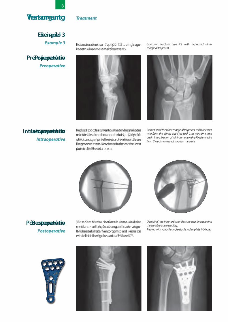
8
كرس العظام الناتج عن املد من نوع )C2( مع جزء زندي مصغر مضغوط
Extension fracture type C2 with depressed ulnar marginal fragment
العادي/ للوضع الزندي الهاميش الجزء إرجاع الخلفي الجانب من كريشنر سلك بواسطة الطبيعي من الجزء هذا تثبيت الوقت ذات ويف )« joy stick «(يف كريشنر سلك بواسطة اليد( )راحة الراحي الجانب
اللوحة.
Reduction of the ulnar marginal fragment with Kirschner wire from the dorsal side (“joy stick”), at the same time preliminary fixation of this fragment with a Kirschner wire from the palmar aspect through the plate.
ثبات من واإلستفادة املفصل داخل الكرس شق تجنب الساعد عظام بلوحة املعالجة التغيري. قابلة الزاوية
ثابتة الزاوية والقابلة للتغيري / ثقب 7/3.
“Avoiding“ the intra-articular fracture gap by exploiting the variable angle stability.Treated with variable angle-stable radius plate 7/3-hole.
مثال رقم 3Example3
Treatment العالج
Intraoperative أثناء العملية الجراحية
Postoperative بعد إجراء العملية الجراحية
Preoperative إجراء العملية الجراحية قبل
Behandling Treatment
PreoperativtPreoperative
PostoperativtPostoperative
IntraoperativtIntraoperative
Extensionsfraktur typ C2 med intryckt ulnart marginellt fragment
Extension fracture type C2 with depressed ulnar marginal fragment
Reposition av det ulnara marginella fragmen-tet med Kirschner tråd från den dorsala sidan („Joy stick“), samtidig preliminär fixering av det här fragmentet med Kirschner tråd från palmar riktning genom plattan.
Reduction of the ulnar marginal fragment with Kirschner wire from the dorsal side (“joy stick”), at the same time preliminary fixation of this fragment with a Kirschner wire from the palmar aspect through the plate.
Lämna den intraartikulära frakturspringan fri med nyttjande av den variabla vinkelsta-biliteten.Behandling med en variabel vinkelstabil radiusplatta / 7/3 hål.
“Avoiding“ the intra-articular fracture gap by exploiting the variable angle stability.Treated with variable angle-stable radius plate 7/3-hole.
Exempel 3Example3
Pré-opératoire Preoperative
Post-opératoire Postoperative
IntraopératoireIntraoperative
Fracture d’extension de type C2 de avec les fragments d’arêtes ulnaires introduits.
Extension fracture type C2 with depressed ulnar marginal fragment
Repositionnement du fragment d’arête ulnaire au moyen du fil de Kirschner, depuis la position dorsale («Joy Stick»), en même temps, la fixation préliminaire de ce frag-ment au moyen du fil de Kirschner depuis le côté palmaire, à travers la plaque.
Reduction of the ulnar marginal fragment with Kirschner wire from the dorsal side (“joy stick”), at the same time preliminary fixation of this fragment with a Kirschner wire from the palmar aspect through the plate.
«Évider» la fissure intra-articulaire de la fracture en utilisant la stabilité angulaire variable. Traitement au moyen d’une plaque radiale à angle stable variable, perforée en 7/3.
“Avoiding“ the intra-articular fracture gap by exploiting the variable angle stability.Treated with variable angle-stable radius plate 7/3-hole.
Exemple 3 Example3
Traitement Treatment Versorgung Treatment
PräoperativPreoperative
PostoperativPostoperative
IntraoperativIntraoperative
Extensionsfraktur Typ C2 mit eingesun- kenem ulnaren Kantenfragment
Extension fracture type C2 with depressed ulnar marginal fragment
Reposition des ulnaren Kantenfragmentes mit Kirschnerdraht von dorsal („Joy Stick“), gleichzeitige praeliminäre Fixation dieses Fragmentes mit Kirschnerdraht von palmar durch die Platte.
Reduction of the ulnar marginal fragment with Kirschner wire from the dorsal side (“joy stick”), at the same time preliminary fixation of this fragment with a Kirschner wire from the palmar aspect through the plate.
„Aussparen“ des intraartikulären Fraktur-spalts unter Ausnutzung der variablen Winkelstabilität. Versorgung mit variabel winkelstabiler Radiusplatte 7/3 Loch.
“Avoiding“ the intra-articular fracture gap by exploiting the variable angle stability.Treated with variable angle-stable radius plate 7/3-hole.
Beispiel 3Example3
Tratamento Treatment
Pré-operatórioPreoperative
Pós-operatório Postoperative
Intra-operatório Intraoperative
Fratura extensiva do tipo C2 com fraga- mento ulnar marginal depressivo
Extension fracture type C2 with depressed ulnar marginal fragment
Redução do fragmento ulnar marginal com arame Kirschner do lado dorsal (“Joy Sti-ck”), concomitante fixação preliminar desse fragmento com arame Kirschner do lado palmar através da placa.
Reduction of the ulnar marginal fragment with Kirschner wire from the dorsal side (“joy stick”), at the same time preliminary fixation of this fragment with a Kirschner wire from the palmar aspect through the plate.
“Evitar” a fenda da fratura intra-articular, mediante utilização da estabilidade angu-lar variável. Tratamento com placa radial de estabilidade angular variável / furo 7/3.
“Avoiding“ the intra-articular fracture gap by exploiting the variable angle stability.Treated with variable angle-stable radius plate 7/3-hole.
Exemplo 3 Example3
До операцииPreoperative
После операции Postoperative
Во время операции Intraoperative
Разгибательный перелом типа C2 с впав-шим локтевым краевым фрагментом
Extension fracture type C2 with depressed ulnar marginal fragment
Репозиция локтевого краевого фраг-мента проволкой Киршнера с тыльной стороны («Joy Stick»), одновременная предварительная фиксация этого фраг-мента проволкой Киршнера через пластину с ладонной стороны.
Reduction of the ulnar marginal fragment with Kirschner wire from the dorsal side (“joy stick”), at the same time preliminary fixation of this fragment with a Kirschner wire from the palmar aspect through the plate.
«Избегание» щелей внутрисуставного перелома за счет использования изме-няемой угловой стабильности пластины.Лечение при помощи лучевой пластины с изменяемой угловой стабильностью,
количество отверстий 7/3
“Avoiding“ the intra-articular fracture gap by exploiting the variable angle stability.Treated with variable angle-stable radius plate 7/3-hole.
Пример 3 Example3
Лечение Treatment Tratamiento Treatment
PreoperativaPreoperative
PostoperativaPostoperative
IntraoperativaIntraoperative
Fractura en extensión tipo C2 con fragmen-to marginal ulnar depresionado
Extension fracture type C2 with depressed ulnar marginal fragment
Reposición del fragmento marginal ulnar con alambre de Kirschner desde el lado dorsal (“joy stick”), al mismo tiempo fijación preliminar de este fragmento con alambre de Kirschner desde el lado palmar a través de la placa.
Reduction of the ulnar marginal fragment with Kirschner wire from the dorsal side (“joy stick”), at the same time preliminary fixation of this fragment with a Kirschner wire from the palmar aspect through the plate.
“Evitar” la fisura de la fractura intraarticular utilizando la estabilidad de ángulo variable.Tratamiento con placa radial de ángulo estable variable de 7/3 orificios.
“Avoiding“ the intra-articular fracture gap by exploiting the variable angle stability.Treated with variable angle-stable radius plate 7/3-hole.
Ejemplo 3 Example3
Trattamento Treatment
Sedepreparatorie
Preoperative
Sedepostoperatoria
Postoperative
Sedeintraoperatoria
Intraoperative
Frattura in estensione di tipo C2 con fram-mento marginale ulnare abbassato.
Extension fracture type C2 with depressed ulnar marginal fragment.
Riduzione del frammento marginale ulnare con filo di Kirschner dal lato dorsale („Joy Stick“), simultaneo fissaggio preliminare di questo frammento con filo di Kirschner dall‘aspetto palmare attraverso la piastra.
Reduction of the ulnar marginal fragment with Kirschner wire from the dorsal side (“joy stick”), at the same time preliminary fixation of this fragment with a Kirschner wire from the palmar aspect through the plate.
Evitare la fessura della frattura intra-articolare, sfruttando in tal senso la stabilità angolare variabile. Trattamento con piastra radiale ad angolo stabile variabile a 7/3 fori.
“Avoiding“ the intra-articular fracture gap by exploiting the variable angle stability.Treated with variable angle-stable radius plate 7/3-hole.
Esempio 3 Example3
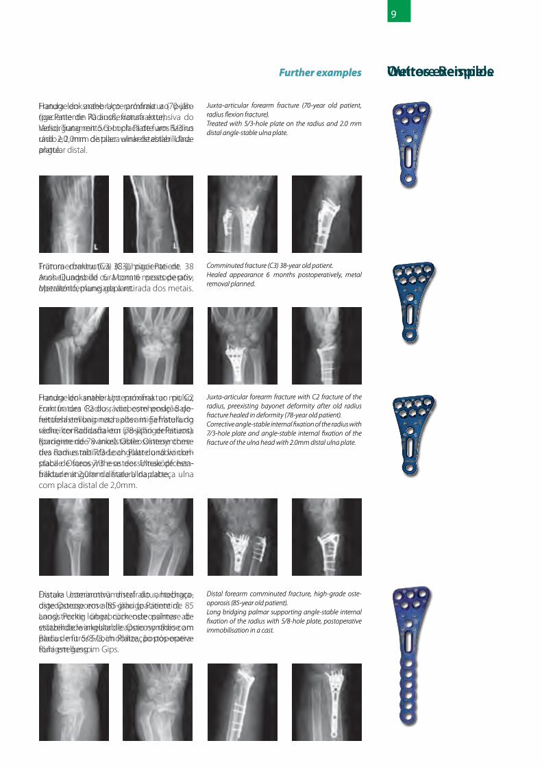
9
كرس يف الساعد بالقرب من املعصم )كرسعظام الساعد الناتجة عن اإلنثناء ملريض البالغ من العمر 70 عاما(.ولوحة ثقوب 5/3 ذات بلوحة الساعد عظام معالجة
زند ثابتة الزاوية.
Juxta-articular forearm fracture (70-year old patient, radius flexion fracture).Treated with 5/3-hole plate on the radius and 2.0 mm distal angle-stable ulna plate.
Furtherexamples أمثلة إضافية
كرس مفتت )C3( ملريض البالغ من العمر 38 عاما.الجراحية العملية إجراء بعد أشهر 6 الشفاء ظواهر
وإعتزام إزالة القطعة املعدنية.
Comminuted fracture (C3) 38-year old patient.Healed appearance 6 months postoperatively, metal removal planned.
عظام يف وكرس املعصم من بالقرب الساعد يف كرس بشكل )بايونت( الحربة إلتئام ، C2 نوع من الساعد الساعد من األقىص الطرف يف كرس بعد صحيح غري البالغ )مريض صحيح بشكل يلتئم لم بدوره والذي
من العمر 78 عاما(.الطرف لتصحيح ثقوب 7/3 ذات الزاوية ثابتة لوحة الزند كرس تثبيت ويتم الساعد عظام من األقىص
بواسطة لوحة للزند ذات 2.0 ملم ثابتة الزاوية.
Juxta-articular forearm fracture with C2 fracture of the radius, preexisting bayonet deformity after old radius fracture healed in deformity (78-year old patient).Corrective angle-stable internal fixation of the radius with 7/3-hole plate and angle-stable internal fixation of the fracture of the ulna head with 2.0mm distal ulna plate.
للعظام شديد وتخلخل الساعد عظام يف مفتت كرس مسافة إجتياز عاما(. 85 العمر من البالغة )مريضة بدورها والتي ثقوب 5/8 ذات لوحة بواسطة طويلة تسند من اليد للتمكن من تثبيت عظام الساعد. الراحة
بالجبس بعد إجراء العملية الجراحية.
Distal forearm comminuted fracture, high-grade oste-oporosis (85-year old patient).Long bridging palmar supporting angle-stable internal fixation of the radius with 5/8-hole plate, postoperative immobilisation in a cast.
Handledsnära underarmsfraktur (70-årig kvinnlig patient, radiusflexionsfraktur).Behandling med 5/3:-håls platta vid radius och 2,0 mm distal vinkelstabil ulnarplatta.
Juxta-articular forearm fracture (70-year old patient, radius flexion fracture).Treated with 5/3-hole plate on the radius and 2.0 mm distal angle-stable ulna plate.
Furtherexamples Ytterligare exempel
Pulveriseringsfraktur (C3) 38-årig patient.Läkningsbild 6 månader postoperativt, avlägsnande av metall planerad.
Comminuted fracture (C3) 38-year old patient.Healed appearance 6 months postoperatively, metal removal planned.
Handledsnåra underarmsfraktur med C2 fraktur av radius, i förväg existerande bajonettdeformation efter en gammal radiusfraktur som läkts i en felaktig position (78- årig patient).Korrigierande vinkelstabil osteosyntes av radius med 7/3-hål platta och vinkelstabil osteosyntes av en ulnahuvudfraktur med 2,0 mm:s distal ulnaplatta.
Juxta-articular forearm fracture with C2 fracture of the radius, preexisting bayonet deformity after old radius fracture healed in deformity (78-year old patient).Corrective angle-stable internal fixation of the radius with 7/3-hole plate and angle-stable internal fixation of the fracture of the ulna head with 2.0mm distal ulna plate.
Distal underarmspulveriseringsfraktur, höggradig osteoporos (85- årig kvinnlig patient).Lång överbryggande volart stödjande osteosyntes vid radius med 5/8-hål platta, postoperativ immobilisering i gips.
Distal forearm comminuted fracture, high-grade oste-oporosis (85-year old patient).Long bridging palmar supporting angle-stable internal fixation of the radius with 5/8-hole plate, postoperative immobilisation in a cast.
Fracture de l’avant-bras proche du poignet (patiente de 70 ans Fracture de flexion du radius). Traitement au moyen d’une plaque à trous 5/3 sur le radius, et plaque ulnaire distale à angle stable de 2,0mm.
Juxta-articular forearm fracture (70-year old patient, radius flexion fracture).Treated with 5/3-hole plate on the radius and 2.0 mm distal angle-stable ulna plate.
Furtherexamples Autres exemples
Fracture comminutive (C3) patients âgé de 38 ans. Photo de la guérison six mois après l’opération, retrait prévu du métal.
Comminuted fracture (C3) 38-year old patient.Healed appearance 6 months postoperatively, metal removal planned.
Fracture de l’avant-bras proche du poignet avec fracture C2 du radius, mauvaise posi-tion pré-existante suite à une fracture du radius guérie dans une mauvaise position (patient âgé de 78 ans). Ostéosynthèse correctrice à angle stable du radius avec plaque à un trous 7/3 et ostéosynthèse à angle de table de la fracture de la tête d’Ulna avec la plaque distale Ulna de 2,0mm.
Juxta-articular forearm fracture with C2 fracture of the radius, preexisting bayonet deformity after old radius fracture healed in deformity (78-year old patient).Corrective angle-stable internal fixation of the radius with 7/3-hole plate and angle-stable internal fixation of the fracture of the ulna head with 2.0mm distal ulna plate.
Fracture distale comminutive de l’avant-bras, ostéoporose très avancée (patiente âgée de 85 ans). Longue ostéosynthèse bridgeante palmaire à angle stable de support sur le radius ayant la plaque à trou 5/8, mis au calme postopératoire dans un plâtre.
Distal forearm comminuted fracture, high-grade oste-oporosis (85-year old patient).Long bridging palmar supporting angle-stable internal fixation of the radius with 5/8-hole plate, postoperative immobilisation in a cast.
Handgelenksnahe Unterarmfraktur (70-jäh-rige Patientin Radiusflexionsfraktur).Versorgung mit 5/3-Loch Platte am Radius und 2,0 mm distaler winkelstabiler Ulna-platte.
Juxta-articular forearm fracture (70-year old patient, radius flexion fracture).Treated with 5/3-hole plate on the radius and 2.0 mm distal angle-stable ulna plate.
Furtherexamples Weitere Beispiele
Trümmerfraktur (C3) 38-jähriger Patient.Ausheilungsbild 6 Monate postoperativ, Metallentfernung geplant.
Comminuted fracture (C3) 38-year old patient.Healed appearance 6 months postoperatively, metal removal planned.
Handgelenksnahe Unterarmfraktur mit C2 Fraktur des Radius, vorbestehende Bajo-nettfehlstellung nach alter in Fehlstellung verheilter Radiusfraktur (78-jähriger Patient). Korrigierende winkelstabile Osteoynthese des Radius mit 7/3-Loch Platte und winkel-stabile Osteosynthese der Ulnaköpfchen-fraktur mit 2,0mm distale Ulnaplatte.
Juxta-articular forearm fracture with C2 fracture of the radius, preexisting bayonet deformity after old radius fracture healed in deformity (78-year old patient).Corrective angle-stable internal fixation of the radius with 7/3-hole plate and angle-stable internal fixation of the fracture of the ulna head with 2.0mm distal ulna plate.
Distale Unterarmtrümmerfraktur, hochgra-dige Osteoporose (85-jährige Patientin).Langstreckig überbrückende palmar ab-stützende winkelstabile Osteosynthese am Radius mit 5/8-Loch Platte, postoperative Ruhigstellung im Gips.
Distal forearm comminuted fracture, high-grade oste-oporosis (85-year old patient).Long bridging palmar supporting angle-stable internal fixation of the radius with 5/8-hole plate, postoperative immobilisation in a cast.
Fratura do antebraço próxima ao pulso (paciente de 70 anos, fratura extensiva do rádio). Tratamento com placa de furos 5/3 no rádio e 2,0mm de placa ulnar de estabilidade angular distal.
Juxta-articular forearm fracture (70-year old patient, radius flexion fracture).Treated with 5/3-hole plate on the radius and 2.0 mm distal angle-stable ulna plate.
Furtherexamples Outros exemplos
Fratura cominutiva (C3), paciente de 38 anos. Quadro de cura com 6 meses de pós-operatório, planejada a retirada dos metais.
Comminuted fracture (C3) 38-year old patient.Healed appearance 6 months postoperatively, metal removal planned.
Fratura do antebraço próxima ao pulso, com fratura C2 do rádio, com posição de-feituosa em baioneta após antiga fratura do rádio, consolidada em posição defeituosa (paciente de 78 anos). Osteossíntese corre-tiva com estabilidade angular do rádio com placa de furos 7/3 e osteossíntese de esta-bilidade angular da fratura da cabeça ulna com placa distal de 2,0mm.
Juxta-articular forearm fracture with C2 fracture of the radius, preexisting bayonet deformity after old radius fracture healed in deformity (78-year old patient).Corrective angle-stable internal fixation of the radius with 7/3-hole plate and angle-stable internal fixation of the fracture of the ulna head with 2.0mm distal ulna plate.
Fratura cominutiva distal do antebraço, osteoporose em alto grau (paciente de 85 anos). Ponte longa, com osteossíntese de estabilidade angular de apoio no rádio com placa de furos 5/8, imobilização pós-opera-tória em gesso.
Distal forearm comminuted fracture, high-grade oste-oporosis (85-year old patient).Long bridging palmar supporting angle-stable internal fixation of the radius with 5/8-hole plate, postoperative immobilisation in a cast.
Перелом предплечья в области запястья (пациент в возрасте 70 лет, флексионный перелом лучевой кости).Лечение при помощи пластины с коли-чеством отверстий 5/3, накладываемой на лучевую кость, и 2,0 мм дистальной
локтевой пластиной с угловой стабильно-стью.
Juxta-articular forearm fracture (70-year old patient, radius flexion fracture).Treated with 5/3-hole plate on the radius and 2.0 mm distal angle-stable ulna plate.
Furtherexamples Дополнительные примеры
Оскольчатый перелом (C3) у 38-летнего пациента. Фотография излеченного пере-лома через 6 месяцев после операции, запланировано удаление металла.
Comminuted fracture (C3) 38-year old patient.Healed appearance 6 months postoperatively, metal removal planned.
Перелом предплечья в области запястья с переломом C2 лучевой кости, имею-щаяся штыковидная деформация пред-плечья после старого перелома лучевой кости, сросшейся неправильно (78-летний пациент). Корректирующий остеосинтез лучевой кости с угловой стабильностью при помощи пластины с количеством отверстий 7/3 и остеосинтез перелома
головки локтевой кости с угловой стабильностью при помощи 2,0 мм дистальной локтевой пластины.
Juxta-articular forearm fracture with C2 fracture of the radius, preexisting bayonet deformity after old radius fracture healed in deformity (78-year old patient).Corrective angle-stable internal fixation of the radius with 7/3-hole plate and angle-stable internal fixation of the fracture of the ulna head with 2.0mm distal ulna plate.
Осколочный перелом в дистальной части костей предплечья, сильный остеопороз (85-летняя пациентка). Перекрывающая большой участок, поддерживающая с ладон-ной стороны внутренняя фиксация лучевой кости с угловой стабильностью при помощи
пластины с количеством отверстий 5/8, по-слеоперационная иммобилизация в гипсе.
Distal forearm comminuted fracture, high-grade oste-oporosis (85-year old patient). Long bridging palmar sup-porting angle-stable internal fixation of the radius with 5/8-hole plate, postoperative immobilisation in a cast.
Fractura yuxtaarticular del antebrazo (paciente de 70 años, fractura distal de flexión del radio). Tratamiento con placa de 5/3 orificios en el radio y placa de cúbito distal de ángulo estable de 2,0 mm.
Juxta-articular forearm fracture (70-year old patient, radius flexion fracture).Treated with 5/3-hole plate on the radius and 2.0 mm distal angle-stable ulna plate.
Furtherexamples Más ejemplos
Frauctura astillada (C3) paciente de 38 años. Cuadro de curación 6 meses postoperati-vos, se planea el desmontaje del metal.
Comminuted fracture (C3) 38-year old patient.Healed appearance 6 months postoperatively, metal removal planned.
Fractura yuxtaarticular del antebrazo con fractura C2 del radio, posición viciosa en ba-yoneta pre-existente después de una fractu-ra vieja del radio, curada en posición viciosa (paciente de 78 años). Osteosíntesis correcti-va de ángulo estable del radio con placa de 7/3 orificios y osteosíntesis de ángulo estable de una fractura de la cabeza ósea del cúbito con una placa ulnar distal de 2 mm.
Juxta-articular forearm fracture with C2 fracture of the radius, preexisting bayonet deformity after old radius fracture healed in deformity (78-year old patient).Corrective angle-stable internal fixation of the radius with 7/3-hole plate and angle-stable internal fixation of the fracture of the ulna head with 2.0mm distal ulna plate.
Frauctura distal del antebrazo, osteoporosis de alto grado (paciente de 85 años).Osteosíntesis de ángulo estable con franqueamiento extendido y soporte pal-mar del radio utilizando una placa de 5/8 ori-ficios, inmovilización postoperativa en yeso.
Distal forearm comminuted fracture, high-grade oste-oporosis (85-year old patient).Long bridging palmar supporting angle-stable internal fixation of the radius with 5/8-hole plate, postoperative immobilisation in a cast.
Frattura dell‘avambraccio in prossimità del polso (paziente settantenne, frattura di flessione del radio).Trattamento con piastra a 5/3 fori e piastra ulnare distale ad angolo stabile da 2,0 mm.
Juxta-articular forearm fracture (70-year old patient, radius flexion fracture).Treated with 5/3-hole plate on the radius and 2.0 mm distal angle-stable ulna plate.
Frattura comminuta (C3), paziente di 38 anni. Quadro di guarigione: 6 mesi posto-peratorie, pianificazione della rimozione del
metallo.Comminuted fracture (C3) 38-year old patient.Healed appearance 6 months postoperatively, metal
Frattura dell‘avambraccio in prossimità del polso con frattura C2 del radio, deformità a baionetta preesistente in seguito a una vecchia frattura radiale guarita in posizione errata (paziente di 78 anni).Osteosintesi correttive ad angolo stabile del radio con piastra a 7/3 fori e osteosintesi ad angolo stabile della frattura della testa ulnare con piastra ulnare distale da 2,0 mm.
Juxta-articular forearm fracture with C2 fracture of the radius, preexisting bayonet deformity after old radius fracture healed in deformity (78-year old patient).Corrective angle-stable internal fixation of the radius with 7/3-hole plate and angle-stable internal fixation of the fracture of the ulna head with 2.0mm distal ulna plate.
Frattura distale dell‘avambraccio, elevata osteoporosi (paziente di 85 anni).Osteosintesi lunga di supporto ad angolo stabile palmare sul radio con piastra a 5/8 fori, immobilizzazione postoperatoria con gesso.
Distal forearm comminuted fracture, high-grade oste-oporosis (85-year old patient).Long bridging palmar supporting angle-stable internal fixation of the radius with 5/8-hole plate, postoperative immobilisation in a cast.
Furtherexamples Ulteriori esempi

10
لوحة ذات 5/3 ثقوب5/3-hole-plate (5.9823.53)
الرأس )5(
23ملم
محور العظم )3(
لوحة ذات 7/3 ثقوب7/3-hole-plate (5.9843.73)
لوحة عظام الساعد الرفيعة ثابتة الزاويةVariable angle-stable distal radius plate, small
رقم الطلب الطول عدد الثقوب عدد الوحدات يف املجموعةCode No titanium Length No of hole Quantity in set
- right 43 5/2 / اليمني 5.9823.52right 51 5/3 1 / اليمني 5.9823.53right 60 5/4 1 / اليمني 5.9823.54- right 77 5/6 / اليمني 5.9823.56- right 94 5/8 / اليمني 5.9823.58- right 111 5/10 / اليمني 5.9823.510- right 136 5/13 / اليمني 5.9823.513- left 43 5/2 / اليسار 5.9833.52left 51 5/3 1 / اليسار 5.9833.53left 60 5/4 1 / اليسار 5.9833.54- left 77 5/6 / اليسار 5.9833.56- left 94 5/8 / اليسار 5.9833.58- left 111 5/10 / اليسار 5.9833.510- left 136 5/13 / اليسار 5.9833.513
الرباغي القرشية، قطر 2.7 ملم، ثابتة الزاوية، قابلة للقطعCortical screws, diameter 2.7 mm, angle-stable, self-tapping
رقم الطلب الطول عدد الوحدات يف املجموعةCode No titanium Length Quantity in set
3.125.12 12 53.125.14 14 53.125.16 16 53.125.18 18 53.125.20 20 53.125.22 22 53.125.24 24 53.125.26 26 53.125.28 28 5
لوحة عظام الساعد العريضة ثابتة الزاويةVariable angle-stable distal radius plate, wide
رقم الطلب الطول عدد الثقوب عدد الوحدات يف املجموعةCode No titanium Length No of hole Quantity in set
- right 43 7/2 / اليمني 5.9843.72right 51 7/3 1 / اليمني 5.9843.73right 60 7/4 1 / اليمني 5.9843.74- right 77 7/6 / اليمني 5.9843.76- right 94 7/8 / اليمني 5.9843.78- right 111 7/10 / اليمني 5.9843.710- right 136 7/13 / اليمني 5.9843.713- left 43 7/2 / اليسار 5.9853.72left 51 7/3 1 / اليسار 5.9853.73left 60 7/4 1 / اليسار 5.9853.74- left 77 7/6 / اليسار 5.9853.76- left 94 7/8 / اليسار 5.9853.78- left 111 7/10 / اليسار 5.9853.710- left 136 7/13 / اليسار 5.9853.713
Optionally only angle-stable distal radius plates are available in narrow and wider version as well.
additionallyOrder طلب إضافي
الرأس )7(
محور العظم )3(
ملمملمملمملمملمملمملمملم ملم ملمملم ملمملم ملم
27ملم
ملمملمملمملمملمملمملمملم ملم ملمملم ملمملم ملم
ملمملمملمملمملمملمملمملم ملم
لوحات عظام الساعد )لوحات للطرف األقىص من عظام الساعد( الفردية ثابتة الزاوية، رفيعة وعريضة الشكلمتوفرة ايضا إختياريا للطلب
5/3-hål platta5/3-hole-plate (5.9823.53)
Huvud (5)Head
23mm
Skaft (3)Shaft
7/3-hål platta7/3-hole-plate (5.9843.73)
Huvud (7)Head
Skaft (3)Shaft
27mm
Efterbeställning Orderadditionally
Variabel vinkelstabil distal radiusplatta smalVariable angle-stable distal radius plate, small
Beställn.-nr. Titan Längd Antal hål Antal i ett setCode No titanium Length No of hole Quantity in set
5.9823.52 höger / right 43 mm 5/2 -5.9823.53 höger / right 51 mm 5/3 15.9823.54 höger / right 60 mm 5/4 15.9823.56 höger / right 77 mm 5/6 -5.9823.58 höger / right 94 mm 5/8 -5.9823.510 höger / right 111 mm 5/10 -5.9823.513 höger / right 136 mm 5/13 -5.9833.52 vänster / left 43 mm 5/2 -5.9833.53 vänster / left 51 mm 5/3 15.9833.54 vänster / left 60 mm 5/4 15.9833.56 vänster / left 77 mm 5/6 -5.9833.58 vänster / left 94 mm 5/8 -5.9833.510 vänster / left 111 mm 5/10 -5.9833.513 vänster / left 136 mm 5/13 -
Cortikala skruvar, D 2,7 mm, vinkelstabil, automatisk gängaCortical screws, diameter 2.7 mm, angle-stable, self-tapping
Beställn.-nr. Titan Längd Antal i ett setCode No titanium Length Quantity in set
3.125.12 12 mm 53.125.14 14 mm 53.125.16 16 mm 53.125.18 18 mm 53.125.20 20 mm 53.125.22 22 mm 53.125.24 24 mm 53.125.26 26 mm 53.125.28 28 mm 5
Variabel vinkelstabil distal radiusplatta bredVariable angle-stable distal radius plate, wide
Beställn.-nr. Titan Längd Antal hål Antal i ett setCode No titanium Length No of hole Quantity in set
5.9843.72 höger / right 43 mm 7/2 -5.9843.73 höger / right 51 mm 7/3 15.9843.74 höger / right 60 mm 7/4 15.9843.76 höger / right 77 mm 7/6 -5.9843.78 höger / right 94 mm 7/8 -5.9843.710 höger / right 111 mm 7/10 -5.9843.713 höger / right 136 mm 7/13 -5.9853.72 vänster / left 43 mm 7/2 -5.9853.73 vänster / left 51 mm 7/3 15.9853.74 vänster / left 60 mm 7/4 15.9853.76 vänster / left 77 mm 7/6 -5.9853.78 vänster / left 94 mm 7/8 -5.9853.710 vänster / left 111 mm 7/10 -5.9853.713 vänster / left 136 mm 7/13 -
Som tillval står även enkelt vinkelstabila distala radiusplattor i smalt och brett utförande till förfogande.Optionally only angle-stable distal radius plates are available in narrow and wider version as well.
Commande Orderadditionally
Plaque à trous 5/35/3-hole-plate (5.9823.53)
Tête (5)
23mm
Tige (3)
Plaque à trous 7/37/3-hole-plate (5.9843.73)
Tête (7)
Tige (3)
27mm
Plaque étroite radiale distale à angle stable variableVariable angle-stable distal radius plate, small
N° de commande Longueur Nombre de trous Quantité contenue dans le setCode No titanium Length No of hole Quantity in set
5.9823.52 droite / right 43 mm 5/2 -5.9823.53 droite / right 51 mm 5/3 15.9823.54 droite / right 60 mm 5/4 15.9823.56 droite / right 77 mm 5/6 -5.9823.58 droite / right 94 mm 5/8 -5.9823.510 droite / right 111 mm 5/10 -5.9823.513 droite / right 136 mm 5/13 -5.9833.52 gauche / left 43 mm 5/2 -5.9833.53 gauche / left 51 mm 5/3 15.9833.54 gauche / left 60 mm 5/4 15.9833.56 gauche / left 77 mm 5/6 -5.9833.58 gauche / left 94 mm 5/8 -5.9833.510 gauche / left 111 mm 5/10 -5.9833.513 gauche / left 136 mm 5/13 -
Vis corticales, D 2,7 mm, à angle stable (autocoupantes)Cortical screws, diameter 2.7 mm, angle-stable, self-tapping
N° de commande Longueur Quantité contenue dans le setCode No titanium Length Quantity in set
3.125.12 12 mm 53.125.14 14 mm 53.125.16 16 mm 53.125.18 18 mm 53.125.20 20 mm 53.125.22 22 mm 53.125.24 24 mm 53.125.26 26 mm 53.125.28 28 mm 5
Plaque large radiale distale à angle stable variableVariable angle-stable distal radius plate, wide
N° de commande Longueur Nombre de trous Quantité contenue dans le setCode No titanium Length No of hole Quantity in set
5.9843.72 droite / right 43 mm 7/2 -5.9843.73 droite / right 51 mm 7/3 15.9843.74 droite / right 60 mm 7/4 15.9843.76 droite / right 77 mm 7/6 -5.9843.78 droite / right 94 mm 7/8 -5.9843.710 droite / right 111 mm 7/10 -5.9843.713 droite / right 136 mm 7/13 -5.9853.72 gauche / left 43 mm 7/2 -5.9853.73 gauche / left 51 mm 7/3 15.9853.74 gauche / left 60 mm 7/4 15.9853.76 gauche / left 77 mm 7/6 -5.9853.78 gauche / left 94 mm 7/8 -5.9853.710 gauche / left 111 mm 7/10 -5.9853.713 gauche / left 136 mm 7/13 -
Des plaques radiales distales à angle stable simples sont également disponibles en option en modèle étroit et large.Optionally only angle-stable distal radius plates are available in narrow and wider version as well.
Variabel winkelstabile distale Radiusplatte, schmalVariable angle-stable distal radius plate, small
Bestell-Nr. Länge Lochanzahl Anzahl im SetCode No titanium Length No of hole Quantity in set
5.9823.52 rechts / right 43 mm 5/2 -5.9823.53 rechts / right 51 mm 5/3 15.9823.54 rechts / right 60 mm 5/4 15.9823.56 rechts / right 77 mm 5/6 -5.9823.58 rechts / right 94 mm 5/8 -5.9823.510 rechts / right 111 mm 5/10 -5.9823.513 rechts / right 136 mm 5/13 -5.9833.52 links / left 43 mm 5/2 -5.9833.53 links / left 51 mm 5/3 15.9833.54 links / left 60 mm 5/4 15.9833.56 links / left 77 mm 5/6 -5.9833.58 links / left 94 mm 5/8 -5.9833.510 links / left 111 mm 5/10 -5.9833.513 links / left 136 mm 5/13 -
5/3-Loch-Platte5/3-hole-plate (5.9823.53)
Kopf (5)Head
23mm
Schaft (3)Shaft
7/3-Loch-Platte7/3-hole-plate (5.9843.73)
Kopf (7)Head
Schaft (3)Shaft
27mm
Kortikalisschrauben, D 2,7 mm winkelstabil selbstschneidendCortical screws, diameter 2.7 mm, angle-stable self-tapping
Bestell-Nr. Titan Länge Anzahl im SetCode No titanium Length Quantity in set
3.125.12 12 mm 53.125.14 14 mm 53.125.16 16 mm 53.125.18 18 mm 53.125.20 20 mm 53.125.22 22 mm 53.125.24 24 mm 53.125.26 26 mm 53.125.28 28 mm 5
Nachbestellung Orderadditionally
Variabel winkelstabile distale Radiusplatte, breitVariable angle-stable distal radius plate, wide
Bestell-Nr. Länge Lochanzahl Anzahl im SetCode No titanium Length No of hole Quantity in set
5.9843.72 rechts / right 43 mm 7/2 -5.9843.73 rechts / right 51 mm 7/3 15.9843.74 rechts / right 60 mm 7/4 15.9843.76 rechts / right 77 mm 7/6 -5.9843.78 rechts / right 94 mm 7/8 -5.9843.710 rechts / right 111 mm 7/10 -5.9843.713 rechts / right 136 mm 7/13 -5.9853.72 links / left 43 mm 7/2 -5.9853.73 links / left 51 mm 7/3 15.9853.74 links / left 60 mm 7/4 15.9853.76 links / left 77 mm 7/6 -5.9853.78 links / left 94 mm 7/8 -5.9853.710 links / left 111 mm 7/10 -5.9853.713 links / left 136 mm 7/13 -
Optional stehen auch einfach winkelstabile distale Radiusplatten in schmaler und breiterAusführung zur Verfügung.Optionally only angle-stable distal radius plates are available in narrow and wider version as well.
Encomenda Orderadditionally
suplementar
Placa de furos 5/35/3-hole-plate (5.9823.53)
Cabeça (5)
23mm
Corpo (3)
Placa de furos 7/37/3-hole-plate (5.9843.73)
Cabeça (7)
Corpo (3)
27mm
Placa radial distal com estabilidade angular variável, estreitaVariable angle-stable distal radius plate, small
N° de encomenda, titânio Comprimento Número de furos Quantidade no KitCode No titanium Length No of hole Quantity in set
5.9823.52 direito / right 43 mm 5/2 -5.9823.53 direito / right 51 mm 5/3 15.9823.54 direito / right 60 mm 5/4 15.9823.56 direito / right 77 mm 5/6 -5.9823.58 direito / right 94 mm 5/8 -5.9823.510 direito / right 111 mm 5/10 -5.9823.513 direito / right 136 mm 5/13 -5.9833.52 esquerdo / left 43 mm 5/2 -5.9833.53 esquerdo / left 51 mm 5/3 15.9833.54 esquerdo / left 60 mm 5/4 15.9833.56 esquerdo / left 77 mm 5/6 -5.9833.58 esquerdo / left 94 mm 5/8 -5.9833.510 esquerdo / left 111 mm 5/10 -5.9833.513 esquerdo / left 136 mm 5/13 -
Parafusos corticais, D 2,7 mm, estabilidade angular (auto-cortantes)Cortical screws, diameter 2.7 mm, angle-stable, self-tapping
N° de encomenda, titânio Comprimento Quantidade no KitCode No titanium Length Quantity in set
3.125.12 12 mm 53.125.14 14 mm 53.125.16 16 mm 53.125.18 18 mm 53.125.20 20 mm 53.125.22 22 mm 53.125.24 24 mm 53.125.26 26 mm 53.125.28 28 mm 5
Placa radial distal com estabilidade angular variável, largaVariable angle-stable distal radius plate, wide
N° de encomenda, titânio Comprimento Número de furos Quantidade no KitCode No titanium Length No of hole Quantity in set
5.9843.72 direito / right 43 mm 7/2 -5.9843.73 direito / right 51 mm 7/3 15.9843.74 direito / right 60 mm 7/4 15.9843.76 direito / right 77 mm 7/6 -5.9843.78 direito / right 94 mm 7/8 -5.9843.710 direito / right 111 mm 7/10 -5.9843.713 direito / right 136 mm 7/13 -5.9853.72 esquerdo / left 43 mm 7/2 -5.9853.73 esquerdo / left 51 mm 7/3 15.9853.74 esquerdo / left 60 mm 7/4 15.9853.76 esquerdo / left 77 mm 7/6 -5.9853.78 esquerdo / left 94 mm 7/8 -5.9853.710 esquerdo / left 111 mm 7/10 -5.9853.713 esquerdo / left 136 mm 7/13 -
Opcionalmente encontram-se à disposição também placas radiais distais de estabilidade angular em modelo estreito ou largo.Optionally only angle-stable distal radius plates are available in narrow and wider version as well.
Дополнительный Orderadditionally
заказ
Пластина с количествомотверстий 5/3 5/3-hole-plate (5.9823.53)
Головка (5)
23mm
Тело (3)
Пластина с количествомотверстий 7/3 7/3-hole-plate (5.9843.73)
Головка (7)
Тело (3)
27mm
Изменяемая, с угловой стабильностью Дистальная лучевая пластина, узкаяVariable angle-stable distal radius plate, small
№ для заказа титан Длина Кол-во отверстий Кол-во в набореCode No titanium Length No of hole Quantity in set
5.9823.52 правая / right 43 mm 5/2 -5.9823.53 правая / right 51 mm 5/3 15.9823.54 правая / right 60 mm 5/4 15.9823.56 правая / right 77 mm 5/6 -5.9823.58 правая / right 94 mm 5/8 -5.9823.510 правая / right 111 mm 5/10 -5.9823.513 правая / right 136 mm 5/13 -5.9833.52 левая / left 43 mm 5/2 -5.9833.53 левая / left 51 mm 5/3 15.9833.54 левая / left 60 mm 5/4 15.9833.56 левая / left 77 mm 5/6 -5.9833.58 левая / left 94 mm 5/8 -5.9833.510 левая / left 111 mm 5/10 -5.9833.513 левая / left 136 mm 5/13 -
Кортикальные винты, диаметр 2,7 мм, угловой стабильностью (саморез)Cortical screws, diameter 2.7 mm, angle-stable, self-tapping
№ для заказа титан Длина Кол-во в набореCode No titanium Length Quantity in set
3.125.12 12 mm 53.125.14 14 mm 53.125.16 16 mm 53.125.18 18 mm 53.125.20 20 mm 53.125.22 22 mm 53.125.24 24 mm 53.125.26 26 mm 53.125.28 28 mm 5
Изменяемая, с угловой стабильностью Дистальная лучевая пластина, широкаяVariable angle-stable distal radius plate, wide
№ для заказа титан Длина Кол-во отверстий Кол-во в набореCode No titanium Length No of hole Quantity in set
5.9843.72 правая / right 43 mm 7/2 -5.9843.73 правая / right 51 mm 7/3 15.9843.74 правая / right 60 mm 7/4 15.9843.76 правая / right 77 mm 7/6 -5.9843.78 правая / right 94 mm 7/8 -5.9843.710 правая / right 111 mm 7/10 -5.9843.713 правая / right 136 mm 7/13 -5.9853.72 левая / left 43 mm 7/2 -5.9853.73 левая / left 51 mm 7/3 15.9853.74 левая / left 60 mm 7/4 15.9853.76 левая / left 77 mm 7/6 -5.9853.78 левая / left 94 mm 7/8 -5.9853.710 левая / left 111 mm 7/10 -5.9853.713 левая / left 136 mm 7/13 -
В качестве опции в распоряжении имеются также простые дистальные лучевыепластины с угловой стабильностью в узком и широком исполнении.Optionally only angle-stable distal radius plates are available in narrow and wider version as well.
Pedido Orderadditionally
adicional
Placa de 5/3 orificios5/3-hole-plate (5.9823.53)
Cabeza (5)
23mm
Vástago (3)
Placa de 7/3 orificios7/3-hole-plate (5.9843.73)
Cabeza (7)
Vástago (3)
27mm
Placa radial distal variable angosta de ángulo estableVariable angle-stable distal radius plate, small
N° de pedido titanio Longitud Número de orificios Cantidad en el juegoCode No titanium Length No of hole Quantity in set
5.9823.52 derecha / right 43 mm 5/2 -5.9823.53 derecha / right 51 mm 5/3 15.9823.54 derecha / right 60 mm 5/4 15.9823.56 derecha / right 77 mm 5/6 -5.9823.58 derecha / right 94 mm 5/8 -5.9823.510 derecha / right 111 mm 5/10 -5.9823.513 derecha / right 136 mm 5/13 -5.9833.52 izquierda / left 43 mm 5/2 -5.9833.53 izquierda / left 51 mm 5/3 15.9833.54 izquierda / left 60 mm 5/4 15.9833.56 izquierda / left 77 mm 5/6 -5.9833.58 izquierda / left 94 mm 5/8 -5.9833.510 izquierda / left 111 mm 5/10 -5.9833.513 izquierda / left 136 mm 5/13 -
Tornillos para la corteza cortical, diámetro 2,7 mm, de ángulo estable (autoroscantes)Cortical screws, diameter 2.7 mm, angle-stable, self-tapping
N° de pedido titanio Longitud Cantidad en el juegoCode No titanium Length Quantity in set
3.125.12 12 mm 53.125.14 14 mm 53.125.16 16 mm 53.125.18 18 mm 53.125.20 20 mm 53.125.22 22 mm 53.125.24 24 mm 53.125.26 26 mm 53.125.28 28 mm 5
Placa radial distal variable ancha de ángulo estableVariable angle-stable distal radius plate, wide
N° de pedido titanio Longitud Número de orificios Cantidad en el juegoCode No titanium Length No of hole Quantity in set
5.9843.72 derecha / right 43 mm 7/2 -5.9843.73 derecha / right 51 mm 7/3 15.9843.74 derecha / right 60 mm 7/4 15.9843.76 derecha / right 77 mm 7/6 -5.9843.78 derecha / right 94 mm 7/8 -5.9843.710 derecha / right 111 mm 7/10 -5.9843.713 derecha / right 136 mm 7/13 -5.9853.72 izquierda / left 43 mm 7/2 -5.9853.73 izquierda / left 51 mm 7/3 15.9853.74 izquierda / left 60 mm 7/4 15.9853.76 izquierda / left 77 mm 7/6 -5.9853.78 izquierda / left 94 mm 7/8 -5.9853.710 izquierda / left 111 mm 7/10 -5.9853.713 izquierda / left 136 mm 7/13 -
Opcionalmente se dispone también de placas radiales distales de ángulo estable simple, en las versiones angosta y ancha.Optionally only angle-stable distal radius plates are available in narrow and wider version as well.
Ordine Orderadditionally
successivo
Piastra a 5/3 fori5/3-hole-plate (5.9823.53)
Testa (5)Head
23mm
Gambo (3)Shaft
Piastra a 7/3 fori 7/3-hole-plate (5.9843.73)
Testa (7)Head
Gambo (3)Shaft
27mm
Piastra radiale distale ad angolo stabile variabile, sottileVariable angle-stable distal radius plate, small
Numero dell‘ordine Titanio Lunghezza Numero di fori Quantità inclusa nel setCode No titanium Length No of hole Quantity in set
5.9823.52 destra / right 43 mm 5/2 -5.9823.53 destra / right 51 mm 5/3 15.9823.54 destra / right 60 mm 5/4 15.9823.56 destra / right 77 mm 5/6 -5.9823.58 destra / right 94 mm 5/8 -5.9823.510 destra / right 111 mm 5/10 -5.9823.513 destra / right 136 mm 5/13 -5.9833.52 sinistra / left 43 mm 5/2 -5.9833.53 sinistra / left 51 mm 5/3 15.9833.54 sinistra / left 60 mm 5/4 15.9833.56 sinistra / left 77 mm 5/6 -5.9833.58 sinistra / left 94 mm 5/8 -5.9833.510 sinistra / left 111 mm 5/10 -5.9833.513 sinistra / left 136 mm 5/13 -
Viti corticali, D 2,7 mm, angolo stabile, automaschiantiCortical screws, diameter 2.7 mm, angle-stable, self-tapping
Numero dell‘ordine Titanio Lunghezza Quantità inclusa nel setCode No titanium Length Quantity in set
3.125.12 12 mm 53.125.14 14 mm 53.125.16 16 mm 53.125.18 18 mm 53.125.20 20 mm 53.125.22 22 mm 53.125.24 24 mm 53.125.26 26 mm 53.125.28 28 mm 5
Piastra radiale distale ad angolo stabile variabile, largaVariable angle-stable distal radius plate, wide
Numero dell‘ordine Titanio Lunghezza Numero di fori Quantità inclusa nel setCode No titanium Length No of hole Quantity in set
5.9843.72 destra / right 43 mm 7/2 -5.9843.73 destra / right 51 mm 7/3 15.9843.74 destra / right 60 mm 7/4 15.9843.76 destra / right 77 mm 7/6 -5.9843.78 destra / right 94 mm 7/8 -5.9843.710 destra / right 111 mm 7/10 -5.9843.713 destra / right 136 mm 7/13 -5.9853.72 sinistra / left 43 mm 7/2 -5.9853.73 sinistra / left 51 mm 7/3 15.9853.74 sinistra / left 60 mm 7/4 15.9853.76 sinistra / left 77 mm 7/6 -5.9853.78 sinistra / left 94 mm 7/8 -5.9853.710 sinistra / left 111 mm 7/10 -5.9853.713 sinistra / left 136 mm 7/13 -
Come opzione sono disponibili anche semplici piastre radiali distali ad angolo stabile nel modello sottile e nel modello largo.Optionally only angle-stable distal radius plates are available in narrow and wider version as well.

11
19.115.00 1Locating sieve (for instruments and implants)
األدوات يف املجموعة، تيتانيوم رقم الطلب عدد الوحدات يف املجموعةInstuments in set, titanium Code No Quantity in set
1 2.954.01 ملقط لشد الرباغيScrew-holding forceps
مفك براغي سدايس األضالع 2.940.25SA 1Hexagonal screw-driver, with handle, for screws diameter 2.7 mm
أداة التثقيب 2.977.11 2Drill guide for angle-stable systems, diameter 2.7 mm, length 45 mm
2.977.10 1Drill guide for variable angle-stable systems, diameter 2.7 mm, length 45 mm
داة التثقيب 2.904.03 1Twist drill (for quick coupler) diameter 2.0 mm, length 105 mm
الرباغي القرشية، قطر 2.7 ملم، قابلة للقطعCortical screws, diameter 2.7 mm, self-tapping
رقم الطلب الطول عدد الوحدات يف املجموعةCode No titanium Length Quantity in set
3.120.12 12 53.120.14 14 53.120.16 16 53.120.18 18 53.120.20 20 5
OP-Set,titanium
رقم الطلب 19.1163.00CodeNo
مجموعة من التيتانيوم للعملية الجراحية
ملمملمملمملمملم
مصفاة )لألدوات والقطع للزرع(
لرباغي قطرها 2،7ملم
للوحة ثابتة الزاوية، قطر2.7 ملم، الطول 45 ملم
للوحة ثابتة الزاوية قابلة للتغيري، قطر2.7 ملم، الطول 45 ملم
أداة التثقيب )لتثقيب رسيع( قطرها 2،0 ملم، الطول 105 ملم
6.031.16 5قطره 1،6 ملم، طوله 150 ملم Kirschner wire with trocar point and round end, diameter 1.6 mm, length 150 mm
سلك كريشنر بقضيب معدني مدور يستعمل للحفر،
أداة لقياس الرباغي Screw gauge, for screws diameter 2.7 mmقطرها 2،7 ملم
2.953.65 1
Operationsset titanOP-Set,titanium
Beställn.-nr. 19.1163.00CodeNo
Skruvhållarpincett 2.954.01 1Screw-holding forceps
Kirschner tråd trocar, rund ända, D 1,6 mm, längd 150 mm 6.031.16 5Kirschner wire with trocar point and round end, diameter 1.6 mm, length 150 mm
Mätinstrument för skruvar 2.953.65 1för skruvar, D 2,7 mmScrew gauge, for screws diameter 2.7 mm
Instrument i ett set Beställn.-nr. Antal i ett setInstuments in set, titanium Code No Quantity in set
Sexkants skruvmejsel, mit Griff,för skruvar D 2,7 mm 2.940.25SA 1Hexagonal screw-driver, with handle, for screws diameter 2.7 mm
Förvaringssil (för instrument och implantat) 19.115.00 1Locating sieve (for instruments and implants)
Borrhylsa
för vinkelstabila system, D 2,7 mm, längd 45 mm 2.977.11 2Drill guide for angle-stable systems, diameter 2.7 mm, length 45 mm
för variabelt vinkelstabila system, D 2,7 mm, längd 45 mm 2.977.10 1Drill guide for variable angle-stable systems, diameter 2.7 mm, length 45 mm
Spiralborr (för snabbkoppling)D 2,0 mm, längd 105 mm 2.904.03 1Twist drill (for quick coupler) diameter 2.0 mm, length 105 mm
Cortikala skruvar, D 2,7 mm, automatisk gängaCortical screws, diameter 2.7 mm, self-tapping
Beställ-nr. titan Längd Antal i ett setCode No titanium Length Quantity in set
3.120.12 12 mm 53.120.14 14 mm 53.120.16 16 mm 53.120.18 18 mm 53.120.20 20 mm 5
Support perforé (pour instruments et implants) 19.115.00 1Locating sieve (for instruments and implants)
Instruments compris dans le set N°commande Quantité contenue dans le setInstuments in set, titanium Code No Quantity in set
Pincette support pour vis 2.954.01 1Screw-holding forceps
Fil métallique de Kirschner Trokar, extrémité ronde, 6.031.16 5D 1,6 mm, longueur 150 mmKirschner wire with trocar point and round end, diameter 1.6 mm, length 150 mm
Instrument de mesure pour vis 2.953.65 1pour vis, D 2,7 mmScrew gauge, for screws diameter 2.7 mm
Tournevis à six pans, avec poignée,pour vis de D 2,7 mm 2.940.25SA 1Hexagonal screw-driver, with handle, for screws diameter 2.7 mm
Douille de perçage, Longueur 45 mm
pour les systèmes à angle stable, D 2,7 mm 2.977.11 2Drill guide for angle-stable systems, diameter 2.7 mm, length 45 mm
pour les systèmes à angle variable stable, D 2,7 mm 2.977.10 1Drill guide for variable angle-stable systems, diameter 2.7 mm, length 45 mm
Foret hélicoïdal (pour raccord rapide)D 2,0 mm, Longueur 105 mm 2.904.03 1Twist drill (for quick coupler) diameter 2.0 mm, length 105 mm
Vis corticales, D 2,7 mm (autocoupant)Cortical screws, diameter 2.7 mm, self-tapping
N° de commande Longueur Quantité contenue dans le setCode No titanium Length Quantity in set
3.120.12 12 mm 53.120.14 14 mm 53.120.16 16 mm 53.120.18 18 mm 53.120.20 20 mm 5
Set opératoire titaneOP-Set,titanium
N°de commande 19.1163.00CodeNo
Schraubenhaltepinzette 2.954.01 1Screw-holding forceps
Kirschnerdraht Trokar, Ende rund, D 1,6 mm, Länge 150 mm 6.031.16 5Kirschner wire with trocar point and round end, diameter 1.6 mm, length 150 mm
Messinstrument 2.953.65 1für Schrauben D 2,7 mmScrew gauge for screws diameter 2.7 mm
Instrumente im Set Bestell-Nr. Anzahl im SetInstuments in set, titanium Code No Quantity in set
Sechskantschraubendreher, mit Griff,für Schrauben D 2,7 mm 2.940.25SA 1Hexagonal screw-driver, with handle, for screws diameter 2.7 mm
Lagerungssieb für Instrumente und Implantate 19.115.00 1Locating sieve for instruments and implants
Bohrbuchse
für winkelstabile Systeme D 2,7 mm, Länge 45 mm 2.977.11 2Drill guide for angle-stable systems diameter 2.7 mm, length 45 mm
für variabel winkelstabile Systeme D 2,7 mm, Länge 45 mm 2.977.10 1Drill guide for variable angle-stable systems diameter 2.7 mm, length 45 mm
Spiralbohrer für SchnellkupplungD 2,0 mm, Länge 105 mm 2.904.03 1Twist drill for quick coupler diameter 2.0 mm, length 105 mm
Kortikalisschrauben D 2,7 mm selbstschneidendCortical screws diameter 2.7 mm self-tapping
Bestell-Nr. Titan Länge Anzahl im SetCode No titanium Length Quantity in set
3.120.12 12 mm 53.120.14 14 mm 53.120.16 16 mm 53.120.18 18 mm 53.120.20 20 mm 5
OP-Set TitanOP-Set,titanium
Bestell-Nr. 19.1163.00CodeNo
Pinça para segurar parafusos 2.954.01 1Screw-holding forceps
Arame Kirschner trocarte, ponta redonda, 6.031.16 5D 1,6 mm, comprimento 150 mmKirschner wire with trocar point and round end, diameter 1.6 mm, length 150 mm
Aparelho de medição de parafusos 2.953.65 1para parafusos, D 2,7 mmScrew gauge, for screws diameter 2,7 mm
Grade de armazenamento (para instrumentos e implantes) 19.115.00 1Locating sieve (for instruments and implants)
Instrumentos do Kit N°de encomenda Quantidade no KitInstuments in set, titanium Code No Quantity in set
Chave sextavada, com cabo,para parafusos D 2,7 mm 2.940.25SA 1Hexagonal screw-driver, with handle, for screws diameter 2.7 mm
Guia rosqueável, comprimento 45 mm,
para sistema de estabilidade angular, D 2,7 mm 2.977.11 2Drill guide for angle-stable systems, diameter 2.7 mm, length 45 mm
para sistema de estabilidade angular variável, D 2,7 mm 2.977.10 1Drill guide for variable angle-stable systems, diameter 2.7 mm, length 45 mm
Broca em espiral (para acoplamento rápido)D 2,0 mm, comprimento 105 mm 2.904.03 1Twist drill (for quick coupler) diameter 2.0 mm, length 105 mm
Parafusos corticais, D 2,7 mm (autocortante)Cortical screws, diameter 2.7 mm, self-tapping
N° de encomenda, titânio Comprimento Quantidade no KitCode No titanium Length Quantity in set
3.120.12 12 mm 53.120.14 14 mm 53.120.16 16 mm 53.120.18 18 mm 53.120.20 20 mm 5
Kit cirúrgico de titânioOP-Set,titanium
N°de encomenda 19.1163.00CodeNo
Инструменты в наборе № для заказа Кол-во в набореInstuments in set, titanium Code No Quantity in set
Решетка для хранения (инструментов и имплантатов) 19.115.00 1Locating sieve (for instruments and implants)
Кортикальные винты, диаметр 2,7 мм (саморез)Cortical screws, diameter 2.7 mm, self-tapping
№ для заказа титан Длина Кол-во в набореCode No titanium Length Quantity in set
3.120.12 12 mm 53.120.14 14 mm 53.120.16 16 mm 53.120.18 18 mm 53.120.20 20 mm 5
Операционный набор, титанOP-Set,titanium
№ для заказа 19.1163.00CodeNo
Щипцы для удержания винтов 2.954.01 1Screw-holding forceps
Проволка Киршнера, трокар, закругленный конец, 6.031.16 5диаметр 1,6 мм, длина 150 ммKirschner wire with trocar point and round end, diameter 1.6 mm, length 150 mm
Инструмент для измерения винтов 2.953.65 1для винтов диаметром 2,7 ммScrew gauge, for screws diameter 2.7 mm
Шестигранная отвертка, с рукояткой,для винтов диаметром 2,7 мм 2.940.25SA 1Hexagonal screw-driver, with handle, for screws diameter 2.7 mm
Направляющая втулка, диаметр 2,7 мм, длина 45 мм,для систем с угловой стабильностью 2.977.11 2Drill guide for angle-stable systems, diameter 2.7 mm, length 45 mm
для систем с изменяемой угловой стабильностью 2.977.10 1Drill guide for variable angle-stable systems, diameter 2.7 mm, length 45 mm
Спиральное сверло (для быстроразъемного соединения),диаметр 2,0 мм, длина 105 мм 2.904.03 1Twist drill (for quick coupler) diameter 2.0 mm, length 105 mm
Instrumentos en el juego N°de pedido Cantidad en el juegoInstuments in set, titanium Code No Quantity in set
Criba de almacenaje (para instrumentos e implantes) 19.115.00 1Locating sieve (for instruments and implants)
Pinzas de retención de tornillos 2.954.01 1Screw-holding forceps
Alambre de Kirschner 6.031.16 5con trocar, extremo redondo, diámetro 1,6 mm, longitud 150 mmKirschner wire with trocar point and round end, diameter 1.6 mm, length 150 mm
Aparato medidor de tornillos 2.953.65 1para tornillos de diámetro 2,7 mmScrew gauge, for screws diameter 2.7 mm
Atornillador hexagonal, con mango,para tornillos de diámetro 2,7 mm 2.940.25SA 1Hexagonal screw-driver, with handle, for screws diameter 2.7 mm
Guía de perforación, longitud 45 mm
para sistema de ángulo estable, diámetro 2,7 mm 2.977.11 2Drill guide for angle-stable systems, diameter 2.7 mm, length 45 mm
para sistema variables de ángulo estable, diámetro 2,7 mm 2.977.10 1Drill guide for variable angle-stable systems, diameter 2.7 mm, length 45 mm
Broca espiral (para anclaje rápido)Diámetro 2,0 mm, longitud 105 mm 2.904.03 1Twist drill (for quick coupler) diameter 2.0 mm, length 105 mm
Tornillos para la corteza cortical, diámetro 2,7 mm (autoroscante)Cortical screws, diameter 2.7 mm, self-tapping
N° de pedido titanio Longitud Cantidad en el juegoCode No titanium Length Quantity in set
3.120.12 12 mm 53.120.14 14 mm 53.120.16 16 mm 53.120.18 18 mm 53.120.20 20 mm 5
Juego OP de titanioOP-Set,titanium
N°de pedido 19.1163.00CodeNo
Set operatorio in titanioOP-Set,titanium
N° dell‘ordine 19.1163.00CodeNo
Pinzetta di tenuta per viti 2.954.01 1Screw-holding forceps
Filo di Kirschner 6.031.16 5con trequarti ed estremità tonda, D 1,6 mm, lunghezza 150 mmKirschner wire with trocar point and round end, diameter 1.6 mm, length 150 mm
Apparecchio di misura per viti 2.953.65 1per viti, D 2,7 mmScrew gauge, for screws diameter 2.7 mm
Strumenti inclusi nel set N° dell‘ordine Quantità inclusa nel setInstuments in set, titanium Code No Quantity in set
Cacciavite per viti a testa esagonale, con impugnatura,per viti D 2,7 mm 2.940.25SA 1Hexagonal screw-driver, with handle, for screws diameter 2.7 mm
Setaccio di deposito (per strumenti e impianti) 19.115.00 1Locating sieve (for instruments and implants)
Bussola per foratura, lunghezza 45 mm
per sistemi ad angolo stabile, D 2,7 mm 2.977.11 2Drill guide for angle-stable systems, diameter 2.7 mm, length 45 mm
Per sistemi ad angolo stabile variabile, D 2,7 mm 2.977.10 1Drill guide for variable angle-stable systems, diameter 2.7 mm, length 45 mm
Punta elicoidale (per accoppiamento rapido)D 2,0 mm, lunghezza 105 mm 2.904.03 1Twist drill (for quick coupler) diameter 2.0 mm, length 105 mm
Viti corticali, D 2,7 mm, automaschiantiCortical screws, diameter 2.7 mm, self-tapping
Numero dell‘ordine Titanio Lunghezza Quantità inclusa nel setCode No titanium Length Quantity in set
3.120.12 12 mm 53.120.14 14 mm 53.120.16 16 mm 53.120.18 18 mm 53.120.20 20 mm 5

2004 - 2008
Supplied by:
Author:
Ltd. OA Dr. med. Klaus-Jürgen Maier
Triamed KlinikBad Aibling
Januar 2010Bestell-Nr.: D.01.9823.10
No part of this publication may be reproduced,stored in a retrieval system or transmitted in any
form or by any means electronic,mechanical, photocopying, recording or other-wise without the prior written permission of the
publisher.
zertifiziert nachEG Richtlinie 93/42/EWGDIN EN ISO 13485: 2007
Hawthorn Business Park, Drybrook,Gloucestershire GL17 9HPTelephone: (01594) 544440
Fax: (01594) 545800Email: [email protected]: www.athrodax.co.uk