anita minnen, frank bürmann, larissa wilhelm, anna ... · control of smc coiled coil architecture...
TRANSCRIPT
Cell Reports, Volume 14
Supplemental Information
Control of Smc Coiled Coil Architecture by the
ATPase Heads Facilitates Targeting to Chromosomal
ParB/parS and Release onto Flanking DNA
Anita Minnen, Frank Bürmann, Larissa Wilhelm, Anna Anchimiuk, Marie-LaureDiebold-Durand, and Stephan Gruber

Minnen, Bürmann et al., Cell Reports 2016 Targeting of Smc/ScpAB to the chromosome
I.) Supplemental Data
Figure S1 Expression, functionality and localization of ATPase mutant Smc proteins. Relatedto Figure 1.(A) Colony formation assay using strains BSG1002, 1007, 1045, 1047, 1046, 1008 and 1083. Notably,Smc(EQ) mutant cells form colonies on minimal medium slightly more slowly than wild type or smcdeletion mutants, suggesting that the mutant protein is mildly toxic when normal Smc function islacking (Figure S1C, D) (Schwartz and Shapiro, 2011). The slow growth is likely due to a defect inreplication origin segregation (Gruber et al., 2014; Schwartz and Shapiro, 2011; Wang et al., 2014),which provides a plausible explanation for the lower number of (replication origin proximal) Smc fociin Smc(EQ) cells (see Figure S1H). (B) Protein extracts stained by Commassie Brilliant Blue.Immunoblotting of identical protein samples is shown in Figure 1B. (C) Same as in (A) with strainsBSG1002, 1007, 1008, 1067, 1068 and 1855. (D) Immunoblotting of extracts from strains BSG1002,1067, 1855, 1857, 1856, 1068, 1881, 1378, 1413, 1677, 1662, 1799 and 1798 with anti GFP antiserum(top panel). SDS PAGE of identical extracts stained by Commassie Brilliant Blue (bottom panel). (E)Immunoblotting against Smc protein using strains BSG1007, 1067, 1002, 1045, 1046, 1008, 2050,2051. (F) Colony formation assay using strains BSG1002, 1007, 1045, 1046, 1008, 2050 and 2051. (G)ChIP qPCR using anti Smc antiserum on strains BSG1002, 1008, 1045, 1046, 2050 and 2051. (H)Quantification of Smc GFP foci in strains shown in Figure 1C. Number of foci is displayed per unit celllength (μm). Standard deviation is derived from four different fields of view for each genotype. ‘n’denotes the total number of individual cells counted.

Figure S1
smc-g
fp
smc K37I D1117A S1090R E1118Qsmc R57A
SMG38 hr
NA14 hr
A
Csmc E1118Q smc E1118Qsmc K37I
smc-gfp*
SMG38 hr
NA14 hr
smc
K37I-g
fp
D1117
A-gfp
S1090
R-gfp
E1118
Q-gfp
R57A-gf
p
smc-g
fp, sc
pA
E1118
Q-gfp,
scpA
mH-gfp
mH-E11
18Q-gf
p,
mH-gfp,
scpA
mH-E11
18Q-gf
p, sc
pA
-GFP
smc
smc-g
fp
smc
K37I
D1117
A
S1090
R
E1118
Q
R57A
0
0.1
0.2
0.3
0.4
0.5
0.6
0.7 -Smc ChIP-qPCR
Smc
parS-359dnaAyocGH
E1118Q K37I S1090R S1090RK37IE1118Q E1118Q
ChI
P (%
of i
nput
)
H
0
0.2
0.4
0.6
0.8
1
1.2
untagged(n = 84)
smc(wt)(n = 85)
E1118Q(n = 35)
R57A(n = 122)
K37I(n = 104)
D1117A(n = 276)
S1090R(n =131)
Sm
c-G
FP fo
ci (p
er
m)
E
smcsmc K37I S1090R E1118Q E1118Q E1118QK37I S1090R
SMG38 hr
NA14 hr
F
smc
smc-g
fp
smc
K37I
S1090
R
E1118
Q
K37I, E
1118
Q
S1090
R, E11
18Q
-Smc
B
D
G
Minnen, Bürmann et al., Cell Reports 2016 Targeting of Smc/ScpAB to the chromosome
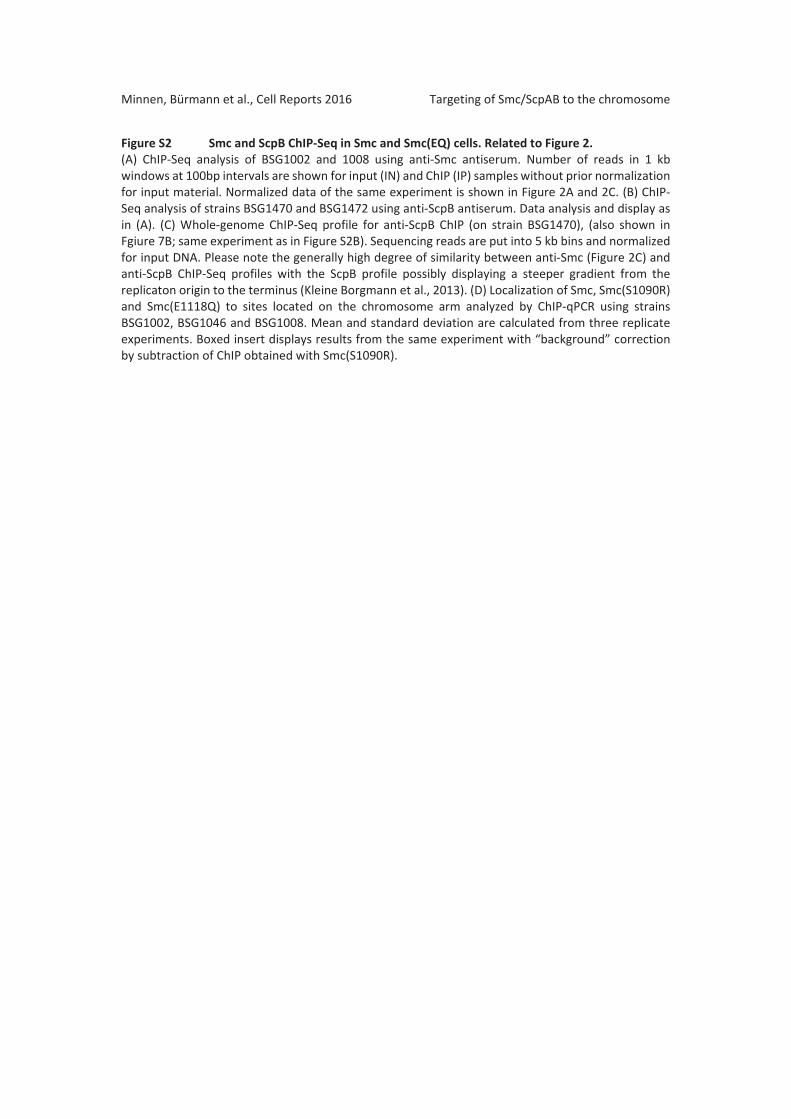
Figure S2 Smc and ScpB ChIP Seq in Smc and Smc(EQ) cells. Related to Figure 2.(A) ChIP Seq analysis of BSG1002 and 1008 using anti Smc antiserum. Number of reads in 1 kbwindows at 100bp intervals are shown for input (IN) and ChIP (IP) samples without prior normalizationfor input material. Normalized data of the same experiment is shown in Figure 2A and 2C. (B) ChIPSeq analysis of strains BSG1470 and BSG1472 using anti ScpB antiserum. Data analysis and display asin (A). (C) Whole genome ChIP Seq profile for anti ScpB ChIP (on strain BSG1470), (also shown inFgiure 7B; same experiment as in Figure S2B). Sequencing reads are put into 5 kb bins and normalizedfor input DNA. Please note the generally high degree of similarity between anti Smc (Figure 2C) andanti ScpB ChIP Seq profiles with the ScpB profile possibly displaying a steeper gradient from thereplicaton origin to the terminus (Kleine Borgmann et al., 2013). (D) Localization of Smc, Smc(S1090R)and Smc(E1118Q) to sites located on the chromosome arm analyzed by ChIP qPCR using strainsBSG1002, BSG1046 and BSG1008. Mean and standard deviation are calculated from three replicateexperiments. Boxed insert displays results from the same experiment with “background” correctionby subtraction of ChIP obtained with Smc(S1090R).

0
2
4
6
8
10
12-ScpB ChIP-SeqoriC
ahpCparS-334
wild type Smc/ScpABC
4 0 213
A05
101520253035
05
101520253035
0
10
20
30
40
05
101520253035
0
5
0
5
0
5
0
5
05
101520253035
05
101520253035
0
20
40
60
80
05
101520253035
0
5
0
5
0
5
0
5
Smc
E1118Q
IP
Input
IP
Input
chromosomal location (kb)
3888 3924
3888 3924
39243888
39243888
4138 4171
4138 4171
4138 4171
4138 4171
4197 5
4197 5
4197 5
4197 5
41 58
41 58
41 58
41 58
*
*
oriCtrnE
* * *
* * *
*
*
*
*
05
101520253035
0
505
101520253035
0
50
102030405060
05
101520253035
0
5
0
5
05
101520253035
0
505
101520253035
0
5
0
5
0
50
20406080
100120
05
101520253035
Smc
E1118Q
IP
Input
IP
Input
num
ber o
f rea
ds p
er k
b (in
100
0)
3888 3924
39243888
39243888
39243888
4138 4171
4138 4171
41714138
41714138
4197 5
4197 5
4197 5
4197 5
41 58
41 58
41 58
41 58
* parS-359
*
*
oriCtrnE
* * *
* * *
* parS-354 -355 -356
*
*
*
*
* parS-4* parS-334
10 kbB
num
ber o
f rea
ds p
er k
b (in
100
0)
Rat
io (C
hIP
/IN)
0.3
0.2
0.1
0.7
0.8
0.9
0
parS-359 (-10 kB)dnaA (0 kB)
yocGH (+2092 kB)
ChI
P (%
of i
nput
)
yolF (-1945 kB)glcC (-1046 kB)murA (- 436 kB)
amyE (+ 328 kB)sacV (+ 531 kB)ftsZ (+ 1598 kB)checC (+1715 kB)
E1118QS1090Rsmc
DamyE
Bs genome
parS359 dnaA
cheCftsZ
sacV
yolF yocGH
murA
glcC
-Smc ChIP-qPCR
0
0.1
0.2
0.3
0.7
0.8
0.9
smc E1118Q
ChI
P (%
of i
nput
)
Signal from [S1090R] subtracted
-ScpB ChIP-Seq
-Smc ChIP-Seq
Figure S2

Minnen, Bürmann et al., Cell Reports 2016 Targeting of Smc/ScpAB to the chromosome
Figure S3 Dimerization at the Smc hinge determines localization of Smc/ScpAB to parS.Related to Figure 3.(A) Immunoblotting of cell extracts from strains BSG1007, 1067, 1002, 1051, 1406, 1052, 1387, 1890,1893, 1889, 1891 and 1892 using anti Smc antiserum. (B) Colony formation Bs strains BSG1007, 1008,1889, 1892 and 1891 on minimal medium (SMG). (C) The hinge mutation (GGGG >AAAA) blocksdimerization of headless Smc protein (BsSmcH CC300). 40 μg of purified proteins was injected onto agel filtration column and analyzed by multi angle light scattering (SEC MALS). Absorbance (at A280) andlight scattering is shown for wild type and hinge mutant BsSmcH CC300 (curves in red and bluecolours, respectively). (D) Analysis of the major peak (in A280 absorbance) in SEC MALS (as in C) of wildtype and hinge mutant BsSmcH CC300 indicates the existence of largely dimeric and monomericprotein species, respectively. (E) Immunoblotting of cell extracts from strains BSG1007, 1067, 1002,1890, 1893, 1892, 1624, 1621 and 1620 using anti Smc antiserum. (F) Colony formation of strainsBSG1007, 1893, 1624, 1621, 1623 and 1620 on minimal medium (SMG). (G) Quantification of Smc GFPfoci in strains shown in Figure 3D. Number of foci is displayed per unit cell length (μm). Standarddeviation is derived from four different fields of view for each genotype. ‘n’ denotes the total numberof individual cells counted. (H) ChIP qPCR analysis of strains BSG1893, 1892, 2144 2147 grown in SMGmedium with anti Smc antiserum.

smc
smc
smc
EQ smc,
parB
EQ, pa
rB
smc,
scpA
smc
EQ smc,
scpB
EQ, sc
pA
-Smc
smc-g
fpA
-Smc
Esm
csm
c-gfp
smc
smc
EQ mH mH-EQ
EQ, sc
pA
-Smc
F
SMG38 hr
smc EQ mH mHEQ
mHscpA
mHEQscpA
C
0.10.100.05
0
4.1
0.6
1.1
1.6
2.1
2.6
3.1
3.6
parS-359dnaAyocGH
ChI
P (%
of i
nput
)
ChIP (%
of input)
hingemHwtheadEQ
scpA scpB scpAB
mHEQ
mHEQ
mHEQEQ
scpA
wtEQ
BsSmcH-CC300
BsSmcH-CC300(mH)
DBsSmcH-CC300 BsSmcH-
CC300(mH)
SMG38 hr
smc EQ smcscpA
EQscpA
smcscpB
B
H
0
0.2
0.4
0.6
0.8
1
1.2
smc
(n = 85)
EQ
(35)
smcscpA
(193)
EQscpA
(135)
mH
(119)
mH-EQ
(64)
mHscpA
(220)
mH-EQscpA(78)
Sm
c-G
FP fo
ci (p
er
m)
G
mH-EQ,
scpA
Figure S3
-Smc ChIP-qPCR

Minnen, Bürmann et al., Cell Reports 2016 Targeting of Smc/ScpAB to the chromosome
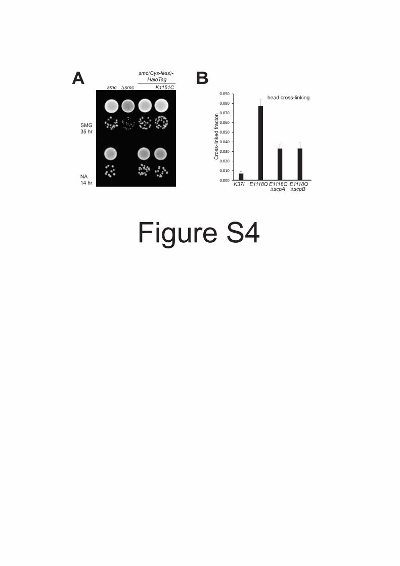
Figure S4 Smc(K1151C) –the reporter for head engagement– is functional. Related to Figure 4.(A) Colony formation of strains BSG1002, 1007, 1360 and 1457 on minimal medium (SMG) and nutrientrich medium (NA). (B) Cross linking of Smc(K1151C) in BSG1607, 1488, 1512 and 1513 with BMOE.Mean values and standard deviation from triplicate experiments are shown.
Asmcsmc
SMG35 hr
NA14 hr
smc(Cys-less)-HaloTag
K1151CB
Cro
ss-li
nked
frac
ton
K37I0.000
0.010
0.020
0.030
0.040
0.050
0.060
0.070
0.080
0.090
E1118Q E1118Q E1118QscpA scpB
head cross-linking
Figure S4

Minnen, Bürmann et al., Cell Reports 2016 Targeting of Smc/ScpAB to the chromosome
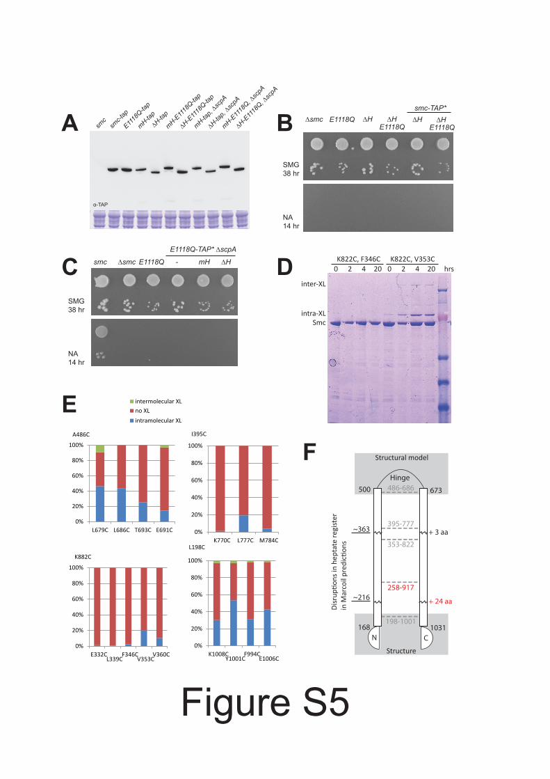
Figure S5 Expression and functionality of hinge less Smc protein. Related to Figure 5.(A) Immunoblotting against the TAP tag on Smc in cell extracts from strains BSG1002, 1016, 1475,1691, 1896, 1671, 1780, 1672, 1895, 1689 and 1779. Commassie staining of the same extracts is shownin the bottom panel. (B) Colony formation assay using strains BSG1007, 1008, 1626, 1619, 1896 and1780. (C) Same as in (B) with strains BSG1002, 1007, 1008, 1520, 1689 and 1779. (D) Exemplary imageof the SDS PAGE analysis of disulfide cross linked BsSmcH CC300 samples harboring pairs of cysteinesas annotated. (E) Quantification of intra and inter molecular disulfide formation (after 4 hrincubation) from Commassie stained SDS PAGE gels for 16 pairs of cysteine mutants. (F) Schematicview of the folding of the Smc coiled coil. Anchor points setting the register of the Smc coiled coils –established by in vitro disulfide formation (see D and E)– are given as dashed lines connecting N andC terminal helix. Disruptions in the coiled coil register were detected by Marcoil prediction. The lengthof extra sequences in the C terminal coiled coil as given by the experimentally determined coiled coilregister are indicated at the corresponding positions. Regions relevant for the targeting of mini Smcto parS are highlighted by labels in red colours.
A B
C smc E1118Qsmc
SMG38 hr
NA14 hr
E1118Q-TAP* scpA
- mH H
smc E1118Q H HE1118Q
H HE1118Q
smc-TAP*
SMG38 hr
NA14 hr
-TAP
smc
smc-t
ap
E1118
Q-tap
mH-tap
H-tap
H-E11
18Q-ta
p
mH-tap,
scpA
H-tap,
scpA
H-E11
18Q,
scpA
mH-E11
18Q,
scpA
mH-E11
18Q-ta
p
Disr
upon
s in
hept
ate
regi
ster
in M
arco
il pr
edic
ons
198-1001
486-686
395-777
353-822
168
500 673
1031
Hinge
N C
258-917
+ 24 aa
+ 3 aa
Structure
Structural model
~363
~216
E
K822C, V353CK822C, F346C
Smcintra-XL
inter-XL
0 2 204 0 2 20 hrs4D
0%
20%
40%
60%
80%
100%
E332CL339C
F346CV353C
V360C
K882C
0%
20%
40%
60%
80%
100%
L679C L686C T693C E691C
intermolecular XLno XLintramolecular XL
A486C
0%
20%
40%
60%
80%
100%
K1008CY1001C
F994CE1006C
L198C
0%
20%
40%
60%
80%
100%
K770C L777C M784C
I395C
F
Figure S5

Minnen, Bürmann et al., Cell Reports 2016 Targeting of Smc/ScpAB to the chromosome
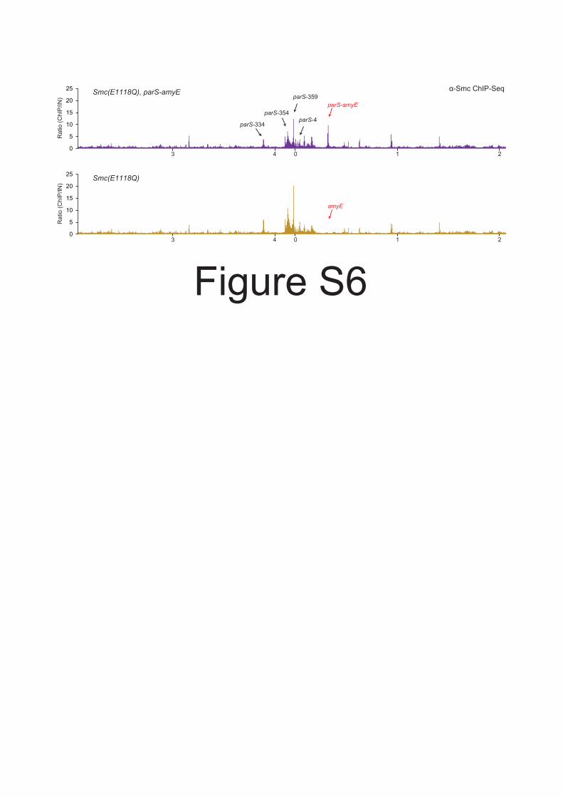
Figure S6 ChIP Seq of Smc(EQ) to an ectopic parS site. Related to Figure 6.Smc(EQ) is efficiently targeted to parS amyE. ChIP Seq analysis of BSG1471 (top panel) and BSG1008(bottom panel) using anti Smc antiserum. ChIP eluate sequence reads were mapped to 5 kb bins andnormalized for input DNA. Please note that Smc(EQ) localization to endogenous parS sites is decreasedby the presence of an extra parS site, being consistent with a titration effect. The bottom panel isidentical to the bottom panel of Figure 2C.
0
5
10
15
20
25
0
5
10
15
20
25
parS-359
parS-354
parS-334
Smc(E1118Q), parS-amyE
Smc(E1118Q)
parS-amyE
parS-4
4 0 213
4 0 213
-Smc ChIP-Seq
amyE
Rat
io (C
hIP
/IN)
Rat
io (C
hIP
/IN)
Figure S6

Minnen, Bürmann et al., Cell Reports 2016 Targeting of Smc/ScpAB to the chromosome
II.) Supplemental Table
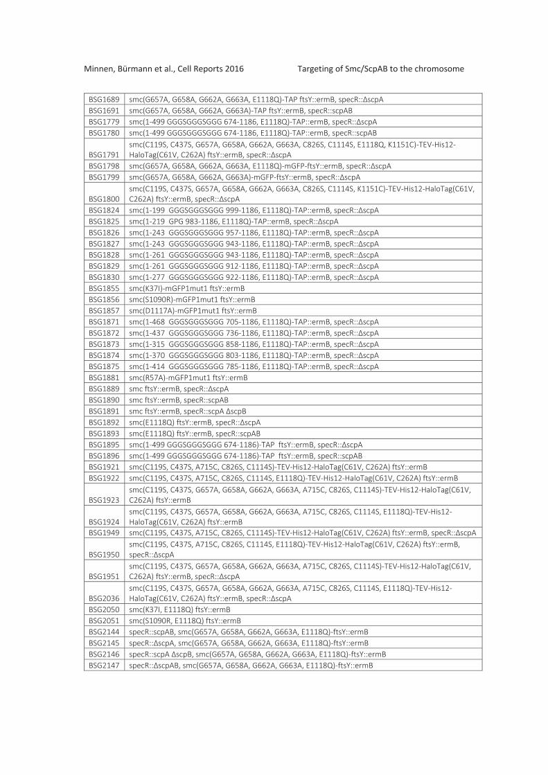
Supplemental Table 1 Genotypes
All strains are derivatives of 1A700 provided by the BGSC ( Genetic StockCenter). All strains are auxotrophic for tryptophan ( ).
BSG1002 smc ftsY::ermBBSG1007 smc ftsY::ermBBSG1008 smc(E1118Q) ftsY::ermBBSG1016 smc TAP ftsY::ermBBSG1045 smc(K37I) ftsY::ermBBSG1046 smc(S1090R) ftsY::ermBBSG1047 smc(D1117A) ftsY::ermBBSG1051 smc ftsY::ermB, parAB::kanRBSG1052 smc ftsY::ermB, parB::kanRBSG1067 smc mGFPmut1 ftsY::ermBBSG1068 smc(E1118Q) mGFP1mut1 ftsY::ermBBSG1083 smc(R57A) ftsY::ermBBSG1360 smc(C119S, C437S, C826S, C1114S) TEV His12 HaloTag(C61V, C262A) ftsY::ermBBSG1378 smc mGFPmut1 ftsY::ermB, specR:: scpABSG1387 smc(E1118Q) ftsY::ermB, parB::kanRBSG1406 smc(E1118Q) ftsY::ermB, parAB::kanRBSG1413 smc(E1118Q) mGFP1mut1 ftsY::ermB, specR:: scpABSG1457 smc(C119S, C437S, C826S, C1114S, K1151C) TEV His12 HaloTag(C61V, C262A) ftsY::ermBBSG1469 smc ftsY::ermB, amyE::parS 359::catBSG1470 smc ftsY::ermB, amyE::mtparS 359::catBSG1471 smc(E1118Q) ftsY::ermB, amyE::parS 359::catBSG1472 smc(E1118Q) ftsY::ermB, amyE::mtparS 359::catBSG1475 smc(E1118Q) TAP ftsY::ermBBSG1488 smc(C119S, C437S, C826S, C1114S, K1151C, E1118Q) TEV His12 HaloTag(C61V, C262A) ftsY::ermB
BSG1509smc(C119S, C437S, C826S, C1114S, K1151C) TEV His12 HaloTag(C61V, C262A) ftsY::ermB,specR:: scpA
BSG1512smc(C119S, C437S, C826S, C1114S, K1151C, E1118Q) TEV His12 HaloTag(C61V, C262A) ftsY::ermB,specR:: scpA
BSG1513smc(C119S, C437S, C826S, C1114S, K1151C, E1118Q) TEV His12 HaloTag(C61V, C262A) ftsY::ermB,specR::scpA scpB
BSG1520 smc(E1118Q) TAP ftsY::ermB, specR:: scpABSG1547 smc(G657A, G658A, G662A, G663A, E1118Q) ftsY::ermB
BSG1597smc(C119S, C437S, G657A, G658A, G662A, G663A, C826S, C1114S, K1151C) TEV His12 HaloTag(C61V,C262A) ftsY::ermB
BSG1598smc(C119S, C437S, G657A, G658A, G662A, G663A, C826S, C1114S, E1118Q, K1151C) TEV His12HaloTag(C61V, C262A) ftsY::ermB
BSG1607 smc(K37I, C119S, C437S, C826S, C1114S, K1151C) TEV His12 HaloTag(C61V, C262A) ftsY::ermBBSG1619 rncS smc(1 499 GGGSGGGSGGG 674 1186, E1118Q) ftsY::ermBBSG1620 smc(G657A, G658A, G662A, G663A, E1118Q) ftsY::ermB, specR:: scpABSG1621 smc(G657A, G658A, G662A, G663A, E1118Q) ftsY::ermB, specR::scpABBSG1624 smc(G657A, G658A, G662A, G663A) ftsY::ermB, specR::scpABBSG1626 rncS smc(1 499 GGGSGGGSGGG 674 1186) ftsY::ermBBSG1662 smc(G657A, G658A, G662A, G663A, E1118Q) mGFP ftsY::ermBBSG1671 smc(G657A, G658A, G662A, G663A, E1118Q) TAP ftsY::ermB, specR::scpABBSG1672 smc(G657A, G658A, G662A, G663A) TAP ftsY::ermB, specR:: scpABSG1677 smc(G657A, G658A, G662A, G663A) mGFP ftsY::ermB
Minnen, Bürmann et al., Cell Reports 2016 Targeting of Smc/ScpAB to the chromosome
BSG1689 smc(G657A, G658A, G662A, G663A, E1118Q) TAP ftsY::ermB, specR:: scpABSG1691 smc(G657A, G658A, G662A, G663A) TAP ftsY::ermB, specR::scpABBSG1779 smc(1 499 GGGSGGGSGGG 674 1186, E1118Q) TAP::ermB, specR:: scpABSG1780 smc(1 499 GGGSGGGSGGG 674 1186, E1118Q) TAP::ermB, specR::scpAB
BSG1791smc(C119S, C437S, G657A, G658A, G662A, G663A, C826S, C1114S, E1118Q, K1151C) TEV His12HaloTag(C61V, C262A) ftsY::ermB, specR:: scpA
BSG1798 smc(G657A, G658A, G662A, G663A, E1118Q) mGFP ftsY::ermB, specR:: scpABSG1799 smc(G657A, G658A, G662A, G663A) mGFP ftsY::ermB, specR:: scpA
BSG1800smc(C119S, C437S, G657A, G658A, G662A, G663A, C826S, C1114S, K1151C) TEV His12 HaloTag(C61V,C262A) ftsY::ermB, specR:: scpA
BSG1824 smc(1 199 GGGSGGGSGGG 999 1186, E1118Q) TAP::ermB, specR:: scpABSG1825 smc(1 219 GPG 983 1186, E1118Q) TAP::ermB, specR:: scpABSG1826 smc(1 243 GGGSGGGSGGG 957 1186, E1118Q) TAP::ermB, specR:: scpABSG1827 smc(1 243 GGGSGGGSGGG 943 1186, E1118Q) TAP::ermB, specR:: scpABSG1828 smc(1 261 GGGSGGGSGGG 943 1186, E1118Q) TAP::ermB, specR:: scpABSG1829 smc(1 261 GGGSGGGSGGG 912 1186, E1118Q) TAP::ermB, specR:: scpABSG1830 smc(1 277 GGGSGGGSGGG 922 1186, E1118Q) TAP::ermB, specR:: scpABSG1855 smc(K37I) mGFP1mut1 ftsY::ermBBSG1856 smc(S1090R) mGFP1mut1 ftsY::ermBBSG1857 smc(D1117A) mGFP1mut1 ftsY::ermBBSG1871 smc(1 468 GGGSGGGSGGG 705 1186, E1118Q) TAP::ermB, specR:: scpABSG1872 smc(1 437 GGGSGGGSGGG 736 1186, E1118Q) TAP::ermB, specR:: scpABSG1873 smc(1 315 GGGSGGGSGGG 858 1186, E1118Q) TAP::ermB, specR:: scpABSG1874 smc(1 370 GGGSGGGSGGG 803 1186, E1118Q) TAP::ermB, specR:: scpABSG1875 smc(1 414 GGGSGGGSGGG 785 1186, E1118Q) TAP::ermB, specR:: scpABSG1881 smc(R57A) mGFP1mut1 ftsY::ermBBSG1889 smc ftsY::ermB, specR:: scpABSG1890 smc ftsY::ermB, specR::scpABBSG1891 smc ftsY::ermB, specR::scpA scpBBSG1892 smc(E1118Q) ftsY::ermB, specR:: scpABSG1893 smc(E1118Q) ftsY::ermB, specR::scpABBSG1895 smc(1 499 GGGSGGGSGGG 674 1186) TAP ftsY::ermB, specR:: scpABSG1896 smc(1 499 GGGSGGGSGGG 674 1186) TAP ftsY::ermB, specR::scpABBSG1921 smc(C119S, C437S, A715C, C826S, C1114S) TEV His12 HaloTag(C61V, C262A) ftsY::ermBBSG1922 smc(C119S, C437S, A715C, C826S, C1114S, E1118Q) TEV His12 HaloTag(C61V, C262A) ftsY::ermB
BSG1923smc(C119S, C437S, G657A, G658A, G662A, G663A, A715C, C826S, C1114S) TEV His12 HaloTag(C61V,C262A) ftsY::ermB
BSG1924smc(C119S, C437S, G657A, G658A, G662A, G663A, A715C, C826S, C1114S, E1118Q) TEV His12HaloTag(C61V, C262A) ftsY::ermB
BSG1949 smc(C119S, C437S, A715C, C826S, C1114S) TEV His12 HaloTag(C61V, C262A) ftsY::ermB, specR:: scpA
BSG1950smc(C119S, C437S, A715C, C826S, C1114S, E1118Q) TEV His12 HaloTag(C61V, C262A) ftsY::ermB,specR:: scpA
BSG1951smc(C119S, C437S, G657A, G658A, G662A, G663A, A715C, C826S, C1114S) TEV His12 HaloTag(C61V,C262A) ftsY::ermB, specR:: scpA
BSG2036smc(C119S, C437S, G657A, G658A, G662A, G663A, A715C, C826S, C1114S, E1118Q) TEV His12HaloTag(C61V, C262A) ftsY::ermB, specR:: scpA
BSG2050 smc(K37I, E1118Q) ftsY::ermBBSG2051 smc(S1090R, E1118Q) ftsY::ermBBSG2144 specR::scpAB, smc(G657A, G658A, G662A, G663A, E1118Q) ftsY::ermBBSG2145 specR:: scpA, smc(G657A, G658A, G662A, G663A, E1118Q) ftsY::ermBBSG2146 specR::scpA scpB, smc(G657A, G658A, G662A, G663A, E1118Q) ftsY::ermBBSG2147 specR:: scpAB, smc(G657A, G658A, G662A, G663A, E1118Q) ftsY::ermB

Minnen, Bürmann et al., Cell Reports 2016 Targeting of Smc/ScpAB to the chromosome
III.) Supplemental Experimental Procedures
In vivo expression of Smc proteins tested by immunoblottingCells were grown in SMG at 37°C to an OD600 of 0.02 0.03, harvested by centrifugation or filtrationsand washed once in 2 ml PBSG (PBS + 0.1% glycerol). The OD600 was measured and equivalent amountof cells for all samples were taken (0.02 ml*OD600). Cells were resuspended in PBSG, ßmercaptoethanol was added to a final concentration of 28.6 mM and kept on ice for 3 min. Lysozyme(12.8U/μl final), Roche Complete protease inhibitor cocktail and Benzonase (0.4 U/μl; Sigma Aldrich)were added and the samples were incubated at 37°C for 20 min. NuPage LDS loading dye (final 1x)and DTT (final conc. 100 mM) were added and the samples incubated at 70°C for 10 min. The extractswere loaded on a 4 12% NuPAGE Bis Tris gel run in MOPS buffer for 50 min at 200 V. Proteins weretransferred to a PVDF membrane which was treated with Smc, GFP (Life Technologies, A6455) orPeroxidase Anti Peroxidase (PAP). Smc and GFP blots were treated with ECL Anti rabbit IgG, HRPlinked whole antibody (from donkey) (GE healthcare). The blots were incubated with Supersignal WestFemto (Thermo Scientific) and were imaged in a LAS4000 scanner.
Chromatin immuno precipitation (ChIP) and qPCRCells were grown in SMG medium at 37ºC overnight and diluted to OD600 0.005 in SMG. At OD 0.020.03 40 ml of fixing solution (50mM Tris/HCl pH 8.0, 100mM NaCl, 1mM EDTA, 0.5mM EGTA, 11%formaldehyde) was added to 400 ml of culture and incubated at room temperature for 30 minutes.Cells were harvested by centrifugation or filtration and washed in 2 ml ice cold PBS and OD600 wasmeasured. Cells were resuspended in 1 ml TESS (50mM Tris/HCl 7.4, 10mM EDTA, 50mM NaCl, 500mMsucrose) and protoplasted by incubating in 1 ml TESS supplemented with 20mg/ml lysozyme (Sigma)and Roche Complete protease inhibitor cocktail for 30 min at 37°C shaking. Cells were washed once in1 ml TESS, aliquoted according to the previously measured OD600 and stored at 80°C.One aliquot of fixed cells was resuspended in 2 ml lysis buffer (50mM Hepes/KOH pH 7.5, 140mMNaCl, 1mM EDTA, 1% (v/v) Triton X 100, 0.1% (w/v) sodium deoxycholate, 100mg/ml RNase, RocheComplete protease inhibitor cocktail) and transferred to a 5 ml round bottom tube. The samples weresonicated 3 x 20 sec on a Bandelin Sonoplus with a MS 72 tip at 90% pulse and 35% power output.Lysates were transferred into 2 ml tubes and centrifuged 5 min at 21000g and the supernatantsubsequently 10 min at 21000g at 4°C.200 μl of the cleared lysates was kept separate as the input sample. 50 μl Protein G coupled dynabead(Invitrogen) were incubated with 50 μl antibody serum ( Smc, ScpB or ParB generated in rabbit)for at least 1 hr rotating at 4°C. Beads were washed in lysis buffer and added to 800 μl of the clearedlysates. For experiments involving TAP tagged strains, rabbit IgG coupled to 50 μl magnetic DynaBeads(Epoxy, M 270) (prepared according to the manufacturer’s protocol) was added to 800 μl clearedlysates. The beads with cleared lysates were incubated at 4°C rotating for 2 4 hours. Beads werewashed once with each of the following buffers, lysis buffer, lysis buffer with high salt (500mM NaCl)and wash buffer (10mM Tris/HCl pH 8.0, 250mM LiCl, 1mM EDTA, 0.5% (w/v) NP 40, 0.5% (w/v)sodium deoxycholate). Beads were resuspended in 520 μl TES (50mM Tris/HCl pH 8.0, 10mM EDTA,1% SDS), the input samples were combined with 300 μl TES and 20 μl 10% SDS solution and incubatedovernight at 65°C shaking. DNA was purified by phenol chloroform extraction and ethanolprecipitation. The DNA was dissolved in 100μl TE at 65°C for 20 min and purified on a Qiagen PCRpurification column and eluted in 30 μl EB. For qPCR 4 μl of the input DNA (diluted 1:200) and IPsamples (diluted 1:20) was used in a 10 μl reaction using 5 μl Takyon no ROX SYBR Mastermix bluedTTP (Eurogentec) and 1 μl primer pair stock solution (3 μM each primer) on a Qiagen Rotor Gene Qin a 72 well rotor according to manufacturer’s instructions. Primer sequences are given in the tablebelow. Curves were analyzed by determining the maximum of the 2nd derivative using the Real timePCR miner software (http://ewindup.info) (Zhao and Fernald, 2005). ChIP efficiencies were calculatedas follows: [(IP/input)*100] for each primer pair.

Minnen, Bürmann et al., Cell Reports 2016 Targeting of Smc/ScpAB to the chromosome
List of primer pairs for qPCR:
parS 356 STG236 tgaaaagaatgcccatcaca
STG237 tgcaagcaacaacccttttac
parS 359 STG097 aaaaagtgattgcggagcag
STG098 agaaccgcatctttcacagg
dnaA STG199 gatcaatcggggaaagtgtg
STG200 gtagggcctgtggatttgtg
trnS STG404 gggttttgacacccttggta
STG405 aagcaaaaggaaatggctga
cheC STG396 tttgcatgaactgggcaata
STG397 tccgaacatgtccaatgaga
yocGH STG099 tccatatcctcgctcctacg
STG100 attctgctgatgtgcaatgg
Protein purification and SEC MALSWild type and hinge mutant BsSmcH CC300 protein (Smc residues 188 1011) were overexpressedfrom plasmid pnEA tH in E. coli with an N terminal HISx6 tag (Diebold et al., 2011). The proteins werepurified via a HisTrap column, concentrated by anion exchange chromatography (MonoQ HiTrap) andeluted from a size exclusion chromatography column (Superdex 200 10/300) in 200mM NaCl, 25mMTris/HCl pH 7.4 (4°C). Multi angle light scattering coupled to size exclusion chromatography (SECMALS) was performed as described previously (Soh et al., 2015).
Viability spotting assayCells were grown in SMG medium overnight into stationary phase, diluted 81 fold and 59049 fold (in9x steps) and spotted on nutrient agar plates (Oxoid) or SMG agar plates. Plates were incubated at37°C for ~12 hr on NA or ~36 hr on SMG agar.
Mapping of the Bs Smc coiled coil register by disulfide formationIntramolecular crosslinking reactions for the determination of the coiled coil register were performedessentially as described in (Waldman et al., 2015) using the BsSmcH CC300 construct (Soh et al., 2015).The protein was expressed from the pnEA tH plasmid as a His Tag fusion protein. For each doublecysteine mutant, 50mL cultures were set up. Cells were lysed by sonication and soluble extract wasincubated for one hour with 300uL of Talon resin (Clontech). Beads were washed three times with thelysis buffer (200 mM NaCl, 50 mM NaPi pH7.4, 5 mM Imidazole) and then resuspended in the lysisbuffer supplemented with 1 mM magnesium chloride. 1uL of benzonase (Roche) was added to thebeads that were shaked for 30min at room temperature to remove the bound DNAs. The beads werewashed two more times with the lysis buffer and the proteins were eluted with elution buffer 200 mMNaCl, 500 mM Imidazole, 50 mM NaPi pH7.4.The proteins were then dialyzed against PBS buffer containing 4 mM DTT for 2 hours. For thecrosslinking reaction, the protein was diluted to a concentration of 5 μM. The non crosslinked samplewas prepared by adding 1mM of iodoacetamide and heating at 70°C for 10 min. Disulfide formationwas set up by adding one volume of PBS supplemented with 5 mM NaAsO2 (Fluka) to the protein and100 μM dithio bisnitrobenzoic acid DTNB (Merck) and 300 μM beta mercaptoethanol. The reactionwas incubated at 4°C under shaking. Samples were taken after 2, 4, 6 and 20h of reaction, andquenched by addition of 10 mM iodoacetamide at 70°C for 10 min.

Minnen, Bürmann et al., Cell Reports 2016 Targeting of Smc/ScpAB to the chromosome
After addition of non reducing SDS Page loading buffer the samples were boiled for 5 min at 95°C andrun on a Bis Tris 4 16% NuPage acrylamide gel (Novex) using MOPS buffer as running buffer for 50 minat 200 V.
Minnen, Bürmann et al., Cell Reports 2016 Targeting of Smc/ScpAB to the chromosome
IV.) Supplemental References
Diebold, M.L., Fribourg, S., Koch, M., Metzger, T., and Romier, C. (2011). Deciphering correct strategiesfor multiprotein complex assembly by co expression: application to complexes as large as the histoneoctamer. Journal of structural biology 175, 178 188.
Gruber, S., and Errington, J. (2009). Recruitment of condensin to replication origin regions byParB/SpoOJ promotes chromosome segregation in B. subtilis. Cell 137, 685 696.Gruber, S., Haering, C.H., and Nasmyth, K. (2003). Chromosomal cohesin forms a ring. Cell 112, 765777.
Kleine Borgmann, L.A., Ries, J., Ewers, H., Ulbrich, M.H., and Graumann, P.L. (2013). The bacterial SMCcomplex displays two distinct modes of interaction with the chromosome. Cell reports 3, 1483 1492.
Schwartz, M.A., and Shapiro, L. (2011). An SMC ATPase mutant disrupts chromosome segregation inCaulobacter. Molecular microbiology 82, 1359 1374.
Soh, Y.M., Burmann, F., Shin, H.C., Oda, T., Jin, K.S., Toseland, C.P., Kim, C., Lee, H., Kim, S.J., Kong,M.S., et al. (2015). Molecular basis for SMC rod formation and its dissolution upon DNA binding.Molecular cell 57, 290 303.
Waldman, V.M., Stanage, T.H., Mims, A., Norden, I.S., and Oakley, M.G. (2015). Structural mappingof the coiled coil domain of a bacterial condensin and comparative analyses across all domains of lifesuggest conserved features of SMC proteins. Proteins 83, 1027 1045.
Wang, X., Tang, O.W., Riley, E.P., and Rudner, D.Z. (2014). The SMC condensin complex is requiredfor origin segregation in Bacillus subtilis. Current biology : CB 24, 287 292.