ankyrin repeat domain(ard) of trpv2 by: marilyn nguyen
Post on 19-Dec-2015
220 views
TRANSCRIPT

Ankyrin Repeat Domain(ARD) of
TRPV2
By: Marilyn Nguyen


Zebrafish

Human


PDB ID: 2ETC, 2ETA, 2ETB

Summary:•Part of the Transient Receptor Potential (TRP) cation channel family and Vanilloid (V) subfamily. •Vanilloid receptors recognize capaisin. The original capaisin receptor is TRPV1. TRPV2 is a homologue of this receptor.•They are broad range cation channels that are crucial for processes ranging from sensory signaling to magnesium homeostasis.•The channels are selective for calcium and magnesium, preferring these two cations over sodium ions.•TRP channels have large N- and C-Terminal cytosolic regions that are believed to contain many unknown protein interactions and regulatory motifs.
•N-terminal region of all TRPV channels contain ankyrin repeats. These repeats are thought to be essential for the proper functioning of the channel. They are involved in protein-protein interactions in bacterial, achaeal, and eukaryotes.•TRPV2 was discovered in 1999 by David Julius’s laboratory in the Department of Cellular and Molecular Pharmacology at the University of California. •TRPV2 channels can be found in the central nervous system, spleen and lung.•Sequence of TRPV2 is highly conserved among mammalian species.

Function:•A thermosensitive receptor that is activated around 52ºC•Believed to be involved in nerve and sensory responses to heat, specifically, stimulation of the sensory ganglia. •Little is known about the true function of the Ankyrin Repeat Domain (ARD) of TRPV2, but it is suspected to be involved in cell surface trafficking.

Structure: (Taken from Rattus norvegicus; common name: brown rat)
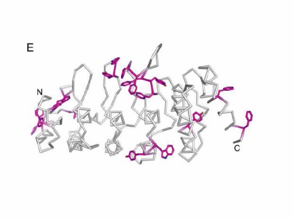
•Ankyrin Repeats Domain (ARD) monomeric in solution.•Ankyrin repeats are approximately 33 amino acid residues in length consisting of two anti-parallel alpha helices separated by intervening finger loop regions.•The three-dimensional structure of TRPV2-ARD consists of 6 ankyrin repeat structure motifs. However, only 4 of these motifs have been identified. •TRPV2-ARD is highly conserved between difference species.•Ankyrin Repeats 1-3 are long flexible fingers with several exposed aromatic residues•Ankyrin Repeats 5-6 have long outer helices (~ 9 residues instead of typical 7 residues).
•Repeats 4-5 exhibits an unusual counterclockwise twist for stacking. This alters the shape of the surface and is believed to be an important site for protein or ligand interaction•Consists of 5 finger loops that are unique in the fact that none contain β-hairpin hydrogen bonding in typical ankyrin repeats.•2 short anti-parallel helices and a finger loop project outwards from the helical axes at ~90º angle that creates an L-shaped cross-section.•Insertions into ARD are most often observed in the finger loop regions.•In humans, Ankyrin groove (ligand recognition site) has extended loops that are sites of exposed hydrophobic residues.

Active Site:•The helical hairpin stacking of the inner helices and fingers create a concave surface that looks like a cupped hand. The “palm” region of this concave cupped hand is considered the active site for protein-protein interactions

Important residues:•The concave face of finger loop regions 2 and 3 contain several Phe and Tyr residues that create the hydrophobic interacting surface.•Homology to TRPV1 indicates that similar amino acid residues; Thr, Ser, and Tyr on the concave face of finger 2 could be phosphorylated through protein kinase A and Src kinase to play crucial role in regulation of the channel.

Diseases:•Muscular Dystrophy – a severe degenerative skeletal muscle disorder. There are several types of muscular dystrophy and all are characterized by progressive muscle weakness. Dystrophy can be caused by a defect in genes encoding for the dystrophin-glycoprotein complex. This defect causes lost of membrane integrity and sustained increase of calcium ion concentration that later results in muscle degeneration. Mutations in TRPV2 in mice showed increased number of central nuclei and fiber size variability/fibrosis/apoptosis and elevated serum creatine kinase levels, and reduced muscle performance. It is suggested that TRPV2 is the principal calcium entry route for the sustained calcium ion concentration increases (2).
References:1. Jin X, Touhey J, and Gaudet R. Structure of the N-terminal ankyrin repeat domain
of the TRPV2 ion channel. J. Biol. Chem. 2006 Sep 1;281(35):25006-10.2. Iwata Y, Katanosaka Y, Arai Y, Shigekawa M, and Wakabayashi S. Dominant-
negative inhibition of Ca2+ influx via TRPV2 ameliorates muscular dystrophy in animal models. Hum Mol Genet. 2009 Mar 1;18(5):824-34
3. McCleverty C, Koesema E, Patapoutian A, and Lesley S. Crystal structure of the human TRPV2 channel ankyrin repeat domain. Protein Science. 2006 Jun 9;15:2201-2206
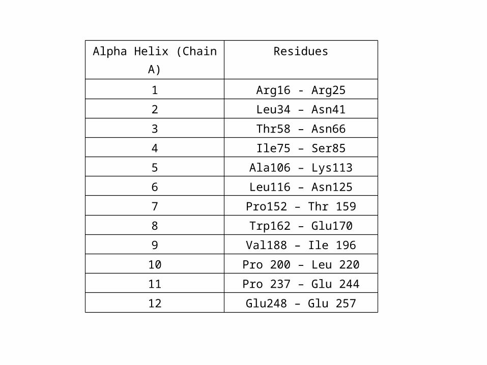
Alpha Helix (Chain A) Residues
1 Arg16 - Arg25
2 Leu34 – Asn41
3 Thr58 – Asn66
4 Ile75 – Ser85
5 Ala106 – Lys113
6 Leu116 – Asn125
7 Pro152 – Thr 159
8 Trp162 – Glu170
9 Val188 – Ile 196
10 Pro 200 – Leu 220
11 Pro 237 – Glu 244
12 Glu248 – Glu 257
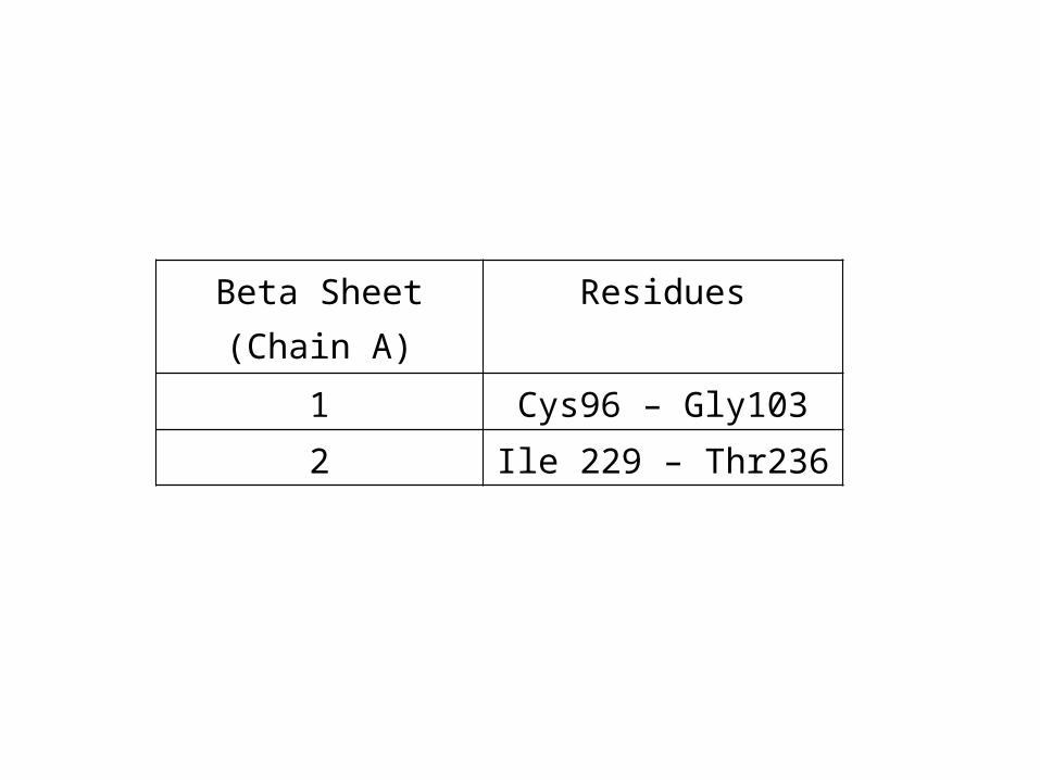
Beta Sheet (Chain A) Residues
1 Cys96 – Gly103
2 Ile 229 – Thr236

Hydrophobic region•Phe 161•Tyr 162•Phe 198•Phe 199•Phe 207•Tyr 208•Phe 209