annex h combined effects of radiation and other agents · annex h: combined effects of radiation...
TRANSCRIPT

ANNEX H
Combined effects of radiation and other agents
CONTENTS
Page
INTRODUCTION . . . . . . . . . . . . . . . . . . . . . . . . . . . . . . . . . . . . . . . . . . . . . . . . . . . 179
I. IDENTIFYING INTERACTIONS AND COMBINED EFFECTS . . . . . . . . . . . 180A. SCOPE OF THE PROBLEM . . . . . . . . . . . . . . . . . . . . . . . . . . . . . . . . . . 180
1. Additivity and deviations from additivity . . . . . . . . . . . . . . . . . . . . . 1802. Radiation effects and effects of other agents . . . . . . . . . . . . . . . . . . . 182
B. EXPOSURE ASSESSMENT . . . . . . . . . . . . . . . . . . . . . . . . . . . . . . . . . . 1831. Dose concepts for physical and chemical agents . . . . . . . . . . . . . . . . 1832. Biochemical monitoring . . . . . . . . . . . . . . . . . . . . . . . . . . . . . . . . . 1843. Gene mutation analysis . . . . . . . . . . . . . . . . . . . . . . . . . . . . . . . . . . 1864. Cytogenetic analysis . . . . . . . . . . . . . . . . . . . . . . . . . . . . . . . . . . . . 1875. Summary . . . . . . . . . . . . . . . . . . . . . . . . . . . . . . . . . . . . . . . . . . . . . 189
II. MECHANISTIC CONSIDERATIONS . . . . . . . . . . . . . . . . . . . . . . . . . . . . . . . 189A. EFFECTS ON THE MOLECULAR LEVEL . . . . . . . . . . . . . . . . . . . . . . 190B. EFFECTS ON THE CELLULAR LEVEL . . . . . . . . . . . . . . . . . . . . . . . . 190
1. Accumulation of (sub)lesions . . . . . . . . . . . . . . . . . . . . . . . . . . . . . . 1912. Cellular repair . . . . . . . . . . . . . . . . . . . . . . . . . . . . . . . . . . . . . . . . . 1923. Cytokinetics . . . . . . . . . . . . . . . . . . . . . . . . . . . . . . . . . . . . . . . . . . . 1934. Toxicological analysis . . . . . . . . . . . . . . . . . . . . . . . . . . . . . . . . . . . 193
C. EFFECTS ON THE TISSUE/ORGAN LEVEL . . . . . . . . . . . . . . . . . . . . 194D. DOSE MODIFIERS AND OTHER INDIRECT INTERACTIONS . . . . . . 197E. SUMMARY . . . . . . . . . . . . . . . . . . . . . . . . . . . . . . . . . . . . . . . . . . . . . . . 197
III. SPECIFIC COMBINED EXPOSURES . . . . . . . . . . . . . . . . . . . . . . . . . . . . . . . 199A. RADIATION AND PHYSICAL AGENTS . . . . . . . . . . . . . . . . . . . . . . . . 200
1. Combinations of ionizing radiation . . . . . . . . . . . . . . . . . . . . . . . . . 2002. Ultraviolet radiation . . . . . . . . . . . . . . . . . . . . . . . . . . . . . . . . . . . . . 2003. Electromagnetic radiation . . . . . . . . . . . . . . . . . . . . . . . . . . . . . . . . 2004. Temperature . . . . . . . . . . . . . . . . . . . . . . . . . . . . . . . . . . . . . . . . . . 2005. Ultrasound . . . . . . . . . . . . . . . . . . . . . . . . . . . . . . . . . . . . . . . . . . . . 2006. Dust, asbestos, and other mineral fibres . . . . . . . . . . . . . . . . . . . . . . 2007. Space flight . . . . . . . . . . . . . . . . . . . . . . . . . . . . . . . . . . . . . . . . . . . 201
B. RADIATION AND CHEMICAL AGENTS . . . . . . . . . . . . . . . . . . . . . . . 2011. Genotoxic chemicals . . . . . . . . . . . . . . . . . . . . . . . . . . . . . . . . . . . . 2012. Non-genotoxic chemicals . . . . . . . . . . . . . . . . . . . . . . . . . . . . . . . . . 2013. Tobacco . . . . . . . . . . . . . . . . . . . . . . . . . . . . . . . . . . . . . . . . . . . . . . 201

ANNEX H: COMBINED EFFECTS OF RADIATION AND OTHER AGENTS
Page
4. Metals . . . . . . . . . . . . . . . . . . . . . . . . . . . . . . . . . . . . . . . . . . . . . . . 2035. Mitogens and cytotoxicants . . . . . . . . . . . . . . . . . . . . . . . . . . . . . . . 2036. Antioxidants, vitamins, and other dietary factors . . . . . . . . . . . . . . . 204
C. RADIATION AND BIOLOGICAL AGENTS . . . . . . . . . . . . . . . . . . . . . 204D. SUMMARY . . . . . . . . . . . . . . . . . . . . . . . . . . . . . . . . . . . . . . . . . . . . . . . 204
IV. COMBINED EXPOSURES IN CANCER THERAPY . . . . . . . . . . . . . . . . . . . . 205A. MECHANISMS OF INTERACTIONS . . . . . . . . . . . . . . . . . . . . . . . . . . . 205B. SECONDARY CANCERS FOLLOWING
COMBINED MODALITY TREATMENT . . . . . . . . . . . . . . . . . . . . . . . . 207C. SUMMARY . . . . . . . . . . . . . . . . . . . . . . . . . . . . . . . . . . . . . . . . . . . . . . . 208
V. EFFECTS OTHER THAN CANCER . . . . . . . . . . . . . . . . . . . . . . . . . . . . . . . . 211A. PRE- AND POST-NATAL EFFECTS . . . . . . . . . . . . . . . . . . . . . . . . . . . 211B. GENETIC AND MULTI-GENERATION EFFECTS . . . . . . . . . . . . . . . . 212C. DETERMINISTIC EFFECTS . . . . . . . . . . . . . . . . . . . . . . . . . . . . . . . . . 212
EXTENDED SUMMARY . . . . . . . . . . . . . . . . . . . . . . . . . . . . . . . . . . . . . . . . . . . . . 213
CONCLUSIONS . . . . . . . . . . . . . . . . . . . . . . . . . . . . . . . . . . . . . . . . . . . . . . . . . . . . 215
FURTHER RESEARCH NEEDS . . . . . . . . . . . . . . . . . . . . . . . . . . . . . . . . . . . . . . . . 217
Appendix:COMBINED EFFECTS OF SPECIFIC PHYSICAL, CHEMICALAND BIOLOGICAL AGENTS WITH IONIZING RADIATION . . . . . . . . . . . . . . . . 218
A. RADIATION AND PHYSICAL AGENTS . . . . . . . . . . . . . . . . . . . . . . . . 2181. Combinations of different types of ionizing radiation . . . . . . . . . . . . 2182. Ultraviolet radiation . . . . . . . . . . . . . . . . . . . . . . . . . . . . . . . . . . . . . 2203. Low- and high-frequency electromagnetic radiation . . . . . . . . . . . . . 2264. Temperature . . . . . . . . . . . . . . . . . . . . . . . . . . . . . . . . . . . . . . . . . . 2275. Ultrasound . . . . . . . . . . . . . . . . . . . . . . . . . . . . . . . . . . . . . . . . . . . . 2286. Dust, asbestos, and other mineral fibres . . . . . . . . . . . . . . . . . . . . . . 2297. Space flight . . . . . . . . . . . . . . . . . . . . . . . . . . . . . . . . . . . . . . . . . . . 231
B. RADIATION AND CHEMICAL TOXICANTS . . . . . . . . . . . . . . . . . . . . 2311. Genotoxic chemicals . . . . . . . . . . . . . . . . . . . . . . . . . . . . . . . . . . . . 2322. Non-genotoxic chemicals . . . . . . . . . . . . . . . . . . . . . . . . . . . . . . . . . 2363. Tobacco . . . . . . . . . . . . . . . . . . . . . . . . . . . . . . . . . . . . . . . . . . . . . . 2394. Metals . . . . . . . . . . . . . . . . . . . . . . . . . . . . . . . . . . . . . . . . . . . . . . . 2465. Mitogens and cytotoxicants . . . . . . . . . . . . . . . . . . . . . . . . . . . . . . . 2486. Antioxidants, vitamins, and other dietary factors . . . . . . . . . . . . . . . 249
C. RADIATION AND BIOLOGICAL AGENTS, MISCELLANEOUS . . . . . 2521. Hormones . . . . . . . . . . . . . . . . . . . . . . . . . . . . . . . . . . . . . . . . . . . . 2522. Viruses, bacteria, and genetic sequences . . . . . . . . . . . . . . . . . . . . . 2543. Miscellaneous factors . . . . . . . . . . . . . . . . . . . . . . . . . . . . . . . . . . . . 254
D. COMBINED MODALITIES IN RADIATION THERAPY . . . . . . . . . . . . 2551. Alkylating agents, nitrosoureas, and
platinum coordination complexes . . . . . . . . . . . . . . . . . . . . . . . . . . . 2552. Antimetabolites . . . . . . . . . . . . . . . . . . . . . . . . . . . . . . . . . . . . . . . . 2573. Antitumour antibiotics . . . . . . . . . . . . . . . . . . . . . . . . . . . . . . . . . . . 2604. Microtubule poisons . . . . . . . . . . . . . . . . . . . . . . . . . . . . . . . . . . . . . 2625. Topoisomerase poisons . . . . . . . . . . . . . . . . . . . . . . . . . . . . . . . . . . 2636. Bioreductive drugs . . . . . . . . . . . . . . . . . . . . . . . . . . . . . . . . . . . . . . 265
Glossary . . . . . . . . . . . . . . . . . . . . . . . . . . . . . . . . . . . . . . . . . . . . . . . . . . . . . . . . . . 267
References . . . . . . . . . . . . . . . . . . . . . . . . . . . . . . . . . . . . . . . . . . . . . . . . . . . . . . . . . 271
178

ANNEX H: COMBINED EFFECTS OF RADIATION AND OTHER AGENTS 179
INTRODUCTION
1. Living organisms are exposed to numerous natural andman-made agents that interact with molecules, cells, andtissues, causing reversible deviations from homeostaticequilibrium or irreversible damage. Many aspects of agingand many diseases are thought to stem from exogenous andendogenous deleterious agents acting on key components ofcells within the body. Because of the worldwide proliferationof a number of man-made agents and the increasing release ofnatural agents due to human activities into the environment,the assessment of toxicity, carcinogenicity, and mutagenicityof a specific chemical, physical, or biological agent is, in fact,a study of combined exposures [G10]. Although this has beenrecognized for a long time, risk assessment is generallyperformed with the simplifying assumption that the agentunder study acts largely independently of other substances.Studies of interactions have indicated, however, that, at leastat high exposures, the action of one agent can be influencedby simultaneous exposures to other agents. The combinedeffects may be greater or smaller than the sum of the effectsfrom separate exposures to the individual agents. The actionat low levels of exposure, which are commonly encounteredin occupational and environmental situations, is less clear.Continued, critical review of studies on the effects ofcombined exposures to radiation and other toxic agents isnecessary, particularly at the lowest levels of exposure, to besure that any modifications of the radiation effects caused byother environmental or occupational agents are recognizedand, as far as possible, taken into account in risk assessments.
2. In the UNSCEAR 1982 Report [U6], the Committeediscussed the problem of the combined action of radiationwith other agents. In reviewing the approaches and the manyreports in which synergisms were claimed, the Committeenoted that, in general, an adequate conceptual framework waslacking. Despite many reports showing the potential import-ance of interactions between different agents under specificconditions, mostly occupational, information on the mechan-isms of action was largely missing, and the methodologies fordata analysis in different branches of the biological scienceswere based on different approaches. The UNSCEAR 1982Report concluded that it was not possible to document clearcases of interaction that could justifysubstantial modificationsto the existing radiation risk estimates. The Committee feltthat systematic investigationsofcombined effects were neededto allow this field to move forward from its early stage ofdevelopment.
3. The objective of this Annex is to update the Com-mittee's previous review of this subject [U6] and to reconsiderwhether interactions of radiation and one or more other agentsshould be taken into account in evaluating radiation risks atlowdoses. To achieve this objective, the following subjects areconsidered:
(a) the concepts of doses, targets, and detriments currentlyused in risk assessments of radiation and chemicalagents;
(b) recent developments from research on the possiblemechanisms of combined effects from low-levelexposures to radiation and other agents;
(c) results and evaluations ofdata from experimental andepidemiological studies;
(d) mechanistic models applied to experimental and epi-demiological results, with generalizations and extra-polations that might be pertinent to low and chronicexposures;
(e) concepts and approaches in other areas of biologicalscience (for example, molecular biology and toxico-logy) that could suggest ways to develop databasesand to identify and assess the effects of interactionsimportant for human populations.
4. Combined effects must be viewed in the light of theconsiderable insights gained from wider studies of cancerinduction (see Annex E, “Mechanisms of radiation onco-genesis", in the UNSCEAR 1993 Report [U3] and Annex G,“Biological effects at low radiation doses”), heritable defects(see Annex G, “Hereditary effects of radiation” in theUNSCEAR 1993 Report [U3]), and DNA integrity (seeAnnex F, “DNA repair and mutagenesis”). Where necessary,the following text refers to these and other Annexes.
5. Since at low levels of exposure, the main endpointsfrom ionizing radiation alone and from its interaction withother agents are stochastic in nature, this Annex will mainlyfocus on this type of effect and consider cancer induction,mutation and the possibility of prenatal effects. Severalspecific areas where the combined action of high doses ofradiation and chemical agents are known to lead toconsiderable deviation from additivity will also be consideredbut only in so far as they help to elucidate the mechanisms ofcombined exposures. These areas include the interaction ofchemotherapeutic compounds and sensitizers to enhanceradiation effects in clinical radiotherapy, the effects ofprotective agents on acute radiation exposure, and stimulatoryresponses to radiation (reviewed in the UNSCEAR 1994Report [U2]). The endpoints of interest in these situations ofhigh-dose exposure are deterministic effects.
6. The Annex begins by introducing the problem ofcombined effects, considering the additivity or non-additivityof biological effects and the possible differences betweenradiation and chemical carcinogenesis. This is followed byconcepts and definitions of physical and biological dosimetryfor radiation and other agents. Interactions of other agents inthe development of radiation-induced cancer are thenconsidered from a mechanistic point of view. A veryimportant part of the Annex is a review of data on the effectsof specific combined exposures on carcinogenesis. This isfollowed by a chapter on interactions in humans that produceeffects other than cancer. Finally, conclusions are drawn andrecommendations are offered. A detailed account of thecombined effects of radiation and specific physical, chemical,and biological agents is provided in the Appendix.

ANNEX H: COMBINED EFFECTS OF RADIATION AND OTHER AGENTS180
I. IDENTIFYING INTERACTIONS AND COMBINED EFFECTS
A. SCOPE OF THE PROBLEM
7. When discussing combined effects, it is of utmostimportance to provide clear definitions and terminology.Multiple-agent toxicology uses many concepts the nomen-clature for which is not unambiguous. Different names aresometimes used for the same phenomenon, and sometimes thesame name is used for different mechanisms. The confusionarises in part because the concepts were developed in differentdisciplines, such as pharmacology, toxicology, biology, stati-stics, epidemiology, and radiation biology. Starting fromdifferent basic assumptions and with different aims in mind,attempts are made to describe the effects of combined expo-sures tochemical and physical agents. The confusing termino-logy inhibits clear understanding and thwarts the comparisonof different investigations and results. In this Chapter somebasic problems concerning combinedexposures are discussed.
1. Additivity and deviations from additivity
8. One of the basic questions surrounding the com-bined effects of two agents is the question of whether theeffect of a combined exposure to two or more agents isthe same as or different from the sum of the effects ofeach agent separately. Many terms and synonyms areused to indicate the result (Table 1). They are, ingeneral, based on deviations from the expected outcome(additivity). On a descriptive level, two classes of com-bined effects can be considered. In the first case, bothionizing radiation and the other agent (or agents) aredeleterious on their own and combine to produce aneffect not directlypredictable from the single exposures.In the second case, only ionizing radiation produces aneffect, but its nature or severity may be modified by theother agent, which is non-toxic by itself.
Table 1Terms and synonyms for combined effects
Effect smaller than anticipated Effect as anticipated Effect larger than anticipated
AntagonismAntergismDepotentiationDesensitationInhibitionInfra-additivityNegative interactionNegative synergismSubadditivity
AdditivityAdditivismIndependenceIndifferenceNon-interactionSummationZero-interaction
AugmentationEnhancementPositive interactionPotentiationSensitationSuperadditivitySupra-additivismSynergismSynergy
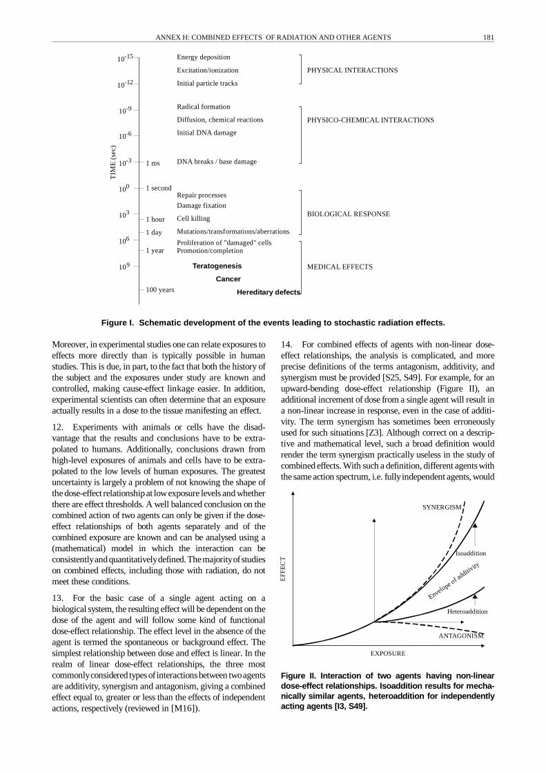
9. On a mechanistic level, insights gained in more recentyears indicate that a much more refined classification may beneeded. The main classes of genotoxic and non-genotoxicagents must be considered in relation to specific targets ofaction. For example, a chemical mayact specificallyat the siteof a radiation-induced lesion, modifying DNA repair fidelity,or it may modify cell growth, strongly influencing the clonalexpansion of precancerous cells. The many possibilities forinteraction are related to the complexityof the development ofthe radiation effect and the many steps involved incarcinogenesis. These steps are prone to the influence ofmanyclasses of agents, both endogenous and environmental. Themulti-step process and the many levels of interaction to beconsidered are schematically depicted in Figure I. In view ofthis complexity, it is not surprising that many models, bothdescriptive and mechanistic, have been developed to describethe combined effects ofexposures todifferent agents [B11, L2,L8, L28, M16, S15, S16, S23, S25, Z1]. In the UNSCEAR1982 Report [U6], the Committee reviewed these approaches.
10. Although classical epidemiology is important inidentifying critical combined effects, it has little potential fordissecting such interactions from the complex interplay
possible among the undocumented (and sometimes unknown)exposures that the individuals in these studies incur duringtheir lifetimes. In epidemiological studies, effects that may beassociated with exposures to specific agents or circumstancesmay be the result of interactions among components of amixture of agents and may have resulted from, or beeninfluenced by, previous exposures. The emerging field ofmolecular epidemiologymaybe able toaddress such questionsin the near future.
11. Most knowledge of interaction effects has been providedbyexperimental studies. These studies have an advantage overepidemiological studies: they retain control of
(a) the population (e.g. selection of systems ranging fromDNA to intact animals and of species, strain, age,gender and previous exposure history);
(b) the exposure (e.g. precise knowledge of the type,dose, dose rate and timing of exposure); and
(c) the endpoints (e.g. selection of sampling time andfrequency, use of invasive and destructive tests,consistency and completeness of health statusevaluations).
ANNEX H: COMBINED EFFECTS OF RADIATION AND OTHER AGENTS 181
10-6
10-12
10-9
10-15
10-3
1 second
1 hour
1 day
1 year
100 years
1 ms
100
109
106
103
Energy deposition
Excitation/ionization
Initial particle tracks
Radical formation
PHYSICAL INTERACTIONS
PHYSICO-CHEMICAL INTERACTIONS
BIOLOGICAL RESPONSE
MEDICAL EFFECTS
Diffusion, chemical reactions
Initial DNA damage
DNA breaks / base damage
Repair processes
Damage fixation
Cell killing
Promotion/completion
Teratogenesis
Cancer
Hereditary defects
Proliferation of "damaged" cells
Mutations/transformations/aberrations
TIM
E(s
ec)
EXPOSURE
SYNERGISM
Isoaddition
Heteroaddition
Envelope of additiv
ity
ANTAGONISM
EF
FE
CT
Figure I. Schematic development of the events leading to stochastic radiation effects.
Moreover, in experimental studies one can relate exposures toeffects more directly than is typically possible in humanstudies. This is due, in part, to the fact that both the history ofthe subject and the exposures under study are known andcontrolled, making cause-effect linkage easier. In addition,experimental scientists can often determine that an exposureactually results in a dose to the tissue manifesting an effect.
12. Experiments with animals or cells have the disad-vantage that the results and conclusions have to be extra-polated to humans. Additionally, conclusions drawn fromhigh-level exposures of animals and cells have to be extra-polated to the low levels of human exposures. The greatestuncertainty is largely a problem of not knowing the shape ofthe dose-effect relationshipat lowexposure levels and whetherthere are effect thresholds. A well balanced conclusion on thecombined action of two agents can only be given if the dose-effect relationships of both agents separately and of thecombined exposure are known and can be analysed using a(mathematical) model in which the interaction can beconsistentlyandquantitativelydefined. Themajorityofstudieson combined effects, including those with radiation, do notmeet these conditions.
13. For the basic case of a single agent acting on abiological system, the resulting effect will be dependent on thedose of the agent and will follow some kind of functionaldose-effect relationship. The effect level in the absence of theagent is termed the spontaneous or background effect. Thesimplest relationship between dose and effect is linear. In therealm of linear dose-effect relationships, the three mostcommonlyconsidered typesofinteractionsbetween twoagentsare additivity, synergism and antagonism, giving a combinedeffect equal to, greater or less than the effects of independentactions, respectively (reviewed in [M16]).
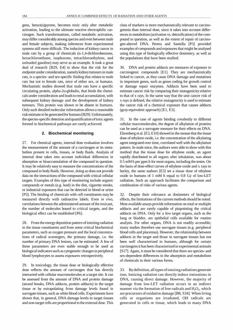
14. For combined effects of agents with non-linear dose-effect relationships, the analysis is complicated, and moreprecise definitions of the terms antagonism, additivity, andsynergism must be provided [S25, S49]. For example, for anupward-bending dose-effect relationship (Figure II), anadditional increment of dose from a single agent will result ina non-linear increase in response, even in the case of additi-vity. The term synergism has sometimes been erroneouslyused for such situations [Z3]. Although correct on a descrip-tive and mathematical level, such a broad definition wouldrender the term synergism practically useless in the study ofcombined effects. With such a definition, different agents withthe same action spectrum, i.e. fullyindependent agents, would
Figure II. Interaction of two agents having non-lineardose-effect relationships. Isoaddition results for mecha-nically similar agents, heteroaddition for independentlyacting agents [I3, S49].

ANNEX H: COMBINED EFFECTS OF RADIATION AND OTHER AGENTS182
produce an apparent synergism in anycombination ofconcen-trations as long as the single dose-effect relationships are bentupwards, as is often the case in the dose range of interest.From a mechanistic point of view, synergism can be definedmore narrowly to imply that agents combine by acting atdifferent rate-limiting steps of a multi-step process or atdifferent sites of a molecule, therebyenhancing the chance fora negative outcome, such as cancer, by different mechanisms[B23]. Such an assessment is often hindered by insufficientknowledge of the underlying mechanism of action andtherefore can rarely be made. Clearly, deviation from additi-vityis a poor indicator of synergism or antagonism, since non-linear dose-effect relationships and threshold phenomena arethe rule rather than the exception for most endpoints inbiological systems, and interaction in the statistical-mathe-matical sense does not define an interaction in a biological-mechanistic sense [B69].
15. In this Annex the term synergism will be used in anarrow sense. The most important question is whether data oncombined effects do show some modification of stochasticradiation effects as a result ofcombined exposure with anotheragent. If not, no interaction will be assumed, and the resultingeffect is additive; if the result of combined exposure isdifferent, some form of interaction has to be assumed, and theresulting effect will be called sub- or supra-additive, depend-ing on whether the effect is lesser or greater, respectively, thanthe sum of the single-agent effects separately.
2. Radiation effects and effects of other agents
16. As far as carcinogenesis is concerned, the primaryeffects of ionizing radiation are on DNA, compromisingcell survival, cell proliferation, and proper physiologicalcell functioning. Although the deposition of energy alongthe track of ionizing radiation can directly affect DNA,most of the damage to DNA from low-LET radiationcomes from the formation of radical intermediates stableenough to diffuse several nanometers and interact withcritical cellular constituents (for details see Annex F,“DNA repair and mutagenesis”). Only a small fraction ofthe radiation-induced molecular modifications occur in theDNA of the cell nucleus, but practically all experimentaland theoretical evidence indicates that DNA, the maincarrier of genetic information in living matter, is thecritical target. Especially at the doses under considerationin this Annex, damage to structural and functional proteinsand lipids has not been shown to contribute noticeably tothe detriment from ionizing radiation. To protect theintegrity of the genetic information, most cells have highlyintricate enzyme systems to repair DNA damage efficientlyand effectively based on information contained within theundamaged complementary DNA strand. Despite that,residual fixed damage may result even from low-doseexposures, especially when both DNA strands aredamaged. Such damage may lead to reproductive celldeath, and therefore possible deterministic effects; tosomatic cell mutation, enhancing the risk of cancer; or tomutations in germ cells, with possible deleterious effects inoffspring.
17. Longer wavelength radiation, such as ultraviolet(UV) light, although not ionizing itself, still acts mainly bymodifying DNA. The UV portion of the electromagneticspectrum covers the wavelengths between 200 and 400 nm.Conventionally, a distinction is made between UV-C(200�280 nm), UV-B (280�320 nm), and UV-A (320�400 nm). The effects of UV light depend on the wavelengthand the absorption properties of the target. Ultravioletradiation mainlycauses the formation ofpyrimidine dimersand 6�4 photoproducts, which may also lead to residualDNA damage after repair. Apart from visible light up to525 nm, which can still interact with photosensitizers togenerate reactive species and, subsequently, oxidativedamage to DNA, infrared, microwave, and low-frequencyelectromagnetic radiation have no direct genotoxic effectsof their own. Indirect effects might arise from local heatingor from charge effects across membranes activating signaltransduction pathways and neurons. Such cellular changesmay be long-lasting or even be passed from one cell to itsprogeny. Sugahara and Watanabe [S10] reviewed theepigenetic aspects of radiation carcinogenesis. Studies usingcell culture systems show that magnetic fields, depending ontheir frequency, amplitude, and wave form, interact withbiological systems. Such effects have been seen on enzymesrelated to growth regulation, on intracellular calcium balance,on gene expression, and on peripheral levels of the oncostatichormonemelatonin [H45]. Theseeffectsarepotentiallyrelatedto tumour promotion. However, the considerable researchconducted thus far has not elucidated critical mechanisms orrevealed important health risks from non-thermal exposures.Other than crude effects present only at high exposures, forexample strong irritations or protein denaturation, cellularperturbations resulting from non-ionizing radiation cannot belabelled harmful per se.
18. Chemical agents may act as genotoxicants by, forexample, forming direct covalent links, by transferringreactive molecular subgroups to DNA, by inducing DNA-DNA or DNA-protein cross-links, or by generating strandbreaks. The mode of action may be direct, by the formation ofsmall or bulky DNA adducts as well as strand breaks, orindirect, by the formation of radicals in the vicinity of DNA,leading to strand breaks or small adducts. On the epigeneticor non-genotoxic level, chemicals may interfere with DNAsynthesis or repair or mayprevent radical scavenging, therebypromoting DNA damage. Non-genotoxic agents may alsoinfluence a broad spectrum of other cellular events. Ofconcern in cancer induction is any interference with cellproliferation, cell differentiation, cell senescence, andapoptosis or with the regulation of these processes.
19. Biological agents may also act at the genetic andepigenetic levels, i.e. theymaybe genotoxic or non-genotoxic,respectively. Virusesareeffectivetransport vectors for genomefragments and may activate or block the expression ofendogenous genetic information. Viral involvement in manyanimal tumours and also in human malignancies is wellestablished, e.g. the DNA tumour viruses of the papillomafamily in cervical carcinoma and the retroviruses HTLV-1 inadult T-cell leukaemia (reviewed in [H13]). In addition,

ANNEX H: COMBINED EFFECTS OF RADIATION AND OTHER AGENTS 183
biological-agent-induced influences on immune responses,inflammation, fever, and endogenous radicals may lead tocytotoxic and/or growth stimulatory responses that are co-carcinogenic, as described later.
B. EXPOSURE ASSESSMENT
20. The most important prerequisite for a comparativeassessment of biological effects of different agents, and also oftheir possible interactions, is the characterization of theexposure or the dosimetry of both agents that may be relatedto subsequent effects. Some of the main concepts used intoxicology and radiation biology to convert exposures intomeaningful measures of dose and health impact are intro-duced in the following paragraphs.
21. The toxicity of an agent can be defined as its inherentability to adversely affect living organisms. The spectrum ofundesired effects is very wide, ranging from local, reversibleeffects to irreversible changes leading to the failure of criticalorgan systems and then to death. The objective of dosimetryis to relate the amount of agent presented to the organism ina way that is relevant to the effects observed and that ismeasurable in a physical, chemical, or biological manner.Identification ofprocessesoccurringat themolecular level, i.e.at a mechanistic level of the effect, would give the most basicindication of a dosimetric measure. The present approachesand possibilities are discussed below. In Section I.B.1,dosimetry based on the measurement of physical or chemicalparameters of the agent itself, the physical or chemicaldosimetry, is considered. In Section I.B.2, measurement ofimmediate biological damage caused by the agent (biochemi-cal monitoring) is discussed; this damage may or may not bedirectly related to the biological effect being considered.
22. Sometimes, when physical, chemical, or biochemicalmeasurements are not possible or cannot be made accuratelyenough, certain biological effects may be detectable. Sucheffects may serve as indicators of the exposure to biologicallyactive agents. These “biological markers“ reflect damageresulting from toxic interaction, either at the target or at ananalogous site that is known or believed to be pathogenicallylinked to health effects. A wide variety of biological markersfall into this category, including gene mutation; alterations inoncogenes and tumour-suppressor genes; DNA single- anddouble-strand breaks; and unscheduled DNA synthesis, sisterchromatid exchanges, chromosomal aberrations; andmicronuclei. None of these markers is highly agent- orexposure-specific, and other factors (lifestyle and environ-ment) that affect these endpoints can act as confoundingvariables in molecular studies. Some possibilities for assaysystems to measure biological markers such as specific genemutations and cytogenetic damage in exposed humans arepresented in Sections I.B.3 and I.B.4, respectively.
1. Dose concepts for physical andchemical agents
23. Ionizing radiation exposure is generally measured interms of absorbed dose, i.e. the average energy deposited
per unit mass. The unit of absorbed dose is the gray (Gy),with 1 Gy equal to 1 J kg�1 [I3]. At the level of a cell or cellnucleus, the minimal dose is determined by the ionizationdensity of a single track. Averaged over the volume of acell nucleus, a single event amounts to between one andseveral milligray (mGy) for electrons and about 300 mGyfor an alpha particle [U3]. Below these dose levels, theprobability of a cell being hit varies but the absorbed doseper cell nucleus does not. For internally deposited radio-nuclides, their location and fate in the organism are usedto calculate the absorbed dose in the organs of interest, andusually the average absorbed dose in the organ is taken asthe relevant dose that causes the biological effect, assuminga rather homogeneous distribution of energy absorption inthe tissue.
24. The definition of exposure or dose for non-ionizingradiation and for most chemical and biological agents is moredifficult than for radiation. Ultraviolet radiation can penetrateinto tissue at most only for several millimetres, depending onwavelength. The energyabsorbed in the tissue of interest, andthus the effectiveness of UV, cannot be easily estimated.Exposure to a toxic agent may be estimated by environmentalmonitoring (referred to as external dose evaluation intoxicology), internal monitoring (internal dose evaluation),and biochemical effect monitoring (tissue dose or biologicallyeffective dose determination) [E1].
25. For chemical and biological agents, the dose can bebased on the time integral of concentration, as for internalexposures with radionuclides. However, in addition to thecommon important question of defining the critical cellulartargets, it is the activation and biodegradation of a chemicalagent in the different compartments of the organism that willdetermine the degree of genetic damage or strength of anepigenetic signal. Although the local concentrations ofreceptors or reactants could possibly be estimated ordetermined, these vary considerably in their response toendogenous and environmental factors, which can lead todifferent sensitivities to the physical or chemical agent. Thismay restrict the use of biochemical markers somewhat,because their concentrations in body fluids will depend on themechanisms of uptake, the formation of reactive molecularspecies, and their breakdown. Somewhat like the dose conceptfor ionizing radiation, exposure can be related to the numberof primary chemical events on DNA leading to the effectunder consideration. The above-mentioned quantitative linkbetween DNA alkylation and the product ofconcentration andtime for ethylene oxide may serve as an example [E3] (seealso paragraph 34). However, only rarely is the nature of suchevents known or quantifiable.
26. To give exposure (or dose) its full biological meaning,the concentration-time product at the level of the cellulartarget structure should be known. Even this is difficult todetermine owing to the many membranes and other barriersto be crossed between the intake port and the place of action.Many chemicals also undergo modifications bydetoxificationin the liver, lung, and other organs, which change both theirtoxicity and their biokinetics. One of the best known carcino-
ANNEX H: COMBINED EFFECTS OF RADIATION AND OTHER AGENTS184
gens, benzo(a)pyrene, becomes toxic only after metabolicactivation, leading to the ultimate reactive electrophilic car-cinogen. Such transformations, called metabolic activation,maydiffer considerablyamong species and even between maleand female subjects, making inferences from experimentalsystems still more difficult. The induction of kidney cancer inmale rats by a group of chemicals (n-1,4-dichlorobenzene,hexachloroethane, isophorone, tetrachloroethylene, andunleaded gasoline) may serve as an example. It took a greatdeal of research [B29, E4] to show that the risk for theendpoint under consideration, namelykidneytumours in malerats, is a species- and sex-specific finding that relates to malerats but not to female rats, mice of either sex, or humans.Mechanistic studies showed that male rats have a specificcirculating protein, alpha-2u-globulin, that binds the chemi-cals under consideration and leads torenal accumulation, withsubsequent kidney damage and the development of kidneytumours. This protein was shown to be absent in humans.Onlysuch detailed molecular information allows a reasonableriskestimatetobe generated for humans[B29]. Unfortunately,thespecies-specificdetection andquantification oftoxicagentsformed in biochemical pathways are rarely achieved.
2. Biochemical monitoring
27. For chemical agents, internal dose evaluation involvesthe measurement of the amount of a carcinogen or its meta-bolites present in cells, tissues, or body fluids. Analysis ofinternal dose takes into account individual differences inabsorption or bioaccumulation of the compound in question.It may be relatively easy to measure the concentrations of thecompound in bodyfluids. However, doing so does not providedata on the interactions of the compound with critical cellulartargets. Examples of this type of monitoring include organiccompounds or metals (e.g. lead) in the diet, cigarette smoke,or industrial exposures that can be detected in blood or urine[P3]. The binding of chemicals with cell constituents may bemeasured directly with radioactive labels. Even in vivo,correlations between the administered amount of the toxicant,the number of molecules bound to critical targets, and thebiological effect can be established [P6].
28. From the energydeposition pattern of ionizing radiationin the tissue constituents and from some critical biochemicalparameters, such as oxygen pressure and the local concentra-tions of radical scavengers, the primary damage, i.e. thenumber of primary DNA lesions, can be estimated. A few ofthese parameters are even stable enough to be used asbiological indicatorssuch as cytogenetic changes in peripheralblood lymphocytes to assess exposures retrospectively.
29. In toxicology, the tissue dose or biologically effectivedose reflects the amount of carcinogen that has directlyinteracted with cellular macromolecules at a target site. It canbe assessed from the amount of DNA and protein damage(strand breaks, DNA adducts, protein adducts) in the targettissue or by extrapolating from damage levels found insurrogate tissues, such as white blood cells. Experiments haveshown that, in general, DNA damage levels in target tissuesand non-target cells are proportional to the external dose. This
class of markers is more mechanistically relevant to carcino-genesis than internal dose, since it takes into account differ-ences in metabolism (activation vs. detoxification) of the com-pound in question, as well as the extent of repair of carcino-gen-altered DNA. Perera and Santella [P3] providedexamples ofcompounds and exposures that might be analysedusing this type of biologically effective dosimetry, as well asthe populations that have been studied.
30. DNA and protein adducts are measures of exposure tocarcinogenic compounds [E1]. They are mechanisticallylinked to cancer, as they cause DNA damage and mutationsin important genes, such as genes coding for growth controlor damage repair enzymes. Adducts have been used toestimate cancer risk by comparing their mutagenicity relativeto that of x rays. In the same way that the unit cancer risk ofx rays is defined, the relative mutagenicity is used to estimatethe cancer risk of a chemical exposure that causes adducts(gray-equivalent approach) [E1, E3].
31. In the case of agents binding covalently to differentcellular macromolecules, the degree of alkylation of proteinscan be used as a surrogate measure for their effects on DNA.Ehrenberg et al. [E3, E10] showed in the mouse that the tissuedose of ethylene oxide, i.e. the concentration of the alkylatingagent integrated over time, correlated well with the alkylationpattern. In male mice, the authors were able to show with thismethod that the tissue dose for ethylene oxide, an agentrapidly distributed to all organs after inhalation, was about0.5 mM h per ppm h for most organs, including the testes. Onthe basis of dose-effect curves of ethylene oxide and x rays inbarley, the same authors [E3] set a tissue dose of ethyleneoxide in humans of 1 mM h equal to 0.8 Gy of low-LETradiation. Such an approach facilitates the comparison andcombination of risks of various agents.
32. Despite their relevance as dosimeters of biologicaleffects, the limitations of the current methods should be noted.Most available assays provide information on total or multipleadducts and are rarely capable of pinpointing the criticaladducts on DNA. Only for a few target organs, such as thelung or bladder, are epithelial cells available for routineanalysis. For other organs, DNA is not readily accessible;many studies therefore use surrogate tissues (e.g. peripheralblood cells and placentas). However, the relationship betweenadducts in the target and those in surrogate tissues has notbeen well characterized in humans, although for certaincarcinogens it has been characterized in experimental animals[S17]. Again, it must be considered that there are species- andsex-dependent differences in the absorption and metabolismof chemicals in their various forms.
33. Bydefinition, all types of ionizing radiations generateions. Ionizing radiation can directly induce ionizations inDNA, causing direct damage. However, the majority ofdamage from low-LET radiation occurs in an indirectmanner via the formation of free radicals and H2O2, whichare precursors of oxidative damage [B8, S34]. When livingcells or organisms are irradiated, OH radicals aregenerated in cells or tissue, which leads to many DNA

ANNEX H: COMBINED EFFECTS OF RADIATION AND OTHER AGENTS 185
lesions, including oxidative DNA base products. Bothionizing radiation and oxidative stress generate freeradicals near DNA. Most of these radicals (�R) interactwith oxygen, forming peroxyl intermediates (�ROO) andfinal products (�P). Most of the products are eliminated bynucleotide excision repair and glycosylases [F10], while asmall fraction remain in the DNA [S35]. Critical arelesions leading to double-strand breaks or even morecomplex local damage.
34. Free radicals are difficult to detect, identify, andmonitor because of their short half-life, particularly in livingorganisms. Such detection and monitoring can be achievedonlybydetecting and measuring the products of their reactionwith endogenous bio-components or exogenous componentsselectively added to a biosystem. Specific products of suchreactions or their metabolites may qualify as markers of aparticular process or specific free radical. In biosystems, theseproducts are called molecular markers, a subclass of bio-markers [G9]. For a product to qualify as a molecular marker,there must be unequivocal proof of an exclusive origin of theproduct. First, a comprehensive understanding of the kinetics,energetics, and mechanisms ofproduct generation is required.Then other possible sources of the product must be excluded[S39].
35. Although a molecular marker can be quantified bymeasurement in vivo, quantification of oxidative stress isconsiderably more complex. The reactivity of all five bases,adenine, cytosine, guanine, thymine and uridine, with OHradicals is extremely high, whereas that of deoxyribose isabout five times lower [B34]. The distribution of damage willtherefore be governed by the relative abundance and reactivityof DNA and RNA components. Each DNA and RNA basecontains more than one site of attack. For example, OH addsto the double bond of thymine at C-5 (56%) and C-6 (35%)and removes hydrogen from the methyl group (9%) [J7]. The5-hydroxythymidine intermediate leads to formation ofthymine glycol. The 6-hydroxythymidine intermediate is anoxidizing radical that gives rise to unstable hydroxy-hydrothymine. The radical on the methyl group of thymine,however, is a reducing radical that yields 5-hydroxymethyl-uracil as the final product (reviewed in [S39]). Addition ofOHto the C-8 position of guanine yields a well-known product,8-hydroxyguanine or 8-oxoguanine, which was discovered byKasai et al. [K1, K47] and described in detail [J4, S39].
Numerous other products have been identified, and thekinetics and mechanisms of their formation have beendescribed [B8, S12, S34, S36].
36. On the basis of extensive studies in radiation chemistryand radiation biology of the kinetics and mechanisms of OHradical reaction with DNA components, it was suggested thatdetection of thymine glycol, thymidine glycol, and 5-hydroxy-methyluracil indicated endogenous OH generation in rats andhumans [C3, H18, W8]. Because thymine glycol can beabsorbed through the gastrointestinal tract and 5-hydroxy-methyluracil maybe generated byenzymatic hydroxylation ofthymine, these products maynot always qualifyas biomarkersfor oxidative damage in organisms. Thymidine glycol is lessprone to such confounders and qualifies as one of the bestendogenous markers of OH [S39]. It was suggested that8-hydroxyguanine could be another OH marker in biosystems[B12, F3, K1, R9, S30, W12]. Enzymatic hydroxylation ofguanine, however, has not been ruled out unequivocally.Hence it is prudent to monitor more than one marker for eachspecific free radical under investigation. 8-Hydroxyguanosinewas analysed in the DNA of peripheral blood leukocytes ofpatients exposed to therapeutic doses of ionizing radiation[W12]. Radiation-generated oxidative DNA base productswere also measured in the DNA of irradiated cells [N2].
37. The chemical reaction products in DNA are excisedfrom damaged DNA over a certain period of time by repairmechanisms and eventually appear in the cell medium orurine. Some oxidative DNA base products have beenmeasured in the urine of irradiated humans and mice. Theradiation yields of these markers, i.e. the increments per unitof energy(mass × dose), were obtained from the level one dayafter irradiation minus the level before irradiation and areshown in Table 2. In contrast to the metabolic levels of thesemarkers, the irradiation yields per unit energyare the same forboth mouse and human, as expected, because the samenumber of OH radicals is generated in both cases [S39]. Themetabolic rate plays an important role in the variability ofrelative rates of oxidative DNA damage. A high metabolicrate, as in rodents, generates a high yield of urinary markers,i.e. higher rates of DNA damage. The rate of DNA damage,however, is not always proportional to the specific metabolicrate because the efficacy of inhibition and scavenging ofoxygen radicals and peroxides as well as of DNA repairsystems varies in different species.
Table 2Yield in urine of biological markers of oxidative DNA damage [B12]
SpeciesSpecific
metabolic rate(kJ kg-1 d-1)
Metabolic yield(nmol kg-1 d-1)
Increment induced by radiation(nmol kg-1 Gy-1)
Thymidine glycol 8-Hydroxy-guanine Thymidine glycol 8-Hydroxy-guanine
Human 100 0.3±0.1 0.3±0.1 3.1±0.8 6.7±1.5
Mouse 750 7.3±1 11±2 3.0±0.6 6.9±1.3
ANNEX H: COMBINED EFFECTS OF RADIATION AND OTHER AGENTS186
3. Gene mutation analysis
38. The analysis and quantification of genomic changes areimportant steps in monitoring and in the elucidation ofmechanisms leading to critical health effects. Functionalchanges, i.e. changes in the phenotype of oncogenes andtumour-suppressor genes, may be of direct relevance to theprocess of carcinogenesis. The ultimate effects of ionizingradiation and other genotoxic agents are genetic changes,which are heritable, i.e. which can be passed in a clonalfashion from one somatic cell to the following cell genera-tions, or from a germ cell to the offspring. Therefore criticalstudies of combined effects should include gene mutationanalysis as one very important biological endpoint forstochastic health effects. Several methods are available for thestudy of gene mutations arising in human somatic cells invivo. These methods allow determination of the frequency ofmutant lymphocytes or erythrocytes or characterization ofmutations at the molecular level in lymphocytes. The study oftypes, frequencies, and mechanisms of human somatic muta-tions in vivo is valuable in its own right and may alsoimprove the understanding of individual variation in sensiti-vity to environmental exposures, the influence of DNA repairand metabolism, and the relationship between mutagenesisand carcinogenesis [L7, M21]. Genetic changes arekeyeventsin carcinogenesis. Most human tumours contain more or lessspecific mutations that are directly or indirectly related to thecarcinogenic process. A description of mutations in humantumours and thescientific background ofmanyof the conceptsand methods addressed in this and the following Chapter arepresented in more detail in Annex F, “DNA repair andmutagenesis”.
39. Early and probably single-step biological end points,such as morphological changes in in vitro cell lines, mightalso serve as indicators of genetic changes. The developmentof cell culture systems has made it possible to assess theoncogenic potential of a variety of agents at the cellular level.Many assays for oncogenic transformation have beendeveloped, ranging from those in established rodent cell lines,where morphological alteration is scored (e.g. loss of contactinhibition in 10T½ cells), to those in human cells growing innude mice, where tumour invasiveness is determined. Themutational changes involved are rarely defined. In general,simple in vitro systems that deliver reproducible results are theleast relevant in terms of human carcinogenesis and humanrisk estimation. The most important potential of these systemslies in the opportunity they offer to identify and quantifyfactors and conditions that prevent or enhance cellulartransformation by radiation and chemicals [H11].
(a) Mutation frequencies
40. Five systems for biomonitoring humans exposed tocarcinogenic agents havebeen developed in which gene muta-tion is the endpoint. Two of these use as markers haemo-globin variants (Hb) [S18, T4] and loss of the cell-surfaceglycoprotein glycophorin A (GPA) in donors heterozygous atthe MN locus in erythrocytes [L1, L3, L6]. The other threeinvolve detection of mutations in T lymphocytes in the
X-linked locus for the purine salvage pathway enzyme,hypoxanthine phosphoribosyltransferase (hprt) [A5, A6, A8,M22, R7, R8, T10], in the autosomal locus for humanleukocyte antigen-A (HLA-A) [J3, M18, T7, T8], and in theautosomal T-cell receptor genes (TCR) [K23, K24, N3, U15].
41. Mean background mutation frequencies in human cellsin vivo, as analysed by the five mutation assays, differ byabout four orders ofmagnitude. In summary, the relative orderof background mutation frequency values from normal adultsfor the five markers are Hb (5 10�8) < hprt (5 10�6) < GPA(1 10�5) < HLA-A (>1 10�5) < TCR (>1 10�4) (reviewed in[C23]). For at least three of these mutation systems, sufficientnumbers of donors have been tested to show that, as a generalrule, the mutant frequency in normal, non-exposed donors islow at birth, increases with age, is often elevated in smokers,and is increased in people who have been exposed to knownmutagens and carcinogens. Despite the great variation inmutant frequency among individuals at each of the locistudied, these findings show the potential relevance of muta-tional analysis in the assessment of combined environmentalexposures. More recently, the polymerase chain reaction(PCR) has also been applied in the analysis of mutationalspectra. A fairly complete database has been compiled byCariello et al. [C51, C52].
42. The frequencies of hprt mutant cells in healthy adultsrange from <0.5 to 112 10�6. In most cases the frequency ofhprt mutant cells is significantly increased after smoking[C17, C19, H2, T4, T11]. There seems to be no effect of sexon the hprt mutant frequency. In most studies, an age-relatedincrease in mutant frequency is seen at the hprt locus,estimated to be 1%�5% per year in adult donors. Radio-chemotherapy for various malignant disorders, includingbreast cancer, hepatoma, other solid tumours, and lymphomaincreased the frequency of hprt mutant T cells by a factor of3�10 [D5, M20, N8]. Cole et al. [C18, C20] examined factoryworkers exposed to styrene or to nitrogen mustard. In contrastto styrene, nitrogen mustard significantly increased thenumber of mutant hprt cells in these donors. Tates et al. [T9]described a significantly increased mutant frequency in agroup of factory workers exposed to ethylene oxide.
(b) Mutational spectrum
43. The spectrum of mutational changes that arisespontaneously or that may be induced by a physical orchemical agent in human cells is broad. At the DNA level itencompasses, at one extreme, single-base events, and at theother, chromosomal rearrangements involving small to largedeletions or translocations. In addition, an important categoryof mutational events in humans involves losses or gains ofwhole chromosomes. The mutation spectrum in themammalian genome is reviewed in Annex F, “DNA repairand mutagenesis”.
44. Many known mutagens form covalent DNA adductsthat are released from DNA either spontaneously or by bio-logical repair processes [H16]. Mutations induced by a largenumber of compounds, e.g. alkylating agents, arylating

ANNEX H: COMBINED EFFECTS OF RADIATION AND OTHER AGENTS 187
agents, and radiation, have been scored and characterizedusing shuttle vectors. These experiments elucidate thesequence specificity of adduct formation and, subsequently,the mutations and the mutational efficiencies of differentadducts [D9, I6, M10].
45. About 15%�20% of hprt mutations in normal adultsresult from gross structural alterations [A8, B31, H8, N7,T10], as detected by Southern blot analysis. These includedeletions, insertions, andrearrangements. The break pointsor alterations are distributed randomly within the gene,with no hot spots having thus far been identified [A8]. Theremaining 80% of the background in vivo hprt mutationsin adults consist of point mutations or small deletions,insertions, and frameshifts beyond the resolution ofSouthern analysis. Considering only the in vivo hprtmutations (46 Lesch-Nyhan germinal, 51 normal adultsomatic, 86 exposed adult somatic), several hot spots ofpoint mutations were observed. In particular, four base-pairsites have been observed to be mutated in all groups [C13].
46. Ionizing radiation is known to induce gross structuralalterations in hprt and other reporter genes in cultured humancells. After exposure to radionuclides for diagnostic purposes,an increase in the frequency of mutants with gross structuralalterations on Southern blots was observed to be 33%,compared with 13% before receiving radionuclides [B31].Mutations from post-radioimmunotherapy patients showedclearly greater frequencies of gross structural alterations thanmutations from pre-radioimmunotherapy patients or normalindividuals. The latter two frequencies are quite similar,suggesting that cancer per se does not produce this sort ofdamage at hprt [A9]. Taken in toto, the data from Albertini etal. [A9] on in vivo hprt T-cell mutations indicate thationizing radiation produces deletions, particularly largedeletions.
47. The yield ofmutations caused byionizing radiation maybe influenced strongly by adaptive responses to othertoxicants or earlier exposures to the same agent. This topicwas reviewed in Annex B, “Adaptive responses to radiation incells and organisms” of the UNSCEAR 1994 Report [U2]. A70% reduction in hprt mutant frequency in radioadaptedhuman lymphoblastoid cells has been reported, as analysed bySouthern blot analysis and multiplex polymerase chainreaction assay [R10, R12]. The treatment was 4 Gy fromgamma rays alone or in addition to an adaptive dose of0.02 Gy. The proportion of deletion-type mutations wasdecreased in adapted cells (42%) compared with that inmutants treated with the high dose alone (77%).
48. Using a shuttle vector system, Kimura et al. [K12]analysed mutational spectra of the human cDNA hprt gene, arecombinant DNA copy of the hprt RNA, arising spontan-eouslyor induced bythe mutagens methylnitrosourea (MNU);3-amino-1-methyl-5H-pyridol[4,3-b-]-indole (Trp-P2), atryptophan pyrolysate; and acetylaminofluorene (AAF). Mostmutations induced by MNU are G:C to A:T transitions. Thiscan be predicted by the major premutagenic lesion in DNAproduced by MNU, namely O6-methyl-guanine that specifi-
cally mispairs with thymine [S41]. Mutations that arise spon-taneously or are caused by x rays, Trp-P2, or AAF give rise toa similar mutation spectrum of c-hprt. Base substitutionsaccount for about one third of all mutations. Mutations otherthan base substitutions make up some two thirds of allmutations. The main mutational event in these cases is dele-tion. A noticeable feature of these deletion mutations is thefrequent presence of short, direct repeats at the site of thedeletion.
49. Mutational alterations in p53, a tumour-suppressorgene, are mostly (more than 85%) missense mutations, whilethose of APC, another tumour-suppressor gene, and hprt arelargely composed of nonsense, frameshift, deletion, andinsertion mutations, resulting in truncated gene products orloss of genes. The mutational spectrum in p53 is thereforeclearlydifferent from that of other genes. Mutations in the p53gene detected in tumours seem to be the result of a functionalselection process for mutant p53 protein that gives growthadvantages to the cell. On the other hand, large deletions inthe p53 region may not be compatible with cell survival. Thissuggests that the mutations of p53 observed in tumours mayreflect only those mutations of the initial events that arecompatible with cell proliferation and may even reflect thosethat give the transformed cell a growth advantage over thesurroundingcells.Mutational selectivityin tumour-suppressorgenes is discussed in detail in Annex F, “DNA repair andmutagenesis”.
50. With respect to interaction mechanisms leading tocombined effects, present knowledge indicates that themutational spectrum found in tumours often reflects not theagent responsible for the primary DNA damage but rathergrowth selection based on specific changes in the phenotypeor general chromosome instability emerging during carcino-genesis. Analysis of marker cells in peripheral lymphocytesmayovercome this problem, albeit at the expense of losing thedirect link to human disease.
4. Cytogenetic analysis
51. The main conceptual basis for using cytogenetic assaysfor biological monitoring is that genetic damage in easilyavailable cells, such as peripheral blood lymphocytes, reflectscomparable events in target cells. The fact that chromosomalabnormalities are often a characteristic feature in malignantcells points to the direct relevance of such markers forclastogenic agents to be considered in combined exposures. Inaddition, long-term follow-up of populations screened forchromosomal aberrations shows a clearly higher cancer riskfor the subgroup with an elevated level of chromosomedamage [B20, H5]. Microscopically recognizable chromo-somal damage includes numerical aberrations and structuralchromosomal aberrations, in which a gross change in themorphologyofa chromosomehas occurred. Chromosome andchromatid breaks, dicentrics, and ring chromosomes areimportant examples of this class of damage [N11]. The yieldof sister chromatid exchanges, which represent apparentlysymmetrical intrachromosomal exchanges between the twoidentical sister chromatids and which are already quite

ANNEX H: COMBINED EFFECTS OF RADIATION AND OTHER AGENTS188
frequent in unexposed cells, is also increased. Micronucleiarising either from acentric chromosome fragments or from alagged whole chromosome with centromere [W15] are alsoimportant markers, although the second production pathwaypoints to a mechanism driven partially by epigenetic factors.
(a) Chromosomal aberrations
52. Induced chromosomal aberrations can be divided intotwo main classes: chromosome-type aberrations, involvingboth chromatids of a chromosome, and chromatid-typeaberrations, involving only one of the two chromatids.Ionizing radiation induces chromosome-type aberrations inthe G0 or G1 stage of the cell cycle (e.g. prior to replication),while chromatid-type aberrationsare produced during the S orG2 stage (e.g. during or after replication of the affectedchromatid segment). In peripheral lymphocytes, most ofwhich are in the G0 stage of the cell cycle, ionizing radiationinduces mainly chromosome-type aberrations.
53. Most chemical mutagens are S-dependent clastogensand therefore produce mainly chromatid-type aberrations.S-dependent compounds have no direct effect on the chromo-somes of peripheral lymphocytes in vivo, because theyreplicate only after stimulation in cell culture. Peripherallymphocytes can, however, carry unrepaired/misrepaired,long-lived lesions that may lead to aberrations duringreplication of DNA in vitro [S28].
54. The classical chromosome aberration assay formeasuring dicentrics is a reasonably good measure of dosedown to 100 mGy whole-body exposure [L31] or, withmuch effort, even lower. However, it is based on a geneticchange that considerably impairs the survival of indicatorcells and their stem cells, so that the signal fades withtime. Reciprocal translocations are considered lessdisruptive to the proliferative future of affected cells. It ispossible to score translocations with G-banding or FISH(fluorescent in situ hybridization) techniques, with thelatter technique having a higher detection limit, about500 mGy. In such systems, the preferential loss of affectedcells may still be a minor problem; in addition, clonalexpansion of cells carrying translocations conferring agrowth advantage may lead to an overestimation of thedose with time. Biological dosimetry using cytogeneticparameters will be discussed later. It seems that all agentsthat apparently induce single-base changes (i.e. basedeletions, transversions, or transitions) also induce grosschromosomal changes that are visible under themicroscope. However, the number of agents clearly shownto induce cytogenetic changes in humans is still relativelylimited [A24, S31]. From known or suspected carcinogenicagents, mixtures, or complex exposures to humans,cytogenetic data are available for 27 compounds inGroup 1 of the IARC classification (known carcinogens tohumans), for 10 compounds in Group 2A (probablecarcinogens to humans), and 15 compounds in Group 2B(possible carcinogens to humans) [I1, I2]. Chromosomedamage in humans was found in 19/27, 6/10, and 5/15cases in these groups, respectively.
55. Most of the informative data on induced chromosomalaberrations in humans arise from high-exposure occupationalsituations. The comparisons of experimental animal data andhuman data for the endpoint of chromosomal aberration aregenerally in good agreement. However, in a few cases thereare discrepancies between animal and human data. Highoccupational exposure to radon induces chromosomalaberrations in humans. Animal experiments with comparableexposures are negative. The most likely explanation is aconfounding by other clastogenic exposures in humans, e.g.smoking.
56. Unlike radiation exposure, chemical exposures havebeen considered in very few cytogenetic follow-up studies.Studies on the induction of chromosomal aberrations afterexposure to alkylating agents expressed in peripherallymphocytes show, like studies after radiation exposure, thatdamage can be conserved over several months or even yearsafter treatment [G3]. The persistence of chromosome damage,however, varies with the type of exposure and the cytogeneticendpoint examined.
(b) Sister chromatid exchange
57. The induction of sister chromatid exchange can beobserved in cells that have undergone two rounds of DNAreplication in the presence of bromodeoxyuridine (BrUdR),which results in chromosomes having sister chromatids thatare chemically different from one another: one is unifilarlylabeled with BrUdRand the other bifilarly labeled. Such sisterchromatids stain differently from one another, and anyexchanges that occur between the sister chromatids can beclearly seen and counted [W7]. A number of studiesconfirmed the ability of low-LET radiation to induce sisterchromatid exchanges in rodent cells [G4, L22, R5, U14] andhuman lymphocytes [G14]. However, in other studies, whennormal human lymphocytes in G0 were assessed for theirability to express sister chromatid exchanges following low-LET radiation exposure, theyfailed to do so, in contrast to thequantifiable induction of chromosomal aberrations [L21,M28, P2]. This difference could possibly be attributed to thepresence of BrUdR, a known radiosensitizer, at the time ofirradiation in the rodent cell studies [L25]. Nevertheless, low-LET ionizing radiation and radiomimetic chemicals are notveryeffectiveat inducing sister chromatidexchanges, contraryto S-dependent agents such as UV light [W11], alkylatingagents [T1, Y4], and cross-linking agents [S4]. High-LETradiation (neutrons and alpha particles), however, inducessister chromatid exchanges in normal human peripherallymphocytes exposed in G0. This suggests that the relativebiological effectiveness for sister chromatid exchangeinduction is very large, since there is little low-LET response[A2, S11]. The induction of sister chromatid exchange as afunction of charged-particle LET in Chinese hamster cellswas recently described [G7]. At each LET examined therewas a dose-dependent increase in the frequency of sisterchromatid exchanges. In contrast to the majority of biologicalendpoints, however, where relative biological effectivenessincreases as LET increases up to a maximum and thendeclines, it was found that sister chromatid exchange

ANNEX H: COMBINED EFFECTS OF RADIATION AND OTHER AGENTS 189
induction already declined as LET changed from 10 to 120keV mm�1 [G7]. These observations can be explained on thebasis of repair differences for DNA damage induced byradiations of different LET, i.e. the faster the repair, the lesslikelihood there will be of unrepaired DNA damage at thetime of replication when sister chromatid exchanges areformed.
(c) Micronuclei induction
58. Micronuclei can be formed from entire chromosomes orchromosomefragments [M36]. Theyresult from chromosomebreakage and/or damage to the mitotic spindle and are used asa measure of genotoxicity [H15]. Techniques to blockcytokinesis in mitogen-stimulated lymphocytes [F4, F5, M24,P19] allow these micronuclei to be observed in binucleatedcells found after the abortive attempt of the cell to divide.There is, however, a large and variable background frequencyof some 5�12 micronuclei per 103 binucleated cells [F5, Y2].The background frequency increases with age from about 4per 103 among those �20 years, to 8 per 103 for those �30years, and nearly 12 per 103 for those �40 years [Y2]. Theincrease is about 4% per year [F5]. The range of variabilityincreases with age as well. Farooqi and Kesavan [F18] alsofound that theyield of radiation-induced micronuclei in mousepolychromatic erythrocytes was strongly influenced by smallconditioning doses (25 mGy). Micronuclei assays are fasterand have a greater potential for automation than the scoringof chromosome aberrations [M36].
59. Caffeinated and alcoholic beverages have no significanteffects on in vivo mean micronuclei frequency in binucleatedlymphocytes. Even the intraperitoneal (ip) injection of largeamounts of caffeine (15 mg kg�1 body weight) did not inducechromosomal aberrations in mice [F19]. However, theestimated number of diagnostic x-ray examinations to anindividual in the year prior to measurement was significantly
correlated to micronuclei frequency [Y2, Y5]. The effect ofage and x rays on lymphocyte micronuclei has been shownrepeatedly [A11, E2, F5, I1, I2]. Tobacco smoke and tobacco-related exposures are listed in the IARC Monograph series[I1, I2] as micronuclei-inducing agents.
60. In an analysis of micronuclei frequency in survivors ofthe atomic bombings, Ban et al. [B4] confirmed the agedependency of background micronuclei levels in peripherallymphocytes. Females showed a somewhat higher frequencyof binucleated cells. Age and sex were independently actingfactors. There is no evidence for an effect of radiation dose onpresent-day background micronuclei frequency in thesurvivors.
5. Summary
61. The primary molecular and cellular effects of the manyagents potentially involved in combined effects are extremelydiverse. No unifying concept of dose can therefore be applied.However, comparisons of toxicity may be based on relevantexperimental and clinical endpoints with sometimes onlyloose and enigmatic links to primary lesions and interactions.A large number of quantitative and semi-quantitativeindicators of exposure are presently available. On the level ofgenotoxicity, DNA damage can be measured up to thefunctional level of single genes, thus allowing a comparisonof the biological activityof different agents and an assessmentof possible interactions on a directly relevant level. Theaccessibility of critical cells and tissues to standard analysisremains a problem. Qualitative and quantitativemonitoring ofbiological effects at the different levels of organization, frommolecules to organisms, not only might allow an assessmentof the exposure to the different agents involved but could alsoform the basis for a better understanding of the mechanismsof combined effects and for the elucidation of dose-effectfunctions for cellular and clinical endpoints.
II. MECHANISTIC CONSIDERATIONS
62. In view of the many different agents that may beinvolved in combined exposures with radiation and thecomplexity of the possible interactions, it is necessary to gainsome insight from the mechanistic point of view. ThisChapter will give a qualitative insight into the interactionprocesses bydescribing important steps in the development ofthe radiation effect and bysuggesting how the radiation effectmight be influenced by other agents. For a quantitativeinsight, various models have been developed to describe thebiological effects. Examples of such models will be brieflydiscussed, in so far as they serve to improve understanding ofthe mechanisms involved in combined effects. However, itshould be kept in mind that models have limited applicability,and agents do not always have only a single mode ofinteraction.
63. Since cancer is the most important health effect forradiation at low doses, the review presented in this Chapter
deals mainly with mechanisms that are central to theemergence of malignant growth. An in-depth review of thescientific background of some of the concepts discussed herewas presented in Annex E, “Mechanisms of radiationoncogenesis”, of the UNSCEAR 1993 Report [U3], Annex F,“DNA repair and mutagenesis” and Annex G, “Biologicaleffects at low radiation doses”.
64. The timescale of events for the various stages ofradiation-induced cancer ranges from less than a second totens of years. Schematically, three crude time-scale-basedphases can be defined on the molecular, the cellular, and thetissue/organ level. The molecular phase ranges from the earlyinteraction of the radiation track until initial damage inbiologically important molecules has occurred (of the order ofseconds). The cellular phase follows and lasts until thebiological reactions of the cells involved have occurred andbiological cellular effects are induced (of the order of a few

ANNEX H: COMBINED EFFECTS OF RADIATION AND OTHER AGENTS190
days). Ultimately, on the tissue/organ level, cellular damagemay progress in due time, with or without cooperation fromother damage, toclinicallydetectable cancer, which can occurup to 40 or more years after the initial irradiation. Thesephases are described below. A schematic representation of theprocesses is given in Figure I. The separation into thesephases is arbitrary; it is time-scale-motivated and serves hereonly to describe the possible interactions of the radiation effectwith other agents. In reality, the processes are not separatedthat rigorously, and interactions with another agent mayoccuron more than one level or phase.
65. Radiation-induced effects other than cancer, such asdeterministic and teratogenic effects, involve similarphases in the development of the radiation damage. Forconciseness, these effects are not explicitly mentioned andconsidered here, but the data in humans are reviewedbriefly in Chapter V.
A. EFFECTS ON THE MOLECULAR LEVEL
66. Following the primary interaction of a radiation trackwith biological matter, an avalanche of events occurs, andvarious reactive species are left after passage of an ionizingparticle or photon: molecules are excited and ionized, radicalsare formed, and secondary electrons progress through thematerial. Most of these species are chemically very reactiveand produce other molecular species. These initial processesdevelop in a very short time (of the order of microseconds)and at short distances from the radiation track. The processesare dependent on the physical and chemical characteristics ofthe material, the type of radiation, and the conditions in theimmediate environment of the target molecule, such as theavailabilityofoxygen, the presence of sensitizing or protectingagents, the ambient temperature, and the ionization densityofthe radiation. The processes involved in the interaction ofradiation at the molecular level are extensively studied inradiation biochemistry and microdosimetry, the concepts ofwhich have been described by the International Commissionon Radiation Units and Measurements (ICRU) [I7].
67. The biological effects of radiation arise mainly fromdamage induced in DNA molecules. Important types of DNAdamage are DNA single- and double-strand breaks, basedamage, intra- and intermolecular cross-links, and multiplydamaged sites (mds) (see Annex F, “DNA repair and muta-genesis”). A review of special models with emphasis on theimportance of the DNA damage is given by Goodhead et al.[G17]. As far as epigenetic damage or modifications of othercell constituents are concerned, cytoplasmic changes andmitochondrial or membrane damage may also play a role incertain types of radiation effects, but the importance of thesefor radiation-induced cancer is disputed. Indirect effectmodifiers such as growth stimulation as a result of stem cellkilling may become important at higher doses.
68. The possibility of another agent interacting with theradiation effect in this early phase is dependent on changes inthe DNA environment. The direct environment of the DNA
defines the fate of radiation-induced reactive species, such aswater radicals, and the possibilityfor direct or indirect damageto the DNA. Interaction leads to changes in the dose-effectrelationship for DNA damage and consequently to changes inthe dose-effect relationship for cellular effects (see SectionII.B). A well known modification of the radiation effect iscaused by a change in the oxygen content. Anoxic cells, ingeneral, are more resistant to radiation than well oxygenatedcells. Typical agents interacting with the radiation effect atthis level are electrophilic compounds, such as N2O, NO2, NO,CO2, SO2, and SO3, and nucleophilic agents, such as cystea-mine and cysteine [G17, O11]. For interaction with theradiation effect, the agents should, in general, be present inthe DNA environment during irradiation. They may modifyradiation effects by a factor of up to 3. More indirect effectsmay result from vasodilators and constrictors modulatingoxygen pressure in irradiated tissue.
69. An important class of agents are hypoxic cellradiosensitizers, also called oxygen-mimetic agents, whichhave potential use in radiotherapy to enhance the effective-ness of the radiation treatment in anoxic or poorlyoxygenatedparts of the tumour. These sensitizers must be present at theinstant of irradiation. The mechanisms are free-radical-based:the compounds, in general, have increased electron affinityand are believed to involve fast electron transfer processes inDNA[A1]. Well-known agents includenitroheterocycliccom-pounds, such as metronidazole, misonidazole, and relatedcompounds, metal-based compounds containing Pt, Rh, Fe,Co, and other metals, and nitro-compounds, such as nitro-soureas [S2].
70. Other chemicals protect healthy cells against theradiation effect. Theymayalso be used in radiotherapy. Theseradioprotectors are mainly sulphur-containing compounds.They act, in part, as radical scavengers and have to be presentat the time of irradiation to produce their protective effect. Theradioprotective effect is a factor of 3 or less. Typicalcompounds of this type are cysteine, cysteamine, aminoethyl-isothiourea (AET), mercaptoethylamine (MEA), and othersulfhydryl-group-containing agents [M4].
B. EFFECTS ON THE CELLULAR LEVEL
71. When the radiation has induced molecular damage, thecell reacts by attempting to remove the damage and restorenormal cellular function. The reaction depends on the type ofdamage. For simplicity, only damage to the DNA isconsidered here, which may be characterized as single-strandor double-strand damage. Single-strand damage, such asbreaks or base damage, may be readily and effectivelyrepaired. Complex localized damage, such as a double-strandbreak, is more difficult to repair and maylead to a biologicallydifferent behaviour of the cell. Repair depends on the cell’sgenotype. It takes place within a few hours after theirradiation. Some of the damage maybe persistent and lead toa radiation effect at the cellular level. The most importantcellular effects are chromosomal aberrations, mutations andcell inactivation, killing, and apoptosis. Changes leading to
ANNEX H: COMBINED EFFECTS OF RADIATION AND OTHER AGENTS 191
E(D) � E0 � αD � βD 2 (1)
Ei (Di) � αi Di � βiD2i
(2)
Ec(D1, D2) � α1 D1 � α2D2 � ( β1 D1 � β2 D2)2 (3)
Ea (D1,D2) � α1D1 � α2D2 � β1D 21 � β2 D 2
2(4)
Ec (D1,D2) � Ea(D1, D2) � 2 β1 β2 D1D2(5)
malignant transformation, which can be considered a specificclass of somatic mutations or chromosomal aberrations, areparticularly important for radiation carcinogenesis.
72. Attempts to characterize the initial biological effect of aradiation exposure and its dose-effect relationship have led tothe development of mechanistic biophysical models ofradiation action. The aim of these models is to present amathematical description of radiation action based on realisticassumptions related to basic mechanisms [G12]. Broadly, acommon characteristic of these models is that they describethe cellular radiation effect E(D) by a linear quadratic dose-effect relationship:
where E(D) is the cellular effect from a dose D, E0 is theeffect without radiation (D = 0), α is the contribution to theeffect per unit dose and β is the contribution to the effectper unit dose squared.
73. The interpretation of the linear and quadratic doseterms depends on the underlying assumptions of the model.The linear term has a single-track nature, sometimes calledintratrack damage. The quadratic term has a dual ormultitrack nature, involving the accumulation of sublethaldamage or sublesions [C16, K5, K8]. Some models do notaccount for repair; in other models, repair is consideredessential for development of the radiation effect. Most modelsdo not specify the initial type of damage [C16, K4], whileothers are more specific [C45]. In general, double-strandbreaks in DNA play an essential role in the radiation effect.
74. Equation (1) broadly describes the dose-effect relation-ships for exposures within one cell cycle and is generallyusedto analyse cellular experimental data, such as chromosomalaberrations, mutations, cellular transformation,andcell killing[I14]. The dose coefficients α and β depend on the effectconsidered, the cell type, the type of radiation, and thedevelopment of the radiation damage during the molecularphase [L11]. For instance, α is particularly dependent on thetype of radiation and, in general, is larger for densely ionizingradiation than for sparsely ionizing radiation. The coefficientβ, in general, tends todecrease with higher-LET radiation. Asfar as irradiation time is concerned, α hardly changes and ismostly invariable, but β changes markedly: it reaches amaximum for acute irradiation, decreases for lower dose rates,and for irradiation times of more than a few hours isnegligible or zero. This implies that for chronic irradiation alinear dose-effect relationship for cellular effects isanticipated.
75. The mechanism of interaction of another agent withthe radiation effect at the cellular level is broadly based onthree types of action: (a) the accumulation of sublesionsand lesions; (b) interference with cellular repair; and (c)changes in cell-cycle kinetics. All types of interaction aremost effective when the potentially interacting agent ispresent in the cell at the time of irradiation or within a fewhours later, roughly as long as the radiation effect is notfixed and repair is still possible.
1. Accumulation of (sub)lesions
76. An important category of combined exposures involv-ing accumulation of sublesions is that of combined exposuresto different types of ionizing radiation. For cellular effectssuch as cell killing, mutations, and chromosomal aberrations,it is well known that the combined exposure to two types ofradiation can lead to a larger than additive effect. Under-standing how cellular damage produced by densely ionizingradiation (high-LET radiation) interacts with that produced bylow-LET radiation is important both in radiation therapy andin evaluating risk.
77. With similarity in the underlying radiation mechanism,interaction between different types of ionizing radiation can beshown to be, in general, of the so-called isoadditive type.Modellers of cellular radiation effects tend to describe thelarger effect of combined radiation exposures in terms ofaccumulation of and interaction between sublethal damagedsites, which may lead to an extra contribution to the radiationeffect (increase of the quadratic term of the linear-quadraticdose-effect relationship) [B35, C15, L10, Z14].
78. In general, if the (additional) radiation effect Ei ofradiation type i is linear-quadratic with dose Di,
then the combined exposure to radiation types 1 and 2 willlead to effect Ec, given by
In the absence of interaction, the effect would be given by
The extra effect is expressed in the difference betweenequations (3) and (4) and can be calculated to be
Equation (5) indicates that the extra effect is dependent onβ1 and β2. Experimental evidence [B5, C6] shows that β ispractically independent of radiation type (i.e. low- or high-LET radiation), so that interaction of sublethal damage canbe expected. Using this assumption, the radiation effect ofcombined exposures of acute high- and low-LET radiationcould well be described by the equations given here [L10].
79. Considering this interaction process, one has to keepin mind the following restrictions:(a) sublethal damage can be repaired by the cell, so that
when there is time between the two exposures, theextra effect will decrease;
(b) the quadratic term for each radiation type separatelyis dependent on dose rate, i.e. irradiation time, whichimplies that the extra term for combined exposures

ANNEX H: COMBINED EFFECTS OF RADIATION AND OTHER AGENTS192
Es(X) � σX � εX 2 (6)
Ec(D, X) � αD � βD 2� σX � εX 2
� ηDX (7)
also vanishes for dose rates below a certain value (i.e.less than 10 mGy min�1);
(c) the interaction process described here is, strictlyspeaking, proven onlyfor exposures occurring withinone cell cycle. Deviations may be expected whenexposures occur over more than one cell cycle; and
(d) for practical applications in risk analysis, deviationsfrom additivity are generally not very large, with themost significant deviations being expected for acuteirradiation exposures such as are used in radiationtherapy; additivity is virtually expected for combinedchronic exposures.
80. As Lam [L47] has shown, the interaction of two typesof radiation can also be described using the linear isobolicrelationship, which is usually used for the combined actionof two toxic agents. The reverse also applies: theinteraction with radiation of a toxic chemical that has asupralinear or quadratic exposure-effect relationship forcellular effects can be similarlydescribed as the interactionof two types of radiation. As described above, if theradiation effect after a dose D is given by equation (1) andthe effect after an exposure X of a second agent is given by
then the effect of a combined exposure will be
This means that the effect of the combined exposure toradiation and the second agent is given by the sum of theeffects of the two agents separately and an extra effect(ηDX), which is proportional to the dose D of radiationand exposure X of the second agent. This extra term is theresult of the interaction of sublethal damage of radiationwith sublethal damage of the second agent.
81. This description of the effect of combined exposures canbe used for a number of compounds with radiation [L51]. Inthis analysis it is assumed that the cellular effect of physicaland chemical agents can be described as a linear-quadraticfunction of exposure X. Examples of such agents are ultra-violet radiation (UV) [L52]; alkylating agents such as thenitrosouric compounds ethylnitrosourea (ENU), 1,3-bis(2-chloroethyl)-1-nitrosourea (BCNU), and 1-(2-chloroethyl)-3-cyclohexyl-1-nitrosourea (CCNU)[L48];benzo[a]pyrene(BP);ethylmethane sulphonate (EMS) [C7] and many more. Theconditions mentioned in paragraph 84 concerning theinteraction of two types of radiation should be kept in mindfor this interaction of radiation with another physical orchemical agent as well. Repair of the sublesions from thefirst agent before the other agent becomes effective canlessen the enhancement effect of the combined exposureand lead to an effect more nearly like additivity. In general,the analysis can be applied to different cellular endpoints,such as cell killing [L48, L50, L51], chromosomalaberrations, and mutations [C7].
2. Cellular repair
82. The speed and fidelity of DNA repair is one of the maindeterminants of the yield of fixed damage. Most moleculardamage to DNA is subject to a sequential series of enzymaticreactions that constitutes the repair process. This topic hasbeen the subject of much recent study, and a spectrum ofanalytical procedures, operative at both the molecular andcellular level, has been developed to monitor DNA repair[F10]. DNA damage may include altered bases, the covalentbindingofbulkyadducts, intrastrand or interstrandcross-linksand the generation of strand breaks. Altered bases may begenerated by spontaneous reactions, most importantly deami-nation of cytosine to form uracil, of adenine to form hypo-xanthine, and of 5-methylcytosine to form thymine. A rangeof alkylated products is formed in DNA as a consequence ofexposure to nitroso compounds and other alkylating agents.Bulky adducts are formed as a consequence of the covalentbinding, to purines in particular, of polycyclic hydrocarbons,aromatic amines, aflatoxins, and similar substances. Twotypes of pyrimidine dimer are induced by exposure to UVradiation: cyclobutyl pyrimidine dimers are most common,and the so-called 6�4 photoproducts are alsoproduced. Cross-linking of DNA strands may occur following exposure tobifunctional alkylating agents and chemicals such as cis-diaminedichloroplatinum. Strand breakage may be caused byionizing radiation, heavy metals, chemicals such asbleomycin, and endogenouslygenerated active oxygen species(reviewed in [S9]).
83. Efficient repair of DNA damage is necessary to retaingenomic stability and to prevent somatic and genetic diseasein humans and other organisms as well. There are severalmodes of repair, and these may also be affected themselves bymutagenic agents. Failure of repair may thus be as much acause of disease as the initial DNA damage. To safeguard thegenome, cells are able to block cell-cycle progression inresponse to DNA damage at specific transition points to allowDNA repair. Most prominent are the so-called checkpointcontrol mechanisms at the G1/S phase and G2/M phasetransition. The subject of DNA repair is reviewed in Annex F,“DNA repair and mutagenesis”.
84. Programmed cell death, known as apoptosis, obviatesthe risks from error-prone repair in heavilydamaged cells andis, accordingly, another important defence mechanism of thecell, preventing the survival of aberrant cells and, hence,tumour development [D13]. Apoptosis can become activatedunder physiological conditions and also after damage to DNA[H44, T17]. p53 plays an important role in DNA damage-induced apoptosis [L32], so the clonal selection of cells withnon-functional p53 by hypoxia [G19] or byUV radiation [Z5]is potentially an important mechanism to increase tumouryield. This was also shown for radiation teratogenesis in mice.Norimura et al. [N18] found that p53-mediated apoptosisstrongly reduced fetal malformations after in utero exposureto ionizing radiation (2 Gy), whereas p53�/� strains displayeda 70% incidence of anomalies. Such effects may lead to anapparent threshold in the dose-effect relationship formalformations after in utero irradiation [N19]. Several other
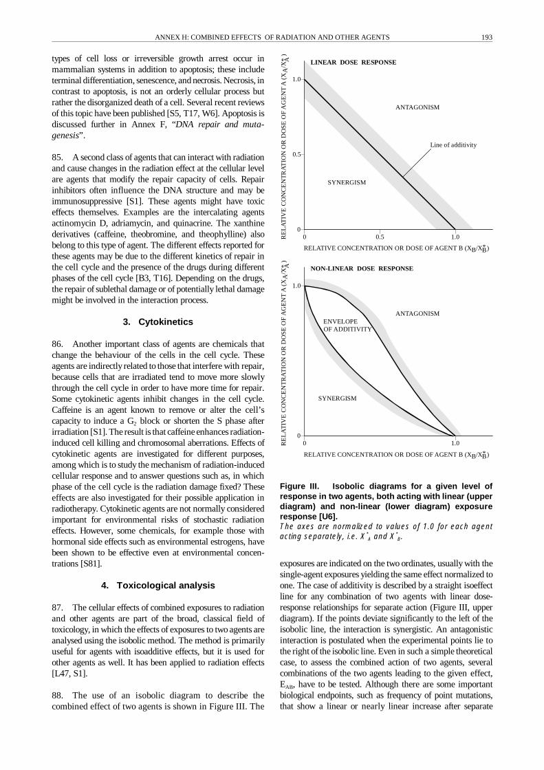
ANNEX H: COMBINED EFFECTS OF RADIATION AND OTHER AGENTS 193
0
0
0.5 1.0
1.0
0
0
0.5
1.0
1.0
RELATIVE CONCENTRATION OR DOSE OF AGENT B (XB/X*B)
RELATIVE CONCENTRATION OR DOSE OF AGENT B (XB/X*B)
LINEAR DOSE RESPONSE
NON-LINEAR DOSE RESPONSE
ANTAGONISM
Line of additivity
ANTAGONISMENVELOPEOF ADDITIVITY
SYNERGISM
SYNERGISM
RE
LA
TIV
EC
ON
CE
NT
RA
TIO
NO
RD
OS
EO
FA
GE
NT
A(X
A/X
* A)
RE
LA
TIV
EC
ON
CE
NT
RA
TIO
NO
RD
OS
EO
FA
GE
NT
A(X
A/X
* A)
types of cell loss or irreversible growth arrest occur inmammalian systems in addition to apoptosis; these includeterminal differentiation, senescence, and necrosis. Necrosis, incontrast to apoptosis, is not an orderly cellular process butrather the disorganized death of a cell. Several recent reviewsof this topic have been published [S5, T17, W6]. Apoptosis isdiscussed further in Annex F, “DNA repair and muta-genesis”.
85. A second class of agents that can interact with radiationand cause changes in the radiation effect at the cellular levelare agents that modify the repair capacity of cells. Repairinhibitors often influence the DNA structure and may beimmunosuppressive [S1]. These agents might have toxiceffects themselves. Examples are the intercalating agentsactinomycin D, adriamycin, and quinacrine. The xanthinederivatives (caffeine, theobromine, and theophylline) alsobelong to this type of agent. The different effects reported forthese agents may be due to the different kinetics of repair inthe cell cycle and the presence of the drugs during differentphases of the cell cycle [B3, T16]. Depending on the drugs,the repair of sublethal damage or of potentially lethal damagemight be involved in the interaction process.
3. Cytokinetics
86. Another important class of agents are chemicals thatchange the behaviour of the cells in the cell cycle. Theseagents are indirectly related to those that interfere with repair,because cells that are irradiated tend to move more slowlythrough the cell cycle in order to have more time for repair.Some cytokinetic agents inhibit changes in the cell cycle.Caffeine is an agent known to remove or alter the cell’scapacity to induce a G2 block or shorten the S phase afterirradiation [S1]. The result is that caffeine enhances radiation-induced cell killing and chromosomal aberrations. Effects ofcytokinetic agents are investigated for different purposes,among which is to study the mechanism of radiation-inducedcellular response and to answer questions such as, in whichphase of the cell cycle is the radiation damage fixed? Theseeffects are also investigated for their possible application inradiotherapy. Cytokinetic agents are not normally consideredimportant for environmental risks of stochastic radiationeffects. However, some chemicals, for example those withhormonal side effects such as environmental estrogens, havebeen shown to be effective even at environmental concen-trations [S81].
4. Toxicological analysis
87. The cellular effects of combined exposures to radiationand other agents are part of the broad, classical field oftoxicology, in which the effects of exposures to two agents areanalysed using the isobolic method. The method is primarilyuseful for agents with isoadditive effects, but it is used forother agents as well. It has been applied to radiation effects[L47, S1].
88. The use of an isobolic diagram to describe thecombined effect of two agents is shown in Figure III. The
Figure III. Isobolic diagrams for a given level ofresponse in two agents, both acting with linear (upperdiagram) and non-linear (lower diagram) exposureresponse [U6].The axes are normalized to values of 1.0 for each agentacting separately, i.e. X*
A and X*B.
exposures are indicated on the two ordinates, usually with thesingle-agent exposures yielding the same effect normalized toone. The case of additivity is described by a straight isoeffectline for any combination of two agents with linear dose-response relationships for separate action (Figure III, upperdiagram). If the points deviate significantly to the left of theisobolic line, the interaction is synergistic. An antagonisticinteraction is postulated when the experimental points lie tothe right of the isobolic line. Even in such a simple theoreticalcase, to assess the combined action of two agents, severalcombinations of the two agents leading to the given effect,EAB, have to be tested. Although there are some importantbiological endpoints, such as frequency of point mutations,that show a linear or nearly linear increase after separate

ANNEX H: COMBINED EFFECTS OF RADIATION AND OTHER AGENTS194
exposures to genotoxic chemicals or radiation, the dose-effectrelationships for health impairments caused bycomplex multi-stage changes in biological systems are often better describedby exponential or sigmoid functions of dose. For these morerealistic circumstances, the line of additivity in the isobolicdiagram becomes curved and transforms into an envelope ofadditivity (Figure III, lower diagram). In general, the order ofexposure to agents with differing dose-response relationshipsthen becomes important as well [R4].
89. Such isobolic analyses are important tools, for examplein optimizing combination therapy [L16, R4], but are of lessvalue in evaluating the effects of chronic exposures in theworkplace and in non-occupational settings [B69]. Anextended review of this approach and its mathematicalbackground was presented in the UNSCEAR 1982 Report[U6]. This approach is based on producing equal effects withdifferent combinations of the two agents under restrictedconditions of time. Owing to the general lack of such rangesof exposures in human populations, this method is notapplicable in epidemiology.
90. The interaction at the cellular level is restricted in time,so that damage from one agent seldom interacts with damagefrom a second one. For low dose rates of radiation and long-term exposures of other agents, the supralinear or quadraticdose terms of the dose-effect relationships tend to diminish,and only a linear dose-effect relationship remains. In thesecases, the possible interaction in combined exposure alsodecreases, and additivity results. This implies that sinceinteraction at the cellular level during low-dose, long-termexposures to radiation and other agents can be expected tohave a low probability of occurrence, it is therefore of limitedimportance for carcinogenesis.
C. EFFECTS ON THE TISSUE/ORGANLEVEL
91. After fixation of the radiation effect at the cellular level,which occurs within a few days, a much longer time is neededbefore an effect at the organ level occurs, i.e. before astochastic radiation effect is evident. The period of occurrenceof a stochastic effect is dependent on the type of effect. Forexample, hereditarydefects mayoccur when a germ cell, afterhaving been irradiated, forms the origin of an organism of thenext generation. For radiation-induced cancer, it is the timebetween the initiation event, or possibly one of the followingsteps of the carcinogenesis process, and the detection of atumour. Full consideration of the mechanistic aspects ofcancer development is given in Annex G, “Biological effectsat low radiation doses”. The events occurring after theinitiation event in the development of tumorigenesis areconsidered to take place on the tissue and organ level and mayoccur years or decades later.
92. It is generally accepted that carcinogenesis is a multi-step process. The usual chain of events is considered to beinitiation of damage, tumour promotion, possibly withactivated proto-oncogenes or deactivated tumour-suppressor
genes, and malignant progression. Each oftheseprocesses canbe related to effects at the cellular level. The basic aspects ofthese processes were reviewed in Annex E, “Mechanisms ofradiation oncogenesis”, of the UNSCEAR 1993 Report [U3].The concepts of multi-stage carcinogenesis have evolved overmany years of cancer research [A17, B10, B14, B15, C8, F7,M27, R6]. Several lines of evidence that support the multi-stage model of cancer derive from studies of pathology,epidemiology, chemical and radiation carcinogenesis inanimals, cell biology, molecular biology, and human genetics[K28, M5]. Germ-line mutations, somatic genetic events, andepigenetic stimulation by the host organism may all playimportant roles in neoplastic development. The definition oftwo broad classes of genes, proto-oncogenes with growth-enhancing functions and tumour- suppressor genes withgrowth-inhibiting functions, brought a biological basis and aunifying concept to the multi-stage theory of cancer [V2].Owing to the functional diversity of the products of thesegenes involving cell surface receptors, protein kinases,phosphatases, and DNA-binding proteins, to mention only afew, this concept does not lend itself directly to a betterunderstanding quantitatively. However, in this area themodifications of the cancer process after exposure to externalagents may be investigated.
93. The number of genetic changes involved in theevolution of a specific malignant neoplasm is not knownwith certainty. In some cancers that occur early in life,soon after exposure, or in genetically susceptibleindividuals, there may be only one rate-limiting changeneeded for malignant disease. Certain forms of leukaemia,e.g. those resulting from reciprocal translocations [B64] orcancer induction in retinoblastoma heterozygotes, seem tofollow this course. Multi-hit models developed on the basisof specific incidence rates of solid cancers from epidemio-logical data often show an exponential increase in theincidence of specific cancers with the fifth to seventhpower of age [K11]. Most colorectal cancers have three ormore altered genes, [F6, V1, V2], and estimates of as manyas 10 or more mutational changes have been proposed tooccur in adult human cancers [B17]. Basically, all thesegenetic changes might be induced by ionizing radiation,other genotoxic agents, and the inherent instabilityof DNAalone.
94. The distinction between proto-oncogenes andtumour-suppressor genes has important repercussions fordose-effect models, because the former class wouldgenerally express its function dominantly, whereas thelatter could fulfill its protective function as long as oneallele is functionally intact, i.e. the tumour-suppressorfunction would be a recessive trait. However, theprobability of developing cancer is in many cases higher inheterozygotes than a pure recessive trait would predict,indicating the importance of penetrance in the genetics ofthe different tumour-suppressor genes. Moreover, muta-tions in some tumour-suppressor genes like p53 and WT1may be of the dominant negative type, in which themutated protein overrides the action of the suppressor wild-type allele [H4, M25].

ANNEX H: COMBINED EFFECTS OF RADIATION AND OTHER AGENTS 195
dNk(t)/dt � λk(t)�t
0�
xk�1
0�
x2
0
N0(x1)λ1 (x1) ...
λk�1(xk�1)dx1 ... dxk�1
(8)
MULTI-STAGE MODEL
Normal cells
Normal cells
Normal cells
Initiatedcells
Initiatedcells
Damagedcells
Damagedcells
Malignantcells
Malignantcells
Stage 1 cells . . . . . . . Stage K cells
TWO-STAGE MODEL
TWO-STAGE MODEL WITH DNA REPAIR
µ1(d)
µ1(t,d)
µ1(t,d) µ
3(t,d)
ρ1(t,d)
β1(t,d)
β2(t,d)
δ2(t,d)
β3(t,d)
ρ3(t,d)
β(t,d)
δ(t,d)
µ2(d)
µ2(t,d)
µk(d)
Figure IV. Models of carcinogenesis.Parameters: µ is the probability of transformation, βi is the birth or replication rate,δi is the death rate, ρi is the repair rate, i is the stage, d the dose, and t the time.
95. Multi-stage cancer models are described in Annex G,“Biological effects at low radiation doses”. The multi-stagemodel proposed by Armitage and Doll [A16, A17, A18]represents one of the first attempts to develop a biologicalmodel ofcarcinogenesis. Theypostulated that cancer developsfrom a single cell that must pass sequentially through aparticular series of transformations to become a malignantcell. The multi-stage Armitage-Doll model is illustrated in theupper portion of Figure IV. This model assumes that a normalcell must pass through k sequential stages before becomingfully malignant. This model has k + 1 types of cells: normalcells, stage 1 cells, stage 2 cells, ..., and stage k (malignant)cells. The model supposes that at age t an individual has apopulation N0(t) of completely normal cells and that thesecells acquire a first mutation at a rate λ1(t). The cells with onemutation acquire a second mutation at a rate λ2(t), and so onuntil at the (k � 1) stage the cells with (k � 1) mutationsproceed at a rate λk(t) to become fully malignant.
96. The instantaneous tumour incidence rate h(t) at timet in the multi-stage model is therefore approximately of theform
where N is the number of cells in the target tissue and λi(t) isthe instantaneous rate of the i-th cellular change (i = 1, ..., k).For simplicity, it is often assumed that the transition rates arelinearly related to the dose di(t) of the carcinogen at time t forthe i-th stage. Therefore the transition rate from one stage tothe next is given by λi(t) = ai + bidi(t). Here, ai denotes thetransition rate in the absence of exposure and bi reflects theeffects of the carcinogen on the transition rate into stage i = 1,..., k. With similar values for spontaneous transition rates (ai
to ak), this model predicts that the age-specific tumourincidence rate will be proportional to the (k � 1)st power oftime and provides a good description of human cancerincidence data with 2 < k < 6 stages [A20, A22].
97. To encompass the growing biological evidence that theprocess of carcinogenesis involves intermediate cells having
ANNEX H: COMBINED EFFECTS OF RADIATION AND OTHER AGENTS196
a growth advantage over normal cells, Armitage and Doll[A17] modified their initial model. The initial model isgenerally viewed as not biologically plausible, because it doesnot account for cell kinetics, more specifically the birth anddeath of cells. The modified model that includes cell kineticsmust sometimes assume very small and, in the opinion ofArmitage and Doll [A17], unlikely values for the growth rateof intermediate cells to fit the data.
98. Moolgavkar and Venzon [M29] and Moolgavkar andKnudson [M23] proposed a two-stage birth-death-mutationmodel to describe the process of carcinogenesis in adults. Byincorporating both cell kinetics and tissue growth, this modelcan be used to describe a broader class of tumour incidencedata than the classical multi-stage model. This model hasthree cell types: normal cells, intermediate or initiated cells,and malignant cells. The middle portion of Figure IV displaysthe general two-stage model of carcinogenesis in which for anormal cell to become malignant, it must pass from thenormal state through the intermediate state and into themalignant state. The simplicity of this model allowsclassifying external agents in three categories of carcinogen:initiators, which stimulate the first transition of a normal stemcell into the intermediate stage; completers, which transforman intermediate cell into a malignant cell by the secondtransition; andpromoters, which enhance cell division and thenet increase of intermediate cells with time [K46]. Ionizingradiation and other genotoxic agents may be both initiatorsand completers.
99. The Moolgavkar-Venzon-Knudson model was laterextended to account for more than two mutational stages[L23, L24, M14]. For the generalized Moolgavkar-Venzon-Knudson model it may be supposed that at age t there are N(t)susceptible stem cells, each subject to mutation toa type ofcellcarrying an irreversible mutation at a rate of µ0(t). The cellswith one mutation divide into two such cells at a rate γ1(t). Ata rate δ1(t) they die or differentiate. Each cell with onemutation can also divide into an equivalent daughter cell andanother cell with a second irreversible mutation at a rate µ1(t).For the cells with two mutations there are also assumed to becompeting processes of cell growth, death and differentiation,and mutation taking place at rates γ2(t), δ2(t), and µ2(t),respectively. This continues until at the (k-1) stage the cellwill have accumulated (k-1) mutations. It will eventuallyacquire another mutation and become fully malignant.
100. With the advent of more sophisticated experimentaltechniques and a growing understanding of the process ofcarcinogenesis, more refined mathematical models have beendeveloped and continue tobe developed toembody the currentscientific knowledge and mechanisms of cancer. Mutation isthe result of DNA damage and the subsequent fixation andpropagation of the damage by DNA replication. This processis included as a single rate constant in the models described inthe previous paragraphs. However, agents can affect DNAdamage rates, cellular replication rates, and/or the processesof DNA repair. Kopp-Schneider and Portier [K19] expandedthe modelling of the mutation process to account explicitly forthe process of cellular damage to DNA, DNA repair, and
DNA replication. The two-stage damage-fixation model hasfive types of cells: normal cells, damaged normal cells anddamaged initiated cells both of which are subject to DNArepair, initiated cells, and malignant cells (in which damagehas been fixed by replication). This model is shown in thelower portion of Figure IV.
101. It is clear that research on quantitative multi-stagemodels is still in progress and that the complexity of thecarcinogenic process inhibits a choice of a universallyaccepted and applicable model. However, it is also clear thatmulti-stage models have a biological basis and could describetumour incidence quantitatively and as such have a future inimproving radiation risk estimates and estimates of combinedeffects. Always important is the question of complexity vs.simplicity. The biology of cancer formation is so complicatedthat an ever-increasing number of parameters are needed tocover all possibilities of tumour formation mathematically. Onthe other hand the available data are limited, so the number ofparameters that can be fixed is limited as well. As far as themathematics and statistics are concerned, it is preferable thatthe number of unknown parameters be as low as possible.
102. Most multi-stage models are used to describe the agedependence of tumour incidence and the influence ofchemical carcinogens in animal experiments. In a few cases,theyhavebeen used todescribe radiation-induced tumours. Asfar as human data are concerned, the induction of lungtumours by radon in miners [L9, M39] and the lifespanstudies of the Japanese atomic bomb survivors [H1, K45, L5]were used to test multi-stage models. In general, ionizingradiation acts mainly as an initiator, although it has someinfluenceon other coefficients. An important conclusion oftheuse of multi-stage models for radiation carcinogenesis is thatradiation generally seems to affect only one step in thecarcinogenesis process; in other words, it is a co-factor ofbackground tumour incidence. This implies that the radiationeffect is dependent on background tumour incidence as wellas on other agents or factors that produce tumours or cancer.
103. The timescale for effects at the tissue or organ level islong and can last for years. The implication is that interactionwith another agent is possible even when the exposures of thetwo agents are separated in time for up to several years. Acomprehensive treatment of the carcinogenic effect ofcombined exposures and the implications for dose-effectrelationships using a two-mutation carcinogenesis model isgiven in Krewski et al. [K46]. They classify carcinogenicagents as initiators, completers, and promoters and concludethat the joint effect of two compounds that both affect thesame stage in the carcinogenic process will be described wellby the additive risk model; however, the effect of combinedexposure to two carcinogens that influence different stageswill not necessarily result in a multiplicative model. Shortexposures that occur close together in time and do notoccur at either very young or very old ages can produce anearly additive relative relationship. Synergism would,however, arise when the contribution to different transi-tions by different carcinogenic agents is large comparedwith the spontaneous rate and when the time course of
ANNEX H: COMBINED EFFECTS OF RADIATION AND OTHER AGENTS 197
a See glossary for definition of terms.
exposure is penalizing if the sequence of steps matters.Brown and Chu [B9] concluded that the observation of amultiplicative relative risk relationship in studies of jointexposure to two carcinogens is evidence of action at twodifferent stages of the carcinogenesis process. Theseexamples illustrate the importance of a full understandingof the timescale of exposure for both agents.
104. An overviewof interactions for simple binaryexposuresto agents with specific effects is given in Table 3. Accordingto the terminology used by Krewski et al. [K46], manyinteractions leading to considerable deviations from additivityare possible although hardly predictable. The effectiveness ofa carcinogenic agent depends not only on the exposure butalso on the time of exposure, age at exposure, time sinceexposure, and duration of exposure. This time dependence iscompletelydifferent from and should not be confused with the
time involved in the cellular dose-rate effect. It is therefore notpossible to quantitatively predict the dose-effect relationshipfor tumour induction after combined exposure.
105. This assessment is based on the evaluation ofcarcinogenesis data using a two-mutation model. Deviationsof the carcinogenesis process in other ways, e.g. by disturbingorgan functions in a crude way, are ignored. The assumptionofotherwise undisturbed functioning of the organ or organismis probably reasonable for low exposures but may complicateanalysis for high doses and exposures. The long-term develop-ment of tumours implies a long period of time over which theprocess can be influenced. For interaction in the genesis ofradiation-induced tumours, it implies that exposure to adifferent agent at a time that is completely separated from thetime of irradiation may influence the radiation effect in oftenpoorly predictable ways.
Table 3Anticipated interaction response of two single-agent carcinogens a
[K29]
Carcinogen A Carcinogen B Interaction response
InitiatorCompleterInitiatorInitiatorInitiatorInitiatorInitiatorPromoter
InitiatorCompleterCompleterPromoter
Promoter and completerInitiator and completerInitiator and promoter
Promoter and completer
AdditiveAdditive
MultiplicativeMultiplicative to supra-multiplicativeMultiplicative to supra-multiplicativeSupra-additive to sub-multiplicative
Supra-additive to supra-multiplicativeSupra-multiplicative
D. DOSE MODIFIERS AND OTHERINDIRECT INTERACTIONS
106. Apart from the direct interference with the develop-ment of the radiation effect, further indirect interactionmechanisms are possible when an agent changes theretention of the radioactive substance following inhalationor ingestion and consequently changes the organ dose. Awell-known case is blockage of 131I uptake to the thyroid bystable iodine, which is used in nuclear medicine andenvisaged in future radiological emergencies to greatlyreduce the dose to the thyroid gland. Other drugs, such asethylenediaminetetracetic acid (EDTA), are used fortherapeutic reasons, to stimulate the metabolic transfer ofinhaled or ingested radionuclides and, consequently, toreduce the relevant organ dose. Natural chelators such ascitrate may also modulate the biological half-lives of metaland actinide ions. Synergistic effects on this level areknown from the inhibition of mucocilliary clearance by asecond agent [F28]. Several examples of dose-modifyingagents are given in the Appendix, which covers specificinteractions. Mechanistic considerations indicate that theirritants and cytotoxicants implicated here generallydisplay an effect threshold and are therefore of littleconcern for combined effects at low exposure levels.
107. Other modifications of the radiation effect arepossible when the physiological condition of the organismis changed, either intentionallyor bychance. Examples arechanges in hormone levels or in the immunological system.Such changes may also be induced by radiation (e.g. UVradiation). Also, novel mechanisms of genetic change suchas radiation- or chemical-induced genetic instability, whichleads to new genetic damage many cell generations afterexposure (see Annex F, “DNA repair and mutagenesis”),may be prone to more-than-additive effects. The results ofthis type of interaction are dependent on the conditions ofthe change and are, in general, poorly predictable.
E. SUMMARY
108. Carcinogenesis and, consequently, also the develop-ment of radiation-induced tumours is a long-term process.Mechanistically, three levels can be distinguished in thedevelopment of the radiation effect on cancer: the molecu-lar, cellular, and tissue/organ levels. On each level, ioniz-ing radiation induces changes and processes, and thesemay be influenced by combined exposures to other agents.A summary of the levels, processes involved in cancerdevelopment, and examples of the many classes of sub-stances and agent with a potential to interfere at different

ANNEX H: COMBINED EFFECTS OF RADIATION AND OTHER AGENTS198
Nucleophylic agents, cysteamine,cysteine, sulphydryl compounds,anoxia, vitaminsGamma rays, x rays,beta particles,alpha particles, UVMetals, platinum complexes,actinomycin, bleomycin,EMSIUdR, BUdR, 5-FU,actinomycin D,adriamycin, quianin
Temperature,microwaves
Cytokinetins, p53
Caffeine, cyclophosphamide,hyperthermia,hydroxyurea
Caffeine
Heat, mineral fibres,dust, lead
Mutagens, tobacco,ENU, BNU, DEN, DBE,BCNU, CCNU, Me-CCNU
Vitamin A, E, K, selenium,low-fat diet, DFMO
Tobacco, asbestos,mineral fibres, hormones,DES, TPA
Mitogens, hormones,DES, oestrogens,mineral fibres, dust
Mutagens, asbestos, tobacco,TPA, benzo(a)pyrene
Tamoxifen, testosterone
UV, Vitamin A, endotoxins,dietary factors
Tobacco, hormones(TSH, oestrogen, DES)
Anoxia, selenium
Mitogens, mechanicalirritation, dust
DNA damage
Damagefixation
Radical scavenging,protecting
_
_
_
_
_
+
+
_+/
_+/
+
+
+
+
+
+
+
Interaction withDNA sub-damage
DNA repair stimulation
DNA repair inhibition
Cell-cyclecontrol change
Apoptosis inhibition
Membrane damage
Interaction withsub-mutation events
Progressioninhibition
Progressionpromotion
Conversionstimulation
Tumour growthinhibition
Immune surveillancechange
Tumour growthstimulation
Cell cyclinginhibition
Cell cyclingstimulation
Cell cycling
Initiation/mutation
Promotion
Tumour growth
Conversion
Metastases
Cancer death
MOLECULAR LEVEL
CELLULAR LEVEL
ORGAN LEVEL
levels of radiation-induced carcinogenesis is presented inFigure V. The biological effect of combined exposures toradiation and other agents at low doses is, in general,expected to be additive, especially in the case of chronicexposures. Deviations from additivity may primarily beexpected from interactions on the tissue/organ level. In thisphase, exposure to other agents may take a long time, up totens of years, and for interaction to occur, the exposures toradiation and the other agent need not be simultaneous.
Thus, interaction can last a relatively long time, and theradiation effect can be influenced to a significant extent bythe interactions that take place during this phase. Studiesusing multi-stage models show that classifying the agentsinvolved in terms of their action as initiators, completers,and/or promoters may help to predict the result of theirinteraction with radiation and other agents. These studiesalso indicate that the radiation effect depends on thebackground tumour incidence.
Figure V. Schematic representation of radiation-induced carcinogenesis and possible interaction mechanismswith examples of agents having shown a potential for more (+) or less (�) than additive effects
for at least one tumour site.

ANNEX H: COMBINED EFFECTS OF RADIATION AND OTHER AGENTS 199
a Assuming 80 and 40 years of exposure for environmental (E) and occupational (O) levels, respectively. Risks are generally upper boundestimates based on linear extrapolations from high exposures.
III. SPECIFIC COMBINED EXPOSURES
109. Recent data on combined exposures to radiation andspecific physical, chemical, or biological agents are reviewedin this Chapter. For each type of interaction, data from epi-demiological studies of the adverse health effects in humansare generally reviewed first. Studies involving experimentalanimal models are considered next. Lastly, various effectsobserved in in vitro systems are reviewed. Carcinogenesis istheprincipal endpoint ofinterest; non-neoplasticendpointsareviewed mainly in relation to mechanistic considerations.
110. Only a minor fraction of the interacting agentsdescribed below are found in the human environment atpotentially critical levels. An overview of agents known orsuspected to affect human health on their own is given inTable 4. Details of the experimental and epidemiologicalconditions and results of studies on specified combinedexposures may be found in the Appendix and aresummarized in Table A.1.
Table 4Exposure conditions and characteristics of prominent environmental and occupational agents andsubstances that may produce combined effects with them[B6]
Agent
Typicalenvironmental
exposure(E)
Occupationallimit for chronic
exposure(O)
Major healthendpoint
Estimatedcontribution tototal incidence/
mortality ofendpoint (%)
Estimatedlifetime risk a
(%)
Substance withknown orsuspected
combined effect
Ionizing radiation 1.5-4 mSv a-1 20 mSv a-1 Cancer 3-8 (E)�10 (O)
0.4-1 (E)�4 (O)
Smoking,asbestos,hormones,arsenic?
UV radiation[W31]
Noontime intensity:UV-A: 40 W m-2
UV-B: 3 W m-2
350 nm:150 kJ m-2 in 8 h300 nm:100 J m-2 in 8 h
Skin agingSkin cancerMelanoma
Important (E)>50 (E)
?
->20 (E)
?
Phototoxicity,allergy withUV sensitizers
Asbestos[I12]
Crocidolite:0.2 fibers cm-3
Other forms:2 fibres cm-3
Lung cancerMesothelioma
Low>50%
Smoking
Benzene[M60, W34]
14 µg m-3 in indoorair
3.2 mg m-3 Leukaemia 2.5 (E) 0.01 (E) Substrates foractivation/detoxificationsystems
Carbon tetra-chloride [W34]
3 µg m-3 in indoor air 65 mg m-3 Cancer 0.05 (E) 0.01 (E) Chloroform
Chloroform [I2] 1.2 µg kg-1 d-1 fromtap water andshowering
50 mg m-3 Cancer 0.15 (E) 0.03 (E) Carbon tetra-chloride
Dioxins/furans[F8]
1.3 µg kg-1 d-1
dietary intake50 pg m-3 Cancer 0.1 (E) 0.02 (E)
Ethylenebisdithio-carbamates(EBDCs) [L49]
n.a. n.a. Cancer, adversereproductiveoutcomes
0.17 (E) 0.034 (E)
Polychlorinatedbiphenyls (PCBs)
[G8]
14 ng kg-1 d-1 1 mg m-3
(42% CI)Cancer 0.06 (E) 0.01 (E)
ANNEX H: COMBINED EFFECTS OF RADIATION AND OTHER AGENTS200
A. RADIATION AND PHYSICALAGENTS
1. Combinations of ionizing radiation
111. Many experiments have been undertaken to investi-gate the cellular effects of combined exposures of two typesof ionizing radiations. In view of their potential radio-therapeutic applications, a wealth of data on the combinedaction of neutrons, heavy ions, and gamma or x rays wasaccumulated in the 1970s and early 1980s. This informa-tion was reviewed in Annex L, “Biological effects ofradiation in combination with other physical, chemical andbiological agents”, in the UNSCEAR 1982 Report [U6].The results generally indicate additive effects of combinedexposures characterized by the so-called isoaddition ofthese agents at the cellular level [L10, L47], as describedin Chapter II. A few data [E5, L26] were reported onradiation-induced tumours; these, in general, includeexposure to internal emitters. The results indicate additiveto slightly supra-additive effects for combined exposures,mainly because of the lower dose rates that are involved inthe internal exposure to alpha radiation in theseexperiments. For estimating the risk of carcinogenesis, theeffects of combined exposures to more than one type ofionizing radiation are expected to be isoadditive, i.e. to addup in the same way as effects of increments of the sameagents, when at least one of the radiations is delivered at alow dose rate (chronic irradiation), as is generally the casefor occupational and environ-mental exposure levels.
2. Ultraviolet radiation
112. Ultraviolet (UV) radiation is recognized as the mostimportant initiator and co-factor for human skin carcino-genesis [S29]. It is mainly the skin that is exposed to UVradiation. A study of combined exposures to gammaradiation and UV radiation was presented by Shore, whoanalysed 12 studies on the incidence of skin cancer inpopulations irradiated with known skin doses [S29]. In theabsence of a proper control (skin exposed to ionizingradiation but not to UV), it was concluded that, at least forcombined exposures, the data are compatible with a lineardose-response relationship for ionizing radiation [S29] butthat the interaction is unclear. The question of whetherrelative risk or absolute risk models are more appropriateremains open. From the mechanistic point of view,interaction at the cellular level may be expected, whichresults in a more or less additive effect for low exposurerates [L52]. The considerable variations in skin canceramong different populations and subgroups seem to reflectthe large differences in UV exposures due to latitude andlifestyle and the differences in genetic predisposition toskin cancer due to skin type. The overwhelming depend-ence of skin cancer on extended exposures to UV preventsconclusive epidemiological data on the interaction of UVwith ionizing radiation. Another important factor to takeinto account in possible interactions is UV-inducedsuppression of the immune system [B76, L58, N26]
3. Electromagnetic radiation
113. Neither low- nor high-frequencyelectromagnetic radia-tion have enough single photon energy to directly damageDNA and therefore cannot be cancer initiators. However,strong electromagnetic fields may modify and stimulategrowth [K16, S33], and this has led to the hypothesis thatelectromagnetic fields may influence cancer development.However, no straightforward inferences from experimentalresults to exposure situations in occupational or environ-mental settings have been found at this stage for the com-bination of electromagnetic and ionizing radiation [B77, B78,B79, U19]. Moreover, there is at present little indication froma mechanistic standpoint for potentially harmful interactionsbetween electromagnetic fields and ionizing radiation atcontrolled exposure levels in the workplace or the clinic. Thepossible modulation of radiation effects by heating producedby strong electromagnetic fields is considered in the nextSection.
4. Temperature
114. Heat can kill mammalian cells in a predictable andstochastic way [D3]. Elevated temperature is used as amodifier of radiation sensitivity in many therapies to controltumour growth. In combination with ionizing radiation, heatcan act synergistically on cell survival, cell proliferation, andcytogenetic damage by, for example, interfering with DNArepair. However, extremely high temperatures, which aregenerally not found in the workplace or in environmentalconditions, are needed in the cells at risk, so heat is notconsidered as potentially enhancing radiation risk.
5. Ultrasound
115. Ultrasound has achieved widespread use in medicaldiagnostic and therapeutic procedures. Studies have shownthat at the intensities used for diagnostic purposes,ultrasound does not interact with ionizing radiation tocause cytogenetic damage in treated cells, although theyield of sister chromatid exchanges was observed to beslightly increased in one study [K20]. Because cavitation-induced mechanical damage by ultrasound shows highthresholds, this mechanism is of little concern forenvironmental exposures. Such damage has tobe preventedin other situations already caused by single-agent effects.
6. Dust, asbestos, and other mineral fibres
116. Mineral dust and fibres such as asbestos generally actthrough non-genotoxic mechanisms such as mechanicalirritation and cell killing [B13]. The combination of radiationexposure and exposure to dusts and fibres is quite common inindustrial settings and in the environment, and these agentsare reported in both animal studies and in vitro studies to actsynergistically at high exposures [B38, H11]. Silicosis wasshown to be a risk factor for human lung cancer in metalminers in the 1940s [H9] and is implicated as a modifier oflung cancer risk in radon-exposed underground workers[K49]. Combined exposure to phosphate ore dust, gamma

ANNEX H: COMBINED EFFECTS OF RADIATION AND OTHER AGENTS 201
radiation, and radon daughter products also resulted inelevated lung cancer risks in earlier practices [B74]. Althoughexposures and thus risks are considerablylower today, mineraldust and fibres still deserve attention because they mayinteract with radiation, including densely ionizing radiationsuch as alpha radiation in mining environments, to enhancethe risk of cancer.
7. Space flight
117. A special form of combined exposure is experienced inspace flights, where a multitude of stressors act in combina-tion on astronauts. This problem has been investigated usinganimals [A13, A14, V3]. The most important environmentalparameter is microgravity. Space radiation effects werecomprehensively reviewed by Kiefer et al. [K54], the inter-action of microgravityand radiation at the cellular level [H52,K55]. No synergistic actions were found. A very importantaspect is a possible reduction of the immune response [S86],which could have an influence on cancer development. Thechanges of many parameters that are normally stable inexperimental work on earth make well designed studies inspace potentially important in addressing combined effects ofphysical agents.
B. RADIATION AND CHEMICAL AGENTS
118. A multitude of natural and man-made chemicals withcancer initiating and promoting potential are present in thehuman environment and may interact with radiation.Classification based on their mode of action is oftendifficult, as many have more than one type of action, but atleast a crude separation can be made into substances thatmainly act by damaging DNA (genotoxic substances) andthose that act in other ways (non-genotoxic substances)[C48]. The former group includes chemically activespecies, or substances that can be activated, bind to ormodify DNA directly, or indirectly via radicals. The non-genotoxic substances range from nonspecific irritants andcytotoxins to natural hormones, growth factors, and theiranalogues. They interact with the regulatory systems ofcells and organs and cannot always be considered toxic bythemselves. Some are clearly protective, e.g. theyscavengereactive species before they interact with DNA.
1. Genotoxic chemicals
119. Numerousexamples ofcombined exposures to radiationand chemical genotoxic agents can be found in the literature,including studies on the improvement of radiation therapy bysimultaneous treatment with a chemical (see Chapter IV). Inmany cases, supra-additive effects are reported, caused byinteraction in the cellular phase and by the high exposurelevels involved. The agents include 1,2-dimethylhydrazine(DMH) [S27], N-methyl-N-nitrosourea (MNU), butyl-nitrosourea (BNU), N-ethyl-N-nitrosourea (ENU) [H6, K15,S13, S20, S21, S22], diethylnitrosamine (DEN) [M8, P26],N-2-fluorenylacetamide (FAA), 4-nitroquinoline-1-oxide(4NQO) [H39], bleomycin [D6], and 1,2-dibromoethane
(DBE) [L13]. The effects are dependent on the species,exposure conditions, time ofexposure, etc., and sometimes thesame chemical is involved in a supra-additive and a sub-additive result. In general, for short exposures to highconcentrations and for low chronic concentrations, deviationsfrom additivity are small, if at all existent. In most epidemio-logical and experimental studies, effects exceeding a levelpredicted from isoaddition have not specifically beendemonstrated.
2. Non-genotoxic chemicals
120. Many chemicals in the human environment or theirmetabolites do not specifically attack DNA but influencecell proliferation and cell differentiation on an epigeneticlevel. These include the tumour promoter 12-O-tetradecanoylphorbol-13-acetate (TPA) [H3, L24], carbontetrachloride, and α-difluoromethylornithine (DFMO)[O12]. These agents act in combination with radiation onthe cellular and tissue level of cancer development and cansignificantly enhance the induction of tumours. Specificmitogens may interfere with regulatory mechanisms andcell-cell signalling, but many substances with a highchemical reactivityact as non-specific irritants or toxicantsvia membranes or proteins. For example, toxin-inducedcell death will induce proliferation in neighbouring cells,which may enhance the progression of premalignant cells.Substances acting in a non-specific manner, for examplelipophilic solvents, quite often show highly non-lineardose-response relationships with apparent thresholds.Other agents may interfere with critical cellular processesinvolved in repairing damage to cellular constituents suchas DNA. The assessment of possible synergistic effects atthe exposure levels relevant for risk estimation remainsvery difficult because of the high exposures used inexperimental systems and the apparent threshold levels.One important group of chemicals, which includes cystea-mine and mexamine, has radioprotective effects; thesechemicals scavenge radicals formed by ionizing radiation[M4]. A considerable number of agents may have bothgenotoxic and epigenetic functionalities such as the baseanalogue 5-bromo-2'-deoxyuridine (BrUdR) [A15].
3. Tobacco
121. Given the large collective dose from radon and its decayproducts in non-occupational and occupational settings andthe prevalence of active smoking, the combined effect of thesetwo exposures on human health deserves special attention[B27]. A large body of epidemiological evidence fromuranium miner studies allows, at least for higher radiationdoses, calculating risks and interaction coefficients directlyfrom human data [C46]. However, the fact that tobaccosmokeis itself a complex mixture of genotoxic and non-genotoxicsubstances and even contains some natural radionuclides (thelong-lived radon progeny 210Po and 210Pb) makes amechanistic assessment difficult.
122. Because of the complex composition of tobacco smoke,the issues surrounding combined exposures to radiation and
ANNEX H: COMBINED EFFECTS OF RADIATION AND OTHER AGENTS202
tobacco smoke are even more difficult to elaborate than forbinary combinations. Some 4,000 individual chemicalcomponents of cigarette smoke have been identified, and thereare probablya number ofadditional important but unidentifiedcomponents, for example, extremely reactive, short-livedcompounds or those present in very low concentrations [G1].The complexity of tobacco smoke means that the action incombined exposures with radiation can take place in both thecellular and organ phase of cancer development. Tobaccosmoke contains only relatively small amounts of DNA-reactive carcinogens such as nitrosamines, polycyclicaromatic hydrocarbons, and pyrolysis products such ascarbolines. Hence enhancing and promoting factors, e.g.catechols, other phenols and terpenes, are important.Discontinuation of smoking progressively reduces the relativerisk of cancer development as time since withdrawalincreases, probably because of reduced pressure from theaction of promoters [W1].
123. In the last few years, joint analyses of original data sets[C1, L18] and meta-analyses of published results [T14] haveyielded a detailed assessment of risk patterns and haveallowed investigators to test risk models. The most com-prehensive and complete analysis of radon-induced healthrisks was published byLubin et al. [L18]. The reviewcontainsa joint analysis of original data from 11 studies of maleunderground miners. Data on smoking were available for 6 ofthe 11 cohorts, but the assessmentswerelimited byincompletedata on lifetime tobacco consumption patterns and thesometimes exotic forms of tobacco use, such as water pipes inthe Chinese study [L18]. Single studies for which smokingdata could be analysed were generallynot informative enoughto allow choosing between an additive or a multiplicative jointrelationship between radon progeny and smoking. TheChinese cohort seemed to suggest an association more con-sistent with additivity, while the Colorado cohort suggested arelationship more consistent with a multiplicative interaction.For all studies taken together, the combined influence ofsmoking and radon progeny exposures on lung cancer wasclearly more than purely additive but less than multiplicativeand compatible with isoadditivity [B69]. The most recentanalyses of the BEIR VI Committee [C46], which were basedon an update of these data, suggested synergism between thetwo agents that is statistically most consistent with a slightlysub-multiplicative interaction. A best estimate from minerdata indicates that the lung cancer risk for smokers expressedin absolute terms is higher by a factor of at least 3. To furthercharacterize the association, more detaileddata on tobaccousewould be needed. Age of starting to smoke, amount andduration of smoking, and type of tobacco were recognized asimportant determinants of risk. A further handicap of presentstudies is that the sub-cohortsoflifetimenon-smokers exposedonly to radon are very small. The statistical power of theconclusions on the radon-tobacco smoke interaction is cor-respondingly low. Data are available from a study by Finch etal. [F28] of smoke exposure and alpha-particle lung irradia-tion over the lifespan of exposed rats. The pulmonary reten-tion of inhaled 239Pu was higher, increasing with the con-centration of the 239Pu, in smoke-exposed rats than in sham-smoke-exposed rats. This effect on retention resulted in
increased alpha-radiation doses to the lung. Assuming anapproximately linear dose-response relationship betweenradiation dose and lung neoplasm incidence, approximateincreases of 20% and 80% in tumour incidence over controlswould be expected in rats exposed to 239PuO2 + low-levelcigarette smoke and 239PuO2 + high-level cigarette smoke,respectively.
124. Hypotheses on the mechanistic interaction betweentobacco smoke and radon were tested by applying the two-mutation clonal expansion model of carcinogenesis ofMoolgavkar to data from the Colorado plateau miners [M39].No interaction between radon and tobacco smoke in anyof thethree steps (the two mutation steps and clonal expansion) isneeded to fit the data, which are clearly supra-additive forradon and smoking combined. The model, however, shows asignificant dependence on age at exposure. Quantitativelysimilar resultswereobtained byLeenhouts and Chadwick[L9,L57]. A highlysignificant decrease in excess relative risk withtime since exposure is found in miner studies in contrast tofindings on lung cancer in survivors of the atomic bombings.This may be explained by microdosimetric considerations. Inthe case of high-LET alpha radiation from radon progeny, theminimal local dose from one single alpha track averaged overa cell nucleus is already in the range of several hundredmilligray, whereas one electron track yields a dose to thenucleus of only 1�3 mGy. This means that even at the lowestpossible nuclear dose from alpha exposure, stem cells that arehit carrya multitude of DNA lesions, which mayconsiderablyimpair long-term cell survival and maintenance ofproliferative capacity [B25, B27].
125. Smoking is also of great importance for non-occupational radon exposures in the indoor environment.Until now, little quantitative evidence has come fromindoor radon studies. Most of the case-control studiespublished are inconclusive [A28, K53, P11]. Only onelarger study [P11] was indicative of an indoor radon riskand its modification by tobacco that is comparable to whatis predicted from miner studies. It remains doubtfulwhether the results from the many case-control studiesunder way will in the near future allow narrowing theuncertainties that surround indoor radon risk and thepossible interactions with smoking. Emerging studyresultsfrom Europe based on much longer residence times mayoffer better statistical power. Several large indoor case-control studies under way will narrow uncertainties in thenext few years. First results from the United Kingdom[D33] and Germany [K53, W35] are indicating a lungcancer risk in the range of ICRP projections. However,confidence intervals are relatively large and still includezero risk in most analyses. Because of the limitation of theindoor radon studies, risk estimates based on miner dataremain the main basis for predicting lung cancer fromindoor radon exposure. A best linear estimate of the riskcoefficients found in the joint analysis of Lubin et al. [L18,L35] for the indoor environment indicates that in theUnited States, some 10%�12%, or 10,000 cases, of thelung cancer deaths among smokers and 28%�31%, or5,000 cases, of the lung cancer deaths among never-

ANNEX H: COMBINED EFFECTS OF RADIATION AND OTHER AGENTS 203
a Global values; percentage of anthropogenic contribution is given in parentheses.
smokers are caused by radon progeny. About half of these15,000 lung cancer deaths traceable to radon would then bethe result of overadditivity, i.e. synergistic interactionsbetween radon and tobacco. Based on the same risk model,Steindorf et al. [S47] estimated an attributable risk forindoor radon of 4%�7% for smokers and 14%�22% fornon-smokers. Because of the many differences betweenexposed persons and exposure situations in mines andhomes and the additional carcinogens such as arsenic, dust,and diesel exhaust in mine air, these figures should beinterpreted with caution.
4. Metals
126. Toxic metals are important trace pollutants in thehuman environment (Table 5). They interact in many wayswith cellular constituents and may produce oxidative DNAdamage or influence enzyme activity at low concentrations,e.g. by competing with essential metal ions [H38]. Carcino-genic transition metals are capable of causing promutagenicdamage, such as DNA base modifications, DNA-proteincross-links, and strand breaks [K7]. The underlying mechan-ism seems to involve active oxygen and other radicals arising
Table 5Metals in the environment and effects on humans[M38, N14, S48]
MetalRelease a
(109 g a-1)Main sources of intake
and typical levels in the bodyCharacteristics affecting health
Arsenic 31 (61) Source: food (seafood up to 120 mg kg-1) anddrinking water
Concentration in body: 0.3 mg kg-1
Mutagenic, teratogenic, co-carcinogenic,As3+causes skin cancer
Cadmium 8.9 (85) Source: inhalation (2 µg cigarette-1) andfood (0.025 mg kg-1)
Mutagenic, teratogenic, co-carcinogenic,causes cancer at multiple sites
Mercury 6.1 (59) Source: metal vapours, food (up to 1 mg kg-1
MeHg+ naturally in fish), tooth fillingsIntake: by inhalation and ingestion
0.2 and 25 µg d-1, respectively
Mutagenic, teratogenic (brain damage), co-carcinogenic,causes sarcomas and renal tumours
Nickel 86 (65) Source: food intake (0.2 mg d-1)Concentration in body: 0.007 mg kg-1
Essential element; allergenic, comutagenic,cocarcinogenic, causes nasal sinus cancer
Lead 12 (96) Source: Food, dust, air (0.15 mg d-1)Amount in body: steady increase
to about 200 mg at age of 60 years
Substitutes for Ca2+, neurobehavioural deficits (decrease infertility, abortifacient)Low mutagenic and carcinogenic potential
(may cause renal adenocarcinoma)
Antimony 5.9 (59) Source: food and tobacco (0.005 mg d-1) Mutagenic as Sb3+, organic antimony compounds used asemetics
Vanadium 114 (75) Source: food (0.01-0.05 mg d-1) Essential elementInhibits Na+/K+ ATPase and drug detoxification
enzymes at low concentrationMutagenic, teratogenic, carcinogenic
Zinc 177 (66) Source: food intake (10-50 mg d-1) Essential element with small window of toleranceClastogenic (causes chromosome aberrations)Causes growth of some tumours
at elevated concentrations
from metal-catalysed redox reactions. Cadmium, nickel,cobalt, lead, and arsenic may also disturb DNA repair pro-cesses [H48]. Only a few data are available from combinedexposures of radiation and metals in human populations; nofirm evidence of interactions was observed. However, metalsand ionizing radiation have been shown to produce combinedeffects in many other biological systems (see the Appendix).Especially in underground mining, possible effects from theepidemiologicallyproven lungcarcinogensarsenic, cadmium,chromium, nickel, and antimony [M65] have to be assessedtogether with high-LET radiation from radon. Arsenic inparticular is a major risk factor in combined exposures to
mineral dust, radon, metals, and diesel fumes [K48, T5]. Therisk-enhancing effects of iron dust seem to be limited to veryhigh dust concentrations, leading to changes in lung function[B74]. The significance of these data for radiation riskestimation at low dose levels remains unclear.
5. Mitogens and cytotoxicants
127. Although many mitogenic and cytotoxic compoundscould have been considered above with genotoxic or non-genotoxic agents, they should be mentioned separatelybecause of their potential to interact with radiation,

ANNEX H: COMBINED EFFECTS OF RADIATION AND OTHER AGENTS204
principally by virtue of their ability to stimulate cellproliferation. From a mechanistic standpoint, they can beexpected to interact in the organ phase of radiation carcino-genesis, but the resulting interaction (sub- or supra-additivity)is not always predictable. Examples of such agents includeN-methylformamide (NMF) [L15], caffeine [M11], theobro-mine, theophylline [Z4], 2-aminopurine, and tributylphosphate. Many studies assessing deviations from additivityin combined exposuresofmitogens/cytotoxicants and ionizingradiation are found in the literature (see Appendix), but thehigh exposure levels applied and the biological endpointstudied generally do not allow directly transferring the resultsto carcinogenesis in humans. However, any endogenous ordietary levels of agents influencing stem-cell population sizeor kinetics will have the potential to modulate response toradiation.
6. Antioxidants, vitamins, and otherdietary factors
128. Diet can modify the effectiveness of chemical carcino-gens, sometimes by a large factor, and interactions withradiation are found as well [B24, C26, H11, W29]. All classesof substances described in the five preceding SectionsIII.B.1�5 are found in human food supplies. Actions rangingfrom subadditive to supra-additive may occur, depending onthe specific agent. The radiation risk may be reduced whengrowth stimuli are reduced as a result of nutritional deficiencyor when repair possibilities are optimized. Synergism can beexpected where lower levels of radical scavengers or thecoenzymes needed for repair increase the yield of effectivedamage from ionizing radiation or impair the speed andaccuracy of cellular recovery from damage. Some of theunderlyingmechanismsofspecificagents havebeen identifiedin animal experiments. Tumour-incidence-enhancing effectshave been noticed with elevated consumption of, for example,riboflavin, ethanol, and marihuana. Tumour-incidence-reducing effects are found for low-caloric diets, vitamins A, C,K, and E, retinoic acid derivatives (but enhancing effects ofartificial beta carotin in some smoker cohorts), selenium, and3-aminobenzamide. Very important in view of populationhealth are behavioural changes and a tendencytomalnutritionin alcohol addicts, which may increase the susceptibility totoxicants in the environment or at the workplace [U18]. Ingeneral, the combined action is not specific for radiation butis also found for other carcinogens, and the interaction isdependent on the dosage.
129. In summary, dietary factors are proven modifiers of riskfrom diverse agents at levels found in human populations andprobably also influence the production and repair of endo-genously arising lesions. Absence or deficiency of importantcoenzymes and nutrients on the one side and high levels ofdirectly or indirectly acting mitogens on the other interferewith molecular, cellular, and tissue responses to ionizingradiation. A modulation in the radiation risk may occur insituations where growth stimuli are reduced or increased,owing to nutritional deficiency or surplus or where thenumber of stem cells at risk is changed. Synergisms are alsoto be expected where reduced levels of radical scavengers or
coenzymes needed for repair increase the yield of primarydamage from ionizing radiation or impair the speed andaccuracy of cellular responses to damage. In general, thesemechanisms apply to most deleterious agents in the humanenvironment.
C. RADIATION AND BIOLOGICAL AGENTS
130. Many hormones are potent growth stimulators, andthere is considerable evidence that they may modify cancerrisk. They include thyroid-stimulating hormone (TSH),oestradiol-17 beta (E2), prolactin, diethylstilbestrol (DES), andandrogens in general. Their effect is dependent on tissue, typeof hormone, and dosage and is important enough to be kept inmind when analysing radiation risks. Tamoxifen, a syntheticanti-oestrogen, has both cancer risk-enhancing functions(endometrium) and protective properties (breast), dependingon the organ [J9]. An important consequence of interactionwith hormones is the sex difference in tumour sensitivity,mainly of organs of the reproductive system.
131. Viruses, bacteria and microbial genetic sequences havebeen shown to play an important role in the development oftumours. Cancer viruses may interact with radiation by muta-tion or translocation of dormant viral sequences. Experimentsso far give no clear indication ofanyinteraction with radiationthat influences cancer development. Viruses may inducegenotypic and functional changes, i.e. they may act as highlysite-specific genotoxic agents in multi-step mechanisms.Highly synergistic effects due to increased sensitivity mayarise for some endpoints. Little information is available atpresent on the mechanism of the induction of gastric cancerby bacteria (Helicobacter pylori).
132. The interaction of several miscellaneous factors withradiation exposure and its role in carcinogenesis has beeninvestigated. Some of these factors are reviewed in theAppendix. The role of others, such as psychosocial factors,remains unclear and is outside the scope of this Annex.
D. SUMMARY
133. Combinations of different types of ionizing radiationshow mainly isoadditive effects. For decreasing doses andchronic exposure, the quadratic terms of the dose-effectrelationships tend to vanish and the linear terms to prevail,indicating additivity for low-level exposures. Also, for thecombination of UV radiation and ionizing radiation, additiveeffects are expected for low exposure levels. Temperature andultrasound arenot considered tosignificantlymodifyradiationrisk. The temperature range and the ultrasound intensitiesnecessary for an interaction with radiation are too high to beof relevance for environmental or occupational settings.Mineral dust and fibres, including asbestos, tend to showsupra-additive interaction with radiation at high exposurelevels. These levels were reached in workplaces in the 1950sand earlier. Today the occupational exposures are lower, butthese agents still deserve attention for their potential toenhance risks after combined exposure.
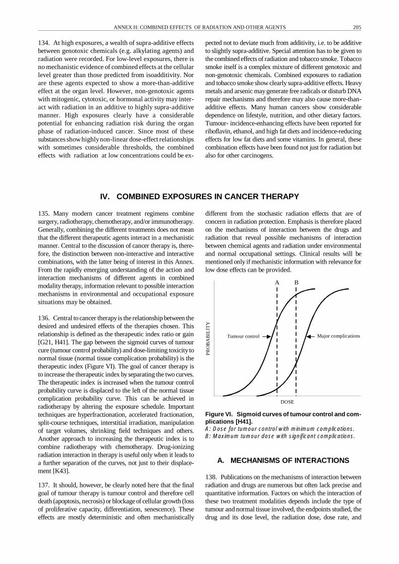
ANNEX H: COMBINED EFFECTS OF RADIATION AND OTHER AGENTS 205
DOSE
Tumour control Major complications
PR
OB
AB
ILIT
Y
A B
134. At high exposures, a wealth of supra-additive effectsbetween genotoxic chemicals (e.g. alkylating agents) andradiation were recorded. For low-level exposures, there isno mechanistic evidence of combined effects at the cellularlevel greater than those predicted from isoadditivity. Norare these agents expected to show a more-than-additiveeffect at the organ level. However, non-genotoxic agentswith mitogenic, cytotoxic, or hormonal activity may inter-act with radiation in an additive to highly supra-additivemanner. High exposures clearly have a considerablepotential for enhancing radiation risk during the organphase of radiation-induced cancer. Since most of thesesubstances showhighlynon-linear dose-effect relationshipswith sometimes considerable thresholds, the combinedeffects with radiation at low concentrations could be ex-
pected not to deviate much from additivity, i.e. to be additiveto slightly supra-additive. Special attention has to be given tothe combined effects of radiation and tobacco smoke. Tobaccosmoke itself is a complex mixture of different genotoxic andnon-genotoxic chemicals. Combined exposures to radiationand tobacco smoke show clearly supra-additive effects. Heavymetals and arsenic maygenerate free radicals or disturb DNArepair mechanisms and therefore may also cause more-than-additive effects. Many human cancers show considerabledependence on lifestyle, nutrition, and other dietary factors.Tumour- incidence-enhancing effects have been reported forriboflavin, ethanol, and high fat diets and incidence-reducingeffects for low fat diets and some vitamins. In general, thesecombination effects have been found not just for radiation butalso for other carcinogens.
IV. COMBINED EXPOSURES IN CANCER THERAPY
135. Many modern cancer treatment regimens combinesurgery, radiotherapy, chemotherapy, and/or immunotherapy.Generally, combining the different treatments does not meanthat the different therapeutic agents interact in a mechanisticmanner. Central to the discussion of cancer therapy is, there-fore, the distinction between non-interactive and interactivecombinations, with the latter being of interest in this Annex.From the rapidly emerging understanding of the action andinteraction mechanisms of different agents in combinedmodality therapy, information relevant to possible interactionmechanisms in environmental and occupational exposuresituations may be obtained.
136. Central tocancer therapy is the relationship between thedesired and undesired effects of the therapies chosen. Thisrelationship is defined as the therapeutic index ratio or gain[G21, H41]. The gap between the sigmoid curves of tumourcure (tumour control probability) and dose-limiting toxicity tonormal tissue (normal tissue complication probability) is thetherapeutic index (Figure VI). The goal of cancer therapy isto increase the therapeutic index byseparating the two curves.The therapeutic index is increased when the tumour controlprobability curve is displaced to the left of the normal tissuecomplication probability curve. This can be achieved inradiotherapy by altering the exposure schedule. Importanttechniques are hyperfractionation, accelerated fractionation,split-course techniques, interstitial irradiation, manipulationof target volumes, shrinking field techniques and others.Another approach to increasing the therapeutic index is tocombine radiotherapy with chemotherapy. Drug-ionizingradiation interaction in therapy is useful only when it leads toa further separation of the curves, not just to their displace-ment [K43].
137. It should, however, be clearly noted here that the finalgoal of tumour therapy is tumour control and therefore celldeath (apoptosis, necrosis) or blockage of cellular growth (lossof proliferative capacity, differentiation, senescence). Theseeffects are mostly deterministic and often mechanistically
different from the stochastic radiation effects that are ofconcern in radiation protection. Emphasis is therefore placedon the mechanisms of interaction between the drugs andradiation that reveal possible mechanisms of interactionbetween chemical agents and radiation under environmentaland normal occupational settings. Clinical results will bementioned only if mechanistic information with relevance forlow dose effects can be provided.
Figure VI. Sigmoid curves of tumour control and com-plications [H41].A: Dose for tumour control with minimum complications.B: Maximum tumour dose with significant complications.
A. MECHANISMS OF INTERACTIONS
138. Publications on the mechanisms of interaction betweenradiation and drugs are numerous but often lack precise andquantitative information. Factors on which the interaction ofthese two treatment modalities depends include the type oftumour and normal tissue involved, the endpoints studied, thedrug and its dose level, the radiation dose, dose rate, and
ANNEX H: COMBINED EFFECTS OF RADIATION AND OTHER AGENTS206
fractionation, and the intervals between and sequencing of thecombined treatments. Chemotherapy could slow the processof cell repopulation after radiotherapy or it could synchronizethe cell cycle. Moreover, tumour reduction by chemotherapycould improve tumour oxygenation, thus increasing the effectof radiotherapy. At the cellular level, inhibition of repair ofsublethal and potentiallylethal radiation damage byanticancerdrugs is probably the most important mechanism of radio-sensitization. Exploitable mechanisms in combined chemo-therapy and radiotherapy treatment can be described underfour headings, as was originally done by Steel and Peckham[S1, S23, S46]: spatial cooperation, independent cytotoxicity,protection of normal tissues, and enhancement of tumourresponse.
139. Spatial cooperation describes a non-interactive com-bination of radiotherapy, chemotherapy, surgery, and othertherapeuticstrategies that act at different anatomical sites. Thecommonest situation is where surgeryand/or radiation is usedto treat the primary tumour and chemotherapy is added as anadjuvant to attack remaining local tumour cells and distantmetastases. There is an analogous situation in the treatment ofleukaemia, wherechemotherapyis themainlinetreatment andradiotherapy is added to deal with the disease in anatomicalsites, e.g. the brain, protected from chemotherapeuticattackbyvascular constraints or by blood barriers. Spatial cooperationstill appears to be one of the main clinical benefits of com-bination modality treatment. This mechanism does notrequire interactive processes between drugs and radiation.
140. Independent cytotoxicitydescribes another form of non-interactive combination of therapeutic modalities. If twomodalities can both be given at full dose, then even in theabsenceofinteractiveprocesses the tumour response should begreater than that achieved with either modalityalone. The costof this improvement on tumour response is that the patient hasto tolerate a wider range of toxic reactions in normal tissues(within and outside of the radiation field). As with spatialcooperation, the mechanism of independent cytotoxicity doesnot require interactive processes between drugs and radiation.Independent cytotoxicitycan even tolerate a subadditive inter-action of the modalities and still produce an increase in thera-peutic gain. The relative extent of reduction in toxicity tonormal tissue within the radiation field is the critical para-meter of this mechanism.
141. The protection of normal tissues requires an antagon-istic interaction of the combined modalities. Since two toxicagents usually tend to produce more damage than either agentalone, it would seem rather unlikely that chemotherapy inconjunction with radiation could reduce the damage to dose-limiting normal tissue. However, there are well-documentedsituations in which certain cytotoxic drugs increase the resist-ance of normal tissue to radiation or to a second cytotoxictreatment.
142. Studiesofthisseeminglycontradictorymechanism haveconcentrated on the bone marrow and the intestinal epithel-ium. It has been shown by Millar et al. [M51, M52] that inthe bone marrow, the most effective cytotoxic agent, cytara-
bine, does not modify stem-cell radiosensitivity; instead, itstimulates enhanced repopulation by the surviving stem cells.This phenomenon is highly dependent on the timing of thetwomodalities. Maximal radioprotection is achieved when thedrug is given two days before radiation. In the small intestine,microcolonysurvival was increasedwhen cytosinearabinosidewas given 12 hours before irradiation [P24]. Other cytotoxicdrugs with radioprotective action are cyclophosphamide,chlorambucil, and methotrexate [M51]. Recently, a topoiso-merase II inhibitor (etoposide) was shown to increase theradioresistance of the bone marrow when given one daybefore whole-body irradiation [Y1].
143. Normal tissue can be protected from radiation effectsby radioprotective agents. Increasing the differentialbetween tumour and normal tissue radiosensitivities wouldgive a therapeutic advantage. Radioprotectors can thus beused as selective protectors against radiation damage tonormal tissue, allowing higher curative doses of radiationto be delivered to tumours.
144. Chemical radioprotectorstarget thedetoxifyingmechan-isms of the cell, in particular the antioxidant enzymes that areavailable for removal or detoxification of the reactive oxygenspecies and their products formed by the action of ionizingradiation. By far the most widely studied class of radio-protective agents is the thiols, and the most important non-protein thiol present in cells is glutathione. Other classes ofagents conferring radioresistance to normal tissue are theeicosanoids, which are biologicallyactive compounds derivedfrom arachidonic acid, the lipoic acids, and calcium antagon-ists (reviewed in [M51, M56]). The effects of biologicalresponse modifiers such as the cytokines IL-1 and TNF
αas
radioprotectors in normal tissue have been discussed in recentreviews [M53, M54, N12, N13, Z11, Z12].
145. Relative enhancement of tumour response is commonlyperceived to be the principal aim of adding chemotherapy toradiotherapy. A wide variety of biological mechanisms havebeen proposed to explain interactions between radiation andtherapeutic agents. In the context of this Annex, this kind ofinteraction is the most important mechanism with respect toenvironmental and normal occupational settings.
146. DNA adduct repair regularly involves strand scissionsby repair enzymes. Conversion of repairable into lethal DNAdamage may occur if a DNA-repair-associated single-strandbreak combines with a radiation-induced single-strand breakto produce new DNA double-strand breaks. This mechanismhas been suggested for the interaction of cisplatin andradiation. A similar mechanism, the production of double-strand breaks by combining single-strand breaks, may occurwhen topoisomerase I or II inhibitors and radiation arecombined.
147. Many drugs inhibit the repair of radiation damage.Antitumour antibiotics (e.g. dactinomycin and doxorubicin),antimetabolites (e.g. hydroxyurea, cytarabine, and arabino-furanosyl-adenine), and alkylating agents and platinum ana-logues (e.g. cisplatin) have been shown to inhibit radiation-

ANNEX H: COMBINED EFFECTS OF RADIATION AND OTHER AGENTS 207
induced DNA damage repair. Repair inhibition has beendetected in a number of ways, including removal of theshoulder on the cell survival curve, inhibition of split-doserecovery, and inhibition of delayed plating recovery.
148. Cell-cycle synchronization exploits the fact that manycytotoxic drugs and radiation show some degree of selectivityin cell killing at certain phases of the cell cycle. Antimeta-bolites show a maximum effect on cells undergoing theS phase. Radiation sensitivity is highest in the G2/M phase.There is, therefore, an attractive possibility of complementaryaction between drugs and radiation. The most attractivepossibilityseem to be the interaction between microtubule andtopoisomerase poisons and primary DNA-damaging agentssuch as radiation.
149. Activation of apoptosis by differential pathwaysincreases cell killing during tumour therapy and is thereforeanother possibility for combined action of radiation andchemotherapeutic drugs. Ionizing radiation may activate theapoptotic process bya DNA damage-p53 dependent pathway,whereas taxoids like paclitaxel may activate a pathwaydownstream of p53 by phosphorylation of Bcl-2. There is,therefore, a possibility that radiation-induced cell killing canincrease, even in p53-deficient tumours. The involvement ofapoptosis in radiation-induced cell killing has recently beenstudied extensively [B75, D34, H44, H49, M69, O19].
150. Reduction of the hypoxic fraction by bioreductive drugstargeted at hypoxic tumour cells increases tumour radio-sensitivity. Most promising here is the development of dual-function drugs specific to hypoxic cells and with intrinsiccytotoxic activity (e.g. alkylating activity).
B. SECONDARY CANCERS FOLLOWINGCOMBINED MODALITY TREATMENT
151. The successful treatment of cancers involves radiationtherapy and/or multi-agent chemotherapy, each of which isused either as primary therapy or as an adjunct to therapy ofthe primary tumour, and it often includes surgery. Withfurther improvements in modern cancer therapy, the durationof survival and the curability of many patients has increasedup to 45%. However, along with this progress has come arecognition of the long-term complications of therapy, such assecondary cancers (reviewed in [T30]). Although otherclinical consequences in non-target tissues are known, themain focus in this Section is on secondary cancers aftercombined modality treatments. Secondary cancers resultingfrom the combined effect of radiotherapy and tobacco smokeare discussed in the Appendix, Section B.3.
152. No one specific type of secondary cancer is seen aftertherapeutic irradiation. Secondary cancers can occur afterany initial cancer, when survival surpasses the latentperiod. Radiation-induced leukaemias begin toappear after3�5 years. Solid cancers typically emerge more than 10years after treatment but may occur earlier in particularlysusceptible individuals [F9, G32, T31, V10]. When the risk
of secondary solid cancer is elevated, it rises withincreasing radiation dose to the site and with increasingtime since treatment and persists as long as 20 years.
153. The predominant secondary cancer associated withchemotherapy is acute non-lymphocytic leukaemia (ANL).Most ANLs have occurred after treatment with alkylatingagents or nitrosoureas. The findings are similar for Hodgkin’sdisease, paediatriccancers, ovarian cancer, multiplemyeloma,polycythemia vera, gastrointestinal cancers, small-cell lungcancer, and breast cancer [B22, B36, B63, B65, C14, F9, G32,G33, R22, T31, T32, V10]. The risk for leukaemia rises withincreasing cumulative dose of the alkylating agent or nitro-sourea. A few ANL cases were reported following combina-tion chemotherapy, including teniposide or etoposide. Theleukaemias differ from those that follow alkylating agents inthat theyoccur sooner and that specific chromosomal abnorm-alities are induced [P9, P29, P30, W32]. Few solid tumourshave been linked to chemotherapy. Bladder cancer has beenassociated with cyclophosphamide treatment, and risk isdependent on the cumulative dose of cyclophosphamide [P4,T33, T36, W33]. Excess bladder cancer risk followingtreatment with both radiotherapy and cyclophosphamide wasas expected from a summation of the individual risks. Bonesarcomas have also followed treatment with alkylating agents[T34]. In general, the risk for solid tumours after chemo-therapy alone has been difficult to evaluate because too fewpatients survived long enough after treatment by chemo-therapyalone. At present, several cohort studies are under wayto assess this risk.
154. Earlier reports indicated a distinctive pattern ofsecondary cancers after treatment of childhood malignancies[M1]. The most common secondarycancer was bonesarcoma,followed by soft tissue sarcomas, leukaemias, and cancer ofthe brain, thyroid, and breast. The cancers showing thehighest increases compared with the usual distribution ofchildhood cancers were retinoblastoma, followed byHodgkin’s disease, soft tissue sarcomas, Wilms’ tumour, andbrain cancer. This difference may reflect both the geneticpredisposition to develop multiple tumours in the case ofheritable retinoblastoma, soft tissue sarcoma of Li-Fraumenisyndrome, and possibly the immune dysfunction associatedwith Hodgkin’s disease. To summarize the findings fromrecent study results [B68, O17, S6, S7], there is littleindication that heritable sensitivity to treatment is asignificant component of secondary cancer, but intensivemultiple agent therapy used in childhood cancer treatmentacts as an independent aetiological factor for a secondtumour. The risk for a second malignant neoplasm aftercancer in childhood is considerable. Absolute risks up to 7%over 15 years following diagnosis of the primary cancer werefound for Hodgkin’s disease [B68]. This amounts to an excessrelative risk (ERR) of about 17, with breast cancercontributing most. A follow-up study in the Nordic countriesshowed a significant increase in the ERR from a low of 2.6 inpatients first diagnosed in the 1940s and 1950s to 5.9 forcohort members included in the late 1970s and 1980s,indicating that newer treatments are not only more successfulbut also carry a higher long-term risk [O17].

ANNEX H: COMBINED EFFECTS OF RADIATION AND OTHER AGENTS208
155. In patients with bone sarcomas as the secondary tumourfollowing childhood cancer therapy, the effects of radiationtherapy, chemotherapy, and combined modality treatmenthave been analysed [H43, T34]. The risk for bone sarcomarose dramatically with increasing doses of radiation with alinear trend. Patients with heritable retinoblastoma had amuch higher risk for secondary bone sarcoma, but theirresponse to radiation was similar to that of patients with otherchildhood cancers. In addition to the radiation dose, theexposure to chemotherapy was evaluated. There was anindependent effect of exposure to alkylating agents in the riskfor bone cancer, i.e. radiation and alkylating agents actedadditively. The risk rose with increasing cumulative dose ofthe alkylating agents. The effect of alkylating agents wasmuch smaller than that of radiation, and in the presence ofradiation at the site of the bone sarcoma, the alkylating agentsadded little to the risk.
156. Thyroid cancer risk after treatment of childhood canceris increased 53-fold compared with general population rates[T35]. The risk for thyroid cancer rose with increasingradiation dose. There was no increased risk of thyroid cancerassociated with alkylating-agent chemotherapy.
157. There was a sevenfold increased risk of secondarycancers after treatment of acute lymphoblastic leukaemia(ALL) [N22]. Most of this risk was due to a 22-foldincrease in brain cancers. The brain cancers occurred inpatients diagnosed with ALL before the age of five yearsand who received cranial or whole-body irradiation.
158. Among 29,552 patients with Hodgkin’s lymphoma, 163cases of secondary leukaemia after treatment of the primarydisease indicated a considerable risk [K44]. There was nodifference in the relative risk of secondary leukaemias fromchemotherapyalone(MOPP regimen) and chemotherapyplusradiotherapy. A relatively small risk for leukaemia was seenafter radiation alone, and this risk increased with radiationdose. The risk did not vary significantlyor consistentlyacrossradiation doses for any given number of chemotherapy cyclesbut increased consistently with more cycles of chemotherapyin each radiation dose range.
159. Significantly elevated risks for secondary solid tumours(lung, non-Hodgkin’s lymphoma, stomach, melanoma, bone,and connective tissue) were reported in patients treated forHodgkin’s disease [T31]. The pattern of secondary tumourswas distinctive and was similar to the distribution of cancersseen in immunosuppressed populations, such as renal trans-plantation patients or patients with non-Hodgkin’s lympho-ma. All cancers of the stomach, bone, and connective tissueoccurred within areas previously treated with radiationtherapy. All those who developed lung cancer had receivedradiation therapy and smoked. For breast cancer, a fourfoldelevated risk was reported in Hodgkin’s disease patients after15 years of follow-up. The highest risk was in womenirradiated before the age of30 [H19]. Comparable results werereported from a Dutch study[V11]. These authors reported anoverall relative risk of 3.5 for secondary cancers afterHodgkin’s disease. Significant increases in relative risk of
34.7, 20.6, 8.8, 4.9, 3.7, 2.4, and 2.0 were reported forleukaemia, non-Hodgkin’s lymphoma, soft tissue sarcoma,melanoma, lung cancer, urogenital cancers, and gastrointesti-nal cancers, respectively. Risk factors for leukaemia werechemotherapyand host factors; for non-Hodgkin’s lymphomathey were combined modality treatment rather than singlemodality treatment or host factors. For lung cancer the riskfactors were strongly related to radiation therapy, while anadditional role for chemotherapy could not be demonstrated.
160. Significant excesses of ANL followed therapy for non-Hodgkin’s disease with either prednimustine, a derivative ofnitrogen mustard, or with regimens containing mechlor-ethamine and procarbazine, for example MOPP therapy,(nitrogen mustard, vincristine, and procarbazine prednisone)[T6]. Chlorambucil and cyclophosphamide were associatedwith smaller increased risk of ANL. In this study, radio-therapy did not add to the leukaemogenicity of alkylatingagents. This finding should be interpreted cautiously,however, because of the small number of patients and thelarge number of parameters evaluated.
161. Few studies have evaluated the late effects of adjuvantchemotherapy and radiotherapy in breast cancer. The inter-action of alkylating agents with radiation in producingleukaemia in women treated for breast cancer was investigatedin a cohort of 82,700 patients in the United States [C29].Based on 74 cases, the risk of ANL was significantlyincreased after radiotherapy alone (relative risk = 2.4, 7.5 Gymean dose to the active marrow) and alkylating agents(melphalan and cyclophosphamide) alone (relative risk = 10).Combined therapy resulted in a more-than-additive relativerisk of 17.4. The most common solid cancer that occurs afterbreast cancer is contralateral breast cancer, but fewer than 3%of these tumours could be attributed to radiation [B2]. The riskwas highest in women treated at young age (under 45 years).The usefulness of such studies is still hampered by the factthat an important proportion of patients developing primarytumours might already belong to a genetically more sensitivesubpopulation [E11]. In addition, combined treatments mightbe more often used in more advanced stages of tumoursneeding higher total doses or more cycles for cure.
C. SUMMARY
162. A large number of chemotherapeutic drugs are usedin clinical cancer therapy in combination with radiation.The main ones in use or proposed for use are described inthe Appendix, with emphasis on the mechanisms ofinteraction between the drugs and radiation that may haverelevance for combined effects between chemical agentsand radiation even at the low exposure levels found incontrolled environmental and occupational settings. Themain findings on modes of action and combined effects aresummarized in Table 6.
163. The predominant secondary cancer associated withchemotherapy is ANL and, to a lesser degree, bladdercancer. No one specific type of secondary cancer follows
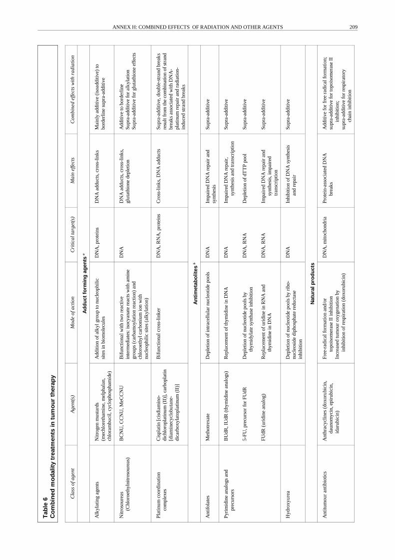
Tab
le6
Co
mb
ined
mo
dal
ity
trea
tmen
tsin
tum
ou
rth
erap
y
Cla
ssof
agen
tA
gent
(s)
Mod
eof
actio
nC
ritic
alta
rget
(s)
Mai
nef
fect
sC
ombi
ned
effe
cts
with
radi
atio
n
Ad
du
ctfo
rmin
gag
ents
a
Alk
ylat
ing
agen
tsN
itrog
enm
usta
rds
(mec
hlor
etha
min
e,m
elph
alan
,ch
lora
mbu
cil,
cycl
opho
spha
mid
e)
Add
ition
ofal
kylg
roup
tonu
cleo
phili
csi
tes
inbi
omol
ecul
esD
NA
,pro
tein
sD
NA
addu
cts,
cros
s-lin
ksM
ainl
yad
ditiv
e(i
soad
ditiv
e)to
bord
erlin
esu
pra-
addi
tive
Nitr
osou
reas
(Chl
oroe
thyl
nitr
osou
reas
)B
CN
U,C
CN
U,M
eCC
NU
Bif
unct
iona
lwith
two
reac
tive
inte
rmed
iate
s:is
ocya
nate
reac
tsw
itham
ine
grou
ps(c
arba
moy
latio
nre
actio
n)an
dch
loro
ethy
lcar
boni
umio
nw
ithnu
cleo
phili
csi
tes
(alk
ylat
ion)
DN
AD
NA
addu
cts,
cros
s-lin
ks,
glut
athi
one
depl
etio
nA
dditi
veto
bord
erlin
eSu
pra-
addi
tive
for
alky
latio
nSu
pra-
addi
tive
for
glut
athi
one
effe
cts
Plat
inum
coor
dina
tion
com
plex
esC
ispl
atin
[cis
diam
ino-
dich
loro
plat
inum
(II)
],ca
rbop
latin
[dia
min
ecyc
lobu
tane
-di
carb
oxyl
atop
latin
um(I
I)]
Bif
unct
iona
lcro
ss-l
inke
rD
NA
,RN
A,p
rote
ins
Cro
ss-l
inks
,DN
Aad
duct
sSu
pra-
addi
tive,
doub
le-s
tran
dbr
eaks
resu
ltfr
omth
eco
mbi
natio
nof
stra
ndbr
eaks
asso
ciat
edw
ithD
NA
-pl
atin
umre
pair
and
radi
atio
n-in
duce
dst
rand
brea
ks
An
tim
etab
olit
esb
Ant
ifol
ates
Met
hotr
exat
eD
eple
tion
ofin
trac
ellu
lar
nucl
eotid
epo
ols
DN
AIm
pair
edD
NA
repa
iran
dsy
nthe
sis
Supr
a-ad
ditiv
e
Pyri
mid
ine
anal
ogs
and
prec
urso
rsB
UdR
,IU
dR(t
hym
idin
ean
alog
s)
5-FU
,pre
curs
orfo
rFU
dR
FUdR
(uri
dine
anal
og)
Rep
lace
men
toft
hym
idin
ein
DN
A
Dep
letio
nof
nucl
eotid
epo
ols
byth
ymid
ylat
esy
ntha
sein
hibi
tion
Rep
lace
men
tofu
ridi
nein
RN
Aan
dth
ymid
ine
inD
NA
DN
A
DN
A,R
NA
DN
A,R
NA
Impa
ired
DN
Are
pair
,sy
nthe
sis
and
tran
scri
ptio
n
Dep
letio
nof
dTT
Ppo
ol
Impa
ired
DN
Are
pair
and
synt
hesi
s,im
pair
edtr
ansc
ript
ion
Supr
a-ad
ditiv
e
Supr
a-ad
ditiv
e
Supr
a-ad
ditiv
e
Hyd
roxy
urea
Dep
letio
nof
nucl
eotid
epo
ols
byri
bo-
nucl
eosi
dedi
phos
phat
ere
duct
ase
inhi
bitio
n
DN
AIn
hibi
tion
ofD
NA
synt
hesi
san
dre
pair
Supr
a-ad
ditiv
e
Nat
ura
lpro
du
cts
Ant
itum
our
antib
iotic
sA
nthr
acyc
lines
(dox
orub
icin
,da
unom
ycin
,epi
rubi
cin,
idar
ubic
in)
Free
-rad
ical
form
atio
nan
d/or
topo
isom
eras
eII
inhi
bitio
nIn
crea
sed
tum
our
oxyg
enat
ion
byin
hibi
tion
ofre
spir
atio
n(d
oxor
ubic
in)
DN
A,m
itoch
ondr
iaPr
otei
n-as
soci
ated
DN
Abr
eaks
Add
itive
for
free
radi
calf
orm
atio
n;su
pra-
addi
tive
for
topo
isom
eras
eII
inhi
bitio
n;su
pra-
addi
tive
for
resp
irat
ory
chai
nin
hibi
tion
ANNEX H: COMBINED EFFECTS OF RADIATION AND OTHER AGENTS 209

Tab
le6
(con
tinue
d)
Cla
ssof
agen
tA
gent
(s)
Mod
eof
actio
nC
ritic
alta
rget
(s)
Mai
nef
fect
sC
ombi
ned
effe
cts
with
radi
atio
n
aE
ffec
tsce
ll-cy
cle
inde
pend
ent.
bE
ffec
tsm
ainl
yce
ll-cy
cle
depe
nden
t.
Ant
itum
our
antib
iotic
s (con
tinue
d)B
leom
ycin
Mito
myc
inC
Act
inom
ycin
D
Prod
uces
activ
eox
ygen
spec
ies
afte
rin
terc
alat
ing
with
DN
A
Bif
unct
iona
lalk
ylat
ing
activ
ityaf
ter
redu
ctio
nof
quin
one
entit
y;hi
ghes
tact
ivity
inre
duci
ngen
viro
nmen
t
Inte
rcal
ates
into
DN
A
DN
A
DN
A
DN
A
Rad
iom
imet
icdr
ug
DN
Aad
duct
s,cr
oss-
links
Inhi
bitio
nof
tran
scri
ptio
n
Isoa
dditi
on
Supr
a-ad
ditiv
ew
hen
give
nbe
fore
radi
atio
n;pr
onou
nced
cyto
toxi
cef
fect
ofth
edr
ugag
ains
tra
dior
esis
tant
hypo
xic
cells
Add
itive
tosu
pra-
addi
tive
Mic
rotu
bule
pois
ons
Vin
caal
kalo
ids
(vin
cris
tine,
vinb
last
ine,
desa
cety
l-vi
nbla
stin
e)
Inhi
bitio
nof
tubu
linpo
lym
eriz
atio
n,in
duct
ion
ofm
icro
tubu
ledi
sass
embl
ySp
indl
eap
para
tus
Cel
lsbl
ocke
din
mito
ticph
ase
Add
itive
tosu
pra-
addi
tive
whe
ngi
ven
befo
rera
diat
ion
Top
oiso
mer
ase
pois
ons
Epi
podo
phyl
loto
xins
(eto
posi
de,
teni
posi
de)
Cam
ptot
heci
n,to
pote
can
Inhi
bitio
nof
topo
isom
eras
-II
Inhi
bitio
nof
topo
isom
eras
-I
DN
A
DN
A
Prot
ein-
asso
ciat
edD
NA
brea
ks
Blo
ckin
gof
repl
icat
ion
fork
s(S
-pha
sesp
ecif
ic),
sing
le-
stra
ndbr
eaks
Supr
a-ad
ditiv
ew
hen
appl
ied
toge
ther
,rep
aira
ble
radi
atio
n-in
duce
dD
NA
dam
age
istr
ansf
orm
edin
tole
thal
dam
age
Supr
a-ad
ditiv
e
Oxy
gen
Hyp
erba
ric
oxyg
enIn
crea
sed
oxyg
enat
ion
oftu
mou
rA
dditi
veto
supr
a-ad
ditiv
e
Bio
redu
ctiv
edr
ugs
Qui
none
alky
latin
gag
ents
(EO
9,po
rfir
omyc
in)
Nitr
oim
idaz
oles
(met
roni
dazo
le,
mis
onid
azol
e,et
anid
azol
e,R
SU10
96)
Ben
zotr
izin
edi
-N-o
xide
s(t
irap
azam
ine)
Met
abol
icre
duct
ion
inan
oxic
tum
our
cells
toal
kyla
ting
agen
ts,m
echa
nism
sim
ilar
tom
itom
ycin
C
Met
abol
icre
duct
ion
inan
oxic
tum
our
cells
tocy
toto
xic
agen
ts;a
dditi
onal
alky
latin
gfu
nctio
nin
RSU
1069
Met
abol
icre
duct
ion
inan
oxic
tum
our
cells
tocy
toto
xic
agen
tsw
ithpr
oduc
tion
offr
eera
dica
ls
DN
A
DN
A
DN
A
DN
Aad
duct
s,cr
oss-
links
Oxi
dativ
est
ress
unde
rae
robi
cco
nditi
ons,
DN
A-c
oval
ent
reac
tion
prod
ucts
unde
rhy
poxi
cco
nditi
ons
DN
Ada
mag
eby
free
radi
cals
Supr
a-ad
ditiv
ew
hen
give
nbe
fore
radi
atio
n,pr
onou
nced
cyto
toxi
cef
fect
ofth
edr
ugag
ains
trad
io-
resi
stan
thyp
oxic
cells
Supr
a-ad
ditiv
e
Supr
a-ad
ditiv
e
ANNEX H: COMBINED EFFECTS OF RADIATION AND OTHER AGENTS210

ANNEX H: COMBINED EFFECTS OF RADIATION AND OTHER AGENTS 211
radiotherapy. In general, there are independent effects ofexposure toalkylating agents and radiotherapy. For secondarysolid tumours, radiation is the main risk factor, while a rolefor chemotherapy has been demonstrated in some cases. Forlung cancer, an additional role of smoking was reported. Hostfactors, for example, age of diagnosis and treatment for breastcancer, are additional risk factors. For secondary leukaemiasthe main risk factors arechemotherapyand host factors. Thereis onlya small increase in this risk due to radiation. The effect
on secondarycancers of increasinglyused adjuvant treatmentswith topoisomerase I or II inhibitors, microtubule poisons(discussed in the Appendix), and hormone treatment is as yetunknown. In summary, secondary, treatment-related cancersare observed increasingly because of the long-term success ofthe initial treatments. At present, no important synergisticeffects between ionizingradiation andother agents are known.Further investigations are needed to assess and to developstrategies to reduce this potential complication.
V. EFFECTS OTHER THAN CANCER
164. Given that there is an overlap in the development ofradiation-induced cancer and other biological effects, such ascellular effects, deterministic effects, and teratogenic effects(see Chapter II), results of combined exposures to radiationand other agents for these other effects might give someinformation on themechanisticaspectsofpossible interactionsfor radiation carcinogenesis. In this Chapter effects other thancancer following combined exposures are reviewed with theaim of concluding whether the agents and low-level radiationinteract. It must be kept in mind that most deterministiceffects and many aspects of teratogenesis are a result ofcytotoxicityand cytolethalityhaving apparent threshold levelsin tissue. Qualitatively, the results reported in this Chapter canbe considered as interactions occurring at the cellular andtissue/organ level of radiation-induced cancer. In view of themany data available and the aim of this Annex, only effects inhumans and mammalian organisms are reviewed.
165. Especially in earlier occupational situations, concomit-ant exposures to other agents may have caused pathologicalchanges in organs such as the lung, with considerable impli-cations for exposure-dose conversion coefficients and possiblyalso for target sensitivity towards stochastic effects fromionizing radiation. For example, in the studies of miners,reduced pulmonary function and early onset of silicosis fromexposure to dust is also correlated with end points of interestin the context of this Annex [K21, K49, N25]. Although thesecombined exposures have little relevance at present, theycontribute to the uncertainties involved in drawing inferencesfrom historicoccupational risksandapplying them tomodern-day working environments and non-occupational settings.
A. PRE- AND POST-NATAL EFFECTS
166. The effects of x-irradiation and hyperthermia at 43°Cboth individuallyand in combination on mouse embryos wereinvestigated by Nakashima et al. [N1]. Cultured eight-dayB6C3F1 embryos were exposed to 0.3�2 Gyfrom x rays, 5�20minutes of heating, or 5 minutes of heating and irradiation at0.3, 0.6, and 0.9 Gy. Irradiation alone at 0.3 Gy showed noapparent effect on embryonic development, but irradiation at0.6�2 Gy caused a dose-dependent increase in malformedembryos. Heating alone for 5 minutes produced nomalformedembryos, while heating for 10�20 minutes caused malforma-
tions as a function of heating time. Combined treatmentsproduced higher frequencies (22%�100%) of malformationsthan would have been expected from considering the sum ofthe separate treatments (0%�42%). The malformationsobserved were primarily microphthalmia, microcephaly, andopen neural tubes. The results indicate that in cultured mouseembryos irradiation combined with a non-teratogenic dose ofhyperthermia increases the formation of malformed embryos.The interaction is most probably in the cellular phase ofeffects development (see Section II.B).
167. The interaction of exposures to heavy metals withradiation was studied during the pre-implantation stage inmice by Müller and Streffer [M3]. At this stage, placentalprotection against chemical attack is lacking, and low cellnumbers limit replacement of damaged cells. Of the metalsarsenic, cadmium, lead, and mercury tested in micromolarconcentrations, arsenic showed no interaction with radiation[M59] and cadmium and lead showed supra-additivity onlyfor singleendpoints: morphological development for cadmiumand micronuclei formation for lead. Mercury, however,showed considerable interaction for morphological develop-ment and cell proliferation. A classical construction of theenvelope of additivity in the range 0�3 Gy from x rays and0�8 µM of mercury chloride showed synergism, i.e. aninteraction effect exceeding isoadditivity. However, there wasno effect on micronuclei formation by mercury. Time factorswere shown to play an important role in these experiments[M57, M58]. For an enhancement of radiation risk, exposureto mercury (3 µM) had to start immediately after irradiationand to last for an extended time period afterwards (112 hours).The interaction is probably in the cellular phase, but the factthat a 24-hour exposure has little effect speaks againstinhibition of repair of radiation-induced DNA damage as theonly mechanism of mercury toxicity in this system.
168. The interaction of ionizing radiation with cadmium,which at higher concentrations is teratogenic by itself, wasstudied by Michel and Balla [M37]. Metal exposure(2 mg kg�1) on day 8 of gestation significantly increasedexencephaly and eye anomalies. They found considerableantagonistic effects on survival, growth retardation, anddevelopmental malformations for combined exposures toCdCl2 and x rays (0.5 and 1 Gy) in NMRI mouse embryos.Since the metal exposure had to precede radiation for an
ANNEX H: COMBINED EFFECTS OF RADIATION AND OTHER AGENTS212
antagonistic effect, induction of maternal metallothioneinwas proposed as a protective mechanism. However, appli-cation of metallothionein shortly before CdCl2 exposureexerted no protective effect. HgCl2 alone induced a low rateof exencephaly, and combined treatment with x raysresulted in additivityof single-exposure effects in the rangetested (0.5 and 1 Gy, 2 mg kg�1). No conclusion on possibleimplications for chronic, low-level exposures and radiationcarcinogenesis is possible.
B. GENETIC AND MULTI-GENERATIONEFFECTS
169. An understanding of mutations in germ cells and ofcarcinogenic and teratogenic effects from germ-cell exposureis of great importance for assessing health risks in futuregenerations [N19, N24]. The same holds true for potentialcombined effects in these exposure situations. The hereditaryeffects of radiation have been considered in most previousreports of the Committee. Experimental studies in animalsand emerging human evidence from epidemiologyconsideredhere deal mainlywith combinedmodalities in tumour therapy.The combined effects of cytotoxic substances used in tumourtherapy on mouse stem cells and gamma-ray doses of 5 and9 Gy were studied using the spermatocyte test [D7]. Most ofthe chemicals tested showed additive effects when combinedwith doses in the ascending part of the dose-response curveand potentiating effects when combined with doses in thecurve’s descending part. This has generally been consideredadditional confirmation that any kind of spermatogoniadepletion is sufficient to modify the genetic response of stemcells. The chemicals mitomycin C and N,N',N"-triethylen-ethiophosphoramide (thiotepa) induced very low yields oftranslocations after single treatments. In combined treatmentswith a dose of 5 Gy, mitomycin C was found to have asubadditive effect and thiotepa, an additive effect. Combinedwith a dose of 9 Gy, the compounds potentiated the effect ofradiation.
170. Based on the generally accepted hypothesis that mostcancers are multifactorial in origin, perinatal and multi-generation carcinogenesis should be considered in depth.Nevertheless, the consequences of prenatal exposures andof prenatal events are often ignored [T15], partiallybecause it is not possible at present to quantify the role ofprenatal exposures tocarcinogens/mutagens in determiningor modulating the risk of cancer in humans. Tomatis [T15]listed prenatal events important to the occurrence of canceras the consequence of one of the following:
(a) the direct exposure of embryonal or fetal cells to acarcinogenic agent;
(b) a prezygotic exposure of the germ cells of one or bothparents to a carcinogen/mutagen before mating; or
(c) a genetic instability and/or a genetic rearrangementresulting from selective breeding, which may favoura deregulation of cellular growth and differentiation.
Because they involve both germ and somatic cells, studiesof prenatal carcinogenesis are sometimes difficult to
interpret but are essential for a more accurate estimation ofthe risks attributable to environmental agents. At the sametime they may contribute to an understanding of some ofthe mechanisms underlying individual variability in thegenetic predisposition to cancer. With regard to combinedeffects, no new or additional mechanisms are apparent forgenetic and multi-generation effects.
C. DETERMINISTIC EFFECTS
171. Deterministic effects of ionizing radiation are theresult of exposures that cause sufficient cell damage or lossof proliferative capacity in stem cells to impair function inthe irradiated tissue or organ. For a given deterministiceffect, a large proportion of cells must generally be affected,so that in most cases there are considerable thresholds in therange from tenths of a sievert to several sievert. Deterministiceffects were reviewed by the Committee in Annex I, “Latedeterministic effects in children”, of the UNSCEAR 1993Report [U3]. Although deterministic effects are practicallyexcluded in controlled settings, side effects in tissueadjacent to treated tumours and localized effects in skin,eyes, and lungs must still be considered when assessinghuman health risks. Since loss of the ability to divide isalso a result of DNA damage, many of the molecularmechanisms that modulate combined effects incarcinogenesis also modulate deterministic effects. In thiscontext, the scavenging of radiation-induced radicals byscavengers such as cysteamine will also exert antagonisticeffects for deterministic endpoints. Because this field is oflimited relevance for this Annex, onlya few examples fromthis poorly explored field are given below.
172. There are suggestions from clinical findings that pre-existing diabetes exacerbates radiation injury to the retinalvasculature. Gardiner et al. [G2] studied this phenomenon instreptozotocin-induced diabetic rats. In both diabetic andcontrol rats, the right eye was irradiated with 90 kVp x raysto 10 Gy and the prevalence of acellular capillaries in trypsindigests of the retinal vasculature was quantified 6.5 monthsafter irradiation. Diabetes as well as irradiation led to astatistically significant higher prevalence of acellularcapillaries. The net increase in acellular capillaries followingirradiation was much greater in rats with an eight-month termof pre-existing diabetes (180%) than in those that had beendiabetic for only three months (36%). These results suggest asynergistic relationship between pre-existing diabetes andionizing radiation in the development of retinal vasculopathythat seems to depend on the duration of diabetes beforeradiation exposure.
173. Ivanitskaia [I4] studied thereduction ofspermatogenesisand of activities of key enzymes as a result of single orcombined action of ionizing radiation and mercury in rats.The combined biological effects seemed to be close to the sumof the effects caused by the single agents.
174. Higher acute radiation doses are known to impairimmune functions at least temporarily. Generally, immune

ANNEX H: COMBINED EFFECTS OF RADIATION AND OTHER AGENTS 213
deficiencies are the condition being considered. However,overstimulation of the immune functions or the emergenceof new antigenic sites as a secondary effect of radiationdamage have to be considered as well. A stimulation effectat high doses was described by Lehnert et al. [L14] ininbred C57BL mice. The mice were irradiated with 10 Gydelivered to the thorax 24 hours prior to the induction ofgraft-versus-host disease by the injection of allogeneiclymphoid cells (2 107 cells). In mice only irradiated or onlyinjected, survival was 100% at 250 days. In contrast, acombination of the two treatments, graft-versus-hostdisease and partial-body irradiation, resulted in a mortalityof 83% and a mean survival time of only 29 days,indicating strong synergybetween graft-versus-host diseaseand partial-body irradiation. From histological studies ofthe lung, it appeared that about 40% of the deathsoccurring after combined graft-versus-host disease andpartial-body irradiation (PBI) treatment might be attri-butable to pneumonia. The cause of death in the remain-ing mice that received combined treatment is unknown.Mice receiving combined PBI/lymphoid cell treatment alsodevelop a characteristic skin lesion that is not seen in non-irradiated mice and that is confined to the irradiated area.A first indication of the mechanism involved is the factthat the amplifying effect of pre-induction partial-bodyirradiation on the timing and severity of graft-versus-hostdisease is similar to the effect that would be produced by anincrease in the number of effector cells. Such aproliferative response should display a highly non-lineardose-response relationship with an apparent thresholdsimilar to immune deficiencies based on widespread stem-cell killing. Therefore, no direct relevance of these findingsfor much lower occupational or environmental exposuresis apparent.
175. Guadagny et al. [G18] described an increase in immu-nogenicity of murine lymphoma cells following exposure togamma rays in vivo. On the basis that mutagenic compoundssuchas5' (3,3'-dimethyl-1-triazeno)-imidazole-4-carboxamide(DTIC) cause a marked increase in immunogenicityin murinelymphoma cells in vivo or in vitro, they then conductedfurther experiments to test whether ionizing radiation wouldbe able to affect the immunogenic properties of cancer cells ina mouse leukaemia model. Male CD2F1 mice were inoculatedwith histocompatible L1210 Ha leukaemia cells and treatedwith 4 Gy of whole-body irradiation. A number of transplantgenerations were carried out with leukaemic cells collectedfrom irradiated donors, generating a radiation-treated line.The immunogenicity of radiation-treated cell lines increasedsignificantlycompared with that of the L1210 Ha line as earlyas after three passages in vivo. However, no strong trans-plantation antigens comparable to those elicited by treatmentwith DTIC were found in radiation-treated cell lines, evenafter a number of transplant generations. The combination ofbis-chloroethyl-nitrosourea and the weakly antigenic radia-tion-treated cell line elicited a strongly synergistic immuneresponse of the host. Moreover, lymphoma induced withradiation-treated cell lines acquired strong immunogenicproperties after a single cycle of DTIC treatment in vivo.Again, these results may well provide an experimental modelfor the exploitation of a radiation-induced increase of tumourcell immunogenicityfor combinedradioimmunochemotherapyin cancer treatment, but no direct relevance for the risk ofradiation carcinogenesis is evident. As with other combinedmodalities in tumour therapy that also enhance and modulatedeterministic effects (described in depth in the Appendix), theabove examples do not indicate mechanisms leading tomarked supra-additivity for effects from low-level exposuresto multiple agents.
EXTENDED SUMMARY
176. In this Annex, the effects of combined exposures toradiation and other agents are considered particularly withrespect to the induction of stochastic effects at low doses.A large amount of information on the combined exposuresof radiation and other physical, chemical, and biologicalagents is reviewed. In many situations agents can interactwith radiation and may significantly modify the biologicalprocesses and outcomes. The implications for radiation riskassessment and limitation of individual and collectivehealth risks are considered.
177. For ionizing radiation, the main potential risk tohumans from exposures at low doses, i.e. at the level ofbackground radiation or a few times that level, is theenhanced incidence of stochastic effects, i.e. carcinogenesisand heritable genetic effects. In this Annex the effects ofcombined exposures to radiation and other agents areconsidered, particularly with respect to the possibility ofenhanced radiation carcinogenesis caused by chronic lowdoses. Many radiobiological experiments, however, used
acute, high radiation doses and high exposures to the otheragent. It is usually not clear how these results might beextrapolated to low and chronic irradiation conditions andto humans. Such data may, however, be informative on thepossible mechanisms of the interactions between variousagents and radiation, and in that sense they may be ofrelevance for combined, low-dose exposures leading tocarcinogenesis.
178. For assessing the effects of chemical agents, thesituation is somewhat more complex than for ionizingradiation. For genotoxic chemicals, the main biologicaleffects from low and chronic exposures are comparable toeffects from low levels of radiation, i.e. stochastic innature. However, as with radiation, most experimental dataare from high, acute exposures. The wealth of epidemio-logical data and risk estimates based on such information,is much greater for radiation protection than for toxi-cology. With onlya few exceptions (e.g. asbestos, smoking,and arsenic) for which human data are available, chemical

ANNEX H: COMBINED EFFECTS OF RADIATION AND OTHER AGENTS214
carcinogenesis data are based solely on biochemical, cellbiological, and/or animal data. The general assumption aboutthe dose-effect relationships for radiation and genotoxicchemicals in the low-dose region for chronic exposures andfor stochastic endpoints is, however, the same. In general, itis assumed that these relationships are linear from high-doseranges with observed effects down to zero dose. For non-genotoxic chemicals, non-linear dose-effect relationships arethe norm, since higher order enzyme reactions are involved inmost cases (uptake of agents, incorporation, metabolization,cell physiological reactions, etc.). These reactions aredominated by sigmoidal dose-effect relationships withapparent thresholds, related to the biochemical Michaelis-Menten kinetics.
179. The starting point for an analysis of combinedexposures is to specify the doses of the agents at the site ofinteraction. For ionizing radiation, absorbed dose is thequantity most generally applied to characterize the exposure,and methods have been developed to calculate the dose intarget cells from the irradiation conditions. For other agents,different methods are used, depending on the characteristicsof the agent involved. Unfortunately, the exposure of the cellsat risk for carcinogenesis is not always clear. No unifyingconcept of dose exists. In this Annex, methods of biochemicaland biological monitoring and dosimetric evaluation arereviewed. The conclusion is that physiologically basedparameters, such as concentration of toxic agents in blood orurine, are often not specific and sensitive enough to begenerallyapplicable for the analysis of the biological action ofa chemical agent. Therefore, biological endpoints at thecellular level, which are more directly related to stochastichealth effects, have been developed and used as measures forgenetic changes in somatic as well as germ cells. To theseendpoints belong biochemical parameters such as DNAadducts, and gene mutation parameters such as mutationfrequency and spectrum, and stable chromosomal alterations.A generallyapplicable method for analysing combined effectsis still lacking, but taking these endpoints as a measure of thegenotoxic burden of radiation or of chemical agents, aunifying risk concept based on genetic burden can beenvisaged.
180. Carcinogenesis is, in general, a slowly developingprocess extending over years and even decades. From amechanistic standpoint, different phases in the developmentof the effects of an agent can be considered. These may becharacterized broadly as changes on the molecular, cellular,and tissue/organ levels. Agents can interact with radiation ineach phase to produce an effect. Radiobiological research hasturned up numerous agents potentially capable of influencingthe progression of early radiation effects towards adversehealth effects. General conclusions are hindered by themultitude and complexity of the possible interactions and thedependence of the combined effect on the sequence of theexposures. More explicitly, because of the long time periodbetween the initial radiation event and the final effect, acombined exposure to radiation and another agent may occurafter simultaneous exposure but also from exposures hours oreven years apart.
181. In the early, molecular phase of the development of theradiation effect, interactions of chemicals with the primaryradiation process can occur that are important for the fixationof the primarymolecular radiation damage. For an interactionto occur, the active agents must be in close proximity to theDNA, which is the most important molecule for radiationcarcinogenesis, at the time of irradiation or during repair andin a sufficientlyhigh concentration. Molecular interactionsarestudied particularly to investigate the early radiation mechan-ism, and changes in the radiation effect from interactions havebeen seen. However, because of the high concentrationsneeded to observe a significant effect, the results from theseinvestigations are not of direct relevance for the low levels ofexposure found in occupational or environmental settings.
182. An impressive amount of information concerninginteractions in the cellular phase can be found in theliterature. For acute exposures, many agents can interact withradiation in this phase, including physical (e.g. UV) andchemical (e.g. alkylating and other genotoxic) agents. Toxicchemicals have been evaluated, using the isobolic method ofanalysis. Different interaction mechanisms are involved,ranging from an accumulation of DNA (sub)lesions, some-times enhanced by repair inhibitors, to modulation of cell-cycle kinetics. The results of the interactions range fromsubadditive to supra-additive; however, for interaction tooccur, the agents must generally be present during or shortlyafter irradiation, and the interaction effects decrease at lowdoses and dose rates. This mode of interaction may haveimplications for radiation carcinogenesis, but at low doses andfor chronic irradiation, deviations from additivityare expectedto be small.
183. In the organ phase of cancer development, interactionsin combined exposures can be significant. The long durationof this phase creates many opportunities for interaction withother agents. As is also concluded for chemical carcino-genesis, these interactions are potentially important, but onlya few data from human epidemiological studies are suitablefor quantitative analysis. Radiation has been found to interactwith physical agents such as UV radiation and mineral fibres;with chemical agents such as alkylating chemicals, tumourpromoters, dietaryfactors, arsenic, and heavymetals; and withbiological agents such as hormones and viruses. Well-definedeffects are summarized in Table 7. Observation and identifi-cation of combination effects in this phase is difficult, becausethe duration of the interaction with the radiation damage maybe long. In view of the many possibilities, it may well be thatdifferent interactions in the organ phase are largely respon-sible for the variations in background cancer incidencebetween populations.
184. A very important combined effect is the interaction ofsmoking and exposure to radon, although even in this casethere is still no unambiguous conclusion on the interactionmechanism. Epidemiological data clearly indicate thatcombined exposure to radon and cigarette smoke leads tomore-than-additive effects on lung cancer. These resultswarrant special consideration in estimating the radiationrisks because a large proportion of the world’s population

ANNEX H: COMBINED EFFECTS OF RADIATION AND OTHER AGENTS 215
is exposed concomitantly to considerable levels of indoorradon and smoking. The combined analysis of 11 minerstudies [L18] indicates that the effect of radon may beenhanced by a factor of about 3 by being combined withsmoking.
185. Since the Committee's previous review of this subject[U6], there have been advances in modelling the multi-stageprocesses involved in carcinogenesis. The development andapplication of mechanistically based, multi-stagecarcinogenesis models promise to give new insights into theinteraction processes, especially because with these models itis possible to analyse interactions at the tissue/organ level ofcarcinogenesis. The results indicate, for example, that the
effect of radiation is dependent on the background tumourincidence; they also show how the interaction of radiationwith other agents might influence carcinogenesis.
186. Information is scarce on combined exposures ofradiation and specific agents that might alter the radiationhealth risks caused by ambient exposures in the humanenvironment. The possible relevance of the interaction ofother agents with the radiation effect is obscured by themany sometimes poorly known or unknown sources ofuncertaintysurrounding radiation-induced carcinogenesis,such as variations in background cancer incidences,population characteristicsandgenetics, diet, and individualsusceptibility.
Table 7Agents that interact with ionizing radiation of importance in radiation carcinogenesis
Interacting agent Interaction Endpoint
Physical agents
External ionizing radiation with internal emittersUltraviolet radiation (UV)Alpha emitters with mineral fibres, including asbestos
Supra-additivePossibly supra-additiveSupra-additive
Bone cancerSkin cancerLung cancer
Chemical agents
Nitroso compounds, such as MNU, DEN, 4NQOTumour promoters, such as TPASmokingVitaminsDiet/fatArsenic
Supra-additiveSupra-additiveSupra-additiveSubadditiveSub- to supra-additiveSupra-additive
Effects shown only in animal experimentsLung cancer
Interaction dependent on comparing levelExtrapolated from chemical carcinogenesis
Biological agents
DESTestosterone
Supra-additiveSupra-additive
Breast cancerProstate cancer
CONCLUSIONS
187. Combined exposures are a characteristic of life. Theenvironment in which organisms reside and the organismsthemselves are complex systems in which a multitude ofinteractionsbetween physical, chemical, andbiological factorsoccur. The specific agents involved in exposures in theenvironment and in occupational settings vary widely, butalmost all physical and chemical agents, both natural andman-made, are capable of producing adverse effects undersome exposure conditions, although individual agents differconsiderably in their capacity to do so. In general, for manyagents essential for life, there is a spectrum of effects asso-ciated with exposure, ranging from deficiency throughsufficiency to adverse effects with increasing levels ofexposure.
188. Although both synergistic and antagonistic combinedeffects are common at high exposures, there is no firmevidence for large deviations from additivity at controlledoccupational or environmental exposures. This holds for
mechanisticconsiderations, animal studies, andepidemiology-based assessments. Therefore, in spite of the potentialimportance of combined effects, results from assessments ofthe effects of single agents on human health are generallydeemed applicable to exposure situations involving multipleagents.
189. With the exception of radiation and smoking, thereis little indication from epidemiological data for a need toadjust for strong antagonistic or synergistic combined effects.The lack of pertinent data on combined effects does notimply per se that interactions between radiation and otheragents do not occur. Indeed, substances with tumour pro-moter and/or inhibitor activities are found in the daily diet,and cancer risk therefore depends on lifestyle, particularlyeating habits. Not only can these agents modify the naturalor spontaneous cancer incidence, but they may also modifythe carcinogenic potential of radiation. Such modificationswould influence the outcome particularly when radiation
ANNEX H: COMBINED EFFECTS OF RADIATION AND OTHER AGENTS216
risks are projected relative to the spontaneous cancerincidence.
190. The analysis of small effects of combined exposures ofother agents with radiation is also inhibited by the lack of welldefined and pertinent harmonized measures of the exposuresto radiation and the other agent. A generally applicablemethod for use in the analysis of combined effects is stilllacking, but taking end points such as measures of thegenotoxic effects of radiation or chemical agents, a unifyingrisk concept based on genetic burden could be envisaged forcombinations ofgenotoxicants. At this stage, the uncertaintiesin the data permit the possible interactions from combinedexposures of radiation with other agents to be onlyqualitatively recognized. A quantitative assessment of theradiation risks at low doses is not yet possible. In other words,even possible deviations from additivityat lower exposuresaregenerally too low to show up in experimental studies orpopulation cohorts.
191. The extent to which the effects of combined exposurescan be elucidated is highly dependent on clarification of thecarcinogenesis process itself and its dependence on environ-mental and lifestyle factors. Interactions of agents with radia-tion can be broadly grouped into three different levels (mole-cular, cellular, and tissue/organ levels) of the radiation effect.The molecular phase lasts onlya fraction of a second until theprimary radiation damage to DNA has occurred. For inter-actions during this phase, the other agent has to be presentconcomitantly and in a high enough concentration.
192. The cellular phase of the radiation effect lasts for oneor a few cell cycles until the primary radiation damage inDNA has been repaired or the remaining damage has beenfixed into heritable genetic damage (mutation in somaticand germ cells). For low doses and dose rates of radiationand lowdoses and chronic exposures togenotoxic chemicalagents, the supralinear or quadratic terms of dose-effectrelationships tend to vanish, and the linear terms dominatefor single-agent effects. In the absence of target specificity,this implies that interaction at the cellular level duringlong-term low-level exposures to radiation and chemicalsis of limited importance.
193. During the tissue/organ phase of radiation-inducedcarcinogenesis, which lasts from the first fixed geneticalteration to the clinically manifested tumour and which mayinclude several genetic and epigenetic changes, combinedeffects can occur from exposures of two and more agentsspread over days or decades, giving a large potential forcombined effects. Besides genotoxic chemicals, many non-genotoxic agents may interact during the organ phase.Tumour promotion, mitogenic stimulation, and hormonalactivation are a few of the important examples of processeswith the potential for more-than-additive effects. Also,radiation-or chemical-inducedgenetic instability, which leadsto new genetic damage after many cell generations, may beprone to more-than-additive effects. An overview of possibleinteraction processes and groups of agents involved in theseprocesses is given in Figure V.
194. Within the framework of the multi-stage mechanismof carcinogenesis, the following general conclusions can bedrawn for the combined action of different carcinogenicand co-carcinogenic agents:(a) genotoxic agents with similar biological and mechan-
istic behaviour and acting at the same time will interactin an isoadditiveor concentration-additivemanner. Thismeans that concurrent exposures to ionizing radiationand other DNA-damaging agents with no specificaffinity to those DNA sequences that are criticallyinvolved in carcinogenesis will generallyresult in effectsnot far from isoadditive. Isoadditivity at this pointincludes “apparent synergisms” or “autosynergisms”resulting from non-linear dose-effect relationshipsofthesingle-agent effects. Supra-additivity of this qualitygenerally does not exceed the expectation value derivedfrom high-exposure, single-agent effects combined withlinear dose-effect models;
(b) for genotoxic agents acting on different rate-limitingsteps of multi-stage stochastic diseases like cancer,strong deviations from additivity might result. Devia-tion from additivity can depend on the specificity of theagents for the different steps, sequence specificity, andthe sequence of exposures. Highly synergistic effectsare, however, only to be expected in cases where bothagents are responsible for a large fraction of the totaltransitions through the respective rate-limiting steps;
(c) in combinations of radiation and non-genotoxic agentsin which the second agent causes promotion, i.e. themultiplication of premalignant cells, highly synergisticeffects may arise. This combined effect is dependent onthe exposure schedule. Thresholds for such combinedeffects are generally implicated from the highly non-linear dose-effect relationship for the non-genotoxicagent acting alone;
(d) for agents acting independently and through differentmechanisms and pathways, heteroadditivity or effectadditivity is predicted. Apparent thresholds will notinterfere with each other, and possible conservatisms inlinear dose-effect extrapolations from high exposureswill not be affected;
(e) in combinations of agents, in which one agent inducesadaptive mechanisms, e.g. increased DNA repaircapacityor increasedradical scavenger function, and theother agent induces DNA damage, antagonistic effectsmay arise. Owing to the generally short half-times ofadaptive mechanisms, the exposures have to occurconcurrently or nearly concurrently.
195. In summary, the following parameters need to beconsidered to address and assess potential combined effects:the mode of action of the agent (genotoxic or non-genotoxic);the shape of the dose-effect relationship for single-agenteffects; the dose or concentration involved (low or high); thetype ofexposure (chronic or acute); and the sequence and timeinterval between exposures (simultaneous or before or afterradiation exposure).
196. There has been little systematic research on possibleinteractions of radiation with other agents. An exception is

ANNEX H: COMBINED EFFECTS OF RADIATION AND OTHER AGENTS 217
the use of combined modalities in tumour therapy, but thehigh doses and deterministic effects involved cannot beeasily related to stochastic, low-level combined effects.Considerable progress in the biological sciences and themany radiological and toxicological disciplines involvedwill be needed to allow predicting the potential presence ofcombined effects at low exposure levels and negative
health outcomes. It can be stated, however, that theconclusion of the Committee’s previous review on com-bined effects [U6] still holds: except for radiation andsmoking, there is no evidence that low-level exposures tomultiple agents yield combined effects far from additivity,or above the estimates resulting from linear extrapolationof single agent effects to lower doses.
FURTHER RESEARCH NEEDS
197. The lack of systematic mechanistic understanding andquantitative assessments of combined exposures and theresulting possible interactions urgently needs to be resolved.This elucidation of interactions of agents in combined effectscritically depends on both a qualitative (mechanistic) and aquantitative knowledge of the action of any single exogenousor endogenous agent involved. The basic tenets ofexperimental toxicology and radiobiology will have to beapplied in studies of the adverse health effects ofcombinationsof exposures. In view of convincing evidence that the criticalstochastic endpoint, cancer, is multifactorial, the manystudiesconcentrating on single carcinogenic agents and attempting toquantify cancer risks as if they were due to single factors haveto be supplemented and extended to address potentialmodifications from joint effects.
198. Present knowledge of the many qualitatively differentinteractions already found in biological systems speaksagainst the emergence of simple unifying concepts to predictmodifications of risk from combined exposures. However,mechanistically based classifications of interactions may behelpful in predicting effects. At present, relevant knowledgeis being gained on interaction mechanisms in different partsof the long process of radiation carcinogenesis. A betterunderstanding of how important these separate physical,chemical, and biological interaction mechanisms are for theultimate endpoint will help to create a basis for riskassessment, and a better understanding of the carcinogenesisprocess itself and the rate-limiting steps involved maycontribute to an understanding of the interactions as well. Thedevelopment of mechanistically based cancer models couldgreatly improve the estimation of quantitative risks.
199. Individual genetic susceptibility is alreadya concern forthe assessment of radiation risks. In addition to those parts ofthe genome susceptible to radiation-inducedeffects, e.g. repairand proofreading genes, heterozygosity for oncogenes andtumour suppressor genes, many additional gene productsdetermining thebiokineticsandbiotransformation ofchemicalagents will have to be considered in the individual response tocombined exposures involving chemicals.
200. Progress in the analysis of interactions between ionizingradiation and toxicants is often hampered by a lack ofscientific data that quantitatively relates chemical exposure tohealth risk or experimental endpoints. The implementation ofstandard protocols and dosimetry to harmonize reportedresearch is urgently needed to allow comparison of data from
studies on different agents. Data will also have to be extendedover a sufficiently large exposure range to allow extrapolatingto the doses relevant in environmental health.
201. Epidemiological studieshavealreadyrevealed importantcombined effects for carcinogenesis, particularly for the jointeffect ofcigarette smoke with either radiation or asbestos. Newtools in molecular biology point the way to the field ofmolecular epidemiology and will provide investigators withmarkers ofexposure and damage that are much more sensitivethan the cruder incidence measures of clinical diseases. Suchapproaches, if successful, can be expected to yield significantnewinformation on interactions between agents at the cellularand molecular levels. Markers of this kind can also be used inmore classical human epidemiological studies of cancer, someof which may help in probing potential interactions betweenagents that may have induced the disease.
202. A mechanistic assessment of combined effects isdependent on progress in the scientific understanding of othergeneric issues, such as extrapolation from high to low levels,transfer of data from laboratory animals to humans, and agedependence of the radiation risk, that are central to the generalrisk assessment process. Current approaches to the riskassessment of complex exposures rely heavily on linear dose-effect relationships and additivity models. However, in thedose and concentration range of interest for human exposures,dose-effect relationships other than linear (sigmoidal andevenU-shaped curves in the case of partially stimulatory oressential agents) are reported as well. This issue must be fullyaddressed in assessing the risks of combined effects.
203. Finally, a comprehensive approach for the study andquantitative assessment of combined effects must bedeveloped. The gap between different conceptual approachesin the assessment of risk in chemical toxicology andradiological protection has to be bridged urgently.Multidisciplinary approaches to research (radiobiology,toxicology, cell and molecular biology, biostatistics,epidemiology) have to be forged. In some instances, recastingand combining results from recent studies and on-going workinto more refined models that take into account additionalmechanisms of responses and that take advantage of themultidisciplinary approach may improve the understandingand quantification of specific interactions. This, together withthe application of refined multi-stage models, will help toreduce uncertainties at the low exposure levels found in thehuman environment.

ANNEX H: COMBINED EFFECTS OF RADIATION AND OTHER AGENTS218
APPENDIX
Combined effects of specific physical, chemical, and biological agentswith ionizing radiation
1. Studies of the combined effects of specific physical,chemical, and biological agents in association with radiationexposure are reviewed in detail in this Appendix. Theintention is to provide an overview of the available literaturein support of the more general findings, summaries, andconclusions of this Annex. First, data from epidemiologicalstudies of the adverse health effects in humans of each groupofagents are reviewed. Studies involvingexperimental animalmodels are then considered. Finally, various effects observedusing in vitro systems are reviewed. Carcinogenesis is theprincipal endpoint ofinterest, but non-neoplasticendpointsarealso discussed. Onlya minor fraction of the interacting agentsdescribed below are found in the human environment atpotentially critical levels. A few such critical agents alreadyknown or suspected to affect human health on their own arelisted in Table 4. The findings of specific combined exposuresand effects described in depth in this Appendix aresummarized in Table A.1.
A. RADIATION AND PHYSICAL AGENTS
1. Combinations of different typesof ionizing radiation
2. Understanding how cellular damage produced by highlinear-energy-transfer (LET) radiation interacts with thatproduced by low-LET radiation is important both in radiationtherapy and in evaluating risk. In view of the possibleradiotherapeutic applications, a wealth of data on thecombined action of neutrons, heavy ions, and gamma orx rays was accumulated in the 1970s and early 1980s. Thisinformation was reviewed in Annex L, “Biological effects ofradiation in combination with other physical, chemical andbiological agents”, in the UNSCEAR 1982 Report [U6].Because the underlying damage mechanism is similar, theinteraction between different types of ionizing radiation is, ingeneral, of the isoadditive type in the case of cell killing.Deviations from additivity are generally not very large andcan be explained in most cases by concomitant changes inexposure rates and in exposures of critical target structures.For example, survival of Chinese hamster V79 cells in vitroafter irradiation first with neon ions (LET = 180 keV µm�1)and then with x rays (225 kVp) was additive, as predictedfrom independent action. The system also showed nodependence on the order of application [N5].
3. In extreme emergencysituations, localized exposures tothe skin or other organs in the presence of elevated externalradiation fields is of considerable concern. Randall andCoggle [R3] studied the deterministic effects of concomitantwhole-body irradiation and localized radiation trauma from
beta activity on the skin. They modelled the immunosuppres-sive effects of whole-bodygamma radiation in the sublethal tolethal range (1�11 Gy) on skin reactions produced by 50 Gyfrom superficial beta radiation from 171Tm in male mice. Forgamma doses below 4 Gy, no interaction effects were detect-able. For gamma doses in the range 4�8 Gy, the skin reactiondeveloped more slowly, but it was not much more severe. Theoverall time for the resolution of the skin reaction, about 45days, was also unaffected by high-dose whole-body irradia-tion. The authors ascribed the absence of any considerabledeviation from additivity in this system to the mismatch intime between maximal immunosuppression and localizedsevere beta burns ranging from 2 to 10 days and 10 to 25days, respectively. Although such beneficial mismatches intime are species-specific, these mechanisms may also beimportant in humans. Deterministic combined effects inChernobyl power plant staff and emergency workers arediscussed in Annex J, “Exposures and effects of theChernobyl accident”.
4. A strong antagonistic combined effect was found in anexperimental study of deterministic effects in rats. When theanimals received high external gamma (about 6 Gy) or beta(about 24 Gy surface dose) radiation, lethality was lower by afactor of 5 when the animals received a concomitant exposureto the thyroid gland of 0.3 kBq g�1 from 131I given orally[M13]. The protective influence of the combined treatmentwas attributed to 131I-induced changes in the hormonal state inthe course of acute radiation sickness. In another study by thesame author with lower sublethal external doses of up to 3 Gyand additional orallyadministered 131I, there was an increasedyield of mammary tumours in the combined treatment groupreceiving low exposures from iodine (0.04�0.8 kBq g�1). Forhigher iodine exposures, however, the reverse was true [M30].The combined effects observed seem to be deterministic andcan be attributed mainly to different organ doses rather thandifferent radiation qualities.
5. Changes in the haematopoietic bone marrow, i.e. in thenumber of colony-forming units (CFU), of rats were observedby Brezani et al. [B1] after a single whole-body neutron doseof 2 Gy and combined single neutron (2 Gy) and continuousgamma irradiation (6 Gy, dailydose rate of 0.57 Gy). Neutronirradiation alone significantly reduced the number ofkaryocytes, including CFU-S in the bone marrow and inducedextensive cytogenetic damage. When followed by continuousgamma irradiation, the primary damage from neutrons wasnot enhanced, however CFU-S remained at a decreased levelfor the whole time of irradiation. Recovery from damagebegan only after termination of the continuous irradiation; itscourse was similar to that after single neutron irradiation. Along-lasting supra-additive influence of the combined

ANNEX H: COMBINED EFFECTS OF RADIATION AND OTHER AGENTS 219
exposure to neutrons and gamma rays is neverthelessmanifested in later periods after irradiation by a reduction inthe total CFU-S number in the bone marrow.
6. Most studiesofstochasticeffects from combined irradia-tions are undertaken in connection with cancer. For bone-seeking radionuclides, a synergistic effect was found in micefor osteosarcomasafter combinedexposuretoshort-lived 227Th(190 Bq g�1, corresponding to about 10 Gy mean skeletalalpha dose) and to longer-lived 227Ac (1.9 Bq g�1) bone-seeking radionuclides. The beta emitter 227Ac produces pro-tracted internal alpha exposures through ingrowth of thedecay product 227Th. At 700 days after intraperitoneal (ip)injection of pure 227Th or 227Th contaminated with 1% 227Ac(combined exposure) in the form of citrate, an osteosarcomaincidence higher than additive was found for the combinedexposure. With incidences of less than 1% for controls, 8% for227Ac alone, and 36% for 227Th alone, the combined effect of62% amounted to an interaction factor of 1.7 [L26]. In termsof the time for 50% tumour appearance, the interaction factorwas reduced to a barely significant 1.3. The authors specu-lated that the increased oncogenetic effectiveness of 227Thcontaminated with 1% 227Ac maybe caused by the continuousstimulation of cell proliferation or by the activation of retro-viruses by protracted low-level alpha irradiation from 227Ac/227Th.
7. Bukhtoiarova and Spirina [B33] studied the combinedeffect of external gamma irradiation and 239Pu on theincidence of osteosarcomas in inbred male rats. Osteo-sarcomas occurred more frequently and at earlier times anddisplayed a more pronounced multicentric pattern of growthand metastatic spreading than the malignancies induced byexposure to only one of the two agents. The differencesresulted from increased development of tumours anddecreased osteogenesis. A quantitative evaluation of the com-bined effect of the same radiation mix on biochemicalparameters of the rat immune system was undertaken byElkina and Lumpov [E5]. The combined effect of externalgamma radiation (137Cs, 1�4 Gy) and incorporated alpharadiation (239Pu nitrate, 9.3�93 kBq kg�1 body mass) wasestimated bydetermining changes in nucleic acid metabolismand the number of cells in rat thymus, spleen, and bonemarrow. The data obtained for the lower end of exposureswere consistent with an additive model. The same researchersalso studied aminotransferase and lactate dehydrogenaseactivity in the blood of dogs exposed to the joint action ofexternal gamma and internal alpha radiation [E6]. After theeffect of external gamma radiation (0.25�2 Gy) and inhaled239Pu submicron oxide containing 25% 241Am (approximately7�10 kBq kg�1) delivered separately and in combination,activities of alanine-aspartate aminotransferase and lactatedehydrogenase changed in an undulatory manner, tending toincrease at later times. The change was a function of type andlevel of radiation as well as time elapsed from the onset ofexposure. Even at the relatively high exposures used in theseexperiments, the combined effect of gamma and alpharadiation did not exceed the additive effect of the two factorsdelivered separately. In view of the deterministic nature of theendpoints studied, no inferences for controlled exposures areapparent.
8. Several authors have shown that large radiation dosesinfluence biokinetics and hence exposure from incorporatedradionuclides. The influence of external gamma radiation on239Pu redistribution in pregnant and lactating rats wasdescribed byOvcharenkoand Fomina [O1]. A quite high doserange of acute external gamma radiation, from 0.5 to 4 Gy,was investigated. Transplacental transfer of 239Pu to theembryo increased with dose to a maximum at 1 Gy and thendeclined. However, transfer of 239Pu via milk to newborn ratswas decreased by external gamma irradiation of lactating ratswith the dose of 0.5 Gy. The nature of the biologicalmechanisms responsible for the changes in biokineticsremains elusive. There are no suggestions by the authors thatthese radiation effects on metabolism are stochastic in natureand would extend to low doses and dose rates.
9. Lundgren et al. [L19, L20] examined the carcino-genicity of a single, acute pernasal inhalation exposure of3,201 male and female F344 rats to 239PuO2, followed one andtwomonths later bywhole-bodyx-irradiation. Plutonium lungburdens were 56 or 170 Bq, and the x-ray exposure wasfractionated into two exposures totalling either 3.8 or 11.5 Gy.Other groups of rats received control (sham) exposures. Minorx-ray-dependent differences in 239Pu lung retention wereobserved; however, exposure to x rays significantly reducedthe median survival times in rats of both sexes [L19]. For agiven level of x-ray exposure (0, 3.8, or 11.5 Gy), the level of239Pu exposure (0, 56, or 170 Bq) had no effect on mediansurvival time. A preliminary histological evaluation ofprimary lung tumours produced has been reported forapproximately two thirds of the rats in this study. The authorsnoted an apparently antagonistic interaction between the twoagents in producing lung tumours; for example, crude tumourincidences were 10.8% in rats receiving 11.5 Gyx-irradiationalone, 9.2% in rats receiving a 170 Bq lung burden of 239PuO2
alone, but only 11.7% in rats receiving a combined exposureat these levels [L20]. The authors cautioned, however, that asimple evaluation of the crude tumour incidence is insufficientbecause of the effect of exposure on lifespan. They furtherstate that analysis of this study is not yet complete.
10. An apparent synergism was described in an in vitrostudy of the combined effect of alpha particles and x rayson cell killing and micronucleus induction in rat lungepithelial cells (LEC) [B39]. The cells were grown onMylar films and exposed to both x rays and alpha particles,separately or simultaneously. X rays and alpha particlesgiven separately caused dose-related increases in cell cycletime, with alpha particles producing greater mitotic delaythan x rays. Damage from alpha particles and x rays givensimultaneously did not interact to further alter the cellcycle. Cell survival data following exposure to x rays andalpha particles, combined or individually, were fitted bylinear-quadratic models. Survival curves followingexposure to alpha particles only, or to 1 Gy from alphaparticles plus graded x-ray doses, were adequatelydescribed using only the linear (alpha) terms with valuesof the coefficients of 0.9±0.04 and 1.03±0.18 Gy�1,respectively. Survival following exposure to x rays only orto 0.06 Gy from alpha particles combined with x rays was

ANNEX H: COMBINED EFFECTS OF RADIATION AND OTHER AGENTS220
best fitted using both alpha and beta terms (0.12±0.03)D +(0.007±0.002)D2, and (0.57±0.08)D + (0.3±0.02)D2,respectively. The numbers of micronuclei in binucleatedcells produced by exposure to alpha particles or x raysalone increased linearly with dose, with slopes of 0.48±0.07 and 0.19±0.05 micronuclei per binucleated cell per Gyfor alpha particles and x rays, respectively. Simultaneousexposure to graded levels of x rays and a constant alphadose of either 1.0 or 0.06 Gy increased micronucleifrequency, with a slope of 0.74±0.05 or 0.58±0.04 micro-nuclei per binucleated cell and Gy, respectively. Theseslopes are similar to that produced byalpha particles alone.These studies demonstrated that both cell killing and theinduction of micronuclei were greater with combinedexposures than with separate exposures.
11. A refined model for the combined effects of mixtures ofionizing radiations was recently published by Lam [L4].Assuming that ionizing radiation is a special group of toxicagents whose general interaction can be calculated, the modelpostulates the existence of a common intermediate lesion andthe relative action of lesions before, at, and after this commonstage. General quantitative dose-effect relationships of mixedradiations can be derived from the dose-effect relationships ofthe components in the mixture. Again, only small deviationsfrom isoadditivity are predicted by this damage function,which allows treating mixed irradiation as two differentincrements of dose from the same radiation source.
12. A unifying concept to predict the expected combinedstochastic radiobiological effects of different ionizingradiations was presented by Scott [S15]. Additive-damagedose-effect models were developed for predicting theradiobiological effects of sequential and simultaneousexposures. These additive-damage dose-effect modelsassume that
(a) each type of radiation in the combined exposureproduces initial damage, called critical damage, thatcould lead to the radiobiological effect of interest; and
(b) doses of different radiations that lead to the same levelof radiobiological effect (or risk) can be viewed asproducing the same amount of critical damage, whichis indistinguishable as far as the effects of subsequentlyadministered radiation are concerned.
The methodologies allow the use of known radiation-specific risk functions to derive risk functions for thecombined effects of different radiations, called global riskfunctions. For sequential exposures to different ionizingradiations, the global risk functions derived depend on howindividual radiation doses are ordered. Global riskfunctions can also differ for sequential and simultaneousexposures. The methodologies are used to account for somepreviouslyunexplained radiobiological effects ofcombinedexposures to high- and low-LET radiations. Since allradiation effects are traced to a common initial damagemainly occurring in DNA, the model is basically additive.
13. At doses lower than those that induce deterministiceffects, no large deviations from additivity are found in the
interaction ofdifferent radiation qualities (see alsoTable A.1).Although the mathematical modelling of mixed radiationshowing non-linear dose-response relationships with a singleradiation qualityyields apparent synergistic interactions whenthe analysis of endpoints like survival is based on somecurrent definitions [Z2], these definitions are clearlyinappropriate for the approach used in this Annex. This pointis also made by Suzuki, who stressed the need for definitionsbased on biological mechanisms [S3].
14. In summary, it can be stated that when dose rates andother possible confounders are taken into account, practicallyall the results from mixed radiation yielding more than thesum of the single agents can be explained by isoaddition, sothat general quantitativedose-responserelationshipsformixedradiations can be derived from the dose-response relationshipsof the components in the mixture [L4, L47]. There is noindication that the influence of external radiation on thebiokinetics of radionuclides found at high doses is relevant atoccupational or environmental exposure levels.
2. Ultraviolet radiation
15. Ultraviolet (UV) radiation is recognized as animportant initiator and co-factor for human skin carcino-genesis. Genetic predisposition, i.e. skin type, age atexposure, and duration of exposures are important deter-minants of risk for UV radiation-induced skin cancer.Shore analysed 12 studies on the incidence of skin cancerin irradiated populations with known skin doses [S29]. Inthe absence of a proper control (skin exposed to ionizingradiation but not to UV), it was concluded that at least forcombined exposures, there was no evidence of a dosethreshold for radiation-induced skin cancer. The data arecompatible with a linear dose-response relationship [S29].The question whether relative risk or absolute risk modelsare more appropriate remains open. Considerable varia-tions in sensitivity to skin cancer induction among demo-graphic and genetic subgroups may be mainly a reflectionof the large differences in UV exposures because oflifestyle, skin type, and tanning.
16. Combined exposure to UV and x rays leads tosynergistic interaction in killing mammalian cells [H53],confirming previous studies in yeast [S87]. Only a smallinteraction was found for mutations at the hprt locus inChinese hamster cells [K56]. A recent study by Spitkovskyet al. [S82] in human peripheral lymphocytes on theinteraction between x-ray doses of 5�250 mGy and20 J m�2 of 254 nm UV light in DNA repair, measured byunscheduled DNA synthesis (UDS), indicated that therepair of UV-induced damage was modulated by previousx-ray exposures. For radiation alone, UDS was highest for20�30 mGy and 150�200 mGy and lowest at 100 mGy.For combined exposures, i.e. ionizing radiation followed byUV, UDS was highest in cells previously exposed to100 mGy and lower than in UV-only controls for cellspreviously exposed to 20�30 or 150�200 mGy. Themechanism of this proposed adaptive response remains tobe elucidated.

Tab
leA
.1C
om
bin
edef
fect
so
fio
niz
ing
rad
iati
on
and
oth
erag
ents
Rad
iatio
nC
ombi
ning
agen
tSt
udy
syst
emE
ndpo
ints
tudi
edN
atur
eof
effe
ctC
omm
ents
Ref
.
Ph
ysic
alag
ents
Bet
ato
skin
Gam
ma,
who
lebo
dyM
ale
mic
eD
eter
min
istic
effe
ctA
dditi
ve[R
3]
Bet
a,13
1 Ito
thyr
oid
Gam
ma,
who
lebo
dyR
ats
Det
erm
inis
ticef
fect
;ch
ange
inho
rmon
est
atus
Suba
dditi
ve[M
13]
Alp
haG
amm
aR
atlu
ngep
ithel
ialc
ells
invi
tro
Cel
lkill
ing
and
mic
ronu
clei
form
atio
nSu
pra-
addi
tive
[B39
]
Alp
ha,22
7 Th
Bet
a,22
7 Ac
Mic
eO
steo
sarc
oma
Supr
a-ad
ditiv
e[L
26]
Alp
ha,23
9 PuG
amm
aM
ale
rats
Ost
eosa
rcom
aSu
pra-
addi
tive
Add
itive
atlo
wex
posu
re[E
5]
Low
LE
TU
Vra
diat
ion
Hum
ans
Skin
canc
er?
Met
a-an
alys
isof
12ep
idem
iolo
gica
lstu
dies
;co
ntro
lsw
ithio
nizi
ngra
diat
ion
only
wer
eno
tav
aila
ble
[S29
]
Alp
haL
aser
(633
nm)
Bac
teri
a(E
.Col
i)Su
rviv
alSu
badd
itive
Mec
hani
smon
lyfo
und
inba
cter
ia[V
6]
Xra
ys(5
.5G
y)M
icro
wav
efi
eld
(200
µW
cm-2
)R
ats
Surv
ival
Suba
dditi
ve[G
13,G
16]
Xra
ysSt
atic
fiel
d(5
8m
T)
Cel
lcul
ture
Gro
wth
and
surv
ival
rate
Supr
a-ad
ditiv
e[K
16]
Gam
ma
(3-9
Gy)
Stat
icfi
eld
(10-
400
mT
)M
ice
Surv
ival
Suba
dditi
veFi
eld
has
topr
eced
eir
radi
atio
nby
7-30
days
for
full
prot
ectio
n[S
33]
Xra
ysT
empe
ratu
reH
uman
,mam
mal
sC
ellk
illin
g,su
ppre
ssio
nof
prol
ifer
atio
nSu
pra-
addi
tive
Com
plex
adap
tive
proc
esse
s,im
port
antf
ortu
mou
rco
ntro
l[G
6,M
15,
Z3]
Xra
ysU
ltras
ound
C3H
T½
cells
Tra
nsfo
rmat
ion
Non
e[H
10]
Alp
ha(r
adon
,6
000
WL
M)
Min
eral
fibr
es(i
ntra
pleu
rally
)R
ats
Lun
gca
rcin
omas
and
mes
othe
liom
asSu
pra-
addi
tive
[B38
]
Alp
ha(r
adon
)M
iner
alfi
bres
Rat
sT
hora
cic
tum
ours
Supr
a-ad
ditiv
e[B
38]
Alp
ha(r
adon
)M
iner
alfi
bres
C3H
T½
cells
Tra
nsfo
rmat
ion
Supr
a-ad
ditiv
e[H
11]
Ch
emic
alto
xica
nts
(gen
oto
xic
chem
ical
s)
Xra
ys(7
.5G
y)A
lkyl
atin
gag
ents
Hum
anSe
cond
ary
non-
lym
phoc
ytic
leuk
aem
iaaf
ter
brea
stca
ncer
ther
apy
Supr
a-ad
ditiv
e[C
29]
ANNEX H: COMBINED EFFECTS OF RADIATION AND OTHER AGENTS 221
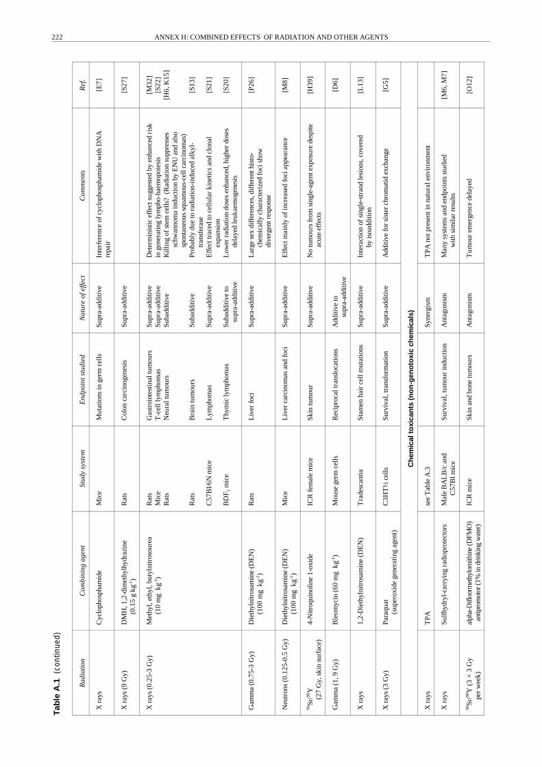
Tab
leA
.1(c
ontin
ued)
Rad
iatio
nC
ombi
ning
agen
tSt
udy
syst
emE
ndpo
ints
tudi
edN
atur
eof
effe
ctC
omm
ents
Ref
.
Xra
ysC
yclo
phos
pham
ide
Mic
eM
utat
ions
inge
rmce
llsSu
pra-
addi
tive
Inte
rfer
ence
ofcy
clop
hosp
ham
ide
with
DN
Are
pair
[E7]
Xra
ys(9
Gy)
DM
H,1
,2-d
imet
hylh
ydra
zine
(0.1
5g
kg-1
)R
ats
Col
onca
rcin
ogen
esis
Supr
a-ad
ditiv
e[S
27]
Xra
ys(0
.25-
3G
y)M
ethy
l,et
hyl,
buty
lnitr
osou
rea
(10
mg
kg-1
)R
ats
Mic
eR
ats
Rat
s
C57
Bl/6
Nm
ice
BD
F 1m
ice
Gas
troi
ntes
tinal
tum
ours
T-c
elll
ymph
omas
Neu
ralt
umou
rs
Bra
intu
mou
rs
Lym
phom
as
Thy
mic
lym
phom
as
Supr
a-ad
ditiv
eSu
pra-
addi
tive
Suba
dditi
ve
Suba
dditi
ve
Supr
a-ad
ditiv
e
Suba
dditi
veto
supr
a-ad
ditiv
e
Det
erm
inis
ticef
fect
sugg
este
dby
enha
nced
risk
inge
nera
ting
lym
pho-
haem
opoi
esis
Kill
ing
ofst
emce
lls?
(Rad
iatio
nsu
ppre
sses
schw
anno
ma
indu
ctio
nby
EN
Uan
dal
sosp
onta
neou
ssq
uam
ous-
cell
carc
inom
as)
Prob
ably
due
tora
diat
ion-
indu
ced
alky
l-tr
ansf
eras
eE
ffec
ttra
ced
toce
llula
rki
netic
san
dcl
onal
expa
nsio
nL
ower
radi
atio
ndo
ses
enha
nced
,hig
her
dose
sde
laye
dle
ukae
mog
enes
is
[M32
][S
22]
[H6,
K15
]
[S13
]
[S21
]
[S20
]
Gam
ma
(0.7
5-3
Gy)
Die
thyl
nitr
osam
ine
(DE
N)
(100
mg
kg-1
)R
ats
Liv
erfo
ciSu
pra-
addi
tive
Lar
gese
xdi
ffer
ence
s,di
ffer
enth
isto
-ch
emic
ally
char
acte
rize
dfo
cish
owdi
verg
entr
espo
nse
[P26
]
Neu
tron
s(0
.125
-0.5
Gy)
Die
thyl
nitr
osam
ine
(DE
N)
(100
mg
kg-1
)M
ice
Liv
erca
rcin
omas
and
foci
Supr
a-ad
ditiv
eE
ffec
tmai
nly
ofin
crea
sed
foci
appe
aran
ce[M
8]
90Sr
/90Y
(27
Gy,
skin
surf
ace)
4-N
itroq
uino
line
1-ox
ide
ICR
fem
ale
mic
eSk
intu
mou
rSu
pra-
addi
tive
No
tum
ours
from
sing
le-a
gent
expo
sure
desp
iteac
ute
effe
cts
[H39
]
Gam
ma
(1,9
Gy)
Ble
omyc
in(6
0m
gkg
-1)
Mou
sege
rmce
llsR
ecip
roca
ltra
nslo
catio
nsA
dditi
veto
supr
a-ad
ditiv
e[D
6]
Xra
ys1,
2-D
ieth
ylni
tros
amin
e(D
EN
)T
rade
scan
tiaSt
amen
hair
cell
mut
atio
nsSu
pra-
addi
tive
Inte
ract
ion
ofsi
ngle
-str
and
lesi
ons,
cove
red
byis
oadd
ition
[L13
]
Xra
ys(3
Gy)
Para
quat
(sup
erox
ide
gene
ratin
gag
ent)
C3H
T½
cells
Surv
ival
,tra
nsfo
rmat
ion
Supr
a-ad
ditiv
eA
dditi
vefo
rsi
ster
chro
mat
idex
chan
ge[G
5]
Ch
emic
alto
xica
nts
(no
n-g
eno
toxi
cch
emic
als)
Xra
ysT
PAse
eT
able
A.3
Syne
rgis
mT
PAno
tpre
sent
inna
tura
lenv
iron
men
t
Xra
ysSu
lfhy
dryl
-car
ryin
gra
diop
rote
ctor
sM
ale
BA
LB
/can
dC
57B
lmic
eSu
rviv
al,t
umou
rin
duct
ion
Ant
agon
ism
Man
ysy
stem
san
den
dpoi
nts
stud
ied
with
sim
ilar
resu
lts[M
6,M
7]
90Sr
/90Y
(3×
3G
ype
rw
eek)
alph
a-D
ifloo
rmet
hylo
rnith
ine
(DFM
O)
antip
rom
otor
(1%
indr
inki
ngw
ater
)IC
Rm
ice
Skin
and
bone
tum
ours
Ant
agon
ism
Tum
our
emer
genc
ede
laye
d[O
12]
ANNEX H: COMBINED EFFECTS OF RADIATION AND OTHER AGENTS222

Tab
leA
.1(c
ontin
ued)
Rad
iatio
nC
ombi
ning
agen
tSt
udy
syst
emE
ndpo
ints
tudi
edN
atur
eof
effe
ctC
omm
ents
Ref
.
Neu
tron
s(1
.7-3
.3G
y)C
CI 4
C57
Bl6
mic
eL
iver
carc
inom
asSu
pra-
addi
tive
No
effe
ctw
ithch
loro
form
[B16
]
Alp
ha(P
uO2)
CC
I 4R
ats
and
ham
ster
sM
odif
icat
ion
ofbi
okin
etic
sR
esul
tsno
tyet
know
n[B
16]
Xra
ys(1
.5G
y)B
rom
o-2'
-deo
xyur
idin
e(B
rdU
rd)
(3.2
mg
per
rat)
Rat
sT
otal
tum
our
yiel
d,la
tenc
ySu
pra-
addi
tive
Pote
ntia
lclin
ical
prob
lem
[A15
]
Xra
ys(4
-10
Gy)
Nic
oran
dil,
inhi
bito
rof
free
radi
cal
prod
uctio
nC
hine
seha
mst
erV
79ce
llsSu
rviv
alSu
badd
itive
[N20
]
Xra
ys(2
-8G
y)C
alyc
ulin
A,p
rote
inph
osph
atas
ein
hibi
tor
(2.5
-10
mM
)B
HK
21ce
llsC
ellk
illin
gSu
pra-
addi
tive
Dis
rupt
ion
ofpr
otei
nki
nase
-m
edia
ted
sign
altr
ansd
uctio
n?[N
21]
To
bac
co
Alp
ha(22
2 Rn)
Act
ive
smok
ing
aH
uman
s(m
iner
s)L
ung
canc
erSu
pra-
addi
tive
Con
foun
ders
:oth
erai
rpo
lluta
nts;
tran
sfer
tom
oder
nw
orkp
lace
san
din
door
expo
sure
[C1,
L18
]
Alp
ha(22
2 Rn)
Act
ive
smok
ing
bH
uman
s(m
iner
s)L
ung
canc
erA
dditi
veto
supr
a-ad
ditiv
eM
ore-
than
-mul
tiplic
ativ
eon
lyfo
rsm
okin
gpr
eced
ing
rado
n[T
18]
Alp
ha(22
2 Rn)
Act
ive
smok
ing
aH
uman
s(h
ome)
Lun
gca
ncer
Supr
a-ad
ditiv
eL
ittle
stat
istic
alpo
wer
[P11
]
Alp
ha(22
2 Rn)
Act
ive
smok
ing
aH
uman
s(t
horo
tras
tpat
ient
s)L
ugca
ncer
Non
ePr
oper
lung
dosi
met
ryla
ckin
g,th
eref
ore
little
stat
istic
alpo
wer
[I5]
Alp
ha(22
2 Rn)
Mai
nstr
eam
smok
eb
Rat
sL
ung
canc
erSu
pra-
addi
tive
Inte
ract
ion
only
for
rado
n[C
9]
Alp
ha(22
2 Rn)
Mai
nstr
eam
smok
eb
Bea
gle
dogs
Lun
gca
ncer
Suba
dditi
veR
educ
eddo
seto
criti
calc
ells
byth
icke
ning
ofbr
onch
ialm
ucus
?[C
21]
Alp
ha(22
2 Rn)
Die
self
umes
bSp
ragu
e-D
awle
yra
tsL
ung
canc
erN
one
Die
self
umes
alon
eha
dno
effe
ct[M
61]
Alp
ha(23
9 Pu)
Mai
nstr
eam
smok
ea
Mic
eL
ung
dose
,his
topa
thol
ogy
Supr
a-ad
ditiv
eL
ung
clea
ranc
ere
duce
d[T
2]
Alp
ha(23
9 Pu)
Mai
nstr
eam
smok
ea
F344
rats
Lun
gle
sion
s,lu
ngca
ncer
Supr
a-ad
ditiv
eL
ung
clea
ranc
ere
duce
d[F
17]
Xra
ys(>
9G
yto
targ
et)
Act
ive
smok
ing
bH
uman
s(H
odgk
in’s
dise
ase
patie
nts)
Lun
gca
ncer
Supr
a-ad
ditiv
eC
linic
alpr
oble
m[V
7]
Xra
ysA
ctiv
esm
okin
gb
Hum
ans
(bre
ast
canc
erpa
tient
s)L
ung
canc
erSu
badd
itive
tosu
pra-
addi
tive
Poss
ible
bias
inpo
sitiv
est
udy
[I9,
I10,
N4]
Met
als
Alp
ha(22
2 Rn)
Ars
enic
Hum
ans
(min
ers)
Lun
gca
ncer
Non
eA
djus
tmen
tfor
arse
nic
expo
sure
redu
ced
the
rado
nri
skes
timat
e(E
RR
WL
M-1
)[L
18,T
5]
ANNEX H: COMBINED EFFECTS OF RADIATION AND OTHER AGENTS 223

Tab
leA
.1(c
ontin
ued)
Rad
iatio
nC
ombi
ning
agen
tSt
udy
syst
emE
ndpo
ints
tudi
edN
atur
eof
effe
ctC
omm
ents
Ref
.
Alp
ha (150
-670
0B
q23
9 Pu)
Ber
ylliu
m(1
-91
µg
lung
burd
en)
Rat
sL
ung
dose
Lun
gtu
mou
rsSu
pra-
addi
tive
Non
eL
ung
clea
ranc
ere
duce
d[S
42]
Alp
ha (60-
170
Bq
239 Pu
)B
eryl
lium
(50-
450
µg
lung
burd
en)
F344
rats
Lun
gdo
seL
ung
tum
ours
Supr
a-ad
ditiv
eSu
pra-
addi
tive
Lun
gcl
eara
nce
redu
ced
[F12
]
Xra
ys(1
,2G
y)B
eryl
lium
(0.2
,1m
M)
CH
Oce
llsC
ell-
cycl
ede
lay
Chr
omos
ome
aber
ratio
nsSu
pra-
addi
tive
Supr
a-ad
ditiv
eM
ultip
licat
ive
inte
ract
ion
prob
ably
limite
dto
San
dG
2ce
lls[B
37]
Gam
ma
Cad
miu
m(1
mg
kg-1
)M
ice
DN
Ada
mag
ein
peri
pher
ally
mph
ocyt
esSu
badd
itive
Cad
miu
mex
posu
reha
sto
prec
ede
radi
atio
nby
seve
ralh
ours
[P27
]
134,
137 C
s( �
250
0B
qkg
- )L
ead
(16-
320
mg
kg-1
)A
rabi
dops
isth
alia
naM
utat
ions
Suba
dditi
veto
supr
a-ad
ditiv
e[K
42]
137 C
s(-
0.01
2G
y)Z
inc
(10-
100
mg
kg-1
)ca
dmiu
m(0
.5-1
6m
gkg
-1)
Soil
mic
robe
sN
itrog
enfi
xatio
n,de
ntri
-fi
catio
n,C
O2fl
uxSu
pra-
addi
tive
Cat
alas
eac
tivity
redu
ced
[E19
,E20
]
Mit
og
ens
and
cyto
toxi
can
ts
Xra
ys(0
.24,
0.94
Gy)
Caf
fein
e(1
-7m
M)
Pre-
impl
anta
tion
mou
seem
bryo
sM
icro
nucl
eiin
duct
ion
Em
bryo
nald
evel
opm
ent
Supr
a-ad
ditiv
eSu
pra-
addi
tive
App
aren
tthr
esho
ldbe
low
1m
M[M
11]
Xra
ysC
affe
ine
Invi
tro
chem
istr
yO
xyge
nra
dica
lco
ncen
trat
ions
Suba
dditi
ve[K
29]
Xra
ysC
affe
ine,
theo
brom
ine,
theo
phyl
line
Tly
mph
oma
cells
TK
Gce
llsA
popt
osis
Apo
ptos
isSu
pra-
addi
tive
Suba
dditi
ve[Z
4]
Xra
ysN
-met
hylf
orm
amid
e(0
-170
mM
)H
uman
colo
ntu
mou
rlin
eSu
rviv
alSu
pra-
addi
tive
Eff
ecto
nal
pha
term
,i.e
.str
onge
rat
low
erex
posu
res
[L15
]
An
tio
xid
ants
,vit
amin
san
do
ther
die
tary
fact
ors
Xra
ysR
estr
icte
dfo
odin
take
aA
nim
als
Tum
our
inci
denc
eSu
badd
itive
Prob
ably
via
redu
ced
grow
thst
imul
i[C
26]
Xra
ys(1
0G
y)N
aCl(
1%in
diet
),et
hano
l(10
%)
bC
D(S
D):
Crj
rats
Inte
stin
alm
etap
lasi
aSu
pra-
addi
tive
No
effe
cton
inci
denc
eof
gast
ric
tum
ours
[W29
]
Gam
ma
rays
(1.5
-4G
y)T
etra
hydr
ocan
nabi
nolb
Rat
sT
umou
rin
cide
nce
Supr
a-ad
ditiv
eB
reas
tade
noca
rcin
oma
incr
ease
dby
afa
ctor
5[M
24]
Gam
ma
rays
(4G
y)V
itam
inA
,E,s
elen
ium
,3-
amin
oben
zam
ide
aC
3H10
T½
cells
Tra
nsfo
rmat
ion
Suba
dditi
veE
pide
mio
logi
cals
tudi
esin
dica
teim
port
ance
ofdi
etar
yfo
rm[B
24,H
11]
Bio
log
ical
agen
ts(h
orm
on
es,v
iru
ses,
oth
er)
Gam
ma,
neut
rons
Oes
trad
iol-
17be
tab
Rat
sM
amm
ary
tum
ours
Que
stio
nabl
eH
igh
horm
one
leve
lsap
plie
d[B
32]
ANNEX H: COMBINED EFFECTS OF RADIATION AND OTHER AGENTS224
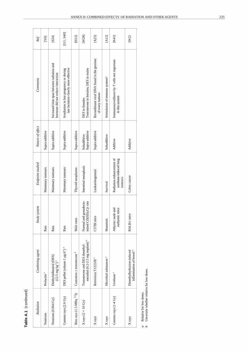
Tab
leA
.1(c
ontin
ued)
Rad
iatio
nC
ombi
ning
agen
tSt
udy
syst
emE
ndpo
ints
tudi
edN
atur
eof
effe
ctC
omm
ents
Ref
.
aR
elat
ive
for
low
dose
s.b
Unc
erta
inw
heth
erre
lativ
efo
rlo
wdo
ses.
Neu
tron
sPr
olac
tina
Rat
sM
amm
ary
tum
ours
Supr
a-ad
ditiv
e[Y
6]
Neu
tron
s(0
.064
Gy)
Die
thyl
stilb
estr
ol(D
ES)
(12.
5m
gkg
-1)
aR
ats
Mam
mar
ytu
mou
rsSu
pra-
addi
tive
Incr
ease
dtim
esp
anbe
twee
nra
diat
ion
and
horm
one
did
notr
educ
ein
tera
ctio
n[S
24]
Gam
ma
rays
(2.6
Gy)
DE
Spe
llet(
rele
ase
1µ
gd-1
)a
Rat
sM
amm
ary
tum
ours
Supr
a-ad
ditiv
eIr
radi
atio
nin
late
preg
nanc
yor
duri
ngla
tela
ctat
ion
clea
rly
mor
eef
fect
ive
[I11
,S40
]
Bet
ara
ys(1
.5M
Bq
131 I)
Cas
trat
ion
±te
stos
tero
neb
Mal
era
tes
Thy
roid
neop
lasm
sSu
pra-
addi
tive
[H12
]
Xra
ys(2
×10
Gy)
Tes
tost
eron
ean
dD
ES
dim
ethy
l-es
trad
iol(
0.2-
2.5
mg
impl
ant)
bN
orm
alan
dgo
nade
cto-
miz
edC
D(S
D);
Cjr
rats
Inte
stin
alm
etap
lasi
aSu
badd
itive
Supr
a-ad
ditiv
eD
ES
infe
mal
esT
esto
ster
one
infe
mal
es;D
ES
inm
ales
[W28
]
Xra
ysR
etro
viru
sT
1223
/Ba
C57
Blm
ice
Leu
kaem
ogen
esis
Supr
a-ad
ditiv
eR
ecom
bina
ntvi
ralD
NA
foun
din
the
geno
me
ofev
ery
tum
our
[A25
]
Xra
ysM
icro
bial
subs
tanc
esa
Mam
mal
sSu
rviv
alSu
badd
itive
Stim
ulat
ion
ofim
mun
esy
stem
?[A
12]
Gam
ma
rays
(1-4
Gy)
Ure
than
ea
Ath
ymic
nude
and
euth
ymic
mic
eR
adia
tion
enha
ncem
ento
fur
etha
ne-i
nduc
edlu
ngtu
mou
rs
Add
itive
Imm
unos
urve
illan
ceby
Tce
llsno
tim
port
ant
inth
issy
stem
[K41
]
Xra
ysD
imet
hylh
ydra
zine
-ind
uced
infl
amm
atio
nof
bow
ela
BA
LB
/cm
ice
Col
onca
ncer
Add
itive
[W2]
ANNEX H: COMBINED EFFECTS OF RADIATION AND OTHER AGENTS 225

ANNEX H: COMBINED EFFECTS OF RADIATION AND OTHER AGENTS226
17. In the earlystudyof molecular genetics, a wealth ofdatawere accumulated on interactions between UV and ionizingradiation in bacterial systems. For a review, see Annex L,“Biological effects of radiation in combination with otherphysical, chemical, and biological agents”, in the UNSCEAR1982 Report [U6]. Recently, laser applications have becomeimportant in industrial settings. A sparing effect from visiblelight on irradiated bacteria was reported by Voskanian et al.[V6]. The study measured the combined effect of laser(helium-neon laser, 633 nm) and alpha radiation on thesurvival of Escherichia coli K-12 cells of different genotypes.Pre- and post-irradiation exposures to laser radiationdiminished the damaging effect of alpha particles. Theincrease in survival was more pronounced for post-irradiationexposure. There isa well-known molecular basis for enhancedDNA repair and hence for survival: photoreactivation withvisible light after UV irradiation. The protective mechanisminvolved in the repair of damage from alpha irradiation,especially the one involved in pre-irradiation exposure to laserlight, remains to be elucidated. However, at this stage thereare no such mechanisms known in mammalian cells. Despitelarge human populations with considerable combinedexposures to ionizing and UV radiation to parts of the skin, noindications of a critical interaction are apparent.
3. Low- and high-frequency electromagneticradiation
18. The photon energies of all frequencies of electro-magnetic radiation below infrared are clearly too low toproduce direct chemical damage to DNA. However, thereis a large body of published data suggesting the presence ofeffects at exposure levels below those from critical thermaleffects, i.e. local heating by several degrees Celsius. (Heatstress is discussed in Section A.4.) The epigenetic influencesof heat stress could only act on later stages of cancerdevelopment. Whether so-called athermic levels of high-frequency non-ionizing radiation may interfere with cellsignalling, levels of cellular calcium, or systemic melatoninremains disputed on the level of the single agent.
19. Tyndall undertook an investigation [T3] to ascertainthe combined effects of magnetic resonance imaging fieldsand x-irradiation on the developing eye in mice from thestrain C57Bl/6J. Dams in groups were subjected toabsorbed doses of 50, 150, and 300 mGy. Other dams wereexposed to T2 spin-echo magnetic resonance imagingfields under clinically realistic conditions followingexposure to 300 mGy from x-irradiation. It was found thatthe 300 mGydose had significant teratogenic effects on theeye of C57BL/6J mice. Groups exposed to both types ofradiation fields demonstrated malformation levels similarto those in animals irradiated only with 300 mGy fromionizing radiation. The results confirmed the teratogeniceffects of low-level x rays but gave no evidence for anenhancement of the teratogenicity of x-irradiation on eyemalformations in the mouse system tested.
20. Somewhat unexpected results of combined effect ofmicrowave exposures ofnon-thermal intensityand ionizing
radiation were reported in rats and chicken embryos byGrigor’ev et al. [G13, G16]. Rats were pre-exposed toelectromagnetic radiation of power flux density (PFD)200 µW cm�2 30 minutes daily for 8 days, followed the nextday by single whole-body gamma irradiation at 5.5 Gy. Pre-exposure to microwave radiation reduced the mortality rate ofthe test animals by33% compared with the controls. Immuno-biological examinations revealed a significant increase in thestimulation index in mitogen (phytohemagglutinin, PHA)induced lymphocytes. The imprinting of chicks was disruptedwhen they were irradiated in early embryogenesis for 5minutes with microwaves (PFD = 40 µW cm�2) and then withgamma rays at a dose of 0.36 Gy.
21. The same group also described changes in humoralimmunity and in autoimmune processes under the combinedaction ofmicrowave, infrasonic, and gamma irradiation [G13,G16]. The exposure regimens for rats and rabbits were9.3 GHz and 0.1 GHz (200 and 1,530 µW cm�2, respectively),infrasound (8 Hz, 115 db), and gamma radiation (cumulativedose of 5.5 Gy). It was shown that pre-irradiation withmicrowaves increased the resistance of the animal to gammaradiation, but microwaves combined with infrasoundenhanced the biological effect of gamma radiation. Since nohypotheses on possible mechanisms are suggested, noinferences applicable to controlled human environments canbe drawn at this stage from the extremelyhigh exposure levelsin this study.
22. A very strong radioprotective effect of static magneticfields of 10, 120, and 350 mT on the survival of mice (CBA× C57Bl/6) after acute 60Co irradiation with a dose of 9 Gywas described by Schein [S33]. The adaptive effect of anexposure of 6 hours in a static field increased with time andwas strongest in animals irradiated 30 days later. The weakestfield, 10 mT, led to a survival of up to 60% of the animals,whereas controls had survival rates of only 0%�4%. Themechanisms behind this antagonism are speculated to beunspecific stress-induced stimulation of endocrine systems bymagnetic fields, an increase in surviving stem cells afterionizing radiation, or a faster proliferation and differentiationof bone marrow stem cells in adapted animals.
23. Growth and survival rates of cultured cells (FM3A)were investigated in a static gradient magnetic field witha strength of 58 mT at the center and a mean gradient of0.6 T m�1 [K16]. The magnetic field alone reduced thegrowth rate by 5% and survival by 20%. The combinedeffect of 60Co irradiation followed by exposure to themagnetic field showed synergism.
24. Magnetic fields have been shown under certainreaction conditions to perturb the rates at which radicalpairs recombine. An example is catalase-catalysed decom-position of H2O2, which is increased by 20% in anextremely high magnetic field of 0.8 T [M35]. In theory,this could lead to changes in the kinetics of free-radicalproduction and recombination [S60]. To measure theinteraction potential of this indirect genotoxic effect ofmagnetic fields with ionizing radiation, the exposures

ANNEX H: COMBINED EFFECTS OF RADIATION AND OTHER AGENTS 227
would have to be simultaneous and not sequential, asdescribed in the preceding paragraphs.
25. In assessing the association between exposure toelectromagnetic fields and cancer, Koifman [K17] definedthe elements necessary for quantitative analysis. Obtainingmore accurate measurements of exposure to electro-magnetic fields is a key to understanding any possibleassociation. In certain circumstances, strong electro-magnetic fields may stimulate growth and hence fulfill thecharacteristics of a cancer-promoter in biomechanisticmodels of carcinogenesis. This leads to the hypothesis thatelectromagnetic fields do not act alone to affect health, asis assumed in many epidemiological studies, but onlywhere their action is combined with that of other initiatoragents.
26. In summary, no straightforward inferences fromexperimental results to exposures in occupational settingsare possible at this stage for the combination of electro-magnetic and ionizing radiation. From the standpoint ofmechanistic considerations, there is little evidence forpotentially harmful interactions between the two radiationmodes for controlled exposure levels in the workplace or inthe clinic.
4. Temperature
27. Heat kills mammalian cells in a predictable andstochastic way [D3]. Heat stress at the cell and tissue levelmay disrupt energy metabolism (local depletion of oxygenand ATP) as a result of the enhanced reactivity of mostenzymes, the production of heat shock proteins, and finallydenaturation and cell death. Critical changes leading to aloss of proliferative capacity involve cell membraneblebbing, probablyowing todetachment of the cytoskeletonfrom the plasma membrane [R23]. A slow mode of cellkilling by hyperthermia in CHO cells involves the forma-tion of multi-nucleated cells from damage to centrioles[D3]. Above 42.5°C, cell-survival curves for Chinesehamster ovary cells in culture where the abscissa is theduration of heat treatment are similar to the curves forx rays. At 42°C and below, the survival curves tend toflatten out with time as tolerance to the elevatedtemperature develops. The cell-cycle dependence ofsensitivity to heat contrasts with that of x rays, with lateS-phase cells being the most sensitive to hyperthermiatreatment. Cells at low pH or deficient in nutrients alsoshow elevated heat sensitivity. Temperature is therefore animportant modifier of radiation sensitivity in many therapiestocontrol tumour growth. In general, hyperthermia increasesthe relative susceptibility of tumour cells to radiationcompared with healthy tissue. Very hot or very coldambient temperatures are rarelyencountered in the modernworkplace and the temperatures that do prevail generallydo not change the body core temperature. No correlationwith elevated radiation exposure is apparent in suchworkplace settings. The same is true for recreationalsettings and even for hot spas with elevated radon levels.Therefore the combined action of high and low tempera-
tures remains in the realm of clinical research, and thefollowing paragraphs give only some cursory remarks onrecent in vitro work.
28. At the mechanistic level, it is important to note that thelarge effects found in hyperthermia treatments cannot beattributed solely to changes in blood flow and concomitantchanges in local oxygen pressure alone. The disruption ofenergy metabolism due to considerably accelerated bio-chemical reactions and a decrease in molecular stability areimportant far below the threshold of protein denaturation.Dauncey and Buttle [D2] found a tendency towards elevatedplasma concentrations of growth hormone and prolactin in14-week-old pigs acclimated to 35° or 10°C, respectively. Inmammalian cell culture (L5178Y), protease inhibitors such asphenylmethylsulfonyl fluoride were shown to potentiatehyperthermic cell killing [Z13]. It is suggested that proteaseinhibitors sensitize by inhibiting the proteases that are neededto degrade denatured proteins induced by heat. In response toheat, cells and tissue produce proteins of mainly 70 and 90kilodaltons. These proteins are called heat-shock proteins,although many other agents such as arsenite and ethanol alsoinduce them. Their appearance coincides with the develop-ment of thermotolerance, an important effect that can in-fluence the slope of the survival curve by a factor of up to 10.The development of thermotolerance and the production ofheat-shock proteins occur during heating at temperatures upto 42°C (CHO cells) but are delayed by several hours for heattreatment with higher temperatures [H36].
29. Skin is the only tissue whose temperature might differconsiderably from the core temperature. Therefore, Zölzer etal. [Z3] studied the influence of radiation and/or hyper-thermia on the proliferation of human melanoma cells invitro. DNA synthesis and content were both determined withtwo-parameter flow cytometry. In controls, most of theS-phase cells showed incorporation of BrUdR. The fraction ofquiescent S-phase cells increased after irradiation (up to 8 Gyfrom x rays) and/or hyperthermia (up to 6 hours at 42°C or upto 2 hours at 43°C). There was a clear dose dependence forradiation and hyperthermia alone or in combination. Ingeneral, the combined effect seemed to be additive.
30. Combination effects of radiation and hyperthermia werefound, however, in several other in vitro cell systems.Matsumoto et al. [M15] treated cultured human retinalpigment epithelial cells by radiation, hyperthermia, or acombination of the two. The effect on cell proliferation wasevaluated by counting the cell number and measuring theuptake of bromodeoxyuridine. x-irradiation with a dose of1 Gy or 3 Gy was not effective in suppressing proliferation ofthe retinal pigment epithelial cells. Similarly, heat treatmentat 42°C for 30 minutes did not suppress proliferation.However, combining hyperthermia at 42°C for 30 minuteswith 3 Gyirradiation suppressed cellular growth of the retinalpigment epithelial cells to 36% of the control, as estimated bycell counting, and to 48% by the bromodeoxyuridine uptakeassay. The effect of radiation combined with heat on threehuman prostatic carcinoma cell lines was investigated byKaver et al. [K6]. Cells were exposed to different radiation

ANNEX H: COMBINED EFFECTS OF RADIATION AND OTHER AGENTS228
doses followed by heat treatment at 43°C for 1 hour. Heattreatment given 10 minutes after radiation significantlyreduced the survival rate of all the cell lines studied. Thecombined effect of radiation and heat produced greatercytotoxicity than predicted from the additive effects of the twoindividual treatment modalities alone. Impairment of DNArepair with elevated temperature is considered an importantmechanism [W36].
31. Growth, cell proliferation, and morphological altera-tions in vivo in mammarycarcinomas of C57 mice exposed tox rays and hyperthermia were followed by George et al. [G6].Radiation doses of 10, 20, or 30 Gy from x rays or heating to43°C for 30 minutes preceded or not by exposure to 10 Gywere studied. Tumour growth, cell proliferation kinetics,induction of micronuclei, and morphological changes innecrosis and vascular density were simultaneously deter-mined. These showed very complex adaptive responses.Treatment with radiation and/or hyperthermia produced onlya delay in tumour growth of between 1 and 3.8 days. How-ever, the effects of the treatments became more apparent whenthe amounts of muscle and necrosis were deducted from theoriginally measured tumour volume. Radiation-induced G2
block of the cells was observed 12 hours after radiation alone.After combined treatment, however, the G2 block was delayedbeyond 12 hours. Whereas the amount of necrosis wasmarkedly enhanced five days after treatment with 10 Gy plusheat, as well as after 30 Gy, no changes in the densityof smallblood vessels could be observed during this period. Theseresults clearly demonstrate that the apparent changes intumour volume after x rays and hyperthermia do not trulyreflect the response of the constituent cells and that there aremany other factors, for instance cell proliferation andmorphological alterations, that influence the combined effectsof radiation and hyperthermia.
32. Heat shock before, during, or immediately afterexposure to ionizing radiation can increase cell killing ina supra-additive manner [B70]. The heat-shock treatmentwas shown to inactivate the Ku auto-antigen binding to
DNA, and this binding capacity of Ku was directly relatedto the hyperthermic radiosensitizing effect. The Ku auto-antigen is the regulatory subunit of the DNA-dependentprotein kinase and is directly involved in DNA double-strand break repair and V(D)J recombination.
33. In general, it can be said that because of the hightemperatures and exposures needed to produce enhanced cellkilling in poorly oxygenated tissue, combined effects fromhyperthermia and ionizing radiation are not relevant outsidethe realm of tumour therapy. Temperature in combinationwith ionizing radiation can act synergisticallyon cell survival,cell proliferation, and cytogenetic damage. However,temperatures higher than those found in the human body areneeded to cause these effects.
5. Ultrasound
34. Possible effects from ultrasound exposures alone or incombination with ionizing radiation are of some concernbecause ultrasound is so widely used in diagnostic pro-cedures. Above a threshold level, ultrasound by itself mayinduce cavitation, leading tomechanical damage tocellularstructures and to microlesions. Kuwabara et al. [K20]studied the effects of ionizing radiation and ultrasound atexposure levels typical for diagnostic purposes on theinduction ofchromosomal aberrations and sister chromatidexchanges in peripheral lymphocytes. No statisticallysignificant increases in the frequencies of dicentric andring chromosomes or sister chromatid exchanges werediscovered after ultrasound exposure alone at the dia-gnostic level (Table A.2). Nor could elevated frequenciesof these phenomena be found following exposure toultrasound before or after ionizing radiation, comparedwith the frequencies found after the same dose of ionizingradiation alone. However, simultaneous exposure to ultra-sound and ionizing radiation seemed to induce a slightenhancement of sister chromatid exchanges, although nosignificant changes were noted in the yields of dicentricand ring chromosomes.
Table A.2Effects of combined exposures to ionizing radiation and ultrasound in peripheral human lymphocytes[K20]
ExposureDicentrics and rings Sister chromatid exchanges
Radiation Ultrasound
None (control) None (control) 6.64±0.40
3 Gy None40 min (immediately following)80 min (immediately following)
30 min (simultaneous)
0.61±0.08
0.52±0.07
7.92±0.546.31±0.537.00±0.479.80±0.91
4 Gy None30 min (simultaneous)
1.12±0.111.10±0.11 9.96±0.50
35. Continuous-wave ultrasound and neoplastic trans-formation was assayed in vitro by Harrison and Balcer-Kubiczek [H10] in C3H10T½ cells in suspension. An
initiation-promotion protocol for neoplastic transformationinduced by continuous-wave ultrasound was used. Cellswere insonated at 1.8 MHz for 40 minutes. Two ultrasonic

ANNEX H: COMBINED EFFECTS OF RADIATION AND OTHER AGENTS 229
intensities were used: 1.3 and 2.6 W cm�2 spatial average.The first intensity was found to be non-cytotoxic; thesecond was above the threshold level for cavitation andresulted in immediate lysis of 20% of the cells (cavitation-induced cell killing), followed by the clonogenic survivalof 64% of the remaining cells. Ultrasound was deliveredalone or in combination with x rays (2 Gy, 240 kVp givenbefore ultrasound) and/or TPA (0.1 µg ml�1 after irradia-tion). Under all treatment conditions, ultrasound had noeffect on transformation at the 95% confidence level. Theeffects of high-energy shock waves, i.e. therapeutic levelsof ultrasound generated by a lithotripter in combinationwith 137Cs gamma rays were shown to act additively orslightlysupra-additivelyin colony-forming assays and cell-cycle analysis [F29]. Both pellets of single cells andmulticellular spheroids of the bladder cancer cell line RT4gave similar results.
36. In conclusion, it can be said that the ultrasoundintensities used for diagnostic purposes and ionizing radiationdid not interact to cause cytogenetic damage in treated cells.However, sister chromatid exchanges were slightly increasedin one study. In vitro transformation rates caused by ionizingradiation were not changed by ultrasound.
6. Dust, asbestos, and other mineral fibres
37. The combination of radiation exposure and exposure todusts and fibres is quite common in important industrialenvironments such as mining, metallurgical industries, andpower plants. Some dusts and fibres are pathogenic orcarcinogenic by themselves. Both experimental results frommammals and epidemiological evidence are available [B9,B13, C22, K13, P1, P5]. In cases where the main biologicaleffect results from soluble toxicants that dissolve from thesurface of dust particles to interact with biological structures,the interaction is basically between radiation and a chemical,which is dealt with in Section B of this Appendix.
38. Silica is often considered to be a co-carcinogen throughthe route of silicosis. Harlan and Costello [H9] studied 9,912metal miners (369 silicotics and 9,543 non-silicotics) toinvestigate the association between silicosis and lung cancermortality. When lung cancer mortality in silicotics and non-silicotics was compared, the age-adjusted rate ratio was 1.56(95%CI: 0.91�2.68). Further adjustment for smokingyieldeda rate ratio of 1.96 (95% CI: 0.98�3.67), and the value foremployment in mines with low levels of radon was 2.59 (95%CI: 1.44�4.68). The statistical power of the study was tooweak toquantifysinglecontributions and interactions betweenmetal, radon, silica, and smoking. For high dust loads andconcomitant exposures to gamma radiation and radon inearlier times, there is indication for an increased lung cancerrisk (standardized mortality ratio = 2.5 with 20 years ofemployment and hired before 1960) in the phosphate industry[B74].
39. The molecular mode of action of mineral fibres is quitedistinct from radiation and genotoxic chemicals interactingdirectly with nuclear DNA. They are relatively ineffective as
mutagens but quite powerful inducers of human meso-theliomas and bronchial cancers. Fibre dimensions, fibredurability, and surface characteristics are important propertiesaffecting their carcinogenicity. In the case of asbestos, there isclear evidence for the induction of chromosomal aberrationsand aneuploidy [B13]. A possible mechanism of asbestos celltoxicity is phagocytosis and accumulation of the fibres in theperinuclear region of cells. During mitosis, the fibres wouldthen interfere with chromosome segregation, and chromo-somal abnormalities would result. In addition, mechanicalirritation and cell killing may lead to growth stimulation andtranscellular epigenetic promotion. The production of activeoxygen species on fibre surfaces was proposed as a directlyacting genotoxic mechanism; however, the relatively longdiffusion length from the site of radical production outside thenucleus to the target structures argues against the importanceof this pathway.
40. Recent reviews of mortality and cancer morbidity inasbestos worker cohorts with large cumulative exposuresshowed an ERR for pleural mesothelioma of about 1 foreach fibre-year ml�1 of air [A3]. For lung cancer, an ERRfrom 0.0009 to 0.08 per fibre-year ml�1 has been found[N9], which, in absolute terms, is considerably higher thanthe mesothelioma risk. The ratio of the number of meso-theliomas to the excess number of cases of lung cancerranges from 0.06 to 0.78.
41. Few epidemiological data exist describing potentialinteractions between mineral fibres and radiation. In acase-control analysis of deaths from lung cancer amongpersons employed at the Portsmouth Naval Shipyard atKittery, Maine, in the United States, elevated odds ratiosfor exposures to ionizing radiation, asbestos, and weldingby-products were found in a first crude assessment. Furtheranalysis of data on radiation exposure, controlling forexposures to asbestos and welding, found no evidence fora risk related to radiation exposure. The low cumulativeradiation doses and the absence of data on cigarettesmoking and socioeconomic status precluded an assess-ment of possible interactions among the three toxic agents[R1].
42. The synergistic effects of the combined exposures toasbestos and smoking in the causation of human lungcancer was one of the first examples of a supra-additiveinteraction of importance for protection in the workplace[S19]. In most studies, very high risk ratios were observedin asbestos-exposed subjects who were heavysmokers. Theinteraction observed in most cases conforms more closelyto a multiplicative model than an additive one. Brown et al.[B7] were able to show in organ cultures derived fromFischer F344 rats that the ability to metabolizebenzo(a)pyrene was significantly reduced after in vivoexposure to crocidolite, thus suggesting possiblemechanisms leading to a departure from linearity. Work byFasske [F2] showed that after the combined instillation of1 mg chrysotile and 0.5 mg benzo(a)pyrene, lung tumoursarose much earlier than after the instillation of only one ofthe carcinogens.

ANNEX H: COMBINED EFFECTS OF RADIATION AND OTHER AGENTS230
Asbestos fibres Alphaparticles
ObservedExpected
Asbestos fibres andalpha particles
Spontaneousfrequency
0
0.01
0.02
0.03
0.04
0.05
0.06
0.07
0.08
TR
AN
SFO
RM
AN
TS
PER
SUR
VIV
ING
CE
LL
S(%
)
43. Regarding animal experimentation, Bignon et al.[B38] inoculated radon-exposed Sprague-Dawley ratsintrapleurally with asbestos fibres, glass fibres, or quartz.In rats given mineral materials, bronchopulmonary car-cinomas and mixed carcinomas were observed, as well astypical mesotheliomas and combined pulmonary pleuraltumours, whereas in rats inhaling radon alone, onlybronchopulmonary carcinomas occurred. A clear co-carcinogenic effect of the insult from the minerals wasestablished for malignant thoracic tumours. Significantdifferences in survival time were found for exposures todifferent types of dust, depending on the additional tumourtypes induced. The same group also studied whethersimilar co-carcinogenic effects would take place overlonger distances, i.e. from subcutaneous injection of chry-sotile fibres. Neither mesotheliomas nor evidence of co-carcinogenic effects were found in the animals treated withboth radon and asbestos fibres [M19]. Three groups ofanimals were used: 109 rats that inhaled radon only (dose= 1,600 working-level months [WLM]); 109 rats given asubcutaneous injection in the sacrococcygeal region of20 mg of chrysotile fibres after inhalation of radonresulting in the same dose; and 105 rats injected with fibresonly. As already stated, no mesotheliomas occurred in anyof the three groups. The incidence of lung cancer was 55%in the second group, 49% in the first, and 1% in the thirdgroup. Statistical analysis using the Pike model showedthat the carcinogenic insult was slightly higher in thesecond group than in the first group. Electron microscopyanalysis of fibre translocation from the injection siteshowed that less than 1% of injected fibres migrated to theregional lymph nodes and only about 0.01% to the lungs.After injection, the mean length of the fibres recovered inlung parenchyma increased with time, suggesting thatshort fibres are cleared by pulmonary macrophages,whereas long fibres remain trapped in the alveolar walls.Kushneva [K49] studied pathological processes in thelungs of white rats exposed intratrachealy to 50 mg offinely dispersed quartz dust and to 3 hours of 3 108 Bq m�3
radon. Supra-additivity is clearly implied but onlydescribed qualitatively.
44. To assess the possible co-carcinogenic effects ofmineral dust in radon-prone mines, five groups of 30Sprague-Dawley rats received minerals typically found inmetal mines (nemalite; biotite, present in many granites;iron pyrite; chlorite) by intratracheal instillations onemonth after the end of a 1,000 WLM radon exposure. Noor onlyslight co-carcinogenic effects were found [M62]. Inearlier work with the same experimental system toinvestigate the effect of intrapleural injection of asbestosfibres (chrysotile), glass fibres, and quartz on the yield ofradon-induced thoracic tumours, a clear promoting effectwas noted [B38].
45. Densely ionizing alpha particles, similar to thoseemitted by radon progeny, are highly effective in inducingtransformations in cell cultures such as CH310T½ cells.The yield of foci from combined alpha/asbestos exposureis clearly greater than would be predicted from the sum of
the effects found with single-agent exposures. Figure A.Ishows a clearly supra-additive interaction with asbestosfibres [H11].
Figure A.I. In vitro transformation of C3H10T½ cellsexposed to asbestos fibres and alpha particles aloneand in combination [H11].
46. In an experimental study, Donham et al. [D10]studied possible combined effects of asbestos ingestion andlocalized x-irradiation of the colon in rats based on thehypothesis that the mucous produced by goblet cells thatnormally coats the normal bowel surface protects againsttissue penetration by ingested asbestos. X-ray treatmentresults in localized damage to the colonic mucosa andtheoreticallydisrupts the normal mucous coating, allowingincreased tissue penetration by the fibres. To study this,segments of the colons of laboratory rats were exposed tox-irradiation. The animals were then divided into threegroups, which were fed a diet containing 10% chrysotileasbestos, a diet containing 10% non-nutritive cellulosefibre, or a standard laboratory diet. Autopsies and histo-pathology were performed on all animals that diedspontaneously and those that were killed at 350 days.Various types of inflammatory and degenerative lesionswere commonly seen, but there was little difference infrequency between the diet groups. Five adenocarcinomasand two sarcomas were seen in the fibre groups (threetumours in the asbestos group and four tumours in thecellulose group), but no tumours were seen in animals onthe standard diet. There was no significant difference intumour rates between the asbestos and cellulose groups,nor was there a significant difference between the com-bined fibre groups and the standard diet group. Ingestedasbestos did not increase the risk of tumour developmentand does not, therefore, seem to be co-carcinogenic or topromote tumours by disrupting the mucous coating.
47. In summary, it can be stated that mineral dust andfibres such as asbestos generallyact through non-genotoxicmechanisms. These include mechanical irritation and cellkilling. However, chromosomal aberrations, especiallyaneuploidy, can be induced by interfering with the spindleapparatus of mitotic cells. At exposure levels found in

ANNEX H: COMBINED EFFECTS OF RADIATION AND OTHER AGENTS 231
workplaces until the early 1940s, there was a clearlysupra-additive interaction between asbestos and tobacco smokeexposure in the causation of lung cancers, with a concomitantshift in the cancer spectrum from mesotheliomas to broncho-pulmonary carcinomas. A similar supra-additive interactionand shift in the cancer spectrum was observed in animalsexposed to both asbestos and radon. The much lower occupa-tional exposures experienced today considerably decrease therisk for potential detrimental interactions between dust/fibresand radiation. However, in view of the proven interactioneffects in humans, any stochastic and/or genotoxic effects ofthese agents merit further consideration.
7. Space flight
48. In space flight, which involves an extreme situationof controlled exposures, a multitude of stressors act incombination on astronauts, the most important beingmicrogravity. Its biological and medical role has beenextensively reviewed [M71]. Microgravity effects mayoccur at all levels of biological organization, and inprinciple can also lead to modifications of radiation action.From an experimental point of view there are no clear-cutresults at the organ and tissue level. With simple organisms,a synergistic action of microgravity and radiation has beenreported for teratogenic effects [B80]. Antipov et al. [A14]analysed structural and functional changes in the centralnervous system of experimental animals exposed to theisolated and combined effects of space flights. Theyevaluated the significance of ionizing and non-ionizingradiation, hyperoxia, hypoxia, acceleration, vibration, andcombined effects of some of these factors for anatomic andphysiological changes in the rat brain. Neuronal functionswere found to be sensitive to ionizing radiation and hypoxia,but these synapses were shown to be highly resistant toshort-term hyperoxia and electromagnetic radiation [A13].Along with radiation, the investigated stressors hadadditive, synergistic, and antagonistic effects on the centralnervous system. However, as significant effects and devia-tions from the sum of effects from exposure to isolatedstressors were always linked to high exposures andexposure rates, they have little relevance for exposuresituations on the ground.
49. In radiobiological experiments in space, a more-than-additive interaction between microgravity and radiationwas reported in several cases (reviewed in [H47]). Insectembryos in particular appear to be susceptible. Conflictingresults were reported for cellular systems. In humanlymphocytes that were exposed to 32P-irradiation in space,chromosomal aberrations were significantly increasedcompared with ground controls [B18]. However, thefollow-up experiment by the same authors did not showthis interaction [B19]. More recently, experiments on theinteraction of space microgravity and DNA repair wereperformed by Hornek et al. [H14]. Microgravity had nomeasurable effect on strand rejoining of x-ray-inducedDNA strand breaks in Escherichia coli (120 Gy) and inhuman fibroblasts (5 and 10 Gy) or on the induction ofSOS reponse in E. coli (300 Gy). In yeast no microgravity
related effects on the repair of DNA double-strand breakswere found both for cells irradiated previously on ground[P31] or during flight using a 63Ni beta source [P32].Therefore, repair of radiation-induced DNA damage seemsnot to be disturbed by microgravity, and other mechanismsmust be involved in the reported interaction betweenradiation and space gravity.
50. At similarly high exposures, Vasin and Semenova[V3] showed synergistic effects for combined stress fromradiation and vibration or normobaric hyperoxia. A studywas made of the combined effect of normobaric hyperoxiaand vibration on the sensitivity of hybrid mice (CBA ×C57Bl)F1 and F2(CBWA) to gamma radiation. Both singleand protracted (for five days, daily) vibration beforeirradiation aggravated acute radiation sickness. Hyperoxiaalso enhanced the development of the intestinal form ofradiation sickness. The combined effect of the two addi-tional factors aggravated the intestinal syndrome of acuteradiation sickness. These deterministic effects have nodirect implication for present-day controlled exposuresituations. Nevertheless, the changes of many parametersthat are normally stable in experimental work on earthmake well-designed studies in space potentially importantin addressing the combined effects of physical agents.
B. RADIATION AND CHEMICALTOXICANTS
51. A multitude of natural and man-made chemicals withcancer-initiating and -promoting potential are present inthe human environment and may interact with radiation.Classification based on their mode of action is oftendifficult, but at least a crude separation can be made intosubstances that mainly act by damaging DNA directly(genotoxic substances) and non-genotoxic substances[C50]. The former group includes chemicallyactive species(activation-independent chemicals) or species dependenton biotransformation and their active metabolites(activation-dependent chemicals). The mode of action iseither direct, by forming covalent links with DNA, orindirect, via radical attack of DNA. The latter groupcomprises chemicals ranging from nonspecific irritantsandcytotoxins to natural hormones and growth factors andtheir analogues that interact with the regulatory systems ofcells and organs. At this point, chemicals that protectagainst ionizing radiation should also be mentioned. Manyendogenous and exogenous sulfhydryl-carrying moleculesas well as other radical-scavenging agents considerablyreduce the primary damage and hence the clinical effectscaused by radiation [M6, M7]. A wealth of experimentaldata is available to describe the action of single chemicalagents, but the literature on interactions between thesesubstances and other agents is far more sketchy. It isimportant to note that recent efforts to quantify tissue dosesof chemical toxicants and their metabolites showed thedecisive importance of interactions in activation anddeactivation/excretion processes. For example, an assess-ment of the toxicity of benzene and its metabolites was
ANNEX H: COMBINED EFFECTS OF RADIATION AND OTHER AGENTS232
shown to depend crucially on the presence of othertoxicants such as toluene, and this effect extended toconcentrations found in human exposures [M60].
1. Genotoxic chemicals
52. The large group of genotoxic chemicals may befurther subdivided on the basis of their need to be activatedby metabolism. Most chemicals require metabolic activa-tion through the generation ofhighlyreactive electrophiles,which form DNA adducts by binding covalently to nucleicacids. The metabolism of any individual chemical can bevery complex, because the chemical can be the substrate ofseveral metabolizing enzymes. Genotoxic chemicals canalso be subdivided based on whether the reactive com-pound acts directly by covalent binding to DNA orindirectly by the generation of free radicals. In the lattercase, effects similar to those of radiation can be envisaged.
(a) Activation-independent alkylating agents
53. Modern cancer therapy involves many combinedtreatments using radiation and genotoxic drugs. Althoughexposures are well known and strong interactions exist,this human experience is of limited direct importance forrisk assessment at low doses, because with therapy, cellkilling is the main endpoint envisaged. Therefore thissubject is considered separately in Section D of thisAppendix. The occurrence of second primary tumours inhealthy tissue adjacent to treated tumours is of great directrelevance.
54. Morishita et al. [M32] examined the effects of x rayson N-methyl-N-nitrosourea (MNU)-induced multi-organcarcinogenesis in both sexes of ACI rats. Rats were treatedwith MNU (25 or 50 mg kg�1) at 6 weeks of age and/orwith x rays (3 Gy) at 10 weeks of age. The incidence ofadenocarcinomas in the small and large intestines of malerats treated with 50 mg kg�1 MNU and x-irradiation (smallintestine, 48%; large intestine, 32%) was significantlyhigher than the sum of the incidences resulting from50 mg kg�1 MNU alone (small intestine, 17%; largeintestine, 8%) and with radiation only(small intestine, 0%;large intestine, 0%) and also higher than the frequency ofadenocarcinomas in the large intestine of males treatedwith 25 mg kg�1 MNU alone (0%). Strongly synergisticeffects in these high-exposure studies were restricted to thegastrointestinal system. When MNU or 1,2-dimethyl-hydrazine (DMH) treatment was started two months afterx-irradiation, no induction ofgastric tumours was observedwith MNU [W3], and only a low incidence was observedwith DMH [A7]. Surprisingly, an inverse relationshipbetween incidences of gastric tumours and intestinalmetaplasias was apparent. These findings again indicatethe importance of the order and timing of the exposures inthe induction of combined effects. It comes as a furthersurprise that the presence of intestinal metaplasia, longconsidered a basis for further malignant growth, does notexert a positive influence on the induction of gastricneoplasia by MNU in the rat.
55. Seidel [S22] studied the effects of radiation onchemicallyinduced T-cell lymphomas (thymomas) in BDF1
mice. N-methyl-N-nitrosourea or butylnitrosourea (BNU)were the main inducers, and x rays in various doseschedules were applied. The radiation was seen to shortenthe latency period between induction and lymphomaemergence in protocols of 12 exposures of 0.25 Gy. Thiseffect was most pronounced compared with chemicallyinduced non-irradiated controls with a prolonged medianinduction time as a result of a dose reduction of thechemical (median induction time 27�36 weeks instead of16�18 weeks under optimal conditions using 50 mg kg�1 ofMNU). Irradiation 2�5 weeks before administrating40 mg kg�1 of MNU also enhanced leukaemogenesis.Again, mice with regenerating lymphohaemopoiesis afterlethal irradiation and bone marrow transplantation weremore sensitive to both chemicals than were the controls.Combined effects from radiation and N-ethyl-N-nitrosourea (ENU) on neural tumours in Wistar rats werereported by Hasgekar et al. [H6]. The animals received2 Gy whole-body irradiation, followed immediately by10 mg kg�1 of ENU on the day of birth. Of 33 rats givenENU alone, 14 developed 22 tumours of the nervoussystem, of which 15 (68%) were gliomas and 7 (32%) wereschwannomas. Of 34 rats given both irradiation and ENU,12 were found to harbour 15 neural tumours, of which 14(93%) were gliomas and 1 (7.1%) was a schwannoma. Thepretreatment with irradiation seems to have resulted inselective suppression of schwannoma induction. Whetherthis antagonistic relationship is a result of overkill orwhether it may be relevant for lower radiation dosesremains to be elucidated.
56. The combined effects of radiation and BNU onmurine T-cell leukaemogenesis was studied by Seidel andBischof [S20] in BDF1 mice. The animals were exposed toBNU (0.02% in drinking water) for 12 weeks, and theydied of thymic lymphomas with median latency periods of12�20 weeks. Groups of mice received weekly radiationdoses of 0.06�1.0 Gy in addition to BNU. Lower doses (12× 0.25 Gy) enhanced leukaemogenesis, high doses (12 ×0.75 Gy) delayed it, and intermediate doses (12 × 0.50 Gy)had no effect. Doses lower than 12 × 0.25 Gy had marginalenhancing effects. After a dose of 12 × 1.0 Gy, the micedied earlier than after treatment with BNU alone, and aswith the dose of 12 × 0.75 Gy, some extrathymic lympho-mas were observed. The numbers of CFU-S in the femurand the spleen showed a dose-dependent depression, inaddition to the decrease from BNU alone. In lymphocytestimulation assays with Con A and LPS and also in themixed lymphocyte reaction, a reduced proliferation wasfound, again dependent on the radiation dose. Thus, therewas an inverse correlation between leukaemogenesis andthe degree of stem-cell reduction or depression of theseimmune parameters.
57. Stammberger et al. [S13] analysed the activity ofO6-alkylguanine-DNA alkyltransferase (AT) in the fetalbrain and liver and made long-term observations of Wistarrats that were treated in utero either with x-irradiation

ANNEX H: COMBINED EFFECTS OF RADIATION AND OTHER AGENTS 233
(1 or 2 Gy), with ENU (50 mg kg�1), or with both in com-bination. They hoped to reveal any relationship betweenthe O6-alkylguanine repair capabilityand tumour incidencein the organs of the offspring. The AT activity in the brainwas affected to the same extent in the fetuses as in thedams. There was a 61% decrease in AT activity in fetuses24 hours after ENU treatment. This correlated with asignificant increase in the incidence of brain tumours inthe treated offspring (44%) compared with control animals.The inductive effects of x-irradiation on AT activity (131%for 1 Gy and 202% for 2 Gy) corresponded with a reduc-tion in the incidence of tumours after the combined treat-ment (27% and 8.3% tumour incidence, 103% and 158%AT activity). Comparing biochemical and morphologicalresults suggests that this antagonistic effect may be theresult of the AT induction by x rays.
58. Yokoro et al. [Y6] found that whole-body irradiationfacilitates chemically initiated T-cell lymphomagenesis inmice. This was attributed to the amplification of the cellpopulation susceptible to a chemical carcinogen in thetarget tissues, bone marrow, and thymus during therecovery phase after irradiation. Split administration ofENU showed different effects in the different phases ofcarcinogenesis leading to T-cell lymphomas. Once morethe authors emphasized that after a cell has been initiatedby a genotoxic agent, its fate is determined by the presenceof promoters and inhibitors and that modifiers of targetcells play a crucial role in the induction yield of tumours.The possibility of synergistic effects in carcinogenesis dueto changes in cellular kinetics brought about by combinedtreatment with radiation and ENU was studied by Seyamaet al. [S21]. Lymphomas in female C57Bl/6N mice wereused as a model system. A single intragastric administrationof 5 mg (about 200 mg kg�1 body weight) of ENU was onlyslightlylymphomagenic, inducing thymic lymphomas in 20%of mice; the incidence was elevated to 92% if the ENUtreatment was preceded (five days earlier) by 4 Gy fromwhole-body x-irradiation, which alone is seldom lympho-magenic. A high yield of lymphoma (84%�93%) was alsoobtained when 5 mg (about 200 mg kg�1) of ENU wasdelivered in two split doses four days apart of 4 mg and1 mg (160 and 40 mg kg�1), indicating that cellularkinetics or clonal expansion, but not two agent-specificdifferent initiation events in the combined treatment, is atthe root of this apparent synergism. Drastic injury to boththe thymus and bone marrow caused by either 4 Gy whole-body x-irradiation or the first dose of ENU (4 mg, or about160 mg kg�1) was followed by a vigorous regenerationwithin a few days. The maximum induction rate oflymphoma was obtained when the subsequent dose of ENU(1 mg, or 40 mg kg�1) was given at the peak of DNAsynthesis in the bone marrow and thymus following thefirst treatment. The data indicate that the principal effectof irradiation or the first dose of ENU was to provide asusceptible cell population, and that a high yield oflymphomas was brought about by the action of thesubsequent dose of ENU on a larger number of potentiallyradiation-modified target cells engaged in heightened DNAsynthesis.
59. A clear antagonistic effect ofENUand x-irradiation wasobserved by Knowles [K14, K15] for neurogenic tumours inneonatal rats. After neonatal injection of rats with 10 mg kg�1
of ENU, whole-body x-irradiation with 1.25 Gy caused areduction in induced neurogenic tumours, which was greatestwhen radiation was given 1 day after ENU and progressivelydecreased with irradiation at 5 and 30 days. Although x-irradiation did not affect the range of histological appear-ances in the tumours, malignant schwannomas, particularlythose of the trigeminal nerve, were significantly reduced by1.25 Gy given after ENU (10 mg kg�1). The mean latency forclinical signs of tumour appearance was not affected byradiation. Another important finding in this study also pointsto the importance of the size of stem cell pools ininteractions: a significant reduction in the high spontaneousincidence of squamous-cell carcinomas of the mouth in theinbred strain used after 1.25 Gy from x-irradiation. Thereduction was greater after irradiation at 5 days of age than at30 days. A large study on the incidence rates of neural,pituitary, and mammary tumours in Sprague-Dawley ratstreated with x-irradiation and ENU during the earlypost-natalperiod was undertaken by Mandybur et al. [M2]. These lateeffects of earlypost-natal treatment with ENU, preceded by x-irradiation to the head, were studied in 226 neonatal CD rats.The animals were divided into six groups, each receiving oneof the following treatments: x-irradiation with 5 Gy to thehead on the third post-natal day; ip injection with 30 mg kg�1
ENU on the fourth post-natal day; ip injection with 30mg kg�1 ENU on the seventh post-natal day; a combination ofx-irradiation with 5 Gyto the head on the third post-natal day,followed by ip 30 mg kg�1 ENU on the fourth post-natal day;a combination of x-irradiation of 5 Gy to the head on the thirdpost-natal day, followed by ip 30 mg kg�1ENU on the seventhpost-natal day; and untreated controls. The results indicatedthat (a) x-irradiation to the head alone significantly extendedthe lifespan of females compared with that of control femalesand did not affect the survival of males; (b) x-irradiation didnot influence the latency period or mortality from neurogenictumours when ENU was given 1 or 3 days afterwards; (c)ENU itself was a factor in shortening latency periods formammary tumours; (d) x-irradiation alone did not increasetheincidence ofmammarytumours and revealed noprotectiveeffect on the ENU-induced mammary carcinogenesis; (e) x-irradiation increased the prevalenceofpituitarytumours in thefemales; (f) no enhancement ofpituitarytumours byENU wasobserved; and (g) there was a statistically significantassociation of pituitary and mammary tumours in females.Again, these widely divergent findings speak against thepossibility of simple concepts for the interaction of differentgenotoxic agents.
60. Post-natal development and cancer patterns in NMRImice after combined treatment with ENU and x-irradiationon different days of the fetal period were studied by Wig-genhauser and Schmahl [W10]. When mice were irradiatedto 1 Gy on day 14, 15, or 16 of gestation, this did not resultin an increased tumour frequency in the offspring until 12months. Mice treated with ENU (45 mg kg�1) on day 15 ofgestation developed a significantly increased tumourfrequency in the lungs and liver and in the ovaries. After

ANNEX H: COMBINED EFFECTS OF RADIATION AND OTHER AGENTS234
combined treatment in the sequence x rays plus ENU withan interval of 4 hours, a significantly increased incidenceof animals with tumours was observed in the offspringtreated on gestation day 14 or 16. Moreover, the treatmenton day 16 exhibited the highest tumour frequency perexamined animal (5.7) of all treatment groups. Althoughthe result was due to a relatively uniform increase of alltumour types, the frequency of liver tumours was mostmarked. In the reverse sequence (ENU plus x rays), thetotal tumour outcome was not significantly altered com-pared with the effects of ENU alone. However, detailedanalysis also showed a significant augmentation of theliver tumour frequency with treatment on day 15.
(b) Metabolism-dependent alkylating agents
61. Maisin et al. [M8] studied the effects of x rays aloneor combined with the initiator diethylnitrosamine (DEN)on liver cancer induction in infant C57Bl/Cnb mice. Thenumber of induced liver foci and carcinomas was found todepend essentially on the dose of DEN. X rays did notproduce any combined effect on the induction of foci orcarcinomas when given seven days before or after admini-stration of DEN [M34]. Using the same system forexposures to DEN and neutrons (average energy =3.1 MeV), it was shown that even high-LET irradiation(0.125�0.5 Gy) initiated only small numbers of nodularlesions, whereas DEN alone increased liver nodulessignificantly and proportional to dose (0.3�2.5 µg). Asupra-additive interaction between the twoinitiatingagentswas found mainly in the increased rate of foci appearanceafter 1.25 µg of DEN and 0.125 Gy of neutrons, both givenseven days before or after DEN exposure [M33]. Peraino etal. [P26] studied three altered hepatocyte foci (elevatedgamma-glutamyl transpeptidase [GG+] and/or iron-exclusion [Fe�]) in Sprague-Dawley rats exposed to DEN(0.15 µmol g�1) and/or gamma rays (0.75, 1.5, and 3 Gy)shortly after birth. The exposure was followed by a pheno-barbital (0.05%) diet to promote focus expression. Radiationalone was a weak hepatocarcinogen. A strong synergismwas seen at the lower radiation doses for the induction of[GG+] foci but not for other focus phenotypes. Aqualitatively different type of genetic damage for DEN(point mutations) and for radiation (rearrangements) ispostulated from the result. Large sex differences in theyield of DEN-induced [GG+/Fe�] foci by a factor of up to10 are additional indicators of the complexity of thissystem.
62. The potential for pulmonary carcinogenic interactionsbetween 239PuO2 and the tobacco-specific nitrosamine 4-(N-methyl-N-nitrosamino)-1-(3-pyridyl)-1-butanone (NNK), agenotoxic lung carcinogen, was studied in 740 male rats[L17]. The animals received 239PuO2 by inhalation to result inlung burdens of 0 or 470 Bq. The NNK was administered bymultiple ip injection at doses of 0, 0.3, 1.0, or 50 mg kg�1. Thehighest dose of NNK markedly reduced the median lifespanof the rats, whereas in the other treatment groups survival wasminimally reduced in comparison with the controls. Resultson carcinogenicity are not yet available from this study.
63. An apparent synergism between low-LET ionizingradiation and the carcinogen 1,2-dimethylhydrazine (DMH)in the induction of colonic tumours in rats has beendescribed by Sharp and Crouse [S27]. They evaluated theinteraction of radiation (9 Gy to the abdomen only) andDMH (150 mg kg�1) with respect to colon carcinogenesisin male Fischer 344 rats. Radiation was administered 3.5days before the DMH. At eight months post-treatment, theincidence of DMH-induced colon tumours was doubled byprior radiation exposure. When the protocol of radiationplus DMH was repeated three times at monthly intervals,a 15-fold increase in tumour incidence (from 5% to 74%)was observed at six months post-treatment. This findingdemonstrated an apparent synergy between radiation andthe chemical carcinogen. Throughout the study, theappearance of carcinomas was associated with pre-existingcolonic lymphoid nodules. The reproducibility of tumourinduction as well as the range of tumour incidence gene-rated by treatment variations in this system appeared to besensitive enough to allow the examination of combinedeffects of much lower doses of radiation and/or chemicalcarcinogens. The model could be used to evaluate therelationship between existing lymphoid aggregates, whichalter local epithelial cell kinetics and are associated withfenestrations in the basement membrane. The quantifi-cation of the development of colon cancer in congruentsites may assist in defining dose-response curves for com-bined agents and may also provide a system for evaluatingthe mechanisms underlying their interactions. When DMHtreatment was started two months after x-irradiation, onlya slight increase in gastric tumour incidence was recorded[A7]. These tumours occurred on top of a background ofradiation-induced gastrointestinal metaplasia.
64. Ehling and Neuhäuser-Klaus [E7] studied theinduction of specific-locus and dominant-lethal mutationsby combined cyclophosphamide (see also Section D.1.a foruses in combined modalities in tumour therapy) and radia-tion treatment in male mice. Unlike radiation, this widelyused antineoplastic agent, used alone, induced recessivemutations in spermatozoa and spermatids but not inspermatocytes and spermatogonia. Pretreatment (with60 mg kg�1) 24 hours before radiation, however, enhancedthe frequencyofspecific-locus mutations in spermatogonia.The mutational spectrum among seven loci remained thesame as in animals treated only with radiation. Thesynergistic interaction was mechanistically explained bythe interference of cyclophosphamide, a strong inhibitor ofDNA and RNA synthesis, with repair of radiation-induceddamage.
65. The effect of radiation on chemical hepatocarcino-genesis has also been examined in male ACI/N rats [M26].The number of neoplastic nodules or hepatocellular carci-nomas in rats given N 2-fluorenylacetamide (FAA) (0.02%in diet for 16 weeks) followed by x-irradiation (3 Gy) wassignificantly greater than in rats given FAA alone (p <0.001). In addition, the incidence ofhepatocellular carcino-mas in rats given the combined treatment was also higherthan in rats given FAA alone (p < 0.003). No liver lesions
ANNEX H: COMBINED EFFECTS OF RADIATION AND OTHER AGENTS 235
were found in animals receiving only an x-ray dose of3 Gy. The authors suggested that these highly supra-additive results indicate that ionizing radiation acts as apromoter in this model.
66. An inhibition of urethane(ethyl carbamate)-inducedpulmonaryadenomas byinhaled 239Pu in random-bredmaleA2G mice was reported as far back as 1973 by Brightwelland Heppleston [B66, B67]. This early study of combinedexposure to alpha radiation and a genotoxic chemicalcomprised four groups, each of 32 animals, receivingplutonium inhalation followed by urethane (PU), pluton-ium followed by saline (PS), mock inhalation followed byurethane (MU), and mock inhalation followed by saline(MS). Exposures consisted of initial lung burdens of925 Bq 239Pu and ip urethane injections of 1 mg g�1 bodyweight two weeks later. Eight weeks after the injections,PS-treated animals showed no increase in pulmonarytumours over control animals (MS), whereas practicallyallanimals in the PU and MU groups had multiple tumours.The number of tumours per animal 8, 16, and 24 weeksafter urethane treatment was clearly lower in the PU group,which had 4.2, 11.4, and 13 as compared with 8, 24.4, and38 in the MU group. An earlier hypothesis, that thisfinding is the result of alpha irradiation counteractingimmuno-suppression by urethane, is rejected on the basisof ultrastructural evidence. Severe morphological changesin mouse type-II cells in the vicinity of alpha particlesindicate that functional impairment of the initiated cells isthe main cause of the effect. The authors said, however,that this apparent antagonism needs to be viewed withcaution; it remains to be determined, they concluded, ifmuch smaller local plutonium doses would augmenturethane tumorigenesis.
67. The transgenerational combined effects of x rays(2.2 Gy) and urethane were studied byNomura [N23, N24]in three different mice strains (ICR, LZ, and N5). Urethanetreatment of F1 offspring of either irradiated males or femalesyielded an 18% incidence of tumour nodule clusters in thelung compared with only 2.8% in offspring of non-irradiatedcontrols. Tumour clusters were defined as having 12 or morenodules. The transgenerational effect of radiation aloneresulted in lung tumours (at least one tumour nodule) in 7.5%of the animals, whereas the value in unexposed controls was4.7%.
68. The interaction of gamma rays with urethane in lungtumorigenesis in mice in relation to the immune status hasbeen studied byKobayashi et al. [K41]. Male athymic nudemice (nu/nu) and their female heterozygous litter mates(nu/+) were treated with 1�4 Gy of 137Cs gamma rays and0.5 mg g�1 of urethane. Gamma-ray exposure alone causedrelatively few lung tumours (in up to 10% of animals);urethane alone caused tumours in 70%�80%. The combinedeffect was supra-additive. There was a tendency towardshigher yields in nu/+ mice, suggesting that impairedimmunosurveillance from T-cell deficiency does not increaselung tumorigenesis in thissystem. Since relativelyradiation-resistant macrophages and natural killer cells had higher
activities in nu/nu mice, the authors concluded that theinfluence of immunological status on tumorigenesis remainedunresolved.
69. A strong synergism was found by Hoshino andTanooka [H39] for skin tumours in beta-irradiated ICRmice painted later with 4-nitroquinoline 1-oxide (4NQO);27 Gy of 90Sr/90Y radiation or 20 applications of 5 mg ml�1
4NQO in benzene to the skin alone did not produce anyskin tumours in groups of 50 mice. Radiation followed by4NQO painting with an interval of 11�408 days betweenthe two treatments resulted in an incidence of malignantskin tumours (squamous-cell carcinomas and papillomas)of up to 17%. There was no significant decrease of thesynergistic effect with increasing interval, the greatesteffect being seen with an interval of 234 days.
70. A notable finding indicating the considerable uncer-tainties and misinterpreting the results of experimentalanimal studies was described by Little et al. [L55], whostudied the potential synergistic interactions between 210Po(185 Bq, resulting in a lifetime lung dose of about 3 Gy)and benzo[a]pyrene (0.3 mg) in the induction of lungcancer in Syrian golden hamsters. It was shown that simul-taneous administration by intratracheal instillation led toadditive effects. A significant apparent synergism wasfound when benzo(a)pyrene was given 4 months after the210Po. Most of this effect could be ascribed, however, to apotentiating effect of the seemingly innocuous 0.9% NaClinstillation solution alone.
71. The effects of repeated low exposures at high doserates such as used in some diagnostic radiologic proceduresat the time of the study were published by Lurie and Cutlerin 1979 [L56]. The induction of lingual tumours by 7,12-dimethylbenz[a]anthracene (DMBA) and radiation to thehead and neck was studied in Syrian golden hamsters.Treatment schedules were topical application of 0.5%DMBA in acetone on the lateral middle third of the tonguethree times a week for 15 consecutive weeks, about 200mGy radiation exposures (x rays with 100 kV peak) of thehead and neck once a week for 15 consecutive weeks, orconcurrent radiation and DMBA treatments for 15 con-secutive weeks. Histopathology was performed 35 weeksafter the start of the treatment. Animals receiving radiationalone had no detectable changes. The combined treatmentled to an excess of lingual papillomas compared withanimals receiving only DMBA (35% versus 15%). In addi-tion, an excess of non-lingual oral tumours (lip, gingiva,and floor of the mouth) was found in the animals receivingthe combined treatment compared with the DMBA-treatedanimals. Whether this radiation enhancement of DMBA-induced tumorigenesis has implications for the lowercombined exposures found for cigarette-smoke-derivedcarcinogens in the bucal cavity of humans and dentalx rays, remains to be elucidated.
72. Studies on chromosome aberrations from the com-bined effect of gamma rays and the mutagen thiotepa onunstimulated human leukocytes showed no significant

ANNEX H: COMBINED EFFECTS OF RADIATION AND OTHER AGENTS236
difference from the sum of their separately induced effects.The sequence of treatment and the interval between them(up to 4 hours) did not affect the frequency of chromosomeaberrations [B21].
73. Leenhouts et al. [L13] investigated the combined effectof 1,2-dibromoethane (DBE) and x rays on the induction ofsomatic mutations in the stamen hair cells of tradescantiaKU 9. At low radiation doses, a synergistic interaction wasfound between the twoagents for both DBE exposure followedby acute x rays and chronic simultaneous exposures. Thesynergism was considered to result from an interaction ofsingle-strand lesions in the DNA. It was concluded that thistype of interaction would not be too important for radiologicalprotection. However, it could be of significance in evaluatingthe effects of chemicals at low exposure rates.
(c) Free-radical-generating chemicals
74. Superoxide (O2�) generating agents such as the
dipyridilium compound paraquat might also interactdirectly with the fixation or repair of radiation-induceddamage. Geard et al. [G5] investigated the combinedeffects of paraquat and radiation on mouse C3H10T½ cells.Effects on oncogenic transformation, chromosome altera-tion, cytokinetics, or cellular survival were the endpointsmeasured. Paraquat alone is a cytotoxic agent and is alsoa weak radiosensitizer. Treatment with 0.1 mM for 24hours results in about 30% cell survival and enhances thecell-killing effects of 137Cs gamma rays by a factor of about1.2. The drug appears to function lethally by initiatinginterphase cell death and also by slowing cell cycling. Incombination with radiation (3 Gy), paraquat acted eitheradditively (sister chromatid exchanges) or with a greater-than-additive effect (cell survival and oncogenic trans-formation).
75. De Luca et al. [D6] studied the induction of reciprocaltranslocations in mouse germ cells (BALB/c) by bleomycinalone or combined with radiation (see also Section D.3 forbleomycin used in combined modalities in tumour therapy).The dose-response relationships after treatments with doses of20, 40, and 60 mg kg�1 of bleomycin as well as the combinedeffect of bleomycin and gamma rays were studied. A positive,significant correlation between the dose of bleomycin and thefrequency of translocations was found. Both potentiation andadditivity were found when the yields of translocationsinduced after combined treatments, separated by a lapse of 24hours, were compared with the sum of translocationfrequencies induced after the correspondingsingle treatments.Potentiation occurred in the treatments with 1 Gy plus 9 Gyand 60 mg kg�1 of bleomycin plus 9 Gy, while additivityoccurred in the treatments with 60 mg kg�1 of bleomycin plus1 Gy and 1 Gy plus 60 mg kg�1 of bleomycin. In miceirradiated with 1 Gy plus 9 Gy and mice treated with 60mg kg�1 of bleomycin plus 9 Gy, similar translocation yieldswere found. The potentiating effect of bleomycin was found tobe similar to that obtained with non-radiomimetic compoundssuch as triethylenemelamine, cyclophosphamide, and adria-mycin. The high doses involved and the erratic changes from
synergistic to additive relationships preclude extending in-ferences from these experiments beyond cancer therapy tooccupational or non-occupational settings.
76. In summary, there are many examples of strong devia-tions from hetero- and isoadditivity in the interactionsbetween genotoxic chemicals and ionizing radiation (TableA.1). Owing to generally high exposures to both agents understudy, deterministic effects were shown or suspected to be thecause of strong deviations from additivity in many studies.Thus, the several cases of synergism found seem to be mostlythe result of modifications of the biokinetics and the meta-bolism of the chemical rather than of agent-specific geno-toxicity at different stages of the pathological processes.Similar considerations hold for antagonistic effects, wheredepletion of stem cells and inhibition of cellular growth maybe a factor in the high dose range. Additional risks, beyondthelevel predicted from isoaddition, from the combinedeffectsof ionizing radiation and genotoxic chemicals at low ex-posures levels are, accordingly, not specifically demonstratedby the many epidemiological and experimental studiesreviewed in this Section.
2. Non-genotoxic chemicals
77. Many chemicals in the human environment or theirmetabolites do not specifically attack DNA but influencecell proliferation and cell differentiation on an epigeneticlevel. Specific mitogens may interfere with regulatorymechanisms and cell-cell signaling, but many substanceswith a high chemical reactivity act as unspecific irritantsor toxicants via membranes or proteins. Toxin-induced celldeath will induce proliferation in neighbouring cells,which may enhance the progression of premalignant cells.Substances acting in a non-specific manner, for examplelipophilic solvents, quite often show highly non-lineardose-response relationships with apparent thresholds.Other agents may interfere with the critical cellularprocesses involved in repairing damage to cellularconstituents such as DNA. The assessment of possiblesynergistic effects at the exposure levels relevant to thisAnnex is very difficult, because of the high exposures usedin experimental systems and the apparent threshold levels.
78. The tumour promoter 12-O-tetradecanoylphorbol-13-acetate (TPA) has the potential to enhance the yield ofradiation-induced tumours. This has been well documentedin vitro and in animal systems. The combined effects ofpaternal x-irradiation and TPA on skin tumours in twogenerations of descendants of male mice was studied byVorobtsova et al. [V5]. Progeny of outbred SHR male micenon-irradiated or exposed to a single dose of whole-body x-irradiation (4.2 Gy) were skin-painted twice a week for 24consecutive weeks from the age of four months onwardswith acetone or with TPA in acetone (6.15 µg ml�1). Theincidence and number of skin papillomas were monitoredbetween week 2 and week 20 after the last application ofthe promoter (TPA). Exposure to acetone was neverfollowed by skin tumour development in the progeny ofeither irradiated or non-irradiated males. Two weeks after
ANNEX H: COMBINED EFFECTS OF RADIATION AND OTHER AGENTS 237
TPA treatment, the incidence of skin tumours in theprogeny of non-irradiated mice was 21% in males and 37%in females, and 20 weeks later it was 12% in males and15% in females. The skin tumour incidence in the progenyof the irradiated male mice 2 and 20 weeks after the lastpainting was clearly elevated: 75% and 68% in males and50% and 43% in females, respectively. Some of the F1
offspring of irradiated male mice were mated before thestart of TPA treatment, and F2 progeny were exposed toacetone or TPA as F1. The incidence of skin papilloma 2weeks after the last TPA painting was 58% in males and40% in females, whereas at 20 weeks after the lastexposure to the promoter it was 53% and 36%,respectively. In the progeny of irradiated male mice therewere more animals with multiple (>4) skin papillomasthan in the progeny of non-irradiated mice. The incidenceof other than skin tumours in offspring was also clearlyincreased in TPA-treated progeny from irradiated malemice. The authors suggested that irradiation of malesbefore mating increases the susceptibility of progeny in atleast two generations to promoters of carcinogenesis as aresult of persisting genomic instability. On the other hand,Brandner et al. [B30] found no influence of ip-admini-stered TPA on the incidence of radiation lymphomas inC57Bl/6 mice. Female C57Bl/6 mice, given four x-irradiations each with 1.7 Gy, developed lethal lymphomasin more than 90% of animals 270 days after irradiation.Intraperitoneal application of TPA, 30 ng g�1 twice weeklyfor 240 days, had no influence on survival of the animalsor on incidence of the malignant lymphomas. However, theincidence in radiation-only treated animals was already sohigh that this test was highly insensitive to the promotingeffects of TPA.
79. Jaffe et al. [J2] studied the effect of proliferation andpromotion time on radiation-initiated tumour incidence inSencar mice. In this system, a single subcarcinogenic doseof ionizing radiation followed by 60 weeks of TPAtreatment led to the formation of squamous-cell carcino-mas. Even TPA pretreatment before irradiation seemed toresult in an overall increase in total tumour incidence,including both epidermal and non-epidermal tumours [J1].Based on these findings, the effect of the proliferative stateof the skin before irradiation and the promotion durationafter irradiation on tumour incidence was further investi-gated in CD-1 mice. To examine the influence of the pro-liferative state of the skin, a 17 nmol TPA solution wasapplied to one half of the mice 24 hours before irradiation.The skin was irradiated using 4 MeV x rays at a dose rateof 0.31 Gy min�1. Animals received a single dose of x raysof 0.5 or 11.3 Gy, followed by twice weekly applications ofTPA (8 nmol). The animals were then promoted for either10 or 60 weeks. All animals promoted with TPA for thesame duration had a similar incidence of papillomasregardless of radiation or TPA pretreatment. Increasing thepromotion duration did not significantlyalter the incidenceof squamous-cell carcinomas at either initiation dose. Atthe lower initiation dose, only animals that were promotedfor 60 weeks developed squamous-cell carcinomas. TPApretreatment at the higher dose resulted in a slight decrease
in tumour incidence; however, this was not statisticallysignificant. The incidence of basal-cell carcinomas wasradiation-dose-dependent and appeared to be independentof TPA promotion. Again, as in many other cases, nocommon pattern emerged for the different tumour types.
80. The interaction between ionizingradiation and TPA hasbeen studied using a three stage model of initiation, promo-tion, and progression. Ionizing radiation is well established asan initiator, whereas its potential for promotion and pro-gression is less well known. Therefore, Jaffe and Bowden [J1]performed a three-stage experiment using ionizing radiationin the third stage of mouse skin carcinogenesis. CD-1 micewere initiated with N-methyl-N'-nitro-N-nitrosoguanidine(MNNG), followed by biweekly promotion with TPA. After20 weeks of promotion, the animals were treated with eitheracetone, TPA (twice a week for two weeks), or eight fractionsof 1 MeV electrons (1 Gy per fraction over a period of 10days). Theconversion ofpapillomas tosquamous-cell carcino-mas was 80% for animals treated with ionizing radiationcompared with 25% for tumour-bearing animals treated withTPA. Ionizing radiation increased the number of cumulativecarcinomas per group. The absence of an increase in thenumber of cumulative papillomas per group due to lateexposure to ionizing radiation suggests that the dose andfractionation protocol used in this study enhanced theprogression of pre-existing papillomas.
81. The tumour-initiating and -promoting effects ofionizing radiation in mouse skin was also studied withTPA by Ootsuyama and Tanooka [O2]. Neither single24 Gy 90Sr/90Y beta irradiation followed by repetitivetreatment with TPA nor single pretreatment with 7,12-dimethylbenz-(alpha)-anthracene (DMBA), followed byrepetitive 4.7 Gy beta irradiation, produced tumours abovethe level of significance within a period of 210 days, whilea positive control, DMBA + TPA, yielded a high incidenceof papilloma in a shorter period. In this system, DMBAseemed to exert an action antagonistic to beta particles inthe induction of malignant tumours. It was concluded thatthe tumour-enhancing activity of repetitive radiation isqualitatively different from the promoting activity of TPA.
82. Nomura et al. [N6] were able to show that in uteroirradiation at early stages of embryogenesis, which was notvisibly carcinogenic by itself in a tester strain of mice (PT ×HT F1), followed by post-natal application of TPA, led to ahigh incidence of skin tumours. Radiation doses in thissystem were 0.3 and 1.0 Gy of 180 kVp x rays, respectively,at about 10.5 days after fertilization. Two dose rates, 0.54 and0.0043 Gymin�1, were used. The incidence ofboth embryonicmutations, determined as spots of different coat color, andtumours increased with in utero doses. Low-dose-rateirradiation led to a large (about 80%) reduction in tumourincidence.
83. TPA also causes enhanced transformation ofirradiated mouse 10T½ cells (Figure A.II). For the loss-of-contact inhibition, two genetic steps and modulation byepigenetically acting substances were proposed by Little
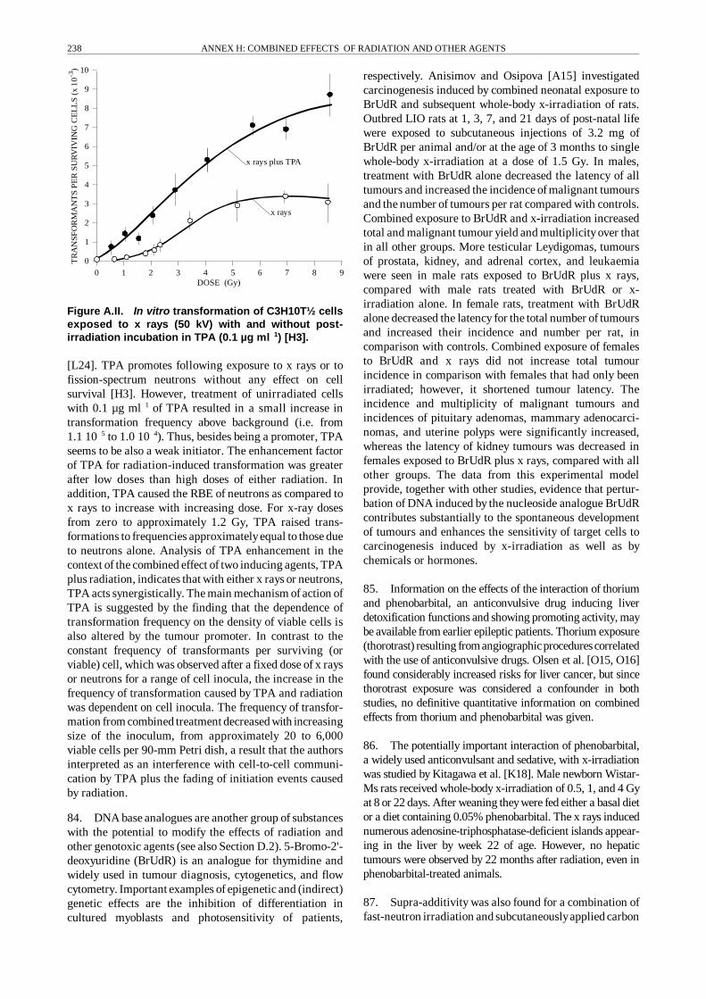
ANNEX H: COMBINED EFFECTS OF RADIATION AND OTHER AGENTS238
0 1 2 3 4 5 6 7 8 9
0
1
2
3
4
5
6
7
8
9
10
DOSE (Gy)
x rays plus TPA
x rays
TR
AN
SFO
RM
AN
TS
PE
RSU
RV
IVIN
GC
EL
LS
(x10
)-3
Figure A.II. In vitro transformation of C3H10T½ cellsexposed to x rays (50 kV) with and without post-irradiation incubation in TPA (0.1 µg ml�1) [H3].
[L24]. TPA promotes following exposure to x rays or tofission-spectrum neutrons without any effect on cellsurvival [H3]. However, treatment of unirradiated cellswith 0.1 µg ml�1 of TPA resulted in a small increase intransformation frequency above background (i.e. from1.1 10�5 to 1.0 10�4). Thus, besides being a promoter, TPAseems to be also a weak initiator. The enhancement factorof TPA for radiation-induced transformation was greaterafter low doses than high doses of either radiation. Inaddition, TPA caused the RBE of neutrons as compared tox rays to increase with increasing dose. For x-ray dosesfrom zero to approximately 1.2 Gy, TPA raised trans-formations to frequencies approximatelyequal to those dueto neutrons alone. Analysis of TPA enhancement in thecontext of the combined effect of two inducing agents, TPAplus radiation, indicates that with either x rays or neutrons,TPA acts synergistically. The main mechanism of action ofTPA is suggested by the finding that the dependence oftransformation frequency on the density of viable cells isalso altered by the tumour promoter. In contrast to theconstant frequency of transformants per surviving (orviable) cell, which was observed after a fixed dose of x raysor neutrons for a range of cell inocula, the increase in thefrequency of transformation caused by TPA and radiationwas dependent on cell inocula. The frequency of transfor-mation from combined treatment decreased with increasingsize of the inoculum, from approximately 20 to 6,000viable cells per 90-mm Petri dish, a result that the authorsinterpreted as an interference with cell-to-cell communi-cation by TPA plus the fading of initiation events causedby radiation.
84. DNA base analogues are another group of substanceswith the potential to modify the effects of radiation andother genotoxic agents (see also Section D.2). 5-Bromo-2'-deoxyuridine (BrUdR) is an analogue for thymidine andwidely used in tumour diagnosis, cytogenetics, and flowcytometry. Important examples of epigenetic and (indirect)genetic effects are the inhibition of differentiation incultured myoblasts and photosensitivity of patients,
respectively. Anisimov and Osipova [A15] investigatedcarcinogenesis induced by combined neonatal exposure toBrUdR and subsequent whole-body x-irradiation of rats.Outbred LIO rats at 1, 3, 7, and 21 days of post-natal lifewere exposed to subcutaneous injections of 3.2 mg ofBrUdR per animal and/or at the age of 3 months to singlewhole-body x-irradiation at a dose of 1.5 Gy. In males,treatment with BrUdR alone decreased the latency of alltumours and increased the incidence of malignant tumoursand the number of tumours per rat compared with controls.Combined exposure to BrUdR and x-irradiation increasedtotal and malignant tumour yield and multiplicityover thatin all other groups. More testicular Leydigomas, tumoursof prostata, kidney, and adrenal cortex, and leukaemiawere seen in male rats exposed to BrUdR plus x rays,compared with male rats treated with BrUdR or x-irradiation alone. In female rats, treatment with BrUdRalone decreased the latency for the total number of tumoursand increased their incidence and number per rat, incomparison with controls. Combined exposure of femalesto BrUdR and x rays did not increase total tumourincidence in comparison with females that had only beenirradiated; however, it shortened tumour latency. Theincidence and multiplicity of malignant tumours andincidences of pituitary adenomas, mammary adenocarci-nomas, and uterine polyps were significantly increased,whereas the latency of kidney tumours was decreased infemales exposed to BrUdR plus x rays, compared with allother groups. The data from this experimental modelprovide, together with other studies, evidence that pertur-bation of DNA induced by the nucleoside analogue BrUdRcontributes substantially to the spontaneous developmentof tumours and enhances the sensitivity of target cells tocarcinogenesis induced by x-irradiation as well as bychemicals or hormones.
85. Information on the effects of the interaction of thoriumand phenobarbital, an anticonvulsive drug inducing liverdetoxification functions and showing promoting activity, maybe available from earlier epileptic patients. Thorium exposure(thorotrast) resulting from angiographicprocedurescorrelatedwith the use of anticonvulsive drugs. Olsen et al. [O15, O16]found considerably increased risks for liver cancer, but sincethorotrast exposure was considered a confounder in bothstudies, no definitive quantitative information on combinedeffects from thorium and phenobarbital was given.
86. The potentially important interaction of phenobarbital,a widely used anticonvulsant and sedative, with x-irradiationwas studied by Kitagawa et al. [K18]. Male newborn Wistar-Ms rats received whole-body x-irradiation of 0.5, 1, and 4 Gyat 8 or 22 days. After weaning theywere fed either a basal dietor a diet containing 0.05% phenobarbital. The x rays inducednumerous adenosine-triphosphatase-deficient islands appear-ing in the liver by week 22 of age. However, no hepatictumours were observed by 22 months after radiation, even inphenobarbital-treated animals.
87. Supra-additivity was also found for a combination offast-neutron irradiation and subcutaneouslyapplied carbon

ANNEX H: COMBINED EFFECTS OF RADIATION AND OTHER AGENTS 239
tetrachloride in male and female C57Bl6 mice. Theanimals received a single whole-body dose of 1.7 or 3.3 Gyfrom fast neutrons, followed nine weeks later by a singlesubcutaneous injection of carbon tetrachloride. Carbontetrachloride markedly increased the incidence of radia-tion-induced liver carcinomas, whereas chloroform, whichwas also tested in this system, did not influence theincidence of radiation-induced tumours [B16].
88. The potential for carbon tetrachloride to modify thebiokinetics of an inhaled, soluble form of plutonium is alsobeing examined in both F344 rats and Syrian hamsters[B16]. Groups of animals were exposed to carbon tetra-chloride in whole-body chambers at concentrations of 0, 5,20, or 100 ppm for 6 hours per day, 5 days per week, for atotal of 16 weeks. After 4 weeks of exposure, approx-imately one half of the animals were exposed by a singlepernasal inhalation exposure to 239Pu nitrate. Serial sacri-fices of groups of animals were conducted at 4 hours and2, 4, 6, or 13 weeks after plutonium exposure for the quan-tification of 239Pu in lung, liver, kidney, and bone (femur)and for the evaluation of histologic changes in varioustissues. Results describing possible carbon tetrachlorideeffects on plutonium disposition are not yet available fromthis study. Another subgroup of rats and hamsters wasexposed to a radioactively labelled insoluble tracer particle.Tracer particle clearance was analysed for 13 weeks follow-ing exposure, and no significant clearance differences wereobserved between carbon-tetrachloride-treated and controlgroups.
89. Since ionizing radiation and tumour-promoting agentsincrease the level of ornithine decarboxylase (ODC) involvedin polyamine biosynthesis, the effect of alpha-difluoromethyl-ornithine (DFMO), an inhibitor of ODC, on tumour yieldfrom beta radiation was tested in female ICR mice [O12]. Thechronic radiation exposure consisted of three times 3 Gy90Sr/90Y surface dose per week to the back. DFMO was addedto the drinking water in a final concentration of 1%. Itsignificantly delayed the time of tumour emergence from 245days with radiation exposure only to 330 days in animals alsogiven DFMO. The antagonistic effect of DFMO was alsoobserved for bone tumours.
90. Monchaux et al. [M61] addressed the importantquestion of possible synergistic contributions from dieselfumes present in mine air to radon-induced lung tumours.Three groups of 50 male Sprague-Dawley rats were exposedto radon (1,000 WLM) and/or diesel exhaust (300 hours;22�25 ppm CO and 4�5 mg m�3 diesel particles), with thediesel exposure succeeding the radon exposure by one month.Contrary to the strong synergistic effect of cigarette smokefound in this system (discussed under tobacco), exhausts hadonly a slight, non-significant effect on the risk for thoracictumours from radon. Diesel exhausts alone were not carcino-genic.
91. Since phosphorylation and dephosphorylation ofproteins play an important role in cellular metabolism,Nakamura and Antoku [N21] studied the effect of
calyculin A (CL-A), a specific inhibitor of proteinphosphatase 1 and 2A isolated from the marine spongeDiscodermia calyx, on x-ray-induced cell killing incultured mammalian cells (BHK21). At concentrationsabove 2.5 nM, CL-A enhanced the radiation effectconsiderably. As also shown in another cell culture systemwith the inhibition of protein kinases [H40], agents thatinterfere with protein-kinase-mediated signal transductionafter radiation exposure may enhance damage andrepresent a new class of radiosensitizers.
92. Many non-genotoxic agents clearly produce strongsynergistic effects with ionizing radiation. The combinedeffects of this class of agent are summarized in Table A.1.Table A.3 lists more detailed effects of TPA, probably thebest-studied modifier of genotoxic agents, on severalendpoints. These studies are of great importance for theelucidation of mechanisms affecting expression of risk. Atthis stage, however, no functional analogues of potentexperimental enhancers of radiation risk, such as TPA orDNA bases, are known to exist in critical concentrations inthe human environment.
3. Tobacco
93. The important interaction of tobacco smoke andradiation was introduced in the main text of this Annex.Epidemiological studies of uranium miners have allowedthe risks and interaction coefficients to be quantified, atleast for higher radiation doses. The complex compositionof tobacco smoke makes the interaction not simplya binarycombination, however. Some 4,000 individual chemicalcomponents of cigarette smoke have been identified, and anumber of additional unidentified components surely exist(for example, extremelyreactive, short-lived compounds orthose present in very low concentrations) [G1]. Identifiedcompounds in smoke include several known carcinogens ofthe polycyclic aromatic hydrocarbon and nitrosamineclasses.
94. The studies reviewed below refer to mainstreamsmoke, sidestream smoke, or environmental tobaccosmoke. Mainstream smoke is defined as the smokeoriginating from the butt end of a cigarette; it is generatedduring the active puffing process. Sidestream smoke is thesmoke released at the burning tip of a cigarette, whetherthe cigarette is being puffed or simply smoldering. Lastly,environmental tobacco smoke is a mixture of sidestreamsmoke and exhaled mainstream smoke. This term mostaccurately describes the smoke that would be found withinan enclosed space with a smoker present. Tobacco smokecontains relatively small amounts of DNA-reactivecarcinogens, such as nitrosamines, polycyclic aromatichydrocarbons, and pyrolysis products, such as carbolines.Hence enhancing and promotional factors, e.g. catechols,other phenols, and terpenes, are an important component.Probably because it reduces pressure from the action ofpromoters, discontinuation of smoking progressivelyreduces the risk of cancer development with time sincewithdrawal [W1].
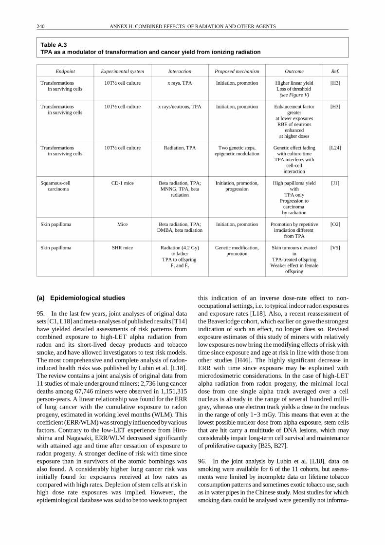
ANNEX H: COMBINED EFFECTS OF RADIATION AND OTHER AGENTS240
Table A.3TPA as a modulator of transformation and cancer yield from ionizing radiation
Endpoint Experimental system Interaction Proposed mechanism Outcome Ref.
Transformationsin surviving cells
10T½ cell culture x rays, TPA Initiation, promotion Higher linear yieldLoss of threshold
(see Figure V)
[H3]
Transformationsin surviving cells
10T½ cell culture x rays/neutrons, TPA Initiation, promotion Enhancement factorgreater
at lower exposuresRBE of neutrons
enhancedat higher doses
[H3]
Transformationsin surviving cells
10T½ cell culture Radiation, TPA Two genetic steps,epigenetic modulation
Genetic effect fadingwith culture time
TPA interferes withcell-cell
interaction
[L24]
Squamous-cellcarcinoma
CD-1 mice Beta radiation, TPA;MNNG, TPA, beta
radiation
Initiation, promotion,progression
High papilloma yieldwith
TPA onlyProgression to
carcinomaby radiation
[J1]
Skin papilloma Mice Beta radiation, TPA;DMBA, beta radiation
Initiation, promotion Promotion by repetitiveirradiation different
from TPA
[O2]
Skin papilloma SHR mice Radiation (4.2 Gy)to father
TPA to offspringF1 and F2
Genetic modification,promotion
Skin tumours elevatedin
TPA-treated offspringWeaker effect in female
offspring
[V5]
(a) Epidemiological studies
95. In the last few years, joint analyses of original datasets [C1, L18] and meta-analyses of published results [T14]have yielded detailed assessments of risk patterns fromcombined exposure to high-LET alpha radiation fromradon and its short-lived decay products and tobaccosmoke, and have allowed investigators to test risk models.The most comprehensive and complete analysis of radon-induced health risks was published by Lubin et al. [L18].The review contains a joint analysis of original data from11 studies of male underground miners; 2,736 lung cancerdeaths among 67,746 miners were observed in 1,151,315person-years. A linear relationship was found for the ERRof lung cancer with the cumulative exposure to radonprogeny, estimated in working level months (WLM). Thiscoefficient (ERR/WLM) was stronglyinfluenced byvariousfactors. Contrary to the low-LET experience from Hiro-shima and Nagasaki, ERR/WLM decreased significantlywith attained age and time after cessation of exposure toradon progeny. A stronger decline of risk with time sinceexposure than in survivors of the atomic bombings wasalso found. A considerably higher lung cancer risk wasinitially found for exposures received at low rates ascompared with high rates. Depletion of stem cells at risk inhigh dose rate exposures was implied. However, theepidemiological database was said to be too weak to project
this indication of an inverse dose-rate effect to non-occupational settings, i.e. to typical indoor radon exposuresand exposure rates [L18]. Also, a recent reassessment ofthe Beaverlodge cohort, which earlier on gave the strongestindication of such an effect, no longer does so. Revisedexposure estimates of this study of miners with relativelylow exposures now bring the modifying effects of risk withtime since exposure and age at risk in line with those fromother studies [H46]. The highly significant decrease inERR with time since exposure may be explained withmicrodosimetric considerations. In the case of high-LETalpha radiation from radon progeny, the minimal localdose from one single alpha track averaged over a cellnucleus is already in the range of several hundred milli-gray, whereas one electron track yields a dose to the nucleusin the range of only 1�3 mGy. This means that even at thelowest possible nuclear dose from alpha exposure, stem cellsthat are hit carry a multitude of DNA lesions, which mayconsiderably impair long-term cell survival and maintenanceof proliferative capacity [B25, B27].
96. In the joint analysis by Lubin et al. [L18], data onsmoking were available for 6 of the 11 cohorts, but assess-ments were limited by incomplete data on lifetime tobaccoconsumption patterns and sometimes exotic tobacco use, suchas in water pipes in the Chinese study. Most studies for whichsmoking data could be analysed were generally not informa-
ANNEX H: COMBINED EFFECTS OF RADIATION AND OTHER AGENTS 241
tive enough to allow deciding between an additive or a multi-plicative joint relationship for radon progeny and smoking.The Chinese cohort seemed to suggest an association moreconsistent with additivity, while the Coloradocohort suggesteda relationship more consistent with a multiplicative inter-action. For all studies combined, the joint relationship ofsmoking and radon progeny exposures with lung cancer wasstable over the different age groups and deviated quite clearlyfrom either a purely additive or a multiplicative relationship.The most recent analyses of the BEIR VI Committee [C46],which were based on an update of these data, suggest that thejoint effect is statistically closer to a multiplicative than anadditive interaction. To further characterize the association,more detailed data on tobacco use would be needed. Age atonset of smoking, amount and duration of smoking, and typeof tobacco were recognized as important determinants of risk.Such a refined analysis of smoking patterns is possible only inthe prospective part of ongoing studies and is subject,furthermore, topotential bias in the affected individuals owingto the rapidly decreasing public acceptance of smoking. Ingeneral, the single-exposure subcohorts of lifetime non-smokers are very small in all studies. The statistical power ofthe conclusions on the interaction between radon and tobaccosmoke is correspondingly small. Applying the two-mutationclonal expansion model ofcarcinogenesis ofMoolgavkar et al.todata from the Coloradoplateau miners shows no interactionbetween radon and tobacco smoke in any of the three steps[M39], but the predicted lung cancer incidence caused byradon and smoking remains more than additive and less thanmultiplicative, an indication of isoadditivity.
97. Microdosimetric considerations are also important inextrapolating the inverse dose-rate effect found foroncogenic endpoints caused by alpha radiation in generaland for lung cancer in miners [L36]. Brenner [B40]postulated that protraction enhancement is a mechanismlimited to cells receiving multiple hits over a humanlifespan. Since a typical domestic exposure to radonprogeny of 14 WLM yields a very small probability ofmultiple traversals in a cell nucleus (<1% for the mosthighly exposed stem cells in the tracheobronchialepithelium), dose-rate effects are probably of no relevance,and lung cancer risk per unit exposure will not increasefurther at low radon levels.
98. Two recent analyses by Yao et al. [Y7] and Thomas etal. ([T18] with erratum) on the radon-smoking interactionshowed a considerable influence of timing of exposures. Theformer study found a higher lung cancer risk for exposure toradon progeny and tobacco use occurring together ascompared to radon exposure preceding tobacco use. Thesecond study on Colorado uranium miners found asignificantly more-than-multiplicative effect for smokingfollowed by radon, whereas radon exposure followed bytobacco use produced an essentially additive effect. Thesefindings are in conflict with earlier notions based onexperimental results in rats, whereby radon is an initiator andtobacco smoke, a promoter [G20]. However the relevance ofthis animal system is questionable, because tobacco smokealone does not produce lung tumours in this system.
99. Despite the remaining uncertainties, it is quite clear thatthe joint effect of radon progeny exposure and smoking isgreater than the sum of each individual effect. The combinedanalysis [L18] shows that a linear exposure-response estimatefor radon and lung cancer is compatible with the data andgives a relative risk that is about three times higher in non-smokers than in smokers. Assuming a 10-fold difference inthe tobacco-caused lung cancer risk between smokers andnon-smokers, this means that the lung cancer risk for smokersexpressed in absolute terms is higher by a factor of about 3.Such a supra-additive effect, if also demonstrated to hold forpresent occupational and non-occupational exposure settings,would be of great importance for the regulation of smokingand radon progeny in the human environment. Until now,little quantitative evidence has come from indoor radonstudies. The few case-control studies published are incon-clusive [A28, P11]. Only one larger study [P11] was indica-tive of an indoor radon risk and its modification by tobaccothat is comparable to what is predicted from miner studies. Itremains doubtful whether the results from the many case-control studies under way will in the near future allownarrowing of the uncertainties that surround indoor radon riskand possible interactions with smoking. Based on inconclu-sive results from 1,000 computer-simulated large case-controlstudies assuming an ERR of 0.015 WLM�1, Lubin et al. [L33]questioned the assumption that epidemiological studies, evenwhen pooled in meta-analyses, will produce reliable estimatesof risk from residential radon exposure. Errors in exposureassessment, migration, and confounding bysmokingareat theroot of this pessimistic assessment. At least for the secondconfounder, studies in Europe based on much longer meanresidence times may offer better statistical power. Severallarge indoor case-control studies under way will narrowuncertainties in the next few years. First results from theUnited Kingdom [D33] and Germany[W35] are indicative ofa lung cancer risk in the range of ICRP projections. However,confidence intervals are relatively large and include zero riskin most analyses.
100. Because of the limitation of the indoor radon studies,risk estimates based on miner data remain the main basisfor predicting lung cancer from indoor radon exposure. Abest linear estimate of the risk coefficients found in thejoint analysis of Lubin et al. [L18, L35] for the indoorenvironment indicates that in the United States, some10%�12%, or 10,000 cases, of the lung cancer deathsamong smokers and 28%�31%, or 5,000 cases, of the lungcancer deaths among never-smokers are caused by radonprogeny. About half of these 15,000 lung cancer deathstraceable to radon would then be the result of overaddi-tivity, i.e. synergistic interactions between radon andtobacco. Based on the same risk model, Steindorf et al.[S47] predicted that about 7% of all lung cancer deaths inthe western part of Germany are due to residential radon.This corresponds to 2,000 deaths per year, 1,600 in malesand 400 in females. The attributable risk estimate was4%�7% for smokers and 14%�22% for non-smokers. Themost recent central estimates for the proportion of radon-attributable lung cancer deaths in the United States in 1995was recently provided by the BEIR VI Committee [C46] in
ANNEX H: COMBINED EFFECTS OF RADIATION AND OTHER AGENTS242
1998, based on an updated data set of the miners studiesreported by Lubin et al. [L18]. The Committee applied asub-multiplicative relation to model the joint effect oftobacco smoking and radon. Depending on two differentmodels (exposure-age-concentration model or exposure-age-duration model) about 14% or 9% of all lung cancerdeaths among ever-smokers and 27% or 19% amongnever-smokers were estimated to be attributable to radon.Because of the many differences between mines and homesand the additional carcinogens such as arsenic, dust anddiesel exhaust in mine air, these figures should beinterpreted with caution. A population-based case-controlstudy of incident lung cancers among women in Missouriwho where lifetime non-smokers or long-term ex-smokersyielded a very low and non-significant estimate of theattributable lung cancer risk from radon in non-smokers[A4].
101. It has been questioned whether toxicants other thanjoint exposures to radon progeny and cigarette smokecontribute considerably to the high lung cancer risk foundin miners [I5]. Heavy exposures to mine dust containingsilicates, diesel exhausts, and fumes from explosives mayadd to or combine with the two main lung carcinogens,radon and cigarette smoke. Patients who received thoro-trast continuously exhale the veryshort-lived 220Rn derivedfrom 232Th deposits in the body and therefore provide amodel for lung carcinogenesis by radon without con-comitant dust exposure. Ishikawa et al. [I5] studied thelung cancer incidence in a Japanese thorotrast cohort andfound 11 lung cancer cases in 359 thorotrast autopsy cases.The analysis revealed that while the proportion of small-cell lung cancer considered to be related to alpha radiationwas significantly increased, the overall lung cancer inci-dence was not significantly higher than in controls, in spiteof the high levels of 220Rn in the patients' breath. Theauthors took this as an indication that the risk for radon-induced lung cancer is not as high as expected from riskcoefficients deduced from miner studies. To substantiatethis hypothesis, the build-up of 220Rn decay products in thelung air space before exhalation and the resulting exposureto critical stem cells would have to be quantified.
102. Owing to the generally good linear correlationbetween radon progeny exposure and lung cancer in themajor miner studies, few additional carcinogens in minedust were considered in depth. Toxic metals are, however,of special concern. Results from the Chinese [X1],Canadian (Ontario), [K21] and Czech [T41] cohortsshowed arsenic to be an important additional risk factor forlung cancer. Adjustment for arsenic exposure reduced theradon risk estimate in these cohorts considerably. Even inthe most recent joint analysis by Lubin et al. [L18], othermine exposures were difficult to interpret, since theinformation was quite limited and of poor quality. In mostcases these concomitant exposures to suspectedcarcinogens or promoters are typically highly correlatedwith radon progeny exposures in a given study andtherefore difficult to assess independently (see alsofollowing Section B.4).
103. The mechanism of interaction between DNA lesionscaused by radon progeny and those caused by chemicaltoxicants contained in tobacco smoke is not known. Thereis clear evidence that the prevalence ofmutations in criticalgenes is dependent on the type of insult. The most commonknown gene mutations in lung cancer cells are found in thetumour-suppressor gene p53, which is thought to be crucialin the initiation of this and many other types of cancer.Several groups analysed the molecular changes in theconserved regions of the p53 gene in lung cancer tissue andreported differences between non-smokers (survivors of theatomic bombings and unirradiated controls), Japanesesmokers, and uranium miners with high radon exposures[T12, T13] (see also Annex F, “DNA repair and muta-genesis”). The non-smokers from Hiroshima showedmainly transition mutations (all G:C to A:T) but no G:C toT:A transversions. By contrast, the changes in 77 Japanesesmokers showed a predominance of G:C to T:Atransversions in which the guanine residues occur in thenon-transcribed DNA [T12]. In 16 of 52 lung cancers ofminers, a specific transversion AGG to ATG at codon 249was reported [T13]. The prevalence of 31% for thismutation in miners was compared with only 1 reportedcase in 241 published p53 mutations from lung cancers inthe general population (mainly smokers). Such a markermight help to define a causal relationship, but even in thefirst study, only a minor fraction of the p53 genes fromlung cancer tissue of miners, all of whom had a uniquegenotoxic exposure, showed the specific change. However,later studies were not able to confirm the initial finding[B73, L53]. As was pointed out, a multitude of differentprimary lesions can lead to the same cellular and clinicalendpoints, in this case a non-functional repressor proteinand lung cancer, respectively, and highly specific mole-cular markers of single agents in all affected individualsare not to be expected.
104. A difficult matter of some concern for the protectionof the public is the combined exposure to indoor radonprogeny and environmental tobacco smoke. The presenceof environmental tobacco smoke in homes has beenimplicated in the causation of lung cancer. In the absenceof direct epidemiological information, the clearly higher-than-additive combined effects of smoking and radonprogeny in mine air may lead to the application of amultiplicative model for risk assessment. While of interestin its own right, environmental tobacco smoke alsoinfluences the risk imposed byradon and its decayproductsthrough its strong influence on aerosol characteristics. Theinteraction between radon progeny and environmentaltobacco smoke alters the exposure, intake, uptake,biokinetics, dosimetry, and radiobiology of those progeny.Crawford-Brown [C10] developed model predictions of thevarious influences of environmental tobacco smoke onthese factors in the population of the United States andprovided estimates of the resulting change in the dose fromaverage levels of radon progeny. It was predicted thatenvironmental tobacco smoke produces a very small, non-measurable increase in the risk of radiation-inducedtracheobronchial cancer in homes with initially very high

ANNEX H: COMBINED EFFECTS OF RADIATION AND OTHER AGENTS 243
Both lungs
Neither
Breast cancer radiation therapy
Smoking
Both
Ipsilateral lung Contralateral lung0
10
20
30
40
50
60
70
80
RE
LA
TIV
ER
ISK
particle concentrations for both active and never-smokersbut that it significantly lowers the dose in homes withinitiallylower particle concentrations for both groups whengeneration 4 of the tracheobronchial tree is considered thetarget site. For generation 16, the presence ofenvironmental tobacco smoke generally increases the lungdose from radon progeny, although the increase should beunmeasurable at high initial particle concentrations.Although the author shows that the dose-modifying effectsof environmental tobacco smoke are negligible, the mainproblem, a potential synergism between environmentaltobacco smoke and radon progeny, was not assessed.
105. A smaller but still considerable cohort may be at riskfrom the combined effects of low-LET radiation and tobaccosmoke, namely cigarette-smoking women who underwentbreast cancer radiation therapy. Ionizing radiation has alreadybeen shown to be a lung carcinogen after breast cancerradiation therapy. Neugut et al. [N4] used a case-control studyto explore whether cigarette smoking and breast cancerradiation therapy have a multiplicative effect on the risk ofsubsequent lung cancer. Case and control women werepersons registered with primary breast cancer in theConnecticut Tumour Registry who developed a secondmalignancy between 1986 and 1989. Cases, i.e. thosediagnosed with a subsequent primary lung cancer, werecompared with controls diagnosed with a subsequent non-smoking, non-radiation-related second malignancy, and age-adjusted odds ratios were calculated with logistic regression.No effects from radiation therapy were observed within 10years of initial primary breast cancer. Among both smokersand non-smokers diagnosed with second primary cancersmore than 10 years after an initial primary breast cancer,radiation therapy was associated with a threefold increasedrisk of lung cancer. A multiplicative effect was observed, withwomen exposed to both cigarette smoking and breast cancerradiation therapy having a relative risk of 32.7 (95% CI:6.9�154) (Figure A.III). Further evidence for a direct causalrelationship was the observation that the carcinogenic effectof radiation was seen only for the ipsilateral lung and not forthe contralateral lung in both smokers and non-smokers. Theauthors concluded that breast cancer radiation therapy, asdelivered before 1980, increased the risk of lung cancer after10 years in non-smokers, and a multiplicative effect wasobserved in smokers. The significance of the findings is,however, strongly reduced by the fact that the study alsoindicates a large difference in the incidence of ipsilateral andcontralateral lung tumours for smokers who had no radiationtherapy(FigureA.III), resulting in concernsabout unidentifiedbias [I10]. A similar case-control investigation was based on61 lung cancer cases from the Connecticut Tumour Registrywho had received radiation therapy for the treatment of breastcancer [I9]. The authors of this study found no indication ofa strong positive association between smoking and radio-therapy in the 27 cases where information on cigarette usewas available. Therefore, it is not possible at this stage todecide whether current treatment practices involving muchlower radiation doses to the lung may need to be reassessed inview of the detriment (late stochastic effects) for young breastcancer patients who smoke.
Figure A.III. Age-adjusted relative risk of lung cancerfor separate or combined exposures to radiation andcigarette smoke [N4].
106. Long-term survivors of Hodgkin's disease display anincreased lung cancer risk. Van Leeuwen et al. [V7] con-ducted a case-control study with 30 lung cancer cases from acohort of 1,939 patients treated for Hodgkin's disease from1966 through 1986 in the Netherlands to investigate theeffects of radiation, chemotherapy, and smoking. Comparingpatients who had a radiation dose of more than 9 Gy to thearea where malignant growth developed with those who hadless than 1 Gy, the relative risk was 9.6 (95% CI: 0.98�98, pfor trend = 0.01). Patients smoking more than 10 pack-years(number of years with more than 1 pack per day) afterdiagnosis had a sixfold higher risk than patients with less than1 pack-year. A multiplicative interaction was observedbetween the lung cancer risk from smoking and fromincreasing levels of radiation. On the other hand, no suchtrend was found with the drugs mechlorethamine or pro-carbazine, either in relation to the number of cycles ofchemotherapy or to cumulative dose. It was suggested thatHodgkin's disease patients should be dissuaded from smokingafter radiotherapy [V7].
(b) Animal studies
107. Although there are no well-suited animal modelsystems in which to examine potential carcinogenic inter-actions between environmental tobacco smoke andradiation, the issue of interactions between exposure tomainstream cigarette smoke and either radon or 239PuO2
has been examined. Relationships between increased riskfor lung cancer in animals and exposure to radon and/orradon progeny [G11] or to 239PuO2 [C2] have recently beenreviewed.
108. Studies conducted in France involved the whole-bodyexposure of rats to diluted mainstream cigarette smokeadministered either before or after exposure to radon [C9].Rats received high-level exposures to smoke for ten 15-minute periods four times weekly for one year. Smokeexposures given before the exposures to radon did notinfluence radon-induced tumour incidence, but smoke
ANNEX H: COMBINED EFFECTS OF RADIATION AND OTHER AGENTS244
exposures given after radon exposure increased the tumourincidence by a factor of 2�3 over rats receiving radonalone. These data indicated that cigarette smoke may haveacted to promote radon-induced carcinogenesis, asreviewed in the UNSCEAR 1982 Report [U6].
109. In contrast, studies conducted on dogs exposed to thesmoke from 10 cigarettes per day for 4�5 years combinedwith radon suggested that the incidence of lung tumourswas less than that in dogs receiving radon alone [C21].Lung tumours were produced in 8 of 20 dogs receivingradon alone, whereas tumours developed in only 2 of 20dogs receiving both agents. The investigators speculatedthat increased mucus flow may have led to a reducedradiation dose to target cells in the smoke-exposed dogs;however, the small number of animals made interpretationof these results difficult.
110. Thus, despite the directly relevant epidemiologicaldata on smoking and albeit high exposure to radonprogeny, a significant problem remains, for example, forextrapolations to lowexposures, in that the epidemiologicaland animal data related to lung cancer are in agreement forrats [C9] but in disagreement for dogs [C21]. Archer [A21]tried to explain this disagreement by advancing ahypothesis based on an additive interaction of the twoagents at the level of initiation and on temporal differencesof cancer expression. The hypothesis is that among cigarettesmokers a given radiation exposure induces a finite number oflung cancers that have shorter latency periods as a result ofthe cancer-promoting activity of smoke.
111. In a study with hamsters exposed to 210Po,benzo(a)pyrene was used as a substitute for tobacco smoke[L24]. As compared with animals exposed only to ionizingradiation (lung cancers incident in about 3% of theanimals) or only to benzo(a)pyrene (no incident cases inover 280 treated animals), animals receiving benzo(a)-pyrene instillations after exposure to ionizing radiationwere at a much higher risk (about 50%) of developing alung tumour. It is noteworthy that the instillation of salineafter radiation exposure also induced lung tumours inabout 30% of the animals.
112. Douriez et al. [D30] investigated the role ofcytochrome P-450 1A1 (CYP1A1) inducers on radon-induced lung cancers in rats. CYP1A1 is the member ofthe cytochrome P-450 gene family producing the mostmutagenic activation products from polycyclic hydro-carbons. All three inducers tested (methylcholanthrene,5,6-benzoflavone, 2,3,7,8-tetrachlorodibenzo-p-dioxin)increased the incidence of epidermoid carcinoma to 100%,independent of whether the inducer itself was converted toa powerful carcinogen or not. Depletion of retinoid acid inCYP1A1-stimulated rats is implicated as a further stepleading to increased susceptibility to lung cancer. Sincetobacco smoke is a powerful inducer of CYP1A1, thismechanism could account for the supra-additive effects inradiation-exposed smokers.
113. Preliminarystudies on an interaction between 239PuO2
and cigarette smoke were reported by Talbot et al. [T2].The experiments were designed to show whether exposureto cigarette smoke for 12 months enhances the incidence oflung tumours in mice that had previously inhaled 239PuO2.The main difference found was a reduced growth rate inboth smoke- and sham-exposed mice relative to that ofcage controls. After 3 months of treatment, histopathologyand morphometry of lung sections found only slightsmoke-induced changes. On a per-unit-area basis, thesechanges included a reduced proportion of alveolar spaceand an increased number of pulmonary alveolarmacrophages that were larger than those from sham-exposed or control mice and had an increased proportionof binucleated cells. All mice in a second study wereinitially exposed to 239PuO2, then subsequently divided intothree treatment groups as above. Cigarette smoke exposurewas shown to increase lung weight and inhibit clearance of239Pu from the lung. The authors pointed out a dosimetricproblem: the group receiving 239PuO2 and subsequentlytobacco smoke would receive a higher radiation dose to thelung than those receiving 239PuO2 alone. Although thisaspect is important for elucidating the mechanisms bywhich synergism or antagonism occur, for radiationprotection, an apparent combined effect traced to amodification of exposure/dose conversion factors by oneagent would still be considered synergism or antagonism.
114. A cigarette-smoke-induced reduction in the lungclearance of inhaled 239PuO2 was also observed in a studyin rats [F15, F28]. Animals were first exposed by a whole-body inhalation mode to diluted mainstream cigarettesmoke at a concentration of 100 or 250 mg m�3 of totalparticulate matter for six hours per day, five days per week.Control rats received filtered air alone. After three months,all groups of rats received a single pernasal exposure toradioactively labelled insoluble tracer particles; then therats were returned to their respective cigarette smoke orfiltered air exposure. External whole-body counting of thetracer was continued for six months, and substantialsmoke-induced clearance inhibition was found. Lifetimeradiation doses were 3.8 Gy, 4.4 Gy, or 6.7 Gy for thecontrol, 100 and 250 mg m�3 total particulate mattergroups, respectively [F28]. The results for the highest levelof cigarette smoke exposure suggested that the radiationdose increased by a factor between 1.6 and 1.7 by thiseffect, compared with the group of rats receiving filteredair alone. It should be noted that cigarette smoking hasbeen shown to reduce the lung clearance of relativelyinsoluble particles in humans as well as in animals [C5].
115. The study described above is part of a carcinogenicityexperiment in which 2,170 male and female F344 ratsreceived exposures tocigarette smoke and/or 239PuO2 [F17].Groups of animals were exposed for up to 30 months tofiltered air or to low or high concentrations of cigarettesmoke. For each of these groups, approximately one half ofthe rats also received a single pernasal inhalation exposureto 239PuO2 that resulted in an initial lung burden ofapproximately 400 Bq. Cigarette smoke exposure did not

ANNEX H: COMBINED EFFECTS OF RADIATION AND OTHER AGENTS 245
No smoke
No plutonium
Plutonium
Low smoke High smoke0 (0%)
INC
IDE
NC
E(%
)
10
20
30
40
50
60
70
80
markedly influence survival, but it did result in decreasedweight gain and a variety of lung lesions such as alveolarmacrophagehyperplasia, interstitial fibrosis, chronic-activeinflammation, hyperplasia of the alveolar epithelium, andbronchial mucous-cell hyperplasia. A preliminary evalua-tion of lung cancer in females indicated that crude lungtumour incidences were approximately 7% in rats exposedto high concentrations of smoke, 20% in rats exposed to239PuO2, and 74% in groups receiving both agents (FigureA.IV). Thus, the interaction was clearly synergistic. Thisstudy illustrates the manner in which a dose from one agentcan be markedly affected by exposure to a second agent,leading to a clear synergism in carcinogenic response. Lesscertain, however, is the extent to which the interactionresulted strictly from the impaired clearance (and associatedincreased radiation dose) in the combined exposure groupsrather than a more fundamental interaction between theradiation and cigarette smoke constituents at the molecular orcellular level. Another mechanism by which synergism couldoccur might relate to the localized radiation dose rather thanthe dose to the whole organ. For example, the synergisticinteraction between smoking and radiation in this examplecould result from the alpha radiation dose delivered at the siteof smoke-induced lung lesions, where the processes of cellhyperplasia, fibrosis, and activated phagocytes were alreadyoccurring.
Figure A.IV. Incidence of lung tumours in female ratsexposed to plutonium dioxide in combination withvarying levels of cigarette smoke [F17].
116. In an early study by Cowdry et al. [C49], the carcino-genicity of 90Sr beta irradiation to the skin of Swiss miceapplied twice weekly and 3 tar paintings per week todistributed skin areas was studied. Surface doses wereabout 2 Gy per fraction and 200 Gy totally. Skin tumourincidences after 30 months were 12.3% for radiation alone,42.9% for tar paint alone, and 61.3% for the combinedtreatment. It was concluded that there is no synergismbetween the two carcinogens in the study system. It isnoteworthy that a monthly skin irradiation of several Gysurface dose did not produce any skin tumours, whereas inthe control group onlypainted with acetone (the solvent forcigarette tar) an incidence of 6.8% was seen after30 months.
117. In view of the many active substances contained incigarette smoke, possible interactions are very numerous.It is outside the scope of this Annex to cover this fully, butthe many reported interactions of caffeine with cigarettesmoke components may merit mention, especially becausecaffeine at higher concentrations also modifies the effectsof ionizing radiation. Rothwell [R13] found an inhibitionof cigarette-smoke-induced carcinogenesis in mouse skinby caffeine. Other recent reports showed an inhibition oftobacco-specific nitrosamine-induced lung tumorigenesisin A/J mice by polyphenols extracted from green tea andcruciferous vegetables [C24, X2]. It is believed that thesedietary compounds act as antioxidants (see also Section B.6).
(c) Cellular studies
118. To examine the interaction between radiation exposureand smoking, Piao and Hei [P8] studied the toxicity andoncogenic transforming incidenceofalpha-particle irradiationwith and without concurrent exposure to cigarette smokecondensate on C3H10T½ cells in vitro. In this system,additive modes of interaction between cigarette smokecondensate and ionizing radiation were observed for theoncogenic transforming potential of both gamma rays andalpha particles. In a recent study made possible by thedevelopment of charged-particle microbeams, it was shownthat even for radon alone, induction of transformation inC3H10T½ cells in vitro from exactly one alpha particle wassignificantly lower than for a Poisson-distributed mean of onealpha particle through a cell nucleus [M66]. This implies thatcells traversed by multiple alpha particles contribute most ofthe risk, and that a linear extrapolation from high exposuresmay overestimate the transforming potential of high-LETradiation in low-level exposures. If generally applicable, suchresults would speak against the potential of combined effectsat low exposures to surpass values expected from linear dose-effect relationships and additivity.
119. The combined genotoxic effect of cigarette smokecondensate and gamma radiation was also studied in a simpleeukaryotic organism [S8]. The induction of gene conversionin diploid yeast (Saccharomyces cerevisiae) strains wasinvestigated following exposure tocigarettesmokecondensateand gamma radiation. Cells exposed to a combination ofcigarette smoke condensate and low-LET radiation showed anadditive response irrespective of the order of treatments. Thesystem also showed large differences in sensitivity dependingon growth status, with log-phase cells being 2�3 times moresensitive than stationary cells. The relevance of these findingsis limited by the fact that critical toxicants in tobacco smokerequire activation by biotransformation, a mechanism that ishighly species- and tissue-specific.
(d) Summary
120. In summing up the many results from the well-studied interaction between tobacco smoking and highlevels of radon exposure, it can be stated that thisimportant combined exposure leads to clearly overadditiveeffects for lung cancer in humans. Some of the more

ANNEX H: COMBINED EFFECTS OF RADIATION AND OTHER AGENTS246
important findings are summarized in Table A.1. The quitedifferent dose-effect relationships for the two agents,apparently linear for radon and clearly non-linear fortobacco smoke, speaks against iso-addition and for a truesynergism. However, large uncertainties remain withregard to quantifying the health effects of these importantagents at prevailing levels of combined exposures in thepresent-dayworkplace and in non-occupational settings. Inview of the complexities involved in the toxicologicalassessment of tobacco smoke, which is itself a combinationof genotoxic and non-genotoxic agents, it is not possible atthis time to extend inferences from mechanistic considera-tions to low combined exposures. Several large case-control studies under way involving non-occupationalexposures may help to solve this enigma by creating betterestimates and reducing the uncertainties surroundingsynergistic effects from smoking and radon.
4. Metals
121. Toxic metals are important trace pollutants in thehuman environment. They interact in many ways with cellconstituents and may produce oxidative gene damage orinfluence enzyme activity at low concentrations, e.g. bycompeting with essential metal ions [H38]. Carcinogenictransition metals are capable ofcausingpromutagenicdamagesuch as DNA base modifications, DNA-protein cross-links,and strand breaks [K7]. The underlying mechanism seems toinvolve active oxygen and other radicals arising from metal-catalysed redox reactions. Cadmium, nickel, cobalt, lead, andarsenic may also disturb DNA repair processes [H48]. Leadneurotoxicity, an example of an important non-genotoxicmetal effect, is a result of intracellular regulatory dysfunctioncaused by this heavy metal. Lead activates calmodulin-dependent phosphodiesterase, calmodulin-sensitivepotassiumchannels,andcalmodulin-independent protein kinaseC(PKC)[G31]. The latter effect alreadyoccurs at picomolar concentra-tions and indicates second messenger metabolism as apotential sensitive site for the disruptive action of lead.Epidemiologicallyproven metal lung carcinogens are arsenic,cadmium, chromium, nickel, and antimony [M65]. In thecritical field of underground mining, possible metal effectshave to be assessed together with high-LET radiation fromradon. Arsenic in particular has been shown to be a major riskfactor in combined exposures to mineral dust, radon, metals,and diesel fumes [K48, T5]. Therisk-enhancing effects of irondust seems to be limited to very high dust concentrations,leading to changes in lung function [B74]. An elevatedstomach cancer risk in Ontario gold miners was statisticallyassociated with chronium exposures but not with arsenic,mineral fibres, or diesel emissions [K50].
122. Multiple exposures to radon, arsenic, and tobaccosmoke were common in several uranium mines (see alsoSection B.3). An assessment of 107 living tin miners whohad lung cancer and an equal number of age-matchedcontrols from tin miners without lung cancer provided noevidence for synergism between radon and arsenic orbetween arsenic and smoking [T5]. That there is noobvious synergism between this heavy metal and radon
progeny exposure is implied by the fact that the risk oflung cancer among workers exposed to arsenic (and radon)in mining only is slightly less than for miners whose exposurecame from smelting operations. In a study on gold minerswith quite low radon exposures, linear regressions indicatedthat exposure to 1 WLM of radon decay products increaseslung cancer mortality rates by 1.2%, a finding comparable toother studies, and that each year of employment in a poorlyventilated mine (before 1946) with an arsenic content of thehost rock of 1% is associated with a 31% increase in lungcancer mortalityrates [K21, K51]. Adding an interaction termto allow for a deviation from additivity for the combined effectof arsenic and radon decay products did not improve the fit.Noteworthy is the fact that the duration of the arsenicexposure seems to be more important than its intensity [T5].
123. The induction of radical-scavenging metallothionein byhigher concentrations of heavy metals may confer protectionagainst ionizing radiation. Single ip injection of cadmium(1 mg kg�1) two hours before radiation exposure increasedthe yield of DNA lesions in peripheral blood lymphocytesof mice, but cadmium injection 24�48 hours in advance ofionizing radiation reduced DNA damage in lymphocytes incomparison with untreated animals [P27]. In this study theprotective effect was due to reduced levels of initial DNAdamage per unit dose of radiation as well as acceleratedDNA repair measured in a single-cell gel assay.
124. Beryllium is another metal that has been examinedfor potential interactions with radiation. Although notconsidered to be a genotoxic metal [A10], beryllium is aknown animal carcinogen and has recently been classifiedas a demonstrated human carcinogen [W9]. The potentialfor carcinogenic interactions between inhaled berylliumoxide and 239PuO2 was examined in rats [S42]. The agentswere administered alone or in combination at initial lungburdens of 1�91 µg beryllium and 0.15�6.7 kBq 239Pu.Beryllium oxide exposure induced few tumours and did notmarkedly influence Pu-induced lung tumorigenicity,despite the fact that beryllium oxide exposure decreased theclearance of 239Pu from the lung and thus served to increasethe total radiation dose to the lung.
125. Another ongoing study investigates the potentialcarcinogenic interactions between inhaled beryllium metaland 239PuO2 in some 5,456 male and female F344 rats [F1].Preliminaryresults from the studydemonstrate that inhaledberyllium metal is a potent rat lung carcinogen; over 90%of rats that survived at least 12 months after inhaling aninitial lung burden of 450 µg beryllium developed benignand/or malignant lung tumours [F12]. At lower lungburdens of approximately 50 µg beryllium, some 65% ofexposed rats (39 of 60 rats) developed at least onemalignant lung tumour. When this level of beryllium wascombined with a lung burden of 60 and 170 Bq 239PuO2,which alone caused crude malignant lung tumourincidences of 8% (6 of 60 rats) and 7% (2 of 27 rats),respectively, crude tumour incidences ranged from 57%(16 of 28 rats) to 90% (54 of 60 rats) [F16, F28]. Thus,indications of a more-than-additive response were observed.

ANNEX H: COMBINED EFFECTS OF RADIATION AND OTHER AGENTS 247
0 0 01 1
DOSE (Gy)
12 2 2
0 mM beryllium
0.2 mM beryllium
1.0 mM beryllium
0
0.1
0.2
0.3
0.4
0.5
0.6
0.7
0.8
RE
LA
TIV
ER
ISK
As was the case for the cigarette smoke or beryllium oxideexposures described above, inhalation of beryllium metalwas found to markedly decrease the clearance of 239PuO2
from the lung [F1], serving to increase the radiation dosein groups receiving combined exposures and to leave inquestion the role of beryllium-induced increased radiationdose vs. a more fundamental interaction between the twoagents at the molecular or cellular level. In addition, theinvestigators reported that exposure to beryllium markedlyreduced the median lifespan of exposed animals, and theynoted that a complete analysis of the combined carcino-genic effects of the two agents would require an analysismore sophisticated than an examination of crude tumourincidence [F15]. Specifically, the authors noted that theage-specific tumour incidence for the two agents alone andin combination should be analysed, and it was noted thatthis analysis is under way.
126. The potential for beryllium and x radiationadministered alone and in combination to affect cell-cyclekinetics, cell killing, and induction of chromosomalaberrations was examined in mammalian cell culture(Chinese hamster ovary cells, Figure A.V) [B37]. Berylliumwas administered in a soluble form (BeSO4) at 0.2 or 1 mMconcentrations, and x rays at levels of 1 or 2 Gy. It was foundthat exposure to beryllium significantly inhibited the capacityof the cells to repair DNA damage induced by x rays. Thecombined exposures were characterized by a multiplicativemodel when total chromosomal aberrations were examinedhours after exposure. Both agents caused an accumulation ofcells in the G2/M stage of the cell cycle, and an analysis usingvarying times between exposures suggested that themultiplicative interaction observed may have been limited tocells in the S and G2 stages of the cell cycle.
Figure A.V. Induction of chromosome aberrations inChinese hamster ovary cells from exposures to x raysand beryllium [B37].
127. Micronucleus formation in mouse bone marrowpolychromatocytes as a measure of the modulation of themutagenic action of gamma rays by chromium and leadsalts was used by Vitvitskii et al. [V12]. Chromium (VI)
ions enhanced radiation effects in acute and chronic experi-ments. Acute exposures of lead (II) ions below 15 mg kg�1
body weight had an antagonistic effect, i.e. they decreased thenumber of gamma-ray-induced micronuclei, whereas higherdoses increased it. Chronic combined action of lead (III) ionsand gamma rays resulted in a lower yield of micronuclei. Foran extrapolation to environmental exposures and humans, anelucidation of the underlying mechanisms, i.e. heavy metalinfluence on cell kinetics and/or on DNA damage and repair,will be necessary.
128. The combined effect of 134,137Cs and lead (Pb2+) at thesoil concentrations found in highly contaminated habitatsin the Russian Federation on the mutation rate in the plantArabidopsis thaliana (L:) Heynh has been investigated[K42]. At concentrations of 220�2,500 Bq kg�1 and 16�320mg kg�1, respectively, both antagonistic and synergistic effectswere seen. The radiation-induced mutation rate wassignificantly reduced in the presence of 16 mg kg�1 Pb2+,whereas higher lead concentrations increased the rate inplants grown in soil with up to 1,000 Bq kg�1 radiocaesium.At the highest radiation level and 32�320 mg kg�1 Pb2+, anapparent decrease in the mutation rate was linked to alarge number of sterile seeds. In an ecological study, thecombined effect of zinc or cadmium and external radiationon microbial activity in soil was determined by measuringnitrogen fixation, dentrification, and CO2 flux [E20]. Atmetal concentrations in soil of 10�100 mg kg�1 for Zn2+
and 0.5�16 mg kg�1 for Cd2+, small radiation doses rangingfrom 3.6 to 12 mGy led to a supra-additive effect in theinhibition of microbial activity in soddy-podzolic soil. Itwas further shown that the enzyme level of invertaseincreased in combined exposures, whereas catalase anddehydrogenase activities were lower [E19].
129. A sensitive assay in spring barley (Hordeumvulgare L.) leaf meristem to record effects from ionizingradiation and/or heavy metals was developed by Gerask’inet al. [G34]. The radiation-induced frequency of cells withaberrant chromosomes in the intercalary meristem allowsdoses to be registered in the range of a few tens ofmilligray [G35]. Irradiations were performed at the shootstage and involved doses of 40, 80, and 200 mGy at a doserate of 2 Gy h�1. Lead (II) and cadmium (II) were appliedas nitrates in two concentrations of 40 and 200, and 3 and20 mg kg�1 of soil, respectively. The authors claimed thatin this system, radiation and heavy metals alone exhibitclearly non-linear relationships, i.e. supralinearity, with ahigher slope for aberrations at lower doses than at higherdoses. Combined exposures show an antagonism for lowdoses of ionizing radiation (40 mGy) and for all leadconcentrations. At doses of 80 and 200 mGy, a slightlysupra-additive effect is reported. For cadmium, supra-additivity is found at low metal concentrations of 4 mg kg�1
for 80 and 200 mGy but not for 40 mGy. At high metalconcentration, less-than-additive effects were found. Al-though these findings may be important for environmentalassessments and potentially extendable to mechanisticstudies, no direct inferences to humans are warranted atthis stage.

ANNEX H: COMBINED EFFECTS OF RADIATION AND OTHER AGENTS248
130. Metals and ionizing radiation have been shown toproduce combined effects in many biological systems(Table A.1). Because metals cause many biological effectswith no or very low thresholds, possible interactions wouldpotentially extend to very low exposures. In the case ofrelatively unspecific damage to DNA, such as oxidativeattack, iso-addition would be predicted. As an example of asynergistic effect at high exposure levels, a thresholdphenomenon, decreased lung clearance of internalradionuclide content byhigh metal concentrations, was foundto be the cause of the combined effect. No supra-additiveeffects are seen in the albeit weak database on combinedoccupational exposure to radon and arsenic. The relativeimportance of different damage-inducing mechanisms ofmetals for combined exposures in human remains to beelucidated.
5. Mitogens and cytotoxicants
131. Although mitogenic and cytotoxic compounds aregenerallynon-genotoxic agents and could have been includedin Section B.2, theyare considered here separately, principallybecause of their ability to stimulate cell proliferation. Thecombination of mitogens or differentiation-inducing agentswith radiation has some potential as a cancer therapeuticstrategy. Experiments in this area employ high doses, butstudies intended to elucidate the mechanisms of interactionmaystill be relevant outside the clinic. Leith and Bliven [L15]investigated the x-ray responses of a human colon tumour cellline after exposure to the differentiation-inducing agentN-methylformamide (NMF). A human colon tumour line wasexposed for three passages to varying concentrations(0�170 mM) of NMF and the change in sensitivityto ionizingradiation was examined in vitro. The linear-quadraticformalism of survival with twoconstants (alpha and beta) wasused tocharacterize the singlegradeddose-survival curves. Asthe NMF concentration increased, the alpha parameterincreased and the beta parameter decreased, yielding aconcentration-dependent radiosensitization that was mostmarked in the low-dose region of the survival curve. Uponremoval of NMF, the original radioresistance was regainedwithin two or three cell culture doubling times.
132. Müller et al. [M11] studied the formation ofmicronuclei in preimplantation mouse embryos in vitroafter combined treatment with x rays and caffeine. Theexposures to caffeine were 0.1 or 2 mM and to x rays, 0.2or 0.9 Gy. X rays as well as caffeine induced micronuclei.The dose-effect curve after irradiation was linear for thedose range measured (0�3.8 Gy). Caffeine only inducedmicronuclei at concentrations higher than 1 mM; between1 mM and 7 mM, however, there was a linear increase inthe number of micronuclei. A considerable enhancement ofthe number of radiation-induced micronuclei was observedwhen irradiation of the embryos was followed by treatmentwith caffeine. The sum of the single effects was clearlyexceeded by the combination effects. An earlier study inthe same laboratory [M12] was on the effects of a combina-tion of x rays (0.2, 0.9, or 1.9 Gy) and caffeine (0.1, 1, or2 mM) on the formation of blastocysts (96 hours post-
conception), hatching of blastocysts (144 hours post-conception), and on the cell numbers of embryos atdifferent times (48, 56, 96, and 144 hours post-conception).The embryos were irradiated in the G2 phase of the two-cellstage (28 or 32 hours post-conception), either 1 hour afteror immediatelybefore application of caffeine. Caffeine waspresent during the whole incubation period (until 144hours post-conception). Specific conditions under whichcaffeine markedly enhanced the radiation risk, i.e. underwhich the combination effect exceeded the sum of thesingle effects, were described. This was the case, inparticular, for embryonal development, for which the riskwas almost doubled, whereas the enhancement of risk wassmaller for the proliferation of cells. The amount ofcaffeine necessary for supra-additivity, however, is so high(at least 1 mM caffeine for rather long times) that it isclearly above the range achievable in vivo by consumptionof caffeine-containing beverages. At physiological levels,caffeine also displays antioxidant properties and inhibitscarcinogenesis induced in rats and mice by various knowncarcinogens. Examples are the inhibition of smoke-condensate-induced carcinogenesis in mouse skin [R13]and gastric tumour promotion by NaCl in rats [N10].Based on these and other findings, Devasagayam et al.[D1] suggest that at lower concentrations, the potency ofthe antioxidant action of caffeine far outweighs thedeleterious effects, if any, from its inhibition of DNArepair.
133. Besides the interference ofcaffeinewith repair processesas a consequence of its effect on cell-cycle blocks at highconcentrations, this ubiquitous substance also scavengesoxygen species induced by radiation and genotoxic chemicals[K27, K28]. The chemical basis of this effect was shown to bethe removal of free electrons and hydroxyl radicals bycaffeine. The reaction rate constants for these two reactionswere shown to be about 1.5 1010 M�1 s�1 and 6.9 109 M�1 s�1,respectively [K27]. The former value is high enough tocompete with oxygen for the scavenging of free electrons andtherefore may reduce oxidative damage involving superoxideanion (O2
�), hydroperoxyl radical (HO2�), and hydrogen
peroxide (H2O2). This mode of action is backed by recentfindings in barleyseeds that caffeine affords protection onlyathigh oxygen concentrations but potentiates radiation damage(albeit less damage) at low oxygen pressures [K29].
134. Both a reduction of the radiation-induced G2/M phasearrest and the antioxidant effect of caffeine may indirectlyinfluence apoptosis and modulate survival and expansionof cells with a modified genome. In different systems, anenhancement of the degree of DNA fragmentation bycaffeine, theobromine, theophylline, and 2-aminopurinewas found in murine T-lymphoma cells [P10], whereas inTKG cells, 2 mM caffeine eliminated the degradation ofDNA entirely [Z4]. At this stage, it is doubtful whetherthese findings have any meaning for risk assessments atcontrolled exposure levels.
135. Caffeine, which may potentiate radiation damage athigher concentrations owing to its release of protective cell-
ANNEX H: COMBINED EFFECTS OF RADIATION AND OTHER AGENTS 249
cycle blocks, seems also to influence the clastogenic effects ofradiation and other genotoxic agents (see also Section B.5).Several studies found an inhibition of oxic radiation damage[K25, K26]. Stoilov et al. [S43] found both potentiation andprotection against radiation-induced chromosomal damage inhuman lymphocytes. Temperature and concentration wereshown to be decisive for the direction of the effect.
136. Kalmykova et al. [K3] evaluated the effectiveness ofjoint exposure to 239Pu and tributyl phosphate on theinduction of leukopenia in Wistar rats. It was shown in thissystem that the additive effect of the two agents deliveredsimultaneously was exceeded only at high doses, i.e. acutelevels. With levels ranging from subacute effective tominimum effective, the effect of the combined treatmentwas less than projected from additivity.
137. Cattanach andRasberry[C27] reviewed theliteratureonthe genetic effects of combined treatments with cytotoxicchemicals and x rays. Some pretreatments clearly enhancedthe yield of genetic damage. With spermatogonial cells,chemicals that kill cells can substantially modify the geneticresponse tosubsequent radiation exposure over several days orweeks. Both enhancement and reduction in the genetic yieldwere found, and the modifications also depended on the typeof genetic damage scored, with specific-locus-mutationresponse differing from that for translocations. Selectivekilling of rapidly dividing cells in the areas most heavilydamaged by radiation was a suggested explanation [C28]. Ingeneral, such interactions based on perturbations of cellkinetics should be of little relevance for lower exposure levels.
138. Cyanate (KOCN)-induced modification of the effect ofgamma radiation and benzo(a)pyrene was studied bySerebryanyi et al. [S80] in cultured CHO-AT3-2 cells.Sensitizing effect was found for radiation and benzo(a)pyreneeffects such as cell viability, micronuclei induction, andmutations in the thymidinekinase and Na+/K+-ATPase loci.The authors suggested that repair inhibition and/or changes inthe cell chromatin structure produced byKOCN is responsiblefor these sensitizing effects. The proposed mechanismsaswellas the concentration and dose ranges used in the experimentpreclude direct transfer to occupational or environmentallevels.
139. In summary, many studies assessing deviations fromadditivity in combined exposures between mitogens/cyto-toxicants and ionizing radiation are found in the literature(Table A.1). In most cases, the high exposure levels appliedand the biological endpoints studied do not allow the transferof results to humans. However, any endogenous or dietarylevels of agents influencing stem-cell population size orkinetics will have the potential to modulate response toradiation.
6. Antioxidants, vitamins, and otherdietary factors
140. The genetic effects of combined treatments of radio-protecting agents and x rays were reviewed by Cattanach and
Rasberry [C27]. Chemicals such as cysteamine, mexamine,and glutathione given in advance of radiation were not alwaysprotective but gave contradictory results, with significantprotection of specific germ-cell stages being restricted todifferent dose ranges. This might be attributable to thedifferent radiation sensitivities and cell-cycle kinetics of thegerm-cell stages tested. Some pretreatments clearly enhancedthe yield of genetic damage.
141. Dietary caloric intake and type of food are importantvariables affecting the rate of spontaneous DNA damage,as was discovered recently in humans [D11, S37, S38].These findings are supported bysimilar findings of reducedoxidative damage to mitochondrial and nuclear DNA infood-restricted rats and mice [C12]. It is known fromexperiments with rats that caloric restriction of food iscorrelated with a lower incidence of cancer, an increasedlifespan, and less free-radical damage to lipids, proteins,and DNA [W4, Y3, Y5, Y14]. Dietary fat is associatedwith increased breast cancer risk. In a study involving 21women at high risk, the level of the oxidized thymine(5-hydroxymethyluracil) per 104 thymine was 9.3±1.9 inthe nucleated peripheral blood cells of women consuming57 g of dietary fat per day compared with 3±0.6 for womenconsuming 32 g per day [D11, F3].
142. Diet can also modify the effectiveness of chemicalcarcinogens, sometimes by a large factor. Some of theunderlying mechanisms have been identified. Rats with adeficiency of riboflavin in their diet become highly sensitiveto liver tumour formation when treated with 4-dimethyl-aminoazobenzene, because reduced levels of a flavin adeninedinucleotide-dependent azo dye reductase increase theeffective dosage of the carcinogen [C25]. On the other hand,a protein-free diet prevents liver toxicity of dimethylnitro-samine in rats, and a fat-restricted diet decreases tumourinduction in mammary glands of rats. Silverman et al. [S32]studied the effect of dietary fat on mammary cancer inductionin Sprague-Dawleyratsgiven 3.5Gywhole-bodyx-irradiationat 50 days of age. Rats on a high-fat diet (20% lard) from 30days of age had more tumours than rats on a low-fat diet (5%lard) and a higher multiplicity of carcinomas per rat. Rats onthe low-fat diet exhibited longer median tumour latencyperiods than did those on the high-fat diet. Spontaneous breastcancer incidence in humans is also influenced bythe level andtype of fat intake. Potential mechanisms in dietary-fat-dependent mammarytumorigenesis were reviewed byWelsch[W5]. Yoshida et al. [Y14] reported that caloric restrictionsignificantly reduced the incidence of x-ray-induced myeloidleukemia in C3H mice. Again, in this system, caloricrestriction either before or after irradiation also significantlyprolonged the lifespan of the animals.
143. In some instances, the degree of tumour formationdepends on the amount of food provided during the pro-moting phase and not on the nutritional status at time ofexposure. Polyunsaturated oils are potent promoters,probably also for humans [W14]. It is now generallyaccepted that restricted food intake, particularly duringdevelopment phases, reduces the incidence of neoplasms

ANNEX H: COMBINED EFFECTS OF RADIATION AND OTHER AGENTS250
-4
Gamma raysonly TPA Vitamin A Vitamin E BenzamideSelenium
Gamma rays plus0
2
4
6
8
10
12
14
16
TR
AN
SFO
RM
AT
ION
PE
RSU
RV
IVIN
GC
EL
LS
(x10
)
and increases longevity. An epigenetic effect, namely ageneral decrease in cell duplication rates, especially inendocrine-sensitive organs, is at the root of this finding[C26].
144. Several vitamins and many food constituents displayradical scavenging activitiesandantioxidant properties. Thereis considerable scientific and economic interest in the stillunresolved question whether diets enriched in vitamins,antioxidants, carotinoids, and selenium reduce the risk ofcancer [W13]. Vitamin A and retinoic acid derivatives areconsidered important micronutrients involved in themodulation of cancer risk in humans. Vitamin A seems toaffect the incidence of lung cancer in smokers and tobaccochewers positively. Hence, clinical trials in Finland and theUnited States randomized the use among smokers of artificialbeta-carotene (precursor of vitamin A) and, in the Finnishstudy, the use of artificial alpha-tocopherol (vitamin E) [H7,O14]. Surprisingly, these two studies found significantincreases in lung cancer risk related to beta-carotene use.Whether this finding is due to the dietary form of theprovitamin remains to be elucidated. Human cervix andbladder cancer are somewhat more frequent in individualswith low vitamin intake [S45]. These beneficial effects arethought to arise from differentiation of epithelial tissues andfrom improved cell-cell communication. Vitamins E and Kare benzo- and naphthoquinones and therefore potentialantioxidants. Reduction of tumour induction by the former inanimal systems was shown only at levels much higher thanare found in the human organism.
145. Selenium also reduces tumour risk in animal systems.Its salts are indicated as a co-factor for glutathioneperoxidase. Vitamins C, E, and K, the latter two in thelipid phase and its boundary, prevent the formation ofnitrosamines and nitrosamides and seem to be important inthe protection of the gastro-intestinal linings, the liver, andthe respiratory tract [M31]. Although any molecule withantioxidant and radical scavenger activity is also a potentialradioprotector, the extreme speed of the interaction of reactivespecies formed by radiation with DNA would require highconcentrations tomake a difference. For combined effects, theavailable information indicates that micronutrients areimportant. The sizeable influence of vitamin A, vitamin E,selenium, and 3-aminobenzamide as radioprotectors in theC3H10T½ transformation assay is shown in Figure A.VI[H11].
146. Borek et al. [B24] studied the anticarcinogenic action ofselenium and vitamin E. The single and combined effects ofthese chemicals were examined on cell transformationsinduced in C3H10T½ cells by x rays and benzo(a)pyrene andon the levels of cellular scavenging and peroxide destruction.Incubation of C3H10T½ cells with 2.5 µM Na2SeO3
(selenium) or with 7 µM alpha-tocopherol succinate(vitamin E) 24 hours prior to exposure to x rays or thechemical carcinogens resulted in an inhibition oftransformation by each of the antioxidants with an additive-inhibitory action when the two nutrients were combined.Cellular pretreatment with selenium resulted in increased
levels of cellular glutathione peroxidase, catalase, and non-protein thiols (glutathione) and in an enhanced destruction ofperoxide. Cells pretreated with vitamin E did not show thesebiochemical effects, and the combined pretreatment withvitamin E and selenium did not augment the effect ofselenium on these parameters. These results support thenotion that free-radical-mediated events play a role inradiation and chemically induced transformation. Theyindicate that selenium and vitamin E act alone and in additivefashion as radioprotecting and chemopreventing agents. Theresults further suggest that selenium confers protection in partby inducing or activating cellular free-radical scavengingsystems and by enhancing peroxide breakdown, whilevitamin E appears to confer its protection through another,complementary mechanism.
Figure A.VI. Effect of chemical and dietary factors onresponse of C3H10T½ cells in combined exposureswith gamma rays giving a dose of 4 Gy [H11].
147. The importance of sulfhydryl groups as an antagonistof or protector against radiation-induced radical attack onDNA is well known from molecular and in vivo studies[M4, M6, M7, V4]. In view of the fast and localized actionof ionizing radiation, these substances have to be smallenough to reach the target to be protective and to bepresent there in considerable concentrations for noticeableeffects. Even for small water-soluble substances displayingsulfhydryl groups, such as cysteamines, this is difficult toachieve in humans. Therefore the use of sulfhydrylradioprotectors is limited by their toxicity and the shortperiod during which they are active [M4]. Atenvironmental levels, no protective effect is to beenvisaged. However, for lipophilic substances such asvitamins A, E, and K or for coenzymes with high-affinitybinding to active centres, local concentrations in specificcompartments might become high enough for a protectiveeffect, even with low dietary intakes. In addition to thesedirectly acting protectors, immunomodulators such asendotoxins and bacterial or yeast polysaccharides areknown to protect against the deleterious effects of radiation[M4]. Their mode of action, stimulation of the reticulo-endothelial system, is probably irrelevant for stochasticeffects.
ANNEX H: COMBINED EFFECTS OF RADIATION AND OTHER AGENTS 251
148. Ethanol consumed in alcoholic beverages is known toincrease the incidence of several cancers of the oral cavityand the oesophagus, especially in combination with activecigarette smoking [I8]. No data from human studies onethanol and ionizing radiation are available, but theirritating effect of higher concentrations of ethanol makesthis form a potential tumour promoter. Mechanistic studiesalso suggest that ethanol modifies the biochemical activa-tion in the oral cavity and the oesophagus of some tobacco-specific carcinogens [S44], a mechanism of no direct rele-vance to radiation. Acetaldehyde is a toxic reactive meta-bolite of ethanol in tissue where biotransformation occurs.Very important in view of population health are behaviouralchanges and a tendency to malnutrition in alcohol addicts,which may increase their susceptibility to toxicants in theenvironment or at the workplace. The many direct andindirect effects of alcohol consumption on radiation-inducedchanges at the cellular, organ, and behavioral levels werediscussed in depth in a review by Ushakov et al. [U18]assessing experiences in human populations affected bytheChernobyl accident.
149. Iodine as a constituent of thyroxine, the hormone ofthe thyroid gland, is often deficient in inland areas, wheregeological factors and the absence of seafood produce a dietlow in iodine. Since its fission yield, relative volatility, andhalf-life make 131I one of the critical fission products thatmay be present in environmental exposures, the potentialincrease of thyroid dose per unit uptake in humans withiodine-deficient diets is a major concern in radiationprotection. It is still not known whether the higherstimulation of the gland in iodine deficiency by endogenoushormones will also alter the radiosensitivity of the stemcells and the risk coefficient for thyroid carcinoma. Thewealth of data, mainly from therapeutic procedures innuclear medicine, showing little to no carcinogenicpotential for 131I, even at high exposures [U2], is somehowcontradicted by recent results showing large increases inthyroid carcinoma in children affected by the Chernobylaccident. Initial measurements of iodine in urine fromBelarus indicated that areas most heavilyaffected byiodinedeposition are also deficient in a dietary supply of iodineand are endemic goitre areas.
150. The modifying influence of sodium chloride (NaCl),miso (Japanese soybean paste), and ethanol on thedevelopment of intestinal metaplasia after x-ray exposurewas examined by Watanabe et al. [W29] in CD(SD):Crjrats. Intestinal metaplasia in the glandular stomach isconsidered a precursor lesion for differentiated gastricadenocarcinoma. Five-week-old rats were treated with twodoses of 10 Gy from x rays to the gastric region at a three-day interval. After exposure, the rats were given NaCl (1%or 10% in diet), miso (10% in diet), or ethanol (10% indrinking water) for 12 months. The number of alkalinephosphatase-positive foci of intestinal metaplasia in ratsgiven a 1% NaCl diet after x rays was significantlyelevated compared with that in rats given x rays alone orx rays with a 10% NaCl diet. In the pyloric gland mucosae,total numbers of metaplastic foci in rats given x rays and
1% NaCl diet were much higher than other combined-treatment groups. The incidence of atypical hyperplasiawas less than 6% in all treatment groups, and no pro-moting effect on gastric tumorigenesis was observed. Theseresults demonstrated that the occurrence of intestinal meta-plasia induced by x rays can be significantly modified bybasic and common food constituents, but this is notassociated with any influence on gastric neoplasia.
151. A potentially important interaction was investigatedby Montour et al. [M24], who studied the modification ofradiation carcinogenesis by marihuana (tetrahydro-cannabinol, delta(9)-tetrahydro-cannabinolic acid). Male,female, and ovariectomized female Sprague-Dawley ratswere irradiated with doses of 1.5, 3, or 4 Gy, respectively,from 60Co gamma rays at between 40 and 50 days of age.The animals were injected three times weekly with eithermarihuana extract or with alcohol-emulphor carrier. Meansurvival time in males was significantly shorter in the 4 Gyplus marihuana group compared with the three othergroups, whose mean survival times did not differ.Throughout the 546-day period in which the male ratswere observed, the total number of tumours other thanfibrosarcomas was significantlygreater following radiationand marihuana administration (22) than followingirradiation alone (6). Fifteen of the tumours originated inbreast or endocrine tissues. No differences were seen in theunirradiated groups. In the females, which were observedfor 635 days, the total number of breast tumours wassignificantly higher in the combined treatment group (38)compared with the group treated with radiation alone (22).This was entirely due to a marked difference in the adeno-carcinoma incidence, which was 21 (radiation plus mari-huana) compared with 4 (radiation alone). The number ofadenofibromas was similar in the two groups. In theunirradiated female groups, the breast adenocarcinomaincidence was 8 in the marihuana group and 2 in thecontrol group. Ovariectomy resulted in a lower breasttumour incidence in all groups. Non-breast tumours weremore frequent in the ovariectomized-irradiated groups.Radiation plus marihuana produced more non-breasttumours (25) than radiation alone (17) in the ovari-ectomized females.
152. Dietary factors are proven modifiers of risk fromdiverse agents at levels found in human populations andprobably also influence the production and repair ofendogenously arising lesions. Absence or deficiency ofimportant coenzymes and nutrients on one side and highlevels of directly or indirectly acting mitogens on the otherinterfere with molecular, cellular, and tissue responses toionizing radiation. In view of the many mechanismsinvolved, the full spectrum of interactions from anta-gonisms to synergisms must be expected (see alsoTable A.1). A reduction in the radiation risk may occur insituations where growth stimuli are reduced owing tonutritional deficiency or where the number of stem cells atrisk are reduced. Synergisms are to be expected wherereduced levels of radical scavengers or coenzymes neededfor repair increase the yield of primary damage from

ANNEX H: COMBINED EFFECTS OF RADIATION AND OTHER AGENTS252
ionizing radiation or impair the speed and accuracy ofcellular responses to damage. In general, the health risksnot only from ionizing radiation but also those from mostother deleterious agents in the human environment will beaffected by deviations from an optimum diet.
C. RADIATION AND BIOLOGICAL AGENTS,MISCELLANEOUS
1. Hormones
153. Many hormones are potent growth stimulators.Considerable evidence is available for the modulation ofcancer risk by hormones. Animal experiments have shownthat increased levels of thyroid-stimulating hormone (TSH)can enhance tumour growth and increase the risk of cancer[D31]. Thyroid stimulating hormone is increased duringpuberty and pregnancy as a result of increased levels offemale sex hormones [H37, P25]. There is epidemiologicalevidence suggesting that the development of thyroid cancerafter high-dose radiation exposure in females can bepotentiated bysubsequent childbearing. Marshall Islanderswho were exposed to radioactive fallout from a nuclearweapons test in 1954 received high thyroid doses fromradioiodines. Women who later became pregnant were athigher risk of thyroid cancer than exposed women whoremained nulliparous [C43]. The numbers, however, weresmall.
154. The same effect was found in a population-basedcase-control study in Connecticut in the United Statesinvolving 159 subjects with thyroid cancer and 285controls [R11]; 12% of the cases but only 4% of thecontrols reported prior radiotherapy to the head and neck.Among women, this risk appeared to be potentiated bysubsequent live births (RR = 2.7). The risk for ever parousalone was, however, higher (1.6) than for priorradiotherapy (1.1). Another case-control study, carried outin Washington in the United States, linked a 16.5-foldincreased risk of thyroid cancer to prior radiotherapy of thehead and neck among 282 females and 394 controls [M17].Overall, 20.2% of the cases but only 1.5% of the controlsreported earlier radiotherapy (RR = 16.5; 95% CI:8.1�33.5). In this study, pregnancyfollowing radiotherapywas associated with only a small additional risk (RR =1.3), which was far from statistically significant (95% CI:0.1�15.7) [M9]. Combined with similar findings fromSweden [W30], these studies suggest that TSH-mediatedtissue proliferation in adolescence and pregnancy may bea risk factor in radiation-induced thyroid cancer.
155. The long-term use of tamoxifen, a synthetic anti-oestrogen that has been shown to reduce mortality frombreast cancer and the occurrence of contralateral breastcancer, increases the risk for endometrial cancer. In a case-control study of woman treated for breast cancer, Sasco etal. [S26] showed that tamoxifen or radiation castration(which included high doses to the uterus as well as theovaries) considerably increased the risk for subsequent
endometrial cancer. The odds ratios for tamoxifen use formore than five years and radiotherapeutic castration were3.5 and 7.7, respectively. Women who had undergonecombined treatment had an odds ratio of only 7.1. Sincethe study was based on small numbers (43 cases and 177controls), the power is not sufficient to postulate anantagonism, but there is enough evidence to reject anenhancement of risk between the two carcinogenic factors.
156. One well-studied interaction is that between radiationand the natural hormone oestradiol-17 beta (E2) in mam-mary carcinogenesis. In a publication by Broerse et al.[B32], the combined effects of irradiation and E2 admini-stration on the mammarygland in different rat strains wereinvestigated. Three rat strains, Sprague-Dawley, WistarWAG/Rij, and Brown Norway, with different suscepti-bilities to the induction of mammary cancer, were irra-diated with x rays and mono-energetic neutrons; increasedhormone levels were obtained by subcutaneous implan-tation of pellets with E2. Mean plasma levels were 100�300pg ml�1 plasma, while normal levels in these rat strainswere about 50 pg ml�1. The latency period for the hormone-treated animals was shown to be considerably shorter thanfor animals with normal endocrinological levels. Admini-stration of the hormone alone also appreciably increasedthe proportion of rats with malignant tumours. At the highlevels of hormones applied in the study, there was littleindication that radiation and hormones produced anysupra-additive effect, but the single-agent effect levels inthis study might have been too high to properly assess thisother effect. The effect of hormone administration andirradiation on mammary tumorigenesis was the same forhormone administration one week prior to or 12 weeksafter irradiation. The RBE values for induction of mammarycarcinomas after irradiation with 0.5 MeV neutrons have amaximum value of 20 and are not strongly dependent onhormone levels.
157. The carcinogenic and co-carcinogenic effects ofradiation on rat mammarycarcinogenesis and mouse T-celllymphomagenesis were studied by Yokoro et al. [Y6]. Forboth experimental models, the study clearly showed theimportance of the promotion stage and of the physiologicalcondition of target cells at the time of initiation. In ratmammary carcinogenesis, prolactin was shown to be apowerful promoter regardless of the initiating agent. Theauthors also suggested that an enhancer like prolactinmight be useful in detecting the carcinogenicity of smalldoses of carcinogens; for example, a high RBE of 2.0 MeVfission spectrum neutrons was demonstrated by theapplication of prolactin to radiation-initiated mammarycarcinogenesis in rats. Because cellular reactions aresomewhat different for different LET values, it remains tobe proven that the sensitizing effect of a hormone isindependent of radiation quality.
158. Shellabarger et al. [S24] investigated the influence ofthe interval between neutron irradiation and diethylstil-bestrol (DES) on mammary carcinogenesis in female ACIrats. Both radiation and DES are carcinogens for the
ANNEX H: COMBINED EFFECTS OF RADIATION AND OTHER AGENTS 253
mammarygland of ACI female rats and act in a synergisticfashion, particularly with regard to the number ofmammary adenocarcinomas per rat when DES is given atabout the same time as radiation. DES, in the form of acompressed pellet containing a mixture of cholesterol andDES, formulated to average 1.25 mg of DES per 100 gbody weight, was given to groups of approximately 28 ratsat 2 days before or 50, 100, or 200 days after 0.064 Gyfrom 0.43 MeV neutron irradiation. Every time DES wasgiven to irradiated rats, it was also given to non-irradiatedrats. When the total number ofmammaryadenocarcinomas375 days after administration of DES was analysed as apercentage of 24 sites per rat at risk, DES and radiationalways produced a response that was larger than the sumof the responses of DES alone and radiation alone. Thesupra-additive interaction between radiation and DES didnot decline as the time interval between irradiation andDES was lengthened, which suggests that neutron-initiatedmammary carcinogenesis is not subject to repair, sinceDES promotion continues to be effective for long times.
159. Irradiation of pregnant Wistar rats at days 7, 14, and20 of pregnancy, followed by DES treatment after nursingfor one year, showed a strong correlation of mammarygland tumours with the hormonal status of the glandduring radiation exposure [I11]. Irradiation alone (2.6 Gy)resulted in a 23% incidence of mammary gland tumours.The additional implantation of a DES pellet (releasingabout 1 mg d�1) increased this value to 35% and 93% forradiation exposure at days 14 and 20, respectively. Thedata suggest that the initiation of tumorigenesis by gammarays is critically dependent on the developmental status ofthe gland at exposure. Since no group with DES exposureonly was included, no direct assessment of the combinedeffects is possible from this study. When the radiationexposure was delayed to day 21 of lactation or day 5 post-weaning, combined treatment with gamma rays and DESresulted in an incidence of mammary gland tumours of94% and 73%, respectively. Since the value from combinedtreatment in virgin animals was only 24%, it is suggestedthat the differentiation state of the radiation-exposed tissueis more relevant than the hormonal and proliferative stateof the cell populations at risk [S40]. Rats with weaningexperience receiving only a gamma dose at day 21 oflactation or DES had tumour incidences of 35% and 27%.When compared with the combined effect of 94%, asynergistic effect has to be postulated.
160. The effect of age and oestrogen treatment on radia-tion-induced mammary tumours in rats was analysed byBartstra et al. [B71, B72]. The excess normalized risk ofmammary carcinoma was 0.9 for 1 Gy and 2.2 for 2 Gy inthe age groups 8, 12, 16, 22, and 36 weeks, with nosignificant differences between the age groups. However,irradiation at 64 weeks yielded fewer carcinomas than inthe controls, the excess normalized risk being 0.7 and �0.3for 1 and 2 Gy, respectively. After oestradiol-17 beta2treatment, the excess normalized risk for carcinomas was7.7 for both 1 and 2 Gy in the age groups 8, 10, 12, and 15weeks, with no significant differences between the age
groups. However, in the age groups 22, 36, and 64 weeks,the excess normalized risk decreased with increasing ageat exposure. Irradiation at 64 weeks yielded fewer carcinomasthan in controls, with an excess normalized risk of �0.6 forboth 1 and 2 Gy. The excess normalized risk was 10�80 inoestrogen-treated controls compared with untreated animals.The findings indicated that administration of oestrogenincreased the radiation sensitivity of the mammary gland inyoung animals considerably. Administration of oestrogeninfluenced the shape of the dose-response curve for radiation-induced mammarycancer in young rats. In untreated animalsthere was a linear dose-effect relationship, whereas inoestrogen-treated ones therelationshipcouldonlybedescribedby a quadratic function. In older rats, radiation dose-effectrelationships in oestrogen-treated and non-treated animalswere best fitted by linear relationships. The reduced risk ofradiation exposure at mid-life was observed in oestrogen-treated and control rats.
161. The influence of androgens in the development ofradiation-induced thyroid tumours in male Long-Evansrats was investigated by Hofmann et al. [H12]. Wheneight-week-old male rats were treated with radiation(1.5 MBq Na131I), thyroid follicular adenomas andcarcinomas were observed at 24 months of age with a highincidence, 94%. Castration of males prior to irradiationsignificantly reduced this tumour incidence to 60%. Whentestosterone was replaced in castrated, irradiated male rats,differentially increased incidences of thyroid tumoursoccurred, depending on the time interval for hormonereplacement. Immediate (age 2�6 months) or early (age6�12 months) testosterone replacement at approximatephysiological levels led to thyroid follicular tumourincidences of 100% and 82%, respectively, whereasintermediate (12�18 months) or late (18�24 months)testosterone treatment led to only 70% and 73% incidences,respectively. Continuous testosterone replacement (2�24months) in castrated, irradiated male rats raised the thyroidtumour incidence to 100%. Only the two 100% values aresignificantly different from the value of 60% in castratedirradiated animals not receiving testosterone replacement.Since elevated TSH is a reported requisite for thedevelopment of radiation-associated thyroid tumours, theeffects of testosterone on serum thyroid-stimulatinghormone levels were examined. Mean serum thyroid-stimulating hormone values in all irradiated animal groupswere significantly elevated and well above those in age-matched, non-irradiated animals at 6, 12, 18, and 24months. Serum thyroid-stimulating hormone levels werehigher in continuous testosterone-replaced irradiatedcastrates than in intact, irradiated males but lower inirradiated castrates without testosterone treatment. Intervaltestosterone replacement in castrated male rats was generallyassociated with increased serum thyroid-stimulatinghormone levels during the treatment interval and withlowered thyroid-stimulating hormone levels afterdiscontinuation of testosterone treatment, particularly inirradiated rats. However, when irradiated, castrated malesreceived late (age 18�24 months) testosterone replacement,there was no elevation of thyroid-stimulating hormone at
ANNEX H: COMBINED EFFECTS OF RADIATION AND OTHER AGENTS254
the end of the treatment interval. Thus an indirect effect oftestosterone via early stimulation of thyroid-stimulatinghormone may be at least partly responsible for the highincidence of radiation-induced thyroid tumours in malerats.
162. Watanabe et al. [W28] examined the influence of sexhormones on the induction of intestinal metaplasia by x raysin five-week-old Crj:CD(SD) rats of both sexes. At the age offour weeks, the animals were gonadectomized and giventestosterone or DES in the form of subcutaneous implantscontaining 0.25�2.5 mg hormone. One week later, they wereirradiated with x rays to give two doses of 10 Gy to the gastricregion at a three-day interval, for a total of 20 Gy. Six monthsafter radiation exposure, the incidence of intestinal metaplasiawith alkaline phosphatase (ALP) positive foci in males wassignificantly higher than in females, in orchidectomizedmales, or orchidectomized plus DES-treated rats. Theincidence of intestinal metaplasia with ALP-positive foci innormal females appeared lower than in ovariectomizedfemales and was increased by treatment with testosterone ordecreased by DES. Numbers of foci of intestinal metaplasiawith Paneth cells and total numbers appeared to increase inmales treated with DES. These results suggest a promotingrole for testosterone in the development of radiation-inducedALPpositivelesionsandalsoindicateconsiderabledifferencesamong intestinal metaplasia subtypes in their response tohormone stimulation.
163. Rat prostate tumours after androgen ablation bycastration showed an increase, from 0.4% to 1.0%, of theapoptotic index as determined by the TUNEL assay. Theapoptotic index did not vary significantly over time aftercastration. Irradiation of intact rats to 7 Gy resulted in anapoptotic response of 2.3%. When castration was initiatedthree days prior to irradiation, peak levels of 10.1% for theapoptotic response were recorded. Androgen restorationwith testosterone implants restored the intact animalresponse [J10].
164. In conclusion, it can be said that many hormones arepowerful regulators of cell proliferation and programmedcell death in specific tissues and organs. The resultinginfluence on radiation risk per unit dose is well proven (seealso Table A.1). An important part of differences in riskslinked to gender or age may be traced to hormones actingas endogenous growth factors.
2. Viruses, bacteria, and genetic sequences
165. Viruses, bacteria, and microbial genetic sequenceshave been shown to play an important role in thepathogenesis of animal tumours. Human malignanciessuch as Burkitt lymphoma and T-cell leukaemia are causedby the Epstein-Barr virus [L12] and the retrovirus HTLV-1[D4], respectively, and a variety of carcinomas, includingcervical, skin, anal, and others, by papilloma viruses[H13]. Hepatitis type B and C virus, the bacteriumHeliobacter pylori, some parasites such as Opistorchisviverrini and Schistosoma haematobium are proven or
putative causes of hepatoma, gastric cancer, cholangio-carcinoma and urinarybladder cancer, respectively [M64].One mechanism of interaction might be the inhibition ofDNA repair by viral proteins. The HBV protein HBx wasshown to interact with cellular DNA repair capacity in ap53-independent manner after ultraviolet C irradiation[G37]. Interaction of cancer viruses with radiation mayalso occur by mutation or translocation of dormant viralsequences. In a multi-stage process, virally infectedorganisms mayalso be much more susceptible to radiation-induced cancer if a virus is causing or facilitating one ofthe genetic transformations leading to the outbreak ofmalignancy. Astier-Gin et al. [A25] investigated the roleof retroviruses in murine radioleukaemogenesis in C57B1mice. The protocol associated the injection of a non-patho-genic retrovirus (T1223/B virus) and a dose from x rays (2× 1.75 Gy), which alone was non-leukaemogenic in thissystem. Thymic lymphomas induced by the combinedeffect of virus and irradiation or irradiation alone wereanalysed for MuLV proviral organization and RNAexpression with the Southern or Northern blottingtechniques, respectively. The active involvement of theretrovirus was shown by the detection of a recombinantprovirus in the chromosomal DNA of every tumourinduced by the combined treatment with virus andradiation. No specific site in the genome was found forprovirus integration and no relationship was observedbetween viral RNA expression and tumour induction.Trisomy 15 was observed in all metaphases irrespective ofthe protocol of tumour induction. The G-banding techniquerevealed an extra band in several thymic lymphomasinduced by irradiation and T1223/B virus injection. Thiscomplex pattern of viral behaviour may pose greatobstacles for diagnosis and for the elucidation of risk fromcombined exposures.
3. Miscellaneous factors
166. Many other sometimes poorly defined biologicalmaterials have also been shown to influence the responseof organisms to ionizing radiation. For example, themodulating effect of microbial substances on survival afteracute radiation doses in mammals (mice, rats, dogs, sheep,and monkeys) was studied by Andrushenko et al. [A12].The highest protection was found for some vaccines con-taining inactivated bacteria and given before the radiationexposure. Polysaccharides, lipopolysaccharides, and protein-lipopolysaccharide complexes were also able to increasethe radioresistance. The mechanisms involved in themodulation of the status and the number of stem cells ofthe immune system remains to be elucidated. Such effectsmight also be of importance at low exposure levels, e.g. formalignancies of the haematopoietic system.
167. To test the hypothesis that low-dose radiation, suchas is used for diagnosis, may act as a co-carcinogen ininflammatory bowel disease, Weinerman et al. [W2]induced inflammation with DMH in a mouse system tostudypotential sensitization towards the radiation exposure(see Section B.1 for genotoxic action of DMH). Four

ANNEX H: COMBINED EFFECTS OF RADIATION AND OTHER AGENTS 255
groups of BALB/c mice (a control, DMH, DMH plus low-dose radiation, and low-dose radiation) were studied. Noprotective or carcinogenic effects of the radiation in com-bination with DMH were found compared with DMHalone. This type of negative experimental finding isdirectly important for radiation protection of the patient, inthat individuals with inflammatory bowel disease undergomany diagnostic x-ray examinations throughout life.
168. A strong effect was, however, found from theinteraction of ionizing radiation with surgical procedureson the stomach. Griem et al. [G15] followed patients withpeptic ulcer who had received radiotherapy to controlexcessive gastric acid secretions, a method used between1937 and 1965 (mean dose to the stomach = 14.8 Gy). Themortality study involved 3,609 patients; 1,831 were treatedwith radiation and 1,778 were treated by other means.Compared with the general population, patients treatedwith or without radiation were at significantly increasedrisk of dying of cancer and non-malignant diseases of thedigestive system. Radiotherapy was linked to significantlyelevated relative risk for all cancers combined (RR = 1.53;95% CI: 1.3�1.8). Radiotherapy and surgery togetherincreased the rate of stomach cancer (RR = 10) above thesum of individual effects. There is no specific informationon co-carcinogenic mechanisms in the post-surgicalreaction of stomach tissue or on tumour location.
169. The influence of pre-immunization with a rectalextract on radiation-induced carcinoma of the rectum wasstudied by Terada et al. [T37] in 4�7-week-old A/HeJmice. The animals received 40 Gy (20 Gy per week fromx rays) in the pelvic region with or without two priorinjections of rectal extract from adult animals of the samestrain emulsified with complete Freud’s adjuvant. Aftereight months, rectal adenocarcinomas were observed insignificantly higher numbers in pre-immunized micecompared with non-immunized animals (62% vs. 18%).The results indicate that local immunological reactionssensitize to the carcinogenic action of ionizing radiation.
170. Finally, the effect ofpsychosocial factors such as fear,anguish, and chronic stress on the health status ofindividuals and populations, both in psychosomaticexpressions and in the subjective perception of radiation-exposed persons, is clearly an important problem duringand after accidents and cases of environmentalcontamination, such as seen in areas affected by theChernobyl accident [I13]. However, a review of theseaspects is beyond the scope of this Annex and involvesprofessional disciplines outside the realm of UNSCEAR.Despite the attention given by the media to the potentialdeleterious effects of ionizing radiation in combinationwith conventional industrial pollutants in such instances,little scientific information is available on specificexposure situations. Some potentiallyimportant modifyingfactors are discussed in connection with dietary factors inSection B.6.
D. COMBINED MODALITIES INRADIATION THERAPY
171. A large number of chemotherapeutic drugs are used inclinical cancer therapy in combination with radiation. Themain ones in use or about to be used are described in thisSection, with emphasis on the mechanisms of interactionbetween the drugs and radiation to reveal possiblemechanisms of interaction between chemical agents andradiation under environmental and normal occupationalsettings. The main findings relating to modes of action andcombined effects are summarized in Table 6. However, itshould be clearly noted here that the final goal of tumour-therapy-related studies is tumour control and therefore celldeath (apopotosis, necrosis) or cell inactivation (loss ofproliferative capacity, differentiation, senescence). Theseeffects are mostly deterministic and often mechanisticallydifferent from the stochastic radiation effects that are ofconcern in radiation protection. Therefore, highly sigmoidaldose-effect relationships and considerable threshold doses arefound for the contribution of many of these agents to theinteraction with radiation. Several groups of agents are alsocovered in the preceding sections, e.g. alkylating agents underthe heading “genotoxic chemicals”.
1. Alkylating agents, nitrosoureas, andplatinum coordination complexes
172. Alkylating agents were among the first compoundsfound to be useful in cancer chemotherapy, and because oftheir variety and relative tumouricidal selectivity, theyremain important components of many modernchemotherapeutic regimens. Although the alkylatingagents are a diverse series of chemical compounds, they allhave the common property of displaying a positivelycharged, electrophilic alkyl group capable of attackingnegativelycharged, electron-rich nucleophilic sites on mostbiologic molecules, thereby adding alkyl groups to oxygen,nitrogen, phosphorus, or sulphur atoms. Theirchemotherapeutic usefulness derives from their ability toform a variety of DNA adducts that sufficiently alter DNAstructure or function, or both, so as to have a cytotoxiceffect [L37]. Many of the pharmacologically useful agentsundergo a complex activation process.
173. The most common site of DNA alkylation is the N-7position of guanine. Alterations at this position are relativelysilent in their effect on DNA function, because these adductsdo not interfere with the base-pairing scheme. In contrast,adducts at the N-3 position of cytosine, the O-6 position ofguanine, and the O-4 position of thymidine interfere with theWatson-Crick base-pairing scheme and are therefore likely tointerfere with fidelity of replication and transcription, leadingto mutagenicity and cytotoxicity. In addition to directinterference with replication and transcription, the formationof DNA adducts leads to a variety of structural lesions,including ring openings, base deletions, and strand scissions[B41, F20, H20]. Many of the DNA adducts and lesions arefurther acted on by repair enzymes that can restore theintegrity of the DNA, or if the repair process is only partially

ANNEX H: COMBINED EFFECTS OF RADIATION AND OTHER AGENTS256
completed, they can cause additional DNA damage, such asthe creation of apurinic or apyrimidinic sites or DNA strandbreaks. Bifunctional alkylating agents with the capacity togenerate two electrophilic groups and to form two adducts arecapable of forming DNA-interstrand and DNA-protein cross-links that interfere directly with DNA replication, repair, andtranscription [L38].
174. Alkylating agents are cell-cycle-dependent but notcell-cycle-specific. They exert their cytotoxic effects oncells throughout the cell cycle but have quantitativelygreater activity against rapidly proliferating cells, possiblybecause these cells have less time to repair damage beforeentering the vulnerable S phase of the cell cycle [T19].Cells in which cross-links occur accumulate and die in theG2 phase of the cell cycle. Persistent DNA strand breaksmay result in lethal chromosomal damage in the mitoticphase of the cell cycle.
(a) Nitrogen mustards (mechlorethamine,melphalan, chlorambucil, cyclophosphamide)
175. Nitrogen mustard, originally studied for its potentialas a vesicant in chemical warfare, is a highly reactiveanalogue of sulphur mustard and was the first alkylatingagent introduced into clinical therapy [G22]. Exposure tothese alkylating agents results in the formation of simpleDNA adducts, DNA-interstrand cross-links, and DNA-protein cross-links [E12].
176. Experimental investigations of interactions betweenderivatives of nitrogen mustard and radiation in vitro showedthat these interactions are additive, independent of sequenceof treatment with the two agents, and not markedlyinfluencedby the interval between treatments [D14, H21]. An isobolicanalysis confirms the additivity, although when radiationprecedes the mustard by4 hours, the effect is on the borderlineof supra-additivity, indicating that the two agents may sharea common mechanism ofcell killing [D14]. Neither radiosen-sitization nor interference with sublethal damage repair hasbeen implicated in these interactions. Hetzel et al. [H21],examining the effects of combined treatment on V79 cellspheroids, put forth the interesting proposal that theenhancement seen with the nitrogen mustard derivativechlorambucil in combination with irradiation may be relatedto its ability to alter the internal oxygen profile in spheroids,resulting in partial reoxygenation.
177. The main use of nitrogen mustards was in thetreatment of lymphomas, breast and ovarian cancer, andcancers of the central nervous system. A prospectiverandomized study examined whether MOPP (nitrogenmustard, vincristine, procarbacine prednisone) therapyalone is superior to combined modality treatment ofextended field radiation and MOPP in patients withHodgkin’s disease [O13]. No significant differences werenoted between the combined modality therapy and therapywith MOPP alone. However, overall toxicity was different.Viral and fungal infections occurred more frequently in thecombined modality. In an overview by Cuzick et al. [C44]
of post-operative radiation therapy of breast cancer, nodifference was seen in mortality over the first 10 yearsbetween patients treated with radiation therapy. After 10years, however, there was a lower survival associated withradiation therapy. In recent years, chemotherapy has beenfavoured for breast cancer treatment. However, the use ofpost-operative radiation therapy needs to be reconsideredin patients who receive adjuvant chemotherapy and inwhom drug resistance develops, leading to failure ofchemotherapy. By decreasing the local tumour burden,adjuvant radiation therapy may decrease the probability ofdrug resistance and increase the probabilityof cure in thosepatients [H42].
(b) Nitrosoureas
178. The chloroethylnitrosoureas, including 1,3-bis(2-chloroethyl)-1-nitrosourea (BCNU), 1-(2-chloroethyl)-3-cyclohexyl-1-nitrosourea (CCNU), andmethyl-CCNU(Me-CCNU), are highly lipophilic and chemically reactivecompounds that are clinically active against a variety oftumours (reviewed in [L39]). Chemical decomposition ofthese agents in aqueous solution yields two reactive inter-mediates, a chloroethyldiazohydroxide and an isocyanategroup [C30, M40]. The latter react with amine groups ina carbamoylation reaction. The isocyanates are believed todeplete glutathione, inhibit DNA repair, and altermaturation ofRNA. The chloroethyldiazohydroxideunder-goes further decomposition to yield reactive chloroethylcarbonium ions that form a variety of adducts with all fourDNA bases and the phosphate groups of DNA. Of majorimportance in the antitumour effects of nitrosoureas is theformation ofDNA interstrand cross-links, as demonstratedby the close correlation between cross-link formation andcytotoxicity [E13, K30, L40]. Alkylation seems to be themore important feature of direct nitrosourea action.
179. Additive or greater-than-additive responses have beenrecorded in in vitro and animal studies, with the greatestenhancement associated with the presence of the drug insome experiments before irradiation and in others afterirradiation. Deen and Williams [D15] provided an isobolicanalysis of the effects of combined BCNU-radiation treat-ment of 9L rat brain tumour that suggested some concen-tration dependence of these interactions. At two levels ofBCNU (1 and 7.5 mg ml�1) all data points fell within theadditivity envelope, indicating similar mechanisms ofaction for the drug and radiation, but at other levels (3 and5 mg ml�1) supra-additivity was noted, suggesting thatalternative mechanisms might be operating. CCNUresulted in less interaction than did BCNU [D16], a findingconfirmed by the study of Kann et al. [K31] on L1210cells. In experimentscomparing the radiosensitizing effectsof four nitrosoureas, the compound without alkylatingactivity, 1,3-bicyclohexyl-1-nitrosourea (BCyNU), was themost effective sensitizer [K31]. BCyNU was reported toselectively inhibit glutathione reductase activity [M41].Kann et al. [K31] concluded that because the agent withoutalkylating activity was the most potent radiation synergist,alkylation was not involved in the enhancing effect, which
ANNEX H: COMBINED EFFECTS OF RADIATION AND OTHER AGENTS 257
may relate instead to differential repair-inhibiting activity.However, even if the enhancement of the cytotoxic effectsof ionizing radiation could be ascribed to repair inhibition,details of the mechanisms of cell killing are still not clear,because inhibition of DNA repair by the nitrosoureas wasnot complete and permanent [K32]. Rather, their effect wasto slow the rate of strand rejoining, prolonging the periodwhen numerous unjoined breaks are present, and lethalitywas considered a consequence of this prolongation.
180. Controlled clinical trials have demonstrated the efficacyof nitrosoureas combined with irradiation as adjuvant therapyfor glioblastoma and anaplastic astrocytoma (reviewed in[L39]).
(c) Platinum coordination complexes
181. Cisplatin and its analogues are an important group ofagents now in use for cancer therapy [R15]. Cisplatin (cis-diaminodichloroplatinum (II)) can bind to all DNA bases,but in intact DNA, there appears to be preferential bindingto the N7 positions of guanine and adenine [B42, M42,P12]. Cisplatin binds to RNA more extensively than toDNA, and to DNA more than to protein [P13]. In thereaction of cisplatin with DNA or other macromolecules,the two chloride ligands can react with two different sitesto produce cross-links [E14, E15, F21, F22]. Studies of theeffects of platinum DNA binding on the three-dimensionalstructure of the DNA double helix revealed that theplatinum lesions cause bending of the DNA double helix,suggesting that the stereochemistry of the platinummolecule is maintained and that DNA is modified in itsthree-dimensional conformation [R14]. The cytotoxicityofcisplatin against cells in culture has been found to berelated directly to total platinum binding to DNA and tointerstrand and intrastrand cross-links. Intrastrandguanine-guanine cross-linkage inhibits DNA replication[G23, P14]. Diaminocyclobutane-dicarboxylatoplatinum(II), carboplatin, and other cisplatin analogues appear tohave subcellular mechanisms of action similar to cisplatin.They form lesions with DNA that are recognized byantibodies reacting with cisplatin-DNA lesions [P15].
182. More than two decades ago, Zak and Drobnik [Z6]reported an apparent interaction between cisplatin andionizing radiation after whole-body irradiation of mice.Since then, cisplatin has been reported to enhance thecytotoxicity of radiation in a number of studies in both cellculture and tumour-bearing animals (reviewed in [B26,B43, C31, D17, D18, D19, H22, H23]). Isobolic analysisprovides some evidence that this interaction can be supra-additive [D20]. A pronounced inhibition of repair of bothradiation-induced potentially lethal damage and sublethaldamage by platinum drugs has been demonstrated inseveral cell lines [B44, C32, D18, D21, O3, Y8]. Thesurvival curves of cells exposed to platinum compoundshave either a reduction or no shoulder, and this effect isinterpreted as evidence for the inhibition of sublethaldamage repair by platinum because of the role of sublethalrepair in the formation of the shoulder of the radiation
survival curve. The enhanced killing of irradiated cells byplatinum compounds may be due to an enhancedproduction of DNA double-strand breaks. Repair of DNA-platinum adducts results in a gap that, in association withradiation-induced DNA single-strand breaks (rejoining ofwhich is retarded by platinum compounds), produces newDNA double-strand breaks [Y9, Y10].
183. DNA-protein cross-links and the binding of high-mobility-group proteins to DNA-platinum lesions seem toplay a role in the radiosensitizing mechanism of cisplatinat moderate doses in hypoxic cells [K33, S50, S51, W16].However, comparable in vivo experiments with RIF-1tumours in mice failed to show the preferential radio-sensitization of hypoxic cells at low radiation doses bycisplatin [S52]. Herman et al. [H24] provided evidence thatintracellular pH is an important variable in the action ofcisplatin as a radiosensitizer of hypoxic cells using murinefibrosarcoma cells in vitro. Radiosensitization of cancercells in vitro and as spheroids was observed when platinumdrugs were delivered before and during irradiation. Inaddition, enhanced cell killing was demonstrated whenthese drugs were added immediately after irradiation(reviewed in [B26, H22, H23, S53]).
184. A number of animal in vivo studies have reportedsequence-dependent positive interactions between the twomodalities. Increased lifespan was reported when cisplatinwas administered before whole abdominal irradiation ofKrebs II ascitic carcinoma-bearing mice compared withcisplatin after irradiation [J8]. Supra-additivity wasreported when cisplatin was given before x rays in SCCVII[T20, Y11] and RIF-1 [L41] or simultaneously in SCCVIIand RIF-1 carcinoma-bearing mice [K34].
185. The platinum coordination complexes are the mostimportant group of agents now in use for cancer treatment.They are curative in combination therapy for testicularcancer and ovarian cancer and play a central role in thetreatment of lung [A29, K52, S54, S84, T21], head andneck [A27, B45, C33, C34, H25, O4, S55], brain [S56,S57], and bladder cancers [C35].
2. Antimetabolites
(a) Antifolates
186. Despite the clinical importance of antifolates incancer therapy, there are only a limited number of reportsof experimental data relating to interactions ofmethotrexate and radiation in vitro. Early studies of Berry[B47, B48] suggested that methotrexate might be useful asa radiosensitizer, with the greatest enhancement occurringwith a cytotoxic drug concentration or in hypoxic cells.Enhancement was influenced by the proliferation status ofthe cells, and although stationary-phase cells showed anenhanced response to radiation, this was accompanied bya decreased response to methotrexate, which cancelled anygain [B49]. The synergistic effects between methotrexateand radiation can be explained by impaired DNA repair

ANNEX H: COMBINED EFFECTS OF RADIATION AND OTHER AGENTS258
owing todepleted intracellular pyrimidine and purine pools[A19]. Methotrexate cytotoxicity may also result fromdrug-induced single- and double-strand breakage of DNA[B46]. These breaks appear come from the methotrexate-induced depletion of intracellular nucleotide pools, withimpairment of the ability to repair DNA damage. Synergisticeffects were observed only when drug and radiation weregiven at the same time. Radioprotective effects of metho-trexate were observed when it was administered hours beforeradiation treatment (see paragraph 119).
187. Effects of intracerebral injections of methotrexate,whole-brain radiation, or a combination of both were analysedon intracerebrally implanted RT-9 gliosarcoma in male CD-Fisher rats. Methotrexate alone and radiation alone eachprolonged survival moderately. Combined methotrexate andradiation caused a significant prolongation of survival in allanimals [W17].
188. In acute lymphoblastic leukaemia, current treatmentis divided into four phases: remission induction bychemotherapy; central nervous system preventive therapyby radiation or combined modality treatment (radiationplus methotrexate); consolidation; and maintenance withchemotherapy. However major adverse effects of centralnervous system preventive therapy have been documented,including CT-detected brain abnormalities, impairedintellectual and psychomotor function, and neuroendocrinedysfunction. These adverse effects have been attributedmainly to radiation therapy[R16, S58]. Several approacheshave been tested to decrease adverse effects, includingreduction of cranial irradiation from 24 to 18 Gy in regimensusing cranial radiation plus intrathecal chemotherapy withmethotrexate or the use of triple intrathecal chemotherapywith methotrexate alone or with methotrexate, cytarabine, andhydrocortisone (reviewed in [P28]).
(b) Pyrimidine analogues and precursors
189. Deoxyuridine analogues that increaseradiosensitivityinclude 5-bromodeoxyuridine (BrUdR), 5-fluoro-2'-deoxyuridine (FUdR), and 5-iododeoxyuridine (IUdR). Inthese compounds, a halogen atom replaces the hydrogen atthe 5 position on the pyrimidine ring of deoxyuridine.Because the van der Waals radii of bromine (1.95 Å) andiodine (2.15 Å) resemble closely the methyl group ofdeoxythymidine (2.00 Å), BrUdR and IUdR are moreaccurately referred to as thymidine analogues. FUdR isconsidered a uridine analogue because the van der Waalsradius of the fluorine atom (1.35 Å) most closely resembleshydrogen (1.20 Å) [S59]. The biological effects of FUdRare significantly different from those of BrUdR/IUdR andwill be discussed separately.
190. 5-Fluorouracil (5-FU) and FUdR are thefluoropyrimidines of greatest clinical interest. The fluoro-pyrimidines require intracellular activation to exert theircytotoxic effects. They are converted by multiplealternative biochemical pathways to one of several activecytotoxic forms. Incorporation of 5-FU into DNA inhibits
DNA replication and alters DNA stability by producingDNA single-strand breaks and DNA fragmentation [C36].Fluoropyrimidines may also induce DNA strand breakswithout being directly incorporated into DNA, possiblythrough the inhibition of DNA repair as a result of dTTP(deoxy-thymidine-triphosphate) depletion [Y12].
191. The synthesis and antitumour activity of 5-FU wasinitially described by Heidelberger et al. in 1957 [H26].Complete tumour regression was observed in mice bearingsarcoma tumours after 5-FU and radiation, not observedafter each treatment alone [H27]. In mice with a trans-planted leukaemic cell line, 5-FU and radiation interactedsynergistically when the drug was given before and afterradiation, with the effects being most noticeable in thelatter situation [V8]. Squamous-cell carcinoma responsesin mice from combined exposures were dependent on totaldrug doses; however, the response was independent of theschedule of drug administration and consistent only withan additive effect [W18].
192. In vitro studies of Nakajima et al. [N15] with mouseL cells showed an enhanced effect of combined treatmentof 5-FU and radiation on cell survival. The results suggestthat maximum enhancement occurred when drug-treatedcells were irradiated in the S phase and also confirmed theimportance of post-irradiation drug treatment. Enhance-ment was dependent on drug concentration, increasingwith increased dosage, and on treatment duration. Theprolonged temporal requirement and the cytotoxic dose of5-FU for the induction of sensitization following x-rayexposure implicates incorporation of 5-FU into RNA as animportant mechanism involved in the combined effect.
193. A series of in vitro combined treatments usingionizingradiation and 5-FUon the human adenocarcinomacell lines HeLa and HT-29 were performed byByfield et al.[B50]. Based on these experiments they concluded that (a)sensitization occurred only with post-irradiation drugtreatment, with prior exposure to 5-FU being strictlyadditive; (b) enhanced cell killing could not be explainedby drug-induced additional acute damage or inhibition ofsublethal damage repair; (c) the effect is maximized if cellsare exposed to 5-FU for prolonged periods followingirradiation; and (d) the concentration of 5-FU required forthese effects is associated with dose-limiting toxicity inclinical studies.
194. Attempts to define more clearly the mechanism ofinteraction of 5-FU have used the derivative FUdR, whichmay limit the complex effects of 5-FU. Radiosensitizationby FUdR in human colon cancer cells (HT-29) wascritically dependent on the timing of exposure, being mostmarked when irradiation occurred 8�12 hours afterexposure toa clinicallyachievable drug concentration, withno effect resulting when the cells were irradiated first[B51]. FUdR impaired sublethal damage repair in a dose-dependent manner but had no effect on the induction ofdouble-strand breaks [H28]. Sensitization correlated withthymidylate synthase inhibition [B51] and depletion of
ANNEX H: COMBINED EFFECTS OF RADIATION AND OTHER AGENTS 259
dTTP pools [H28] and was blocked by co-incubation withthymidine [B51]. These findings strongly suggest thatFUdR acts by inhibiting thymidylate synthase.
195. In more recent studies by Miller and Kinsella [M43],a 2-hour exposure to low doses of FUdR resulted inextended thymidylate synthase inhibition after the drugwas removed (up to 30 hours after treatment). Althoughthe enzyme was nearly completely inhibited (>90%), anincrease in radiosensitivity of cells was not evident until 16hours after removal of the drug. Therefore, no directcorrelation between thymidylate synthase inhibition andradiosensitization was observed. Parallel analysis of cell-cycle kinetics showed that cells accumulated during theearly S phase after drug exposure and the rise and fall ofradiosensitivity of the entire cell population over timefollowed the change of proportion of cells in early S phase[M44], a relatively radiosensitive phase of the cell cycle[T22]. These data suggest that radiosensitization by FUdRis in part caused by alterations in cell kinetics and aredistribution of cells through the cell cycle.
196. Concomitant radiotherapy with 5-FU has beenevaluated in patients with cancers of the oesophagus,rectum, anus, bladder, and advanced laryngeal tumours(reviewed in [K35, M45, O5]). A recent consensusconference at the National Institute of Health reviewed thedata from clinical trials and has recommended combinedpost-operative 5-FU and radiotherapy as the most effectivemanagement of patients with stage II or III surgicallyresected rectal cancer [N16]. In general, 5-FU andradiation in combined modality treatment is superior toradiation alone in the treatment of intestinal tumours.
197. Differential sensitization of tumours with bromated oriodinated pyrimidines, including BrUdR and IUdR, has beenobserved. These analogues influence only proliferating cellsand may therefore preferentially sensitize rapidly growingtumours surrounded by more slowly proliferating normaltissue. BrUdR and IUdR are readily incorporated into theDNA of mammalian cells. The incorporation follows thethymidine salvage pathway. The extent of thymidinereplacement in DNA, however, is not simply a function ofcompetition within the salvage pathway, because the preferredpathway for thymidine incorporation is through the de novosynthesis of pyrimidine nucleotides.
198. Steric hindrance resulting from analogue incorporationinto DNA appears minimal. In contrast, the physicochemicalproperties of altered DNA are influenced by thymidinereplacement. Incorporation ofBrUdRincreases the forces thatbind the strands of DNA together [P16]. This may alter DNAtranscription and replication. The affinity of chromosomalproteins for BrUdR- and IUdR-substituted DNA is increased.This increased affinity has been associated with therepression or induction of cellular proteins, receptors, andgrowth factors (reviewed in [M45]). BrUdR and IUdRcause a dose-dependent delay of cells in the S and G2
phases of the cell cycle, as demonstrated in human ilealand spleen cells in vitro [P17].
199. The physicochemical properties of IUdR- andBrUdR-containing DNA have been implicated in itsincreased sensitivity to radiation. The large, highlyelectro-negative halogen atoms greatlyincrease the cross-sectionalarea available for trapping radiation-produced electrons. Inaddition, migration of absorbed energy to a halogenatedbase has been demonstrated [F23, L43]. Highly reactiveuracilyl radicals may result from these reactions.
200. Erikson and Szybalski [E16, E17] reported radiosen-sitization of human cells exposed to BrUdR and IUdR.These studies revealed that the incorporation of halo-genated pyrimidine radiosensitizes the cell through a directeffect on DNA [S59]. It was demonstrated that BrUdRresulted in greater thymidine replacement than did IUdR.However, IUdR was a more effective sensitizer to x rays,even at lower levels of incorporation [E18, M46, M47].The distribution between the two DNA strands was not acritical factor in radiosensitization. Sensitization was alsoshown to be independent of the presence of oxygen [H29].
201. Recent analysis of radiosensitization by IUdR andBrUdR in two exponentially growing human colon cancercell lines (HCT116 and HT29) using the linear-quadraticmodel revealed that an increase in the initial slope of thecell survival curve is the predominant mode of radiosensi-tization [M46, M47]. This suggests that the radio-sensitizing effect may be the result of an increase in theamount of initial DNA damage. However, other recent invitro studies with plateau-phase cells (CHO cells) suggestthat IUdR and BrUdR are, in fact, potentially lethaldamage repair inhibitors [F27, W19]. These differentproposed mechanisms of radiosensitization of BrUdR andIUdR in exponentiallygrowing and plateau-phase cells arenot inconsistent and may reflect a bimodal mechanism.
202. Significant systemic toxicity was noted in animals[B52, G24] and humans, suggesting minimal tumourselectivity for these analogues. For clinical investigationsin humans, therefore, tumours were selected that weresurrounded by practically non-proliferating normal tissue(brain, bone, and muscle), thereby limiting the incorpora-tion of BrUdR and IUdR into normal cells within theirradiated volume (reviewed in [M44]).
203. Gemcitabine (2',2'-difluorodeoxycytidine) is a newantimetabolite. It is a pyrimidine analogue and appears toprevent the addition of other nucleotides by DNApolymerase (masked chain termination) and to impairDNA repair. Gemcitabine has been shown to be a potentradiosensitizer in a variety of tumour cell lines, includingHT-29 colorectal carcinoma, pancreatic cancer, breast,non-small-cell lung and head and neck cancer cell lines. Itwas most effective when administered prior to radiation.For most cell lines, sensitization was evident at non-cytotoxic concentrations of gemcitabine. For most celllines, the primary radiosensitizing effect seems to beassociated with depletion of endogeneous nucleotide pools[L54, S83, S85]. Radiosensitization by gemcitabine wasobserved in mice bearing tumours in vivo [M70]. In
ANNEX H: COMBINED EFFECTS OF RADIATION AND OTHER AGENTS260
clinical trials gemcitabine seems to be a powerful radiationenhancer in the treatment of non-small-cell lung cancer[H51, V13].
(c) Hydroxyurea
204. Hydroxyurea, a relatively simple compound, is arepresentative of a group of compounds that have as theirprimary site of action the enzyme ribonucleosidediphosphate reductase. This enzyme, which catalyses thereactive conversion of ribonucleotides to deoxyribo-nucleotides, is a crucial and rate-limiting step in thebiosynthesis of DNA. The mechanism of cytotoxicity fromhydroxyurea is related to direct inhibition of DNAsynthesis and repair. Hydroxyurea causes cells to arrest atthe G1/S phase transition [S61]. It selectively kills cellssynthesizing DNA at concentrations that have no effect oncells in other stages of the cell cycle [S62].
205. Additive or greater-than-additive responses havegenerally been reported for combined treatment withhydroxyurea and ionizing radiation in vitro. Phillips andTolmach [P7], using synchronized HeLa cells, reportedthat enhancement occurred onlywhen the drug was presentpost-irradiation. They demonstrated that hydroxyureainhibits potentially lethal damage repair. In synchronizedV79 cells, hydroxyurea treatment was necessarybefore andafter irradiation to be effective as a radiosensitizer [S61].Sensitizing by hydroxyurea resulted from its inhibitoryaction at the G1/S-phase transition or its lethal actionduring the S phase. Kimler and Leeper [K36] showed thatthe enhancement of radiation-induced lethality observedwhen hydroxyurea was present after irradiation wasspecific for G1 and S phase cells, but that the drug did notinterfere with recovery from radiation-induced divisiondelay in the G2 phase. Non-cytotoxic doses of hydroxyureasignificantly increased the early S-phase population in ahuman bladder cancer cell line (647V) [K37]. Exposure tothese non-toxic concentrations of hydroxyurea beforeirradiation resulted in radiosensitization. In the humancervix carcinoma cell line Caski, the radiosensitizing effectof hydroxyurea was mainly due to a significantly longer G2
block, indicating effects on DNA repair [K2].
206. Hydroxyurea has been shown to inhibit the repair ofradiation-induced single-strand breaks in HeLa cells. Thetime course for repair of radiation-induced, single-strandDNA breaks is partially inhibited by exposure tohydroxyurea before and after irradiation [F24]. The effectsof hydroxyurea on DNA repair after UV irradiation havealso been studied, and depletion of the triphosphate pools(except dTTP) appears to be responsible for the observedalterations in DNA repair and enhanced cytotoxicity[C38].
207. Piver et al. [P18] showed a significant reduction inthe radiation dose needed to control mammary tumours inmice when hydroxyurea was given with fractionatedradiation exposure. However, a similar study on implantedsquamous cells of cervix carcinoma in nude mice showedno radiosensitizing effect [X3].
208. Clinical trials ofhydroxyurea radiosensitization haveinvolved patients with head and neck malignancies [R17,S63] and primary brain tumours [L27]. The most convinc-ing trials, suggesting radiosensitization and improved localcontrol with hydroxyurea, involved patients with uterinecervical carcinoma [P19, P20, S64]. Although these studiessuggested improved results, none of the trials consideredcell cycle times of the tumour and normal tissue orhydroxyurea concentrations in the relevant tissues [S65].
(d) Other antimetabolites
209. Other antimetabolites ofclinical relevanceare arabinosenucleosides, including cytarabine (cytosine arabinoside,ara-C); ara-C, a cytidine analogue, has important clinicalactivity against acute myelocytic leukaemia. It is an inhibitorof DNA synthesis and kills cells selectivelyduring the S phaseof the cell cycle. Synergism between cytarabine and anumber of antitumour agents, including alkylating agents,platinum coordination complexes, and topoisomerase IIinhibitors, has been observed in vitro and in animalmodels. ara-C enhances the activity of these compounds byinhibiting the repair of strand breaks associated with theseagents.
210. Another class of antimetabolites is the purineanalogues, including 6-mercaptopurine and 6-thioguanine,which act as guanine analogues, and adenosine analogues,including arabinofuranosyladenine (9-ß-arabinofurano-syladenine, ara-A). All these compounds have antileukaemicactivity and are used in combination chemotherapy. Theiractivity is directed against DNA replication and repair.
211. The potential of the thymidylate synthase inhibitortomudex to interact with ionizing radiation was assessed byTeicher et al. [T38]. Tomudex (1 µM) decreased theshoulder of the radiation survival curve in both oxygenatedand hypoxic HT-29 cells (human colon carcinoma) andSCC-25 cells (squamous-cell carcinoma of the head andneck), respectively. The effect was more significant inoxygenated cells. In tumour-bearing animals, tomudex incombination with radiation showed an additive to supra-additive effect on tumour control. The interaction effectwas dependent on the fractionation schedule of drug andradiation. In each assay, the results obtained with tomudexwere equal to or exceeded the results of comparableexperiments with 5-fluorouracil.
3. Antitumour antibiotics
212. Anthracyclines such as doxorubicin, daunomycin,epirubicin, and idarubicin cause a range of biochemicaleffects in tumour cells. The antitumour activityand toxicityare the result of free-radical formation and/or triggering oftopoisomerase-II-dependent DNA fragmentation. Theenzyme is prevented from finishing its cycle with the reli-gation of the broken strands. In addition, the alteration ofthe DNA helical structure that occurs on DNA inter-calation by anthracyclines may trigger enhanced topoiso-merase II activity. The net result is that addition of

ANNEX H: COMBINED EFFECTS OF RADIATION AND OTHER AGENTS 261
anthracyclines to tumour cells dramatically increasesprotein-associated breaks. There is strong evidence that thetopoisomerase II mechanism is the means by whichdoxorubicin and other anthracyclines kill leukaemia andlymphoma cells.
213. As a second mechanism, anthracyclines are able toform oxygen radicals. Evidence suggests a role for anthra-cycline-induced radical formation by virtue of its killing ofovary, breast, and colon tumour cells. Much of thisevidence depends on the key roles that gluthathione andgluthathione peroxidase playin detoxifying hydrogen pero-xide and organic peroxides. Doxorubicin is an inhibitor ofmitochondrial and cell respiration and reduces oxygenconsumption by cells in the outer layers of the tumour.This may lead to improved oxygenation and radiosensi-tivity of hypoxic areas of the tumour [D23]. On the otherhand, aclarubicin, an anthracycline differing in its sugarmoiety from doxorubicin, was shown to exert its enhance-ment effect on x-ray-induced cell killing in HeLa cells onlywhen given after radiation exposure (5 µg ml�1 for onehour) [M63]. The authors hypothesized that this potentia-tion, which is visible through 10 cell divisions, is due tothe interaction between radiation and drug damage, amechanism probably relevant only for very high acuteexposures.
214. Bleomycin is another important antitumour anti-biotic. Its action has been associated primarily with itsability to produce single- and double-strand breaks inDNA. The sequence of events leading to DNA breakagebegins with the metabolic activation of bleomycin. Theactivated agent binds to DNA as the result of intercalation.Highly toxic oxygen intermediates, such as the superoxideor hydroxyl radicals, are then formed that attack DNA.There is indirect evidence that the same processes requiredto repair ionizing radiation damage also are used inbleomycin repair [C39]. The lesions caused by bleomycininclude chromosome breaks and deletions, very similar tothe action of ionizing radiation. Bleomycin is thereforecalled a radiomimetic drug. There is, however, some basesequence specificity for the site of DNA cleavage. Bleo-mycin binds preferentially to the DNA strand opposing thesequences GpT and GpC to attack and cleave the strand atthe 3' side of G [P21, S66]. A primary point of attack innon-mitotic cells is considered to be the link regions ofDNA between nucleosomes [K38].
215. The effects of the interaction between bleomycin andionizing radiation on cell survival have been reported torange from additive to greater than additive [B26, H30,T28, T29]. These effects are schedule-dependent, withmaximum interaction occurring when there is only a shorttime interval between administration of the two agents orwhen they are administered simultaneously, possiblyreducing the extent of repair of any induced damage orsimilar lesions by these two agents. Although both ionizingradiation and bleomycin induce G2 arrest, their damage isindependent and purely additive [K39]. Thus, in contrastto the sometimes greater-than-additive effects observed for
cell lethality, bleomycin and radiation do not interact in theinduction of cell-cycle blocks.
216. Bleomycin and radiation have been combinedfrequently in the treatment of head and neck cancer. Thereare several randomized clinical trials (reviewed in [S67]),some of which showed a benefit in response rate and/orsurvival; others, however, including the largest trials, didnot reveal any benefit from the use of bleomycin andradiotherapy.
217. Mitomycin C is a bioreductive alkylating agent thatis inactive in its original form but is activated to analkylating species by reduction of the quinone and sub-sequent loss of the methoxy group. Recent studies indicatethat bifunctional alkylation by mitomycin C occurspreferentially in a reducing environment [T23]. In anaerobic environment, the reduction of mitomycin Cinitiates a chain of electron transfers that leads ultimatelyto the formation of toxic hydroxyl and superoxide radicals[B54].
218. Mitomycin C significantly reduced the radiation-resistant subpopulation of KHT carcinomas growing intra-muscularly in C3H/HeJ mice when administered 24 hoursbefore radiation. Isobolic analysis indicated that this treat-ment combination led to supra-additive cell killing in thetumour [S68]. Combined treatment with mitomycin C andradiation of C3H mouse mammary carcinoma in vivoshowed that the drug significantly enhanced the radiation-induced growth delay when administered before radiation[G25]. Isobolic analysis revealed that pre-irradiationtreatment with mitomycin C resulted in a supra-additiveresponse, whereas post-irradiation treatment resulted inonlyan additive response. The enhancement appeared to berelated to both a direct radiosensitization and a pronouncedcytotoxic effect of the drug against radioresistant hypoxiccells.
219. A randomized trial using mitomycin C with radio-therapy for head and neck cancer showed a disease-freesurvival benefit [W21]. However, a high incidence ofpulmonary complications was reported. Mitomycin C isincluded in many multimodality therapy regimens forgastrointestinal tumours in combination with 5-FU andradiation. For cancers of the anal region, chemotherapywith 5-FU and mitomycin C plus irradiation have beenwidely accepted as the conventional treatment for mostpatients, and surgery may not be required in many cases(reviewed by [S14]).
220. Actinomycin D (dactinomycin) binds to DNA byintercalation. The intercalation depends on a specificinteraction between the polypeptide chains of the antibioticand deoxyguanosine and blocks the ability of DNA to actas a template for RNA and DNA synthesis [R17]. Thepredominant effect is selective inhibition of DNA-dependent RNA synthesis. In addition to these effects,actinomycin D causes single-strand breaks in a mannersimilar to doxorubicin.

ANNEX H: COMBINED EFFECTS OF RADIATION AND OTHER AGENTS262
221. Experimental investigations of interactions betweenactinomycin D and radiation were reviewed by Hill andBellamy [B26, H30]. The overall conclusion of this reviewwas that irrespective of the sequence employed, the twoagents are at least additive and that a shorter rather than alonger interval between the two agents is the most bene-ficial. Because actinomycin D generally leads to adecreased Dq and a decrease in split-dose recovery, itinhibits sublethal damage repair but does not effectpotentially lethal damage repair in exponentially growingcells. This increased radiation damage expression, result-ing from residual non-repaired single- and double-strandbreaks in DNA, has been proposed as a possiblemechanism for interaction between actinomycin D andionizing radiation.
222. Actinomycin D is effective in the treatment of Wilms’tumour, Ewing’s sarcoma, embryonal rhabdomyosarcoma,and gestational choriocarcinoma. It enhances radiationeffects in clinical therapy when both are given simul-taneously. When given after radiation therapy, actinomycinD, like doxorubicin, can recall the irradiation volumes byerythema of the skin or by producing pulmonary reactions[D32]. It is not known whether this is due to interactionbetween the damage done by radiation and that by the drugor whether it represents only additivity of the effects. Therecall effect can be observed even after a period of severalmonths between radiation and drug treatments.
4. Microtubule poisons
223. Many antineoplastic agents currently in use arebiosynthetic products and were initially isolated fromplants [D24]. In this Section, the mode of action alone andin combination with radiation of the vinca alkaloids,epipodophyllotoxins, and taxanes is reviewed.
224. Vinca alkaloids are present naturally in minutequantities in the common periwinkle plant, Catharanthusroseus. Vincristine (VCR), vinblastine (VBL), desacetylvinblastine (vindesine), and vinorelbine are in clinical use.Vinca alkaloids exert their cytotoxic effects by binding toa specific site on tubulin and preventing polymerization oftubulin dimers, disrupting the formation of microtubules[M55]. The binding occurs at sites that are distinct frombinding sites of other antimicrotubule agents, such ascolchicine, podophyllotoxin, and paclitaxel [B55].
225. The effect of single doses of VCR on mice spermato-gonia was investigated byHansen and Sorensen [H31], andthe influence of these drugs on the radiation response ofmurine spermatogonial stem cells was examined. VCRsignificantly reduced the survival in the differentiatedspermatogonia and to a lesser extent in the stem cells. VCRradiosensitized spermatogonial stem cells, with the effectbeing most prominent when it was administered afterirradiation. Grau et al. [G26] evaluated the interactionbetween VCR and x rays in a murine C3H mammarycarcinoma and its surrounding skin. VCR caused atemporary blockage of cells in the mitotic phase. The
tumour control studies, however, showed a lack ofcorrelation between the VCR-induced accumulation ofcellsin the G2/M cell-cycle phase and enhancement of tumourradiation response. Nevertheless, pre-irradiation VCRcaused radiosensitization in both tumours and skin,whereas post-irradiation VCR mostly resulted in responsesequal to radiation only.
226. The effect of combining VBL and ionizing radiation ontumour response was investigated in CDF1 mice bearing theMO4mousefibrosarcoma [V9]. Different treatment schedulesfor the combination of VBL and radiation all resulted inadditive tumour responses. The maximum percentage oftumour cells that could be accumulated in mitosis by a singleintravenous bolus of VBL was around 13%. The results showthat this will probably be insufficient for significant radiationenhancement.
227. Paclitaxel (commercial name Taxol), another micro-tubule poison, was first isolated from the Pacific yew, Taxusbrevifolia. Paclitaxel promotes microtubule assembly in vitroand stabilizes microtubules in mouse fibroblast cells exposedto the drug [S69, S70]. It binds preferentially to microtubulesrather than to tubulin dimers [P22]. Although the binding sitefor paclitaxel on microtubules is distinct from the binding sitefor exchangeable guanosine triphosphate (GTP) and forcolchicine, podophyllotoxin, and VBL, the specific bindingsite for paclitaxel on microtubules has not been identified.Unlike other antimicrotubule agents such as colchicine andthe vinca alkaloids, which induce microtubule disassembly,paclitaxel shifts theequilibrium towardsmicrotubuleassemblyand stabilizes microtubules. Distinct morphological effectssuggest that paclitaxel adversely affects critical microtubulefunctions during interphase and mitosis. Paclitaxel belongs tothegroup oftaxanes, microtubuli stabilizingagentscontaininga taxane ring. Microtubuli stabilizing agents without taxanering are called taxoids.
228. Choy et al. [C40] evaluated the possible radiosensi-tizing effects of paclitaxel on the human leukaemic cellline (HL-60). When HL-60 cells were treated with paclitaxel,up to 70% of them were blocked in the G2/M phase. Isobolicanalysis of the data revealed that the combined effects ofionizing radiation and paclitaxel fell within the range betweenadditivity and synergism. Reasoning that paclitaxel couldfunction as a cell-cycle-selective radiosensitizer, Tishler et al.[T24, T25] examined the consequences of combineddrug/radiation exposures on the radioresistant human grade 3astrocytoma cell line, G18, under oxic conditions. Survivalcurve analysis showed a dramatic interaction betweenpaclitaxel and ionizing radiation, with the degree of enhancedcell killing dependent on paclitaxel concentration and on thefraction of cells in the G2 or M phases of the cell cycle.
229. Three human ovarian cancer cell lines were used toexamine the radiosensitizing effects of paclitaxel: BG-1,SKOV-3, and OVCAR-3 [S71]. Paclitaxel was found tohave a significant radiosensitizing effect on all cell lines.Proliferating cells were more sensitive to paclitaxel,radiation, and the combination than confluent cells.
ANNEX H: COMBINED EFFECTS OF RADIATION AND OTHER AGENTS 263
Treatment of proliferating cells with paclitaxel 48 hoursprior to irradiation had a greater radiosensitizing effectthan treatment 24 hours prior to irradiation.
230. Liebmann et al. [L44] examined the radiosensitizingeffects of paclitaxel in four cell lines: MCF-7, A549,OVG-1, and V79. All cell lines developed a G2/M blockafter paclitaxel exposure. Paclitaxel acted as a radio-sensitizer in human breast cancer cells (MCF-7), in humanovary adenocarcinoma cells (OVG-1), and in Chinesehamster lung fibroblast cells (V79). However, paclitaxelwas unable to enhance the radiation sensitivity of humanlung adenocarcinoma cells (A549). Paclitaxel increased thelinear component of the radiation survival curves in all celllines. The quadratic component was unaffected by paclitaxelin the rodent cell line. The cells that were sensitized toradiation by paclitaxel had a relatively small baseline linearcomponent, while A549 cells had a large linear component.Asynchronous and synchronous cells from carcinomas of thehuman uterine cervix were irradiated alone and afterpaclitaxel treatment [G27, T39]. Irradiating paclitaxel-treatedcells resulted in a strictly additive response, like the responsein lung adenocarcinoma cells and in contrast to the earliersupra-additive results with astrocytoma cells, breast cancercells, and ovarian cancer cells. Paclitaxel affected the cervicalcarcinoma cells at stages of the cell cycle other than G2/M.This may explain the failure to observe paclitaxel radio-sensitization with these cells, and it may indicate thatpaclitaxel has a multiplicity of actions, with differences ineffectiveness likely between cells of different origins. Similarcell-line-specific results on the cell-cycle specificity of thecombined paclitaxel radiation effects were reported for othertumour cell lines. In non-synchronized and synchronizedhuman fibroblasts, however, the combined effect was additiveto even subadditive [G36]. Subadditive effects on cell survivalbetween radiation and paclitaxel were reported for the humanlaryngeal squamous-cell carcinoma cell line SCC-20 [I15].
231. Besides having cell-cycle effects, paclitaxel is able toinduce apoptosis by a p53-independent mechanism. On amolecular level, paclitaxel effects primarily involve phos-phorylation of the product of the bcl-2 gene downstream ofp53 [M68].
232. Hei et al. [H32, H33] assessed the potential oncogeniceffects of paclitaxel either alone or in combination withgamma irradiation in C3H10T½ cells. In contrast to humancells in vitro, the mitotic block induced by paclitaxel in 10T½cells was only partial. While paclitaxel was ineffective intransformant induction, it enhanced the oncogenic trans-forming potential of gamma rays in a supra-additive manner.
233. In vivo experiments with animal tumours showed thatenhanced tumour radiosensitivity after paclitaxel treatmentwas attributable to two distinct mechanisms. Paclitaxel wasable to enhance the radioresponse of apoptosis-sensitive and-resistant tumours but not the normal tissue radioresponse,thus providing true therapeutic gain. Tumour reoxygenationand antiangiogenic properties occurring as a result ofpaclitaxel-induced apoptosis in apoptosis-sensitive tumours
and mitotic arrest after paclitaxel treatment in apoptosis-resistant tumoursare twodistinct radiosensitizingmechanismsof paclitaxel [M67]. In mice bearing spontaneous mammarycarcinoma, paclitaxel and radiation interacted in a supra-additive manner in controlling tumour growth. However, nosupra-additive response has been observed in normal tissue,indicating a favourable therapeutic gain [C48].
234. Antitumour activity of paclitaxel has been observed inadvanced ovarian cancer and metastatic breast cancer. Theinitial activity reported in refractory ovarian cancer has nowbeen confirmed in threesubsequent studieswith response ratesranging from 21% to 40% (reviewed in [Y13]). Significantactivity (56%�62%) has also been observed in metastaticbreast cancer [H34]. Docetaxel, a paclitaxel derivative, hasbeen shown to be 100-fold more potent than paclitaxel inbcl-2 phosphorylation and apoptotic cell death [H50]. Theradiosensitizing activity of docetaxel has been reported inclinical trials with head and neck cancer [S79] and non-small-cell lung cancer [G38, O20].
5. Topoisomerase poisons
235. Epipodophyllotoxins from extracts of the mandrakeplant (Podophyllum pelatum) have been used for medicalpurposes for centuries as cathartics or as treatment forparasites or venereal warts. Podophyllotoxin, an antimitoticagent that binds to a site on tubulin distinct from thatoccupied bythe vinca alkaloids or paclitaxel, was identifiedas the main constituent possessing cytostatic activity. Theseearly tubulin-binding podophyllotoxins possessed a pro-hibitively high degree of clinical toxicity. For example, aconsiderable risk of pneumonitis was observed followingirinotecan and radiotherapy for lung cancer [Y15]. How-ever, two glycosidic derivatives of podophyllotoxin,etoposide (VP-16) and teniposide (VM-26), have verysignificant clinical activity against a wide variety ofneoplasms. Their main target is DNA topoisomerase II.
236. Epipodophyllotoxins were found to arrest cells in thelate S or early G2 phase of the cell cycle rather than theG2/M border that would have been expected of anantimicrotubule agent [K40]. It was noted that these agentshad no effect on microtubule assembly at concentrationsthat were highlycytotoxic [L45]. It was subsequently foundthat these drugs produced DNA strand breaks in intactcells but that these effects were not seen when theepipodophyllotoxins were incubated in vitro with purifiedDNA, suggesting that direct chemical cleavage in DNAwas not occurring [W20]. The epipodophyllotoxins exerttheir cytotoxic effects by interfering with the scission-reunion reaction of the enzyme DNA topoisomerase II[Y10]. The enzyme binds to DNA covalently and formssingle-strand, protein-associated breaks. On a molar basis,teniposide is approximately 10 times more effective thanetoposide at inducing DNA strand breaks [L46]. Inaddition, the epipodophyllotoxins inhibit the catalytic or“strand-passing” activity of topoisomerase II that permitsthe enzyme to catenate DNA circles and disentangletopologically constrained DNA.

ANNEX H: COMBINED EFFECTS OF RADIATION AND OTHER AGENTS264
237. Isobolic analysis of the combined modality treatmentof etoposide and radiation on asynchronous growing V79fibroblasts showed that considerable potentiation occursupon concomitant radiation/drug exposure [G28]. Syner-gistic cell killing was observed as radiation was appliedbefore or concomitantly with etoposide. Rapidly repairableradiation-induced DNA damage was fixed into lethallesions by etoposide, giving rise to supra-additive inter-action under concomitant radiation/drug exposure. Theshoulder of the radiation survival curve was eliminated. Asecond interaction mechanism was that cells arrested in theG2 phase of the cell cycle byirradiation were hypersensitiveto the cytotoxic effects of the drug. Recently, Goswami etal. [G29] reported that the synthesis of topoisomerase II issuppressed as cells accumulate in G2 following irradiation.Ng et al. [N17] investigated the ability of etoposide topotentiate the x-irradiation response and to inhibit therepair of potentially lethal damage and sublethal damagein confluent cultures of a radioresistant (Sk-Mel-3) and aradiosensitive (HT-144) human melanoma cell line. Inboth cell lines, etoposide inhibited sublethal damage repair;however, in contrast to camptothecin, a topoisomerase Iinhibitor, it also inhibited potentially lethal damage repairin HT-144 cells but not in the radioresistant cell lineSk-Mel-3.
238. Non-cytotoxic concentrations of etoposide (1.7 mM)caused little or no effect in V79 cells when combined withradiation [S72]. Even at highly toxic doses of etoposide,human bladder carcinoma cells were not radiosensitized bythe drug [M48]. Etoposide and teniposide have demon-strated highly significant clinical activity against a widevarietyofneoplasms, including non-Hodgkin's lymphomas,germ-cell malignancies, leukaemias, and small-cell lungcarcinoma [B56, O6, W22].
239. Camptothecin, a heterocyclic alkaloid, and itsanalogues are inhibitors of topoisomerase I and possessantitumour activity. Camptothecin was first isolated fromthe stem wood of Camptotheca acuminata, a tree native tonorthern China. Characterization of the molecular structureofcamptothecin critical for antitumour activity has led to thedevelopment of the camptothecin analogue topotecan andothers with greater solubilityand improved therapeutic indicesin preclinical models.
240. DNA topoisomerase I is the unique target forcamptothecin [S73]. Topoisomerase I transiently breaks asingle strand ofDNA, therebyreducing torsional strain andunwinding DNA ahead of the replication fork. HumanDNA topoisomerase I binds to its nucleic acid substratenon-covalently. The bound enzyme then creates a transientbreak in one strand and concomitantly binds covalently tothe 3'-phosphoryl end of the broken DNA strand. Topo-isomerase I then allows passage of the unbroken DNAstrand through the break site and religates the cleavedDNA. Camptothecin blocks the topoisomerase I in the formthat is covalently bound to DNA [C41]. Camptothecin-induced DNA strand breaks have been detected frequentlyat replication forks close to growth points. The cytotoxicity
of camptothecin, a highly S-phase-specific agent, may beexplained by the collision of drug-stabilized topoisomeraseI-DNA complexes with moving replication forks, leadingto replication arrest and conversion of topoisomerase-I-bound transient DNA strand breaks into persistent breaks[H35]. A direct stereospecific interaction between campto-thecin and DNA topoisomerase is essential for the radio-sensitizing effect of the inhibitor [C47].
241. Exposure to camptothecin under conditions of low-dose-rate irradiation (1 Gy h�1) induced the accumulationof cells in the S phase in V79 and HeLa cells. Isobolicanalysis of survival data consistently showed supra-additivityof cell killing in both cell lines upon concomitantexposure to camptothecin and low-dose-rate irradiation.Cytokinetic cooperation appears to be the main deter-minant of cell survival in treatments associating campto-thecin and radiation in growing cells. Non-cytotoxicconcentrations of camptothecin produced a reproducibleeffect at x-ray doses of up to 2 Gy; however, like cellstreated with etoposide at non-toxic concentrations, theradiation survival curves for drug-treated and untreatedV79 cells were comparable at higher radiation doses [S72].X-irradiation of camptothecin-treated SV40 transformednormal (MRC5CVI)and ataxia-telangiectasia (AT5BIVA)fibroblast cells resulted in additive prolongation of S-phasedelay in MRC5CVI cultures and additive effects for cellkilling in both cell lines [F11]. Hypersensitivity ofAT5BIVA to camptothecin was not attributable to elevatedlevels of complex trapping.
242. HT-29 human colon adenocarcinoma cells growingin spheroids were more resistant to both SN-38, a meta-bolite ofa derivative ofcamptothecin (irinotecan: CPT-11),and radiation than HT-29 monolayers. SN-38 at a subtoxicconcentration (2.5 µg ml�1) increased the lethal effects ofradiation on spheroids in a supra-additive manner but onlyacted additively on monolayers. The mechanism of radio-sensitization of SN-38 is due to the inhibition of potentiallylethal damage repair in spheroids [O18]. In both small-celllung cancer and small-cell/large-cell lung carcinoma xeno-grafts, combination treatment with SN-38 and radiationresulted in a significant tumour regression compared withthe use of SN-38 or radiation alone [T40].
243. Gamma-rayirradiation ofAS-30D rat hepatoma cellsfollowed bya 2-hour exposure to camptothecin in vitro wasfound to act additively at low radiation doses and syner-gistically at higher radiation doses, as shown by isobolicanalysis [R18]. Treatment of established ascites tumours inrats with either camptothecin or 131I-labelled monoclonalantibody RH1, which specifically localizes in hepatomaascites, prolonged rat survival but was ineffective at curinganimals of tumours. In contrast, combined therapy con-sisting of camptothecin followed by the injection of 131I-labelled monoclonal antibody RH1 cured 86% of animals.These results suggest that topoisomerase I inhibitors maybe useful for increasing the efficacy of radioimmuno-conjugates for the treatment of cancer.
ANNEX H: COMBINED EFFECTS OF RADIATION AND OTHER AGENTS 265
244. Subtoxic concentrations of topotecan potentiatedradiation-induced killing ofexponentiallygrowingChinesehamster ovary or P388 murine leukaemia cultured cells[M49]. Survival curve shoulders were reduced; the slopesof the exponential portions of the curves were slightlydecreased. Potentiation of radiation-induced cell killing bytopotecan was absolutely dependent on the presence of thetopoisomerase I inhibitor during the first few minutes afterirradiation. A dose-dependent reduction in cell survivalwas obtained with a 4-hour exposure of topotecanfollowing irradiation of human carcinoma cells in cultureand murine fibrosarcoma in mice [K22]. No enhancementof cell killing was seen when cells were treated with thedrug before irradiation. In vivo tumour studies showed asignificant radiosensitizing effect of topotecan that wasdependent on both drug dose and time sequence (beforeirradiation). There was no enhanced skin reactionfollowing the combined treatments [K22].
6. Bioreductive drugs
245. The oxygenation status of clonogenic cells in solidtumours is believed to be one of the main factors adverselyaffecting tumour response in radiotherapy. In totallyhypoxic cells, the radiation dose must be raised by a factoras great as 3 to achieve the effects obtained in fully oxiccells. The presence of 2%�3% of such resistant cells maydouble the total radiation dose required for eradication ofall tumour cells [G30, T26]. It appears that solid tumourscan contain two distinct classes of hypoxic cells: chroni-callyand transientlyhypoxic cells [C42, T26]. However, inclinical radiotherapy, treatment is usually sufficientlyprotracted to allow a significant re-oxygenation.
246. Results of clinical studies on the use of hyperbaricoxygen in combination with radiotherapy to increaseoxygenation ofhypoxic tumour cells have been conflicting.Nine randomized trials have been reported, of which onlythree gave statistically significant positive results for theuse of hyperbaric oxygen, particularly in tumours of thehead and neck region and advanced carcinoma of thecervix [D25, D26, F13, W23]. A second approach towardsincreased delivery of oxygen to tumours involved the useof erythrocyte transfusions. Retrospective studies of cancerpatients with anaemia showed some indications for anegative correlation between anaemia and the outcome ofradiotherapy [B57, D27, D28]. Use of the perfluoro-chemical oxygen-carrying emulsion Fluosol-DA and 100%oxygen as an adjunct to radiotherapy is a third approach toincreased oxygen delivery. Clinical trials in the treatmentof head and neck cancer showed a benefit of this combinedmodality treatment [L42, R19].
247. Clinical trials with hypoxic cell radiosensitizers relyon a different approach [R2]. Drugs that replace oxygen inchemical reactions that lead to radiation-induced DNAdamage are used as adjuncts to radiotherapy. These drugssensitize hypoxic tumour cells to radiation but do notsensitize normal tissue, which is already maximallysensitized by oxygen. Hypoxia-directed drugs would have
limited use as single agents, because they would notdestroy the normally oxygenated tumour cells; however,they could be extremely valuable in combination withradiotherapy or drugs that selectively kill aerobic cells.Optimal use of hypoxia-directed drugs would thereforerequire the development of regimens in which concomitanttherapies with agents attacking each cell population werecombined effectively to eradicate all the different cellpopulations within the tumour. Drugs that are selectivelytoxic to hypoxic cells should be relatively non-toxic tohealthynormal tissue, which is generallywell perfused andwell oxygenated.
248. Bioreductive drugs are activated by metabolicreduction in tumour cells to form highly effective cyto-toxins. Tumour selectivity exploits the presence of hypoxiain tumours, since oxygen can reverse the activating step,thereby greatly reducing drug activity in most normaltissues. Selectivity can also depend on the level ofexpression in tumour cells of the particular reductase forwhich the drug can act as a substrate. These include DT-diaphorase, various P450 isozymes, cytochrome P450reductase, xanthine oxidase, and doubtless other enzymesas well.
(a) Quinone alkylating agents
249. Quinone alkylating agents, as well as various nitrocompounds and the benzotriazine di-N-oxides, have theability to undergo metabolic reduction in such a way as toselectively kill hypoxic cells. When quinones are reducedunder normal aerobic conditions, the cell is placed inoxidative stress due to a process known as redox cycling[P23, T27]. Although oxidative stress due to cycling isconsidered important in the toxicity of quinones and otherredox labile agents to normal oxic cells, this pathway is infact less damaging than the highly toxic metabolites thatpredominate in hypoxic cells. This is partly because of theprotective enzymes that detoxify superoxide, that is,superoxide dismutase and catalase. Another pathway thatprotects oxic cells from the toxic action of quinones isdirect reduction by DT-diaphorase. Unlike other reduct-ases, DT-diaphorase catalyses a concerted two-electronreduction step, which is therefore not reversible by oxygen.Radiosensitizing effects of EO9, an analogue of mito-mycin C, and porfiromycin, another quinone alkylatingagent, were reported in experimental animal tumourmodels [A26, R20].
(b) Nitroimidazoles
250. Nitroimidazoles are reduced intracellularly, but in theabsence of adequate supplies of oxygen they undergofurther reduction to more reactive products [E9]. Theformation of these products is initiated by an enzyme-mediated single-electron reduction of the nitro group to afree radical that is an anion at neutral pH. The reductionpathway can proceed in successive steps past the nitro-radical anion (one electron addition), the nitroso (twoelectrons), and the hydroxylamine (four electrons) to

ANNEX H: COMBINED EFFECTS OF RADIATION AND OTHER AGENTS266
terminate at the relatively inactive amine derivative (sixelectrons). In aerobic conditions the predominant reactionis redox cycling through radical anion analogous toquinone bioreduction, and oxidative stress may result fromthis pathway. The precise molecular nature of the covalentreaction products that predominate under hypoxia have notbeen identified, but these products almost certainly derivefrom the nitroso- or hydroxylamine reduction level or theirring cleavage products such as glyoxal [W24].
251. The first compound tested in clinical trials was the5-nitroimidazole, metronidazole [D29, U16, U17]. It wasselected because of its known activity both in vitro [C37,F25] and in experimental murine tumours [B58, R21,S74]. Misonidazole was the first in a series of 2-nitro-imidazole compounds to be used in the clinic. Because the2-nitroimidazole compounds are more electron-affinitivethan metronidazole, they are more efficient as hypoxic cellsensitizers. Misonidazole was shown to be more efficientas a radiosensitizer in experimental tumour systems thanmetronidazole [F26]. Clinical experience with misoni-dazole as a radiosensitizer showed some benefit of the drugin some head and neck cancer and pharyngeal cancerstudies [D12, D22, F14, O7]. However, clinical use islimited because it induces cumulative peripheral neuro-pathy.
252. Neurotoxicity is linked to the lipophilic properties ofthe compound [B59, B60, B61]. The less lipophilicmisonidazole analogue SR 2508 (etanidazole), withradiosensitizing activity comparable to that of misoni-dazole, was subsequently used. Adding etanidazole toconventional radiotherapy was beneficial for patients whohad squamous-cell carcinoma of the head and neck withoutregional lymph node metastasis [L34]. Nimorazole, aweakly basic 5-nitroimidazole with an electron affinitylower than that of the 2-nitroimidazoles, was evaluated ina randomized trial in patients with squamous-cellcarcinoma of the larynx and pharynx [O8]. Resultsdemonstrated a statistically significant improvement inlocoregional control. Nimorazole was much less toxic thanetanidazole, and the toxicity was reversible.
253. RSU 1069 is the leading compound of dual-functionhypoxic cell radiosensitizers. It is a 2-nitroimidazolecontaining a monofunctional, alkylating aziridine ring.RSU 1069 has radiosensitizing properties and can be up to100 times more toxic to hypoxic cells than to aerobic cells[S75]. The increased differential toxicity compared withthat of other simple nitroimidazoles is due to the alkylatingfunction in the molecule [W25]. Following bioreduction,therefore, the drug is converted into a bifunctional agentthat can cause both DNA strand breaks and cross-links [J5,O9, S76, W26]. In mice injected with RSU 1069, aerobiccells exhibited large numbers of DNA single-strand breaks,while toxic DNA interstrand cross-links were producedonly in hypoxic cells. Cells from bone marrow and spleenshowed extensive numbers of DNA single-strand breaksbut minimal cross-linking compared with tumours [O10].However, clinical testing revealed severe gastrointestinal
toxicity at doses below those needed for therapeutic benefit[H17].
254. A series of pro-drugs (e.g. RB 6145) have beendeveloped that release RSU 1069 spontaneously underphysiological conditions [J6]. In vitro and in vivo animaldata showed that the hypoxic cell specificity and cytotoxicactivity are retained but that at the same time the acutetoxicity is reduced in animal models [A23, C4, C11, C44,S77, S78]. The efficacy of the combined treatment ofSCCVII transplantable tumours issignificantlyhigher thanthat of treatment with radiation alone.
(c) Benzotriazine di-N-oxides
255. Brown and collaborators [M50, Z7, Z8] introducedthe benzotriazine di-N-oxide tirapazamine (SR 4233) andanalogues into the field of bioreductive drugs. Like thenitro compounds and quinones, the benzotriazine di-N-oxides are reduced to one-electron reduced free radicals[B62, K10, L30, Z9]. Tirapazamine is highly efficient inkilling hypoxic cells in vitro and in vivo [B53, K9, Z7, Z8].Unlike the toxicity of other bioreductive drugs studied, thetoxicity of SR 4233 does not level off at normal oxygenconcentrations but continues to decrease as the oxygenconcentration increases.
256. The drug appears to induce DNA strand breaks bymeans of an oxidative damage to pyrimidines [E8].Analysis of DNA and chromosomal breaks after hypoxicexposure to SR 4233 suggests that DNA double-strandbreaks are the primary lesion causing cell death [B28].More DNA single-strand breaks and a greaterheterogeneity in DNA damage were observed in tumourcells than in spleen and marrow cells of mice exposed totirapazamine, consistent with the presence of hypoxic cellsand the greater bioreductive capacity of tumours [O10].
257. SR 4233 is also extremely active when used incombination with fractionated radiation schedules [B62,E8, Z9]. This enhancement is seen when SR 4233 is givenbefore and after irradiation [Z10]. In two animal tumourmodels (KHT and SCCVII), SR 4233 with radiationproduced a significantly greater enhancement than didnicotinamide with carbogen, a combination that has beenshown to improve tumour oxygenation. In RIF-1 tumour,which has the lowest hypoxic fraction of the three, theresponse was comparable for the two modalities [D8]. SR4233 was able to enhance the tumour growth delayproduced by radioimmunotherapy in severe combinedimmunodeficient phenotype mice with human cutaneousT-cell lymphoma xenografts [W27]. In a study by Lartigauand Guichard [L29], the pO2 dependence of the survival ofthree human cell lines (HRT 18, Na11+, and MEWO)exposed to tirapazamine (SR4233) alone or combined withionizing radiation, was studied in vitro. There was amarked increase in cell killing when tirapazamine wascombined with radiation, compared with eithertirapazamine or radiation given alone.

ANNEX H: COMBINED EFFECTS OF RADIATION AND OTHER AGENTS 267
Glossary
Absolute risk Excess risk attributed to an agent and usually expressed as the numeric differencebetween exposed and unexposed populations (e.g. five cancer deaths over a lifetimeper 100 people, each irradiated with 1 Sv).
Additivity Effect of a combined exposure equaling the sum of the effects from single-agentexposures.
Absorbed dose Chemicals The amount of an applied dose of chemical absorbed into the body orinto organs and tissues of interest.
Radiation The average energy imparted to matter in an element of volume byionizing radiation divided by the mass of that element of volume. The SI unit forabsorbed dose is joule per kilogramme (J kg�1) and its special name is gray (Gy).
Alkylating agents compounds that transfer an alkyl group to DNA.
Antagonism General A combined effect of two or more interacting agents that is smaller thanthe addition of the single-agent effects with known dose-effect relationships.
Chemical antagonism or inactivation Chemical reaction between two compoundsto produce a less toxic product. Example: toxic metal and chelator.
Dispositional antagonism Alteration of absorption, biotransformation, distribution, orexcretion of one agent in such a way that the time-concentration product in the targetorgan is diminished. Example: prevention of absorption with charcoal.
Functional antagonism Two agents balance each other by producing opposite effectson the same function. Example: drug with vasodepressing side effect and vasopressor.
Receptor antagonism Competitive binding to the same receptor producing a smallereffect. Examples: oxygen in carbon monoxide poisoning, ethanol in methanolpoisoning.
Antioxidants substances preventing oxidation.
Biochemical effect monitoring Monitoring of biochemical and molecular effects, i.e. changes in sequence, structure,and/or function of biologically relevant molecules caused by an exposure to an agentor a mixture of agents. Biochemical effect monitoring determines tissue dose orbiologically effective dose. Examples are direct measurement of DNA adducts andstrand breaks. Biochemical effect monitoring takes into account individual differencessuch as genetic background and deficiencies in DNA repair. A disadvantage is thedifficulty of directly monitoring changes in target cell populations. Most analyses aretherefore done on surrogate tissue such as blood cells.
Biological indicator Measurable biological effect that is clearly, specifically, and quantifiably relatedto an exposure.
Biological effective dose Biological effect in cells or tissues at risk with direct relevance to the initiation orprogression of a disease; see also Biochemical effect monitoring.
Biological monitoring Continuous or repeated monitoring of potentially toxic agents or their metabolitesin cells, tissues, body fluids, or excretions (internal dose). Biological monitoringtakes into account individual differences in absorption or bioaccumulation ofagents in question. It has the advantage of being comparatively easy to monitor.
Biomonitoring monitoring the environment or a population with biological markers.
Carcinogen An agent, chemical, physical, or biological, that can act on living tissue in such away as to cause a malignant neoplasm.
Solitary or complete The agent does not need additional action of furtherexogenous cancer risk factors to cause a neoplasm.
Indirect or precarcinogen The agent has to be transformed to its active molecularform (ultimate carcinogen) in the metabolism.

ANNEX H: COMBINED EFFECTS OF RADIATION AND OTHER AGENTS268
Co-factor A substance or agent that acts with another substance to bring about certain effects; e.g.coenzyme, a low-molecular entity needed for enzymatic activity of the apoenzyme.
Combined effect The joint effects of two or more agents on the level of molecules, cells, organs, andorganisms in the production of a biological effect.
Concentration additivity Combined effect is predicted by addition of concentrations of different agents ona normalized concentration-effect graph; valid in the case of isoaddition. In thiscase, a combined effect can arise even when all single-agent concentrations arebelow their threshold for the endpoint under study (see also effect additivity).
Confounder A variable that can cause or prevent the outcome of interest, is not an intermediatevariable, and is not associated with the factor under investigation. Such a variablemust be controlled in order to obtain an undistorted estimate of the effect of thestudy factor(s) on risk.
Confounding A situation in which the effects of two processes are not separated. The distortionof the apparent effect of an exposure on risk brought about by the association withother factors that can influence the outcome. Distortion of a measure of the effectbecause of the association of exposure with other factor(s) that influence theoutcome under study (WHO).
Dependent action Action of two and more agents, in which the effect of a second agent depends onthe effect of a first agent. Dependent action leads to combined effects different fromheteroadditivity.
Deterministic effect Effect on sufficient proportion of cells to disrupt tissue or organ function. Theprobability of causing observable damage will be zero at small doses but willincrease steeply to unity above a threshold. Above the threshold, the severity ofdamage will also increase with dose. Examples include cataracts, skin erythema,and stem-cell depression in bone marrow or the small intestine.
Dose Radiation See Absorbed dose radiation.Chemicals The amount of a chemical administered to an organism. See alsoAbsorbed dose chemicals.
Dose-response relationship The relationship between the magnitude of exposure to a chemical, biological, orphysical agent (dose) and the magnitude or frequency and/or severity of associatedadverse effects (response).
Effect additivity Combined effect is predicted by adding the effects of different agents; valid in thecase of heteroaddition. In this case, the combined effect is zero as long as allsingle-agent concentrations are below their threshold.
Environmental monitoring Quantitative determination of a potentially detrimental agent in the environment(external dose).
Epigen, epigenetic Changes in an organism brought about by alterations in the expression of geneticinformation without any change in the genome itself; the genotype is unaffectedby such a change but the phenotype is altered.
Exposure Concentration, amount, or intensity of a particular physical or chemical orenvironmental agent that reaches the target population, organism, organ, tissue, or cell,usually expressed in numerical terms of substance concentration, duration, andfrequency (for chemical agents or microorganisms) or intensity (for physical agentssuch as radiation). In the radiation field, exposure mayalso denote the electrical chargeof ions caused by x or gamma rays per unit mass of air; however the term is used in itsmore general sense as described here.
External dose Radiation Dose from an external radiation source; obtained from being within aradiation field.
Chemicals Concentration of an agent in an exposure medium, i.e. air or water; seealso Environmental monitoring.
Genotoxicity Ability to cause damage to genetic material. Such damage may be mutagenicand/or carcinogenic.

ANNEX H: COMBINED EFFECTS OF RADIATION AND OTHER AGENTS 269
Hazard Set of inherent properties of a substance, mixture of substances, or a processinvolving substances that, under production, usage, or disposal conditions, makeit capable of having adverse effects on organisms or the environment, dependingon the degree of exposure; in other words, a source of danger.
Heteroadditivity Additive effect from two independently acting agents with different modes ofaction and therefore different dose-effect relationships. See also Effect additivity.
Initiator In the multi-stage model of carcinogenesis, initiators are defined by their ability toinduce persistent changes (probablydue togenotoxic effects) in the cell (initiation).If there is subsequent promotion, these changes may result in tumour formation.
Independent action Action of two and more agents in which the effect of one agent is independent ofthe effect of the other agent. Independent action leads to combined effects definedas heteroadditive.
Interaction Combined, mutual effects between agents on a molecular and/or cellular levelwithin a short time.
Internal dose Radiation Dose from radioactive material deposited in the body.
Chemicals (a) Amount of a chemical recently absorbed; measured, e.g. as metalconcentration in blood; (b) amount of chemical stored in one or several bodycompartments or in the whole body (body burden); used mainly for cumulativetoxicants; (c) in the case of ideal biological monitoring, amount of active chemicalspecies bound to the critical sites of action (target dose; e.g. carbon monoxidebinding to haemoglobin).
Isoadditivity Additive effect from two similarly acting agents or from two increments of thesame agent on an upward bent dose-effect relationship. See also Concentrationadditivity. On a descriptive level without detailed information about dose-effectrelationships, isoadditivityis sometimes indistinguishable from supra-additivityorsynergism.
Mitogens substances with a mitogenic effect on cells.
Multiplicative response Effect of two agents for which the single-agent response coefficients or relativerisks have to be multiplied to describe the combined response.
Mutagen A substance that can induce heritable changes (mutations) of the genotype in a cellas a consequence of alteration or loss of genes or chromosomes (or parts thereof).
Mutation A hereditary change in genetic material. A mutation can be a change in a singlebase (point mutation) or a single gene or it can involve larger chromosomalrearrangements such as deletions and translocations.
Non-genotoxic effect Effect of an agent at the cellular, organ, or organism level without direct effects onthe genome such as DNA damage.
Non-stochastic effect see Deterministic effect.
No Observed Adverse Effect Level The greatest concentration or amount of a substance, found by experiment or(NOAEL) observation, that causes no detectable adverse alteration of morphology, functional
capacity, growth, development, or lifespan of the target organism under definedconditions of exposure. Alterations of morphology, functional capacity, growth,development, or lifespan of the target can be detected at this level but may bejudged not to be adverse.
No Observed Effect Level The greatest concentration or amount of a substance, found by experiment or(NOEL) observation, that causes no detectable adverse alteration of morphology, functional
capacity, growth, development, or lifespan of the target organisms distinguishablefrom those observed in normal (control) organisms of the same species and strainunder the same defined conditions of exposure.
Potentiation Synergism.
Precursor Substance from which another, usually more biologically active, substance isformed.

ANNEX H: COMBINED EFFECTS OF RADIATION AND OTHER AGENTS270
Progression Increase in autonomous growth and malignancy; used in particular to describe thetransition from benign to malignant tumours and the progression of malignancy.There are probablynumerous stages of progression during neoplastic development.The process of progression features in the general model of carcinogenesis as wellas in the multi-stage model.
Promoter Risk factors of cancer that are capable of triggering preferential multiplication ofa cell changed by initiation. Often, following initiation, a long-term action on thetarget tissue is necessary. Promoters often cause enzyme induction, hyperplasia,and/or tissue damage. The essential primary effects are considered to be reversible.As a rule, promoters do not bind covalently to cell components and do not exert animmediate genotoxic action.
Relative risk (RR) Ratio between the cancer cases in the exposed population to the number of casesin the unexposed population. A relative risk of 1.5 indicates a 50% increase incancer due to the agent under consideration. Excess relative risk (ERR) is RR � 1.
Sensitizer An agent or substance that is capable of causing a state of abnormal responsivenessin an individual. In most cases, initial exposure results in a normal response, butrepeated exposures lead to progressively strong and abnormal responses.
Stochastic effect Effect of an agent on a cell of a random or statistical nature in which the cell ismodified rather than killed. If this cell is able to transmit the modification to latercell generations, any resulting effect, of which there may be many different kindsand severity, are expressed in the progeny of the exposed cell. The probability ofsuch a transmittable effect resulting from an exposure to a genotoxic agentincreases with increments of dose, at least for doses well below the threshold fordeterministic effects. The severity of the damage is not affected by the dose. Whenthe modified cell is a germ cell, the stochastic effect is called a hereditary effect.
Subadditivity Less than additive; effect of a combined exposure being less than the sum of effectsfrom single-agent exposures.
Supra-additivity More than additive; effect of a combined exposure exceeding the sum of the effectsfrom single-agent exposures.
Synergism A combined effect of two or more interacting agents that is greater than theaddition of the single-agent effects with known dose-effect relationships.
Target (biological) Any organism, organ, tissue, or cell that is subject to the action of a pollutant orother chemical, physical, or biological agent.
Threshold dose The minimum dose that will produce a biological effect. Dose below which noeffects occur (“true”, mechanistically derived threshold) or are measurable(apparent threshold). For a given agent there can be multiple threshold doses, inessence one for each definable effect.
Tissue dose Local dose in an organ or a functional or structural entity of an organ. See alsoAbsorbed dose and Internal dose-chemicals.
Topoisomerase ubiquitous enzymes that alter DNA configuration or topology.
Toxicity Capacity of an agent to cause injury to a living organism. Toxicity can only bedefined in quantitative terms with reference to the quantity of substanceadministered or absorbed, the way in which this quantity is administered (e.g.inhalation, ingestion, or injection) and distributed in time (e.g. single or repeateddoses), the type and severity of injury, and the time needed to produce the injury.

ANNEX H: COMBINED EFFECTS OF RADIATION AND OTHER AGENTS 271
References
A1 Adams, G.E. Redox, radiation and bioreductivebioactivation. Radiat. Res. 132: 129-139 (1992).
A2 Aghamolammadi, S.Z., D.T. Goodhead and J.R.K. Savage.Induction of sister chromatid exchanges (SCE) in G0
lymphocytes by plutonium-238 alpha particles. Int. J.Radiat. Biol. 53: 909-915 (1988).
A3 Albin, M., K. Jakobsson, R. Attewell et al. Mortality andcancer morbidity in cohorts of asbestos cement workersand referents. Br. J. Ind. Med. 47(9): 602-619 (1990).
A4 Alvanja, M.C.R., R.C. Brownson, J. Benichou et al.Attributable risk of lung cancer in lifetime nonsmokers andlong-term ex-smokers (Missouri, United States). CancerCases and Control 6: 209-216 (1995).
A5 Albertini, R.J., K.L. Castle and W.R. Borcherding. T-cellcloning to detect the mutant 6-thioguanine-resistantlymphocytes present in human peripheral blood. Proc. Natl.Acad. Sci. U.S.A. 79(21): 6617-6621 (1982).
A6 Albertini, R.J., J.P. O'Neill, J.A. Nicklas et al. Alterationsof the hprt gene in human in vivo-derived 6-thioguanine-resistant T lymphocytes. Nature 316: 369-371 (1985).
A7 Ando, Y., H. Watanabe and M. Tatematsu. Gastric tumori-genicity of 1,2-dimethylhydrazine on the background ofgastric intestinal metaplasia induced by X-irradiation inCD (SD) rates. Jpn. J. Cancer Res. 87: 433-436 (1996).
A8 Albertini, R.J., J.P. O'Neill, J.A. Nicklas et al. HPRTmutations in vivo in human T-lymphocytes, frequencies,spectra and clonality. p. 15-20 in: Mutation and theEnvironment, Fifth International Conference onEnvironmental Mutagens, Part C: Somatic and HeritableMutation, Adduction and Epidemiology(M.L. Mendelsohnand R.J. Albertini, eds.). Wiley/Liss, New York, 1990.
A9 Albertini, R.J., J.A. Nicklas, J.C. Fuscoe et al. In vivomutations in human blood cells: biomarkers for molecularepidemiology. Environ. Health Perspect. 99: 135-141 (1993).
A10 Ashby, J., M. Ishidate, G.D. Stoner et al. Studies on thegenotoxicity of beryllium sulphate in vitro and in vivo.Mutat. Res. 240: 217-225 (1990).
A11 Amos, N., D. Bass and D. Roe. Screening human popula-tions for chromosome aberrations. Mutat. Res. 143: 155-160 (1985).
A12 Andrushenko, V.N., A.A. Ivanov and V.N. Maltsev. Theradioprotective effect of microbial substances. J. Radia-tionbiol. Radioecol. 36(2): 195-208 (1996). (In Russian).
A13 Antipov, V.V., V.S. Tikhonchuk, I.B. Ushakov et al. Statusof the synapses of the end brain of rats exposed to thefactors of space flight. Kosm. Biol. Aviakosm. Med. 22(4):54-61 (1988). (In Russian).
A14 Antipov, V.V., B.I. Davydov, I.B. Ushakov et al. Theeffects of space flight factors on the central nervoussystem. Structural-functional aspects of radiomodifyingaction. Probl. Kosm. Biol. 66: 1-328 (1989). (In Russian).
A15 Anisimov, V.N. and G.Y. Osipova. Carcinogenesis inducedbycombined neonatal exposure to5-bromo-2'-deoxyuridineand subsequent total-body X-ray irradiation in rats. CancerLett. 70(1-2): 81-90 (1993).
A16 Armitage, P. and R. Doll. The age distribution of cancerand a multistage theory of carcinogenesis. Br. J. Cancer 8:1-12 (1954).
A17 Armitage, P. and R. Doll. A two-stage theory of carcino-genesis in relation to the age distribution of human cancer.Br. J. Cancer 11: 161-169 (1957).
A18 Armitage, P. and R. Doll. Stochastic models for carcino-genesis. p. 19-37 in: Proceedings of the Fourth BerkeleySymposium on Mathematical Statistics and Probability (J.Neyman, ed.). Berkeley, University of California, 1961.
A19 Allegra, C.J. Antifolates. p. 110-153 in: Cancer Chemo-therapy: Principles and Practice (B.A. Chabner and J.M.Collins, eds.). J.B. Lippincott Co., Philadelphia, 1990.
A20 Armitage, P. The assessment of low-dose carcinogenicity.Biometrics 28 (Suppl.): 119-129 (1982).
A21 Archer, V.E. Enhancement of lung cancer by cigarettesmoking in uranium and other miners. Carcinog. Compr.Surv. 8: 23-37 (1985).
A22 Armitage, P. Multistage models of carcinogenesis.Environ. Health Perspect. 63: 195-201 (1985).
A23 Adams, G.E. and I.J. Stratford. Bioreductive drugs forcancer therapy: the search for tumor specificity. Int. J.Radiat. Oncol. Biol. Phys. 29: 231-238 (1994).
A24 Ashby, J. and C.R. Richardson. Tabulation and assessmentof 113 human surveillance cytogenetic studies conducted1965-1984. Mutat. Res. 154(2): 111-133 (1985).
A25 Astier-Gin, T., M. Galiay, E. Legrand et al. Murine thymiclymphomas after infection with a B-ecotropic murineleukemia virus and/or X-irradiation: proviral organizationand RNA expression. Leuk. Res. 10(7): 809-817 (1986).
A26 Adams, G.E., I.J. Stratford, H.S. Edwards et al.Bioreductive drugs as post-irradiation sensitizers:comparison of dual function agents with SR 4233 and themitomycin C analogue EO9. Int. J. Radiat. Oncol. Biol.Phys. 22: 717-720 (1992).
A27 Al Sarraf, M., T.F. Pajak, V.A. Marcial et al. Concurrentradiotherapyand chemotherapywith cisplatin in inoperablesquamous cell carcinoma of the head and neck. An RTOGStudy. Cancer 59: 259-265 (1987).
A28 Alavanja, M.C., R.C. Brownson, J.H. Lubin et al.Residential radon exposure and lung cancer amongnonsmoking women [see comments]. J. Natl. Cancer Inst.86(24): 1829-1837 (1994).
A29 Amsari, R., R. Tokars, W. Fisher et al. A phase III study ofthoracic irradiation with or without concomitant cisplatinin locoregional unresectable non-small cell lung cancer: aHoosier Oncology Group Protocol. Proc. Am. Soc. Clin.Oncol. 10: 241 (Abstract) (1991).
B1 Brezani, P., I. Kalina and A. Ondrussekova. Changes incellularity, CFU-S number and chromosome aberrations inbone marrow and blood of rats after neutron andcontinuous gamma irradiation. Radiobiol. Radiother. 30:431-438 (1989).
B2 Boice, J.D. Jr., E.B. Harvey, M. Blettner et al. Cancer inthe contralateral breast after radiotherapy of breast cancer.N. Engl. J. Med. 326: 781-785 (1992).
B3 Busse, P.M., S.K. Bose, R.W. Jones et al. The action ofcaffeine on x-irradiated HeLa cells. III. Enhancement ofx-ray-induced killing during G2 arrest. Radiat. Res. 76:292-307 (1978).
B4 Ban, S., J.B. Cologne, S. Fujita et al. Radiosensitivity ofatomic bomb survivors as determined with a micronucleusassay. Radiat. Res. 134: 170-178 (1993).
B5 Blakely, E.A., C.A. Tobias, T.C.H. Yang et al. Inactivationof human kidney cells by high-energy monoenergeticheavy-ion beams. Radiat. Res. 80: 122-160 (1979).
ANNEX H: COMBINED EFFECTS OF RADIATION AND OTHER AGENTS272
B6 Burkart, W., G.L. Finch and T. Jung. Quantifying healtheffects from the combined action of low-level radiation andother environmental agents: Can new approaches solve theenigma? Sci. Total Environ. 205: 51-70 (1997).
B7 Brown, R.C., A. Poole and G.T. Flemming. Themetabolism of benzo(a)pyrene by the lungs of asbestosexposed rats. Exp. Pathol. 26(2): 101-105 (1984).
B8 Bensasson, R.V., E.J. Land and T.G. Truscott. FlashPhotolysis and Pulse Radiolysis. Pergamon Press, NewYork, 1983.
B9 Brown, C.C. and K.C. Chu. Additive and multiplicativemodels and multistage carcinogenesis theory. Risk Anal. 9:99-105 (1989).
B10 Boweri, T. Zur Frage der Entstehung maligner Tumoren.Gustav Fischer, Jena, 1914.
B11 Berenbaum, M.C. Antagonism between radiosensitizingagents. Br. J. Radiol. 61: 975-976 (1988).
B12 Bergtold, D.S., M.G. Simic, H. Alessio et al. Urinebiomarkers for oxidative DNA damage. p. 483-490 in:Oxygen Radicals in Biology and Medicine (M.G. Simic etal., eds.). Plenum Press, New York, 1988.
B13 Barrett, J.C., P.W. Lamb and R.W. Wiseman. Multiplemechanisms for the carcinogenic effects of asbestos andother mineral fibers. Environ. Health Perspect. 81: 81-89(1989).
B14 Bishop, J.M. Molecular themes in oncogenesis. Cell 64:235-248 (1991).
B15 Berenblum, I. The mechanism of carcinogenic actionrelated phenomena. Cancer Res. 1: 807-814 (1941).
B16 Benson, J.M., E.B. Barr, D.L. Lundgren et al. Pulmonaryretention and tissue distribution of239Pu nitrate in F344 ratsand syrian hamsters inhaling carbon tetrachloride. p. 146-148 in: Annual Report 1993-1994, Inhalation ToxicologyResearch Institute (S.A. Belinsky et al., eds.). ITRI-144(1994).
B17 Boyd, J.A. and J.C. Barret. Genetic and cellular basis ofmultistep carcinogenesis. Pharmacol. Ther. 46: 469-486(1990).
B18 Bender, M.A., P.C. Gooch and S. Kondo. The Gemini-3S-4 space flight-radiation interaction experiment. Radiat.Res. 31: 91-111 (1967).
B19 Bender, M.A., P.C. Gooch and S. Kondo. The Gemini XIS-4 space flight-radiation interaction experiment: Thehuman blood experiment. Radiat. Res. 34:228-238 (1968).
B20 Bonassi, S., A. Abbondandolo, L. Camurri et al. Arechromosome abberations in circulating lymphocytespredictive of future cancer onset in humans? Cancer Genet.Cytogenet. 79: 133-135 (1995).
B21 Bochkov, N.P., K.N. Yakovenko and N.I. Voskoboiynik.Dose and concentration dependence of chromosomeaberrations in human cells and the combined action ofradiation and chemical mutagens. Cytogenet. Cell Genet.33(1): 42-47 (1983).
B22 Boice, J.D. Jr., M.H. Greene, J.Y. Killen Jr. et al.Leukemia and preleukemia after adjuvant treatment ofgastrointestinal cancer with semustine (methyl-CCNU). N.Engl. J. Med. 309: 1079-1084 (1983).
B23 Burkart, W. Molecular mechanisms in radiation-inducedcancer. p. 141-160 in: Epidemiology of ChildhoodLeukemia (J. Michaelis, ed.). Gustav Fischer Verlag,Stuttgart/New York, 1993.
B24 Borek, C., A. Ong, H. Mason et al. Selenium and vitaminE inhibit radiogenic and chemically induced trans-formation in vitro via different mechanisms. Proc. Natl.Acad. Sci. U.S.A. 83(5): 1490-1494 (1986).
B25 Burkart, W., P. Heusser and V. Vijayalaxmi. Micro-dosimetric constraints on specific adaptation mechanismsto reduce DNA damage caused by ionizing radiation.Radiat. Prot. Dosim. 31: 69-274 (1990).
B26 Bellamy, A.S. and B.T. Hill. Interactions betweenclinically effective antitumor drugs and radiation inexperimental systems. Biochim. Biophys. Acta 738: 125-166 (1984).
B27 Burkart, W. Radiation biology of the lung. Sci. TotalEnviron. 89: 1-230 (1989).
B28 Biedermann, K.A., J. Wang, R.P. Graham et al. SR 4233cytotoxicity and metabolism in DNA repair-competent andrepair-deficient cell cultures. Br. J. Cancer 63: 358-362(1991).
B29 Borghoff, S.J. α2u-Globulin-mediated male ratnephropathy and kidney cancer: relevance to human riskassessment. CIIT Activities 13(4): 1-8 (1993).
B30 Brandner, G., A. Luz and S. Ivankovic. Incidence ofradiation lymphomas in C57BL/6 mice is not promotedafter intra-peritoneal treatment with the phorbolester tetra-decanoylphorbol acetate. Cancer Lett. 23(1):91-95 (1984).
B31 Bradley, W.E., J.L. Gareau, A.M. Seifert et al. Molecularcharacterization of 15 rearrangements among 90 human invivo somatic mutants shows that deletions predominate.Mol. Cell. Biol. 7(2): 956-960 (1987).
B32 Broerse, J.J., L.A. Hennen, W.M. Klapwijk et al.Mammary carcinogenesis in different rat strains afterirradiation and hormone administration. Int. J. Radiat. Biol.Relat. Stud. Phys. Chem. Med. 51(6): 1091-1100 (1987).
B33 Bukhtoiarova, Z.M. and S.S. Spirina. Characteristics ofosteosarcomas and morphological changes in rat thymusduring the combined action of gamma-radiation and 239Pu. Radiobiologiya 29(6): 804-808 (1989). (In Russian).
B34 Buxton, G.V., C.L. Greenstock, W.P. Helman et al.Critical review of rate constants for reactions of hydratedelectrons, hydrogen atoms and hydroxyl radicals. Phys.Chem. Ref. Data 17: 513-517 (1988).
B35 Barendsen, G.W. Interpretation of the LET dependence ofradiation induced lethal and sublethal lesions inmammalian cells. p. 13-20 in: Biophysical Modelling ofRadiation Effects (K.H. Chadwick, G. Moschini and M.N.Varma, eds.). Adam Hilger, Bristol, 1992.
B36 Berk, P.D., J.D. Goldberg, M.N. Silverstein et al.Increased incidence of acute leukemia in polycythemia veraassociated with chlorambucil therapy. N. Engl. J. Med.304: 441-447 (1981).
B37 Brooks, A.L., W.C. Griffith, N.F. Johnson et al. Theinduction of chromosome damage in CHO cells byberyllium and radiation given alone and in combination.Radiat. Res. 120: 494-507 (1989).
B38 Bignon, J., G. Monchaux, J. Chameaud et al. Incidence ofvarious types of thoracic malignancy induced in rats byintrapleural injection of 2mg of various mineral dusts afterinhalation of 222Rn. Carcinogenesis 4(5): 621-628 (1983).
B39 Brooks, A.L., G.J. Newton, L.J. Shyr et al. The combinedeffects of alpha-particles and X-rays on cell killing andmicronuclei induction in lung epithelial cells. Int. J.Radiat. Biol. 58(5): 799-811 (1990).
B40 Brenner, D.J. The significance of dose rate in assessing thehazards of domestic radon exposure. Health Phys. 67(1):76-79 (1994).
B41 Bohr, V.A., D.H. Phillips and P.C. Hanawalt. Hetero-geneous DNA damage and repair in the mammaliangenome. Cancer Res. 47: 6426-6436 (1987). [publishederratum appeared in Cancer Res. 48: 1377 (1988)].

ANNEX H: COMBINED EFFECTS OF RADIATION AND OTHER AGENTS 273
B42 Bau, R., R.W. Gellert and S.M. Lehovec. Crystallographicstudies on platinum-nucleoside and platinum-nucleotidecomplexes. J. Clin. Hematol. Oncol. 7: 51-63 (1977).
B43 Begg, A.C. Cisplatin and radiation: interactionprobabilities and therapeutic possibilities. Int. J. Radiat.Oncol. Biol. Phys. 19: 1183-1189 (1990).
B44 Begg, A.C., P.J. van der Kolk, L. Dewit et al. Radio-sensitization by cisplatin of RIF1 tumour cells in vitro. Int.J. Radiat. Biol. Relat. Stud. Phys. Chem. Med. 50:871-884(1986).
B45 Bachaud, J.M., J.M. David, G. Boussin et al. Combinedpostoperative radiotherapy and weekly cisplatin infusionfor locally advanced squamous cell carcinoma of the headand neck: preliminary report of a randomized trial. Int. J.Radiat. Oncol. Biol. Phys. 20: 243-246 (1991).
B46 Borchers, A.H., K.A. Kennedy and J.A. Straw. Inhibitionof DNA excision repair by methotrexate in Chinesehamster ovary cells following exposure to ultravioletirradiation or ethylmethanesulfonate. Cancer Res. 50:1786-1789 (1990).
B47 Berry, R.J. A reduced oxygen enhancement ratio for x-raysurvival of HeLa cells in vitro, after treatment with'Methotrexate'. Nature 208: 1108-1110 (1965).
B48 Berry, R.J. Some observations on the combined effects ofx-rays and methotrexate on human tumor cells in vitro withpossible relevance to their most useful combination inradiotherapy. Am. J. Roentgenol., Radium Ther. Nucl.Med. 102: 509-518 (1968).
B49 Berry, R.J. and J.M. Huckle. Radiation and methotrexateeffects on stationary phase cells. Br. J. Radiol. 45: 710-711(1972).
B50 Byfield, J.E., P. Calabro Jones, I. Klisak et al. Pharma-cologic requirements for obtaining sensitization of humantumor cells in vitro to combined 5-Fluorouracil or florafurand X rays. Int. J. Radiat. Oncol. Biol. Phys. 8: 1923-1933(1982).
B51 Bruso, C.E., D.S. Shewach and T.S. Lawrence. Fluoro-deoxyuridine-induced radiosensitization and inhibition ofDNA double strand break repair in human colon cancercells. Int. J. Radiat. Oncol. Biol. Phys. 19: 1411-1417 (1990).
B52 Brown, J.M., D.R. Goffinet, J.E. Cleaver et al. Preferentialradiosensitization of mouse sarcoma relative to normal skinbychronic intra-arterial infusion of halogenated pyrimidineanalogs. J. Natl. Cancer Inst. 47: 75-89 (1971).
B53 Baker, M.A., E.M. Zeman, V.K. Hirst et al. Metabolism ofSR 4233 by Chinese hamster ovary cells: basis of selectivehypoxic cytotoxicity. Cancer Res. 48: 5947-5952 (1988).
B54 Bachur, N.R., S.L. Gordon and M.V. Gee. A generalmechanism for microsomal activation of quinoneanticancer agents to free radicals. Cancer Res. 38: 1745-1750 (1978).
B55 Bryan, J. Definition of three classes of binding sites inisolated microtubule crystals. Biochemistry 11: 2611-2616(1972).
B56 Blume, K.G., G.D. Long, R.S. Negrin et al. Role ofetoposide (VP-16) in preparatory regimens for patientswith leukemia or lymphoma undergoing allogeneic bonemarrow transplantation. Bone Marrow Transplant (Suppl.4) 14: 9-10 (1994).
B57 Bush, R.S. The significance of anemia in clinical radiationtherapy. Int. J. Radiat. Oncol. Biol. Phys. 12: 2047-2050(1986).
B58 Begg, A.C., P.W. Sheldon and J.L. Foster. Demonstrationof radiosensitization of hypoxic cells in solid tumours bymetronidazole. Br. J. Radiol. 47: 399-404 (1974).
B59 Brown, J.M. and W.W. Lee. Pharmacokinetic considera-tions in radiosensitizer development. p. 2-13 in: RadiationSensitizers - Their Use in the Clinical Management ofCancer (L.M. Brady, ed.). Masson, New York, 1980.
B60 Brown, J.M. and P. Workman. Partition coefficient as aguide to the development of radiosensitizers which are lesstoxic than misonidazole. Radiat. Res. 82: 171-190 (1980).
B61 Brown, J.M., N.Y. Yu, D.M. Brown et al. SR-2508: a 2-nitroimidazole amide which should be superior tomisonidazole as a radiosensitizer for clinical use. Int. J.Radiat. Oncol. Biol. Phys. 7: 695-703 (1981).
B62 Brown, J.M. and M.J. Lemmon. Potentiation by thehypoxic cytotoxin SR 4233 of cell killing produced byfractionated irradiation of mouse tumors. Cancer Res. 50:7745-7749 (1990).
B63 Bergsagel, D.E., A.J. Bailey, G.R. Langley et al. Thechemotherapyof plasma-cell myeloma and the incidence ofacute leukemia. N. Engl. J. Med. 301: 743-748 (1979).
B64 Boice, J.D. Jr. and P.D. Inskip. Radiation-inducedleukemia. p. 195-209 in: Leukemia, 6th edition (E.S.Henderson, T.A. Lister and M.F. Greaves, eds.). W.B.Saunders Co., Philadelphia, 1996.
B65 Boice, J.D. Jr., M.H. Greene, J.Y. Killen Jr. et al.Leukemia after adjuvant chemotherapy with semustine(methyl-CCNU) - evidence of a dose-response effect. N.Engl. J. Med. 314: 119-120 (1986).
B66 Brightwell, J. and A.G. Heppleston. Inhibition of urethane-induced pulmonary adenomas by inhaled plutonium-239.Br. J. Radiol. 46: 180-182 (1973).
B67 Brightwell, J. and A.G. Heppleston. Effect of inhaledplutonium dioxide on development of urethane-inducedpulmonary adenomas. Br. J. Cancer 35: 433-438 (1977).
B68 Bhatia, S., L.L. Robison, O. Oberlin et al. Breast cancerand other second neoplasms after childhood Hodgkin’sdisease. N. Engl. J. Med. 334: 745-751 (1996).
B69 Burkart, W. and T. Jung. Health risks from combinedexposures: mechanistic considerations on deviations fromadditivity. Mutat. Res. 411: 119-128 (1998).
B70 Burgaman, P., H. Ouyang, S. Peterson et al. Heatinactivation of Ku autoantigen: possible role in hyper-thermic radiosensitization. Cancer Res. 57(14):2847-2850(1997).
B71 Bartstra, R.W., P.A. Bentvelzen, J. Zoetelief et al.Induction of mammary tumours in rats by single-dosegamma irradiation at different ages. Radiat. Res. 150(4):442-450 (1998).
B72 Bartstra, R.W., P.A. Bentvelzen, J. Zoetelief et al. Theinfluence of estrogen treatment on induction of mammarycarcinoma in rats by single-dose gamma irradiation atdifferent ages. Radiat. Res. 150(4): 451-458 (1998).
B73 Bartsch, H., M. Hollstein, R. Mustonen et al. Screening forputative radon-specific p53 mutation hotspot in Germanuranium miners. Lancet 346(8967): 121 (1995).
B74 Block, G., G.M. Matanoski, R. Seltser et al. Cancermorbidityand mortality in phosphate workers. Cancer Res.48: 7298-7303 (1988).
B75 Brown, J.M. and B.G. Wouters. Apoptosis, p53, and tumorcell sensitivity to anticancer agents. Cancer Res. 59(7):1391-1399 (1999).
B76 Beissert, S. and T. Schwarz. Mechanisms involved inultraviolet light-induced immunosuppression. J. Invest.Dermatol. 4(1): 61-64 (1999).
B77 Boorman, G.A., R.D. Owen, W.G. Lotz et al. Evaluation ofin vitro effects of 50 and 60 Hz magnetic fields in regionalEMF exposure facilities. Radiat. Res. 153:648-657 (2000).
ANNEX H: COMBINED EFFECTS OF RADIATION AND OTHER AGENTS274
B78 Boorman, G.A., C.N. Rafferty, J.M. Ward et al. Leukemiaand lymphoma incidence in rodents exposed to low-frequency magnetic fields. Radiat. Res. 153: 627-636(2000).
B79 Boorman, G.A., D.L. McCormick, J.M. Ward et al.Magnetic fields and mammary cancer in rodents: a criticalreview and evaluation of published literature. Radiat. Res.153: 617-626 (2000).
B80 Bücker, H., R. Facius, G. Horneck et al. Embryogenesisand organogenesis of Carausius morosus under spaceflightconditions. Adv. Space Res. 6: 115-124 (1986).
C1 Committee on the Biological Effects of Ionizing Radiations(BEIR V). Health Effects of Exposure to Low Levels ofIonizing Radiation. United States National Academy ofSciences, National Research Council. National AcademyPress, Washington, 1990.
C2 Committee on the Biological Effects of Ionizing Radiations(BEIR IV). Health Risks of Radon and Other InternallyDeposited Alpha-Emitters. United States NationalAcademyof Sciences, National Research Council. NationalAcademy Press, Washington, 1988.
C3 Cathcart, R., E. Schwiers, R.L. Saul et al. Thymine glycoland thymidine glycol in human and rat urine: a possibleassay for oxidative DNA damage. Proc. Natl. Acad. Sci.U.S.A. 81: 5633-5637 (1984).
C4 Cole, S., I.J. Stratford, E.M. Fielden et al. Dual functionnitroimidazoles less toxic than RSU 1069: selection ofcandidate drugs for clinical trial (RB 6145 and/or PD130908. Int. J. Radiat. Oncol. Biol. Phys. 22: 545-548 (1992).
C5 Cohen, D., S.F. Arai and J.D. Brain. Smoking in pairslong-term dust clearance from the lung. Science 204: 514-517 (1979).
C6 Chapman, J.D., E.A. Blakely, K.C. Smith et al. Radio-biological characterization of the inactivating eventsproduced in mammalian cells by helium and heavy ions.Int. J. Radiat. Oncol. Biol. Phys. 3: 97-102 (1977).
C7 Cebulska-Wasilewska, A., H.P. Leenhouts and K.H.Chadwick. Synergism between EMS and X-rays for theinduction of somatic mutations in Tradescantia. Int. J.Radiat. Biol. 40: 163-173 (1981).
C8 Case, R.A.M., M.E. Hosker, D.B. McDonald et al. Tumorsof the urinary bladder in workmen engaged in themanufacture and use of certain dyestuf intermediates in theBritish chemical industry. I. The role of aniline, benzidine,alpha-naphtylamine and beta-naphtylamine. Br. J. Ind.Med. 11: 75-104 (1954).
C9 Chameaud, J., R. Perraud, J. Chretien et al. Lungcarcinogenesis during in vivo cigarette smoking and radondaughter exposure in rats. Cancer Res. 82: 11-20 (1982).
C10 Crawford-Brown, D.J. Modeling the modification of therisk of radon-induced lung cancer by environmentaltobacco smoke. Risk Anal. 12(4): 483-493 (1992).
C11 Cole, S., I.J. Stratford, J. Bowler et al. Oral (po) dosingwith RSU 1069 or RB 6145 maintains their potency ashypoxic cell radiosensitizers and cytotoxins but reducessystemic toxicity compared with parenteral (ip)administration in mice. Int. J. Radiat. Oncol. Biol. Phys.21: 387-395 (1991).
C12 Chung, M.H., H. Kasei, S. Nishimura et al. Protection ofDNAdamage bydietaryrestriction. Free Radic. Biol. Med.12: 523-525 (1992).
C13 Craft, T.R., N.F. Cariello and T.R. Skopek. Mutationalprofile of the human hprt gene: summary of mutationsobserved to date. Environ. Mol. Mutagen. 17(19): 18-25(1991).
C14 Chak, L.Y., B.I. Sikic, M.A. Tucker et al. Increasedincidence of acute nonlymphocytic leukemia followingtherapy in patients with small cell carcinoma of the lung.J. Clin. Oncol. 2: 385-390 (1984).
C15 Curtis, S.B. Application of the LPL model to mixedradiations. p. 21-28 in:Biophysical Modelling of RadiationEffects (K.H. Chadwick, G. Moschini and M.N. Varma,eds.). Adam Hilger, Bristol, 1992.
C16 Curtis, S.B. Lethal and potentially lethal lesions inducedby radiation - a unified repair model. Radiat. Res. 106:252-270 (1986).
C17 Cole, J., M.H.L. Green, S.E. James et al. A furtherassessment of factors influencing measurements ofthioguanine-resistant mutant frequency in circulating T-lymphocytes. Mutat. Res. 204(3): 493-507 (1988).
C18 Cole, J., C.F. Arlett, M.H.L. Green et al. Measurement ofmutant frequencyto 6-thioguanine resistance in circulatingT-lymphocytes for human population monitoring. p. 175-203 in: New Trends in Genetic Risk Assessment (G. Jollesand A. Cordier, eds.). Academic Press, New York, 1989.
C19 Cole, J., M.H.L. Green, G. Stephens et al. HPRT somaticmutation data. p. 25-35 in: Mutation and the Environment,Fifth International Conference on EnvironmentalMutagens, Part C: Somatic and Heritable Mutation,Adduction and Epidemiology (M.L. Mendelsohn and R.J.Albertini, eds.). Wiley/Liss, New York, 1990.
C20 Cole, J., C.F. Arlett, M.H.L. Green et al. An assessment ofthe circulating T-lymphocyte 6-thioguanine resistancesystem for population monitoring. in: Biomonitoring andCarcinogen Risk Assessment (R.C. Garner, ed.). OxfordUniversity Press, Oxford, 1991.
C21 Cross, F.T., R.F. Palmer, R.E. Filipy et al. Carcinogeniceffects of radon daughters, uranium ore dust and cigarettesmoke in Beagle dogs. Health Phys. 42: 33-52 (1982).
C22 Cross, F.T., R.F. Palmer, R.E. Filipy et al. Study of thecombined effects of smoking and inhalation of uranium oredust, radon daughters and diesel oil exhaust fumes inhamsters and dogs. PNL-2744 (1978).
C23 Cole, J. and T.R. Skopek. International Commission forProtection Against Environmental Mutagens andCarcinogens. Working paper no. 3. Somatic mutantfrequency, mutation rates and mutational spectra in thehuman population in vivo. Mutat. Res. 304(1): 33-105(1994).
C24 Chung, F.L., M.A. Morse, K.I. Eklind et al. Inhibition oftobacco-specific nitrosamine-induced lung tumorigenesisby compounds derived from cruciferous vegetables andgreen tea. Ann. N.Y. Acad. Sci. 686: 186-201 (discussion201-202) (1993).
C25 Conney, A.H. Induction of microsomal enzymes by foreignchemicals and carcinogenesis by polycyclic aromatichydrocarbons: G.H.A. Clowes Memorial Lecture. CancerRes. 42(12): 4875-4917 (1982).
C26 Clayson, D.B., E.A. Nera and E. Lok. The potential for theuse of cell proliferation studies in carcinogen risk assess-ment. Regul. Toxicol. Pharmacol. 9(3): 284-295 (1989).
C27 Cattanach, B.M. and C. Rasberry. Genetic effects ofcombined chemical X-ray treatments in male mouse germcells. Int. J. Radiat. Biol. Relat. Stud. Phys. Chem. Med.51(6): 985-996 (1987).
C28 Cattanach, B.M. and J.H. Barlow. Evidence for the re-establishment of a heterogeneity in radiosensitivity amongspermatogonial stem cells repopulating the mouse testisfollowing depletion by X-rays. Mutat. Res. 127(1): 81-91(1984).

ANNEX H: COMBINED EFFECTS OF RADIATION AND OTHER AGENTS 275
C29 Curtis, R.E., J.D. Boice Jr., M. Stovall et al. Risk ofleukemia after chemotherapy and radiation treatment forbreast cancer. N. Engl. J. Med. 326(26): 1745-1751 (1992).
C30 Colvin, M., R.B. Brundrett, W. Cowens et al. A chemicalbasis for the antitumor activity of chloroethylnitrosoureas.Biochem. Pharmacol. 25: 695-699 (1976).
C31 Coughlin, C.T. and R.C. Richmond. Biologic and clinicaldevelopments of cisplatin combined with radiation:concepts, utility, projections for new trials and theemergence of carboplatin. Semin. Oncol. 16: 31-43 (1989).
C32 Carde, P. and F. Laval. Effect of cis-dichlorodiammineplatinum II and X rays on mammalian cell survival. Int. J.Radiat. Oncol. Biol. Phys. 7: 929-933 (1981).
C33 Choi, K.N., M. Rotman, H. Aziz et al. Locally advancedparanasal sinus and nasopharynx tumors treated withhyperfractionated radiation and concomitant infusioncisplatin. Cancer 67: 2748-2752 (1991).
C34 Crispino, S., G. Tancini, S. Barni et al. Simultaneouscisplatinum (CDDP) and radiotherapy in patients withlocally advanced head and neck cancer. Proc. Am. Soc.Clin. Oncol. 6: 123 (Abstract) (1987).
C35 Coppin, C. Improved local control of invasive bladdercancer by concurrent cisplatin and preoperative or radicalradiation. Proc. Am. Soc. Clin. Oncol. 11: 198 (Abstract)(1992).
C36 Cheng, Y.C. and K. Nakayama. Effects of 5-fluoro-2'-deoxyuridine on DNA metabolism in HeLa cells. Mol.Pharmacol. 23: 171-174 (1983).
C37 Chapman, J.D., A.P. Reuvers and J. Borsa. Effectivenessof nitrofuran derivatives in sensitizing hypoxic mammaliancells to x rays. Br. J. Radiol. 46: 623-630 (1973).
C38 Collins, A. and D.J. Oates. Hydroxyurea: effects ondeoxyribonucleotide pool sizes correlated with effects onDNA repair in mammalian cells. Eur. J. Biochem. 169:299-305 (1987).
C39 Chafouleas, J.G., W.E. Bolton and A.R. Means.Potentiation of bleomycin lethality by anticalmodulindrugs: a role for calmodulin in DNA repair. Science 224:1346-1348 (1984).
C40 Choy, H., F.F. Rodriguez, S. Koester et al. Investigation oftaxol as a potential radiation sensitizer. Cancer 71: 3774-3778 (1993).
C41 Covey, J.M., C. Jaxel, K.W. Kohn et al. Protein-linkedDNA strand breaks induced in mammalian cells bycamptothecin, an inhibitor of topoisomerase I. Cancer Res.49: 5016-5022 (1989).
C42 Chaplin, D.J., P.L. Olive and R.E. Durand. Intermittentblood flow in a murine tumor: radiobiological effects.Cancer Res. 47: 597-601 (1987).
C43 Conard, R.A. Late radiation effects in Marshall Islandersexposed to fallout 28 years ago. p. 57-71 in: RadiationCarcinogenesis:Epidemiologyand Biological Significance(J.D. Boice Jr. and J.R. Fraumeni Jr., eds.). Raven Press,New York, 1984.
C44 Cuzick, J., H. Stewart, R. Peto et al. Overview ofrandomised trials of postoperative adjuvant radiotherapyinbreast cancer. Cancer Treat. Rep. 71: 15-29 (1987).
C45 Chadwick, K.H. and H.P. Leenhouts. The MolecularTheory of Radiation Biology. Springer Verlag, Berlin,1981.
C46 Committee on the Biological Effects of Ionizing Radiations(BEIR VI). The Health Effects of Exposure to IndoorRadon. United States National Academy of Sciences,National Research Council. National Academy Press,Washington, 1998.
C47 Chen, A.Y., P. Okunieff, Y. Pommier et al. MammalianDNA topoisomerase I mediates the enhancement ofradiation cytotoxicity by camptothecin derivatives. CancerRes. 57(8): 1529-1536 (1997).
C48 Cividalli, A., G. Arcangeli, G. Cruciani et al. Enhancementof radiation response by paclitaxel in mice according todifferent treatment schedules. Int. J. Radiat. Oncol. Biol.Phys. 40(15): 1163-1170 (1998).
C49 Cowdry, E.V., A. Pild, M.A. Croninger et al. Combinedaction of cigarette tar and beta radiation on mice. Cancer2(14): 344-352 (1961).
C50 Cohen, S.M. and L.B. Ellwein. Cell proliferation incarcinogenesis. Science 249: 1007-1011 (1990).
C51 Cariello, N.F., G.R. Douglas, N.J. Gorelick et al.Databases and software for the analysis of mutations in thehuman p53 gene, human hprt gene and both the lacl andlacZ gene in transgenic rodents. Nucleic Acids Res. 26(1):198-199 (1998).
C52 Cariello, N.F., T.R. Craft, H. Vrieling et al. Human HPRTmutant database: software for data entry and retrieval.Environ. Mol. Mutagen. 20: 81-83 (1992).
D1 Devasagayam, T.P.A., J.P. Kamat, H. Mohan et al.Caffeine as an antioxidat: inhibition of lipid peroxidationinduced by reactive oxygen species. Biochim. Biophys.Acta 1282: 63-70 (1996).
D2 Dauncey, M.J. and H.L. Buttle. Differences in growthhormone and prolactin secretion associated withenvironmental temperature and energy intake. Horm.Metab. Res. 22(10): 524-527 (1990).
D3 Dewey, W.C. The search for critical cellular targetsdamaged by heat. Radiat. Res. 120: 191-204 (1989).
D4 Dalgleish, A.G. Human T cell leukaemia/lymphoma virusand blood donation. Br. Med. J. 307: 1224-1225 (1993).
D5 Dempsey, J.L., R.S. Seshadri and A.A. Morley. Increasedmutation frequency following treatment with cancerchemotherapy. Cancer Res. 45(6): 2873-2871 (1985).
D6 De Luca, J.C., F.N. Dulot and J.M. Andrieu. The inductionof reciprocal translocations in mouse germ cells bychemicals and ionizing radiations. I. Dose-responserelationships and combined effects of bleomycin with thio-tepa and gamma-rays. Mutat. Res. 202(1): 65-70 (1988).
D7 De Luca, J.C., F.N. Dulot, M.A. Ulrich et al. The inductionof reciprocal translocations in mouse germ cells bychemicals and ionizing radiations. II. Combined effects ofmitomycin C or thio-tepa with two doses of gamma-rays.Mutat. Res. 232(1): 11-16 (1990).
D8 Dorie, M.J., D. Menke and J.M. Brown. Comparison of theenhancement of tumor responses to fractionated irradiationby SR 4233 (tirapazamine) and by nicotinamide with car-bogen. Int. J. Radiat. Oncol. Biol. Phys. 28: 145-150 (1994).
D9 Dixon, K., E. Roilides, J. Hauser et al. Studies on directand indirect effects of DNA damage on mutagenesis inmonkey cells using an SV40-based shuttle vector. Mutat.Res. 220(2-3): 73-82 (1989).
D10 Donham, K.J., L.A. Will, D. Denman et al. The combinedeffects of asbestos ingestion and localized X-irradiation ofthe colon in rats. J. Environ. Pathol., Toxicol. Oncol. 5(4-5): 299-308 (1984).
D11 Djuric, Z., L.K. Heilbrun, B.A. Reading et al. Effects oflow-fat diet on levels of oxidative damage to DNA inhuman peripheral nucleated blood cells. J. Natl. CancerInst. 83: 766-769 (1991).
D12 Dische, S. Chemical sensitizers for hypoxic cells: a decadeof experience in clinical radiotherapy. Radiother. Oncol. 3:97-115 (1985).

ANNEX H: COMBINED EFFECTS OF RADIATION AND OTHER AGENTS276
D13 Deng, C., P. Zhang, J.W. Harper et al. Mice lackingp21CIP1/WAF1 undergo normal development, but aredefective in G1 checkpoint control. Cell 82: 675-684(1995).
D14 Deen, D.F., P.M. Bartle and M.E. Williams. Response ofcultured 9L cells to spirohydantoin mustard and X-rays.Int. J. Radiat. Oncol. Biol. Phys. 5: 1663-1667 (1979).
D15 Deen, D.F. and M.E. Williams. Isobologram analysis of X-ray-BCNU interactions in vitro. Radiat. Res. 79: 483-491(1979).
D16 Deen, D.F., M.E. Williams and K.T. Wheeler. Comparisonof the CCNU and BCNU modification of the in vitroradiation response in 9L brain tumor cells of rats. Int. J.Radiat. Oncol. Biol. Phys. 5: 1541-1544 (1979).
D17 Dewit, L. Combined treatment of radiation andcisdiamminedichloroplatinum (II): a review ofexperimental and clinical data. Int. J. Radiat. Oncol. Biol.Phys. 13: 403-426 (1987).
D18 Douple, E.B. Interactions between platinum coordinationcomplexes and radiation. p. 171-190 in: InteractionsBetween Antitumor Drugs and Radiation (B.T. Hill and A.Bellamy, eds.). CRC Press, Boca Raton, 1990.
D19 Douple, E.B. Platinum chemotherapy for cancer treatment.p. 349-376 in: Noble Metals and Biological Systems (R.R.Brooks, ed.). CRC Press, Boca Raton, 1993.
D20 Double, E.B. and R.C. Richmond. A review of interactionsbetween platinum coordination complexes and ionizingradiation: implications for cancer therapy. p. 125-147 in:Cisplatin - Current Status and New Developments (A.W.Prestayko, S.T. Crooke and S.K. Carter, eds.). AcademicPress, New York, 1980.
D21 Dritschilo, A., A.J. Piro and A.D. Kelman. The effect ofcis-platinum on the repair of radiation damage in plateauphase Chinese hamster (V-79) cells. Int. J. Radiat. Oncol.Biol. Phys. 5: 1345-1349 (1979).
D22 Dische, S., M.I. Saunders, I.R. Flockhart et al.Misonidazole – a drug for trial in radiotherapy andoncology. Int. J. Radiat. Oncol. Biol. Phys. 5: 851-860(1979).
D23 Durand, R.E. and S.L. Vanderbyl. Sequencing radiationand adriamycin exposures in spheroids to maximizetherapeutic gain. Int. J. Radiat. Oncol. Biol. Phys. 17: 345-350 (1989).
D24 Donehower, R.C. and E.K. Rowinsky. Anticancer drugsderived from plants. p. 409-417 in: Cancer: Principles andPractice of Oncology, 4th edition (V.T. DeVita Jr., S.Hellman and S.A. Rosenberg, eds.). J.B. Lippincott Co.,Philadelphia, 1993.
D25 Dische, S., P.J. Anderson, R. Sealy et al. Carcinoma of thecervix-anaemia, radiotherapy and hyperbaric oxygen. Br.J. Radiol. 56: 251-255 (1983).
D26 Dische, S. A review of hypoxic cell radiosensitization. Int.J. Radiat. Oncol. Biol. Phys. 20: 147-152 (1991).
D27 Dische, S., M.I. Saunders and M.F. Warburton.Hemoglobin, radiation, morbidity and survival. Int. J.Radiat. Oncol. Biol. Phys. 12: 1335-1337 (1986).
D28 Dische, S. Radiotherapy and anaemia - the clinicalexperience. Radiother. Oncol. 20 (Suppl. 1):35-40 (1991).
D29 Deutsch, G., J.L. Foster, J.A. McFadzean et al. Humanstudies with "high dose" metronidazole: a non-toxic radio-sensitizer of hypoxic cells. Br. J. Cancer 31: 75-80 (1975).
D30 Douriez, E., P. Kermanach, P. Fritsch et al. Cocarcinogeniceffect of cytochrome P-450 1A1 inducers for epidermoidlung tumour induction in rats previously exposed to radon.Radiat. Prot. Dosim. 56(1-4): 105-108 (1994).
D31 Doniach, I. Effects including carcinogenesis of I-131 andx-rays on the thyroid of experimental animals - a review.Health Phys. 9: 1357-1362 (1963).
D32 Donaldson, S.C., J.M. Click and J.R. Wilburn. Adriamycinactivating a recall phenomenon after radiation therapy.Ann. Intern. Med. 81: 407-408 (1974).
D33 Darby, S., E. Whitley, P. Silocks et al. Risk of lung cancerassociated with residential radon exposure in south-westEngland:a case-control study. Br. J. Cancer 78(3):394-408(1998).
D34 Dewey, W.C., C.C. Ling and R.E. Meyn. Radiation-induced apoptosis: relevance to radiotherapy. Int. J. Radiat.Oncol. Biol. Phys. 33(4): 781-796 (1995).
E1 European Chemical Industry Ecology and ToxicologyCentre. DNA and Protein Adducts: Evaluation of their Usein Exposure Monitoring and Risk Assessment. MonographNo. 13. European Chemical Industry, Brussels, 1989.
E2 Eastmond, D.A. and J.D. Tucker. Identification ofaneuploidy-inducing agents using cytokinesis-blockedhuman lymphocytes and an antikinetochore antibody.Environ. Mol. Mutagen. 13(1): 34-43 (1989).
E3 Ehrenberg, L., K.D. Hiesche, S. Osterman-Golkar et al.Evaluation of genetic risks of alkylating agents: tissuedoses in the mouse from air contaminated with ethyleneoxide. Mutat. Res. 24(2): 83-103 (1974).
E4 Environmental Protection Agency. Alpha-2u-globulin:association with chemically-induced renal toxicity andneoplasia in the male rat. EPA-625/3-91/019F (1991).
E5 Elkina, N.I. and A.I. Lumpov. Quantitative evaluation ofthe combined effect of external gamma radiation andinternal alpha radiation from 239Pu based on biochemicalindices of the organs of the rat immune system.Radiobiologiya 28(5): 695-699 (1988). (In Russian).
E6 Elkina, N.I. and A.I. Maksutova. Aminotransferase andlactate dehydrogenase activity in the blood of dogs exposedto the joint action of external gamma- and internal alpha-radiation. Radiobiologiya 28(5): 657-659 (1988). (InRussian).
E7 Ehling, U.H. and A. Neuhäuser-Klaus. Induction ofspecific-locus and dominant-lethal mutations by cyclo-phosphamide and combined cyclophosphamide-radiationtreatment in male mice. Mutat. Res. 199: 21-30 (1988).
E8 Edwards, D.I. and N.S. Virk. Repair of DNA damageinduced by SR 4233. p. 229-230 in: Proceedings of theSeventh International Conference on Chemical Modifiersof Cancer Treatment. Clearwater, FLorida, 1991.
E9 Edwards, D.I. Nitroimidazole drugs - action and resistancemechanisms. I. Mechanisms of action. J. Antimicrob.Chemother. 31: 9-20 (1993).
E10 Ehrenberg, L., E. Moustacchi and S. Osterman-Golkar.International Commission for Protection AgainstEnvironmental Mutagens and Carcinogens. Dosimetry ofgenotoxic agents and dose-response relationships of theireffects. Mutat. Res. 123(2): 121-182 (1983).
E11 Eng, C., F.P. Li, D.H. Abramson et al. Mortality fromsecond tumors among long-term survivors ofretinoblastoma [see comments]. J. Natl. Cancer Inst.85(14): 1121-1128 (1993).
E12 Ewig, R.A. and K.W. Kohn. DNA damage and repair inmouse leukemia L1210 cells treated with nitrogen mustard,1,3-bis(2-chloroethyl)-1-nitrosourea, and othernitrosoureas. Cancer Res. 37: 2114-2122 (1977).
E13 Ewig, R.A. and K.W. Kohn. DNA-protein cross-linkingand DNA interstrand cross-linking by haloethylnitro-soureas in L1210 cells. Cancer Res. 38:3197-3203 (1978).
ANNEX H: COMBINED EFFECTS OF RADIATION AND OTHER AGENTS 277
E14 Eastman, A. Characterization of the adducts produced inDNA by cis-diamminedichloroplatinum(II) and cis-dichloro(ethylene-diamine)platinum(II). Biochemistry 22:3927-3933 (1983).
E15 Eastman, A. Reevaluation of interaction of cis-dichloro(ethylene-diamine)platinum(II) with DNA. Biochemistry25: 3912-3915 (1986).
E16 Erikson, R.L. and W. Szybalski. Molecular radiobiologyofhuman cells. I: comparative sensitivity of x-ray and ultra-violet light of cells containing halogen-substituted DNA.Biochem. Biophys. Res. Commun. 4: 258-261 (1961).
E17 Erikson, R.L. and W. Szybalski. Molecular radiobiology ofhuman cell lines. V: comparative radiosensitizingproperties of 5-halodeoxycytidines and 5-halodeoxy-uridines. Radiat. Res. 20: 252-262 (1963).
E18 Erikson, R.L. and W. Szybalski. Molecular radiobiology ofhuman cell lines. III: radiation-sensitizing properties of 5-iododeoxyuridine. Cancer Res. 23: 122-130 (1963).
E19 Egorova, E.I. and S.M. Polyakova. Enzyme activity of soilfor the condition of combined γ-irradiation and heavymetalcontamination. J. Radiationbiol. Radioecol. 36(2):227-233(1996). (In Russian).
E20 Egorova, E.I. Nitrogen fixation, dentrification and Co2 fluxunder condition of combined γ-irradiation and heavy metalcontamination in soil. J. Radiationbiol. Radioecol. 36(2):219-226 (1996). (In Russian).
F1 Finch, G.L., P.J. Haley, M.D. Hoover et al. Interactionsbetween inhaled beryllium metal and plutonium dioxide inrats: effects on lung clearance. p. 49-51 in: Proceedings ofthe Fourth International Conference on the CombinedEffects of Environmental Factors (L. Fechter, ed.). JohnHopkins University, Baltimore, 1991.
F2 Fasske, E. Experimental lung tumours following specificintrabronchial application of chrysotile asbestos.Longitudinal light and electron microscopic investigationin rats. Respiration 53(2): 111-127 (1988).
F3 Floyd, R.A. The role of 8-hydroxydeoxyguanosine incarcinogenesis. Carcinogenesis 11: 1447-1450 (1990).
F4 Fenech, M. and A.A. Morley. Measurement of micronucleiin lymphocytes. Mutat. Res. 147: 29-36 (1985).
F5 Fenech, M. and A.A. Morley. Cytokinesis-blockmicronucleus method in human lymphocytes: effect of invivo ageing and low dose X-irradiation. Mutat. Res.161(2): 193-198 (1986).
F6 Fearon, E. and B. Vogelstein. Agentic model for colorectaltumorgenesis. Cell 61: 759-767 (1990).
F7 Foulds, L. The experimental study of tumor progression: areview. Cancer Res. 14: 327-339 (1954).
F8 Fingerhut, M.A., W.E. Halferin, D.A. Marlow et al.Cancer mortality in workers exposed to 2,3,7,8-tetrachlorodi-benzo-p-dioxin. N. Engl. J. Med. 324: 212-496 (1991).
F9 Fisher, B., H. Rockette, E.R. Fisher et al. Leukemia inbreast cancer patients following adjuvant chemotherapy orpostoperative radiation: the NSABP experience. J. Clin.Oncol. 3: 1640-1658 (1985).
F10 Friedberg, E.C. DNA Repair. W.H. Freeman & Co., NewYork, 1985.
F11 Falk, S.J. and P.J. Smith. DNA damaging and cell cycleeffects of the topoisomerase I poison camptothecin inirradiated human cells. Int. J. Radiat. Biol. 61: 749-57(1992).
F12 Friedberg, E.C. and P.C. Hanawalt. DNA Repair. ALaboratory Manual of Research Procedures. MarcelDekker Inc., New York, 1981.
F13 Fischer, J.J., S. Rockwell and D.F. Martin. Perfluoro-chemicals and hyperbaric oxygen in radiation therapy. Int.J. Radiat. Oncol. Biol. Phys. 12: 95-102 (1986).
F14 Fazekas, J., T.F. Pajak, T. Wasserman et al. Failure ofmisonidazole-sensitized radiotherapy to impact upon out-come among stage III-IV squamous cancers of the head andneck. Int. J. Radiat. Oncol. Biol. Phys. 13: 1155-1160 (1987).
F15 Finch, G.L., F.F. Hahn, W.W. Carlton et al. Combinedexposure of F344 rats to beryllium metal and 239PuO2
aerosols. p. 81-84 in:Annual Report 1993-1994, InhalationToxicology Research Institute (S.A. Belinsky et al., eds.).ITRI-144 (1994).
F16 Fishel, R., M.K. Lescoe, M.S.R. Rao et al. The humanmutator gene homolog MSH2 and its assiciation withhereditary nonpolyposis colon cancer. Cell 75: 1027-1038(1993).
F17 Finch, G.L., K.J. Nikula, E.B. Barr et al. Exposure of F344rats to aerosols of 239PuO2 and chronically inhaled cigarettesmoke. p. 75-77 in: Annual Report 1993-1994, InhalationToxicology Research Institute (S.A. Belinsky et al., eds.).ITRI-144 (1994).
F18 Farooqi, Z. and P.C. Kesavan. Low-dose radiation-inducedadaptive response in bone marrow cells of mice. Mutat.Res. 302(2): 83-89 (1993).
F19 Farooqi, Z. and P.C. Kesavan. Radioprotection by caffeinepre- and post-treatment in the bone marrow chromosomesof mice given whole-body gamma-irradiation. Mutat. Res.269: 225-230 (1992).
F20 Friedberg, E.C., G.C. Walker and W. Siede. DNA RepairAnd Mutagenesis. American Society for MicrobiologyPress, Washington, D.C., 1995.
F21 Fichtinger-Schepman, A.M., A.T. van Oosterom, P.H.Lohman et al. cis-diamminedichloroplatinum(II)-inducedDNA adducts in peripheral leukocytes from seven cancerpatients: quantitative immunochemical detection of theadduct induction and removal after a single dose of cis-diamminedichloroplatinum(II). Cancer Res. 47:3000-3004(1987).
F22 Fichtinger-Schepman, A.M., F.J. van Dijk, W.H. De Jonget al. In vivo cis-diamminedichloroplatinum (II)-DNAadduct formation and removal as measured with immuno-chemical techniques. p. 32-46 in: Platinum and OtherMetal Coordination Compounds in Cancer Chemotherapy(M. Nicolini, ed.). Martinum Nijhoff, Boston, 1988.
F23 Fielden, E.M., S.C. Lillicrap and A.B. Robins. The effectof 5-bromouracil on energy transfer in DNA and relatedmodel systems: DNA with incorporated 5-BUdR. Radiat.Res. 48: 421-431 (1971).
F24 Fram, R.J. and D.W. Kufe. Effect of 1-beta-D-arabino-furanosyl cytosine and hydroxyurea on the repair of X-ray-induced DNA single-strand breaks in human leukemicblasts. Biochem. Pharmacol. 34: 2557-2560 (1985).
F25 Foster, J.L. and R.L. Willson. Radiosensitization of anoxiccells by metronidazole. Br. J. Radiol. 46: 234-235 (1973).
F26 Fowler, J.F., G.E. Adams and J. Denekamp. Radio-sensitizers of hypoxic cells in solid tumors. Cancer Treat.Rev. 3: 227-256 (1976).
F27 Franken, N.A., C.V. Van-Bree, J.B. Kipp et al. Modifi-cation of potentially lethal damage in irradiated Chinesehamster V79 cells after incorporation of halogenatedpyrimidines. Int. J. Radiat. Biol. 72(1): 101-109 (1997).
F28 Finch, G.L., D.L. Lundgren, E.B. Barr et al. Chroniccigarette smoke exposure increases the pulmonaryretention and radiation dose of 239Pu inhaled as 239PuO2 byF344 rats. Health Phys. 75(6): 597-609 (1998).

ANNEX H: COMBINED EFFECTS OF RADIATION AND OTHER AGENTS278
F29 Fickweiler, S., P. Steinbach et al. The combined effects ofhigh-energy shock waves and ionising radiation on ahuman bladder cancer cell line. Ultrasound Med. Biol.22(8): 1097-1102 (1996).
G1 Guerin, M.R. The Chemistry of Environmental TobaccoSmoke: Composition and Measurements. Lewis Publish-ing, Boca Raton, 1992.
G2 Gardiner, T.A., W.M. Amoaku and D.B. Archer. Thecombined effect of diabetes and ionising radiation on theretinal vasculature of the rat. Curr. Eye Res. 12(11): 1009-1014 (1993).
G3 Gebhart, E., J. Losing and F. Wopfner. Chromosomestudies on lymphocytes of patients under cytostatic therapy.I. Conventional chromosome studies in cytostatic intervaltherapy. Hum. Genet. 55(1): 53-63 (1980).
G4 Geard, C.R., M. Rutledge-Freeman, R.C. Miller et al.Antipain and radiation effects on oncogenic transformationand sister chromatid exchanges in Syrian hamster embryoand mouse C3H10T½ cells. Carcinogenesis 2: 1229-1233(1981).
G5 Geard, C.R., C.M. Shea and M.A. Georgsson. Paraquatand radiation effects on mouse C3H10T½ cells. Int. J.Radiat. Oncol. Biol. Phys. 10(8): 1407-1410 (1984).
G6 George, K.C., D. van Beuningen and C. Streffer. Growth,cell proliferation and morphological alterations of a mousemammary carcinoma after exposure to X-rays and hyper-thermia. Recent results. Cancer Res. 107: 113-117 (1988).
G7 Geard, C.R. Induction of sister chromatid exchange as afunction of charged-particle linear energy transfer. Radiat.Res. 134: 187-192 (1993).
G8 Gartrell, M.J., J.C. Craun, D.S. Podrebarac et al.Pesticides, selected elements, and other chemicals in adulttotal diet samples, October 1980 - March 1982. J. Assoc.Off. Anal. Chem. 69: 146-159 (1986).
G9 Gledhill, B.L. and F. Mauro (eds.). New Horizons inBiological Dosimetry. Wiley/Liss, New York, 1991.
G10 German Radiation Protection Commission. Synergism andradiological protection. Comment of the RadiologicalProtection Commission, September 1977. Bundesanzeiger29: 1-3 (1977).
G11 Guilmette, R.A., N.F. Johnson, G.J. Newton et al. Risksfrom radon progeny exposure: what we know, and what weneed to know. Annu. Rev. Pharmacol. Toxicol. 31:569-601(1991).
G12 Goodhead, D.T. Biophysical models of radiation action;introductory review. p. 306-311 in: Proceedings of the 8thInternational Congress on Radiation Research (E.M.Fielden et al., eds.). Taylor and Francis, London, 1987.
G13 Grigor'ev, I.G., L.I. Beskhlebnova, Z.I. Mitiaeva et al.Combined effect of microwaves and gamma-rays on theimprinting of chickens, irradiated in early embryogenesis.Radiobiologiya 24(2): 204-207 (1984). (In Russian).
G14 Grundy, S., L. Varga and M.A. Bender. Sister chromatidexchange frequency in human lymphocytes exposed toionizing radiation during in vivo and in vitro. Radiat. Res.100: 47-54 (1984).
G15 Griem, M.L., R.A. Kleinermann, J.D. Boice Jr. et al.Cancer following radiotherapy for peptic ulcer. J. Natl.Cancer Inst. 86(11): 824-829 (1994).
G16 Grigor'ev, I.G., V.S. Stepanov, G.V. Batanov et al. Com-bined effect of microwave and ionizing radiation. Kosm.Biol. Aviakosm. Med. 21(4): 4-9 (1987). (In Russian).
G17 Goodhead, D.T., H.P. Leenhouts, H.G. Paretzke et al.Track structure approaches to the interpretation ofradiation effects of DNA. Radiat. Prot. Dosim. 52: 217-223(1994).
G18 Guadagny, F., M. Roselli, M.P. Fuggetta et al. Increasedimmunogenicity of murine lymphoma cells followingexposure to gamma rays in vivo. Chemiotheraoia 3(6):358-364 (1984).
G19 Graeber, T.G., C. Osmanian, T. Jacks et al. Hypoxia-mediated selection of cells with diminished apoptoticpotential in solid tumours. Nature 379: 88-91 (1996).
G20 Gray, R., J. LaFuma, S.E. Parish et al. Lung tumors andradon inhalation in over 2000 rats: Approximate linearityacross a wide range of doses and potention by tobaccosmoke. p. 592-607 in: Life-Span Radiation Effects InAnimals: What Can They Tell Us? (R.C. Thompson andJ.A. Mahaffey, eds.). Department of Energy, Washington,D.C., 1986.
G21 Goodman, L.S. and A. Gilman. The Pharmacological Basisof Therapeutics. Macmillan, London, 1970.
G22 Goodman, L.S., M.M. Wintrobe, W. Dameshek et al.Nitrogen mustard therapy: use of methylbis(B-chlorethyl)-aminohydrochloride for Hodgkin's disease, lympho-sarcoma, leukemia and certain allied and miscellaneousdisorders. J. Am. Med. Assoc. 132: 126-132 (1946).
G23 Gralla, J.D., S. Sasse Dwight and L.G. Poljak. Formationof blocking lesions at identical DNA sequences by thenitrosourea and platinum classes of anticancer drugs.Cancer Res. 47: 5092-5096 (1987).
G24 Goffinet, D.R. and J.M. Brown. Comparison of intravenousand intra-arterial pyrimidine infusion as a means of radio-sensitizing tumors in vivo. Radiology124:819-822 (1977).
G25 Grau, C. and J. Overgaard. Radiosensitizing and cytotoxicproperties of mitomycin C in a C3H mouse mammarycarcinoma in vivo. Int. J. Radiat. Oncol. Biol. Phys. 20:265-269 (1991).
G26 Grau, C., M. Hoyer and J. Overgaard. The in vivointeraction between vincristine and radiation in a C3Hmammary carcinoma and the feet of CDF1 mice. Int. J.Radiat. Oncol. Biol. Phys. 30: 1141-1146 (1994).
G27 Geard, C.R. and J.M. Jones. Radiation and taxol effects onsynchronized human cervical carcinoma cells. Int. J.Radiat. Oncol. Biol. Phys. 29: 565-569 (1994).
G28 Giocanti, N., C. Hennequin, J. Balosso et al. DNA repairand cell cycle interactions in radiation sensitization by thetopoisomerase II poison etoposide. Cancer Res. 53: 2105-2111 (1993).
G29 Goswami, P.C., M. Hill, R. Higashikubo et al. Thesuppression of the synthesis of a nuclear protein in cellsblocked in G2 phase: identification of NP-170 astopoisomerase II. Radiat. Res. 132: 162-167 (1992).
G30 Gray, L.H., A.O. Conger, M. Ebert et al. The concentrationof oxygen dissolved in tissues at the time of irradiation asa factor in radiotherapy. Br. J. Radiol. 36: 638-648 (1953).
G31 Goldstein, G.W. Evidence that lead acts as calciumsubstitute in second messenger metabolism.Neurotoxicology 14(2-3): 97-102 (1993).
G32 Greene, M.H., E.L. Harris, D.M. Gershenson et al.Melphalane may be a more potent leukemogen than cyclo-phosphamide. Ann. Intern. Med. 105: 360-367 (1986).
G33 Greene, M.H., J.D. Boice Jr., B.E. Greer et al. Acute non-lymphocytic leukemia after therapy with alkylating agentsfor ovarian cancer. N. Engl. J. Med. 307: 1416-1421(1982).
G34 Gerask’in, S.A., V.G. Dikarrev, A.A. Udalova et al. Thecombined effect of ionizing irradiation and heavymetals onthe frequency of chromosome abberations in spring barleyleaf meristem. Russian J. Gen. 32(2): 246-254 (1996).

ANNEX H: COMBINED EFFECTS OF RADIATION AND OTHER AGENTS 279
G35 Gerask’in, S.A., V.G. Dikarrev, N.S. Dikareva et al. Effectof ionizing or heavy metals on the frequency ofchromosome abberations in spring barley leaf meristem.Russian J. Gen. 32(2): 240-245 (1996).
G36 Gorodetsky, R., L. Levdansky, I. Ringel et al. Paclitaxel-induced modification of the effect of radiation andalterations in the cell cycle in normal and tumourmammalian cells. Radiat. Res. 150: 283-291 (1998).
G37 Groisman, I.J., R. Koshy, F. Henkler et al. Downregulationof DNA excision repair by the hepatitis B virus-x proteinoccurs in p53-proficient and p53-deficient cells.Carcinogenesis 20: 479-483 (1999).
G38 Gordon, G.S. and E.E. Vokes. Chemoradiation for locallyadvanced, unresectable NSCLC. New standard of care,emerging strategies. Oncology 13(8): 1075-1088 (1999).
H1 Heidenreich, W.F., P. Jacob and H.G. Paretzke. Exactsolutions of the clonal expansion model and theirapplication to the incidence of solid tumors of atomic bombsurvivors. Radiat. Environ. Biophys. 36: 45-58 (1997).
H2 Huttner, E., U. Mergner, R. Braun et al. Increasedfrequency of 6-thioguanine-resistant lymphocytes inperipheral blood of workers employed in cyclophos-phamide production. Mutat. Res. 243(2): 101-107 (1990).
H3 Han, A. and M.M. Elkind. Enhanced transformation ofmouse 10T½ cells by12-0-tetradecanoylphorbol-13-acetatefollowing exposure to X-rays or to fission-spectrumneutrons. Cancer Res. 42(2): 477-483 (1982).
H4 Hann, B.C. and D.P. Lane. The dominating effect ofmutant p53. Nature Genet. 9: 221 (1991).
H5 Hagmar, L., A. Brogger, I.L. Hansteen et al. Cancer risk inhumans predicted by increased levels of chromosomalaberrations in lymphocytes: Nordic Study Group on theHealth Risk of Chromosome Damage. Cancer Res. 54:2919-2922 (1994).
H6 Hasgekar, N.N., A.M. Pendse and V.S. Lalitha. Effect ofirradiation on ethyl nitrosourea induced neural tumors inWistar rat. Cancer Lett. 30(1): 85-90 (1986).
H7 Heinonen, O.P., D. Albanes, P.R. Taylor et al. Alpha-tocopherol and beta-carotene supplements and lung-cancerincidence in the alpha-tocopherol, beta-carotene cancerprevention study - effects of base-line characteristics andstudy compliance. J. Natl. Cancer Inst. 88(N21): 1560-1570 (1996).
H8 Hakoda, M., Y. Hirai, S. Kyoizumi et al. Molecularanalyses of in vivo hprt mutant T cells from atomic bombsurvivors. Environ. Mol. Mutagen. 13(1): 25-33 (1989).
H9 Harlan, A. and J. Costello. Silicosis and lung cancer inU.S. metal miners. Arch. Environ. Health 46(2): 82-86(1991).
H10 Harrison, G.H. and E.K. Balcer-Kubiczek. Continuous-wave ultrasound and neoplastic transformation in vitro.Ultrasound Med. Biol. 15: 335-340 (1989).
H11 Hall, E.J. and T.K. Hei. Modulation factors in theexpression of radiation-induced oncogenetic trans-formation. Environ. Health Perspect. 88: 149-155 (1990).
H12 Hofmann, C., R. Oslapas, R. Nayyar et al. Androgen-mediated development of irradiation-induced thyroidtumors in rats: dependence on animal age during intervalof androgen replacement in castrated males. J. Natl.Cancer Inst. 77(1): 253-260 (1986).
H13 Hesketh, R. The Oncogene Handbook. Academic Press,London, 1994.
H14 Hornek, G., P. Rettberg, S. Kozubek et al. The influence ofmicrogravity on repair of radiation-induced DNA damagein bacteria and human fibroblasts. Radiat. Res. 147: 376-384 (1997).
H15 Heddle, J.A., A.B. Kreijnsky and R.R. Marshall. Cellularsensitivity to mutagens and carcinogens in thechromosome-breakage and other cancer-prone syndroms.p. 203-234 in: Chromosome Mutation and Neoplasia (J.German, ed.). A.R. Liss, New York, 1983.
H16 Hemminki, K. Nucleic acid adducts of chemical carcino-gens and mutagens. Arch. Toxicol. 52(4): 249-285 (1983).
H17 Horwich, A., S.B. Holliday, J.M. Deacon et al. A toxicityand pharmacokinetic study in man of the hypoxic-cellradiosensitiser RSU-1069. Br. J. Radiol. 59: 1238-1240(1986).
H18 Hegi, M.E., P. Sagelsdorff and W.K. Lutz. Detection by32P-postlabeling of thymidine glycol in γ-irradiated DNA.Carcinogenesis 10: 43-47 (1991).
H19 Hancock, S.L., M.A. Tucker and R.T. Hoppe. Breastcancer after treatment for Hodgkin’s disease. J. Natl.Cancer Inst. 85: 25-31 (1993).
H20 Hanawalt, P.C., P.K. Cooper, A.K. Ganesan et al. DNArepair in bacteria and mammalian cells. Annu. Rev.Biochem. 48: 783-836 (1979).
H21 Hetzel, F.W., M. Brown, N. Kaufman et al. Radiationsensitivity modification by chemotherapeutic agents.Cancer Clin. Trials 4: 177-182 (1981).
H22 Hill, B.T. Overview of experimental laboratory investi-gations ofantitumor drug-radiation interactions. p. 225-246in: Antitumor Drug Radiation Interactions (B.T. Hill andA.S. Bellamy, eds.). CRC Press, Boca Raton, 1990.
H23 Hill, B.T. Interactions between antitumour agents andradiation and the expression of resistance. Cancer Treat.Rev. 18: 149-190 (1991).
H24 Herman, T.S., B.A. Teicher, M.R. Pfeffer et al. Effect ofacidic pH on radiosensitization of FSaIIC cells in vitro bymisonidazole, etanidazole, or cisdiamminedichloro-platinum (II). Radiat. Res. 124: 28-33 (1990).
H25 Haselow, R.E., M.G. Warshaw, M.M. Oken et al.Radiation alone versus radiation with weekly low dosecisplatinum in unresectable cancer of the head and neck. p.279-281 in: Head and Neck Cancer, Volume 2 (W.E. FeeJr., H. Goepfert, M.E. Johns et al., eds.). B.C. Decker,Philadelphia, 1990.
H26 Heidelberger, C., N.K. Chaudhuari, P. Danenberg et al.Fluorinated pyrimidines: a new class of tumor inhibitorycompounds. Nature 179: 663-666 (1957).
H27 Heidelberger, C., L. Griesbach, B.J. Montag et al. Studieson fluorinated pyrimidines. II: effects on transplantabletumors. Cancer Res. 18: 305-317 (1958).
H28 Heimburger, D.K., D.S. Shewach and T.S. Lawrence. Theeffect of fluorodeoxyuridine on sublethal damage repair inhuman colon cancer cells. Int. J. Radiat. Oncol. Biol. Phys.21: 983-987 (1991).
H29 Humphrey, R.M., W.C. Dewey and A. Cork. Effect ofoxygen in mammalian cells sensitized by incorporation of5-bromo-deoxyuridine into DNA. Nature 198: 268-269(1963).
H30 Hill, B.T. and A.S. Bellamy. An Overview of experimentalinvestigations of interaction between certain antitumordrugs and X-irradiation in vitro. Adv. Radiat. Biol. 11:211-267 (1984).
H31 Hansen, P.V. and D. Sorensen. Effect of vincristine orbleomycin on radiation-induced cell killing of micespermatogonial stem cells: the importance of sequence andtime interval. Int. J. Radiat. Oncol. Biol. Phys. 20: 339-341(1991).
H32 Hei, T.K. and E.J. Hall. Taxol, radiation and oncogenictransformation. Cancer Res. 53: 1368-1372 (1993).

ANNEX H: COMBINED EFFECTS OF RADIATION AND OTHER AGENTS280
H33 Hei, T.K., C.Q. Piao, C.R. Geard et al. Taxol and ionizingradiation: interaction and mechanisms. Int. J. Radiat.Oncol. Biol. Phys. 29: 267-271 (1994).
H34 Holmes, F.A., R.S. Walters, R.L. Theriault et al. Phase IItrial of taxol, an active drug in the treatment of metastaticbreast cancer. J. Natl. Cancer Inst. 83: 1797-1805 (1991).
H35 Hsiang, Y.H., M.G. Lihou and L.F. Liu. Arrest ofreplication forks by drug-stabilized topoisomerase. I. DNAcleavable complexes as a mechanism of cell killing bycamptothecin. Cancer Res. 49: 5077-5082 (1989).
H36 Hall, E.J. Radiobiology for the Radiologist, Third edition.J.B. Lippincott Co., Philadelphia, 1988.
H37 Hempelmann, L.H. and J. Furth. Etiology of thyroidcancer. p. 37-49 in: Thyroid Cancer (L.D. Greenfield, ed.).CRC Press, West Palm Beach, FL., 1978.
H38 Hartwig, A. Current aspects in metal genotoxicity.Biometals 8(1): 3-11 (1995).
H39 Hoshino, H. and H. Tanooka. Interval effect ofβ-irradiation and subsequent 4-nitroquinoline 1-oxidepainting on skin tumor induction in mice. Cancer Res. 35:3663-3666 (1975).
H40 Hallahan, D.E., S. Virudachalam, J.L. Schwartz et al.Inhibition of protein kinases sensitizes human tumor cellsto ionizing radiation. Radiat. Res. 129: 345-350 (1992).
H41 Hellman, S. Principles of radiation therapy. p. 248-275 in:Cancer: Principles and Practice of Oncology, 4th edition(V.T. DeVita Jr., S. Hellman and S.A. Rosenberg, eds.).J.B. Lippincott Co., Philadelphia, 1993.
H42 Harris, J.R., M. Morrow and G. Bonadonna. Cancer of thebreast. p. 1264-1332 in: Cancer: Principles and Practice ofOncology, 4th edition (V.T. DeVita Jr., S. Hellman andS.A. Rosenberg, eds.). J.B. Lippincott Co., Philadelphia,1993.
H43 Hawkins, M.M., L.M. Wilson, H.S. Burton et al. Radio-therapy, alkylating agents, and risk of bone cancer afterchildhood cancer. J. Natl. Cancer Inst. 88: 270-278 (1996).
H44 Harms-Ringdahl, M., P. Nicotera and I.R. Radford. Radia-tion induced apoptosis. Mutat. Res. 366: 171-179 (1996).
H45 Holmberg, B. Magnetic fields and cancer: animal andcellular evidence an overview. Environ. Health Perspect.(Suppl. 2) 103: 63-67 (1995).
H46 Howe, G.R. and R.H. Stager. Risk of lung cancer mortalityafter exposure to radon decay products in the Beaverlodgecohort based on revised exposure estimates. Radiat. Res.146(1): 37-42 (1996).
H47 Hornek, G. Radiobiological experiments in space: areview. Nucl. Tracks Radiat. Meas. 20: 185-205 (1992).
H48 Hartwig, A. Carcinogenicity of metal compounds: possiblerole of DNA repair. Toxicol. Lett. 102/103: 235-239 (1998).
H49 Hendry, J.H. and C.M. West. Apoptosis and mitotic celldeath: their relative contributions to normal-tissue andtumour radiation response. Int. J. Radiat. Biol. 71(6): 709-719 (1997).
H50 Hortobagyi, G.N. Recent progress in clinical developmentof decetaxel (Taxotere). Semin. Oncol. 26 (3 Suppl. 9):32-36 (1999).
H51 Herbst, R.S. and R. Lilenbaum. Gemcitabine andvinorelbine combinations in the treatment of non-small celllung cancer. Semin. Oncol. 26 (5 Suppl. 16):67-70 (1999).
H52 Horneck, G. Impact of microgravity on radiobiologicalprocesses and efficiency of DNA repair. Mutat. Res. 430:221-228 (1999).
H53 Han, A. and M.M. Elkind. Ultraviolet light and x-raydamage interaction in Chinese hamster cells. Radiat. Res.74: 88-100 (1978).
I1 International Agency for Research on Cancer. Genetic andrelated effects:An updating of selected IARC Monographs,Volumes 1-42. IARC Monographs on the Evaluation ofCarcinogenic Risks to Humans, Suppl. 6 (1987).
I2 International Agency for Research on Cancer. Overallevaluations of carcinogenicity: an updating of IARCMonographs, Volumes 1-42. IARC Monographs on theEvaluation of Carcinogenic Risks to Humans, Suppl. 7(1987).
I3 International Commission on Radiation Units andMeasurements. Quantitative concepts and dosimetry inradiobiology. ICRU Report 30: 14-15 (1979).
I4 Ivanitskaia, N.F. Evaluation of combined effects ofionizing radiation and mercuryon the reproductive functionof animals. Gig. Sanit. 12: 48-52 (1991).
I5 Ishikawa, Y., T. Mori, Y. Kato et al. Lung cancersassociated with thorotrast exposure: high incidence ofsmall-cell carcinoma and implications for estimation ofradon risk. Int. J. Cancer 52(4): 570-574 (1992).
I6 Ingle, C.A. and N.R. Drinkwater. Mutational specificitiesof 1'-acetoxysafrole, N-benzoyloxy-N-methyl-4-aminoazobenzene, and ethyl methanesulfonate in humancells. Mutat. Res. 220: 133-142 (1989).
I7 International Commission on Radiation Units andMeasurements. Microdosimetry. ICRU Report 36 (1983).
I8 International Agency for Research on Cancer. Cancer:causes, occurrence and control (L. Tomatis, A. Aitio, N.E.Day et al., eds.). IARC Scientific Publication 100 (1990).
I9 Inskip, P.D., M. Stovall and J.T. Flannery. Lung cancerrisk and radiation dose among women treated for breastcancer. J. Natl. Cancer Inst. 86(13): 983-988 (1994).
I10 Inskip, P.D. and J.D. Boice Jr. Radiotherapy-induced lungcancer among women who smoke. Cancer 73(6): 1541-1543 (1994).
I11 Inano, H., K. Suzuki, H. Ishii-Ohba et al. Pregnancy-dependent initiation in tumorigenesis of Wistar ratmammaryglands by60Co-irradiation. Carcinogenesis 12(6):1085-1090 (1991).
I12 International Agency for Research on Cancer. Monographson the evaluation of carcinogenic risk to humans, man-made fibers and radon. IARC Publication 43 (1988).
I13 Institute of Biophysics. Problems in Establishing Norms forIonizing Radiation in the Presence of Modifying Factors.Ministry of Health, Moscow, 1991. (In Russian).
I14 International Commission on Radiological Protection. 1990Recommendations of the International Commission onRadiological Protection. ICRP Publication 60. Annals ofthe ICRP 21(1-3). Pergamon Press, Oxford, 1991.
I15 Ingram, M.L. and J.L. Redpath. Subadditive interaction ofradiation and taxol in vitro. Int. J. Radiat. Oncol. Biol.Phys. 37(5): 1139-1144 (1997).
J1 Jaffe, D.R. and G.T. Bowden. Ionizing radiation as aninitiator: effects of proliferation and promotion time ontumor incidence in mice. Cancer Res. 47: 6692-6696(1987).
J2 Jaffe, D.R., J.F. Williamson and G.T. Bowden. Ionizingradiation enhances malignant progression of mouse skintumors. Carcinogenesis 8(11): 1753-1755 (1987).
J3 Janatipour, M., K.J. Trainor, R. Kutlaca et al. Mutations inhuman lymphocytes studied by an HLA selection system.Mutat. Res. 198(1): 221-226 (1988).
J4 Jovanovic, S.V. and M.G. Simic. The DNA-guanyl radical:kinetics and mechanisms of generation and repair.Biochim. Biophys. Acta 1008: 39-44 (1989).

ANNEX H: COMBINED EFFECTS OF RADIATION AND OTHER AGENTS 281
J5 Jenner, T.J., P. O'Neill, M.A. Naylor et al. The repair ofDNA damage induced in V79 mammalian cells by thenitroimidazole-aziridine, RSU-1069. Implications for radio-sensitization. Biochem. Pharmacol. 42: 1705-1710 (1991).
J6 Jenkins, T.C., M.A. Naylor, P. O'Neill et al. Synthesis andevaluation of alpha-[[(2-haloethyl)amino]methyl]-2-nitro-1H-imidazole-ethanols as prodrugs of alpha-[(1-aziridinyl)methyl]-2-nitro-1H-imidazole-1-ethanol (RSU-1069) andits analogues which are radiosensitizers and bioreductivelyactivated cytotoxins. J. Med. Chem. 33:2603-2610 (1990).
J7 Jovanovic, S.V. and M.G. Simic. Mechanisms of OHradical reaction with thymine and uracil derivatives. J. Am.Chem. Soc. 108: 5968-5972 (1986).
J8 Julia, A.M., P. Canal, D. Berg et al. Concomitantevaluation of efficiency, acute and delayed toxicities ofcombined treatment of radiation and CDDP on an in vivomodel. Int. J. Radiat. Oncol. Biol. Phys. 20: 347-350(1991).
J9 Jordan, V.C. Designer estrogens. Sci. Am. 279(Oct):60-67(1998).
J10 Joon, D.L., M. Hasegawa, C. Sikes et al. Supraadditiveapoptotic response of R3327-G rat prostate tumours toandrogen ablation and radiation. Int. J. Radiat. Oncol. Biol.Phys. 38(5): 1071-1077 (1997).
K1 Kasai, H., P.F. Crain, Y. Kuchino et al. Formation of 8-hydroxyguanine moiety in cellular DNA by agentsproducing oxygen radicals and evidence for its repair.Carcinogenesis 7: 1849-1851 (1986).
K2 Kuo, M.L., K.A. Kunugi, M.J. Lindstrom et al. Theinteraction of hydroxyurea and ionizing radiation in humancervix carcinoma cells. Cancer J. Sci. Am. 3(3): 163-173(1997).
K3 Kalmykova, Z.I., V.A. Chudin and Z.S. Men'shikh.Quantitative evaluation of the effectiveness of exposure ofthe joints to 239Pu and tributyl phosphate by the degree ofleukopenia in rats. Radiobiologiya 28(3): 420-424 (1988).(In Russian).
K4 Kellerer, A.M. and H.H. Rossi. A generalized formulationof dual radiation action. Radiat. Res. 75: 471-488 (1978).
K5 Kellerer, A.M. and H.H. Rossi. The theory of dual action.Curr. Top. Radiat. Res. 8: 85-158 (1972).
K6 Kaver, I., J.L. Ware, J.D. Wilson et al. Effect of radiationcombined with hyperthermia on human prostatic carcinomacell lines in culture. Urology 38(1): 88-92 (1991).
K7 Kasprzak, K.S. Possible role of oxidative damage in metal-induced carcinogenesis. Cancer Invest. 13(4): 411-430(1995).
K8 Katz, R., B. Ackerson, M. Homayoonfar et al. Inactivationof cells by heavy ion bombardment. Radiat. Res. 47: 402-425 (1971).
K9 Koch, C.J. Unusual oxygen concentration dependence oftoxicity of SR-4233, a hypoxic cell toxin. Cancer Res. 53:3992-3997 (1993).
K10 Khan, S. and O.B. PJ. Molecular mechanisms oftirapazamine (SR 4233, Win 59075)-induced hepatocytetoxicity under low oxygen concentrations. Br. J. Cancer 71:780-785 (1995).
K11 Kaldor, J.M. and N.E. Day. Interpretation of epidemiolo-gical studies in context of the multistage model ofcarcinogenesis. p. 21-57 in:Mechanisms of EnvironmentalCarcinogenesis: Multistep Models of Carcinognesis,Volume 2 (J.C. Barret, ed.). CRC Press, Boca Raton, 1987.
K12 Kimura, H., H. Iyehara-Ogawa and T. Kato. Slippage-misalignment: to what extent does it contribute to mamma-lian cell mutagenesis. Mutagenesis 9(5): 395-400 (1994).
K13 Knizhnikov, V.A., V.A. Grozovskaya, N.N. Litvinov et al.Appearance of malignant tumors in the lungs of mice afterinhalation of shistous ash, 3,4-benzopyzene and 210Po. in:Hygienic Problems of Radiation and ChemicalCarcinogenesis (L.A. Iljin and V.A. Knizhnikov, eds.).Institute of Biophysics, Moscow, 1979. (In Russian).
K14 Knowles, J.F. The effect of x-radiation given after neonataladministration of ethylnitrosourea on incidence of inducednervous system tumors. Neoropathol. Applied Neurobiol.8(4): 265-276 (1982).
K15 Knowles, J.F. Changing sensitivity of neonatal rats totumorigenic effects of N-nitroso-N-ethylurea and x-radiation, given singly or combined. J. Natl. Cancer Inst.74(4): 853-857 (1985).
K16 Kobayashi, H. and S. Sakuma. Biological effects of staticgradient magnetic field on cultured mammalian cells andcombined effects with 60Co gamma rays. Nippon ActaRadiologica 52(12): 1679-1685 (1992).
K17 Koifman, S. Electromagnetic fields: a cancer promoter?Med. Hypotheses 41(1): 23-27 (1993).
K18 Kitagawa, T., K. Nomura and S. Sasaki. Induction by X-irradiation of adenosine triphosphatase-deficient islands inthe rat liver and their characterization. Cancer Res. 45:6078-6082 (1985).
K19 Kopp-Schneider, A. and C. Portier. Distinguishing betweenmodels of carcinogenesis: the role of clonal expansion.Fundam. Appl. Toxicol. 17: 601-613 (1991).
K20 Kuwabara, Y., S. Matsubara, S. Yoshimatsu et al.Combined effects of ultrasound and ionizing radiation onlymphocyte chromosomes. Acta Radiol. Oncol. 25(4-6):291-294 (1986).
K21 Kusiak, R.A., J. Springer, A.C. Ritchie et al. Carcinoma ofthe lung in Ontario gold miners: possible aetiologicalfactors. Br. J. Ind. Med. 48: 808-817 (1991).
K22 Kim, J.H., S.H. Kim, A. Kolozsvary et al. Potentiation ofradiation response in human carcinoma cells in vitro andmurine fibrosarcoma in vivo by topotecan, an inhibitor ofDNA topoisomerase I. Int. J. Radiat. Oncol. Biol. Phys. 22:515-518 (1992).
K23 Kyoizumi, S., M. Akiyama, Y. Hirai et al. Spontaneousloss and alteration of antigen receptor expression in matureCD4+ T cells. J. Exp. Med. 171(6): 1981-1999 (1990).
K24 Kyoizumi, S., S. Umeki, M. Akiyama et al. Frequency ofmutant T lymphocytes defective in the expression of the T-cell antigen receptor gene among radiation-exposed people.Mutat. Res. 265(2): 173-180 (1992).
K25 Kesavan, P.C., G.J. Sharma and S.M.J. Afzal. Differentialmodification of oxic and anoxic radiation damage bychemicals (1) Simulation of the action of caffeine bycertain inorganic radical scavengers. Radiat. Res. 75:18-30(1978).
K26 Kesavan, P.C. and A.T. Natarajan. Protection andpotentiation of radiation clastogenesis by caffeine: natureof possible initial events. Mutat. Res. 143: 61-68 (1985).
K27 Kesavan, P.C. and E.L. Powers. Differential modificationof oxic and anoxic components of radiation damage inBacillus megaterium spores by caffeine. Int. J. Radiat.Biol. Relat. Stud. Phys. Chem. Med. 48: 223-233 (1985).
K28 Kesavan, P.C. Protection by caffeine against oxic radiationdamage and chemical carcinogens: mechanisticconsiderations. Curr. Sci. 62: 791-797 (1992).
K29 Kesavan, P.C., S.P. Singh and N.K. Sah. Chemicalmodification of postirradiation damage under varyingoxygen concentrations in barley seeds. Int. J. Radiat. Biol.59(3): 729-737 (1991).

ANNEX H: COMBINED EFFECTS OF RADIATION AND OTHER AGENTS282
K30 Kohn, K.W. Interstrand cross-linking of DNAby1,3-bis(2-chloroethyl)-1-nitrosourea and other 1-(2-haloethyl)-1-nitrosoureas. Cancer Res. 37: 1450-1454 (1977).
K31 Kann, H.E. Jr., M.A. Schott and A. Petkas. Effects ofstructure and chemical activity on the ability of nitro-soureas to inhibit DNA repair. Cancer Res. 40: 50-55(1980).
K32 Kann, H.E. Jr., B.A. Blumenstein, A. Petkas et al.Radiation synergism by repair-inhibiting nitrosoureas inL1210 cells. Cancer Res. 40: 771-775 (1980).
K33 Korbelik, M. and K.A. Skov. Inactivation of hypoxic cellsby cisplatin and radiation at clinically relevant doses.Radiat. Res. 119: 145-156 (1989).
K34 Kallman, R.F., D. Rapacchietta and M.S. Zaghloul.Schedule-dependent therapeutic gain from the combinationof fractionated irradiation plus c-DDP and 5-FU or plus c-DDP and cyclophosphamide in C3H/Km mouse modelsystems. Int. J. Radiat. Oncol. Biol. Phys. 20: 227-232(1991).
K35 Kelsen, D.P. and D.H. Ilson. Chemotherapyand combined-modality therapy for esophageal cancer. Chest 107: 224s-232s (1995).
K36 Kimler, B.F. and D.B. Leeper. Effect of hydroxyurea onradiation-induced division delayin CHO cells. Radiat. Res.72: 265-276 (1977).
K37 Kuo, M.L., K.A. Kunugi, M.J. Lindstrom et al. Theinteraction of hydroxyurea and iododeoxyuridine on theradiosensitivityof human bladder cancer cells. Cancer Res.55: 2800-2805 (1995).
K38 Kuo, M.T. and T.C. Hsu. Bleomycin causes release ofnucleosomes from chromatin and chromosomes. Nature271: 83-84 (1978).
K39 Kimler, B.F. The effect of bleomycin and irradiation on G2progression. Int. J. Radiat. Oncol. Biol. Phys. 5: 1523-1526(1979).
K40 Krishan, A., K. Paika and E. Frei III. Cytofluorometricstudies on the action of podophyllotoxin andepipodophyllotoxins (VM-26, VP-16-213) on the cell cycletraverse of human lymphoblasts. J. Cell Biol. 66: 521-530(1975).
K41 Kobayashi, S., H. Otsu, Y. Noda et al. Comparison ofdose-dependent enhancing effects of γ-ray irradiation onurethan-induced lung tumorigenesis in athymic nude(nu/nu) mice and euthymic (nu/+) littermates. J. CancerRes. Clin. Oncol. 122: 231-236 (1996).
K42 Kryukov, V.I., V.A. Shishkin and S.F. Sokolenko.Chronical influence of radiation and lead on mutation ratesin plants of arabidopsis thaliana (L.) heyhn. J.Radiationbiol. Radioecol. 36(2):209-218 (1996). (InRussian).
K43 Kallman, R.F. The importance of schedule and drug doseintensity in combinations of modalities. Int. J. Radiat.Oncol. Biol. Phys. 28: 761-771 (1994).
K44 Kaldor, J.M., N.E. Day, A. Clarke et al. Leukemiafollowing Hodgkin’s disease. N. Engl. J. Med. 322: 7-13(1990).
K45 Kai, M., E.G. Luebeck and S.H. Moolgavkar. Analysis ofsolid cancer incidence among atomic bomb survivors usinga two-stage model of carcinogenesis. Radiat. Res. 148:348-358 (1997).
K46 Krewski, D., M.J. Goddard and J.M. Zielinski. Dose-response relationships in carcinogenesis. p. 579-599 in:Mechanisms of Carcinogenesis in Risk Identification (H.Vainio, P.N. Magee, D.B. McGregor et al., eds.). IARCScientific Publication 116 (1992).
K47 Kasai, H. and S. Nishimura. Hydroxylation of deoxy-guanosine at C-8 position by ascorbic acid and otherreducing agents. Nucleic Acids Res. 12:2137-2145 (1984).
K48 Kusiak, R.A., A.C. Ritchie et al. Mortality from lungcancer in Ontario uranium miners. Br. J. Ind. Med. 50:920-928 (1993).
K49 Kushneva, V.S. On the problem of the long-term effects ofthe combined injury to animals of silicon dioxide andradon. p. 21-28 in: Long-term Effects of Injuries Caused bythe Action of Ionizing Radiation (D.I. Zatukinski, ed.).AEC-tr-4473 (1959).
K50 Kusiak, R.A., A.C. Ritchie et al. Mortality from stomachcancer in Ontario miners. Br. J. Ind. Med. 50: 117-126(1993).
K51 Kusiak, R.A. Lung cancer mortality in Ontario goldminers. Chron. Dis. Canada 13(6): 23-26 (1992).
K52 Komaki, R., C. Scott, J.S. Lee et al. Impact of addingconcurrent chemotherapyto hyperfractionated radiotherapyfor locally advanced non-small cell lung cancer (NSCLC):comparison of RTOG 83-11 and RTOG 91-06. Am. J. Clin.Oncol. 20(5): 435-440 (1997).
K53 Kreienbrock, L., M. Kreuzer, M. Gerken et al. Case-control study on lung cancer and residential radon in WestGermany. Am. J. Epidemiol. (2000, in press).
K54 Kiefer, J., M. Kost and K. Schenk-Meuser. Radiationbiology. p. 300-367 in:Biological and Medical Research inSpace (D. Moore, P. Bie and H. Oser, eds.). Springer-Verlag, New York, 1996.
K55 Kiefer, J. and H.D. Pross. Space radiation effects andmicrogravity. Mutat. Res. 430: 307-313 (1999).
K56 Kiefer, J., A. Schreiber, F. Gutermuth et al. Mutationinduction by different types of radiation at the HPRT locus.Mutat. Res. 431: 429-448 (1999).
L1 Langlois, R.G., W.L. Bigbee and R.H. Jensen.Measurements of the frequencyof human erythrocytes withgene expression loss phenotypes at the glycophorin Alocus. Hum. Genet. 74(4): 353-362 (1986).
L2 Lam, G.K.Y. The survival response of a biological systemto mixed radiations. Radiat. Res. 110: 232-243 (1987).
L3 Langlois, R.G., W.L. Bigbee, S. Kyoizumi et al. Evidencefor increased somatic cell mutations at the glycophorin Alocus in atomic bomb survivors. Science 236: 445-448(1987).
L4 Lam, G.K.Y. The combined effects of mixtures of ionizingradiations. J. Theor. Biol. 134(4): 531-546 (1988).
L5 Little, M. and C. Muirhead. Mechanistic models ofcarcinogenesis. Radiol. Prot. Bull. 164: 10-19 (1995).
L6 Langlois, R.G., N. Bigbee and R.H. Jensen. Theglycophorin A assay for somatic cell mutations in humansHPRT somatic mutation data. p. 47-56 in: Mutation andthe Environment, Fifth International Conference onEnvironmental Mutagens, Part C: Somatic and HeritableMutation, Adduction and Epidemiology (M.L. Mendelsonand R.J. Albertini, eds.). Wiley/Liss, New York, 1990.
L7 Lambert, B. Biological markers in exposed humans: genemutation. p. 535-542 in: Mechanisms of Carcinogenesis inRisk Identification (H. Vainio, P.N. Magee, D.B. McGre-gor et al., eds.). IARC Scientific Publication 116 (1992).
L8 Loewe, S. The problem of synergism and antagonism ofcombined drugs. Arzneim.-Forsch. 3: 285-290 (1953).
L9 Leenhouts, H.P. and K.H. Chadwick. Use of a two-mutation carcinogenesis model for analysis ofepidemiological data. p. 145-149 in: Health Effects of LowDose Radiation: Challenges of the 21st Century. BritishNuclear Energy Society, London, 1997.
ANNEX H: COMBINED EFFECTS OF RADIATION AND OTHER AGENTS 283
L10 Leenhouts, H.P. and K.H. Chadwick. Analysis of theinteraction of different radiations on the basis of DNAdamage. p. 29-36 in: Biophysical Modelling of RadiationEffects (K.H. Chadwick, G. Moschini and M.N. Varma,eds.). Adam Hilger, Bristol, 1992.
L11 Leenhouts, H.P. and K.H. Chadwick. The influence of doserate on the dose-effect relationship. J. Radiol. Prot. 10: 95-102 (1990).
L12 Lenoir, G.M., G.T. O'Conor and C.L.M. Olweny. BurkittsLymphoma: A Human Cancer Model. WHO/IARC, 1985.
L13 Leenhouts, H.P., M.J. Sijsma, A. Cebulska-Wasilewska etal. The combined effect of DBE and X-rays on theinduction of somatic mutations in Tradescantia. Int. J.Radiat. Biol. Relat. Stud. Phys. Chem. Med. 49(1): 109-119 (1986).
L14 Lehnert, S., W.B. Rybka and T.A. Seemayer.Amplification of the graft-versus-host reaction by partialbody irradiation. Transplantation 41(6): 675-679 (1986).
L15 Leith, J.T. and S.F. Bliven. X ray responses of a humancolon tumor cell line after exposure to the differentiation-inducing agent N-methylformamide: concentrationdependence and reversibility characteristics. Int. J. Radiat.Oncol. Biol. Phys. 14(6): 1231-1237 (1988).
L16 Lystsov, V.N. and I.I. Samolinenko. Quantitativeassessments of synergism. Radiobiologiya 25(1): 43-46(1985). (In Russian).
L17 Lundgren, D.L., S.A. Belinsky, K.J. Nikula et al. Effects ofcombined exposure of F344 rats to inhaled 239PuO2 and achemical carcinogen. p. 78-80 in: Annual Report 1993-1994, Inhalation Toxicology Research Institute (S.A.Belinsky et al., eds.). ITRI-144 (1994).
L18 Lubin, J.H., J.D. Boice Jr., C. Edling et al. Radon and lungcancer risk: A joint analysis of 11 underground minersstudies. NIH Publication No. 94-3644 (1994).
L19 Lundgren, D.L., F.F. Hahn, W.C. Griffith et al. Effects ofcombined exposure of F344 rats to 239PuO2 and whole-bodyx-irradiation. p. 61-63 in: Annual Report 1992-1993,Inhalation Toxicology Research Institute (K.J. Nikula etal., eds.). ITRI-140 (1993).
L20 Lundgren, D.L., F.F. Hahn, W.C. Griffith et al. Effects ofcombined exposure of F344 rats to 239PuO2 and whole-bodyx-irradiation. p. 115-117 in: Annual Report 1991-1992,Inhalation Toxicology Research Institute (G.L. Finch et al.,eds.). LMF-138 (1992).
L21 Littlefield, L.G., S.P. Colyer, E.E. Joiner et al. Sisterchromatid exchanges in human lymphocytes exposed toionizing radiation during G0. Radiat. Res. 78: 514-521(1979).
L22 Livingston, G.K. and L.A. Dethlefsen. Effects ofhyperthermia and x irradiation on sister chromatidexchange (SCE) frequency in Chinese hamster ovary(CHO) cells. Radiat. Res. 77: 512-520 (1979).
L23 Little, J.B. Influence of noncarcinogenic secondary factorson radiation carcinogenesis. Radiat. Res. 87: 240-250(1981).
L24 Little, J.B. Low-dose radiation effects: interactions andsynergism. Health Phys. 59(1): 49-55 (1990).
L25 Luchnik, N.V., N.A. Projadkova, T.V. Kondrashova et al.Production of sister chromatid exchange by irradiationduring the G1 stage. The probable role of 5-bromo-deoxyuridine. Mutat. Res. 190: 149-152 (1987).
L26 Luz, A., W.A. Linzner, V. Müller et al. Synergisticosteosarcoma induction by incorporation of the short-livedalpha-emitter 227Th and low-level activity of the long-lived227Ac. p. 141-151 in: Biological Implications of Radio-
nuclides Released from Nuclear Industries, Volume 1.IAEA Vienna, 1979.
L27 Levin, V.A., C.B. Wilson, R. Davis et al. A phase IIIcomparison of BCNU, hydroxyurea, and radiation therapyto BCNU and radiation therapy for treatment of primarymalignant gliomas. J. Neurosurg. 51: 526-532 (1979).
L28 Loewe, S. Die quantitativen Probleme der Pharmakologie.Ergebn. Physiol. 27: 47-187 (1928).
L29 Lartigau, E. and M. Guichard. Does tirapazamine (SR-4233) have any cytotoxic or sensitizing effect on threehuman tumour cell lines at clinically relevant partialoxygen pressure? Int. J. Radiat. Biol. 67: 211-216 (1995).
L30 Laderoute, K., P. Wardman and A.M. Rauth. Molecularmechanisms for the hypoxia-dependent activation of 3-amino-1,2,4-benzotriazine-1,4-dioxide (SR 4233).Biochem. Pharmacol. 37: 1487-1495 (1988).
L31 Lloyd, D. and R. Purrot. Chromosome aberration analysisin radiological protection dosimetry. Radiat. Prot. Dosim.1(1): 19-28 (1981).
L32 Lane, D.P., C.A. Midgley, T.R. Hupp et al. On theregulation of the p53 tumour suppressor, and its role in thecellular response to DNA damage. Philos. Trans. R. Soc.Lond., Ser. B: Biol. Sci. 347(1319): 83-87 (1995).
L33 Lubin, J., J.D. Boice Jr. and J. Samet. Errors in exposureassessment, statistical power and the interpretation of resi-dential radon studies. Radiat. Res. 144: 329-341 (1995).
L34 Lee, D.J., D. Cosmatos, V.A. Marcial et al. Results of anRTOG phase III trial (RTOG 85-27) comparing radio-therapyplus etanidazole with radiotherapyalone for locallyadvanced head and neck carcinomas [see com-ments]. Int.J. Radiat. Oncol. Biol. Phys. 32: 567-576 (1995).
L35 Lubin, J., J.D. Boice Jr., C. Edling et al. Lung cancer inradon-exposed miners and estimation of risk from indoorexposure. J. Natl. Cancer Inst. 87(11): 817-827 (1995).
L36 Lubin, J.H. and K. Steindorf. Cigarette use and theestimation of lung cancer attributable to radon in theUnited States. Radiat. Res. 141(1): 79-85 (1995).
L37 Ludlum, D.B. Alkylating agents and the nitrosoureas. p.285-307 in: Cancer: A Comprehensive Treatise, Volume 5(F.F. Becker, ed.). Plenum Press, New York, 1977.
L38 Lawley, P.D. and P. Brookes. Molecular mechanism of thecytotoxic action of difunctional alkylating agents and ofresistance to this action. Nature 206: 480-483 (1965).
L39 Levin, V.A., P.H. Gutin and S. Leibel. Neoplasms of thecentral nervous system. p. 1679-1737 in:Cancer:Principlesand Practice of Oncology, 4th edition (V.T. DeVita Jr., S.Hellman and S.A. Rosenberg, eds.). J.B. Lippincott Co.,Philadelphia, 1993.
L40 Ludlum, D.B., B.S. Kramer, J. Wang et al. Reaction of 1,3-bis(2-chloroethyl)-1-nitrosourea with syntheticpolynucleotides. Biochemistry 14: 5480-5485 (1975).
L41 Lelieveld, P., M.A. Scoles, J.M. Brown et al. The effect oftreatment in fractionated schedules with the combinationof X-irradiation and six cytotoxic drugs on the RIF-1 tumorand normal mouse skin. Int. J. Radiat. Oncol. Biol. Phys.11: 111-121 (1985).
L42 Lustig, R., N. McIntosh Lowe, C. Rose et al. Phase I/IIstudy of Fluorosol-DA and 100% oxygen as an adjuvant toradiation in the treatment of advanced squamous celltumors of the head and neck. Int. J. Radiat. Oncol. Biol.Phys. 16: 1587-1593 (1989).
L43 Lillicrap, S.C. and E.M. Fielden. The effect of 5-bromouracil on energy transfer in DNA and related modelsystems. Purine: pyrimidine crystal complexes. Radiat.Res. 48: 432-446 (1971).
ANNEX H: COMBINED EFFECTS OF RADIATION AND OTHER AGENTS284
L44 Liebmann, J., J.A. Cook, J. Fisher et al. Changes inradiation survival curve parameters in human tumor androdent cells exposed to paclitaxel (Taxol). Int. J. Radiat.Oncol. Biol. Phys. 29: 559-564 (1994).
L45 Loike, J.D. and S.B. Horwitz. Effects of podophyllotoxinand VP-16-213 on microtubule assembly in vitro andnucleoside transport in HeLa cells. Biochemistry 15:5435-5443 (1976).
L46 Long, B.H. and M.G. Brattain. The activity of etoposide(VP-16-213) and teniposide (VM-26) against human lungtumor cells in vitro: cytotoxicity and DNA breakage. p. 63-86 in: Etoposide (VP-16): Current Status and NewDevelopments (B.F. Issell, F.M. Muggia and S.K. Carter,eds.). Academic Press, New York, 1984.
L47 Lam, G.K.Y. An isoeffect approach to the study ofcombined effects of mixed radiations - the nonparametricanalysis of in vivo data. Radiat. Res. 119: 424-431 (1989).
L48 Leenhouts, H.P., K.H. Chadwick and D.F. Deen. Ananalysis of the interaction between two nitrosoureacompounds and X-radiation in rat brain tumour cells. Int.J. Radiat. Biol. 37: 169-181 (1980).
L49 Lu, M.-H. and G.L. Kennedy. Teratogenic evaluation ofmancozeb in the rat following inhalation exposure.Toxicol. Appl. Pharmacol. 84: 355-368 (1986).
L50 Leenhouts, H.P. and K.H. Chadwick. A molecular modelfor the cytotoxic action of UV and ionizing radiation. p. 71-81 in: Photobiology (E. Riklis, ed.). Plenum Press, NewYork, 1991.
L51 Leenhouts, H.P. and K.H. Chadwick. A quantitativeanalysis of the cytotoxic action of chemical mutagens.Mutat. Res. 129: 345-357 (1984).
L52 Leenhouts, H.P. and K.H. Chadwick. Fundamental aspectsof the dose-effect relationship for ultraviolet radiation. p.21-25 in: Human Exposure to Ultraviolet Radiation: Risksand Regulations (W.F. Passchier and B.F. M. Bosnjakovic,eds.). Elsevier Science Publishing B.V. (BiomedicalDivision), Amsterdam, 1987.
L53 Lo, Y.M., S. Darby, L. Noakes et al. Screening for codon249 p53 mutation in lung cancer associated with domesticradon exposure. Lancet 345(8941): 60 (1995).
L54 Lawrence, T.S., A. Eisbruch and D.S. Shewach.Gecitabine-mediated radiosensitization. Semin. Oncol. 24(2 Suppl. 7): 24-28 (1997).
L55 Little, J.B., R.B. McGandy and A. Kennedy. Interactionsbetween polonium-210 a-radiation, benzo(a)pyrene, and0.9% NaCI solution instillations in the induction of experi-mental lung cancer. Cancer Res. 38: 1929-1935 (1978).
L56 Lurie, A.G. and L.S. Cutler. Effects of low-levelx-radiation on 7,12-dimethylbenz[a]anthracene-inducedlingual tumors in Syrian golden hamsters. J. Natl. CancerInst. 63(1): 147-152 (1979).
L57 Leenhouts, H.P. Radon-induced lung cancer in smokersand non-smokers: risk implications using a two-mutationcarcinogenesis model. Radiat. Environ. Biophys. 38:57-71(1999).
L58 Longstreth, J., F.R. de Gruijl, M.L. Kripke et al. Healthrisks. J. Photochem. Photobiol. B 46(1-3): 20-39 (1998).
M1 Meadows, A.T. Risk factors for second malignantneoplasms: report from the Late Effects Study Group. Bull.Cancer (FR) 75: 125-130 (1988).
M2 Mandybur, T.I., I. Ormsby, S. Samuels et al. Neural,pituitary and mammary tumors in Sprague-Dawley ratstreated with X irradiation to the head and N-Ethyl-N-nitrosourea (ENU) during the early postnatal period: astatistical study of tumor incidence and survival.Radiat. Res. 101(3): 460-472 (1985).
M3 Müller, W.-U. and C. Streffer. Risk to preimplantationmouse embryos of combinations of heavy metals andradiation. Int. J. Radiat. Biol. 51(6): 997-1006 (1987).
M4 Maisin, J.R. Chemical radioprotection: past, present andfuture prospects. Int. J. Radiat. Biol. 73: 443-450 (1998).
M5 Mihara, K., L. Bal, Y. Kano et al. Malignant trans-formation of human fibroblasts previously immortalizedwith 60Co gamma rays. Int. J. Cancer 50: 639-643 (1992).
M6 Maisin, J.R., G. Mattelin and M. Lambiet-Collier.Chemical protection against the long-term effects of asingle whole-body exposure of mice to ionizing radiation.I. Life shortening. Radiat. Res. 71: 119-131 (1977).
M7 Maisin, J.R., A. Declève, G.B. Gerber et al. Chemicalprotection against the long-term effects of a single whole-body exposure of mice to ionizing radiation. II. Causes ofdeath. Radiat. Res. 74: 415-435 (1978).
M8 Maisin, J.R., L. De Saint-Georges, M. Janowski et al.Effect of X-rays alone or combined with diethylnitrosamineon cancer induction in mouse liver. Int. J. Radiat. Biol.Relat. Stud. Phys. Chem. Med. 51(6): 1049-1057 (1987).
M9 McTiernan, A.M., N.S. Weiss and J.R. Daling. Incidenceof thyroid cancer in women in relation to reproductive andhormonal factors. Am. J. Epidemiol. 120: 423-435 (1984).
M10 Maher, V.M., J.L. Yang, M.C. Mah et al. Comparing thefrequencyand spectra ofmutations induced when an SV-40based shuttle vector containing covalently bound residuesof structurally-related carcinogens replicates in humancells. Mutat. Res. 220(2-3): 83-92 (1989).
M11 Müller, W.U., C. Streffer and R. Wurm. Supraadditiveformation of micronuclei in preimplantation mouseembryos in vitro after combined treatment with X-rays andcaffeine. Teratog. Carcinog. Mutagen. 5(2): 123-131(1985).
M12 Müller, W.U., C. Streffer and C. Fischer-Lado. Effects ofa combination of X-rays and caffeine on preimplantationmouse embryos in vitro. Radiat. Environ. Biophys. 22(2):85-93 (1983).
M13 Moskalev, Y.I., I.K. Reitarovsky and I.K. Petrovich. Theinfluence of 131I on the development of lesions brought bycombined action of external and internal irradiation. p.224-227 in: Distribution, Kinetic of Metabolism andBiological Action of Radioactive Isotopes of Iodine (Y.I.Moskalev, ed.). Medizina Moscow, 1970. (In Russian).
M14 Moolgavkar, S.H. and E.G. Luebeck. Multistagecarcinogenesis: population-based model for colon cancer.J. Natl. Cancer Inst. 84: 610-618 (1992).
M15 Matsumoto, M., H. Takagi and N. Yoshimura. Synergisticsuppression of retinal pigment epithelial cell proliferationin culture by radiation and hyperthermia. Invest.Ophthalmol. Visual Sci. 34(6): 2068-2073 (1993).
M16 Mauderly, J.L. Toxicological approaches to complexmixtures. Environ. Health Perspect. 101 (Suppl.): 155-165(1993).
M17 McTiernan, A.M., N.S. Weiss and J.R. Daling. Incidenceof thyroid cancer in women in relation to previousexposure to radiation therapy and history of thyroiddisease. J. Natl. Cancer Inst. 73: 575-581 (1984).
M18 McCarron, M.A., A. Kutlaca and A.A. Morley. The HLA-Amutation assay: improved technique and normal results.Mutat. Res. 225(4): 189-193 (1989).
M19 Monchaux, G., J. Chameaud, J.P. Morlier et al.Translocation of subcutaneously injected chrysotile fibres:potential cocarcinogenic effect on lung cancer induced inrats byinhalation of radon and its daughters. p. 161-166 in:IARC Scientific Publication 90 (1989).

ANNEX H: COMBINED EFFECTS OF RADIATION AND OTHER AGENTS 285
M20 Messing, K. and W.E. Bradley. In vivo mutant frequencyrises among breast cancer patients after exposure to highdoses of gamma-radiation. Mutat. Res. 152(1): 107-112(1985).
M21 Mohrenweiser, H.W. and I.M. Jones. Review of themolecular characteristics of gene mutations of the germlineand somatic cells of the human. Mutat. Res. 231(1): 87-108 (1990).
M22 Morley, A.A., K.J. Trainor, R. Seshadri et al.Measurement of in vivo mutations in human lymphocytes.Nature 302: 155-156 (1983).
M23 Moolgavkar, S. and A. Knudson. Mutation and cancer: amodel for human carcinogenesis. J. Natl. Cancer Inst. 66:1037-1052 (1981).
M24 Montour, J.L., W. Dutz and L.S. Harris. Modification ofradiation carcinogenesis by marihuana. Cancer 47(6):1279-1285 (1981).
M25 Milner, J. Conformational hypothesis for the suppressorand promotor functions of p53 in cell growth control and incancer. Proc. R. Soc. London 245: 139-145 (1991).
M26 Mori, H., H. Itawa, Y. Morishita et al. Synergistic effect ofradiation on N-2-fluorenylacetamide-induced hepato-carcinogenesis in male ACI/N rats. Jpn. J. Cancer Res. 81:975-978 (1990).
M27 Mottram, J.C. The origin of tar tumours in mice, whetherfrom single cells or many cells. J. Pathol. 40: 407-409(1935).
M28 Morgan, W.F. and P.E. Crossen. X-irradiation and sisterchromatid exchanges in cultured human lymphocytes.Environ. Mutagen.2: 149-155 (1980).
M29 Moolgavkar, S. and D. Venzon. Two-event models forcarcinogenesis: incidence curves for childhood and adulttumors. Math. Biosci. 47: 55-77 (1979).
M30 Moskalev, Y.I. and S.V. Kalistratova. Late effects for theyield of mammary tumors after incorporation of iodine-131and combined treatment. Government Report of the USSRto UNSCEAR 78-4 (1978). (In Russian).
M31 Mirvish, S.S. Effects of vitamins C and E on N-nitrosocompound formation, carcinogenesis and cancer. Cancer 58(Suppl): 1842-1850 (1986).
M32 Morishita, Y., T. Tanaka, H. Mori et al. Effects of X-irradiation on N-methyl-N-nitrosourea-induced multiorgancarcinogenesis in rats. Jpn. J. Cancer Res. 84: 26-33(1993).
M33 Maisin, J.R., J. Vankerkom, L. De Saint-Georges et al.Effect of neutrons alone or combined with diethyl-nitrosamine on tumor induction in the livers of infantC57BL mice. Radiat. Res. 142: 78-84 (1995).
M34 Maisin, J.R., J. Vankerkom, L. De Saint-Georges et al.Effect of X rays alone or combined with diethyl-nitrosamine on tumor induction in infant mouse liver.Radiat. Res. 133: 334-339 (1993).
M35 Molin, Y.N., R.Z. Sagdeev, T.V. Leshina et al. MagneticResonance and Related Phenomena (E. Kundla, E. Lipp-maa, T. Saluvere, eds.). Springer Verlag, Berlin, 1979.
M36 Müller, W. and C. Streffer. Micronucleus assays. p. 1-134in:Advances in Mutagenesis Research, Volume 5 (G. Obe,ed.). Springer Verlag, Berlin, 1994.
M37 Michel, C. and I. Balla. Interaction between radiation andcadmium or mercury in mouse embryos duringorganogenesis. Int. J. Radiat. Biol. Relat. Stud. Phys.Chem. Med. 51(6): 1007-1019 (1987).
M38 Merian, E. Metals and their Compunds in theEnvironment.VCH Verlagsgesellschaft, Weinheim/NewYork/Basel, 1991.
M39 Moolgavkar, S.H., E.G. Luebeck, D. Krewski et al. Radon,cigarette smoke and lung cancer: a re-analysis of theColorado Plateau uranium miners' data [see comments].Epidemiology 4(3): 204-217 (1993).
M40 Montgomery, J.A., R. James, G.S. McCaleb et al. Themodes of decomposition of 1,3-bis(2-chloroethyl)-1-nitrosourea and related compounds. J. Med. Chem. 10:668-674 (1967).
M41 Miller, A.C. and W.F. Blakely. Inhibition of glutathionereductase activity by a carbamoylating nitrosourea: effecton cellular radiosensitivity. Free Radic. Biol. Med. 12: 53-62 (1992).
M42 Macquet, J.P., K. Jankowski and J.L. Butour. Massspectrometry study of DNA-cisplatin complexes:perturbation of guanine-cytosine base-pairs. Biochem.Biophys. Res. Commun. 92: 68-74 (1980).
M43 Miller, E.M. and T.J. Kinsella. Radiosensitization byfluorodeoxyuridine: effects of thymidylate synthase inhibi-tion and cell synchronization. Cancer Res. 52: 1687-1694(1992).
M44 McGinn, C.J., E.M. Miller, M.J. Lindstrom et al. The roleof cell cycle redistribution in radiosensitization: implica-tions regarding the mechanism of fluorodeoxy-uridineradiosensitization. Int. J. Radiat. Oncol. Biol. Phys. 30:851-859 (1994).
M45 McGinn, C.J. and T.J. Kinsella. The clinical rationale forS-phase radiosensitization in human tumors. Curr. Probl.Cancer 17: 273-321 (1993).
M46 Miller, E.M., J.F. Fowler and T.J. Kinsella. Linear-quadratic analysis of radiosensitization by halogenatedpyrimidines. I. Radiosensitization of human colon cancercells by iododeoxyuridine. Radiat. Res. 131: 81-89 (1992).
M47 Miller, E.M., J.F. Fowler and T.J. Kinsella. Linear-quadratic analysis of radiosensitization by halogenatedpyrimidines. II. Radiosensitization of human colon cancercells by bromodeoxyuridine. Radiat. Res. 131: 90-97 (1992).
M48 Musk, S.R. and G.G. Steel. The inhibition of cellularrecovery in human tumour cells by inhibitors oftopoisomerase. Br. J. Cancer 62: 364-367 (1990).
M49 Mattern, M.R., G.A. Hofmann, F.L. McCabe et al.Synergistic cell killing by ionizing radiation andtopoisomerase I inhibitor topotecan (SK&F 104864).Cancer Res. 51: 5813-5816 (1991).
M50 Minchinton, A.I., M.J. Lemmon, M. Tracy et al. Second-generation 1,2,4-benzotriazine 1,4-di-N-oxide bioreductiveanti-tumor agents: pharmacology and activity in vitro andin vivo. Int. J. Radiat. Oncol. Biol. Phys. 22: 701-705(1992).
M51 Millar, J.L., N.M. Blackett and B.N. Hudspith. Enhancedpost-irradiation recovery of the haemopoietic system inanimals pretreated with a variety of cytotoxic agents. CellTissue Kinet. 11: 543-553 (1978).
M52 Millar, J.L. and T.J. McElwain. Combinations of cytotoxicagents that have less than expected toxicity on normaltissues in mice. Antibiot. Chemother. 23: 271-282 (1978).
M53 Michalowski, A. On radiation damage to normal tissuesand its treatment. I. Growth factors. Acta Oncol. 29: 1017-1023 (1990).
M54 Michalowski, A.S. On radiation damage to normal tissuesand its treatment. II. Anti-inflammatorydrugs. Acta Oncol.33: 139-157 (1994).
M55 Madoc Jones, H. and F. Mauro. Interphase action ofvinblastine and vincristine: differences in their lethalaction through the mitotic cycle of cultured mammaliancells. J. Cell Physiol. 72: 185-196 (1968).
ANNEX H: COMBINED EFFECTS OF RADIATION AND OTHER AGENTS286
M56 Murray, D., A. Prager, R.E. Meyn et al. Radioprotectiveagents as modulators of cell and tissue radiosensitivity.Cancer Bull. 44: 137-144 (1992).
M57 Müller, W.-U. and C. Streffer. Enhancement of radiationeffects by mercury in early postimplantation mouseembryos in vitro. Radiat. Environ. Biophys. 25: 213-217(1986).
M58 Müller, W.-U. and C. Streffer. Time factors in combinedexposures of mouse embryos to radiation and mercury.Radiat. Environ. Biophys. 27: 115-121 (1988).
M59 Müller, W.-U., C. Streffer and C. Fischer-Lahdo. Toxicityof sodium arsenite in mouse embryos in vitro and itsinfluence on radiation risk. Arch. Toxicol. 59: 172-175(1986).
M60 Medinsky, M.A., P.M. Schlosser and J.A. Bond. Criticalissues in benzene toxicity and metabolism: the effect ofinteractions with other organic chemicals on risk assess-ment. Environ. Health Perspect. 102(9): 119-124 (1994).
M61 Monchaux, G., J.P. Morlier, M. Morin et al. Carcinogeniceffects on rats of exposure to mixtures of diesel exhausts,radon and radon daughters. Am. Occup. Hyg. 38(1): 281-288 (1994).
M62 Monchaux, G., J.P. Morlier, M. Morin et al. Carcinogeniceffects in rats of exposure to different minerals frommetallic mine ores, radon and radon daughters. p.159-164in: Cellular and Molecular Effects of Mineral andSynthetic Dusts and Fibres (J.M.G. Davis and M.-C.Jaurand, eds.). Nato ASI Series, Volume H85. SpringerVerlag, Berlin-Heidelberg, 1994.
M63 Miyamoto, T., M. Wakabayashi and T. Terasima.Aclarubicin (aclacinomycin A) and irradiation: evaluationusing HeLa cells. Radiology 149: 835-839 (1983).
M64 Maeda, H. Carcinogenesis via microbial infection. Gan toKagaku Ryoho 25(10): 1474-1485 (1998).
M65 Magos, L. Epidemiological and experimental aspects ofmetal carcinogenesis: physiochemical properties, kineticsand the active species. Environ. Health Perspect. 95: 157-189 (1991).
M66 Miller, R.C., G. Randers-Person et al. The oncogenictransforming potential of the passage of single α particlesthrough mammalian cell nuclei. Proc. Natl. Acad. Sci.U.S.A. 96: 19-22 (1999).
M67 Milross, C.G., K.A. Mason, N.R. Hunter et al. Enhancedradioresponse ofpaclitaxel-sensitive and -resistant tumoursin vivo. Eur. J. Cancer 33(8): 1299-1308 (1997).
M68 Milas, L., M.M. Milas and K.A. Mason. Combination oftaxanes with radiation: preclinical studies. Semin. Radiat.Oncol. 9 (2 Suppl. 1): 12-26 (1999).
M69 Muschel, R.J., D.E. Sato, W.G. McKenna et al. Radio-sensitization and apoptosis. Oncogene 17(25): 3359-3363(1998).
M70 Milas, L., T. Fujii, N. Hunter et al. Enhancement of tumorradioresponse in vivo by gemcitabine. Cancer Res. 59(1):107-114 (1999).
M71 Moore, D., P. Bie and H. Oser (eds.). Biological andMedical Research in Space. Springer-Verlag, New York,1996.
N1 Nakashima, K., A. Kawamata, I. Fujiki et al. Theindividual and combined effects of X-irradiation andhyperthermia on early somite mouse embryos in culture.Teratology 44(6): 635-639 (1991).
N2 Nackerdien, Z., R. Olinski and M. Dizdaroglu. DNA basedamage in chromatin of γ-irradiated cultured human cells.Free Radic. Res. Commun. 16: 259-273 (1992).
N3 Nakamura, N., S. Umeki, Y. Hirai et al. Evaluation of foursomatic mutation assays for biological dosimetry ofradiation-exposed people including atomic bomb survivors.p. 341-350 in: Progress in Clinical and BiologicalResearch, Volume 372 (B.L. Gledhill and F. Mauro, eds.).Wiley/Liss, New York, 1991.
N4 Neugut A.I., T. Murray, J. Santos et al. Increased risk oflung cancer after breast cancer radiation therapy incigarette smokers. Cancer 73(6): 1615-1620 (1994).
N5 Ngo, F.Q.H., E.A. Blakely and C.A. Tobias. Sequentialexposures of mammalian cells to low- and high-LETradiations. I. Lethal effects following X-ray and Neon-ionirradiation. Radiat. Res. 87: 59-78 (1981).
N6 Nomura, T., H. Nakajiama, T. Hatanaka et al. Embryonicmutation as a possible cause of in utero carcinogenesis inmice relevated by postnatal treatment with 12-O.tetra-decanoylphorbol-13-acetate. Cancer Res. 50: 2135-2138(1990).
N7 Nicklas, J.A., T.C. Hunter, L.M. Sullivan et al. Molecularanalyses of in vivo hprt mutations in human T-lympho-cytes. I. Studies of low frequency 'spontaneous' mutants bySouthern blots. Mutagenesis 2(5): 341-347 (1987).
N8 Nicklas, J.A., M.T. Falta, T.C. Hunter et al. Molecularanalysis of in vivo hprt mutations in human T lymphocytes.V. Effects of total body irradiation secondary to radio-immunoglobulin therapy(RIT). Mutagenesis 5(5):461-468(1990). [published erratum appeared in Mutagenesis 6(1):101 (1991)].
N9 Nicholson, W.I. Comparative dose-response relationshipsof asbestos fiber types: magnitudes and uncertainties. Ann.N.Y. Acad. Sci. 643: 74-84 (1991).
N10 Nishikawa, A., F. Furukawa, T. Imazawa et al. Effects ofcaffeine on glandular stomach carcinogenesis induced inrats by N-methyl-N-nitro-N-nitrosoguanidine and sodiumchloride. Food Chem. Toxicol. 33: 21-26 (1995).
N11 Natarajan, A.T., J.D. Tucker and M.S. Sasaki. Monitoringcytogenetic damage in vivo. in: Methods to Assess DNADamage and Repair, Chapter 8 (R.G. Tardiffet, ed.). JohnWiley, London, 1994.
N12 Neta, R. and J.J. Oppenheim. Radioprotection withcytokines: a clarification of terminology. Cancer Cells 3:457 (1991).
N13 Neta, R. and J.J. Oppenheim. Radioprotection withcytokines-learning from nature to cope with radiationdamage. Cancer Cells 3: 391-396 (1991).
N14 Nriagu, J. A global assessment of natural sources ofatmospheric trace metals. Nature 338: 47-49 (1989).
N15 Nakajima, Y., T. Miyamoto, M. Tanabe et al.Enhancement of mammalian cell killing by 5-fluorouracilin combination with X-rays. Cancer Res. 39: 3763-3767(1979).
N16 National Institute of Health Consensus Conference (NIH).Adjuvant therapy for patients with colon and rectal cancer.J. Am. Med. Assoc. 264: 1444-1450 (1990).
N17 Ng, C.E., A.M. Bussey and G.P. Raaphorst. Inhibition ofpotentially lethal and sublethal damage repair bycamptothecin and etoposide in human melanoma cell lines.Int. J. Radiat. Biol. 66: 49-57 (1994).
N18 Norimura, T., S. Nomoto, M. Katsuki et al. p53-dependentapoptosis suppresses radiation-induced teratogenesis. Nat.Med. 2(5): 577-580 (1996).
N19 Nomura, T. Quantitative studies on mutagenesis, terato-genesis and carcinogenesis in mice. p. 27-34 in: Problemsof Threshold in Chemical Mutagenesis (Y. Tazima et al.,eds.). The Environmental Mutagen Society, Japan, 1984.
ANNEX H: COMBINED EFFECTS OF RADIATION AND OTHER AGENTS 287
N20 Nakamura, K. and S. Antoku. Radioprotective effects ofnicorandil in cultured mammalian cells. Med. Sci. Res. 21:501-502 (1993).
N21 Nakamura, K. and S. Antoku. Enhancement of x-ray cellkilling in cultures mammalian cells by the proteinphosphatase inhibitor calyculin A. Cancer Res. 54: 2088-2090 (1994).
N22 Neglia, N.P., A.T. Meadows, L.L. Robinson et al. Secondneoplasms after acute lymphoblastic leukemia inchildhood. N. Engl. J. Med. 325: 1330-1336 (1991).
N23 Nomura, T. X-ray induced germ-line mutation leading totumors. Its manifestation in mice given urethane post-natally. Mutat. Res. 121: 59-65 (1983).
N24 Nomura, T. Role of radiation induced mutations inmultigeneration carcinogenesis. p. 375-387 in: Perinataland Multigeneration Carcinogenesis (N.P. Napalkov andJ.M. Rice, eds.). IARC, Lyon, 1989.
N25 National Council on Radiation Protection andMeasurements. Evaluation of occupational andenvironmental exposures to radon and radon daughters inthe United States. NCRP Report No. 78 (1984).
N26 Nishigori, C., D.B. Yarosh, C. Donawho et al. The immunesystem in ultraviolet carcinogenesis. J. Invest. Dermatol.1(2): 143-146 (1996).
O1 Ovcharenko, E.P. and T.P. Fomina. Combined effect of239Pu and external gamma radiation on pregnant andlactating rats. Radiobiologiya 23(2): 275-277 (1983).
O2 Ootsuyama, A. and H. Tanooka. The tumor-initiating andpromoting effects of ionizing radiations in mouse skin. Jpn.J. Cancer Res. 78(11): 1203-1206 (1987).
O3 O'Hara, J., E.B. Douple and R.C. Richmond. Enhancementof radiation-induced cell kill by platinum complexes(carboplatin and iproplatin) in V79 cells. Int. J. Radiat.Oncol. Biol. Phys. 12: 1419-1422 (1986).
O4 Osoba, D., A.D. Flores, J.H. Hay et al. Phase I study ofconcurrent carboplatin and radiotherapy in previouslyuntreated patients with stage III and IV head and neckcancer. Head Neck 13: 217-222 (1991).
O5 O'Connel, M., J. Martenson, T. Rich et al. Protractedvenous infusion (PVI) 5-fluorouracil (5FU) as a componentof effective combined modality post-operative surgicaladjuvant therapyfor high-risk rectal cancer. Proc. Am. Soc.Clin. Oncol. 12: 193 (1993).
O6 O'Dwyer, P., B. Leyland Jones, M.T. Alonso et al.Etoposide (VP-16-213): current status of an activeanticancer drug. N. Engl. J. Med. 312: 692-700 (1985).
O7 Overgaard, J., H.S. Hansen, A.P. Andersen et al.Misonidazole combined with split-course radiotherapy inthe treatment of invasive carcinoma of larynx and pharynx:report from the DAHANCA 2 study. Int. J. Radiat. Oncol.Biol. Phys. 16: 1065-1068 (1989).
O8 Overgaard, J., H.S. Hansen, B. Lindelov et al. Nimorazoleas a hypoxic radiosensitizer in the treatment of supra-glottic larynx and pharynx carcinoma. First Report fromthe Danish Head and Neck Cancer Study (DAHANCA)Protocol 5-85. Radiother. Oncol. 20 (Suppl. 1): 143-149(1991).
O9 O'Neill, P., S.S. McNeil and T.C. Jenkins. Induction ofDNA crosslinks in vitro upon reduction of thenitroimidazole-aziridines RSU-1069 and RSU-1131.Biochem. Pharmacol. 36: 1787-1792 (1987).
O10 Olive, P.L. Use of the comet assay to detect hypoxic cellsin murine tumours and normal tissues exposed tobioreductive drugs. Acta Oncol. 34: 301-305 (1995).
O11 O'Neill, P. and E.M. Fielden. Primary free radicalprocesses in DNA. Adv. Radiat. Biol. 17: 53-120 (1993).
O12 Ootsuyama, A. and H. Tanooka. Effect of an inhibitor oftumor promotion, α-difluoromethylornithine, on tumorinduction by repeated beta irradiation in mice. Jpn. J.Cancer Res. 84: 34-36 (1993).
O13 O’Dwyer, P.J., P.H. Wiernik, M.B. Steward et al.Treatment of early stage Hodgkin’s disease: a randomisedtrial of radiotherapy plus chemotherapy versuschemotherapyalone. p. 329-336 in:Malignant Lymphomasand Hodgkin’s Disease: Experimental and TherapeuticAdvances (F. Cavalli, G. Bonadonna, M. Rozencweig,eds.). Martinus Nijhoff, Boston, 1985.
O14 Omenn, G.S., G.E. Goodman, M.D. Thornquist et al.Effects of a combination of beta carotene and vitamin A onlung cancer and cardiovascular disease. N. Engl. J. Med.334(18): 1150-1155 (1996).
O15 Olsen, J.H., J.D. Boice Jr., J.P.A. Jensen et al. Canceramong epileptic patients exposed to anticonvulsant drugs.J. Natl. Cancer Inst. 81(10): 803-808 (1989).
O16 Olsen, J.H., G. Schulgen, J.D. Boice Jr. et al. Antiepileptictreatment and risk for hepatobiliary cancer and malignantlymphoma. Cancer Res. 55(2): 294-297 (1995).
O17 Olsen, J.H., S. Garwicz, H. Hertz et al. Second malignantneoplasms after cancer in childhood or adolescence. Br.Med. J. 307: 1030-1036 (1993).
O18 Omura, M., S. Torigoe and N. Kubota. SN-38, a metaboliteof the camptothecin derivative CPT-11, potentiates thecytotoxic effect of radiation in human colon adeno-carcinoma cells grown as spheroids. Radiother. Oncol.43(2): 197-201 (1997).
O19 Olive, P.L. and R.E. Durand. Apoptosis: an indicator ofradiosensitivity in vitro? Int. J. Radiat. Biol. 71(6): 695-707 (1997).
O20 Ornstein, D.L., A.M. Nervi and J.R. Rigas. Docetexel(Taxotere) in combination chemotherapyand in associationwith thoracic radiotherapy for the treatment of non-smallcell lung cancer. Thoracic Oncology Program. Ann. Oncol.10 (Suppl. 5): S35-S40 (1999).
P1 Panov, D., V. Višnji�, Lj. Novak et al. Radiobiologicaleffects of joint action of alpha radiation (Po-210) andquartz dust in the rats respiratory and renal system. p.1165-1168 in: Proceedings of the Fourth Congress of Inter-national Radiation Protection Association, Vol. 4 (1977).
P2 Painter, R.B. and W.F. Morgan. SCE induced by ionizingradiation are not the result of exchanges between homo-logous chromosomes. Mutat. Res. 121: 205-210 (1983).
P3 Perera, F.P. and R.M. Santella. Carcinogenesis. p. 227-300in:Molecular Epidemiology:Principles and Practices (P.A.Schulte and F.P. Perera, eds.). Academic Press, San Diego,1993.
P4 Pedersen-Bjergaard, J., J. Ersboll, V.L. Hansen et al.Carcinoma of the urinary bladder after treatment withcyclophosphamide for non-Hodgkin’s lymphoma. N. Engl.J. Med. 318: 1028-1032 (1988).
P5 Ponomareva, V.L., P.P. Lyarskii, L.N. Burykina et al. Theresults of experimental studies of the late effects of dustcombined with radiation and the problems of hygienicstandardization. in: Proceedings of the RadiobiologicalConference of Socialist Countries, Varna, 1978. (InRussian).
P6 Pereira, M.A., F.J. Burns and R.E. Albert. Dose responsefor benzo(a)pyrene adducts in mouse epidermal DNA.Cancer Res. 39: 2556-2559 (1979).
P7 Phillips, R.A. and L.J. Tolmach. Repair of potentiallylethal damage in x-irradiated HeLa cells. Radiat. Res. 29:413-432 (1966).

ANNEX H: COMBINED EFFECTS OF RADIATION AND OTHER AGENTS288
P8 Piao, Ch.Q. and T.K. Hei. The biological effectiveness ofradon daughter alpha particles. I. Radon, cigarette smokeand oncogenic transformation. Carcinogenesis 14(3): 497-501 (1993).
P9 Pedersen-Bjergaard, J., P. Philip, S.O. Larson et al.Chromosome aberrations and prognostic factors in therapy-related myelodysplasia and acute nonlymphocyticleukemia. Blood 76: 1083-1091 (1990).
P10 Palayoor, S.T., R.M. Macklis, E.A. Bump et al.Modulation of radiation-induced apoptosis and G2/M blockin murine T-lymphoma cells. Radiat. Res. 141(3): 235-243(1995).
P11 Pershagen, G., G. Akerblom, O. Axelson et al. Residentialradon exposure and lung cancer in Sweden. N. Engl. J.Med. 330(3): 159-164 (1994).
P12 Pinto, A.L. and S.J. Lippard. Binding of the antitumor drugcis-diamminedichloroplatinum(II) (cisplatin) to DNA.Biochim. Biophys. Acta 780: 167-180 (1985).
P13 Pascoe, J.M. and J.J. Roberts. Interactions betweenmammalian cell DNA and inorganic platinum compounds.I. DNA interstrand cross-linking and cytotoxic propertiesof platinum(II) compounds. Biochem. Pharmacol. 23:1359-1365 (1974).
P14 Pinto, A.L. and S.J. Lippard. Sequence-dependent termina-tion of in vitro DNA synthesis by cis- and transdiammine-dichloro-platinum (II). Proc. Natl. Acad. Sci. U.S.A. 82:4616-4619 (1985).
P15 Poirier, M.C., M.J. Egorin, A.M. Fichtinger Schepman etal. DNA adducts of cisplatin and carboplatin in tissues ofcancer patients. p. 313-320 in: IARC Scientific Publication89 (1988).
P16 Prusoff, W.H. A review of some aspects of 5-iododeoxy-uridine and azauridine. Cancer Res. 23: 1246-1259 (1963).
P17 Post, J. and J. Hoffman. The effects of 5-iodo-2'-deoxyuridine upon the replication of ileal and spleen cellsin vivo. Cancer Res. 29: 1859-1863 (1969).
P18 Piver, M.S., A.E. Howes, H.D. Suit et al. Effect ofhydroxyurea on the radiation response of C3H mousemammary tumors. Cancer 29: 407-412 (1972).
P19 Piver, M.S., J.J. Barlow, V. Vongtama et al. Hydroxyurea:a radiation potentiator in carcinoma of the uterine cervix.A randomized double-blind study. Am. J. Obstet. Gynecol.147: 803-808 (1983).
P20 Piver, M.S., M. Khalil and L.J. Emrich. Hydroxyurea pluspelvic irradiation versus placebo plus pelvic irradiation innonsurgically staged stage IIIB cervical cancer. J. Surg.Oncol. 42: 120-125 (1989).
P21 Povirk, L.F., Y.H. Han and R.J. Steighner. Structure ofbleomycin-induced DNA double-strand breaks:predominance of blunt ends and single-base 5' extensions.Biochemistry 28: 5808-5814 (1989).
P22 Parness, J. and S.B. Horwitz. Taxol binds to polymerizedtubulin in vitro. J. Cell Biol. 91: 479-487 (1981).
P23 Powis, G. Metabolism and reactions of quinoid anticanceragents. Pharmacol. Ther. 35: 57-162 (1987).
P24 Phelps, T.A. and N.M. Blackett. Protection of intestinaldamage by pretreatment with cytarabine (cytosine arabino-side). Int. J. Radiat. Oncol. Biol. Phys. 5: 1617-1620 (1979).
P25 Pacchiarotti, A., E. Martino, L. Bartalena et al. Serumthyrotropin byultra sensitive immunoradiometric assayandserum free thyroid hormones in pregnancy. J. Endocrinol.Invest. 9: 185-189 (1986).
P26 Peraino, C., D.J. Grdina and B.A. Carnes. Synergisticinduction of altered hepatocye foci by combined gammaradiation and diethylnitrosamine administered to neonatalrats. Carcinogenesis 7(3): 445-448 (1986).
P27 Privezentzev, C.V., N.P. Sirota and A.I. Gaziev. Theinfluence of combined treatment of Cd and γ-irradiation onDNA damage and repair in lymphoid tissues of mice. J.Radiationbiol. Radioecol. 36(2): 234-240 (1996).
P28 Poblack, D.G., I.T. Magrath, L.E. Kun et al. Leukemiasand lymphomas of childhood. p. 1792-1818 in: Cancer:Principles and Practice of Oncology, 4th edition (V.T.DeVita Jr., S. Hellman and S.A. Rosenberg, eds.). J.B.Lippincott Co., Philadelphia, 1993.
P29 Pui, C.H., R.C. Ribeiro, M.L. Hancock et al. Acutemyeloid leukemia in children treated withepipodophyllotoxins for acute lymphoblastic leukemia. N.Engl. J. Med. 325: 1682-1687 (1991).
P30 Pedersen-Bjergaard, J., G. Daugaard, S.W. Hansem et al.Increased risk of myelodysplasia and leukemia afteretoposide, cisplatin and bleomycin for germ-cell tumours.Lancet 338: 359-363 (1991).
P31 Pross, H.D. and J. Kiefer. Repair of cellular radiationdamage in space under microgravity conditions. Radiat.Environ. Biophys. 38: 133-138 (1999).
P32 Pross, H.D., A. Casares and J. Kiefer. Induction and repairof DNA double-strand breaks under irradiation andmicrogravity. Radiat. Res. 153: 521-525 (2000).
R1 Rinsky, R.A., J.M. Melius, R.W. Hornung et al. Case-control study of lung cancer in civilian employees at thePortsmouth Naval Shipyard, Kittery, Maine. Am. J.Epidemiol. 127(1): 55-64 (1988).
R2 Rockwell, S. Use of hypoxia-directed drugs in the therapyof solid tumors. Semin. Oncol. 19: 29-40 (1992).
R3 Randall, K. and J.E. Coggle. The effect of whole-bodygamma-irradiation on localized beta-irradiation-inducedskin reactions in mice. Int. J. Radiat. Biol. 62(6): 729-733(1992).
R4 Redpath, J.L. Mechanisms in combination therapy:isobologram analysis and sequencing. Int. J. Radiat. Biol.38: 355-356 (1980).
R5 Renault, G., A. Gentil and I. Chouroulinkov. Kinetics ofinduction of sister-chromatid exchanges by X-rays throughtwo cell cycles. Mutat. Res. 94: 359-368 (1982).
R6 Rous, P. and J.W. Beard. The progression to carcinoma ofvirus-induced rabbit papillomas (shape). J. Exp. Med. 62:523-548 (1935).
R7 Rossi, A.M., J.C. Thijssen, A.D. Tates et al. Mutationsaffecting RNA splicing in man are detected morefrequently in somatic than in germ cells. Mutat. Res.244(4): 353-357 (1990).
R8 Recio, L., J. Cochrane, D. Simpson et al. DNA sequenceanalysis of in vivo hprt mutation in human T lymphocytes.Mutagenesis 5(5): 505-510 (1990).
R9 Richter, C., J.W. Park and B.N. Ames. Normal oxidativedamage to mitochondrial and nuclear DNA is extensive.Proc. Natl. Acad. Sci. U.S.A. 85: 6465-6467 (1988).
R10 Rigaud, O., D. Papadopoulo and E. Moustacchi. Decreaseddeletion mutation in radioadapted human lymphoblasts.Radiat. Res. 133: 94-101 (1993).
R11 Ron, E., R.A. Kleinermann, J.D. Boice Jr. et al. Apopulation-based case-control study of thyroid cancer. J.Natl. Cancer Inst. 79: 1-12 (1987).
R12 Rigaud, O. and E. Moustacchi. Radioadaptation to themutagenic effect of ionizing radiation in humanlymphoblasts:molecular analysis of HPRT mutants. CancerRes. 54 (Suppl.): 1924-1928 (1994).
R13 Rothwell, K. Dose-related inhibition of chemicalcarcinogenesis in mouse skin by caffeine. Nature 252: 69-70 (1974).

ANNEX H: COMBINED EFFECTS OF RADIATION AND OTHER AGENTS 289
R14 Rice, J.A., D.M. Crothers, A.L. Pinto et al. The majoradduct of the antitumor drug cis-diamminedichloro-platinum(II) with DNA bends the duplex by approximatelyequal to 40 degrees toward the major groove. Proc. Natl.Acad. Sci. U.S.A. 85: 4158-4161 (1988).
R15 Reed, E. Platinum analogs. p. 390-400 in: Cancer:Principles and Practice of Oncology, 4th edition (V.T.DeVita Jr., S. Hellman and S.A. Rosenberg, eds.). J.B.Lippincott Co., Philadelphia, 1993.
R16 Roman, D.D. and P.W. Sperduto. Neuropsychologicaleffects of cranial radiation: current knowledge and futuredirections. Int. J. Radiat. Oncol. Biol. Phys. 31: 983-998(1995).
R17 Reich, E., R.M. Franklin, A.J. Shatkin et al. Action ofactinomycin D on animal cells and viruses. Proc. Natl.Acad. Sci. U.S.A. 48: 1238-1245 (1962).
R18 Roffler, S.R., J. Chan and M.Y. Yeh. Potentiation of radio-immunotherapy by inhibition of topoisomerase I. CancerRes. 54: 1276-1285 (1994).
R19 Rose, C., R. Lustig, N. McIntosh et al. A clinical trial ofFluosol DA 20% in advanced squamous cell carcinoma ofthe head and neck. Int. J. Radiat. Oncol. Biol. Phys. 12:1325-1327 (1986).
R20 Rockwell, S., S.R. Keyes and A.C. Sartorelli. Preclinicalstudies of porfiromycin as an adjunct to radiotherapy.Radiat. Res. 116: 100-113 (1988).
R21 Rauth, A.M. and K. Kaufman. In vivo testing of hypoxicradiosensitizers using the KHT murine tumour assayed bythe lung-colony technique. Br. J. Radiol. 48: 209-220(1975).
R22 Reimers, R.R., R.N. Hoover, J.F. Fraumeni Jr. et al. Acuteleukemia after alkylating-agent therapy for ovarian cancer.N. Engl. J. Med. 307: 1416-1421 (1982).
R23 Roti Roti, J.L. and A. Laszlo. The effects of hyperthermiaon cellular macromolecules. p. 13-56 in: Hyperthermia andOncology, Volume 1 (M. Urano and E. Douple, eds.). VSPBV, The Netherlands, 1988.
S1 Steel, G.G. The search for therapeutic gain in thecombination of radiotherapyand chemotherapy. Radiother.Oncol. 11: 31-53 (1988).
S2 Stratford, I.J. Concepts and developments inradiosensitization of mammalian cells. Int. J. Radiat.Oncol. Biol. Phys. 22: 529-532 (1992).
S3 Suzuki, S. The 'synergistic' action of mixed irradiation withhigh-LET and low-LET radiation. Radiat. Res. 138: 297-301 (1994).
S4 Sahar, E., C. Kittrel, S. Fulghum et al. Sister-chromatidexchange induction in Chinese hamster ovary cells by 8-methoxypsoralen and brief pulses of laser light.Assessment of the relative importance of 8-methoxypsoralen-DNA monoadducts and crosslinks.Mutat. Res. 83: 91-105 (1981).
S5 Steller, H. and S.J. Korsmeyer. Regulators of cell death.Trens. Genet. 11: 101-105 (1995).
S6 Scaradavou, A., G. Heller, C.A. Sklar, et al. Secondmalignant neoplasms in long-term survivors of childhoodrhabdomyosarcoma. Cancer 76(10): 1860-1867 (1995).
S7 Sankila, R. et al. Risk of subsequent malignant neoplasmsamong 1641 Hodgkin’s disease patients diagnosed inchildhood and adolescence: a population-based cohortstudy in the five Nordic countries. J. Clin. Oncol. 14: 1442-1446 (1996).
S8 Sankara Narayanan, N. and B.S. Rao. Interaction betweencigarette smoke condensate and radiation for the inductionof genotoxic effects in yeast. Mutat. Res. 208: 45-49 (1988).
S9 Stewart, B.W. Role of DNA repair in carcinogenesis. p.307-320 in: Mechanisms of Carcinogenesis in RiskIdentification (H. Vainio, P.N. Magee, D.B. McGregor etal., eds.). IARC Scientific Publication 116 (1992).
S10 Sugahara, T. and M. Watanabe. Epigenetic nature ofradiation carcinogenesis at low doses. Int. J. Occup. Med.Toxicol. 3: 129-136 (1994).
S11 Savage, J.R.K. and M. Holloway. Induction of sister-chromatid exchanges by d(42 MeV)-Be neutrons inunstimulated human-blood lymphocytes. Br. J. Radiol. 61:231-234 (1988).
S12 Steenken, S. Purine bases, nucleosides and nucleotides:aqueous solution redox chemistry and transformation oftheir radical cations e- and OH adducts. Chem. Rev. 89:503-520 (1989).
S13 Stammberger, I., W. Schmahl and L. Nice. The effects ofx-irradiation, N-ethyl-N-nitrosourea or combined treatmenton O6-alkylguanine-DNA alkyltransferase activity in fetalrat brain and liver and the induction of CNS tumours.Carcinogenesis 11(2): 219-222 (1990)
S14 Shank, B., A.M. Cohen and D. Kelsen. Cancer of the analregion. p. 1006-1022 in: Cancer: Principles and Practice ofOncology, 4th edition (V.T. DeVita Jr., S. Hellman andS.A. Rosenberg, eds.). J.B. Lippincott Co., Philadelphia,1993.
S15 Scott, B.R. Methodologies for predicting the expectedcombined stochastic radiobiological effects of differentionizing radiations and some applications. Radiat. Res.98(1): 182-197 (1984).
S16 Streffer, C. and W.-U. Müller. Dose-effect relationshipsand general mechanisms of combined exposures. Int. J.Radiat. Biol. 51: 961-969 (1987).
S17 Stowers, S.J. and M.W. Anderson. Formation andpersistence of benzo(a)pyrene metabolite-DNA adducts.Environ. Health Perspect. 62: 31-39 (1985).
S18 Stamatoyannapoulos, G., P.E. Nute, D. Lindsley et al.Somatic cell mutation monitoring system based on humanhemoglobin mutants. p. 1-35 in: Single Cell MonitoringSystems (Topics in Chemical Mutagenesis) (A.A. Ansariand F.J. deSerres, eds.). Plenum Press, New York, 1984.
S19 Selikoff, I.J., E.C. Hammond and H. Seidman. Mortalityexperience of insulation workers in the United States andCanada, 1943-1976. Ann. N.Y. Acad. Sci. 330: 91-116(1979).
S20 Seidel, H.J. and S. Bischof. Effects of radiation on murineT-cell leukemogenesis induced by butylnitrosourea. J.Cancer Res. Clin. Oncol. 105(3): 243-249 (1983).
S21 Seyama, T., T. Kajitani, A. Inoh et al. Synergistic effect ofradiation and N-nitrosoethylurea in the induction oflymphoma in mice: cellular kinetics and carcinogenesis.Jpn. J. Cancer Res. 76(1): 20-27 (1985).
S22 Seidel, H.J. Effects of radiation and other influences onchemical lymphomagenesis. Int. J. Radiat. Biol. Relat.Stud. Phys. Chem. Med. 51(6): 1041-1048 (1987).
S23 Steel, G.G. and M.J. Peckham. Exploitable mechanisms incombined radiotherapy-chemotherapy: the concept of addi-tivity. Int. J. Radiat. Oncol. Biol. Phys. 5: 85-91 (1979).
S24 Shellabarger, C.J., J.P. Stone and S. Holtzman. Effect ofinterval between neutron radiation and diethylstilbestrol onmammary carcinogenesis in female ACI rats. Environ.Health Perspect. 50: 227-232 (1983).
S25 Streffer, C. and W.-U. Müller. Radiation risk fromcombined exposures to ionizing radiations and chemicals.Adv. Radiat. Biol. 11: 173-209 (1984).

ANNEX H: COMBINED EFFECTS OF RADIATION AND OTHER AGENTS290
S26 Sasco, A.J., G. Chaplain, E. Amoros et al. Endometrialcancer following breast cancer: effect of tamoxifen andcastration by radiotherapy. Epidemiology 7(1): 9-13 (1996).
S27 Sharp, J.G. and D.A. Crouse. Apparent synergism betweenradiation and the carcinogen 1,2-dimethyl-hydrazine in theinduction of colonic tumors in rats. Radiat. Res. 117(2):304-317 (1989).
S28 Sorsa, M., J. Wilbourn and H. Vainio. Human cytogeneticdamage as a predictor of cancer risk. p. 543-554 in:Mechanisms of Carcinogenesis in Risk Identification (H.Vainio, P.N. Magee, D.B. McGregor et al., eds.). IARCScientific Publication 116 (1992).
S29 Shore, R.E. Overview of radiation-induced skin cancer inhumans. Int. J. Radiat. Biol. 57(4): 809-827 (1990).
S30 Shigenaga, M.K. and B.N. Ames. Assays for 8-hydroxy-2'-deoxyguanosine: a biomarker of in vivo oxidative DNAdamage. Free Radic. Biol. Med. 10: 211-216 (1991).
S31 Sorsa, M. and J.W. Yager. Cytogenetic surveillance ofoccupational exposures. p. 345-360 in: Cytogenetics (G.Obe and A. Basler, eds.). Springer Verlag, Berlin, 1987.
S32 Silverman, J., C.J. Shellaberger, S. Holtzman et al. Effectof dietary fat on X-ray-induced mammary cancer inSprague-Dawley rats. J. Natl. Cancer Inst. 64(3): 631-634(1980).
S33 Schein, V.I. Kombinierte Wirkung eines konstantenMagnetfeldes und ionisierender Strahlung. Radiobiologiya28: 703-706 (1988). (In Russian).
S34 Sonntag, von C. The Chemical Basis of Radiation Biology.Taylor and Francis, New York, 1987.
S35 Simic, M.G. Introduction to mechanisms of DNA damageand repair. p. 1-8 in: Mechanisms of DNA Damage andRepair (M.G. Simic, L. Grossman and A.C. Upton, eds.).Plenum Press, New York, 1986.
S36 Simic, M.G. DNA damage, environmental toxicants andrate of aging. J. Environ. Carcinog. Ecotoxicol. Rev. C9:113-153 (1991).
S37 Simic, M.G. and D.S. Bergtold. Urinary biomarkers ofoxidative DNA base damage and human caloric intake. p.217-225 in: Biological Effects of Dietary Restriction (L.Fishbein, ed.). Springer Verlag, Berlin, 1991.
S38 Simic, M.G. and D.S. Bergtold. Dietary modulation ofDNA damage in human. Mutat. Res. 250: 17-24 (1991).
S39 Simic, M.G. DNA markers of oxidative processes in vivo:relevance to carcinogenesis and anticarcinogenesis. CancerRes. 54 (Suppl.): 1918-1923 (1994).
S40 Suzuki, K., H. Ishii-Ohba, H. Yamanouchi et al.Susceptibility of lactating rat mammary glands to gamma-ray-irradiation-induced tumorigenesis. Int. J. Cancer 56:413-417 (1994).
S41 Snow, E.T., R.S. Foote and S. Mitra. Base-pairing proper-ties of O6-methylguanine in template DNA during in vitroDNA replication. J. Biol. Chem. 259(13): 8095-8100 (1984).
S42 Sanders, C.L., W.C. Cannon and G.J. Powers. Lungcarcinogenesis induced by inhaled high-fired oxides ofberyllium and plutonium. Health Phys. 35:193-199 (1978).
S43 Stoilov, L.M., L.H. Mullenders and A.T. Natarajan.Caffeine potentiates or protects against radiation-inducedDNA and chromosomal damage in human lymphocytesdepending on temperature and concentration. Mutat. Res.311(2): 169-174 (1994).
S44 Seitz, H.K. and U.A. Simanowski. Alcohol andcarcinogenesis. Annu. Rev. Nutr. 8: 99-119 (1988).
S45 Stich, H.F., K.D. Brunnemann, B. Mathew et al. Chemo-preventive trials with vitamin A and beta-carotene: someunresolved issues. Prev. Med. 18(5): 732-739 (1989).
S46 Steel, G.G. Principles for the combination of radiotherapyand chemotherapy. p. 14-22 in: Combined Radiotherapyand Chemotherapyin Clinical Oncology (A. Horwich, ed.).Edward Arnold, London, 1992.
S47 Steindorf, K., J. Lubin, H.E. Wichmann et al. Lung cancerdeaths attributable to indoor radon exposure in WestGermany. Int. J. Epidemiol. 24(3): 485-492 (1995).
S48 Seiler, H., H. Sigel and A. Sigel. Handbook on Toxicity ofInorganic Compounds (H. Seiler, ed.). Marcel Dekker Inc.,New York/Basel, 1988.
S49 Sinclair, W.K. Radiation, chemicals and combined effects.in:The Future of Human Radiation Research (G.B. Gerber,D.M. Taylor, E. Cardis et al., eds.). British Institute ofRadiology, Report 22 (1991).
S50 Skov, K. and S. MacPhail. Interaction of platinum drugswith clinically relevant x-ray doses in mammalian cells: acomparison of cisplatin, carboplatin, iproplatin, and tetra-platin. Int. J. Radiat. Oncol. Biol. Phys. 20: 221-225(1991).
S51 Skov, K.A., H.S. MacPhail and B. Marples. The effect ofradiosensitizers on the survival response of hypoxicmammalian cells: the low X-ray dose region,hypersensitivity and induced radioresistance. Radiat. Res.138: S113-S116 (1994).
S52 Sun, J.R. and J.M. Brown. Lack of differentialradiosensitization of hypoxic cells in a mouse tumor at lowradiation doses per fraction by cisplatin. Radiat. Res. 133:252-256 (1993).
S53 Schwachöfer, J.H., R.P. Crooijmans, J. Hoogenhout et al.Effectiveness in inhibition of recovery of cell survival bycisplatin and carboplatin: influence of treatment sequence.Int. J. Radiat. Oncol. Biol. Phys. 20: 1235-1241 (1991).
S54 Schaake-Koning, C., W. van den Bogaert, O. Dalesio et al.Effects of concomitant cisplatin and radiotherapy oninoperable non-small-cell lung cancer. N. Engl. J. Med.326(8): 524-530 (1992).
S55 Schnabel, T., N. Zamboglou, H. Pape et al. Phase II trialwith carboplatin and radiotherapy in previously untreatedadvanced squamous cell carcinoma of the head and neck(SCCHN). Proc. Am. Soc. Clin. Oncol. 9: 176 (Abstract)(1990).
S56 Stewart, D.J. The role of chemotherapy in the treatment ofgliomas in adults. Cancer Treat. Rev. 16: 129-160 (1989).
S57 Stewart, D.J., J.M. Molepo, L. Eapen et al. Cisplatin andradiation in the treatment of tumors of the central nervoussystem:pharmacological considerations and results of earlystudies. Int. J. Radiat. Oncol. Biol. Phys. 28: 531-542(1994).
S58 Stehbens, J.A., T.A. Kaleita, R.B. Noll et al. CNSprophylaxis of childhood leukemia: what are the long-termneurological, neuropsychological, and behavioral effects?Neuropsychol. Rev. 2: 147-177 (1991).
S59 Szybalski, W. Properties and applications of halogenateddeoxyribonucleic acids. p. 147-171 in: The MolecularBasis of Neoplasia; Program and Abstracts of Papers;Fifteenth Annual Symposium on Fundamental CancerResearch, Houston, 1961. University of Texas Press,Austin, 1962.
S60 Steiner, U.E. and T. Ulrich. Magnetic effects in chemicalkinetics and related phenomena. Chem. Rev. 89: 51-147(1989).
S61 Sinclair, W.K. The combined effect of hydroxyurea and x-rays on Chinese hamster cells in vitro. Cancer Res. 28:198-206 (1968).

ANNEX H: COMBINED EFFECTS OF RADIATION AND OTHER AGENTS 291
S62 Sinclair, W.K. Hydroxyurea: differential lethal effects oncultured mammalian cells during the cell cycle. Science150: 1729-1731 (1965).
S63 Stefani, S., R.W. Eells and J. Abbate. Hydroxyurea andradiotherapy in head and neck cancer. Radiology 101: 391-396 (1971).
S64 Stehman, F.B., B.N. Bundy, G. Thomas et al. Hydroxyureaversus misonidazole with radiation in cervical carcinoma:long-term follow-up of a Gynecologic Oncology Grouptrial. J. Clin. Oncol. 11: 1523-1528 (1993).
S65 Sinclair, W.K. Hydroxyurea revisited: a decade of clinicaleffects studies. Int. J. Radiat. Oncol. Biol. Phys. 7:631-637(1981).
S66 Sugiyama, H., C. Xu, N. Murugesan et al. Chemistry of thealkali-labile lesion formed from iron(II) bleomycin andd(CGCTTTAAAGCG). Biochemistry 27: 58-67 (1988).
S67 Schantz, S.P., L.B. Harrison and W.K. Hong. Tumors ofthe nasal cavity and paranasal sinuses, nasopharynx, oralcavity and oropharynx. in: Cancer: Principles and Practiceof Oncology, 4th edition (V.T. DeVita Jr., S. Hellman andS.A. Rosenberg, eds.). J.B. Lippincott Co., Philadelphia,1993.
S68 Siemann, D.W. and P.C. Keng. Responses of tumor cellsubpopulations to single modality and combined modalitytherapies. Natl. Cancer Inst. Monogr. 6: 101-105 (1988).
S69 Schiff, P.B., J. Fant and S.B. Horwitz. Promotion ofmicrotubule assembly in vitro by taxol. Nature 277: 665-667 (1979).
S70 Schiff, P.B. and S.B. Horwitz. Taxol stabilizesmicrotubules in mouse fibroblast cells. Proc. Natl. Acad.Sci. U.S.A. 77: 1561-1565 (1980).
S71 Steren, A., B.U. Sevin, J. Perras et al. Taxol as a radiationsensitizer: a flow cytometric study. Gynecol. Oncol. 50: 89-93 (1993).
S72 Skov, K., H. Zhou and B. Marples. The effect of twotopoisomerase inhibitors on low-dose hypersensitivity andincreased radioresistance in Chinese hamster V79 cells.Radiat. Res. 138: S117-S120 (1994).
S73 Schneider, E., Y.H. Hsiang and L.F. Liu. DNAtopoisomerases as anticancer drug targets. Adv. Pharmacol.21: 149-183 (1990).
S74 Stone, H.B. and H.R. Withers. Metronidazole: effect onradiosensitivity of tumor and normal tissues in mice. J.Natl. Cancer Inst. 55: 1189-1194 (1975).
S75 Stratford, I.J., P. O'Neill, P.W. Sheldon et al. RSU 1069,a nitroimidazole containing an aziridine group. Bio-reduction greatly increases cytotoxicity under hypoxicconditions. Biochem. Pharmacol. 35: 105-109 (1986).
S76 Stratford, I.J., J.M. Walling and A.R. Silver. Thedifferential cytotoxicity of RSU 1069: cell survival studiesindicating interaction with DNA as a possible mode ofaction. Br. J. Cancer 53: 339-344 (1986).
S77 Sebolt-Leopold, J.S., P.W. Vincent, K.A. Beningo et al.Pharmacologic/pharmacokinetic evaluation of emesisinduced by analogs of RSU 1069 and its control byantiemetic agents. Int. J. Radiat. Oncol. Biol. Phys. 22:549-551 (1992).
S78 Sebolt-Leopold, J.S., W.L. Ellicot, H.D. Showater et al.Rationale for selection of PD 144872, the R isomer of RB6145, for clinical development as a radiosensitizer. Proc.Am. Assoc. Cancer Res. 34: 362 (Abstract 2155) (1993).
S79 Schrijvers, D. and J.B. Vermorken. Update on the taxoidsand other new agents in head and neck cancer therapy.Curr. Opin. Oncol. 10(3): 233-241 (1998).
S80 Serebryanyi, A.M., A.L.E. Salnikov, A.L.M. Bakhitov etal. Potassium Cyanate-induced modification of toxic andmutagenic effects of γ-radiation and benzo(A)pyrene. Wiss.Akad. CCCP Radiologia 29(2): 235-240 (1989).
S81 Sumpter, J.P. Reproductive effects from oestrogen activityin polluted water. Arch. Toxicol. (Suppl.) 20: 143-150(1998).
S82 Spitkovsky, D.M., A.V. Ermakov, A.I. Gorin et al.Peculiarities of unscheduled DNA synthesis and humanlymphocyte nuclear parameter alterations induced byx-raysand with following uv-irradiation. Radiat. Biol. Radioecol.34(1): 23-30 (1994).
S83 Shewach, D.S. and T.S. Lawrence. Radiosensitization inhuman solid tumor cell lines with gemcitabine. Semin.Oncol. 23 (5 Suppl. 10): 65-71 (1996).
S84 Schaake-Koning, C., W. van den Bogaert, O. Dalesio et al.Radiosensitization by cytotoxic drugs. The EORTCexperience by the Radiotherapy and Lung CancerCooperative Group. LungCancer 10 (Suppl. 1):S263-S270(1994).
S85 Shewach, D.S. and T.S. Lawrence. Gemcitabine and radio-sensitization in human tumor cells. Invest. New Drugs 14(3): 257-263 (1996).
S86 Sonnenfeld, G. Immune responses in space flight. Int. J.Sports Med. 19 (Suppl. 3): S195-S202 (1998).
S87 Schneider, E. and J. Kiefer. Interaction of ionizing andultraviolet light in diploid yeast strains of differentsensitivity. Photochem. Photobiol. 24: 573-578 (1976).
T1 Takehisa, S. and S. Wolff. Induction of sister chromatideexchanges in Chinese hamster cells by carcinogenicmutagens requiring metabolic activation. Mutat. Res. 45:263-270 (1977).
T2 Talbot, R.J., A. Morgan, S.R. Moores et al. Preliminarystudies of the interaction between 239Pu02 and cigarettesmoke in the mouse lung. Int. J. Radiat. Biol. Relat. Stud.Phys. Chem. Med. 51(6): 1101-1110 (1987).
T3 Tyndall, D.A. MRI effects on the teratogenecity of x-irradiation in the C57BL/6J mouse. Magn. Reson. Imaging8(4): 423-433 (1990).
T4 Tates, A.D., L.F. Bernini, A.T. Natarajan et al. Detectionof somatic mutants in man: HPRT mutations inlymphocytes and hemoglobin mutations in erythrocytes.Mutat. Res. 213(1): 73-82 (1989).
T5 Taylor, P.R., Y.L. Quiao, A. Schatzkin et al. Relation ofarsenic exposure to lung cancer among tin miners inYunnan Province, China. Br. J. Ind. Med. 46: 881-886(1989).
T6 Travis, L.B., R.E. Curtis, M. Stovall et al. Risk ofleukemia following treatment for non-Hodgkin’slymphoma. J. Natl. Cancer Inst. 19: 1450-1457 (1994).
T7 Turner, D.R. and A.A. Morley. Human somatic mutationat the HLA-A locus. p. 25-35 in: Mutation and theEnvironment, Fifth International Conference onEnvironmental Mutagens (M.L. Mendelson and R.J.Albertini, eds.). Wiley/Liss, New York, 1990.
T8 Turner, D.R., S.A. Grist, M. Janatipour et al. Mutations inhuman lymphocytes commonly involve gene duplicationand resemble those seen in cancer cells. Proc. Natl. Acad.Sci. U.S.A. 85(9): 3189-3192 (1988).
T9 Tates, A.D., T. Grummt, M. Tornqvist et al. Biological andchemical monitoring of occupational exposure to ethyleneoxide. Mutat. Res. 250(1-2): 483-497 (1991).[published erratum appeared in Mutat. Res. 280(1): 73(1992)].

ANNEX H: COMBINED EFFECTS OF RADIATION AND OTHER AGENTS292
T10 Turner, D.R., A.A. Morley, M. Haliandros et al. In vivosomatic mutations in human lymphocytes frequently resultfrom major gene alterations. Nature 315(6017): 343-345(1985).
T11 Tates, A.D., F.J. van Dam, H. van Mossel et al. Use of theclonal assay for the measurement of frequencies of HPRTmutants in T-lymphocytes from five control populations.Mutat. Res. 253(2): 199-213 (1991).
T12 Takeshima, Y., T. Seyama, W.P. Bennett et al. p53mutations in lung cancers from non-smoking atomic-bombsurvivors. Lancet 342: 1520-1521 (1993).
T13 Taylor, J.A., M.A. Watson, T.R. Devereux et al. p53mutation hotspot in radon-associated lung cancer. Lancet343: 86-87 (1994).
T14 Thomas, D.C., K.G. McNeill and C. Dougherty. Estimatesof lifetime lung cancer risks resulting from Rn progenyexposures. Health Phys. 49: 825-846 (1985).
T15 Tomatis, L. Overview of perinatal and multigenerationcarcinogenesis. p. 1-15 in: IARC Scientific Public. 96 (1989).
T16 Tolmach, L.J. and P.M. Busse. The action of caffeine onx-irradiated HeLa cells. IV. Progression delays andenhanced cell killing at high caffeine concentrations.Radiat. Res. 82: 374-392 (1980).
T17 Thompson, C.B. Apoptosis in the pathogenesis andtreatment of disease. Science 267: 1456-1462 (1995).
T18 Thomas, D., J. Pogoda, B. Langholz et al. Temporalmodifiers of the radon-smoking interaction. Health Phys.66(3): 257-262 (1994).[published erratum in Health Phys. 67(6): 675 (1994)].
T19 Tannock, I. Cell kinetics and chemotherapy: a criticalreview. Cancer Treat. Rep. 62: 1117-1133 (1978).
T20 Tanabe, M., D. Godat and R.F. Kallman. Effects offractionated schedules of irradiation combined with cis-diamminedichloro-platinum IIon the SCCVII/St tumor andnormal tissues of the C3H/KM mouse. Int. J. Radiat.Oncol. Biol. Phys. 13: 1523-1532 (1987).
T21 Trovò, M.G., E. Minatel, G. Franchin et al. Radiotherapyversus radiotherapy enhanced by cisplatin in stage III non-small cell lung cancer: randomized cooperative study. LungCancer 7: 158 (Abstract) (1991).
T22 Terasima, T. and L.J. Tolmach. Variations in severalresponses of HeLa cells to X-irradiation during thedevision cycle. Biophys. J. 3: 11-33 (1963).
T23 Tomasz, M., A.K. Chawla and R. Lipman. Mechanism ofmonofunctional and bifunctional alkylation of DNA bymitomycin C. Biochemistry 27: 3182-3187 (1988).
T24 Tishler, R.B., P.B. Schiff, C.R. Geard et al. Taxol: a novelradiation sensitizer. Int. J. Radiat. Oncol. Biol. Phys. 22:613-617 (1992).
T25 Tishler, R.B., C.R. Geard, E.J. Hall et al. Taxol sensitizeshuman astrocytoma cells to radiation. Cancer Res. 52:3495-3497 (1992).
T26 Thomlinson, R.H. and L.H. Gray. The histological struc-ture of some human lung cancers and the possible implica-tions for radiotherapy. Br. J. Cancer 9: 539-549 (1955).
T27 Thor, H., M.T. Smith, P. Hartzell et al. The metabolism ofmenadione (2-methyl-1,4-naphthoquinone) by isolatedhepatocytes. A study of the implications of oxidative stressin intact cells. J. Biol. Chem. 257: 12419-12425 (1982).
T28 Terasima, T., Y. Takabe and M. Yasakuwa. Combinedeffect of x-rayand bleomycin on cultured mammalian cells.GANN Monogr. Cancer Res. 66: 701-703 (1975).
T29 Takabe, Y., T. Miyamoto, M. Watanabe et al. Synergysmof x-rays and bleomycin on ehrlich ascites tumour cells. Br.J. Cancer 36: 196-200 (1977).
T30 Tucker, M.A. Secondary cancers. p. 2407-2416 in: Cancer:Principles and Practice of Oncology, 4th edition (V.T.DeVita Jr., S. Hellman and S.A. Rosenberg, eds.). J.B.Lippincott Co., Philadelphia, 1993.
T31 Tucker, M.A., N.C. Coleman, R.S. Cox et al. Risk ofsecondary malignancies following Hodgkin’s disease after15 years. N. Engl. J. Med. 318: 76-81 (1988).
T32 Tucker, M.A., A.T. Meadows, J.D. Boice Jr. et al.Leukemia after therapywith alkylating agents of childhoodcancer. J. Natl. Cancer Inst. 78: 459-464 (1987).
T33 Travis, L.B., R.E. Curtis, J.D. Boice Jr. et al. Bladdercancer after chemotherapy for non-Hodgkin’s lymphoma.N. Engl. J. Med. 321: 544-545 (1989).
T34 Tucker, M.A., G.J. D’Angio, J.D. Boice Jr. et al. Bonesarcomas linked to radiotherapy and chemotherapy inchildren. N. Engl. J. Med. 317: 588-593 (1987).
T35 Tucker, M.A., P.H. Morries Jones, J.D. Boice Jr. et al.Therapeutic radiation at a young age is linked to secondarythyroid cancer. Cancer Res. 51: 2885-2888 (1991).
T36 Travis, L.B., R.E. Curtis, B. Glimelius et al. Bladder andkidney cancer following cyclophosphamide therapy fornon-Hodgkin’s lymphoma. J. Natl. Cancer Inst. 87: 524-530 (1995).
T37 Terada, Y., H. Watanabe and N. Fujimoto. Enhancingeffects of immunization with rectal extract on rectalcarcinogenesis bylocal X-irradiation caused in male A/HeJmice. Oncol. Rep. 3: 707-712 (1996).
T38 Teicher, B.A., G. Ara, Y.N. Chen et al. Interaction oftomudex with radiation in vitro and in vivo. Int. J. Oncol.13(3): 437-442 (1998).
T39 Talwar, N. and J.L. Redpath. Schedule dependence of theinteraction of radiation and taxol in HeLa cells. Radiat.Res. 148(1): 48-53 (1997).
T40 Tamura, K., M. Takada, I. Kawase et al. Enhancement oftumor radio-response by irinotecan in human lung tumorxenografts. Jap. J. Cancer Res. 88(2): 218-223 (1997).
T41 Tomásek, L., S.C. Darby, T. Fearn et al. Patterns of lungcancer mortality among uranium miners in West Bohemiawith varying rates of exposure to radon and its progeny.Radiat. Res. 137: 251-261 (1994).
U2 United Nations. Sources and Effects of Ionizing Radiation.United Nations Scientific Committee on the Effects ofAtomic Radiation, 1994 Report to the General Assembly,with Scientific Annexes. United Nations sales publicationE.94.IX.11. United Nations, New York, 1994.
U3 United Nations. Sources and Effects of Ionizing Radiation.United Nations Scientific Committee on the Effects ofAtomic Radiation, 1993 Report to the General Assembly,with scientific annexes. United Nations sales publicationE.94.IX.2. United Nations, New York, 1993.
U6 United Nations. Ionizing Radiation:Sources and BiologicalEffects. United Nations Scientific Committee on theEffects of Atomic Radiation, 1982 Report to the GeneralAssembly, with annexes. United Nations sales publicationE.82.IX.8. United Nations, New York, 1982.
U14 Uggla, A.H. and A.T. Natarajan. X-ray-induced SCEs andchromosomal aberrations in CHO cells. Influence ofnitrogen and air during irradiation in different stages of thecell cycle. Mutat. Res. 122: 193-200 (1983).
U15 Umeki, S., S. Kyoizumi, Y. Kusunoki et al. Flow cyto-metric measurements of somatic cell mutations in Thoro-trast patients. Jpn. J. Cancer Res. 82(12): 1349-1353 (1991).
U16 Urtasun, R.C., P. Band, J.D. Chapman et al. Radiation andhigh-dose metronidazole in supratentorial glioblastomas.N. Engl. J. Med. 294: 1364-1367 (1976).
ANNEX H: COMBINED EFFECTS OF RADIATION AND OTHER AGENTS 293
U17 Urtasun, R.C., J. Sturmwind, H. Rabin et al. "High-dose"metronidazole: a preliminary pharmacological study priorto its investigational use in clinical radiotherapy trials. Br.J. Radiol. 47: 297-299 (1974).
U18 Ushakov, I.B., V.E. Lapaev, Z.A. Vorontsova et al.Radiation and alcohol (the essays of radiation narcology oralcoholic „Chernobyl“). Voronezh „Istoki“ 248 (1998).
U19 United States National Research Council. Possible HealthEffects of Exposure to Residential Electric and MagneticFields. National Academy Press, Washington, 1997.
V1 Vogelstein, B., E.R. Fearon, S.R. Hamilton et al. Geneticalterations during colorectal-tumor development. N. Engl.J. Med. 319: 525-532 (1988).
V2 Vogelstein, B., E.R. Fearon, S.E. Kern et al. Allelotype ofcolorectal carcinomas. Science 244: 207-211 (1989).
V3 Vasin, M.V. and L.A. Semenova. Combined effect of nor-mobaric hyperoxia and vibration on the radiosensitivity ofanimals. Radiobiologiya 27(5): 704-705 (1987). (In Russian).
V4 Vasin, M.V., V.V. Antipov, G.A. Chernov et al.Investigation of radioprotective effect of indraline onhematopoietic system in different species of animal. J.Radiationbiol. Radioecol. 36(2): 168-189 (1996). (InRussian).
V5 Vorobtsova, I.E., L.M. Aliyakparova and V.N. Anisimov.Promotion of skin tumors by 12-O-tetradecanoylphorbol-13-acetate in two generations of descendants of male miceexposed to X-ray irradiation. Mutat. Res. 287(2): 207-216(1993).
V6 Voskanian K.S., N.V. Simonian, T.M. Avakian et al.Modification of the damaging effect of alpha-particles onEscherichia coli K-12 by low intensity laser irradiation.Radiobiologiya 26(3): 375-377 (1986). (In Russian).
V7 van Leeuwen, F., W. Klokmann, M. Stovall et al. Roles ofradiotherapy and smoking in lung cancer followingHodgkin's disease. J. Natl. Cancer Inst. 87(20): 1530-1537(1995).
V8 Vietti, T., F. Eggerding and F. Valeriote. Combined effectof x radiation and 5-fluorouracil on survival of transplantedleukemic cells. J. Natl. Cancer Inst. 47: 865-870 (1971).
V9 Van Belle, S., L. Fortan, M. De Smet et al. Interactionbetween vinblastine and ionizing radiation in the mouseMO4 fibrosarcoma in vivo. Anticancer Res. 14:1043-1048(1994).
V10 Valgussa, P., A. Santoro, F. Bellani-Fossati et al. Secondacute leukemia and other malignancies following treatmentfor Hodgkin’s disease. J. Clin. Oncol. 4: 830-837 (1986).
V11 van Leeuwen, F.E., W.J. Klokman, A. Hagenbeek et al.Second cancer risk following Hodgkin's disease: a 20-yearfollow-up study. J. Clin. Oncol. 12: 312-325 (1994).
V12 Vitvitskii, V.N., L.M. Bakhitova, L.S. Soboleva et al.Modification of γ-rays mutagenic effects by heavy metalsalts. Russ. Acad. Sci. 4: 495-498 (1996).
V13 Vokes, E.E., A. Gregor and A.T. Turrisi. Gemcitabine andradiation therapy for non-small cell lung cancer. Semin.Oncol. 25 (4 Suppl. 9): 66-69 (1998).
W1 Williams, G.M. and J.H. Weisburger. Chemicalcarcinogenesis. p. 127-194 in: Casaret & Doull’sToxicology. Pergamon Press, New York, 1992.
W2 Weinerman, B.H., K.B. Orr, L. Lu et al. Low-dose(diagnostic-like) x-ray as a cocarcinogen in mouse coloncarcinoma. J. Surg. Oncol. 31(3): 163-165 (1986).
W3 Watanabe, H., Y. Ando and K. Yamada. Lack of anypositive effect of intestinal metaplasia on induction ofgastric tumors in Wistar rats with N-methyl-N-nitrosoureain their drinking water. Jpn. J. Cancer 85: 892-896 (1994).
W4 Weindruch, R. and R.L. Walford. The Retardation ofAging and Disease by Dietary Restriction. Charles C.Thomas Publishers, Springfield, 1988.
W5 Welsch, C.W. Enhancement of mammarytumorigenesis bydietary fat: review of potential mechanisms. Am. J. Clin.Nutr. 45: 192-202 (1987).
W6 Wyllie, A.H. The genetic regulation of apoptosis. Curr.Opin. Genet. Dev. 5: 97-104 (1995).
W7 Wolff, S. Biological dosimetry with cytogenetic endpoints.Prog. Clin. Biol. Res. 372: 351-362 (1991).
W8 Weinfeld, M. and K.-J.M. Soderlind. 32P-postlabellingdetection of radiation-induced DNAdamage: identificationand estimation of thymine glycols and phosphoglycolatetermini. Biochemistry 30: 1091-1097 (1991).
W9 World Health Organization and International Agency forResearch on Cancer (WHO/IARC). Beryllium and berylliumcompounds. Beryllium, cadmium, mercury and exposuresin the glass manufacturing industry, Working Group viewsand Expert opinions. p. 41-117 in: IARC ScientificPublication 58 (1993).
W10 Wiggenhauser, A. and W. Schmahl. Postnatal developmentand neoplastic disease pattern in NMRI mice aftercombined treatment with ethylnitrosourea and x-irradiationon different days of the fetal period. Int. J. Radiat. Biol.Relat. Stud. Phys. Chem. Med. 51(6): 1021-1029 (1987).
W11 Wolff, S., J. Bodycote and R.B. Painter. Sister chromatideexchanges induced in chinese hamster cells by UV irradia-tion of different stages of the cell cycle: the necessity ofcells to pass through S. Mutat. Res. 25: 73-81 (1974).
W12 Wilson, V.L., B.G. Taffe, P.G. Shields et al. Detection andquantification of 8-hydroxydeoxyguanosine adducts inperipheral blood of people exposed to ionizing radiation.Environ. Health Perspect. 99: 261-263 (1993).
W13 Weisburger, J.H. Nutritional approach to cancer preventionwith emphasis on vitamins, antioxidants and carotenoids.Am. J. Clin. Nutr. 53(Suppl. 1): 226s-237s (1991).
W14 Weisburger, J.H. and C. Horn. Causes of cancer. in:American Cancer Society Textbook on Clinical Oncology,6th edition, Chapter 7 (A. Holleb and D. Fink, eds.).American Cancer Society, Atlanta, GA, 1990.
W15 Weissenborn, U. and C. Streffer. Micronuclei withkinetochores in human melanoma cells and rectalcarcinomas. Int. J. Radiat. Biol. 59(2): 373-383 (1991).
W16 Walter, P., M. Korbelik, I. Spadinger et al. Investigationsinto mechanisms of the interaction between platinumcomplexes and irradiation at low (2 Gy) doses in hypoxiccells. I. The role of single-strand breaks. Radiat. Oncol.Invest. 1: 137-147 (1993).
W17 Wilkinson, H.A., T. Fujiwara and S. Rosenfeld. Synergisticeffect between intraneoplastic methotrexate and radiationon experimental intracerebral rat gliosarcoma.Neurosurgery 34: 665-668 (1994).
W18 Weinberg, M.J. and A.M. Rauth. 5-Fluorouracil infusionsand fractionated doses of radiation: studies with a murinesquamous cell carcinoma. Int. J. Radiat. Oncol. Biol. Phys.13: 1691-1699 (1987).
W19 Wang, Y. and G. Iliakis. Effects of 5'-iododeoxyuridine onthe repair of radiation induced potentially lethal damageinterphase chromatin breaks and DNA double strandbreaks in Chinese hamster ovary cells. Int. J. Radiat.Oncol. Biol. Phys. 23: 353-360 (1992).
W20 Ward, J.F., W.F. Blakeley and E.I. Jones. Effects ofinhibitors of DNA strand break repair of HeLa cellradiosensitivity. Int. J. Radiat. Oncol. Biol. Phys. 8: 811-819 (1982).
ANNEX H: COMBINED EFFECTS OF RADIATION AND OTHER AGENTS294
W21 Weissberg, J.B., Y.H. Son, R.J. Papac et al. Randomizedclinical trial of mitomycin C as an adjunct to radiotherapyin head and neck cancer. Int. J. Radiat. Oncol. Biol. Phys.17: 3-9 (1989).
W22 Warde, P. and D. Payne. Does thoracic irradiation improvesurvival and local control in limited-stage small-cellcarcinoma of the lung? A meta-analysis. J. Clin. Oncol. 10:890-895 (1992).
W23 Watson, E.R., K.E. Halnan, S. Dische et al. Hyperbaricoxygen and radiotherapy: a Medical Research Council trialin carcinoma of the cervix. Br. J. Radiol. 51: 879-887(1978).
W24 Workman, P. Bioreductive mechanisms. Int. J. Radiat.Oncol. Biol. Phys. 22: 631-637 (1992).
W25 Walton, M.I., C.R. Wolf and P. Workman. Molecularenzymology of the reductive bioactivation of hypoxic cellcytotoxins. Int. J. Radiat. Oncol. Biol. Phys. 16: 983-986(1989).
W26 Whitmore, G.F. and S. Gulyas. Studies on the toxicity ofRSU-1069. Int. J. Radiat. Oncol. Biol. Phys. 12:1219-1222(1986).
W27 Wilder, R.B., J.K. McGann, W.R. Sutherland et al. Thehypoxic cytotoxin SR 4233 increases the effectiveness ofradioimmuno-therapy in mice with human non-Hodgkin'slymphoma xenografts. Int. J. Radiat. Oncol. Biol. Phys. 28:119-126 (1994).
W28 Watanabe, H., T. Okamoto, M. Matsuda et al. Effects ofsex hormones on induction of intestinal metaplasia by X-irradiation in rats. Acta Pathol. Jpn. 43: 456-463 (1993).
W29 Watanabe, H., T. Okamoto, T. Takahashi et al. The effectsof sodium, chloride, miso or ethanol on development ofintestinal metaplasia after x-irradiation of the rat glandularstomach. Jpn. J. Cancer Res. 83: 1267-1272 (1992).
W30 Wingren, G., T. Hatschek and O. Axelson. Determinantsof papillary cancer of the thyroid. Am. J. Epidemiol. 138:482-491 (1993).
W31 World Health Organization. Ultraviolet radiation.Environmental Health Criteria 160 (1994).
W32 Whitlock, J.A., J.P. Greer and J.N. Lukens. Epipodo-phyllotoxin-related leukemia: identification of a new subsetof secondary leukemia. Cancer 68: 600-604 (1991).
W33 Wall, R.L. and K. Clausen. Carcinoma of the urinarybladder in patients receiving cyclophosphamide. N. Engl.J. Med. 293: 271-273 (1975).
W34 Wallace, L. Personal exposures, indoor and outdoor airconcentrations, and exhaled breath concentrations ofselected volatile organic compounds measured for 600residents of New Jersey, North Dakota, North Carolina,and California. Toxicol. Environ. Chem. 12: 215-236(1986).
W35 Wichmann, H.E., L. Kreienbrock, M. Kreuzer et al.Lungenkrebsrisiko durch Radon in der BundesrepublikDeutschland (West). in:Fortschritte in der Umweltmedizin(H.E. Wichmann, H.W. Schlipköter und G. Fülgraf, eds.).Ecomed Verlagsgesellschaft, Landsberg, 1998.
W36 Woudstra, E.C., A.W. Konings, P.A. Jeggo et al. Role ofDNA-PK subunits in radiosensitization by hyperthermia.Radiat. Res. 152(2): 214-218 (1999).
X1 Xuan, X.Z., J.H. Lubin, J.Y. Li et al. A cohort study insouthern China of workers exposed to radon and radondecay products. Health Phys. 64: 120-131 (1993).
X2 Xu, Y., C.T. Ho, S.G. Amin et al. Inhibition of tobacco-specific nitrosamine-induced lung tumorigenesis in A/Jmice bygreen tea and its major polyphenol as antioxidants.Cancer Res. 52(14): 3875-3879 (1992).
X3 Xynos, F.P., I. Benjamin, R. Sapiente et al. Adriamycin andhydroxyurea as radiopotentiators in the treatment of squamouscell carcinoma of the cervix implanted in nude mice: apreliminary report. Gynecol. Oncol. 9: 170-176 (1980).
Y1 Yamada, S., K. Ando, S. Koike et al. Accelerated bonemarrow recovery from radiation damage in etoposide-pretreated mice. Int. J. Radiat. Oncol. Biol. Phys. 29: 621-625 (1994).
Y2 Yager, J.W. The effect of background variables on humanperipheral lymphocyte micronuclei. p. 147-150 in: IARCScientific Publication 104 (1990).
Y3 Yu, B.P. Free radicals and modulation by dietaryrestriction. Age Nutr. 2: 84-89 (1991).
Y4 Yager, J.W., W.M. Paradisin, E. Symanski et al. Sisterchromatid exchanges induced in peripheral lymphocytes ofworkers exposed to low concentrations of styrene. Prog.Clin. Biol. Res. 340C: 347-356 (1990).
Y5 Youngman, L.D., J.-Y.K. Park and B.N. Ames. Proteinoxidation associated with aging is reduced by dietaryrestriction of proteins or calories. Proc. Natl. Acad. Sci.U.S.A. 89: 9112-9116 (1992).
Y6 Yokoro, K., O. Niwa, K. Hamada et al. Carcinogenic andco-carcinogenic effects of radiation in rat mammarycarcinogenesis and mouse T-cell lymphomagenesis: areview. Int. J. Radiat. Biol. Relat. Stud. Phys. Chem. Med.51(6): 1069-1080 (1987).
Y7 Yao, S.X., J.H. Lubin, Y.L. Qiao et al. Exposure to radonprogeny, tobacco use and lung cancer in a case-controlstudy in southern China. Radiat. Res. 138(3): 326-336(1994).
Y8 Yang, L-X., E.B. Douple, J.A. O'Hara et al. Enhancedradiation-induced cell killing by carboplatin in cells ofrepair-proficient and repair-deficient cell lines. Radiat.Res. 144: 230-236 (1995).
Y9 Yang, L-X., E.B. Douple, J.A. O'Hara et al. Carboplatinenhances the production and persistence of radiation-induced DNA single-strand breaks. Radiat. Res. 143: 302-308 (1995).
Y10 Yang, L-X., E.B. Douple, J.A. O'Hara et al. Production ofDNA double-strand breaks by interactions betweencarboplatin and radiation: a potential mechanism forradiopotentiation. Radiat. Res. 143: 309-315 (1995).
Y11 Yan, R.D. and R.E. Durand. The response of hypoxic cellsin SCCVII murine tumors to treatment with cisplatin andx rays. Int. J. Radiat. Oncol. Biol. Phys. 20: 271-274(1991).
Y12 Yoshioka, A., S. Tanaka, O. Hiraoka et al. Deoxyribo-nucleoside triphosphate imbalance 5-Fluorodeoxy-uridine-induced DNA double strand breaks in mouse FM3A cellsand the mechanism of cell death. J. Biol. Chem. 262:8235-8241 (1987).
Y13 Young, R.C., C.A. Perez and W.J. Hoskins. Cancer of theovary. p. 1226-1263 in: Cancer: Principles and Practice ofOncology, 4th edition (V.T. DeVita Jr., S. Hellman andS.A. Rosenberg, eds.). J.B. Lippincott Co., Philadelphia,1993.
Y14 Yoshida, K., T. Inoue and K. Nojima. Caloric restrictionreduces the incidence of myeloid leukemia induced by asingle whole-body radiation in C3H/He mice. Proc. Natl.Acad. Sci. 94: 2615-1619 (1997).
Y15 Yamada, M., S. Kudoh and K. Hirata. Risk factors ofpneumonitis following chemoradiotherapy for lung cancer.Eur. J. Cancer 34: 71-75 (1998).

ANNEX H: COMBINED EFFECTS OF RADIATION AND OTHER AGENTS 295
Z1 Zaider, M. and H.H. Rossi. The synergistic effect ofdifferent radiations. Radiat. Res. 83: 732-739 (1980).
Z2 Zaider, M. and D.J. Brenner. Comments on "V79 survivalfollowing simultaneous or sequential irradiation by 15-MeV neutrons and 60Co photons" by Higgins et al. [Radiat.Res. 95: 45-56 (1983)]. Radiat. Res. 99: 438-441 (1984).
Z3 Zölzer, F., C. Streffer and T. Pelzer. Induction of quiescentS-phase cells by irradiation and/or hyperthermia. I. Timeand dose dependence. Int. J. Radiat. Biol. 63(1): 69-76(1993).
Z4 Zhen, W. and A.T. Vaughan. Effect of caffeine onradiation-induced apoptosis in TK6 cells. Radiat. Res.141(2): 170-175 (1995).
Z5 Ziegler, A., A.S. Jonason, D.J. Leffell et al. Sunburn andp53 in the onset of skin cancer. Nature 372: 773-776(1994).
Z6 Zak, M. and J. Drobnik. Effect of cisdichlorodiamineplatinum (II) on the post-irradiation lethality in mice afterirradiation with X-rays. Strahlentherapie 142:112-115 (1971).
Z7 Zeman, E.M., J.M. Brown, M.J. Lemmon et al. SR-4233:a new bioreductive agent with high selective toxicity forhypoxic mammalian cells. Int. J. Radiat. Oncol. Biol. Phys.12: 1239-1242 (1986).
Z8 Zeman, E.M., M.A. Baker, M.J. Lemmon et al. Structure-activity relationships for benzotriazine di-N-oxides. Int. J.Radiat. Oncol. Biol. Phys. 16: 977-981 (1989).
Z9 Zeman, E.M., V.K. Hirst, M.J. Lemmon et al.Enhancement of radiation-induced tumor cell killing bythehypoxic cell toxin SR 4233. Radiother. Oncol. 12: 209-218(1988).
Z10 Zeman, E.M. and J.M. Brown. Pre- and post-irradiationradiosensitization by SR 4233. Int. J. Radiat. Oncol. Biol.Phys. 16: 967-971 (1989).
Z11 Zucali, J.R. Mechanisms of protection of hematopoieticstem cells from irradiation. Leuk. Lymphoma 13: 27-32(1994).
Z12 Zucali, J.R., A. Suresh, F. Tung et al. Cytokine protectionof hematopoietic stem cells. Prog. Clin. Biol. Res. 389:207-216 (1994).
Z13 Zhu, W.-G., S. Antoku, K. Shinobu et al. Enhancement ofhyperthermic killing in L5178Y cells by proteaseinhibitors. Cancer Res. 55: 739-742 (1995).
Z14 Zaider, M. The application of the principle of “dualradiation action” in biophysical modelling. p. 37-46 in:Biophysical Modelling of Radiation Effects (K.H.Chadwick, G. Moschini and M.N. Varma, eds.). AdamHilger, Bristol, 1992.