anterior cmpartment of thigh
TRANSCRIPT


Anterior Compartment of Thigh
Dr. Atif Raza

Fascial Compartments of the Thigh• Three fascial septa pass from the inner
aspect of the deep fascial sheath of the thigh to the linea aspera of the femur.
• By this means, the thigh is divided into three compartments, each having muscles, nerves, and arteries.
• The compartments are
Anterior Compartment (Femoral nerve)
Medial Compartment (Obturator Nerve)
Posterior Compartment (Sciatic Nerve)

■ Muscles:
Sartorius, iliacus, psoas,
pectineus, and quadriceps
femoris
■ Blood supply:
Femoral artery
■ Nerve supply:
Femoral nerve
■ Action:
Flexion of Hip &
Extension of Knee
Anterior Fascial Compartment

Ilio-Psoas:• Origin: Iliacus from Iliac Fossa
Psoas from T-12 & Lumbar Vertebrae
• Insertion: Lesser Trochanter of Femur
• Nerve Supply: Psoas Major by ant. Rami of L1 to L3 &
Iliacus by Femoral Nerve
• Action: Iliopsoas is a powerful flexor of the thigh at
the hip joint
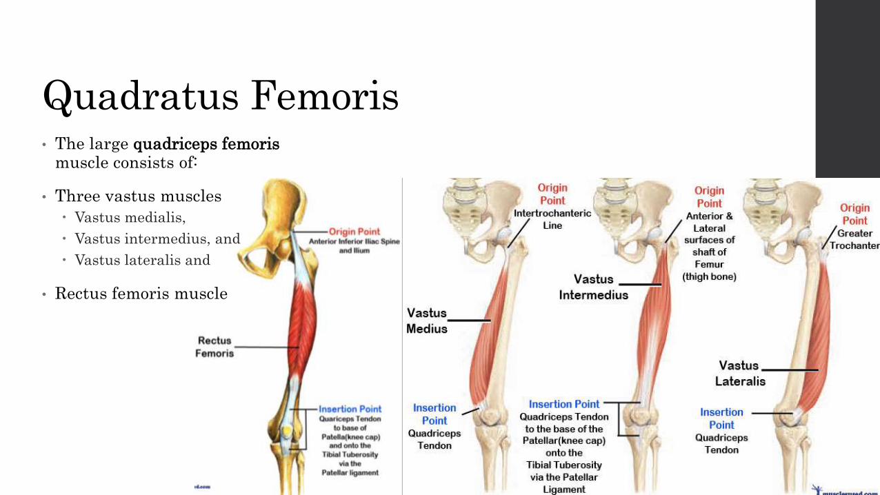
Quadratus Femoris• The large quadriceps femoris
muscle consists of:
• Three vastus muscles
Vastus medialis,
Vastus intermedius, and
Vastus lateralis and
• Rectus femoris muscle

Quadratus Femoris• Nerve Supply: Femoral Nerve (L2, L3, L4)
• Action: Extend leg at knee joint; rectus femoris also steadies hip joint and helps iliopsoas
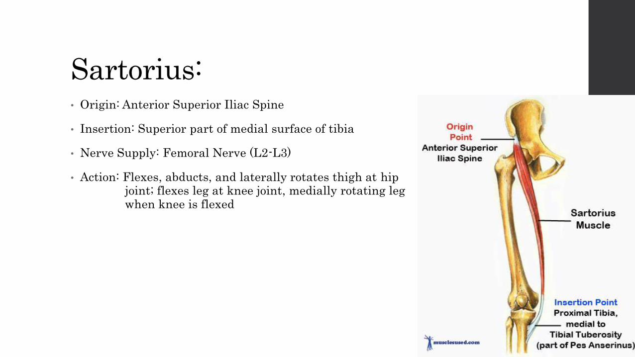
Sartorius:• Origin: Anterior Superior Iliac Spine
• Insertion: Superior part of medial surface of tibia
• Nerve Supply: Femoral Nerve (L2-L3)
• Action: Flexes, abducts, and laterally rotates thigh at hip joint; flexes leg at knee joint, medially rotating leg when knee is flexed
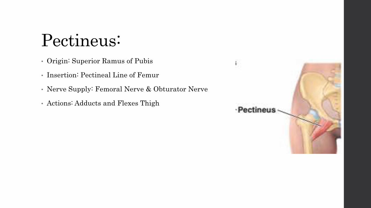
Pectineus:• Origin: Superior Ramus of Pubis
• Insertion: Pectineal Line of Femur
• Nerve Supply: Femoral Nerve & Obturator Nerve
• Actions: Adducts and Flexes Thigh

Flexors of Hip Joint:

Extensors of Knee Joint:
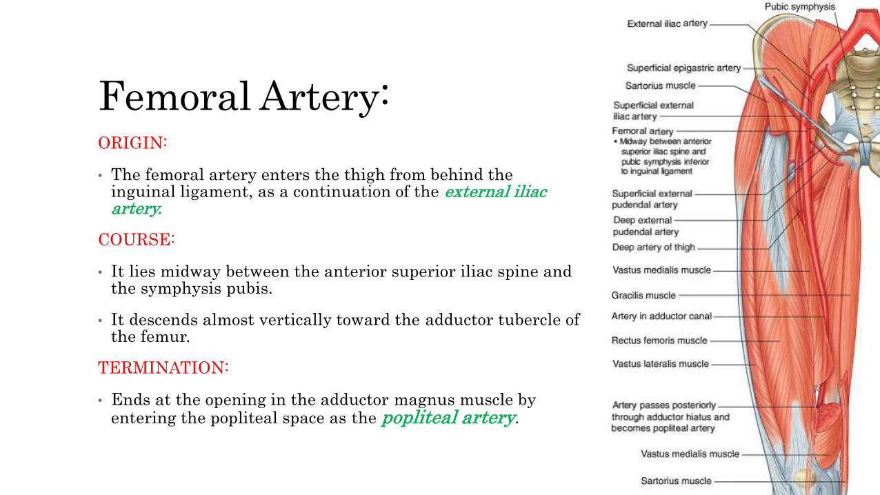
ORIGIN:
• The femoral artery enters the thigh from behind the inguinal ligament, as a continuation of the external iliac artery.
COURSE:
• It lies midway between the anterior superior iliac spine and the symphysis pubis.
• It descends almost vertically toward the adductor tubercle of the femur.
TERMINATION:
• Ends at the opening in the adductor magnus muscle by entering the popliteal space as the popliteal artery.
Femoral Artery:

Branches:• The superficial circumflex iliac artery is a small branch that runs up to
the region of the anterior superior iliacspine.
• The superficial epigastric artery is a small branch that crosses the inguinal ligament and runs to the region of the umbilicus.
• The superficial external pudendal artery is a small branch that runs medially to supply the skin of the scrotum (or labium majus).
• The deep external pudendal artery runs medially and supplies the skin of the scrotum (or labium majus).
• The profunda femoris(Deep) artery is a large and important branch that arises from the lateral side of the femoral artery below the inguinal ligament. It passes medially behind the femoral vessels and enters the medial fascial compartment of the thigh. It ends by becoming the fourth perforating artery. At its origin, it gives off the medial and lateral femoralcircumflex arteries, and during its course it gives off three perforating arteries.
• The descending genicular artery is a small branch that arises from the femoral artery near its termination. It assists in supplying the knee joint.
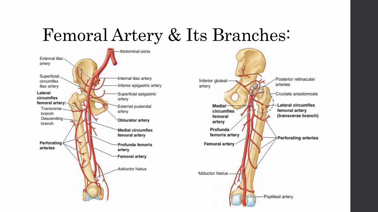
Femoral Artery & Its Branches:
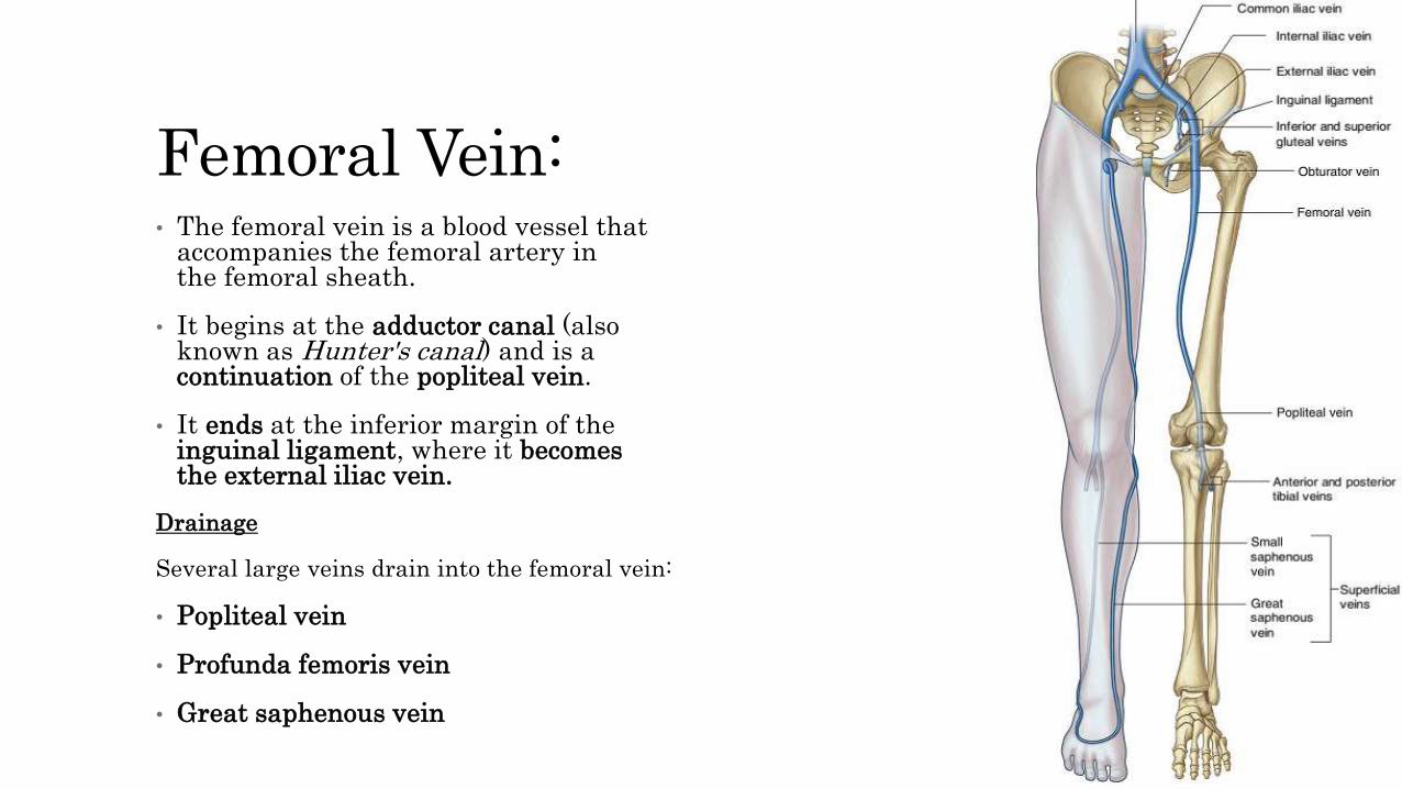
Femoral Vein:• The femoral vein is a blood vessel that
accompanies the femoral artery in the femoral sheath.
• It begins at the adductor canal (also known as Hunter's canal) and is a continuation of the popliteal vein.
• It ends at the inferior margin of the inguinal ligament, where it becomesthe external iliac vein.
Drainage
Several large veins drain into the femoral vein:
• Popliteal vein
• Profunda femoris vein
• Great saphenous vein

Femoral Nerve:• The femoral nerve originates from the lumbar plexus (spinal cord
segments L2-L4) on the posterior abdominal wall
• Enters the femoral triangle of the thigh by passing under the inguinal ligament.
• In the femoral triangle the femoral nerve lies on the lateral side of the femoral artery and is outside the femoral sheath, which surrounds the vessels.
• Before entering the thigh, the femoral nerve supplies branches to the iliacus and pectineus muscles.
• Immediately after passing under the inguinal ligament, the femoral nerve divides into anterior and posterior branches.
Branches of the Femoral Nerve:• Anterior cutaneous branches, which penetrate deep fascia to supply skin on the front of the thigh and knee;
• Numerous motor nerves, which supply the quadriceps femoris muscles (rectus femoris, vastus lateralis, vastus intermedius, and vastusmedialis muscles) and the sartorius muscle; and
• One long cutaneous nerve, the saphenous nerve, which supplies skin as far distally as the medial side of the foot.
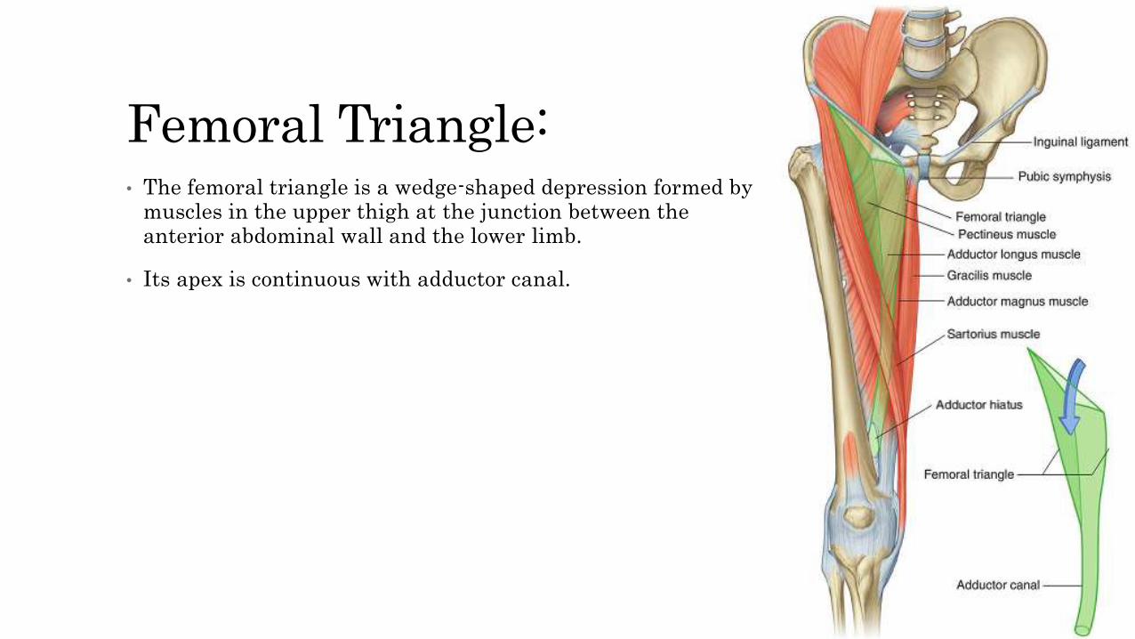
Femoral Triangle:• The femoral triangle is a wedge-shaped depression formed by
muscles in the upper thigh at the junction between the anterior abdominal wall and the lower limb.
• Its apex is continuous with adductor canal.
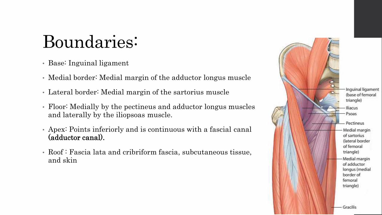
Boundaries:• Base: Inguinal ligament
• Medial border: Medial margin of the adductor longus muscle
• Lateral border: Medial margin of the sartorius muscle
• Floor: Medially by the pectineus and adductor longus muscles and laterally by the iliopsoas muscle.
• Apex: Points inferiorly and is continuous with a fascial canal (adductor canal).
• Roof : Fascia lata and cribriform fascia, subcutaneous tissue, and skin
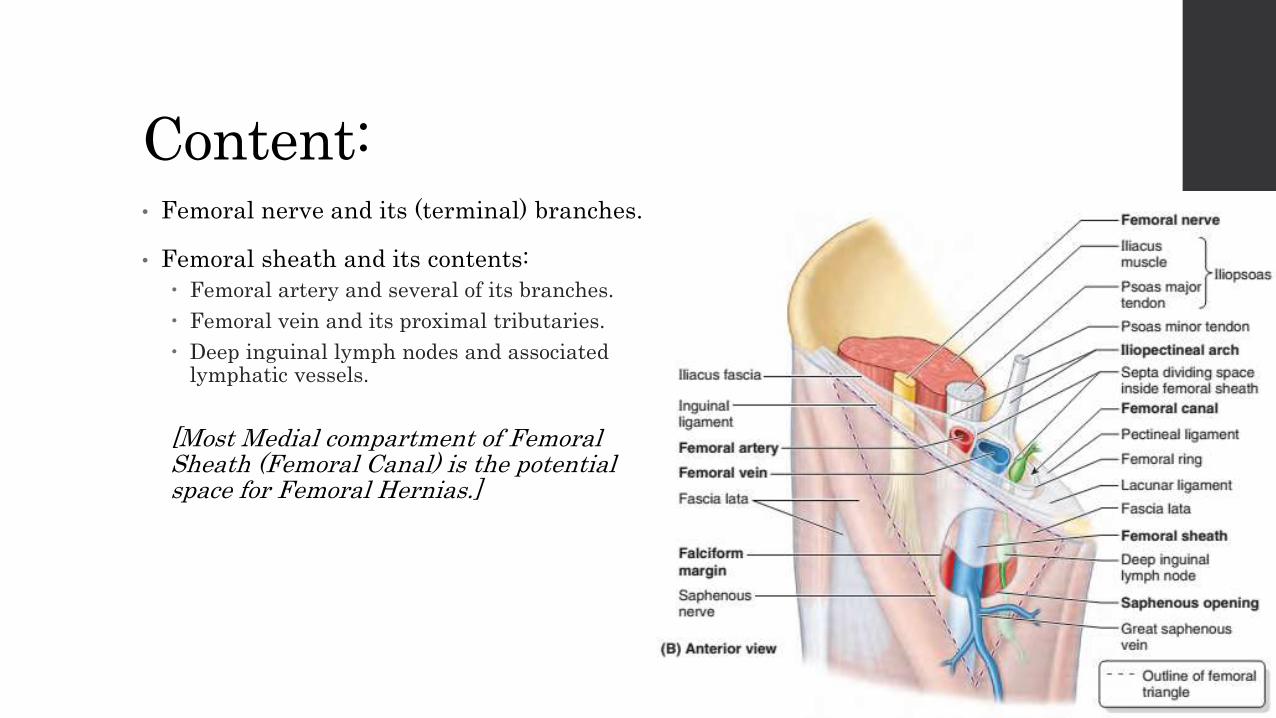
Content:• Femoral nerve and its (terminal) branches.
• Femoral sheath and its contents:
Femoral artery and several of its branches.
Femoral vein and its proximal tributaries.
Deep inguinal lymph nodes and associated lymphatic vessels.
[Most Medial compartment of Femoral Sheath (Femoral Canal) is the potential space for Femoral Hernias.]

Adductor Canal:• It is an intermuscular cleft situated on the medial aspect of the middle third of
the thigh beneath the sartorius muscle.
• It commences above at the apex of the femoral triangle and ends below at the opening in the adductor magnus.
• In cross section, itis triangular, having an anteromedial wall, a posterior wall,and a lateral wall.
■ The anteromedial wall is formed by the sartorius muscle and fascia.■ The posterior wall is formed by the adductor longus and magnus.■ The lateral wall is formed by the vastus medialis.
Contents:• Terminal part of the femoral artery,
• Femoral vein,
• Deep lymph vessels,
• Saphenous nerve,
• Nerve to the vastus medialis, and
• Terminal part of the obturator nerve.
