anterior cruciate ligament-deficient
TRANSCRIPT

89
Neuromuscular Performance in Normal andAnterior Cruciate Ligament-DeficientLower Extremities*
Edward M. Wojtys,† MD, and Laura J. Huston, MS
From MedSport at the University of Michigan, Ann Arbor, Michigan
ABSTRACT
The neuromuscular function of the lower extremity in 40normal and 100 anterior cruciate ligament-deficient vol-unteers was evaluated by physical examination, KT-1000 arthrometer measurements, isokinetic strengthand endurance testing, subjective functional assess-ment, and an anterior tibial translation stress test. Aspecially designed apparatus delivered an anteriorly di-rected step force to the posterior aspect of the leg whileanterior tibial translation was monitored and electro-
myographic signals were recorded at the medial andlateral quadriceps, medial and lateral hamstrings, andgastrocnemius muscles. Testing was done at 30° ofknee flexion with the foot fixed to a scale to monitor
weightbearing, while the tibia remained unconstrained.Results indicate that muscle timing and recruitment or-der in response to anterior tibial translation are affectedby anterior cruciate ligament injury. These alterations inmuscle performance change with time from injury, cor-relate with an individual’s physical activity level, affectsubjective functional parameters, and are directly re-lated to the degree of dynamic anterior tibial laxity seenwith stress testing.
Over the last 25 years, a tremendous research efforthas been focused on the biomechanics of theACL.13,15, 29, 30, 42, 46, 51-53, 76 The principles learned have beenapplied clinically in the hope of improving the natural his-tory of a knee with an injured ACL. In fact, impressivestrides in treatment have followed the course of ACL re-search. A torn ACL in an athlete is not necessarily the
&dquo;beginning of the end,&dquo; as it was once described.&dquo; The prog-nosis for an athletic individual with an ACL injury withproper care appears to be much improved, at least over theshort term. 16,56
Despite these clinical and biomechanical advances, ourcurrent information base does not explain the functionalstatus of several types of individuals with increased ante-rior laxity of the knee. One of these types is the athlete whohas excess anterior laxity but who functions at a very highlevel of activity without evidence of instability. 51,51,11 Someof these athletes have normal ligaments that are just notas tight as most, while a few represent asymptomatic ACL-deficient (ACL-D) extremities. Despite a lack of passive re-straint, no instability is apparent, and a high level of func-tion is maintained. Secondly, there is the individual whohas had an ACL reconstruction and has returned to a highlevel of activity without instability in spite of considerableresidual anterior laxity. Paradoxically, there are thosewhose laxities are within &dquo;normal parameters&dquo; yet in whomsymptoms of instability persist.22 These examples empha-size the dual nature of knee stability: the passive restraintsystem, which is composed primarily of ligaments and cap-sule, and the dynamic system, which is composed of theneuromuscular elements. The interaction between the dy-namic and static systems remains unclear. An improvedunderstanding of the neuromuscular component of stabi-lization is needed to augment our treatment of injuries tothe passive restraints of the knee.
This investigation was not performed to alter the currentstandards in decision-making regarding nonoperative ver-sus operative treatment of ACL tears. These decisionsshould be made based on the age and activity level of thesubject while keeping in mind the known risk factors fordegenerative disease. This research was designed to aug-ment both directions of treatment by maximizing the po-tential of the neuromuscular control system. This was at-tempted by studying the neuromuscular response toanterior tibial translation (ATT) in a specifically designedapparatus that allowed monitoring of spinal reflexes andcortical control activity. Unfortunately, at this time, we do
* Presented at the 18th annual meeting of the AOSSM, San Diego, Cali-forma, July 6, 1992
t Address correspondence and repnnt requests to: Edward M. Wojtys, MD,MedSport, POB 363, Ann Arbor, Mi 48106
No author or related mstitution has received any financial benefit from aproduct named or used in this study.
90
not fully understand the complexities of dynamic knee jointcontrol, which makes the interpretation of this data diffi-cult and subject to criticism.
BACKGROUND
In 1944, Ivar Palmars2 wrote about the theory that liga-ments supply the central nervous system (CNS) input thatmakes neuromuscular control of knee joints possible. Sev-eral years later, in 1956, Cohen and Cohen&dquo; popularizedthe idea of an &dquo;arthrokinetic reflex.&dquo; Based on their workwith decerebrate cats, they suggested that the origin ofimportant protective afferent input was the knee joint cap-sule. Cohen and Cohen decided that a quadriceps-hamstrings tension balance was necessary for knee jointstability. Andersson and Stener’ concurred with Cohenand Cohen’s work after localizing important mechanore-ceptors in the anteromedial region of the cat knee jointcapsule. Subsequently, Petersen and Stener 63 suggestedthat these capsular receptors were not mechanoreceptorsbut merely nociceptors responding to large joint loads.These capsular receptors were later shown to be sensitiveto very low joint pressures and to tensile loads in the rangeof fractions of Newtons. 17,36
In hopes of isolating the origin of proprioceptive afferentinput, several neuroanatomic studies have focused directlyon the ACL. Kennedy et al. 41 isolated free nerve endings inGolgi-like receptors in the synovium covering the ACL.Schultz et al. 71 found these same receptors on the surfaceof the ACL beneath the synovial sheath. Schutte et al.’2described an extensive network of sensory receptors in theACL, including Pacinian and Ruffini corpuscles and freenerve endings. Gomez-Barrena et aI,32 isolated direct neu-ral pathways from the ACL to spinal ganglia by using trac-ers to study axonal transport.Despite these anatomic advancements, the precise origin
of the afferent input needed to protect specific ligamentsand maintain joint stability is still not agreed on.2,3,25,6l.65, 73In 1987 Solomonow et al .73 reported a direct ligament-muscle reflex arc from their work with ACLs in cats andhumans. They suggested that ACL injury in humans in-terrupts this ligament-muscle reflex arc, triggering a sec-ond slower pathway needed to modulate the quadricepsand hamstrings muscles from muscle and capsular recep-tors. In a comparison of normal and ACL-D human ex-tremities, Solomonow et al. were able to show a substantialdifference in the EMG activity of the ACL agonist ham-string muscles when the ACL was severed. The meaning ofthese EMG changes remains in question.Electromyographic work by Draganich et al.20 with six
male subjects with ACL-D extremities also suggested thatthe hamstrings act synergistically with the ACL. Specifi-cally, they reported increased EMG activity in the bicepsfemoris muscle from 30° to 0°, which is precisely whereACL force and strain increase. 14,15,70,76
In contrast, work done recently by Pope et al.s5 in 1990questioned the existence of a direct ligament-muscle reflexarc; they attributed the activity in the posterior articularnerve of cats after tugging on the ACL to receptors in theperiarticular tissues not in the ligament itself. Interest-
ingly, recent work by Pitman et al. 64 continues this debate.By using an arthroscope in vivo, they demonstratedsomatosensory-evoked potentials (SSEP) in the cerebralcortex after direct electrical stimulation of an intact ACLin nine patients. The SSEPs represent proprioceptive inputfrom peripheral nerves transmitted along the posterior col-umns.
While a direct ligament-muscle protection system maybe difficult to demonstrate in all animal and human modelsin all joint positions, there is increasing evidence that sup-ports a modulating system of knee joint afferent input onmotoneuron output. Alpha motoneurons are affected byboth high 14 and low3’°4$ threshold joint afferents. Flexorreflex pathways,24 Ia interneurons,2s°33 and Ib interneu-rons48 have all demonstrated the capability of modulatingdifferent efferent pathways.Even though a precise diagram of the neurocircuitry of
dynamic knee joint control is not yet agreed on by inves-tigators, there is no doubt that the neuromuscular systemis capable of altering the strains imposed on the passiverestraints of the knee.70 White and Raphael8° reported thata quadriceps contraction could reduce the strain producedin the medial collateral ligament when a valgus force wasapplied to the knee. Goldfuss et al.3l showed that the stiff-ness of the medial side of the knee could be increased up to
48% with contraction of the quadriceps and hamstringmuscles. Renstrbm et al. 70 used cadavers and a Hall effecttransducer to show that the hamstring muscles could de-crease ACL strain in all positions tested.Timely muscle contraction, or &dquo;dynamization&dquo; of the tib-
iofemoral joint, adds a new dimension to the concept ofknee joint stability. It is this juxtaposing of the joint sur-faces through muscle contraction or loadbearing that al-lows the geometry of the tibiofemoral joint to become anintegral part of joint stability.38 In the unloaded knee,which does not mimic most ligament injury situations, allexternally applied forces or moments are internally re-sisted by ligaments and capsule.15 When the knee joint issubjected to axial loading, joint contour becomes an im-portant stabilizing factor that is frequently underappreci-ated. Markolf et a1.5° tested axial joint loads up to 925 N at0° and 20° of knee flexion in cadavers to demonstrate the
stabilizing effect of joint contact force; their work in well-conditioned athletes showed a tenfold increase in knee jointstiffness with muscle contraction. Work done by Wang andWalker,&dquo; Hsieh and Walker,38 and Olmstead et al.59 con-curred with the findings of Markolf et al. Wang and Walkerdemonstrated an 80% reduction in rotatory laxity with 938N of compressive force. This stabilizing capacity is impor-tant for activities of daily living as well as for physicallydemanding sports.When an injury occurs to the passive restraint system,
new demands are placed on dynamic restraints, and re-training becomes the key to adaptation. Abbott et al.,l intheir extensive review of knee ligament injuries, statedthat ligaments are the first link in the kinetic chain thatprovides rich sensory input to the nervous system. Fortu-nately, the neuromuscular &dquo;servomechanism&dquo; that modu-lates hamstring-quadriceps activity is truly dynamic. The

91
central nervous system processes incoming afferent pro-prioceptive input by comparing actual movement with in-tended performance.67 The discrepancy between actual anddesired movement then can trigger efferent output to cor-rect the error (servocontrol).Brand12 and Wroble and Brand8l questioned the tradi-
tional view of ligaments as merely mechanical restraintsand speculated that the neurosensory function of the liga-ments may, in fact, approach that of their mechanical ef-fect. Because voluntary movements initiated at the cere-bral cortex may be too slow to prevent injury, 55,66 questionsexist about short loop (spinal) reflexes that may be capableof a more timely response.48 Triggering protective spinalreflexes during a dangerous maneuver may play an evengreater role in knee joint stabilization than the voluntaryresponse.67,69 Regardless of where the afferent input origi-nates, a timely protective response is the key to knee jointprotection in the injury situation.
MATERIALS AND METHODS
Subjects
Forty healthy, athletically active volunteers (26 men and14 women) with no known knee injuries served as the nor-mal control group; the average age was 23.5 years. Onehundred consecutive ACL-D individuals (70 men and 30women) who were identified by athletic trainers and phy-sicians were tested; the average age was 25.7 years. AllACL tears had been arthroscopically documented. Ini-
tially, no attempt was made to eliminate combination liga-ment injuries or those with chondral or meniscal problems;however, all of these individuals were considered to be &dquo;re-covered&dquo; from the injuries before their participation in thisstudy. To be included in the study, individuals were re-quired to perform an isokinetic strength test with no morethan minimal discomfort. Those with significant discom-fort from underlying knee problems were excluded. Neitherthe physicians’ recommendations for treatment nor the in-dividuals’ selection of nonoperative versus operative treat-ment were used as selection criteria. Success of treatmentdid not affect participation in this study. Several ACL-Dindividuals were highly successful intercollegiate athletes,while others could not walk without episodes of giving way.The ACL-D individuals were subdivided into three
groups based on the time from ACL injury to evaluation. Inthe acute injury group, the duration of time was less than6 months. In the semiacute injury group the duration oftime was 6 to 18 months, and in the chronic injury groupit was greater than 18 months. All individuals had a physi-cal examination; a subjective functional evaluation includ-ing activity level, pain, swelling and giving way56; KT-1000arthrometer measurements; isokinetic dynamometerevaluation of knee flexion and extension peak torque andendurance testing at 60° and 240° per second; and a kneejoint stress test on a specifically designed apparatus. Thesubjective evaluation 56 was used to subdivide each timegroup (acute, semiacute, and chronic groups) into best andworst subsets.
Knee testing apparatus
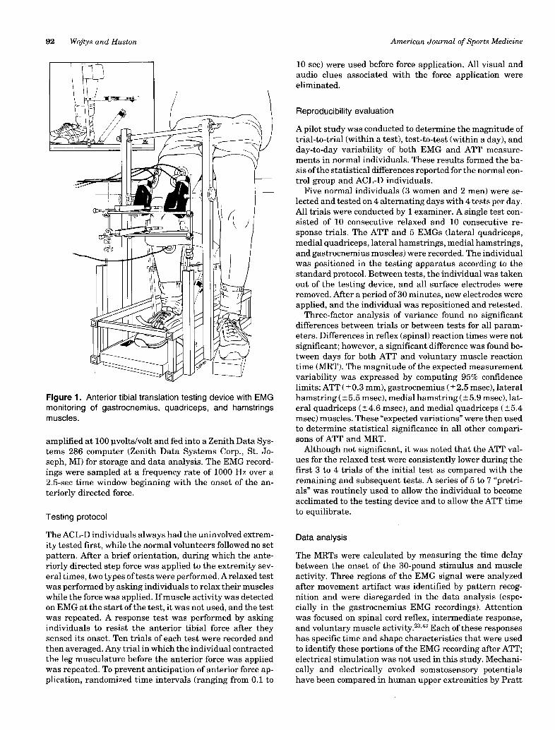
The testing apparatus was designed to measure ATT inreference to the femur in response to an anteriorly directed30-pound step force applied to the posterior aspect of theleg, while lower extremity muscle function was recordedusing surface EMG at five muscle locations. The inducedATT stimulated efferent activity originating at both thespinal cord and cortical level. Because the anteriorly di-rected force was applied to the gastrocnemius muscle belly,care was taken to ensure that motion artifact and the ef-fects of muscle deformation were not confused with a spinallevel reflex to the gastrocnemius muscle in response to thedisplacement. This was accomplished by several trials withand without a windowed short leg cast in place. This modelallowed for ATT but would not permit gastrocnemius de-formation, allowing differentiation of these two factors.Also, it is important to note that the displacing force wasapplied identically in every trial. Therefore, if the gastroc-nemius EMG activity detected was caused by the directstimulation of the device, the EMG pattern should havebeen nearly the same in each trial, but in fact it variedsignificantly.During testing, the individuals were comfortably posi-
tioned with the knee maintained at 30° of flexion by verticaladjustment of an ischial support specifically designed toallow uninhibited hamstring muscle activity (Fig. 1). Thefoot was fixed with the ankle at 10° to 15° of dorsiflexionon a standard scale to monitor the weightbearing statusthat was maintained in the 20- to 30-pound range. Tibialtranslations, rotations, and subluxations were not re-
strained in any way. Two linear potentiometers were em-ployed to measure ATT (Fig. 1). Relative tibial displace-ment was quantified by placing one potentiometer on thepatella with the second placed on the tibial tuberosity.A validation study comparing the accuracy of the KT-
1000 arthrometer with this system was performed usingmachined aluminum plates of known differentials.44 In thisin vitro setting, the correlation coefficient between the KT-1000 arthrometer and the dual linear potentiometers was0.98; however, it is important to note that differences existin the degree of sensitivity of each device. The dual poten-tiometer system was accurate to 0.01 mm throughout a fullrange of displacements (-20.00 to 20.00 mm), while theKT-1000 arthrometer measurement was accurate to 0.16mm with a much higher level of variability.44,77,82
Electromyographic recordings
Surface EMG recordings were taken from five locations:lateral quadriceps, medial quadriceps, lateral hamstrings,medial hamstrings, and gastrocnemius muscles. Surfaceelectrodes produced more reproducible and repeatable re-cordings than indwelling wire electrodes in previous test-ing by Kadaba et a1.4° Bipolar electrodes (Hewlett-Packard,Waltham, MA) were consistently placed over the midregionof each muscle group oriented along the muscle belly, 3 cmapart. Before electrode placement, the skin was preparedwith sandpaper and cleaned with isopropyl alcohol to en-sure adequate surface contact. The raw EMG signal was
92
Figure 1. Anterior tibial translation testing device with EMGmonitoring of gastrocnemius, quadriceps, and hamstnngsmuscles.
amplified at 100 uvolts/volt and fed into a Zenith Data Sys-tems 286 computer (Zenith Data Systems Corp., St. Jo-seph, MI) for storage and data analysis. The EMG record-ings were sampled at a frequency rate of 1000 Hz over a2.5-sec time window beginning with the onset of the an-teriorly directed force.
Testing protocol
The ACL-D individuals always had the uninvolved extrem-ity tested first, while the normal volunteers followed no setpattern. After a brief orientation, during which the ante-riorly directed step force was applied to the extremity sev-eral times, two types of tests were performed. A relaxed testwas performed by asking individuals to relax their muscleswhile the force was applied. If muscle activity was detectedon EMG at the start of the test, it was not used, and the testwas repeated. A response test was performed by askingindividuals to resist the anterior tibial force after theysensed its onset. Ten trials of each test were recorded andthen averaged. Any trial in which the individual contractedthe leg musculature before the anterior force was appliedwas repeated. To prevent anticipation of anterior force ap-plication, randomized time intervals (ranging from 0.1 to
10 sec) were used before force application. All visual andaudio clues associated with the force application wereeliminated.
Reproducibility evaluation
A pilot study was conducted to determine the magnitude oftrial-to-trial (within a test), test-to-test (within a day), andday-to-day variability of both EMG and ATT measure-ments in normal individuals. These results formed the ba-sis of the statistical differences reported for the normal con-trol group and ACL-D individuals.
Five normal individuals (3 women and 2 men) were se-lected and tested on 4 alternating days with 4 tests per day.All trials were conducted by 1 examiner. A single test con-sisted of 10 consecutive relaxed and 10 consecutive re-
sponse trials. The ATT and 5 EMGs (lateral quadriceps,medial quadriceps, lateral hamstrings, medial hamstrings,and gastrocnemius muscles) were recorded. The individualwas positioned in the testing apparatus according to thestandard protocol. Between tests, the individual was takenout of the testing device, and all surface electrodes wereremoved. After a period of 30 minutes, new electrodes wereapplied, and the individual was repositioned and retested.
Three-factor analysis of variance found no significantdifferences between trials or between tests for all param-eters. Differences in reflex (spinal) reaction times were notsignificant; however, a significant difference was found be-tween days for both ATT and voluntary muscle reactiontime (MRT). The magnitude of the expected measurementvariability was expressed by computing 95% confidencelimits: ATT (±0.3 mm), gastrocnemius (±2.5 msec), lateralhamstring (±5.5 msec), medial hamstring (±5.9 msec), lat-eral quadriceps (±4.6 msec), and medial quadriceps (±5.4msec) muscles. These &dquo;expected variations&dquo; were then usedto determine statistical significance in all other compari-sons of ATT and MRT.
Although not significant, it was noted that the ATT val-ues for the relaxed test were consistently lower during thefirst 3 to 4 trials of the initial test as compared with theremaining and subsequent tests. A series of 5 to 7 &dquo;pretri-als&dquo; was routinely used to allow the individual to becomeacclimated to the testing device and to allow the ATT timeto equilibrate.
Data analysis
The MRTs were calculated by measuring the time delaybetween the onset of the 30-pound stimulus and muscleactivity. Three regions of the EMG signal were analyzedafter movement artifact was identified by pattern recog-nition and were disregarded in the data analysis (espe-cially in the gastrocnemius EMG recordings). Attentionwas focused on spinal cord reflex, intermediate response,and voluntary muscle activity.23°43 Each of these responseshas specific time and shape characteristics that were usedto identify these portions of the EMG recording after ATT;electrical stimulation was not used in this study. Mechani-cally and electrically evoked somatosensory potentialshave been compared in human upper extremities by Pratt

93
TABLE 1
Activity level, strength, displacements, and subjective evaluation versus time from injury
a 10 = Competitive jumping, turning, twisting sports.8 = Recreational jumping, turning, twisting sports.6 = Jog, bike, swim, occasional pivoting sports.4 = No jumping, turning, twisting sports; swim, bike, jog regularly.2 = No jumping, turning, twisting, occasional jog, swim, bike.0 = Couch potato.
b Significant difference at P = 0.05 level; in each subgroup best is compared with worst. Best and worst are determined by subjectiveevaluation. Chronics are best/worst 15% = 8/50; semiacutes are best/worst 25% = 5/20; and acutes are best/worst 15% = 5/30.
c Strength = (Peak torque/Body weight) x 100%.
TABLE 2Muscle recruitment preference (in percent) in response to
anterior tibial translation (response test)
a Spinal cord.
et al. 68 In general, mechanically induced potentials were oflower amplitude and contained fewer components, suggest-ing that the electric stimulus activated more fibers syn-chonously. There was less temperal dispersion with themechanical potentials, suggesting that they originatedfrom a more uniform fiber population.The initial spinal cord reflex appears to be monosynaptic,
bypasses muscle spindle receptors, resembles a tendon tapreflex on manual physical examination,43 and it occurredbetween 20 and 119 msec after the onset of ATT in this
study. The large range in reflex response time may be ex-plained by the level of presynaptic inhibition present andby the level of motoneuron excitability. This initial spinalreflex on the EMG recording was usually monophasic,while the amplitude was approximately 5% of that seen involuntary activity. The initial spinal level response ap-pears to be similar to an H-reflex that requires an electricstimulus to be delivered to the afferent nerve in a reflex arc,sufficient to depolarize the large sensory fibers but insuf-ficient to activate the smaller motor fibers. Deschuytere etaI.l9 have previously recorded H-reflex activity from thequadriceps, hamstrings, and gastrocnemius muscles.During the early phase of ATT, if the motor nerves are
depolarized instead of the sensory fiber, and antidromicconduction occurs along the motor nerve, a biplasic re-sponse of larger amplitude similar to an F-wave (electricalstimulation) may result.43 During preliminary testing, thepossibility that this response was generated solely by a di-rect stimulation of the muscle fibers was ruled out by theapplication of a short leg cast and by repeat testing. Thecast allowed ATT but did not allow deformation of the gas-trocnemius muscle.The intermediate response appears to be a spinal reflex
with interneuronal input from centers higher than the spi-nal cord and resembles the late response produced elec-trically.23 It is very reproducible, occurring between spinalcord and voluntary activity. This response is biphasic,larger in amplitude than the spinal cord reflex but smallerin amplitude than voluntary activity and routinely occursjust before voluntary activity (130 to 170 msec). Interest-ingly, Pitman et al.64 reported SSEP occurring with 38.6 to81.6 msec, which may represent the afferent portion of thisloop if the signal reached the cerebral cortex.Voluntary muscle activity was identified by the time of
occurrence (220 to 360 msec) and pattern of activity. It wasalways biphasic, of the largest amplitude and of longestduration.

94
TABLE 3Muscle timing in response to anterior tibial translation normal (control) group
d Spinal cord.b Significant difference (P = 0.05).
TABLE 4Male/female comparison of ACL-deficient and normal
a Strength = (Peak torque/Body weight) x 100%.b Significant difference at P = 0.05 level.
Because most traditional EMG testing data are gener-ated electrophysiologically, mechanically induced EMGmuscle activity requires careful interpretation. Therefore,only those signals that were reproducible were analyzedand incorporated into the data base.
Statistics
All data were first tested for normality, and nonparametricand parametric hypothesis testing was then applied whenappropriate. The analysis of data included t-test compari-sons between dominant and nondominant lower extremi-ties in both the normal and ACL-D groups. Comparativeresults between the ACL-D subgroups and the controlgroup were statistically compared using multiway analysisof variance with Bonferroni’s correction factor for repeatedmeasures. Tukey’s post hoc tests were also incorporated
when appropriate. In several instances where the data vio-lated the definition of &dquo;normality,&dquo; the Friedman test (arobust, nonparametric measure) was used. In all tests, a Pvalue of less than 0.05 was considered significant.
Results summary-normal controls
1. The average ATT with muscles relaxed was 5.4 mmwhen a 30-pound anteriorly directed step force was appliedto the proximal leg (Table 1).
2. Quadriceps strength averaged 86% of body weight(torque foot-pounds/body weight pounds), while the ham-strings averaged 47% (Table 1).
3. The average activity level for the normal group was 5.6(scale 0 to 10) (Table 1).
4. At the spinal cord level, the initial response to ATT wasusually seen in the gastrocnemius muscle, while the ham-

95
TABLE 5Muscle timing in response to anterior tibial translation
° Spinal cord.b Significant difference (P = 0.05); leg dominance not considered.
TABLE 6Muscle timing in response to anterior tibial translation
a Spinal cord.b Significant difference (P = 0.05) when compared with the normal group; leg dominance not considered.
strings were most often recruited first during the inter-mediate and voluntary phases (Table 2).
5. There was no significant difference in the muscle re-cruitment order or MRT between dominant and nondomi-nant extremities in the spinal cord reflex or intermediateresponse; however, during voluntary activity the medialhamstrings were statistically faster on the dominant ex-tremity (Table 3).
6. There was no significant difference between male andfemale normals in terms of age, activity level, hamstrings
strength, ATT, and subjective evaluation score; however,the quadriceps strength of normal men was significantlybetter than that of women (Table 4).
Uninvolved extremities-ACL-D individuals
1. The spinal cord response to ATT of the uninvolvedextremity was not significantly different from normal(Table 5).
2. The intermediate response in the gastrocnemius
96
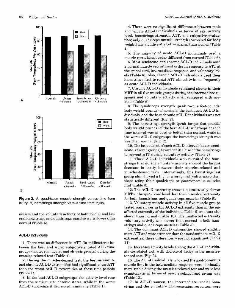
Figure 2. A, quadriceps muscle strength versus time frominjury. B, hamstrings strength versus time from injury.
muscle and the voluntary activity of both medial and lat-eral hamstrings and quadriceps muscles were slower thannormal (Table 5).
ACL-D individuals
1. There was no difference in ATT (in millimeters) be-tween the best and worst subjectively rated ACL timegroups (acute, semiacute, and chronic groups) during themuscles-relaxed test (Table 1).
2. During the muscles-tensed test, the best semiacuteand chronic ACL-D extremities had significantly less ATTthan the worst ACL-D extremities at those time periods(Table 1).
3. In the best ACL-D subgroups, the activity level rosefrom the semiacute to chronic states, while in the worstACL-D subgroups it decreased minimally (Table 1).
4. There were no significant differences between maleand female ACL-D individuals in terms of age, activitylevel, hamstrings strength, ATT, and subjective evalua-tion ; only quadriceps muscle strength (corrected for bodyweight) was significantly better in men than women (Table4).
5. The majority of acute ACL-D individuals used amuscle recruitment order different from normal (Table 6).
6. Most semiacute and chronic ACL-D individuals useda normal muscle recruitment order in response to ATT atthe spinal cord, intermediate response, and voluntary lev-els (Table 6). Also, chronic ACL-D individuals used theirhamstrings first to resist ATT almost twice as frequentlyas acute ACL-D individuals.
7. Chronic ACL-D individuals remained slower in theirMRT in all five muscle groups during the intermediate re-sponse and voluntary activity when compared with nor-mals (Table 6).
8. The quadriceps strength (peak torque foot-pounds/body weight pounds) of normals, the best acute ACL-D in-dividuals, and the best chronic ACL-D individuals was notstatistically different (Fig. 2).
9. The hamstrings strength (peak torque foot-pounds/body weight pounds) of the best ACL-D subgroups at eachtime interval was as good or better than normal, while inthe worst ACL-D subgroups, the hamstrings strength wasless than normal (Fig. 2).
10. The best subset of each ACL-D interval (acute, semi-acute, chronic groups) favored initial use of the hamstringsto prevent ATT during voluntary activity (Table 7).
11. Those ACL-D individuals who recruited the ham-
strings first during voluntary activity showed the largestdecrease in laxity between their muscles-relaxed andmuscles-tensed tests. Interestingly, this hamstring-firstgroup also showed a higher average subjective score thanthose using their quadriceps or gastrocnemius musclesfirst (Table 8).
12. The ACL-D extremity showed a statistically slowerMRT at the spinal cord level than the uninvolved extremityfor both hamstrings and quadriceps muscles (Table 9).
13. Voluntary muscle activity in all five muscle groupstested was slower in the ACL-D extremity than in the un-affected extremity of the individual (Table 9) and was alsoslower than normal (Table 10). The unaffected extremityvoluntary activity was slower than normal in both ham-strings and quadriceps muscles (Table 5).
14. The dominant ACL-D extremities showed slightlymore ATT and were stronger than the nondominant ACL-Dextremities; these differences were not significant (Table11).
15. Increased activity levels among the ACL-D individu-als correlated well with decreased laxity in the muscles-tensed test (Fig. 3).
16. The ACL-D individuals who used the gastrocnemiusmuscle first in the intermediate response were minimallymore stable during the muscles-relaxed test and were lesssymptomatic in terms of pain, swelling, and giving way(Table 12).
17. In ACL-D women, the intermediate medial ham-
string and the voluntary gastrocnemius responses were

97
TABLE 7Muscle timing-best and worst of ACL time groups versus average muscle response times (msec)’
a Significant at P = 0.05 level; best in terms of subjective evaluation compared with worst in each subgroup.b Significant with the normal control group only.~ Significant with both groups 1) best versus worst, and 2) normal control groups.
TABLE 8ACL-deficient subjects-voluntary activity analysis of initial muscle recruitment versus anterior tibial displacement, strength, activity
level, and subjective evaluation
a Significant with gastrocnemius only.b Strength = (Peak torque/Body weight) x 100%.c Significant with both groups.
significantly slower than in ACL-D men. Women with nor-mal extremities demonstrated faster intermediate lateral
quadriceps and voluntary medial hamstring response thandid men with normal extremities (Table 13).
DISCUSSION
Dynamic protection of the knee joint during injury-producing activities requires the recognition of the dan-gerous force through peripheral or central receptors, af-ferent transmission to either the spinal cord or highercenters in the CNS, processing of the signal, and then anappropriate response. Beard et a1.8 defined these proprio-ceptive abilities in terms of static awareness of joint posi-tion in space, kinesthetic awareness (detection of limb
movement and acceleration), and closed-loop efferent ac-tivity that is required for a reflex response and the regu-lation of muscle stiffness. The speed of a protective re-sponse is determined by the site of the afferent reception(central versus peripheral site) and the location of thesignal-processing center (spinal cord versus cerebral cor-tex). The location of the afferent signal processing center(spinal cord versus cortex) is important because it deter-mines the distance a signal must travel to generate an ef-ferent response. Visual afferent input to the cortex can pro-duce a more timely voluntary response than peripheralafferent input because an injury-producing situation maybe recognized in advance; peripheral mechanoreceptorscannot begin their afferent signal until the onset of theinjury-producing force. While cortical recognition may be

98
TABLE 9Muscle timing in response to anterior tibial translation
a Spinal cord.b Significant difference (P = 0.05); leg dominance not considered.
TABLE 10Muscle timing in response to anterior tibial translation
a Spinal cord.b Significant difference (P = 0.05); leg dominance not considered.c Significant difference (P = 0.05); leg dominance considered.
advantageous in many situations, local stimuli at the spi-nal cord level may prove to be adequate to generate a pro-tective muscle response. This dynamic local response maynot generate any limb movement. In fact, a generalizedlimb stiffening through muscle cocontraction may be allthat is needed to prevent knee joint injury.
Purposeful muscle activity can be classified as auto-matic, semiautomatic, or voluntary, depending on the levelof cortical involvement.28 Automatic or reflex responses
originating at the spinal cord are usually generated by localstimuli and can be characterized as gross, quick move-ments that require no cortical input or sensory feedback.Semiautomatic movement (rhythmic behavior) usually re-quires supraspinal initiation and termination but proceedsautomatically in terms of neural control (i.e., chewing,walking). These semiautomatic motions are intermediatesbetween reflex and voluntary activity. They are controlledby neural networks in the spinal cord and brain stem.&dquo;

99
TABLE 11 1ACL-deficient extremities-dominant versus nondominant
a Strength = (Peak torque/Body weight) x 100%.’ Spinal cord.c Significant at the P = 0.05 level.
Figure 3. Activity level versus knee laxity with musclestensed. Activity levels 0 through 10 are defined in Table 1.
These neural networks are &dquo;pattern generators,&dquo; composedof reciprocal connections between pools of neurons waitingto be triggered.&dquo;
Voluntary movements, which have the most cortical con-trol, are affected by attention and motivation. They arelearned and require practice for perfection. Once learned,complicated voluntary movements can be used to form amotor program by which complex tasks are accomplishedwithout thinking about each step (i.e., typing). The amountof conscious effort needed to accomplish a task is an im-portant determinant of the speed at which it can be ac-complished.The speed of limb movement is an important factor in
many athletic activities. Several studies have focused onthe level of neuromuscular activity during rigorous sports,such as skiing, and have asked this question: &dquo;Can musclesreact fast enough to offer protection to the static restraintsof the knee joint?&dquo;5,21,55 Ligament injuries can occur in afraction of a second. Yasuda et al. 84 recently reported thatpeak strains occur in the ACL at 40 to 70 msec after theapplication of a damaging valgus force to the knee. Con-sequently, most investigators would agree that a voluntaryresponse to a damaging force is too slow to protect liga-ments.28 More specifically, Pope et al.65 concluded that amusculoprotective reflex initiated by pain or tendonstretch could not be quick enough to protect knee ligamentsfrom injury. However, this work did not take into accountthe anticipation of the damaging force, allowing activationof a voluntary response before the injury-producing event.If the injury situation can be anticipated and a coordinatedmuscle response formulated before the peak strain pro-duced by a damaging force, adequate voluntary protectionmay be available to the static restraints of the knee. In the
100
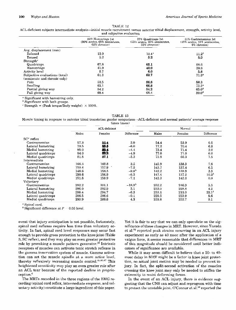
TABLE 12ACL-deficient subjects intermediate analysis-imtial muscle recruitment versus anterior tibial displacement, strength, activity level,
and subjective evaluation
a Significant with hamstring only.b Significant with both groups.c Strength = (Peak torque/Body weight) x 100%.
TABLE 13Muscle timing in response to anterior tibial translation gender comparison-ACL-deficient and normal patients’ average response
times (msec)
d Spinal cord.b Significant difference at P = 0.05 level.
event that injury anticipation is not possible, fortunately, Yet it is fair to say that we can only speculate on the sig-spinal cord reflexes require less time than voluntary ac- nificance of these changes in MRT. However, since Yasudativity. In fact, spinal cord level responses may occur fast et al. 84 reported peak strains occurring in an ACL injuryenough to provide gross protection to the knee joint (Table experiment as early as 40 msec after the application of a3, SC reflex), and they may play an even greater protective valgus force, it seems reasonable that differences in MRTrole by provoking a muscle pattern generator.28 Intrinsic of this magnitude should be considered until better indi-receptors of muscles can activate tonic stretch reflexes in cators of significance are available.the gamma innervation system of muscle. Gamma activa- While it may seem difficult to believe that a 20- to 40-tion can set the muscle spindle at a more active level, msec delay in MRT might be a factor in knee joint protec-thereby reflexively increasing muscle control .48,60,67 This tion, no actual joint motion may be needed to prevent in-heightened sensitivity may play an even greater role after jury. In fact, the split-second activation of the musclesan ACL tear because of the reported decline in proprio- crossing the knee joint may only be needed to stiffen theception.6 6 extremity to resist deforming forces.The MRTs recorded in the three regions of the EMG re- In the event of an ACL injury, there is evidence sug-
cording (spinal cord reflex, intermediate response, and vol- gesting that the CNS can adjust and reprogram with timeuntary activity) constitute a large ingredient of this paper. to protect the unstable joint. O’Connor et al.58 reported the
101
effects of dorsal root ganglionectomy before, and 52 weeksafter, sectioning of the ACL in dogs. The group of dogs thatexperienced 52 weeks of CNS reconditioning after section-ing of the ACL before the dorsal root ganglionectomy didmuch better during gait analysis and in the prevention ofosteoarthritic degeneration with time.
Normal extremities
The spinal cord reflex generated in response to ATT wasusually seen first in the gastrocnemius muscle (Table 2).This was somewhat a surprise to the investigators since thehamstrings and the quadriceps muscles are usually con-sidered the primary muscle stabilizers of the knee. In thisinvestigation, the gastrocnemius muscle appears to play aneven greater role in ACL-D extremities. It had been iden-tified as a primary knee stabilizer in a previous gait studyof ACL-D knees.45
Intermediate and voluntary response to ATT usually fa-vored the initial use of the hamstrings (Table 2). There wasvery little difference in MRT between the dominant andnondominant extremities at the spinal cord and corticallevels except for the voluntary activity of the medial ham-strings ; this was statistically faster on the dominant ex-tremity (Table 3).
In normal extremities, the degree of ATT during themuscles-tensed test correlated statistically with the indi-vidual’s lifestyle: the more active the individual, the tighterthe knee. Those participating in competitive, jumping,twisting sports (high-activity level) demonstrated the leastATT (Fig. 3).
Uninvolved extremities-ACL-D individuals
A normal pattern of muscle recruitment and timing wasseen at the spinal cord level in response to ATT (Table 5);however, when cortical level EMG activity was examined,differences began to appear. The intermediate response ofthe gastrocnemius muscle was significantly slower whencompared with normal response, while the voluntary re-sponse in both the hamstrings and quadriceps muscles wasalso significantly slower. A quadriceps-hamstrings-gastrocnemius recruitment pattern was favored in inter-mediate response, while normals preferred initial use ofthe hamstrings. The voluntary response favored the initialuse of the hamstrings (Table 5), which was similar to thenormal responses. Interestingly, the voluntary recruit-ment of both the hamstrings and quadriceps muscles inthese uninvolved extremities was slower than normal.The slowing of the MRT in the uninvolved extremity was
unexpected but can be explained in several ways. One pos-sibility is that the decreased activity of the ACL-D indi-viduals as a group affects the normal conditioning of theseprotective responses, thus producing slower MRTs in theiruninvolved extremities. More importantly, the initial useof the quadriceps muscle in the intermediate response maybe suggestive of a more profound problem: a quadriceps-dependent extremity. Since the quadriceps muscle is anACL antagonist muscle, its initial recruitment in the in-termediate response may actually represent a risk factor
for ACL injury and may not represent a postinjury alter-ation in muscle recruitment order. Preseason testing ofseveral athletic groups at risk for ACL injury may resolvethis question.
ACL-D extremities
Intermediate response activity apparently undergoes adramatic shift during the 18 months after an ACL tear.Acute injuries (less than 6 months from injury) favor theinitial use of the quadriceps muscle (55% quadriceps and30% hamstrings), while the semiacute ACL tears (6 to 18months postinjury) shift over to the hamstrings (65% ham-strings and 20% quadriceps muscle) (Table 2). This patternof hamstring substitution in ACL-D individuals has beenreported in walking, stair climbing, and more stressful ac-tivities.39,83 These findings also agree with EMG work byBranch et al.11 that showed decreased activity of the ACLantagonist muscle (quadriceps) and increased activity ofthe ACL agonist muscle (hamstrings) in the gait cycle of theACL-D individuals an average of 66.5 months after injury.
Regardless of the time from injury, ACL-D individuals asa group usually showed spinal cord and cortical level re-sponses to ATT slower than normal and slower than theuninvolved extremity (Tables 9 and 10). We do not knowwhether this pattern of generalized MRT slowing is pecu-liar to ACL injury or reflective of knee trauma in general.
Other investigators have indicated that loss of the ACLcan result in a slower MRT. Beard et al.8 studied the la-tency of the hamstring reflex in 30 individuals with a uni-lateral ACL injury and in 20 normal individuals. The meanlatency in the injured extremity was almost twice that inthe uninjured extremity (99 msec versus 53 msec). In thenormal patients, the mean interlimb differential in ATTwas 4.2 mm, and the average reflex hamstring latency was43.2 msec. Twenty-nine of the 30 ACL-D individuals dem-onstrated an increase in reflex hamstring contraction la-tency with a mean noninjured-injured latency differentialof 46.4 msec. Beard et al. reported a significant correlationbetween the hamstrings reflex latency and the frequencyof giving-way episodes, and they suggested that a relativeincrease in reflex hamstring contraction latency is a meas-ure of proprioception and can be used to provide objectivedata for decision-making for ACL-D individuals. Interest-ingly, the reflex response to passive tibial movement wasreported to be significantly slower in a recently injuredACL-D extremity than in the contralateral extremity or anormal extremity.
In this investigation, the generalized MRT slowing inACL-D individuals is most impressive at the voluntarylevel in the best subjectively rated, chronic ACL subgroup(Table 7). This group averaged a knee rating score of 91 ona scale of 100 (Table 1) and represents primarily highlycompetitive intercollegiate athletes. These individualshave the advantage of year-round, intense, sophisticatedtraining. If these athletes cannot return their MRTs to nor-mal 18 months after injury, it seems either that the train-ing and conditioning of the athlete need alteration to betteraddress deficits in MRT, or that the goal of normal MRT isunattainable after ACL injury. If the afferent arm of the

102
protective reflex system has truly been damaged, as somehave suggested,32>73 then normal neuromuscular function
may not be achievable. Further investigation of trainingtechniques after ACL injury may answer this question.Muscle recruitment patterns in response to ATT appear
to change with time from injury. Acutely injured ACL-Dextremities initiate their intermediate and voluntary re-sponse most often in the antagonist quadriceps muscles,while normals favor the agonist hamstrings (Table 6). Thisseemingly unwanted quadriceps response may be the re-sult of postinjury dysfunction caused by the loss of protec-tive afferent input previously originating in theACL.32, 39, 41, 71-73
Lorentzon et al.47 documented profound quadriceps dys-function in chronic ACL-D individuals that was caused by&dquo;nonoptimal quadriceps activation&dquo; during voluntary ac-tivity secondary to impaired afferent input. There is nodoubt that body movement control is dependent on feed-back from peripheral receptors and the existence of
learned, pattern behavior. If there is a defect in afferentinput system to the CNS, or if a recent injury necessitatesa pattern of integrated muscle response that has not beenused previously, then muscle control could be diminishedor ineffective.35 Since semiacute and chronic ACL-D ex-tremities favor initial intermediate and voluntary responsein the hamstrings (Table 6), there may be a learnable butslower compensatory mechanism that uses secondary af-ferent receptors other than those in the ACL.The work of Solomonow et a1.73 on the synergistic action
of the ACL and thigh muscles concluded that there is aprimary, fast-to-respond reflex arc from the mechanore-ceptors in the normal ACL to the hamstrings. Further-more, Solomonow et al. reported that when this arc is in-terrupted by an ACL injury, a secondary, slower reflex arc,originating from mechanoreceptors in muscle and jointcapsule, takes control and inhibits the quadriceps muscles.These findings are in agreement with Birac et al.l° andBerchuck et a1.9 who, through EMG studies, characterizedthe ACL-D gait pattern by its quadriceps avoidance. Also,Andriacchi et al.4 reported increases in hip flexion inACL-D extremities during a sidestep cutting maneuver.Increased hip and knee flexion (approximately 90°) wouldput the hamstrings in a better position to stabilize theACL-D extremity. All of these findings are supported by thework of Basmajian’ who showed that men can be trainedto alter muscle recruitment order with proper training us-ing biofeedback.The role of the secondary stabilizers of the knee, includ-
ing the menisci, should not be overlooked when analyzingthe use of a particular muscle recruitment pattern. The roleof these secondary structures in generating afferent inputfor protective muscle activity remains under investigation.Variables other than those investigated in this study mayimprove our understanding of the muscle recruitment pat-terns reported here.When the members of each ACL time group were ranked
on the basis of their total subjective functional score, a bestand worst subgroup could be identified (Table 1). A highersubjective functional rating correlated well with betterhamstrings and quadriceps strength and a higher activity
level. The ATT in the muscles-tensed test was significantlyless in the best subgroup of the semiacute and chronicACL-D extremities. This ATT control correlates with the
higher muscle strength (Table 1) and faster MRT (Table 7)seen in these &dquo;best&dquo; subgroups. However, the apparent lackof knee motion produced by these initial muscle responsesmay indicate that the MRT is not as important as the abil-ity to coordinate the nearly simultaneous activation (co-contraction) of antagonist muscles to stiffen the knee. Thispossibility is currently being investigated.The best ACL-D individuals in terms of their total sub-
jective functional score favored the same voluntary recruit-ment order as most normals-hamstrings-quadriceps-gastrocnemius muscles (Table 7)-while the worst ACLsubgroup at each time interval (acute, semiacute, andchronic intervals) favored their ACL antagonist quadricepsmuscle first in their voluntary response. Also, hamstringsand quadriceps strength in the best chronic groups was asgood as or better than in the normal group (Table 1). Thedegree of ATT in the muscles-relaxed test did not appearto affect the subjective outcome. In fact, ATT in themuscles-relaxed test showed no significant difference be-tween the best and the worst of each ACL-D time interval.
Interestingly, in the muscles-tensed test, ATT was signifi-cantly less in the semiacute and chronic groups, suggestingthat the active ability to stabilize the knee is somewhatdependent on time from injury (Table 1).The muscle recruitment order in response to ATT may be
partially dependent on the integrity of the secondary re-straints in terms of their mechanical characteristics and
afferent input. Grigg and Greenspan34 showed that jointafferents act as capsular stretch receptors with the level ofinput frequently proportional to the muscle torque gener-ated. These studies implied that the afferent input is re-lated to the function of the tissues to which it is associated.Those ACL-D individuals who responded first with theirgastrocnemius muscle (70 men and 30 women) during theintermediate response (intermediate response, Table 13)showed statistically the least ATT in the muscles-relaxedtest. The significance of these findings remains unclear.There were gender differences noted in both the subjec-
tive and objective evaluations (Tables 4 and 13). The mostimpressive difference is the greater quadriceps strength ofmen even when corrected for body weight (normals, 18.0%;ACL-D, 9.5%). Unfortunately, these differences do not eas-ily explain the epidemic of ACL injuries recently reportedin women. 49Hamstring rehabilitation has been recognized as an in-
tegral factor in the recovery from ACL injury and from sur-gery for some time. Unfortunately, excellent hamstringsand quadriceps strength alone is obviously not enough. 14This shortcoming may be explained by the work of Barracket al.~ that showed that the decline in proprioceptive abilityof an ACL-D extremity does not correlate with strengthloss, lending support to the belief that proprioceptive losswas the cause of, rather than the result of, ACL injury.
Despite all the technical improvements in our treatmentof a torn ACL in the last 20 years, the challenge remains:to return an ACL-D or reconstructed extremity to a normalpattern of function. To accomplish this goal, rehabilitation
103
and training must focus on more than muscle strengthen-ing. Muscle timing, coordination, and integration need tobe addressed.
CONCLUSIONS
1. Significant differences exist between normal andACL-D lower extremities in terms of muscle recruitmentorder at the spinal cord and cortical level in response to anATT.
2. The timing of many muscle responses to an ATT inACL-D extremities is delayed.
3. In ACL-D extremities, both muscle timing and recruit-ment order change with time from injury.
4. Muscle timing and recruitment order in the ACL-Dextremity directly affect the individual’s physical activitylevel, subjective functional performance, and dynamicATT.
REFERENCES
1. Abbott LC, Saunders JB, Bost FC, et al Injuries to the ligaments of the kneejoint J Bone Joint Surg 26: 503-521, 1944
2 Andersson S, Stener B Experimental evaluation of the hypothesis ofligamento-muscular protective reflexes II A study in cats using the medialcollateral ligament of the knee joint. Acta Physiol Scand 48(Suppl 166)27-49, 1959
3 Andrew B: The sensory innervation of the medial ligament of the knee joint.J Physiol 123: 241-250, 1954
4 Andriacchi TP, Kramer GM, Landon GC The biomechanics of running andknee injuries, in Finerman G (ed). The AAOS, Symposium on Sports Medi-cine The Knee. St Louis, CV Mosby Co, 1985, pp 23-32
5. Bahniuk E, MacLaughlin TL, Van Der Meulen JP, et al EMG response ofthe lower extremities to the patellar tendon tap Am Soc Mechan Engineers73-WA/BIO 30 2-7, 1973
6 Barrack RL, Skinner HB, Buckley SL Proprioception in the antenor cruciatedeficient knee Am J Sports Med 17 1-6, 1989
7. Basmajian JV Control and training of Individual motor units Science 141 440-441, 1963
8 Beard DJ, Kyberd PJ, Fergusson CM, et al. Proprioception after rupture ofthe antenor cruciate ligament: An objective indication of the need for sur-gery J Bone Joint Surg 75B 311-315, 1993
9 Berchuck M, Andriacchi TP, Bach BR, et al: Functional adaptation in ACL-deficient patients Trans Orthop Res Soc 13 84, 1988
10 Birac DA, Andriacchi TP, Bach BR Time related changes following ACLrupture Trans Orthop Res Soc 16 231, 1991
11 Branch TP, Hunter R, Donath M Dynamic EMG analysis of antenor cru-ciate deficient legs with and without bracing dunng cutting. Am J SportsMed 17: 35-41, 1989
12 Brand RA Knee ligaments: A new view Trans Am Soc Mechan Engineers108: 106-110, 1986
13 Brantigan OC, Voshell AF: The mechanics of the ligaments and menisci ofthe knee joint J Bone Joint Surg 23 44-66, 1941
14. Butler DL Antenor cruciate ligament: Its normal response and replace-ment. J Orthop Res 7 910-921, 1989
15 Butler DL, Noyes FR, Grood ES Ligamentous restraints to antenor-
postenor drawer in the human knee A biomechanical study J Bone JointSurg 62A: 259-270, 1980
16. Clancy WG, Nelson DA, Reider B, et al. Antenor cruciate ligament recon-struction using one-third of the patellar ligament, augmented by extra-articular tendon transfers. J Bone Joint Surg 64A 352-359, 1982
17. Clark F: Information signaled by sensory fibers in medial articular nerve. JNeurophysiol 38: 1464-1475, 1975
18 Cohen LA, Cohen ML: Arthrokinetic reflex of the knee. Am J Physiol 184433-437, 1956
19 Deschuytere J, DeKeyser C, Deschuttere M, et al H reflexes in musclesof the lower and upper limbs in man Identification and clinical significance,in Desmedt JE (ed): Motor Control Mechanisms in Health and Disease.New York, Raven Press, 1983, pp 951-960
20 Draganich LF, Jaeger R, KralJ A: EMG activity of the quadnceps and ham-stnngs dunng monoarticular knee extension and flexion. Trans Orthop ResSoc 12 283, 1987
21 Duncan AM, Wynck W, Miller EL: Instrumentation for obtaining fractionatedelectromyographical response times to a joint displacement stimulus. ResQ 45: 452-459, 1974
22 duToit GT: Knee joint cruciate ligament substitution: The Lindemann pro-cedure. S Afr J Surg 5 25-30, 1987
23 Eccles JC Evolution of the brain Creation of the self, in Evolution of theBram. New York, Routledge, 1989, pp 61-67
24 Eccles R, Lundberg A Synaptic actions in motoneurones by afferentswhich may evoke the flexion reflex Arch Ital Biol 97 199-221, 1959
25 Ekholm J, Eklund G, Skoglund S: On the reflex effects from the knee jointof the cat Acta Physiol Scand 50 167-174, 1960
26 Fedina L, Hultborn H Facilitation from ipsilateral pnmary afferents of in-terneuronal transmission in the la inhibitory pathway to motoneuronesActa Physiol Scand 86: 59-81, 1972
27 France EP, Paulos LE, Jayaraman G, et al: The biomechanics of lateralknee bracing Part II Impact response of the braced knee Am J Sports Med15 430-438, 1987
28 Fuchs AF, Anderson ME, Binder MD, et al: The neural control of movement,in Patton HD (ed): Textbook of physiology Philadelphia, WB Saunders Co,1989, pp 503-509
29 Fukubayashi T, Torzilli PA, Sherman MF, et al: An in vitro biomechanicalevaluation of antenor-postenor motion of the knee. J Bone Joint Surg 64A258-264, 1982
30 Girgis FG, Marshall JL, Al Monajem ARS: The cruciate ligament of the kneejoint. Anatomical, functional and experimental analysis. Clin Orthop 106216-231, 1975
31 Goldfuss AJ, Morehouse CA, LeVeau BF: Effect of muscular tension onknee stability Med Sci Sports 5: 267-271, 1973
32 Gomez-Barrena E, Munuera L, Martinez-Moreno E Neural pathways ofantenor cruciate ligament traced to the spinal ganglia. Trans Orthop ResSoc 17 503, 1992
33. Goslow G, Reinking R, Stuart D: The cat step cycle: Hind limb joint anglesand muscle lengths during unrestrained locomotion. J Morphol 141: 1-42,1973
34. Gngg P, Greenspan BJ: Response of pnmate joint afferent neurons tomechanical stimulation of knee joint. J Neurophysiol 40: 1-8, 1977
35 Gngg P, Harngan EP, Fogarty KE: Segmental reflexes mediated by jointafferent neurons in cat knee J Neurophysiol 41. 9-14, 1978
36. Gngg P, Hoffman AH. Calibrating joint capsule mechanoreceptors as invivo soft tissue load cells J Biomech 22. 781-785, 1989
37 Hongo T, Jankowska E, Lundberg A The rubrospinal tract II Facilitationof interneuronal transmission in reflex paths to motoneurons Exp BrainRes 7 365-391, 1969
38 Hsieh HH, Walker PS: Stabilizing mechanisms of the loaded and unloadedknee joint. J Bone Joint Surg 58A: 87-93, 1976
39 Jonsson H, Karrholm J Kinematics of the weight-bearing knee with andwithout ACL injury. Trans Orthop Res Soc 17 664, 1992
40 Kadaba MP, Wootten ME, Gainey J, et al: Repeatability of phasic muscleactivity Performance of surface and intramuscular wire electrodes in gaitanalysis. J Orthop Res 3 350-359, 1985
41 Kennedy JC, Alexander IJ, Hayes KC Nerve supply of the human knee andits functional importance Am J Sports Med 10 329-335, 1982
42 Kennedy JC, Weinberg HW, Wilson AS The anatomy and function of theantenor cruciate ligament. As determined by clinical and morphologicalstudies. J Bone Joint Surg 56A 223-235, 1974
43 Kimura J: Electrodiagnosis in Diseases of Nerve and Muscle: Principlesand Practice Second edition Philadelphia, FA Davis Co, 1989
44 Kowalk DL, Wojtys EM, Disher J, et al Quantitative analysis of the mea-sunng capabilities of the KT-1000 knee ligament arthrometer. Am J SportsMed 21. 744-747, 1993
45 Lass P, Kaalund S, leFevre S, et al: Muscle coordination following ruptureof the antenor cruciate ligament Electromyographic studies of 14 patientsActa Orthop Scand 62 9-14, 1991
46 Lewis JL, Shybut GT. In vivo forces in the collateral ligaments of canineknees Trans Orthop Res Soc 6 4, 1981
47 Lorentzon R, Elmqvist LG, Sjostrom M, et al: Thigh musculature in relationto chronic anterior cruciate ligament tear Muscle size, morphology, andmechanical output before reconstruction. Am J Sports Med 17: 423-429,1989
48 Lundberg A, Malmgren K, Schomburg E: Role of joint afferents in motorcontrol exemplified by effects on reflex pathways from Ib afferents J
Physiol 284 327-343, 197849 Malone TR, Hardaker WT, Garrett WE, et al Relationship of gender in
antenor cruciate ligament injuries in intercollegiate basketball players JSouth Orthop Assoc 2 36-39, 1993
50 Markolf KL, Bargar WL, Shoemaker SC, et al. The role of joint load in kneestability J Bone Joint Surg 63A 570-585, 1981
51 Markolf KL, Graff-Radford A, Amstutz HC In vivo knee stability J BoneJoint Surg 60A. 664-674, 1978
52 Markolf KL, Kochan A, Amstutz HC Measurement of knee stiffness andlaxity in patients with documented absence of the antenor cruciate liga-ment. J Bone Joint Surg 66A: 242-253, 1984
53 Markolf KL, Mensch JS, Amstutz HC: Stiffness and laxity of the knee Thecontributions of the supporting structures A quantitative in vitro study. JBone Joint Surg 58A: 583-594, 1976

104
54 McDaniel WJ, Dameron TB Untreated ruptures of the antenor cruciateligament J Bone Joint Surg 62A 696-705, 1980
55 Miyashita M, Miura M, Matsui H, et al Measurement of the reaction timeof muscular relaxation Ergonomics 15 555-562, 1972
56 Noyes FR, Barber SD, Mangine RE Bone-patellar ligament-bone and fas-cia lata allografts for antenor cruciate ligament reconstruction J Bone JointSurg 72A. 1125-1136, 1990
57 Noyes FR, Mooar PA, Matthews DS, et al The symptomatic antenorcruciate-deficient knee Part I The long-term functional disability in ath-
letically active individuals J Bone Joint Surg 65A 154-162, 198358 O’Connor BL, Visco DM, Brandt KD Gait alterations in dogs with unstable
knee joints Evidence that the central nervous system is reprogrammed toprotect unstable joints Trans Orthop Res Soc 17 478, 1992
59 Olmstead TG, Wevers HW, Bryant JT, et al Effect of muscular activity onvalgus/varus laxity and stiffness of the knee J Biomech 19 565-577, 1986
60 Osternig LR, Robertson R, Troxel R, et al Muscle activation dunng pro-prioceptive neuromuscular facilitation (PNF) stretching techniques Am JPhys Med 66 298-307, 1987
61 Palmar I Pathophysiology of the medial ligament of the knee joint Acta ChirScand 115 312-318, 1958
62 Palmar I Plastic surgery of the ligaments of the knee. Acta Chir Scand 91 37-48, 1944
63 Petersen I, Stener B Experimental evaluation of the hypothesis of
ligamento-muscular protective reflexes III A study in man using the medialcollateral ligament of the knee joint Acta Physiol Scand 48(Suppl 166)51-61, 1958
64 Pitman MI, Nainzadeh N, Menche D, et al The intraoperative evaluationof the neurosensory function of the antenor cruciate ligament in humansusing somatosensory evoked potentials Arthroscopy 8 442-447, 1992
65 Pope DF, Cole KJ, Brand RA Physiologic loading of the antenor cruciateligament does not activate quadnceps or hamstrings in the anesthetizedcat Am J Sports Med 18 595-599, 1990
66. Pope MH, Johnson RJ, Brown DW, et al The role of the musculature ininjuries to the medial collateral ligament J Bone Joint Surg 61A. 398-402,1979
67. Powers WR: Nervous system control of muscular activity, in Knuttgen HG(ed) Neuromuscular Mechanisms for Therapeutic and Conditioning Ex-ercise Baltimore, University Park Press, 1976, pp 1-30
68 Pratt H, Starr A, Amlie RN, et al Mechanically and electrically evokedsomatosensory potential in normal humans Neurology 29 1236-1244,1979
69 Prochazka A: Proprioception dunng voluntary movement Can J PhysiolPharmacol 64 499-504, 1986
70 Renstrom P, Arms SW, Stanwyck TS, et al Strain within the antenor cru-ciate ligament during hamstring and quadnceps activity Am J Sports Med14 83-87, 1986
71 Schultz RA, Miller DC, Kerr CS, et al Mechanoreceptors in human cruciateligaments A histological study J Bone Joint Surg 66A 1072-1076
72 Schutte MJ, Dabezies EJ, Zimny ML, et al Neural anatomy of the humanantenor cruciate ligament J Bone Joint Surg 69A 243-247, 1987
73 Solomonow M, Baratta R, Zhou BH, et al The synergistic action of theantenor cruciate ligament and thigh muscles in maintaining joint stabilityAm J Sports Med 15 207-213, 1987
74 Tibone JE, Antich TJ, Fanton GS, et al Functional analysis of anteriorcruciate ligament instability Am J Sports Med 14 276-284, 1986
75 Torg JS, Conrad W, Kalen V: Clinical diagnosis of antenor cruciate ligamentInstability in the athlete Am J Sports Med 4 84-93, 1976
76 Torzilli PA, Greenberg RL, Insall J An in vivo biomechanical evaluation ofantenor-postenor motion of the knee J Bone Joint Surg 63A 960-968,1981
77 Torzdli PA, Panariello RA, Forbes A Measurement reproducibility of twocommercial knee test devices J Orthop Res 9 730-737, 1991
78 Walla DJ, Albright JP, McAuley E, et al Hamstnng control and the unstableantenor cruciate ligament-deficient knee Am J Sports Med 13: 34-39,1985
79. Wang CJ, Walker PS Rotatory laxity of the human knee joint J Bone JointSurg 56A 161-170, 1974
80 White AA, Raphael IG The effect of quadnceps loads and knee positionon strain measurements of the tibial collateral ligament. An experimentalstudy on human amputation specimens Acta Orthop Scand 43 176-187,1972
81 Wroble RR, Brand RA Function of knee ligaments A historical review oftwo perspectives lowa Orthop J 8 67-72, 1988
82 Wroble RR, Van Ginkel LA, Grood ES, et al Repeatability of the KT-1000arthrometer in a normal population Am J Sports Med 18 396-399, 1990
83 Wu CD, Birac D, Andriacchi TP, et al A study of compensatory function inACL-deficient knees in walking and more stressful activities Trans OrthopRes Soc 17 659, 1992
84 Yasuda K, Erickson AR, Johnson RJ, et al Dynamic strain behavior in themedial collateral and antenor cruciate ligaments during lateral impact load-ing Trans Orthop Res Soc 17 127, 1992