anthocyanins increase antioxidant enzyme activity … · anthocyanins increase antioxidant enzyme...
TRANSCRIPT

ANTHOCYANINS INCREASE ANTIOXIDANT ENZYME ACTIVITY IN HT-29
ADENOCARCINOMA CELLS
by
MARTHA KATHLEEN TURNER
(Under the Direction of Joan G. Fischer)
ABSTRACT
Anthocyanins are thought to have antioxidant effects in the body. The effects of two
anthocyanins, malvidin and peonidin, on activity of antioxidant enzymes, glutathione-S-
transferase (GST), glutathione reductase (GR), and glutathione peroxidase (GPx), were
examined in HT-29 human adenocarcinoma cells. Cells were treated with each anthocyanin
or a combination of both at concentrations of 0, 5, and 10 µg/mL in study one and 0, 2.5 and
5 µg/mL in study two. While the data suggests that these anthocyanin concentrations may
increase activity of each enzyme, effects were often anthocyanin and dose-dependent. A
synergistic effect between malvidin and peonidin was observed. At 2.5 µg/mL, the
anthocyanins did not individually increase enzyme activity, however, a combined dose of 2.5
µg/mL significantly increased activity, GR by 55%, GPx by 21%, and GST by 42%. This
study demonstrated that malvidin and peonidin have the potential to increase antioxidant
enzyme activity at 10 µg/mL and below.
INDEX WORDS: Anthocyanin, Antioxidant, Glutathione peroxidase, Glutathione reductase,
Glutathione-S-transferase

ANTHOCYANINS INCREASE ANTIOXIDANT ENZYME ACTIVITY IN HT-29
ADENOCARCINOMA CELLS
by
MARTHA KATHLEEN TURNER
B.S., Clemson University, 2007
A Thesis Submitted to the Graduate Faculty of The University of Georgia in Partial
Fulfillment of the Requirements for the Degree
MASTER OF SCIENCE
ATHENS, GEORGIA
2009

© 2009 Martha Kathleen Turner
All Rights Reserved

ANTHOCYANINS INCREASE ANTIOXIDANT ENZYME ACTIVITY IN HT-29
ADENOCARCINOMA CELLS
by
MARTHA KATHLEEN TURNER
Major Professor: Joan G. Fischer
Committee: Arthur Grider James Hargrove
Electronic Version Approved: Maureen Grasso Dean of the Graduate School The University of Georgia August 2009

iv
DEDICATION
To Dean Kindle He fought the good fight.
His spirit will live on forever in the hearts of those who were privileged to know him.

v
ACKNOWLEDGEMENTS
Thank you to Dr. Fischer for all of her support and guidance throughout this
entire process. Thanks also to my committee members, Dr. Arthur Grider and Dr. James
Hargrove, for all of their help. Thanks to Dr. Grider for the frequent use of his lab.
Thank you to Julie Tokarev for all of her help in the lab.
Thank you to my family and friends who always feigned interest in my research
and without whose constant encouragement and support I would not be where I am today.

vi
TABLE OF CONTENTS
Page
ACKNOWLEDGEMENTS.................................................................................................v
LIST OF TABLES...........................................................................................................viii
LIST OF FIGURES...........................................................................................................ix
CHAPTER
I INTRODUCTION.......................................................................................1
II LITERATURE REVIEW…………………………………………………4
Background………………………………………………………..4
Cancer……………………………………………………………..4
Oxidative Stress…………………………………………………...6
Antioxidant Enzymes……………………………………………...9
Polyphenols………………………………………………………10
Anthocyanins…………………………………………………….11
Hypothesis………………………………………………………..17
III DO BLUEBERRY ANTHOCYANINS INCREASE THE ACTIVITY OF
ANTIOXIDANT ENZYMES?..................................................................21
Abstract…………………………………………………………..22
Introduction………………………………………………………23
Methods…………………………………………………………..25
Results……………………………………………………………28

vii
Discussion………………………………………………………..30
IV SUMMARY AND CONCLUSIONS……………………………………39
Purpose…………………………………………………………...39
Findings…………………………………………………………40
Limitations……………………………………………………….41
Implications………………………………………………………42
Future Research…………………………….……………………43
REFERENCES…………………………………….…………………….44
APPENDICES………………………………………………………...…48
A Anthocyanidin Serial Dilutions…….……………………49

viii
LIST OF TABLES
Page
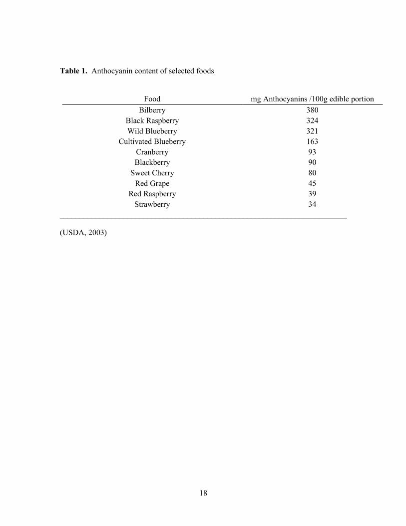
Table 1: Anthocyanin content of selected foods…………………..……………..………18
Table 2: Antioxidant enzyme activity in HT-29 cells treated with 0, 5 or 10 µg/mL
malvidin and/or peonidin.………………………………………..………………………37
Table 3: Antioxidant enzyme activity in HT-29 cells treated with 0, 2,5 or 5 µg/mL
malvidin and/or peonidin………………………………………...………………………38

ix
LIST OF FIGURES
Page
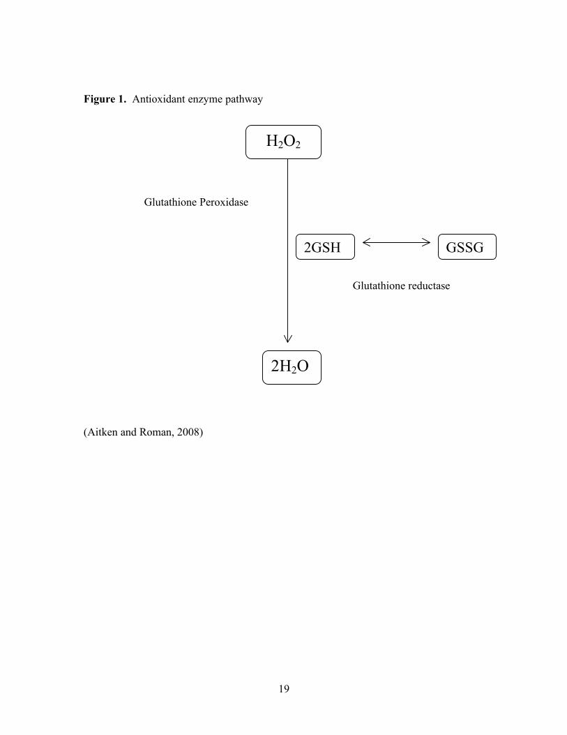
Figure 1: Antioxidant enzyme pathway…………………...……..………………………19
Figure 2: Chemical structure of anthocyanidins…………………………………………20
Figure 3: Glutathione reductase activity (U/mg protein) in HT-29 cells treated with 0, 5,
or 10 µg/mL malvidin and/or peonidin.………………..…………...…………...………34
Figure 4: Glutathione reductase activity (U/mg protein) in HT-29 cells treated with 0,
2.5, or 5 µg/mL malvidin and/or peonidin………………………………...….…………34
Figure 5: Glutathione peroxidase activity (U/mg protein) in HT-29 cells treated with 0, 5,
or 10 µg/mL malvidin and/or peonidin…………………………..………………………35
Figure 6: Glutathione peroxidase activity (U/mg protein) in HT-29 cells treated with 0,
2.5, or 5 µg/mL malvidin and/or peonidin.………………………………………………35
Figure 7: Glutathione-S-transferase activity (U/mg protein) in HT-29 cells treated with 0,
5, or 10 µg/mL malvidin and/or peonidin ………………..……………………………..36
Figure 8: Glutathione-S-transferase activity (U/mg protein) in HT-29 cells treated with 0,
2.5, or 5 µg/mL malvidin and/or peonidin..…….……..….…………………………..…36

1
CHAPTER I
INTRODUCTION
Cancer is the second leading cause of death in the United States. Research shows
that diets high in fruits and vegetables can decrease the risk of some cancers and other
chronic diseases. While plant foods are high in fiber, vitamins, and minerals, it has been
suggested that the phytochemicals found in plants contribute to their ability to decrease
disease risk in humans. Phytochemicals are secondary plant metabolites and are
abundant in plant foods. Some of the most abundant phytochemicals are flavonoids.
Anthocyanins, a class of flavonoids, are the red, blue, and purple pigments found in
plants. Foods high in anthocyanins include aubergine, blackberries, black currants,
blueberries, black grapes, cherries, rhubarb, strawberries, red wine, plums, and red
cabbage (Manach and others 2004). Average consumption of anthocyanins in the United
States is estimated to be 12.5 mg/day/person (Wu and others 2006).
Oxidative stress is an improper balance between free radicals and antioxidants,
which leads to oxidative damage. Free radicals are highly reactive molecules due to an
unpaired electron. Anthocyanins act as antioxidants and counteract oxidative damage in
plants (Wu and Prior, 2008). They have this same effect in the human body. When high
levels of free radicals are present in the body, whether from normal metabolic processes
and the immune system, or from environmental sources, antioxidants may not be able to
offset all the potential oxidative damage. It is widely postulated that this damage to the
cells in the body is linked to the progression of many chronic diseases, including cancer
(Jacob and Burri, 1996). Damaged cells are normally repaired or destroyed by the body;

2
however, cancer cells bypass this normal process, resulting in uncontrolled cell
proliferation and tumor growth (WCRF/AICR, 2007).
Phytochemicals found in fruits and vegetables may counteract these processes,
thus preventing oxidative damage, protecting against disease progression, and interfering
with the development of cancer (Cooke and others 2005). Anthocyanins have been found
to be especially potent antioxidants. They function as antioxidants in a number of ways.
Not only do they donate electrons to free radicals, but it is also thought that another
mechanism of action involves increasing the activity of antioxidant enzymes (Youdim
and others 2000). Antioxidant enzymes neutralize free radicals present in the tissues.
These enzymes include superoxide dismutase, catalase, glutathione peroxidase,
glutathione-S-transferase, and glutathione reductase (Thomas, 2006).
This study investigated the two common anthocyanidins, malvidin and peonidin,
in an in vitro HT-29 human adenocarcinoma cell model. Anthocyanin bioavailability is
low, thus the colon is exposed to high amounts of anthocyanins (Mazza and others 2002).
We tested the hypothesis that the anthocyanins, malvidin and peonidin, would increase
the activity of the antioxidant enzymes glutathione peroxidase, glutathione-S-transferase,
and glutathione reductase, in human colon cancer cells. We further hypothesized that the
single anthocyanins would individually increase the activity of the enzymes, and an
anthocyanin mixture of equal parts of the two anthocyanins would have a synergistic
effect. The study examined the use of anthocyanins at a physiological level in order to
determine whether or not an effect was attainable at an anthocyanin dose that could be
realistically achieved in a human diet.

3
This study demonstrated that malvidin and peonidin have the ability to increase
antioxidant enzyme activity, although the effects are anthocyandin- and concentration-
dependent. Also, at the low concentration of 2.5 µg/mL, the combined anthocyanidins
synergistically increased GR, GPx and GST activities. Individual anthocyanidins,
however, had little or no effect on enzyme activity.
4
CHAPTER II
LITERATURE REVIEW
Background
Cancer is the second leading cause of death in the United States. Diets high in
fruits and vegetables have been shown to decrease the risk of some cancers, including
cancers of the gastrointestinal tract and lung (WCRF/AICR, 2007). There are many
compounds in fruits and vegetables that may be protective, including the phytochemicals
(Kris-Etherton and others 2002). Phytochemicals have no nutritive value, but are thought
to protect against cellular damage associated with the development of cancer.
Polyphenols are one class of phytochemicals that have been extensively studied for their
suggested impact on disease prevention. Though widely studied, there are still many
questions to be answered regarding the mechanisms by which polyphenols impact overall
health and potentially reduce the risk of certain diseases. Anthocyanins have been
identified as one group of polyphenols that may contribute to a reduction in disease risk.
Cancer
Cancer is defined as group of over 100 diseases that are characterized by
uncontrolled cellular growth which results from changes in the genetic information of the
cells (WCRF/AICR, 2007). When cells replicate, they are normally destroyed by the
body if abnormalities are detected. This controlled cell death is called apoptosis
(Johnstone and others 2002). The system is carefully regulated to prevent the growth of
cancerous tumors, and normally works very effectively. However, the human body is

5
made up of a vast number of cells, which are replaced at a rapid pace. Errors are
inevitable.
Most of the errors observed when cells divide can be attributed to genetic
variation and mutation due to a deletion, strand breakage, or crossovers, but some of
them are caused by the cellular environment. This environment is dictated by many
lifestyle factors. High levels of cancer causing agents, carcinogens, can cause changes to
the cell’s DNA, which could potentially be copied when the cell is replicated, and if the
error is not detected and removed, more mutated cells will be formed. Carcinogens are
introduced into the body by a number of different factors including: ultraviolet light,
cigarette smoke, ozone, chlorinated hydrocarbons, heavy metals, and x-rays (Jones and
DeLong, 2000). These environmental factors along with other normal, metabolic
chemicals produced in the body can cause damage due to their highly reactive nature,
oxidizing cellular components at the site of exposure (Halliwell and Gutteridge, 1999).
Although there are many different types of cancer, the pathogenesis of the disease
is the same throughout and the multi-stage process can be classified into initiation,
promotion, and progression (WCRF/AICR, 2007). During initiation, the cell or tissue is
exposed to carcinogen and DNA mutation results. If unrepaired, or cellular apoptosis
does not occur this damage is passed on. Promotion occurs during replication of the
initiated cell, and requires additional promotional factors. Once the replicated cells have
become a tumor mass, unless interrupted, the progression stage occurs as they continue
growing larger and ultimately metastasizing to distant organs. Cancer cells will display
all or some of the six characteristics that have come to be know as the hallmarks of
cancer (Hanahan and Weinberg, 2000). It is these hallmarks that make cancer such a
6
difficult disease to treat and control. These six hallmarks are growth signal autonomy,
insensitivity to antigrowth signals, evasion of apoptosis, limitless replicative potential,
sustained angiogenesis, and tissue invasion and metastasis. Unlike normal cells, cancer
cells do not need growth signals to grow. Growth signal autonomy refers to the fact that
the cells can replicate without mitogenic growth signals. Cancer cells are also insensitive
to antigrowth signals. Cancer cells also evade apoptosis, whereas normal cells, even
without similar mutations, are only allowed to live for so long before they undergo cell
death. Cancer cells have the ability to replicate indefinitely. Since cancer cells grow
rapidly and without restraint, they require a large supply of nutrients. Angiogenesis
refers to the development of new blood vessels and is needed by the tumor in order to
supply enough nutrients. Lastly, unlike normal cells, cancer cells invade their
surrounding tissue. When the cancer successfully invades the vascular or lymphatic
systems, it can spread and metastasize to the rest of the body (Hanahan and Weinberg,
2000). This development is a common characteristic of most cancer deaths
(WCRF/AICR, 2007).
Oxidative Stress
It is theorized that free radicals can damage DNA, ultimately leading to cancer
development (Jacob and Burri, 1996; Juranic and Zizac, 2005). Free radicals are highly
reactive molecules with an upaired electron. This makes them unstable and very potent
oxidizers. They oxidize other molecules they come into contact with in the body by
removing electrons. This then creates a new molecule that has an unpaired electron,
causing the cycle to start over. Free radicals may be generated by environmental

7
carcinogens. However, many free radicals are formed naturally in the body as by-
products of normal metabolic and immune system processes (Halliwell, 2007).
Reactive oxygen species (ROS) and reactive nitrogen species (RNS) are both free
radicals found naturally in the body. ROS are byproducts of various metabolic and
immunological processes and include superoxide (O2-• ), hydrogen peroxide (H2O2),
hydroxyl( •OH), ozone (O3), and singlet oxygen (Jones and DeLong, 2000). Nitric oxide
(NO) and peroxynitrite are both RNS. While all of these molecules are free radicals, they
also contribute to normal metabolism and are therefore essential to proper cell function
(Halliwell and Gutteridge, 1999).
Free radicals can damage carbohydrates, nucleic acids, proteins, and lipids.
Damage to proteins and carbohydrates can lead to improper enzyme function
(WCRF/AICR, 2007). Oxidation of proteins can cause them to fragment and alter
protein binding. These protein changes are sometimes reversible. Damage to lipids can
alter membrane functioning, disrupting the fluidity of the membranes (Thomas, 2006).
As mentioned previously, damage to nucleic acids can cause mutations to the DNA
(Halliwell and Gutteridge, 1999).
Oxidative stress is the term used to describe an improper balance between free
radicals and antioxidants. Oxidative stress is defined as an imbalance between oxidizing
agents and cellular antioxidant systems, which include antioxidant vitamins, cellular
reduced glutathione and reduced coenzymes, as well as the enzymes that control
glutathione dependent processes. It can occur if there are not enough antioxidants
present, as in the case of a poor diet, or if there are simply too many free radicals

8
generated from metabolism and the environment (Halliwell and Gutteridge, 1999; Cooke
and others, 2005).
Dietary antioxidants are defined as “a substance in foods that significantly
decreases the adverse effects of reactive oxygen species, reactive nitrogen species or both
on normal physiological function in humans” (FNB, IOM, 2009). Antioxidants
counteract the effects of free radicals by donating an electron to neutralize the free
radical, thus rendering them harmless. Like free radicals, antioxidants are found both
naturally in the body and in the environment. Food contains many antioxidants in the
form of vitamins and phytochemicals. Many researchers believe that it is the antioxidants
in fruits and vegetables that decrease chronic disease risk. Vitamins E and C are well
known for their antioxidant properties. More recently, researchers have begun to study
phytochemicals and their impact on cancer prevention and cancer treatment. Although
their mechanisms of action are not yet fully understood, many of them have been shown
to act as antioxidants in the body (Issa and others 2006; Moller and Loft, 2006). It is also
thought that some may act as pro-oxidants in certain situations, particularly with cancer
cells, possibly contributing to the apoptosis (Srivastava and others 2007). There are
enzymatic and non-enzymatic antioxidants. Non-enzymatic antioxidants are typically
“chain breaking” antioxidants, so named because they scavenge ROS and RNS in the
body and break the chain of oxidation being caused by free radicals after it has already
begun. Vitamin E and glutathione are “chain breaking” antioxidants and break the cycle
of oxidation (Ou and others 2002).

9
Antioxidant Enzymes
The body has an extensive system of antioxidant enzymes to safely remove free
radicals formed by metabolism and the immune system. These enzymes protect the
body’s cells from ROS, RNS, and any other environmental free radicals by directly
inhibiting the formation of, or metabolize, free radicals. Many of these antioxidant
enzymes function together (Figure 1) (Thomas, 2006). Antioxidant enzymes include
superoxide dismutase, catalase, glutathione peroxidase (GPx), glutathione-S-transferase
(GST), and glutathione reductase (GR). Superoxide anions, one type of free radical, are
neutralized by superoxide dismutase into hydrogen peroxide and oxygen. Catalase
converts hydrogen peroxide to water (Thomas, 2006).
GR, GST, and GPx are found primarily in the cytosol of cells. These enzymes
function individually, but also together, to remove pro-oxidants from the cell. GPx, like
catalase, converts hydrogen peroxide to water (Paglia and Valentine, 1967). It uses
reduced glutathione to reduce hydrogen peroxide to water. This then produces oxidized
glutathione. GR catalyzes the reduction of oxidized glutathione and NADPH back to
reduced glutathione (Xia and others 1985; Carlberg and Mannervik, 1975). The reduced
glutathione can be used to chelate metals or again as a substrate for GPx. GST is a
conjugation enzyme that transfers glutathione to other molecules. Glutathione is itself a
reducing agent. It makes molecules water-soluble, which allows them to be easily
excreted. Making the molecules water-soluble is one way for the body to expel mutagens
and carcinogens (Habig and others 1974). GST and GPx both remove peroxides from the
body (Paglia and Valentine, 1967; Shih and others 2007). Thus GST is considered to be
both a phase 2 enzyme and an antioxidant enzyme.

10
The antioxidant response element (ARE) reacts to chemical stress in the cell and
induces GST and NADPH: quinone oxidoreductase, another antioxidant enzyme. It has
been determined that ARE responds to increased ROS levels or decreased antioxidant
levels, such as a decrease in glutathione. Activation of the ARE is primarily controlled
by nuclear factor E2-related factor 2 (Nrf2). Nrf2 is normally bound to Kelch-like
erythroid CNC homologue (ECH)-associated protein 1 (Keap1). ROS react with cysteine
bonds on the Keap1 protein and as a result the Nrf2 disassociates with Keap1. The Nrf2
is then free to move into the nucleus where is activates ARE, so ARE can in turn activate
antioxidant enzymes. When the ARE is activated, it directs the formation of messenger-
RNA to code for peptides and proteins to make antioxidant enzymes. Nrf2 is sensitive to
levels of ROS and other pro-oxidants, but also antioxidants, including phenolic
compounds, like anthocyanins. Therefore, the Nrf2/ARE pathway could be one more
way anthocyanins increase the activities of GST, GR and GPx in the cells (Shih and
others, 2007; Nguyen and others 2009).
Polyphenols
Phytochemicals are non-nutritive molecules found in plant foods that convey a
beneficial function in the body (Liu, 2004). There are many different classes of
phytochemicals (Bravo, 1998). One class, the polyphenols, consists of secondary
metabolites produced by plants during metabolism. Over 8000 polyphenols have been
identified to date (Bravo, 1998). Polyphenols are found in a wide variety of foods such
as berries, grapes, tea, coffee, chocolate, wine, olive oil, walnuts, peanuts, pomegranates,
and many other fruits and vegetables. Within the polyphenols class, the structures of
11
different polyphenols vary widely and they are further broken down into different groups
based on these structures. General polyphenolic classifications include phenolic acids,
stillbenes, coumarins, tannins, and flavonoids (Liu, 2004). The flavonoids include a
number of different subgroups, including anthocyanins, flavonols, flavones, flavanols,
flavanones, and isoflavones. Flavonoids are widely studied since they are so commonly
distributed in our foods (Manach and others 2004).
Studies have shown that flavonoids may have antioxidant and anti-inflammatory
properties that may decrease disease risk. Flavonoids act as direct antioxidants by
reducing free radicals. They directly quench free radicals, chelate transition metals, and
stimulate the activity of antioxidant enzymes (Duthie and others 2000). They have also
been shown to increase levels of glutathione. This in turn acts as an antioxidant directly
by removing free radicals. Glutathione is also involved in many other enzymatic
antioxidant pathways. Therefore, consumption of flavonoids can have a positive health
impact by decreasing chronic disease risk. As with other complex disease processes,
flavonoids impact many different levels of disease progression and therefore have
numerous effects (Kris-Etherton and others 2004).
Anthocyanins
This study will focus on one specific class of flavonoids, the anthocyanins. These
red, blue and purple pigments are found in many foods, namely aubergine, blackberries,
black currants, blueberries, black grapes, cherries, rhubarb, strawberries, red wine, plums,
and red cabbage (Table 1) (Manach and others 2004). Although many foods contain
anthocyanins, some in very high amounts, it is estimated that consumption of

12
anthocyanins in the western diet is approximately 12.5 mg/day (Wu and others 2006).
Due to the high antioxidant capacity of anthocyanins, they are frequently studied for their
antioxidant roles (Harris, 2006; Neto, 2007). Their presence in the diet in relatively high
concentrations is another reason they are often studied (Cooke and others 2005). They
have also garnered attention for anti-inflammatory and anti-tumor properties, and for the
reduction in risk of cardiovascular diseases (Shih and others 2007). Their effects on
control of cancer cell proliferation have been studied (Yi and others 2005). These studies
have focused mainly on controlling cancer proliferation and progression, but few of them
study the mechanisms by which these changes occur. In order to better understand the
antioxidant activities of anthocyanins and their role in cancer treatment and prevention, it
is important to know the mechanisms by which these chemicals cause these effects.
Anthocyanins vary in structure and are typically found glycosylated with one or
more sugars (Figure 2). Anthocyanidins are aglycones. Structure is an important
determining factor of the way in which ingested anthocyanins are utilized in the body.
Some of the common anthocyanins are peonidin, malvidin, cyanidin, delphinidin,
pelargonidin, and petunidin (Prior, 2003).
The bioavailability of anthocyanins has been examined by a number of research
groups. In humans, Charron and others (2007), examined the concentration of
anthocyanins being excreted in the urine of twelve, healthy volunteers. They were fed an
anthocyanin free diet and then one of three treatments of red cabbage, 100, 200, or 300 g
of cooked red cabbage. The results showed a linear response, with those eating higher
levels of cabbage having greater excretion of anthocyanins, but the levels seen in the
urine from the 200 or 300 g diet were not two and three times higher than the 100 g

13
treatment group (Charron and others 2007). It was also found that non-acylated
anthocyanins were excreted at a significantly higher level in the urine. Another study by
Kurilich and others (2005) also used twelve, healthy volunteers who were fed an
anthocyanin free diet and then placed in treatment groups and fed 250 g raw purple
carrots, 250 g cooked purple carrots, or 500 g of cooked purple carrots. This study
measured blood levels of anthocyanins rather than urine concentrations. All three
treatment groups showed very similar levels of anthocyanins in the blood (Kurilich and
others 2005). And, although there are different forms of anthocyanins that may not have
been measured using these tests, they do demonstrate that anthocyanins have a low
bioavailability. Factors affecting bioavailability of anthocyanins include acylation and
structure (Charron and others 2007; Yi and others 2006b). When anthocyanins are
absorbed into the bloodstream, the process of absorption and excretion is fairly rapid.
Both the liver and kidney modify anthocyanins by the processes of methylation and also
mono-glucuronidation. This is done more by the liver than the kidneys (Talavera and
others 2005). Excretion begins around 20 minutes after absorption, with bloodstream
levels reaching their peak between 30 minutes and 2 hours. By entering the blood steam,
anthocyanins are circulated throughout the entire body and levels can be measured in
other tissues (Prior and Wu, 2006; Talavera and others 2005).
Although anthocyanins are absorbed to an extent, they are excreted to a large
extent unmetabolized. The amount of anthocyanins seen in the urine compared to the
amount ingested demonstrates the low levels of absorption (Cao and others 2000). The
anthocyanins are passing through the body unabsorbed. The bioavailability of the
various anthocyanins differs dependent upon the structure of the anthocyanin. The
14
specific anthocyanins are absorbed and excreted to differing degrees (McGhie and others
2003). This is true in both rat and human models. If the bioavailability of anthocyanins
is relatively low, it follows that the colon would have a relatively high concentration of
undigested anthocyanins present after ingestion of an anthocyanin rich food. Using the
colon cancer cell model replicates these conditions, however the form that ingested
anthocyanins are in when they reach the colon is still unclear.
Anthocyanins may have a protective role at various stages in the cancer process.
They can act as antioxidants by directly reducing free radicals and preventing oxidative
damage to DNA from carcinogens (Shih and others 2007). A number of studies have
been conducted associating consumption of fruits and vegetables with decreased cancer
risk and mortality (Doll, 1990; Dragsted, 1993). Antioxidant capacity and polyphenols
were measured in a study looking at 100 foods to determine a correlation between the
two. Fruits and vegetables showed a strong correlation between the two parameters (Wu
and others 2006). Anthocyanins have been shown to act as antioxidants and
chemopreventive agents during initiation, promotion, and progression of cancer (Hou,
2003). Studies have also found that they are stronger antioxidants than both vitamins E
and C (Duthie, 2007). It has also been theorized that anthocyanins can increase apoptosis
in mutated cells. Yi and others (2005, 2006a) demonstrated an increase in apoptosis of
human liver and colon cancer cells in response to treatment with blueberry anthocyanins.
The studies also showed inhibition of cell growth at high concentrations of blueberry
extracts. Srivastava and others (2007) confirmed the enhancement of apoptosis by
blueberry anthocyanin extracts. Other possible anticancer effects of anthocyanins include
decreased cell proliferation, decreased cell growth, increased DNA damage, and

15
increased enzyme activity (Feng and others 2007; Cooke and others 2006). Preventative
changes anywhere in the stages of cancer could lead to prevention of the development of
cancer altogether.
Others have proposed that anthocyanins upregulate the phase 2 enzymes. It is
well known that many other flavonoid compounds such as lycopene, quercetin and
resveratrol can enhance GST activity (Breinholt and others 2003). Srivastava and others
(2007) examined the effect of blueberry anthocyanins and tannins on the activation of
GST and quinone reductase, another Phase II/antioxidant enzyme, in HT-29 colon cancer
cells. They measured the effects that anthocyanin extracts had on the cells at
concentrations of 50 to 150 mg/ml. These high doses were selected because they induced
apoptosis in the colon cancer cells. However, the activity of the GST and quinone
reductase decreased as a result of these high doses (Srivastava and others, 2007).
Dulebohn and others (2007) examined the effects of whole freeze dried
blueberries and blueberry extracts on the enzyme activity in the colon and liver of rats.
DNA and lipid damage were the main focus of the study. However, Dulebohn also
examined the effect of these compounds on the activity of GST. Changes in GST
activity seen in the colon were not significant and although liver GST activity was 26%
higher in rats fed blueberries or blueberry extracts, this was not significant. Boateng and
others (2007) treated rats with azoxymethane to induce colon cancer and then
supplemented them with blueberries to 5% of their total diet or blueberry juice at
100mL/day. The treated rats in both groups had increased GST activity in the liver and
decreased precancerous aberrant crypt foci. Reen and others (2006) conducted a similar
study in which rats were supplemented with diets of 5% or 10% dried black raspberries.

16
They found a significant increase in GST activity. Patterson and others (2008) examined
a concentration-dependent effect of malvidin and peonidin in human intestinal
adenocarcinoma cells stressed with H2O
2 as measured by DNA damage and apoptosis.
They hypothesized that low concentrations would scavenge H2O
2 and prevent DNA
damage; and higher concentrations would induce oxidative stress and increase DNA
damage. The results of the study showed some apoptosis, although at much lower
concentrations than expected and not a significant increase at the very high
concentrations. It has been hypothesized that increased activity in antioxidant enzymes
actually protects the cells from apoptosis (Shih and others 2007). Shih and others (2007)
examined the effects of 50 µmol/L of 10 different anthocyanins on GST, GR, GPx, and
apoptosis. They found increases in all enzymes from nearly all the anthocyanins used
and decreased apoptosis. In this case, the enzymes were thought to be performing
antioxidant roles in the cancer cells to shield them from harm, including apoptosis, or the
cells could have been undergoing necrosis. If anthocyanins increase antioxidant enzyme
activity in cancer cells, it will be important to determine whether or not this would be a
protective effect for the cancer cells or prevent apoptosis, and therefore be a negative
impact for the body. The current study will attempt to determine whether or not
anthocyanins increase the activity of the antioxidant enzymes, GST, GPx, and GR in HT-
29 colon cancer cells, thereby increasing the overall antioxidant effects of the
anthocyanins.

17
Hypothesis
We tested the hypothesis that the blueberry anthocyanins, malvidin and peonidin,
would increase the activity of the antioxidant enzymes, glutathione-S-transferase,
glutathione peroxidase, and glutathione reductase, in human adenocarcinoma HT-29
cells. We further hypothesized that the single anthocyanins would individually increase
the activity of the antioxidant enzymes, and that an equal mixture of the anthocyanins
would have a synergistic effect on the antioxidant enzyme activity.
18
Table 1. Anthocyanin content of selected foods
Food mg Anthocyanins /100g edible portion
Bilberry 380 Black Raspberry 324 Wild Blueberry 321
Cultivated Blueberry 163 Cranberry 93 Blackberry 90
Sweet Cherry 80 Red Grape 45
Red Raspberry 39 Strawberry 34
________________________________________________________________________
(USDA, 2003)
19
Figure 1. Antioxidant enzyme pathway
(Aitken and Roman, 2008)
H2O2
2H2O
Glutathione Peroxidase
Glutathione reductase
GSSG 2GSH

20
Figure 2. Chemical structure of anthocyanidins
(USDA, 2003)

21
CHAPTER III
DO BLUEBERRY ANTHOCYANINS INCREASE THE ACTIVITY OF
ANTIOXIDANT ENZYMES?
_______________________________
Turner, M. To be submitted for publication in the Journal of Nutritional Biochemistry.

22
Abstract
Anthocyanins, red, blue, and purple polyphenols found in high amounts in
blueberries, are thought to have an antioxidant effect in the body. We examined the
effects of the anthocyanins, malvidin and peonidin, on activity of the antioxidant
enzymes, GST, GR, and GPx in HT-29 human adenocarcinoma cells. The cells were
treated with malvidin and peonidin or a combination of malvidin and peonidin in
concentrations of 0, 5, and 10 µg/mL in study one and 0, 2.5 and 5 µg/mL in study two.
While data suggests that anthocyanins at these concentrations may increase activity of all
three enzymes, the effects were often anthocyanin and dose-dependent. A consistent
finding suggested a synergistic effect between malvidin and peonidin on enzyme activity.
For all three enzymes, while malvidin and peonidin alone at 2.5 µg/mL did not increase
enzyme activity, the combination of the two anthocyanidins significantly increased
enzyme activity above control. Malvidin and peonidin combined at a dose of 2.5 µg/mL
increased GR activity by 55%, GPx activity by 21%, and GST activity by 42%. This
study demonstrated that malvidin and peonidin do have the potential to increase
antioxidant enzyme activity at concentrations of 10 µg/mL and below.

23
Introduction
It is estimated that about 35% of cancer occurrences are directly related to diet
(ACS, 2007). Diets high in fruits and vegetables have been shown to decrease the risk of
many chronic diseases, including some types of cancers (WCRF/AICR, 2007). There are
many theories as to which compounds in fruits and vegetables are protective. One
possibility is the phytochemicals in fruits and vegetables. Phytochemicals have no
nutritive value, but are thought to protect against cellular damage associated with the
development of cancer. Many phytochemicals may act as antioxidants in the body,
preventing damage from oxidative stress (Kris-Etherton and others 2002).
Oxidative stress refers to an improper balance between prooxidant free radicals
and antioxidants. Free radicals are molecules that are unstable and are highly reactive
due to an unpaired electron. It has been theorized that oxidative damage to DNA causes
mutations ultimately leading to cancer (Jacob and Burri, 1996; Juranic and Zizac, 2005).
Although antioxidant activity varies, anthocyanins seem to be especially potent
antioxidants, shown by one study to be 2-6 times more potent than some other
phytochemicals (Prior and others 1998). They function as direct antioxidants by donating
electrons to free radicals, thus rendering them harmless. It is also theorized that
anthocyanins induce antioxidant enzymes (Shih and others 2007). Antioxidant enzymes
are the natural defense system of the body against reactive oxygen and nitrogen species
and include GPx, GST, and GR (Thomas, 2006). GPx and GR work in conjunction to
reduce free radicals. GPx converts hydrogen peroxide to water, using reduced
glutathione. Glutathione reductase then reduces the oxidized glutathione (Thomas, 2006).
GST transfers the antioxidant glutathione to other molecules. Activity of these enzymes

24
may be upregulated by anthocyanins via the antioxidant response element (Shih and
others 2007; Nguyen and others 2009).
There has been little research on the effects of anthocyanins on antioxidant
activity. A previous study in our lab by Dulebohn and others (2008), examined the effect
of blueberry supplementation on GST activity in Sprague-Dawley rats. Although a
significant effect was not shown, there was a slight trend seen in GST activity. The
blueberry, polyphenols, and 1% flavonoid groups had roughly 27% higher GST activity
in liver tissue when compared to the control group. She suggested low anthocyanin
bioavailability as one reason for the lack of significant results. Others (Boateng and
others 2007; Reen and others 2006) found significant increases of GST activity in rats
supplemented with high anthocyanin fruits. Srivastava and others (2007) examined the
effect of blueberry anthocyanins and tannins on the activities of GST and quinone
reductase in colon cancer cells. They used high concentrations that had previously been
shown to increase apoptosis, but the activities of GST and quinone reductase decreased at
these levels of supplementation.
This study investigated the use of the blueberry anthocyanins, malvidin and
peonidin, in an in vitro colon cancer model. We tested the hypothesis that malvidin and
peonidin would increase the activity of the antioxidant enzymes, GPx, GST, and GR, in
HT-29 human adenocarcinoma cells. We further hypothesized that the single
anthocyanins would individually increase the activity of the enzymes, and that an
anthocyanin mixture of equal amounts of malvidin and peonidin would have a synergistic
effect. The study examined the use of anthocyanins at a physiological level in order to

25
determine whether or not an effect was attainable at an anthocyanin dose that could be
realistically achieved in a human diet.
Methods
Cells. The colon adenocarcinoma HT-29 cell line was purchased from ATCC
(Manassas, VA). Colon cancer cells were chosen because of the exposure of the colon to
nonabsorbed anthocyanins. Cells were maintained in McCoy’s 5A modified medium
with L-glutamine (ATCC, Manassas, VA) with fetal bovine serum added at a final
concentration of 10% in 75 cm3 flasks. Cells were incubated at 37° C in 5% CO2, 95%
air and controlled humidity. Cells were passaged when they became confluent, and the
media was changed every 2-3 days, as needed.
Anthocyanin treatment. Two anthocyanins, malvidin chloride and peonidin
chloride, or equal mixture of malvidin and peonidin chloride were mixed with media and
applied to the cells in concentrations of 0, 5 (13.5 µmol/L) and 10 µg/ml (27 µmol/L) for
study 1 and 0, 2.5 (6.75 µmol/L) and 5 µg/mL for study 2 (15). The anthocyanin media
mixtures and control media contained 0.1% DMSO. The anthocyanins were purchased
from Chromadex (Irvine, CA). Each treatment was performed in triplicate flasks. The
treated cells were then incubated for 22 hours.
Cell Collection. Following the incubation period, cells were trypsinized,
collected, added to homogenizing buffer (pH 7.4, 50 mmol/L potassium phosphate, 5
mmol/L EDTA) and homogenized for 5 seconds at 4° C. The cell lysate was then
centrifuged in a J2-HS Beckman Centrifuge (Beckman Instruments Inc., Fullerton, CA)
for 20 minutes at 4˚C at 10,000g. The supernatant was ultracentrifuged in a Beckman

26
Optima LE-80K Ultracentrifuge (Beckman Instruments Inc., Fullerton, CA) at 4˚C,
100,000 G for 1.16 hours. The supernatant following ultracentrifugation, which was the
cytosol, was then removed and stored at -80˚C until analysis. The protein concentrations
of the cytosolic fractions were measured using methods by Lowry and others (1951).
Glutathione Reductase. The activity of GR was assessed using the method of Xia
and others (1985), measuring the reduction of oxidized glutathione by NADPH. Briefly,
0.1 ml dilute cytosol was placed in a cuvette. 0.8 ml of 0.05 mol/L potassium phosphate
buffer with 0.001 mol/L EDTA and 0.25 mmol/L NADPH was added to the cuvette and
then 0.1 ml 5 mmol/L oxidized glutathione was added for a total volume of 1.0 ml in the
cuvette. The substrate, oxidized glutathione, was added immediately preceding the
spectrophotometric readings. Change in absorbance at 340 nm was read every minute for
4 minutes using a spectrophotometer (Beckman Instruments Inc, Fullerton CA). Change
in absorbance was used to determine the concentration of NADPH that had been
oxidized. One unit of enzyme activity was equal to 1 µmole of NADPH oxidized per min
per mg protein (Xia and others 1985).
Glutathione S-Transferase. The activity of GST was assessed using
spectrophotometric methods as described by Habig and others (1974) with 10 mmol/L 1-
Chloro-2,4-dinitrobenzene as the substrate. This assay used 0.1 mol/L potassium
phosphate buffer (pH 6.5), 6.24 mmol/L GSH, and 10 mmol/L 1-Chloro-2,4-
dinitrobenzene. Briefly, 0.1 ml diluted cytosol was added to a cuvette. 0.8 ml of buffer
with glutathione (final concentration 5 mmol/L) and 0.1 ml of 1-Chloro-2,4-
dinitrobenzene in ethanol (final concentration 1 mmol/L) were added for a total volume
of 1 ml. The velocity of formation of S-2,4-dinitrophenylglutathione at 25°C was
27
measured for 3 minutes at 340 nm. One unit of enzyme activity is equal to 1 nmol
conjugate formed/minute/mg protein. The molar extinction coefficient for 1-Chloro-2,4-
dinitrobenzene at 340 nm is 9.6/mM/cm.
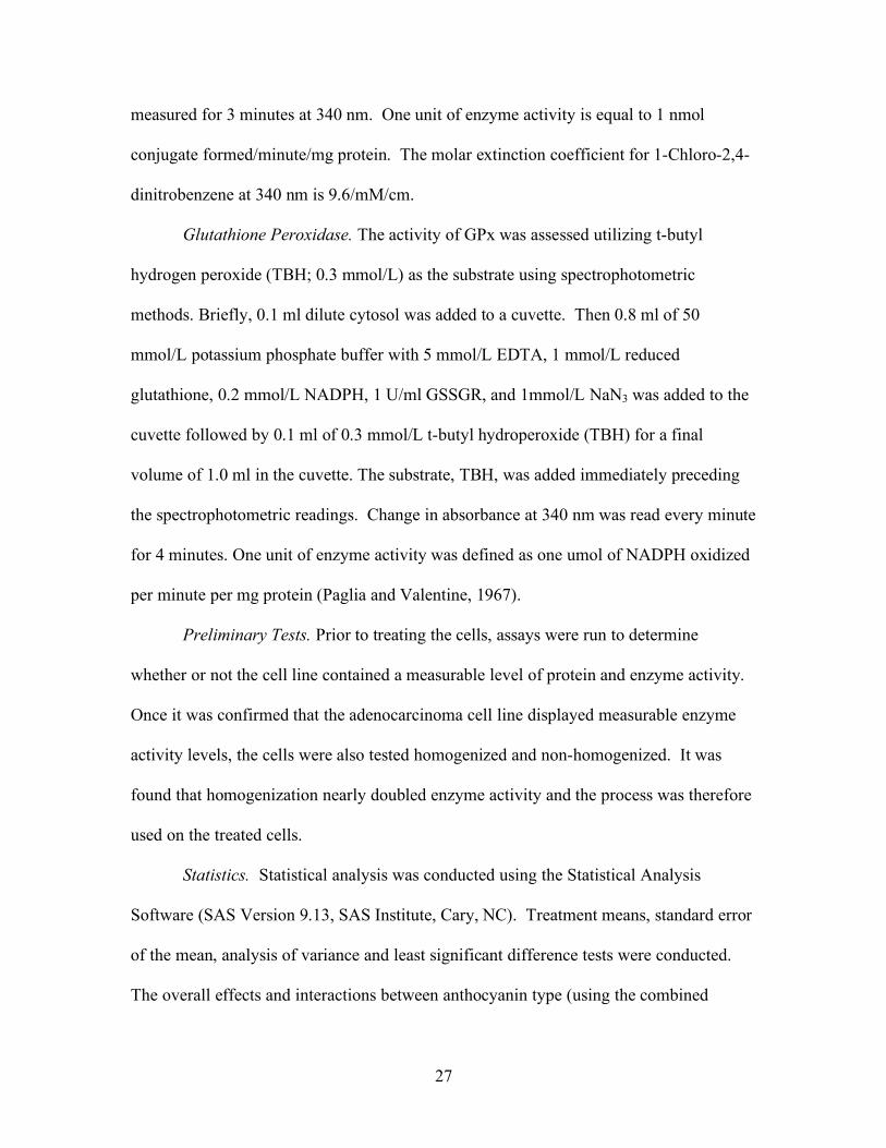
Glutathione Peroxidase. The activity of GPx was assessed utilizing t-butyl
hydrogen peroxide (TBH; 0.3 mmol/L) as the substrate using spectrophotometric
methods. Briefly, 0.1 ml dilute cytosol was added to a cuvette. Then 0.8 ml of 50
mmol/L potassium phosphate buffer with 5 mmol/L EDTA, 1 mmol/L reduced
glutathione, 0.2 mmol/L NADPH, 1 U/ml GSSGR, and 1mmol/L NaN3 was added to the
cuvette followed by 0.1 ml of 0.3 mmol/L t-butyl hydroperoxide (TBH) for a final
volume of 1.0 ml in the cuvette. The substrate, TBH, was added immediately preceding
the spectrophotometric readings. Change in absorbance at 340 nm was read every minute
for 4 minutes. One unit of enzyme activity was defined as one umol of NADPH oxidized
per minute per mg protein (Paglia and Valentine, 1967).
Preliminary Tests. Prior to treating the cells, assays were run to determine
whether or not the cell line contained a measurable level of protein and enzyme activity.
Once it was confirmed that the adenocarcinoma cell line displayed measurable enzyme
activity levels, the cells were also tested homogenized and non-homogenized. It was
found that homogenization nearly doubled enzyme activity and the process was therefore
used on the treated cells.
Statistics. Statistical analysis was conducted using the Statistical Analysis
Software (SAS Version 9.13, SAS Institute, Cary, NC). Treatment means, standard error
of the mean, analysis of variance and least significant difference tests were conducted.
The overall effects and interactions between anthocyanin type (using the combined
28
anthocyanins as one type) and concentration were determined using two way ANOVA.
All data was tested for normality. When non-parametric tests were run, the analysis
yielded a similar level of significance as ANOVA, so the data presented uses ANOVA.
Fisher’s least significance difference test was used to assess the difference between
means for treatment groups. The statistical significance was p < 0.05.
Results
The effects of the anthocyanins malvidin and peonidin on antioxidant enzyme
activity at concentrations of 0, 5, and 10 µg/mL are shown in Table 2 and Figures 3, 5,
and 7. While there was a significant (p<0.05) interaction between anthocyanin type and
dose, there was no effect of anthocyanin type or anthocyanin concentration alone on GR
activity (Figure 3). Peonidin, at a concentration of 10 µg/mL, increased GR activity
291% above control, while 5 µg/mL malvidin increased GR activity 198% about control.
The combined malvidin and peonidin treatment increased GR activity about 118% above
control for both 5 and 10 µg/mL. Similar to GR, there were no main effects of
anthocyanin type or dose on GPx activity (Figure 5). However, there was a significant
(p<0.05) interaction between anthocyanin type and dose on GPx activity. The
combination of anthocyanins increased the GPx enzyme activity slightly above that of
control, but only at 5 µg/mL. GST activity (Figure 7) was not significantly affected by
anthocyanins at 5 or 10 µg/mL concentrations. There was a slight trend for anthocyanin
concentration (p=0.082) in GST activity such that lower concentrations tended to show a
larger increase in enzyme activity.
Table 3 and figures 4, 6, and 8 show the effects of malvidin and peonidin on

29
antioxidant enzyme activity at concentrations of 0, 2.5, and 5 µg/mL. GR activity
(Figure 4) was significantly (p<0.05) affected by anthocyanin type with highest activity
found with the combined anthocyanins. There was also a significant (p<0.05) interaction
between anthocyanin type and dose. While malvidin and peonidin alone did not increase
activity above control, the mixture of both anthocyanidins at a concentration of 2.5
µg/mL significantly increased GR activity about 55% above control. Like GR, using 2.5
and 5 µg/mL concentrations, GPx activity (Figure 6) was significantly (p<0.05) affected
by anthocyanin type and there was a significant (p<0.05) interaction between anthocyanin
type and dose. Peonidin increased GPx activity slightly more than malvidin. The
mixture of the two anthocyanins resulted in the highest GPx activity and a concentration
of 2.5 µg/mL significantly increased activity about 21% above control. GST activity
(Figure 8) was only significantly (p<0.05) affected by anthocyanin type. Once more, the
combination of anthocyanins resulted in the largest increase in GST activity with a 2.5
µg/mL concentration increasing enzyme activity about 42% above control.
We hypothesized that a combination of the two anthocyanins would have a
synergistic effect. For all three enzymes, 2.5 µg/mL of the combined malvidin and
peonidin had the greatest effect. The 3x3 ANOVA analysis appeared to be synergistic,
however, a 2x2 analysis using only cells treated with 0 and 2.5 µg/ml for malvidin and
peonidin was conducted. This examined all of the 2.5 µg/mL treatment groups and the
control, and then compared the single anthocyanins to the mixture and the control.
Slinker (1998) recommends this analysis with the suggestion that synergism has occurred
only when the interaction between malvidin and peonidin is significant. The analysis
confirmed a significant synergistic malvidin x peonidin interaction. The individual

30
anthocyanins did not show a significant effect above control. The mixture of malvidin
and peonidin was significant (p<0.05) for GR, GPx, and GST. This confirmed the
ANOVA results and showed that there was a synergistic effect of the two anthocyanins.
Discussion
The results of our study indicate that anthocyanins, malvidin and peonidin
specifically, do have the potential to impact the activity of GST, GR, and GPx. Our
results show that these effects are dependent on the specific anthocyanin and dose used.
One significant finding was that anthocyanin concentration did not have a consistent
effect on enzyme activity across anthocyanin types. This result was unexpected and
shows how important anthocyanin structure is. In interpreting the results, it is important
to remember that structure plays a very significant role in bioavailability and metabolism
of anthocyanins (Prior and Wu, 2006). Our results confirm that different anthocyanins
may have completely different biological effects. Thus it is important to study many
different anthocyanins and anthocyanin mixtures before making conclusions about the
effects of anthocyanins on biological processes.
The synergistic effect found between malvidin and peonidin suggests that
although individual anthocyanins do have an effect, a combination of anthocyanins, or
maybe even a whole berry extract, would have a greater or lesser effect. Individual
anthocyanins have been shown to have an effect on antioxidant enzymes, but there may
be unknown factors within the metabolism of the anthocyanins that cause them to have
this synergistic effect. And while it is essential to study individual anthocyanins to
determine mechanism of action, studying whole berries is also important because it can
31
be more directly correlated to blueberries and other high anthocyanin foods in the diet
(Bravo, 1998).
The highest levels of anthocyanins tested in this study did not have an effect on
enzyme activity. Instead, the lower concentrations of anthocyanins tended to have the
greatest effects, increasing antioxidant enzyme activity. Since the lowest concentration
of 2.5 µg/mL is close to feasible physiological levels, this may be similar to results of
others that have shown slight increases in GST activity in rats fed high anthocyanin foods
(Boateng and others 2007; Dulebohn and others 2008; Reen and others 2006). Previous
research (Srivastava and others, 2007) showed that high concentrations of anthocyanins
(50-150 µg/mL) decreased GST activity. While some of our results did not vary greatly
from the control, there were no significant decreases from the control, even at the higher
levels. This could be due to the fact that 10 µg/mL was the highest concentration used in
this study and the much higher concentrations have a very different effect on the
enzymes.
In contrast, Shih and others (2007) examined the effects of 50 µmol/L of 10
different anthocyanins on GST, GR, GPx activities. They found increases in all enzymes
with nearly all the anthocyanins used. It is possible that different concentrations have
different effects on the ARE, a control point for expression of these enzymes, causing
differing results. It is also important to note that the cell type used by Shih et al. was the
clone 9 rat liver cell, which may metabolize anthocyanins differently. Lastly, the effects
found in the Shih et al. study varied among the 10 different anthocyanins, showing again
that effects are based on structural differences among anthocyanins.

32
It is important to note that the results are not always consistent among studies for
a number of possible reasons, including: use of in vivo versus in vitro models, different
cell types, different lengths of treatment, different anthocyanins and different
concentrations. These variations can make results difficult to compare. Our results show
that anthocyanin type and dose interact significantly, and this should be considered when
looking at results from different studies. Cell type is also of particular importance,
whether in vivo or in vitro. Different tissue will metabolize the anthocyanins differently
and potentially have different results. Metabolites found within liver tissue, for example,
could quite possibly be very different than the metabolites that exist within the colon.
These metabolites will most likely have differing effects on enzyme activity. This is of
utmost important in vivo where the environment is not as controlled and other factors
may play a greater role in the results that are obtained. The concentration of antioxidant
enzymes will also vary with different tissue types.
One area of current interest is the study of the impact of flavonoid-induced
antioxidant enzyme activity on cellular apoptosis. Apoptosis, a beneficial process, may
be induced via oxidative stress (Shih and others 2007). There is interest in whether
flavonoid induction of antioxidant enzymes is associated with a decrease in the apoptotic
process. However, results of these studies, thus far, are conflicting and may be dependent
on concentration and flavonoid structure (Leunga and others 2006; Srivastava and others
2007; Shih and others 2007). This is an area for further research.
In conclusion, GPx, GST, and GR activities were significantly increased by the
anthocyanins malvidin and peonidin. The lower concentrations showed a greater impact
on enzyme activity. The combination of the two anthocyanins at 2.5 µg/mL had the

33
greatest effect in all three enzymes and proved to be synergistic. There was less of an
effect from dose than anticipated, and it seems that the interaction between dose and
anthocyanin type was more indicative of effect than dose alone. It is always important to
remember that results from in vitro studies cannot be directly applied to dietary
recommendations. For that reason, more in vivo research, both animal and human, needs
to be done to confirm results from in vitro studies and to further examine the relationship
between anthocyanin type and dose.

34
Figure 3. Glutathione reductase activity (U/mg protein) in HT-29 cells treated with 0, 5, or 10 µg/mL malvidin and/or peonidin.
Figure 4. Glutathione reductase activity (U/mg protein) in HT-29 cells treated with 0, 2.5, or 5 µg/mL malvidin and/or peonidin.

35
Figure 5. Glutathione peroxidase activity (U/mg protein) in HT-29 cells treated with 0, 5, or 10 µg/mL malvidin and/or peonidin.
Figure 6. Glutathione peroxidase activity (U/mg protein) in HT-29 cells treated with 0, 2.5, or 5 µg/mL malvidin and/or peonidin.

36
Figure 7. Glutathione-S-transferase activity (U/mg protein) in HT-29 cells treated with 0, 5, or 10 µg/mL malvidin and/or peonidin.
Figure 8. Glutathione-S-transferase activity (U/mg protein) in HT-29 cells treated with 0, 2.5, or 5 µg/mL malvidin and/or peonidin.

37

38

39
CHAPTER IV
SUMMARY AND CONCLUSIONS
Diets high in fruits and vegetables have been shown to decrease disease incidence
possibly due to phytochemicals found in plant foods that act as antioxidants. The
anthocyanins found in blueberries have been extensively studied due to their high
antioxidant capacity. Studies have examined their antioxidant and prooxidant effects in
healthy and carcinoma cell models. It is theorized that one mechanism by which these
anthocyanins act as antioxidants is to increase the activity of antioxidant enzymes in
vitro. Three antioxidant enzymes of interest, GST, GR, and GPx, have not been
sufficiently examined.
Purpose
The purpose of this research was to study the effect of the blueberry
anthocyanins, malvidin and peonidin, on HT-29 adenocarcinoma cell antioxidant enzyme
activity. The study examined whether or not the anthocyanins increased the activity of
the antioxidant enzymes, GST, GR, and GPx. It attempted to determine at what
concentration an increase was displayed and whether combining the anthocyanins would
have a synergistic effect or not.

40
Findings
In this study, we hypothesized that the blueberry anthocyanins, malvidin and
peonidin, would increase the activity of the antioxidant enzymes, glutathione-S-
transferase, glutathione peroxidase, and glutathione reductase, in human HT-29
adenocarcinoma cells. We anticipated that the single anthocyanins would individually
increase the activity of the antioxidant enzymes and that combining the anthocyanins
would have a synergistic effect on the enzymes.
Individual anthocyanins have been shown to have an effect on antioxidant
enzymes, but there may be unknown factors within the metabolism of the anthocyanins
that cause them to have a synergistic effect. Our study found that significant effects from
individual anthocyanins, even at low concentrations, were plausible. However, the
synergistic effect found between malvidin and peonidin was most notable. It suggests
that although individual anthocyanins do have an effect, a combination of anthocyanins
or maybe even a whole berry extract, would have a greater effect. And while it is
essential to study individual anthocyanins to determine mechanisms of action, studying
whole berries is also important because it can be more directly correlated to blueberries
and other high anthocyanin foods in the diet.
Also of importance to note, this study did not find the anthocyanin dose to have a
consistent effect on enzyme activity. A more consistent dose response was expected.
However, this draws attention to the fact that the mechanism of action by which
anthocyanins increase enzyme activity is still largely speculative. The various
41
concentrations could affect the enzymes differently, at one dose having an antioxidant
effect, at another dose having a prooxidant effect, and at a third dose have no effect.
Our study demonstrated that malvidin and peonidin do significantly increase
antioxidant enzyme activity. The results varied based on anthocyanin type and on
anthocyanin and dose together, but not dose alone.
Limitations
While in vitro studies are an excellent means for determining mechanisms of
action in various processes, they do have some innate limitations. The first is that the
growing environment of an incubator, while intended to be similar, is not exactly the
same as the environment within the human body. There are many other factors in an in
vivo model that must be taken into account and cannot be emulated in an in vitro model.
However, the environment is conducive to cell growth and is at this time the best possible
model to be used in an in vitro study.
Cell culture studies are also limited in their ability to be applied to broad
recommendations. This research cannot be used to extrapolate any conclusions regarding
dietary intake of anthocyanins. Results cannot be applied to other cell types, or other
anthocyanins. However, these can be used to explain mechanism of action.
Little is known about the form anthocyanins are in once they reach the colon.
Therefore, the anthocyanins used here may not be the same as those experienced by the
42
colon during normal digestion. The concentration of anthocyanins in the colon is also
estimated. However, using colon cancer cells and the low anthocyanin concentration
makes it much more likely to be a realistic situation. Furthermore, in the diet, blueberries
may vary in anthocyanin concentration depending on growing and processing conditions.
Also, many other phytochemicals are present, as well as vitamins, that could change the
effects of the malvidin and peonidin. This variation could be additive, competitive, or
synergistic.
Implications
The results of the study showed mixed findings. The anthocyanins did impact
the antioxidant enzyme activity in the cells, although the results were not entirely
consistent. The lower doses of anthocyanins tended to show a greater increase in
antioxidant activity. This implies that low, physiologically realistic concentrations of
anthocyanins are all that is needed to show a significant increase in enzyme activity.
Also, the combination of anthocyanins appeared to increase activity more. This is an
important point to note since when eaten as whole berries, the anthocyanins will be
present as a mixture.

43
Future Research
As with all research, the results from this study elucidate other questions and
areas of research that should be explored in future studies. This study showed that the
two anthocyanins examined did impact the antioxidant enzyme activities. Expanding on
these results and exploring other anthocyanins and anthocyanin mixtures, or even
complete blueberry portions, would be helpful to show an effect that would more closely
resemble blueberries in the human diet. Further studies should be done to determine if
there is a dose response and at what concentrations. Previous studies have shown
prooxidant effects of anthocyanins at high levels. Studies should be conducted using
very high levels of anthocyanins to see if there is an inhibitory effect on antioxidant
enzyme activity at very high levels. Human and animal in vivo studies that examine
these same effects are greatly needed. It is important to see if these enzymes are
similarly affected by anthocyanins in the body and whether or not similar results can be
replicated in a dietary model and not simply in cell culture. This would allow the
findings to be applied to dietary recommendations.

44
REFERENCES
American Cancer Society (ACS). 2007. Diet and Physical Activity: What's the Cancer Connection? American Cancer Society.
Aitken R & Roman S. 2008. Antioxidant Systems and Oxidative Stress in the Testes. In: Yan, C., editor. Molecular Mechanisms in Spermatogenesis. New York: Landes Bioscience.
Boateng J, Verghese M, Shackelford L, Walker L, Khatiwada J, Ogutu S, Williams D, Jones J & Guyton M. 2007. Selected fruits reduce azoxymethane (AOM)-induced aberrant crypt foci (ACF) in Fisher 344 rats. Food and Chemical Toxicology 45:725-732.
Bravo L. 1998. Polyphenols: Chemistry, Dietary Sources, Metabolism, and Nutritional Significance. Nutrition Reviews 56(11):317-333.
Breinholt V, Molck A, Svendsen G, Daneshvar B, Vinggaard A, Poulsen M & Dragsted L. 2003. Effects of dietary antioxidants and 2-amino-3-methylimidazo[4,5-f]-quinoline (IQ) on preneoplastic lesions and on oxidative damage, hormonal status, and detoxification capacity in the rat. Food and Chemical Toxicology 41:1315-1323.
Cao G, Muccitelli H, Sanchez-Moreno C & Prior R. 2000. Anthocyanins are absorbed in glycated forms in elder women: a pharmacokinetic study. American Journal of Clinical Nutrition 73:920-926.
Carlberg I & Mannervik B. 1975. Purification and characterization of the flavoenzyme glutathione reductase from rat liver. Journal of Biological Chemistry 250:5475-5480.
Charron C, Clevidence B, Britz S & Novotny J. 2007. Effect of dose size on bioavailability of acylated and nonacylated anthocyanins from red cabbage. Journal of Agriculture and Food Chemistry 55(13):5354-5362.
Cooke D, Schwarz M, Boocock D, Winterhalter P, Steward W, Gescher A & TH M. 2006. Effect of cyanidin-3-glucoside and an anthocyanin mixture from bilberry on adenoma development in the ApcMin mouse model of intestinal carcinogenesis--relationship with tissue anthocyanin levels. International Journal of Cancer 119(9):2213-2220.
Cooke D, Steward WP, Gescher AJ & Marczylo T. 2005. Anthocyans from fruits and vegetables - Does bright colour signal cancer chemopreventive activity? European Journal of Cancer 41:1931-1940.
Doll R. 1990. An overview of the epidemiological evidence linking diet and cancer. Proceedings of the Nutrition Society 49:119-131.
Dragsted L. 1993. Cancer-protective factors in fruits and vegtables: biochemical and biological background. Pharmacology and Toxicology 72(1):116-135.
Dulebohn RV, Yi W, Srivastava A, Akoh CC, Krewer G, Fischer JG. 2008. Effects of blueberry (Vaccinium ashei) on DNA damage, lipid peroxidation, and Phase II enzyme activities in rats. Journal of Agricultural and Food Chemistry 56:11700-11706.
45
Duthie G, Duthie S & Kyle J. 2000. Plant polyphenols in cancer and heart disease: implications as nutritional antioxidants. Nutrition Research Reviews 13:79-106.
Duthie S. 2007. Berry phytochemicals, genomic stability and cancer: evidence for chemoprevention at several stages in the carcinogenic process. Molecular Nutrition & Food Research 2007(51):665-674.
Feng R, Ni H, Wang S, Tourkova I, Shurin M, Harada H & Yin X. 2007. Cyanidin-3-rutinoside, a natural polyphenol antioxidant, selectively kills leukemic cells by induction of oxidative stress. Journal of Biological Chemistry 282:13468-12476.
Food and Nutrition Board. Institute of Medicine. 1998. Proposed Definition and Plan for Review of Dietary Antioxidants and Related Compounds. Dietary Reference Intakes:1-23. National Academy Press, Washington, DC.
Food and Nutrition Board. Institute of Medicine. 2000. Dietary reference intakes for vitamin C, vitamin E, selenium and carotenoids. National Academy Press, Washington, DC.
Habig W, Pabst M & Jakoby W. 1974. Glutathione S-transferases: The first enzymatic step in mercapturic acid formation. Journal of Biological Chemistry 249:7130-7139.
Halliwell B. 2007. Oxidative stress and cancer: have we moved forward? The Biochemical journal 401:1-11.
Halliwell B & Gutteridge J. 1999. Free radicals in biology and medicine,3rd ed. New York: Oxford University Press.
Hanahan D & Weinberg R. 2000. The hallmarks of cancer. Cell 100:57-70. Harris D. 2006. Phytochemicals in Health and Disease. American Journal of Clinical
Nutrition 85:1556-1557. Hou D. 2003. Potential mechanism of cancer chemoprevention by anthocyanins. Current
molecular medicine 3:149-159. Issa AY, Volate SR & Wargovich MJ. 2006. The role of phytochemicals in inhibition of
cancer and inflammation: New directions and perspectives. Journal of Food Composition and Analysis 19:405-419.
Jacob R & Burri B. 1996. Oxidative damage and defense. American Journal of Clinical Nutrition 63:985S-990S.
Johnstone R, Ruefli A & Lowe S. 2002. Apoptosis: a link between cancer genetics and chemotherapy. Cell 2:153-164.
Jones DP & DeLong MJ. 2000. Detoxification and Protective Functions of Nutrients. In: Biochemical & Physiological Aspects of Human Nutrition. (Stipanuk, MH, ed.) St. Louis: W.B. Saunders Company. p. 901-916.
Juranic Z & Zizac Z. 2005. Biological activities of berries: From antioxidant capacity to anti-cancer effects. Biofactors 23:207-211.
Kris-Etherton PM, Hecker KD, Bonanome A, Coval SM, Binkoski AE, Hilpert KF, Griel AE & Etherton TD. 2002. Bioactive Compounds in Foods: Their Role in the Prevention of Cardiovascular Disease and Cancer. The American Journal of Medicine 113:71S-88S.
Kris-Etherton PM, Lefevre M, Beecher GR, Gross MD, Keen CL & Etherton TD. 2004. Bioactive Compounds in Nutrition and Health-Research Methodologies for Establishing Biological Function: The Antioxidant and Anti-inflammatory Effects of Flavonoids on Atherosclerosis. Annual Review of Nutrition 24:511-538.
46
Kurilich A, Clevidence B, Britz S, Simon P & Novotny J. 2005. Plasma and urine responses are lower for acylated vs nonacylated anthocyanins from raw and cooked purple carrots. Journal of Agriculture and Food Chemistry 53:6537-6542.
Leung H, Kuo C, Yang W, Lin C & Lee H. 2006. Antioxidant enzymes activity involvement in luteolin-induced human lung squamous carcinoma CH27 cell apoptosis. European Journal of Pharmacology 534:12-18.
Liu R. 2004. Potential Synergy of Phytochemicals in Cancer Prevention: Mechanism of Action. The Journal of Nutrition 134:3479S-3485S.
Lowry O, Rosebrough N, Farr A & Randall R. 1951. Protein measurement with the Folin phenol reagent. Journal of Biological Chemistry 193:265-275.
Manach C, Scalbert A, Morand C, Remesy C & Jimenez L. 2004. Polyphenols: food sources and bioavailability. The American Journal of Clinical Nutrition 79:727-742.
Mazza G, Kay C, Cottrell T & Holub B. 2002. Absorption of anthocyanins from blueberries and serum antioxidant status in human subjects. Journal of Agriculture and Food Chemistry 50:850-857.
McGhie T, Ainge G, Barnett L, Cooney J & Jensen D. 2003. Anthocyanin Glucosides from Berry Fruit Are Absorbed and Excreted Unmetabolized by Both Human and Rats. Journal of Agriculture and Food Chemistry 51:4539-4548.
Moller P & Loft S. 2006. Dietary antioxidants and beneficial effect on oxidatively damaged DNA. Free Radical Biology & Medicine 41:388-415.
Neto CC. 2007. Cranberry and blueberry: Evidence for protective effects against cancer and vascular disease. Molecular Nutrition & Food Research 51:652-664.
Nguyen T, Nioi P & Pickett C. 2009. The Nrf2-Antioxidant Response Element Signaling Pathway and Its Activation by Oxidative Stress. Journal of Biological Chemistry 284:13291-13295.
Ou B, Huang D, Hampsch-Woodill M, Flanagan J & Deemer E. 2002. Analysis of antioxidant activities of common vegetables employing oxygen radical absorbance capacity (ORAC) and ferric reducing antioxidant power (FRAP) assays: a comparative study. Journal of Agriculture and Food Chemistry 50:3122-3128.
Paglia DE & Valentine WN. 1967. Studies on the quantitative and qualitative characterization of erythrocyte glutathione peroxidase. Journal of Laboratory and Clinical Medicine 70:158-169.
Patterson SJ. 2008. Effects of anthocyanidins on intestinal adenocarcinoma cells under oxidative stress. M.S. Thesis, The University of Georgia, Athens, Georgia.
Prior R, Cao G, Martin A, Sofic E, McEwen J, O'Brien C, Lischner N, Ehlenfeldt M, Kalt W, Krewer G & Mainland C. 1998. Antioxidant Capacity as Influenced by Total Phenolic and Anthocyanin Content, Maturity, and Variety of Vaccinium Species. Journal of Agriculture and Food Chemistry 46:2686-2693.
Prior RL. 2003. Fruits and vegetables in the prevention of cellular oxidative damage. American Journal of Clinical Nutrition 78 (suppl):570S-578S.
Prior RL & Wu X. 2006. Anthocyanins: structural characteristics that result in unique metabolic patterns and biological activities. Free Radical Research 40:1014-1028.

47
Reen R, Nines R & Stoner G. 2006. Modulation of N-nitrosomethylbenzylamine metabolism by black raspberries in the esophagus and liver of Fisher 344 rats. Nutrition and Cancer 54:47-57.
Shih P-H, Yeh C-T & Yen G-C. 2007. Anthocyanins induce the activation of phase II enzymes through the antioxidant response element pathway against oxidative stress-induced apoptosis. Journal of Agriculture and Food Chemistry 55:9427-9435.
Slinker B. 1998. The Statistics of Synergism. Journal of Molecular and Cellular Cardiology 30:723-731.
Srivastava A, Akoh CC, Fischer J & Krewer G. 2007. Effect of anthocyanin fractions from selected cultivars of Georgia-grown blueberries on apoptosis and phase II enzymes. Journal of Agriculture and Food Chemistry 55:3180-3185.
Talavera S, Felgines C, Texier O, Besson C, Gil-Izquierdo A, Lamaison J & Remesy C. 2005. Anthocyanin metabolism in rats and their distribution to digestive area, kidney, and brain. Journal of Agriculture and Food Chemistry 53:3902-3908.
Thomas JA. 2006. Oxidant Defense in Oxidative and Nitrosative Stress. In: Modern Nutrition in Health and Disease. 10 ed. (Shils ME, Shike M, Ross AC, Caballero B & Cousins RJ, eds.) Hagerstown: Lippincott Williams & Wilkins. pp. 685-694.
USDA. 2003. USDA Database for the Flavonoid Content of Selected Foods. Accessed from http://www.nal.usda.gov/fnic/foodcomp/Data/Flav/flav.html.
World Cancer Research Fund American Institute for Cancer Research (WCRF/AICR). 2007. Food, Nutrition, Physical Activity, and the Prevention of Cancer: a Global Perspective. Washington, DC: AICR.
Wu X, Beecher GR, Holden JM, Haytowitz DB, Gebhardt SE & Prior RL. 2006. Concentrations of anthocyanins in common foods in the united states and estimation of normal consumption. Journal of Agriculture and Food Chemistry 54:4069-4075.
Wu X & Prior R. 2008. Anthocyanins: Analysis and distribution in selected medicinal plants. Asian Chemistry Letters 12(1):9-22.
Xia Y, Hill K & Burk R. 1985. Effect of selenium deficiency on hydoperoxide-induced glutathione release from the isolated perfused rat heart. Journal of Nutrition 115:733-742.
Yi W, Akoh CC, Fischer J & Krewer G. 2006a. Effects of phenolic compounds in blueberries and muscadine grapes on HepG2 cell viability and apoptosis. Food Research International 29:628-638.
Yi W, Akoh CC, Fischer J & Krewer G. 2006b. Absorption of Anthocyanins from Blueberry Extracts by Caco-2 Human Intestinal Cell Monolayers. Journal of Agriculture and Food Chemistry 54:5651-5658.
Yi W, Fischer J, Krewer G & Akoh CC. 2005. Phenolic Compounds from Blueberries Can Inhibit Colon Cancer Cell Proliferation and Induce Apoptosis. Journal of Agriculture and Food Chemistry 53:7320-7329.
Youdim KA, Martin A & Joseph JA. 2000. Incorporation of the Elderberry Anthocyanins by Endothelial Cells Increases Protection Against Oxidative Stress. Free Radical Biology & Medicine 29(1):51-60.

48
APPENDICES

49
APPENDIX A
Anthocyanidin Serial Dilution
NOTES:
• Solutions must be tightly covered with foil and lights must remain off during entire procedure to prevent degradation of compounds
• Beakers must be labeled before starting dilution to keep track of every thing. • Use caution when opening the extract bottle as contents are powder and vacuum
sealed. Opening too quickly will pull some of the powder out. • Add DMSO to anthocyanins first as it will not dissolve in media. • All steps must be done aseptically, under the hood.
PROCEDURE:
• Aseptically transfer 350 mL media into a beaker under the hood • Make 150 ml of 0.1% dimethylsulfoxide (DMSO) = 0.15ml DMSO and 149.85ml
media • Make anthocyanin mixtures as follows:
First Run: M Make 10 µg/mL Malvidin Mix 0.1 ml DMSO + 1 mg Malvidin Mix 99.9 ml media + 0.1 ml DMSO/Malvidin Make 5 µg/mL Malvidin Mix 40 ml of 10 µg/mL Malvidin + 40 ml 0.1 DMSO media Make 2.5 µg/mL Malvidin Mix 12.5 ml 5 µg/mL Malvidin + 12.5 ml 0.1 DMSO Media P Make 10 µg/mL Peonidin Mix 0.1 ml DMSO + 1 mg Peonidin Mix 99.9 ml media + 0.1 ml DMSO/Peonidin Make 5 µg/mL Peonidin Mix 40 ml of 10 µg/mL Peonidin + 40 ml 0.1 DMSO media

50
Make 2.5 µg/mL Peonidin Mix 12.5 ml 5 µg/mL Peonidin + 12.5 ml 0.1 DMSO Media M&P Make 10 µg/mL Malvidin + Peonidin Mix 22.5 ml of 5 µg/mL Malvidin+ 22.5 ml 5 µg/mL Peonidin Make 5 µg/mL Malvidin + Peonidin Mix 22.5 ml 2.5 µg/mL Malvidin + 22.5 ml 2.5 µg/mL Peonidin Second Run: M Make 10 µg/mL Malvidin Mix 0.1 ml DMSO + 1 mg Malvidin Mix 99.9 ml media + 0.1 ml DMSO/Malvidin Make 5 µg/mL Malvidin Mix 45 ml of 10 µg/mL Malvidin + 45 ml 0.1 DMSO media Make 2.5 µg/mL Malvidin Mix 40 ml 5 µg/mL Malvidin + 40 ml 0.1 DMSO Media P Make 10 µg/mL Peonidin Mix 0.1 ml DMSO + 1 mg Peonidin Mix 99.9 ml media + 0.1 ml DMSO/Peonidin Make 5 µg/mL Peonidin Mix 45 ml of 10 µg/mL Peonidin + 45 ml 0.1 DMSO media Make 2.5 µg/mL Peonidin Mix 40 ml 5 µg/mL Peonidin + 40 ml 0.1 DMSO Media

51
Make 5 µg/mL Malvidin + Peonidin Mix 22.5 ml of 2.5 µg/mL Malvidin+ 22.5 ml 2.5 µg/mL Peonidin Make 2.5 µg/mL Malvidin + Peonidin Mix 11.25 ml 2.5 µg/mL Malvidin + 11.25 ml 0.1 DMSO media + 11.25 ml 2.5 µg/mL Peonidin + 11.25 ml 0.1 DMSO media