anti-inflammatory effect of lactobacillus casei on ...s dillies,† jean-yves coppe´e,†...
TRANSCRIPT

of July 13, 2018.This information is current as
Epithelial Cells-Infected Human IntestinalShigella on casei
LactobacillusAnti-Inflammatory Effect of
PédronRaphaëlle Bourdet-Sicard, Philippe J. Sansonetti and ThierryLionel Le Bourhis, Marie-Agnès Dillies, Jean-Yves Coppée, Meng-Tsung Tien, Stephen E. Girardin, Béatrice Regnault,
http://www.jimmunol.org/content/176/2/1228doi: 10.4049/jimmunol.176.2.1228
2006; 176:1228-1237; ;J Immunol
MaterialSupplementary http://www.jimmunol.org/content/suppl/2006/01/04/176.2.1228.DC1
Referenceshttp://www.jimmunol.org/content/176/2/1228.full#ref-list-1
, 22 of which you can access for free at: cites 41 articlesThis article
average*
4 weeks from acceptance to publicationFast Publication! •
Every submission reviewed by practicing scientistsNo Triage! •
from submission to initial decisionRapid Reviews! 30 days* •
Submit online. ?The JIWhy
Subscriptionhttp://jimmunol.org/subscription
is online at: The Journal of ImmunologyInformation about subscribing to
Permissionshttp://www.aai.org/About/Publications/JI/copyright.htmlSubmit copyright permission requests at:
Email Alertshttp://jimmunol.org/alertsReceive free email-alerts when new articles cite this article. Sign up at:
Errata
/content/176/6/3841.3.full.pdfor:
next pageAn erratum has been published regarding this article. Please see
Print ISSN: 0022-1767 Online ISSN: 1550-6606. Immunologists All rights reserved.Copyright © 2006 by The American Association of1451 Rockville Pike, Suite 650, Rockville, MD 20852The American Association of Immunologists, Inc.,
is published twice each month byThe Journal of Immunology
by guest on July 13, 2018http://w
ww
.jimm
unol.org/D
ownloaded from
by guest on July 13, 2018
http://ww
w.jim
munol.org/
Dow
nloaded from
by guest on July 13, 2018http://w
ww
.jimm
unol.org/D
ownloaded from
by guest on July 13, 2018
http://ww
w.jim
munol.org/
Dow
nloaded from
by guest on July 13, 2018http://w
ww
.jimm
unol.org/D
ownloaded from
by guest on July 13, 2018
http://ww
w.jim
munol.org/
Dow
nloaded from

Anti-Inflammatory Effect of Lactobacillus casei onShigella-Infected Human Intestinal Epithelial Cells1
Meng-Tsung Tien,2*† Stephen E. Girardin,2* Beatrice Regnault,† Lionel Le Bourhis,‡
Marie-Agnes Dillies,† Jean-Yves Coppee,† Raphaelle Bourdet-Sicard,§ Philippe J. Sansonetti,3*and Thierry Pedron*
Shigella invades the human intestinal mucosa, thus causing bacillary dysentery, an acute recto-colitis responsible for lethalcomplications, mostly in infants and toddlers. Conversely, commensal bacteria live in a mutualistic relationship with the intestinalmucosa that is characterized by homeostatic control of innate responses, thereby contributing to tolerance to the flora. Cross-talkestablished between commensals and the intestinal epithelium mediate this active process, the mechanisms of which remain largelyuncharacterized. Probiotics such as Lactobacillus casei belong to a subclass of these commensals that modulate mucosal innateresponses and possibly display anti-inflammatory properties. We analyzed whether L. casei could attenuate the pro-inflammatorysignaling induced by Shigella flexneri after invasion of the epithelial lining. Cultured epithelial cells were infected with L. casei,followed by a challenge with S. flexneri. Using macroarray DNA chips, we observed that L. casei down-regulated the transcriptionof a number of genes encoding pro-inflammatory effectors such as cytokines and chemokines and adherence molecules induced byinvasive S. flexneri. This resulted in an anti-inflammatory effect that appeared mediated by the inhibition of the NF-�B pathway,particularly through stabilization of I-�B�. In a time-course experiment using GeneChip hybridization analysis, the expression ofmany genes involved in ubiquitination and proteasome processes were modulated during L. casei treatment. Thus, L. casei hasdeveloped a sophisticated means to maintain intestinal homeostasis through a process that involves manipulation of the ubiquitin/proteasome pathway upstream of I-�B�. The Journal of Immunology, 2006, 176: 1228–1237.
M ore than 400 species of bacteria are present in the hu-man gut, ranging from 105 to 107 CFU/g of intestinalcontents in the jejunum to 1011 to 1012 CFU/g in the
colon (1). To maintain the homeostatic mechanisms responsiblefor tolerance of the intestinal mucosa vis-a-vis its commensal flora,and at the same time to permit the development of an innate re-sponse to pathogenic bacteria, intestinal epithelial cells (IECs),4
which are in close contact with luminal bacteria, are expected toplay a key regulatory role (2, 3). Intestinal homeostasis has beenshown to be under the control of TLRs. Indeed, mice deficient inTLR2, TLR4, or the adaptor molecule MyD88 are more prone todevelop intestinal inflammation after dextran sodium sulfate-in-
duced colitis (4). Tolerance to the commensal flora is similarlydysregulated in the case of inflammatory bowel disease (IBD) (5).
Lactobacilli and Bifidobacteria are natural components of thecolonic microbiota, and as probiotic agents, they have been testedin the prevention and treatment of IBD (6, 7). A strain of Lacto-bacillus casei, DN-114 001, has been shown to decrease the se-cretion of TNF-� from the inflammed ileum of Crohn’s diseasepatients (8, 9). The beneficial effect of probiotics is proposed to bedue, at least in part, to interference with the innate immune systemand possibly the orientation of adaptive immunity. Indeed, the pro-biotic preparation VSL#3 (a mixture of eight different Gram-pos-itive bacteria) as well as different lactobacilli strains were shown toincrease production of the anti-inflammatory cytokine IL-10 bydendritic cells (10, 11). This anti-inflammatory effect was also ob-served using DNA extracted from these probiotics; VSL#3 DNAwas able to reduce IL-8 production by epithelial cells exposed topro-inflammatory stimuli via a mechanism involving I-�B stabili-zation (12). Using a nonvirulent Salmonella strain, Neish and col-leagues showed that regulation of epithelial responses occurs byinhibition of I-�B� ubiquitination (13), which is likely mediatedby the Salmonella-secreted protein AvrA (14). Finally, regulationof multiple intestinal functions, such as nutrient absorption, mu-cosal barrier enhancement, and angiogenesis, is also modulated bythe commensal Bacteroides thetaiotaomicron (15, 16).
Shigella flexneri, an entero-invasive, Gram-negative bacterium,causes the inflammatory destruction of the intestinal epithelium(17), leading to bacillary dysentery, an acute recto-colitis causinglethal complications, mostly in infants and toddlers in the mostimpoverished areas of the planet. The annual mortality attributedto bacillary dysentery is about one million. The mechanisms lead-ing to acute mucosal inflammation need to be understood to de-velop efficient methods of prevention and cure. A number of stud-ies have demonstrated that S. flexneri infection leads to the
*Pathogenie Microbienne Moleculaire Unit, Institut National de la Sante et de laRecherche Medicale U389, †DNA Chip Platform, Genopole, and ‡Imunite Innee etSignalisation, Pasteur Institute, Paris, France; and §Danone Vitapole, Nutrivaleur,Palaiseau, France
Received for publication March 25, 2005. Accepted for publication October 25, 2005.
The costs of publication of this article were defrayed in part by the payment of pagecharges. This article must therefore be hereby marked advertisement in accordancewith 18 U.S.C. Section 1734 solely to indicate this fact.1 This work was supported by grants from Danone-Vitapole (Palaiseau, France), theAPEX program of assistance to exceptional proposals from Institut National de laSante et de la Recherche Medicale, and the Genopole of Pasteur Institute. The Af-fymetrix station of the Pasteur Institute was purchased with a donation from Dr. R.Nunnikhoven. M.-T.T. was supported by fellowships from the French Research Min-istry and Taıwan Chao-Tung University. P.J.S. is a Howard Hughes Medical Institutescholar.2 M.-T.T. and S.E.G. contributed equally to this work.3 Address correspondence and reprint requests to Prof. Philippe J. Sansonetti, Pathog-enie Microbienne Moleculaire Unit, Institut National de la Sante et de la RechercheMedicale U389, Pasteur Institute, 28 rue du Docteur Roux, 75015 Paris, France.E-mail address: [email protected] Abbreviations used in this paper: IEC, intestinal epithelial cell; IBD, inflammatorybowel disease; MRS, Mann-Rogosa-Sharpe; MOI, multiplicity of infection.
The Journal of Immunology
Copyright © 2006 by The American Association of Immunologists, Inc. 0022-1767/06/$02.00
by guest on July 13, 2018http://w
ww
.jimm
unol.org/D
ownloaded from

induction of several markers of acute inflammation, such as thechemokines IL-8 and CCL20. These pro-inflammatory genes areunder the control of the NF-�B pathway (18), and recently, themechanism of NF-�B activation in Shigella-infected cells has beenuncovered. Indeed, our group has demonstrated that after infection,epithelial cells sense the presence of invading S. flexneri throughNod1, an intracellular pattern recognition molecule that specifi-cally detect peptidoglycan from Gram-negative bacteria (19, 20).
Because probiotics such as L. casei represent a subclass of com-mensals that modulate mucosal innate responses and possibly ex-hibit an anti-inflammatory function (8), we aimed to analyze theeffect of L. casei on epithelial inflammatory signaling induced byS. flexneri. Our results demonstrate that L. casei potently modu-lates pro-inflammatory pathways induced by S. flexneri, and byusing global microarray analyses, we identify the ubiquitin/pro-teasome complex as the main target of L. casei action. Together,this study allows for a better understanding of how the commensalmicroflora contributes to the homeostasis of the host intestinaltract.
Materials and MethodsCells lines, bacterial strains, and infections
Caco-2 and HEK293T epithelial cells lines were cultured with DMEMsupplemented with 10% FCS, 100 U/ml penicillin, 100 �g/ml streptomy-cin, and 1% nonessential amino acids (Invitrogen Life Technologies).
Invasive colonies of the S. flexneri serotype 5a strain M90T were iso-lated on Congo red agar plates (Congo red-positive colonies). One colonywas then grown overnight in 7 ml of Trypticase soy broth (Difco) mediumat 37°C under rotation. After dilution in fresh Trypticase soy broth, a sub-culture was then performed for 2 h to bring the culture to the exponentialphase (OD, 600 nm; O.4). The strain SC301, a derivative of M90T after itstransformation with plasmid pIL22 (encoding the afimbrial adhesin AfaE)from uropathogenic Escherichia coli, was used in this study (21).
The probiotic strain L. casei DN-114 001 was provided by DanoneVitapole. A colony of L. casei isolated on a Mann-Rogosa-Sharpe (MRS)agar plate was picked and cultured overnight at 37°C in 10 ml of MRSbroth without agitation in absence of oxygen.
To obtain a polarized monolayer of IECs, Caco-2 cells were grown toconfluency and then for 2 additional weeks to reach full differentiation andpolarization. The culture medium was changed every 2 d. For the infectionstep, Caco-2 cells were incubated overnight without serum, with L. casei ata multiplicity of infection (MOI) of 100, before the addition of strainM90T-AfaE at the same MOI for 4 h. For the time-course experiment,nonpolarized Caco-2 cells were incubated at a MOI of 100 with L. casei for2, 6, or 24 h. Biological duplicates were done for this time-course exper-iment. After washing Caco-2 cells with cold PBS, RNAs were extractedwith the RNEasy mini kit (Qiagen). The quantity and quality of RNApreparations were determined, respectively, by absorbance lecture andelectrophoresis using the Agilent nanochip technology.
To detect and quantify cell invasion, overnight culture of HEK cellsseeded on coverslips, in the presence or absence of L. casei, were infectedwith M90T-AfaE as described above. After 1 h of infection, cells werewashed in PBS and fixed with 4% paraformaldehyde. To identify intracel-lular vs extracellular Shigella, after saturation with BSA (1% in PBS), afirst staining was made in the absence of permeabilization with a rabbitpolyclonal Ab against S. flexneri LPS, followed by the addition, after wash-ing, of a rhodamine-labeled goat Ab against rabbit Ig. The cells were thenpermeabilized with 0.1% Triton X-100. The same incubation with the Abwas performed, with a FITC-labeled goat Ab. Once labeled intracellularbacteria appeared green-labeled, whereas twice-labeled extracellular bac-teria appeared yellow-labeled. Fluorescence detection was done using anOlympus BX50 microscope (at a �400 magnification) and a LeicaDC350F camera. Pictures were analyzed using the Leica IM50 software(version 1.2; (Leica Microsystems). Determination of the green and redpixels were performed using Photoshop 7.0 software.
Macroarrays hybridization and analysis
The PCR products of 1050 human genes were spotted in duplicate onpositively charged nylon membranes as described previously (22). Themacroarray design can be found on the ArrayExpress web site (www.ebi.ac.uk/arrayexpress) with the accession number A-MEXP-141. cDNAlabeling and hybridization scanning were also as described previously (22).
After recording of the signals for each gene with ArrayVision and qual-ity control of hybridizations using the luciferase signal intensity, data cor-responding to all of the membranes were transformed in a log2 scale andnormalized by a method derived from the variance analysis (ANOVA) togive to all membranes an equal mean signal. This statistical method esti-mates the weight and significance of variability sources on experimentaldata. For each condition, eight biological replicates were performed andhybridized onto macroarrays in which PCR products were spotted in du-plicate; 16 signals for one gene were used for one experimental condition.Comparative analyses between baseline (noninfected cells) and experiment(infected cells) were done with the dChip software (23), using an unpairedWelch t test with a p-value threshold of 0.05. This software was also usedfor hierarchical clustering using Euclidian distance and average as a link-age method. Before clustering, the expression values for one gene acrossall samples are standardized to have a mean of zero. Increased or decreasedvalues are then ranged compared with this mean. In the clustering picture,each row represents a gene and each column represents a sample. For thecolor scale, blue and red represent, respectively, low and high signal ex-pression values.
GeneChip hybridization and analysis
Starting from 5 �g of total RNA, the first strand of cDNA was synthetizedusing T7-(dT24) oligonucleotides and Superscript II reverse transcriptase(Invitrogen Life Technologies) for 1 h at 37°C. The double-strand cDNAwas then synthetized at 16°C for 2 h by the addition of DNA polymerase,DNA ligase, and RNaseH. A terminal step by T7 DNA polymerase wasperformed for 5 min. The resulting DNA was used to synthetize biotin-labeled cRNA via an in vitro transcription labeling kit (Enzo Diagnostics).Ten micrograms of fragmented cRNA were hybridized with the GeneChipU133A for 16 h at 60 rpm in a 45°C hybridization oven. The chips werewashed and stained with streptavidin PE (Molecular Probes) in the Af-fymetrix fluidics station. To amplify staining, an anti-streptavidin biotin-ylated Ab was introduced between two streptavidin PE staining steps. Afterhybridization, staining, and scanning (Agilent), the chip images were an-alyzed for quality control before signal analysis. The chips were then nor-malized using the scaling factor method (with a target intensity of 100).The analysis was performed using the statistical algorithms from theMAS-5 software (Affymetrix) to obtain absolute signals, detection calls,and the associated p values. Transcripts that were absent both in controland experimental conditions were removed from further analysis. Compar-ative analysis, between control and experimental conditions, which give asignal log ratio, a change call, and an associate p value, was performed withMAS-5 software using the t test and Mann-Whitney U test. Because theexperiments were done in duplicate, to choose the modulated genes, weselected the probe set, the change call (increase or decrease) of whichappeared to be four times (each experimental point vs each control point).Hierarchical clustering was performed using dChip software with the sameparameters than those described above.
PCR analysis
cDNAs were synthetized from a template of 5 �g of total RNA usingoligo(dT) primers (Promega) and Superscript II (Invitrogen Life Technol-ogies) for 1 h at 42°C. PCR was performed using 5 �l of a 1/20 dilutionof the cDNA as a template in a final volume of 50 �l. The standard pro-gram used was as follows: denaturation for 5 min at 94°C, 35 cycles of 45 streatment at 94°C, 45 s at 55°C, and 90 s at 72°C, followed by a finalelongation for 7 min at 72°C. One unit of Eurobiotaq (Eurobio) was usedfor one PCR. After electrophoresis in a 2% agarose gel containing ethidiumbromide, PCR products were quantified using the ImageQuant software(Amersham Biosciences). The primers used are described in Table I.
NF-�B reporter gene assays and I-�B� evaluation
Experiments were conducted as reported previously (19). Briefly, 5 � 105
HEK293 cells were transiently transfected using 75 ng of the NF-�B lu-ciferase reporter system, and NF-�B-dependent luciferase activity wasmeasured 20 h after transfection. NF-�B-dependent luciferase assays wereperformed in duplicate, and data represent at least three independent ex-periments. Data show mean � SE.
For experiments in which L. casei was added before stimulation witheither S. flexneri or TNF-�, the following procedure was set up: HEK293cells were seeded at 5 � 105 cells/ml and either left unstimulated (forWestern blotting experiments) or transfected with 75 ng of the NF-�Bluciferase reporter gene (for NF-�B-dependent luciferase assays) as de-scribed above. Twenty hours later, the incubation medium was changedand replaced by fresh medium containing either MRS medium alone or L.casei grown in MRS buffer (5 � 107 bacteria/ml). After an overnight in-cubation, cells were rinsed three times with PBS, and fresh medium was
1229The Journal of Immunology
by guest on July 13, 2018http://w
ww
.jimm
unol.org/D
ownloaded from

added. HEK293 cells were then left unstimulated, stimulated with TNF-�(100 ng/ml), or infected with S. flexneri M90T-AfaE (5 � 107 bacteria/ml)for variable periods (4 h in the case of NF-�B luciferase reporter assays),before lysis of the cells.
Western blot analysis
After overnight incubation of Caco-2 cells in the presence or absence of L.casei, the cells were stimulated either with M90T-AfaE or with TNF beforelysis by the addition of 200 �l of Laemmli solution. After heating 5 min at90°C, 10 �l of lysate were loaded in a 10% acrylamide SDS-PAGE. Aftermigration, proteins were transferred onto nitrocellulose by semidry trans-fer. After blocking by PBS/5% milk, the membrane was incubated over-night with anti-I-�B�, antiphosphorylated I-�B�, anti-Rbx1, or anti-poly-ubiquitin Abs (all purchased from Santa Cruz Biotechnology) or with
anti-�-tubulin (Sigma-Aldrich) at 1/500 in PBS/milk. After three washes inPBS/Tween, the membrane was incubated with a peroxydase-labeled sec-ondary Ab (1/1000) for 1 h. After washing, the membrane was incubatedfor 5 min with ECL� chemiluminescence reagent (Amersham Bio-sciences). Kodak film was then exposed for different time periods to themembrane.
ResultsGene expression patterns in Caco-2 cells during infection withShigella
Until recently, GeneChip studies have been hampered by sometechnical concerns, including the absence of key genes and redun-dancy. In an effort to characterize with precision the global
Table II. Macroarray analysis of polarized Caco-2 cell gene expression during Shigella infectiona
Probe SetNI
(mean � SD)M90T
(mean � SD) P valueFold
Change
Vimentin 14.62 � 0.18 14.15 � 0.13 0.041027 �1.39Postmeiotic segregation 1 14.32 � 0.09 13.97 � 0.09 0.008549 �1.27Human chemokine � 3 (CKA-3) 13.44 � 0.1 13.1 � 0.09 0.015804 �1.27Collagen, type II, � 1 14.23 � 0.12 13.91 � 0.09 0.038098 �1.25PAC 179D3 13.8 � 0.08 13.55 � 0.05 0.009966 �1.20Uk-13 13.59 � 0.05 13.39 � 0.06 0.015796 �1.16v-erb-a avian erythroblastic leukemia viral oncogene 13.35 � 0.09 13.14 � 0.03 0.045152 �1.16Bullous pemphigoid antigen 1 13.25 � 0.06 13.05 � 0.05 0.014533 �1.15FK506-binding protein 1A (12 kDa) 13.75 � 0.07 13.55 � 0.06 0.043947 �1.15Flavin containing monooxygenase 4 13.11 � 0.03 12.95 � 0.04 0.004456 �1.13FK506-binding protein 4 (59 kDa) 13.36 � 0.06 13.19 � 0.04 0.031667 �1.12Bcl-2 binding protein 13.35 � 0.05 13.2 � 0.03 0.012662 �1.11Heparan sulfate proteoglycan 2 13.15 � 0.05 13.02 � 0.05 0.041943 �1.10IL-2 13.09 � 0.09 13.32 � 0.04 0.023695 1.18CXCL1 13.09 � 0.08 13.33 � 0.08 0.042154 1.19IEX 13.21 � 0.05 13.45 � 0.08 0.016412 1.19Connective tissue growth factor 13.05 � 0.05 13.36 � 0.07 0.000867 1.24CXCL3 12.9 � 0.07 13.22 � 0.12 0.037018 1.24Coproporphyrinogen oxidase 14.19 � 0.08 14.53 � 0.1 0.01293 1.27SOCS-3 13.34 � 0.12 13.68 � 0.1 0.032277 1.27CL100 mRNA for protein tyrosine phosphatase 12.9 � 0.07 13.24 � 0.08 0.003215 1.27IRF-1 12.99 � 0.06 13.37 � 0.14 0.019869 1.30Ribosomal protein S3 17.22 � 0.11 17.63 � 0.09 0.009622 1.32Ryudocan 13.99 � 0.07 14.4 � 0.14 0.02105 1.32MAD-3 (IK-B like activity) 12.95 � 0.07 13.37 � 0.13 0.007291 1.34CCL20 (MIP-3 �) 13.61 � 0.12 14.1 � 0.18 0.032425 1.39GRO-2 oncogene 12.91 � 0.1 13.42 � 0.18 0.018873 1.42Heat shock 70 KDa protein 6 14.09 � 0.12 14.61 � 0.12 0.005595 1.42Homo sapiens chemokine exodus-1 12.73 � 0.1 13.27 � 0.22 0.03742 1.45TNF ip20 13.16 � 0.03 13.74 � 0.14 0.000984 1.49Amphiregulin 13.92 � 0.16 14.52 � 0.1 0.003459 1.52CXCL2 13.37 � 0.06 14.01 � 0.23 0.015317 1.56EST-2 14.46 � 0.2 15.11 � 0.14 0.011017 1.58v-jun 13.4 � 0.12 14.14 � 0.13 0.000343 1.67Superoxide dismutase 2, mitochondrial 16.83 � 0.25 17.66 � 0.3 0.041135 1.77Heat shock 70 KDa protein 1A 15.12 � 0.18 16.28 � 0.12 0.000013 2.23
a Signal intensity resulting from macroarray hybridizations done with eight biological replicates of M90T infected Caco-2 cells were normalized and log2 transformed. Dataanalysis using the unpaired Welch t test from the dChip software gives a fold change between experimental point and baseline in a linear scale and an associated p value. Resultswith a p � 0.05 were considered as statistically significant.
Table I. PCR primers used for PCRs
Gene Name Forward Reverse Length
GAPDH TGAAGGTCGGAGTCAACGGATTTGGT CATGTGGGCCATGAGGTCCACCAC 983Amphiregulin CTAGTAGTGAACCGTCCTCG CTCCTTCATATTTCCTGACG 489CXCL1 TGTCAACCCCAAGTTAGTTC TCAATAATTAAGCCCCTTTG 400CXCL2 CCAAAGTGTGAAGGTGAAGT ATGGGAGAGTGTGCAAGTAG 400IL-8 ATGACTTCCAAGCTGGCCGTGGCT TCTCAGCCCTCTTCAAAAACTTCTC 289ICAM-1 AGTCACCTATGGCAACGACTCC GGCCATACAGGACACGAAGCT 401Rbx-1 AAGAAGCGCTTTGAAGTGAA GGTAACAGCAGGGAAAGTCA 339Skp-1 GGAAATTGCCAAACAATCTG TTGAAGGTCTTGCGAATCTC 366Proteasome ATPase 1 CAATCATGCCATCGTGTCTA GAGCCAACCACTCTCAAGAA 405Proteasome ATPase 6 CAGCTGGACTGCAATTTCTT GCGAACATACCTGCTTCAGT 494
1230 PROTECTIVE EFFECT OF L. casei TO Shigella INFECTION
by guest on July 13, 2018http://w
ww
.jimm
unol.org/D
ownloaded from

response of cells infected by a bacterial pathogen, we have devel-oped a macroarray-based procedure in which PCR products of1050 genes of interest, including cytokines, chemokines, transcrip-tion factors, etc. (22), have been spotted. Using this tool, we aimedto identify the repertoire of genes modulated after infection ofpolarized Caco-2 cells with the invasive S. flexneri strain M90T-AfaE. Radioactive cDNA was hybridized onto macroarray nylonmembranes, and after normalization plus comparative analysis us-ing dChip, 36 genes modulated during infection with M90T withstatistical significance ( p � 0.05) were identified (Table II). In-terestingly, genes encoding chemoattractant chemokines with pro-inflammatory functions such as CXCL1–3 and CCL20 were foundup-regulated after Shigella infection. Importantly, results obtainedby macroarray analyses confirm and further expand results ob-tained during Shigella infection of nonconfluent Caco-2 cells usingAffimetrix technology (24).
Modulation of the response of Caco-2 cells infected withShigella by L. casei
To observe the effect of L. casei DN-114 001 on the inflammatoryprocess occurring during Shigella infection of IECs, polarizedCaco-2 cells were preincubated overnight with L. casei before in-fection with M90T-AfaE. After RNA extraction, hybridizations tothe macroarray membranes were performed. Interestingly, hierar-chical clustering identified L. casei-mediated modulation of Shi-gella-induced responses of epithelial cells (Fig. 1). Among these,
some key pro-inflammatory genes were found to be down-regu-lated when Caco-2 cells were preincubated with the probiotic bac-teria before infection with Shigella. This was the case for the ex-pression of CXCL1 and CXCL2, two chemokines involved inchemoattraction of polymorphonuclear cells, and for the expres-sion of the dendritic cell chemoattractant CCL20.
When macroarray-based technologies are used, it is crucial tofurther confirm the results obtained by individual RT-PCR. Usingthis approach, we could validate that expression of CXCL1 andCXCL2 was down-regulated (both by �50%) in conditions ofcoinfection with L. casei and M90T-AfaE compared with M90T-AfaE alone (Fig. 2). Moreover, in a previous study (24), we dem-onstrated that the expressions of ICAM-1 and amphiregulin wereup-regulated during Shigella infection. In this study, we report thatpreincubation of Caco-2 cells with L. casei prevented increasedtranscription of these genes after infection with Shigella (Fig. 2).
The above results pointed to a role of L. casei in the specificmodulation of a selected repertoire of Shigella-induced genes.However, to ascertain that these observations were not an indirectconsequence of the experimental set-up used, we aimed to inves-tigate whether coculture of Caco-2 cells with L. casei interfereswith Shigella invasion. Using double-fluorescence labeling, fol-lowed by counting of intracellular vs extracellular Shigella, weobserved that regardless of the absence or presence of L. casei inthe culture medium, the ratio of intracellular vs extracellular Shi-gella remained unchanged (1.91 � 0.74 or 1.70 � 0.66,respectively).
Together, these results demonstrate that L. casei selectivelydown-regulates the expression of some key factors induced afterinfection of IECs with Shigella.
L. casei blocks NF-�B activation induced by S. flexneri
Because a number of the Shigella-induced genes identified aboveare under the control of the NF-�B transcription complex, weaimed to investigate whether L. casei acted directly on this sig-naling pathway. Strikingly, using a NF-�B-dependent luciferasereporter gene assay, we observed that overnight preincubation ofHEK293 cells with L. casei repressed NF-�B activation inducedby Shigella (Fig. 3). We then investigated the nature of this block-age at the molecular level. In epithelial cells, the signaling pathwayleading to the activation of NF-�B after S. flexneri infection wasrecently identified. It involves the intracellular peptidoglycan-rec-ognition molecule Nod1 and a signaling cascade involving Rip2/RICK/Cardiak, the IKK complex, and the inhibitory protein I-�B�
FIGURE 1. Hierarchical clustering modulated genes during infection ofCaco-2 cells with Shigella, in the presence or absence of L. casei. Caco-2cells were incubated overnight in the presence or absence of L. casei andchallenged for 3 h with M90T-AfaE, the virulent strain of S. flexneri. Ra-dioactive nucleotides were incorporated during cDNA synthesis. The la-beled probes were hybridized onto macroarrays consisting of 1050 humangenes represented by PCR products spotted onto nylon membranes. Afterwashings and scanning, signal detection and normalization were performedallowing group comparisons. Modulated genes were then clustered usingdChip software. Each row represents a gene, and each column representsthe mean expression for each replicate. The red color represents an ex-pression level above the mean expression of a gene across all samples, andthe blue color represents an expression level lower than the mean. For eachcondition, eight biological replicates were made.
FIGURE 2. Anti-inflammatory effect of L. casei. The cells were treatedas described in Material and Methods and in the legend to Fig. 1. AfterRNA extraction, cDNA was synthetized using Oligo-dT and reverse tran-scriptase enzyme. Five microliters of a 1/20 dilution were used as a tem-plate for the PCR. A, Confluent, polarized Caco-2 cells were used. B, Den-sitometric quantification of PCR products ((gene/GAPDH) � 100).
1231The Journal of Immunology
by guest on July 13, 2018http://w
ww
.jimm
unol.org/D
ownloaded from

(25). We analyzed whether preincubation with L. casei could mod-ulate the expression of selected members of this signaling cascade.Although the expressions of Rip2 and IKK� were not modified bypretreatment with L. casei (Fig. 4A), we noticed a substantial in-crease of I-�B� expression after overnight coculture of HEK293cells with L. casei (Fig. 4A).
Infection of epithelial cells with S. flexneri induces rapid anddramatic degradation of I-�B�, resulting in the release of a freeNF-�B complex that translocates to the nucleus to induce tran-scription of NF-�B-dependent pro-inflammatory genes. Indeed, wecould confirm these observations in a kinetic study of I-�B� deg-radation ranging from 10 to 40 min after infection (Fig. 4B, toppanel). In similar conditions, overnight preincubation of HEK293
cells with L. casei resulted in a strong inhibition of S. flexneri-induced I-�B� degradation. Instead, degradation of I-�B� was ob-served only at later time points and remained partial (Fig. 4B,bottom panel). Interestingly, a slower migrating band likely cor-responding to a phosphorylated form of I-�B� was transiently de-tected, but only in cells preincubated with L. casei. I-�B� degra-dation is mediated by the ubiquitin-proteasome system and thephosphorylation of I-�B� is a signal that targets the protein todegradation. In unstimulated cells, phospho-I-� B� is hardly de-tectable because the protein is immediately targeted for degrada-tion. Because in epithelial cells incubated with L casei we coulddetect phospho-I-�B� (Fig. 5), we anticipated that the effect of L.casei on I-�B� may be related to altered degradation of the proteinby the ubiquitin-proteasome system.
These results suggested that the anti-inflammatory effect of L.casei is largely mediated through specific modulation of the I-�B�protein level. However, I-�B� lays relatively downstream in theNF-�B signaling cascade, and several pro-inflammatory stimulilead to its degradation. We therefore investigated whether L. caseicould down-regulate NF-�B activation induced by TNF-�, anotherwell characterized pro-inflammatory stimulus. Indeed, we couldobserve that overnight preincubation of HEK293 cells with L. ca-sei potently repressed NF-�B activation induced by TNF-� (Fig.6A). Moreover, a kinetic study of I-�B� degradation ranging from
FIGURE 4. L. casei infection results in up-regulation of I-�B� proteinand inhibits Shigella-induced I-�B� degradation. A, Overnight preincuba-tion of HEK293 cells with L. casei induces the up-regulation of I-�B�protein, while leaving other proteins of the Nod1-dependent signaling path-way unchanged. B, Overnight (O/N) preincubation of HEK293 cells withL. casei blocks I-�B� degradation induced by the invasive bacteria S. flex-neri. CTR, Control.
FIGURE 5. L. casei infection results in up-regulation of the phosphor-ylated form of the I-�B� protein. Overnight (O/N) preincubation ofHEK293 cells with L. casei (ranging from 2.5 106 to 5.107 bacteria/ml)induced a dose-dependent increase of I-�B� protein phosphorylation asdetermined by Western blotting using a specific polyclonal anti-humanphospho-I-�B� Ab. CTR, Control.
FIGURE 3. Inhibition of Shigella-induced NF-�B activation by L. ca-sei. Overnight preincubation of HEK293 cells with L. casei blocks NF-�Bactivation induced by the invasive bacteria S. flexneri. CTR, Control.
FIGURE 6. Inhibition of TNF-�-induced NF-�B activation by L. casei.A, HEK293 cells were preincubated overnight with L. casei or MRS buffer(control medium) before stimulation for 4 h with TNF-�. Three bacterialconcentrations were used: 5.106, 2.107, or 5.107 bacteria/ml. B, HEK293cells were preincubated overnight with L. casei or MRS buffer (controlmedium) before stimulation for various times (15, 30, or 45 min) withTNF-�, followed by lysis of the cells. The I-�B� protein content wasdetermined by Western blotting using a polyclonal anti-human I-�B� Ab.CTR, Control.
1232 PROTECTIVE EFFECT OF L. casei TO Shigella INFECTION
by guest on July 13, 2018http://w
ww
.jimm
unol.org/D
ownloaded from

15 to 45 min after stimulation with TNF-� showed that I-�B�degradation was strongly decreased if HEK293 cells had been pre-incubated overnight with L. casei (Fig. 6B). Together, these resultsstrongly suggest that the anti-inflammatory properties of L. caseilargely depend on the modulation of I-�B� expression and/orstability.
Global transcriptomic analysis of epithelial cells incubated withL. casei
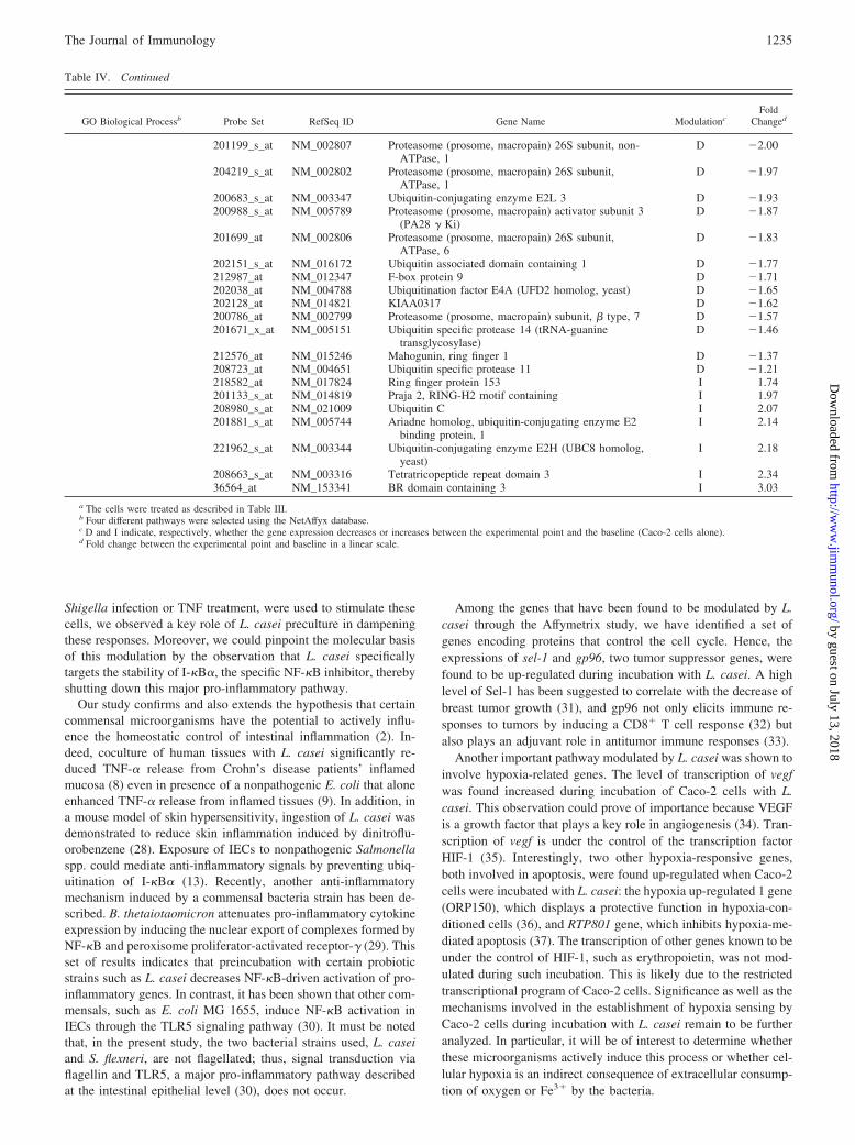
The above results demonstrate a role for L. casei in the specificmodulation of NF-�B-driven genes induced after Shigella infec-tion. However, it remains possible that other key signaling path-ways are targets of L. casei-induced modulatory properties. Togain more insight into the global effects mediated by L. casei onhost cell signaling, we performed a large-scale gene expressionstudy (14,500 well characterized human genes; Affymetrix Gene-chips U133A) on Caco-2 cells cocultured with the probiotic strainfor 2, 6, or 24 h. The entire set of results is available online (sup-plemental Table S1)5, and Table III represents the number of probesets modulated during Caco-2 cell culture in the presence of L.casei. Interestingly, most of the modulation was observed after24 h of incubation. Because all of the results are available online,and to avoid to present an extensive list of modulated genes, wehave focused in this study on four functional clusters (defined us-ing NettAffyx, the database from Affymetrix) that appeared to bemodulated most extensively by L. casei (Table IV and see below).
Group 1: genes involved in control of the cell cycle. L. caseiinduced both positive and negative modulation of expression ofgenes involved in the regulation of the cell cycle. Indeed, tumorsuppressor genes, such as tumor rejection Ag (gp96) and sel-1suppressor of lin-12, were up-regulated during incubation with L.casei, whereas the expression of the gene encoding prohibitin, anegative regulator of cell proliferation, was down-regulated.
Group 2: genes involved in the control of apoptosis. Expressionof two genes encoding proteins involved in the inhibition of apo-ptosis (Fas apoptotic inhibitory molecule and cytokine-induced ap-optosis inhibitor) were down-regulated, and expression of BCL2/adenovirus E1B was increased. It should be noted that theexpression of Nalp2, encoding a part of the inflammasome (26),decreased during the incubation of Caco-2 cells with L. casei, sug-gesting its potential involvement in the anti-inflammatory proper-ties of the probiotic bacteria. In addition, the expression of pro-grammed cell death 4, a tumor suppressor (27), was positivelymodulated.
Group 3: genes involved in hypoxia. Expression of two genesunder the control of the hypoxia-inducible factor-1 (HIF-1) tran-scription factor (hypoxia up-regulated 1 and HIF-1 response
RTP801) was induced during the incubation of Caco-2 cells withL. casei.
Group 4: genes belonging to the ubiquitination/degradationpathway. Twenty-five genes involved in the protein ubiquitina-tion and/or degradation process were found to be positively ornegatively modulated during incubation of Caco-2 cells with L.casei. The transcription of genes encoding three ubiquitin-conju-gating enzymes, E2D, E2L, and E2N, and four subunits of the 26Sproteasome were down-regulated. Another gene, the expression ofwhich decreased during this incubation, was rbx-1/roc-1/hrt-1,which encodes a protein belonging to the E3 ligase complex. Weconfirmed by individual PCR on a set of these selected genes theirdown-regulated expression after treatment with L. casei (Fig. 7A).It is striking to note that, among all of the genes modulated by L.casei, the group of genes implicated in ubiquitination/degradation(group 4) is, by far, the most represented. This observation sug-gests that L. casei is capable of establishing a program leading tothe down-regulation of the ubiquitin-mediated degradation of pro-teins. Moreover, because the ubiquitin system is responsible forthe degradation of I-�B�, this observation strongly supports ourconclusions that modulating the stability of I-�B� represents amajor target of L. casei.
Among the 25 genes of the ubiquitination/degradation pathwaythat were identified, we noticed that Rbx-1 expression was down-regulated to a greater extent (�4-fold decrease) by L. casei thanthe others (Table IV). This prompted us to investigate whether theeffects of L. casei on the NF-�B pathway could be accounted forby the specific down-regulation of Rbx-1. First, the expression ofRbx-1 at the protein level was monitored in L. casei-treated cellsand was found to be reduced by �50% (Fig. 7B). Second, over-expression of exogenous epitope-tagged Rbx-1 in L. casei-treatedcells was performed, and TNF-mediated (as well as Shigella-me-diated) degradation of I-�B� was monitored (data not shown).However, restoring Rbx-1 expression was found to be insufficientto recapitulate the effects of L. casei on the ubiquitin/degradationpathway. As a consequence, it is likely that L. casei down-regu-lates this cellular process via the targeting of multiple effectors.Accordingly, we observed that the global pattern of ubiquitinatedproteins after L. casei treatment closely resembles the one of cellstreated with MG132, a broad inhibitor of multiple ubiquitin/pro-teasome-dependent pathways (Fig. 7C). Together, this large-scaleanalysis of L. casei-modulated genes in Caco-2 cells strongly sup-ports the conclusion that this probiotic bacteria displays anti-inflammatory properties through the targeting of the ubiquitin/pro-teasome system.
DiscussionIn this study, we have investigated the effect of coculture of IECswith the probiotic bacteria L. casei. We were interested in twoquestions of fundamental importance: 1) Does this probiotic bac-teria display protective properties on IECs infected with a patho-genic entero-invasive bacteria? and 2) What is the pattern of tran-scriptional responses of the epithelial cells cocultured for variousperiods with L. casei? To gain insight into these questions, weembarked on a project to identify the global repertoire of hostresponses to L. casei using, for the first time on this topic, thetechnologies of macroarray analysis (1,050 genes selected) asso-ciated with DNA chip hybridization (Affymetrix; 14,500 genes)and biochemical analysis. Our results clearly identify a role for L.casei in the specific modulation of a repertoire of genes associatedwith the ubiquitin/proteasome pathway. Importantly, the down-regulation of this set of ubiquitin system-associated genes appearsto play a crucial role in the modulation of pro-inflammatory path-ways in IECs. Indeed, when pro-inflammatory stimuli, such as5 The online version of this article contains supplemental material.
Table III. Determination of modulated probe sets during time courseculture of Caco-2 cells with L. caseia
Time Course
Probe Set Number
Decrease Increase
2 h 53 356 h 63 59
24 h 357 237
a Caco-2 cells were cultured 2, 6, or 24 h with L. casei. After RNA extraction,labeling and hybridizations onto U133A Affymetrix GeneChip were performed. Thenumber represents the modulated probe set with a fold change � 2.
1233The Journal of Immunology
by guest on July 13, 2018http://w
ww
.jimm
unol.org/D
ownloaded from

Table IV. Gene expression modulation during L. casei time coursea
GO Biological Processb Probe Set RefSeq ID Gene Name ModulationcFold
Changed
Group 1: Cell growth/cellcycle
201278_at NM_001343 Disabled homolog 2, mitogen-responsivephosphoprotein (Drosophila)
D �4.68
201202_at NM_002592 Proliferating cell nuclear antigen D �4.29216237_s_at NM_006739 MCM5 minichromosome maintenance deficient 5, cell
division cycle 46D �4.07
203625_x_at NM_005983 S-phase kinase-associated protein 2 (p45) D �3.93204318_s_at NM_016426 G-2 and S-phase expressed 1 D �3.42204709_s_at NM_004856 Kinesin family member 23 D �3.14200659_s_at NM_002634 Prohibitin D �3.08209662_at NM_004365 Centrin, EF-hand protein, 3 (CDC3 1 homolog, yeast) D �2.98204162_at NM_006101 Kinetochore associated 2 D �2.88214710_s_at NM_031966 Cyclin B1 D �2.83207165_at NM_012484 Hyaluronan-mediated motility receptor (RHAMM) D �2.73202883_s_at NM_002716 Protein phosphatase 2 (formerly 2A), regulatory
subunit A (PR 65), �D �2.73
208712_at NM_001758 Cyclin D1 (PRAD1: parathyroid adenomatosis 1) D �2.51213222_at NM_015192 Phospholipase C, � 1 (phosphoinositide-specific) D �2.51203145_at NM_006461 Sperm associated antigen 5 D �2.46204444_at NM_004523 Kinesin family member 11 D �2.46202240_at NM_005030 Polo-like kinase 1 (Drosophila) D �2.42200959_at NM_004960 fusion (involved in t(12;16) in malignant liposarcoma) D �2.38218350_s_at NM_015895 geminin, DNA replication inhibitor D �2.34203078_at NM_003591 Cullin 2 D �2.30203362_s_at NM_002358 MAD2 mitotic arrest deficient-like 1 (yeast) D �2.22212426_s_at NM_006826 Tyrosine 3-monooxygenase/tryptophan 5-
monooxygenase activationD �2.22
212949_at NM_015341 Barren homolog (Drosophila) D �2.18208727_s_at NM_001791 cell division cycle 42 (GTP binding protein, 25 kDa) D �2.18201938_at NM_004642 CDK2-associated protein 1 D �2.14203740_at NM_005792 M-phase phosphoprotein 6 D �2.03201664_at NM_0012799 SMC4 structural maintenance of chromosomes 4-like
1 (yeast)D �1.90
218009_s_at NM_003981 Protein regulator of cytokinesis 1 D �1.90201725_at NM_006023 Chromosome 10 open reading frame 7 D �1.87220789_s_at NM_004749 Transforming growth factor � regulator 4 D �1.87204817_at NM_012291 Extra spindle poles like 1 (S. cerevisiae) D �1.87201186_at NM_002337 Low-density lipoprotein receptor-related protein
associated protein 1D �1.65
221509_at NM_003677 Density-regulated protein D �1.57201173_x_at NM_006600 Nuclear distribution gene C homolog (A. nidulans) D �1.46217839_at NM_006070 TRK-fused gene I 1.87219910_at NM_007076 Huntingtin interacting protein E I 2.38203226_s_at NM_005981 Sarcoma amplified sequence I 2.55202205_at NM_003370 Vasodilator-stimulated phosphoprotein I 3.86202061_s_at NM_005065 sel-1 suppressor of lin-12-like (C. elegans) I 3.86210513_s_at NM_003376 Vascular endothelial growth factor I 4.07205569_at NM_014398 Lysosomal-associated membrane protein 3 I 4.92200598_s_at NM_003299 Tumor rejection antigen (gp96) 1 I 5.46
Group 2: Apoptosis 220643_s_at NM_018147 Fas apoptotic inhibitory molecule D �5.10221690_s_at NM_017852 NACHT, leucine rich repeat and PYD containing 2
(Nalp2)D �4.68
202268_s_at NM_003905 Amyloid � precursor protein binding protein 1, 59KDa
D �2.14
220044_x_at NM_006107 Cisplatin resistance-associated overexpressed protein D �2.14208424_s_at NM_020313 Cytokine induced apoptosis inhibitor 1 D �2.11219275_at NM_004708 Programmed cell death 5 D �1.65203489_at NM_006427 CD27-binding (Siva) protein D �1.49221479_s_at NM_004331 BCL2/adenovirus E1B 19 kDa interacting protein 3-
likeI 2.30
202731_at NM_014456 Programmed cell death 4 (neoplastic transformationinhibitor)
I 3.36
202014_at NM_014330 Protein phosphatase 1, regulatory (inhibitor) subunit15A
I 7.21
Group 3: Hypoxia 200825_s_at NM_006389 Hypoxia up-regulated 1 I 3.86202887_s_at NM_019058 HIF-1 response RTP801 I 4.92
Group 4: Ubiquitination/ 218117_at NM_014248 Ring-box 1 D �4.07degradation 212751_at NM_003348 Ubiquitin-conjugating enzyme E2N (UBC13 homolog,
yeast)D �2.88
201377_at NM_014847 Ubiquitin associated protein 2-like D �2.42201498_at NM_003470 Ubiquitin specific protease 7 (herpes virus-associated) D �2.34211764_s_at NM_003338 Ubiquitin-conjugating enzyme E2D 1 (UBC4/5
homolog, yeast)D �2.22
1234 PROTECTIVE EFFECT OF L. casei TO Shigella INFECTION
by guest on July 13, 2018http://w
ww
.jimm
unol.org/D
ownloaded from
Shigella infection or TNF treatment, were used to stimulate thesecells, we observed a key role of L. casei preculture in dampeningthese responses. Moreover, we could pinpoint the molecular basisof this modulation by the observation that L. casei specificallytargets the stability of I-�B�, the specific NF-�B inhibitor, therebyshutting down this major pro-inflammatory pathway.
Our study confirms and also extends the hypothesis that certaincommensal microorganisms have the potential to actively influ-ence the homeostatic control of intestinal inflammation (2). In-deed, coculture of human tissues with L. casei significantly re-duced TNF-� release from Crohn’s disease patients’ inflamedmucosa (8) even in presence of a nonpathogenic E. coli that aloneenhanced TNF-� release from inflamed tissues (9). In addition, ina mouse model of skin hypersensitivity, ingestion of L. casei wasdemonstrated to reduce skin inflammation induced by dinitroflu-orobenzene (28). Exposure of IECs to nonpathogenic Salmonellaspp. could mediate anti-inflammatory signals by preventing ubiq-uitination of I-�B� (13). Recently, another anti-inflammatorymechanism induced by a commensal bacteria strain has been de-scribed. B. thetaiotaomicron attenuates pro-inflammatory cytokineexpression by inducing the nuclear export of complexes formed byNF-�B and peroxisome proliferator-activated receptor-� (29). Thisset of results indicates that preincubation with certain probioticstrains such as L. casei decreases NF-�B-driven activation of pro-inflammatory genes. In contrast, it has been shown that other com-mensals, such as E. coli MG 1655, induce NF-�B activation inIECs through the TLR5 signaling pathway (30). It must be notedthat, in the present study, the two bacterial strains used, L. caseiand S. flexneri, are not flagellated; thus, signal transduction viaflagellin and TLR5, a major pro-inflammatory pathway describedat the intestinal epithelial level (30), does not occur.
Among the genes that have been found to be modulated by L.casei through the Affymetrix study, we have identified a set ofgenes encoding proteins that control the cell cycle. Hence, theexpressions of sel-1 and gp96, two tumor suppressor genes, werefound to be up-regulated during incubation with L. casei. A highlevel of Sel-1 has been suggested to correlate with the decrease ofbreast tumor growth (31), and gp96 not only elicits immune re-sponses to tumors by inducing a CD8� T cell response (32) butalso plays an adjuvant role in antitumor immune responses (33).
Another important pathway modulated by L. casei was shown toinvolve hypoxia-related genes. The level of transcription of vegfwas found increased during incubation of Caco-2 cells with L.casei. This observation could prove of importance because VEGFis a growth factor that plays a key role in angiogenesis (34). Tran-scription of vegf is under the control of the transcription factorHIF-1 (35). Interestingly, two other hypoxia-responsive genes,both involved in apoptosis, were found up-regulated when Caco-2cells were incubated with L. casei: the hypoxia up-regulated 1 gene(ORP150), which displays a protective function in hypoxia-con-ditioned cells (36), and RTP801 gene, which inhibits hypoxia-me-diated apoptosis (37). The transcription of other genes known to beunder the control of HIF-1, such as erythropoietin, was not mod-ulated during such incubation. This is likely due to the restrictedtranscriptional program of Caco-2 cells. Significance as well as themechanisms involved in the establishment of hypoxia sensing byCaco-2 cells during incubation with L. casei remain to be furtheranalyzed. In particular, it will be of interest to determine whetherthese microorganisms actively induce this process or whether cel-lular hypoxia is an indirect consequence of extracellular consump-tion of oxygen or Fe3� by the bacteria.
Table IV. Continued
GO Biological Processb Probe Set RefSeq ID Gene Name ModulationcFold
Changed
201199_s_at NM_002807 Proteasome (prosome, macropain) 26S subunit, non-ATPase, 1
D �2.00
204219_s_at NM_002802 Proteasome (prosome, macropain) 26S subunit,ATPase, 1
D �1.97
200683_s_at NM_003347 Ubiquitin-conjugating enzyme E2L 3 D �1.93200988_s_at NM_005789 Proteasome (prosome, macropain) activator subunit 3
(PA28 � Ki)D �1.87
201699_at NM_002806 Proteasome (prosome, macropain) 26S subunit,ATPase, 6
D �1.83
202151_s_at NM_016172 Ubiquitin associated domain containing 1 D �1.77212987_at NM_012347 F-box protein 9 D �1.71202038_at NM_004788 Ubiquitination factor E4A (UFD2 homolog, yeast) D �1.65202128_at NM_014821 KIAA0317 D �1.62200786_at NM_002799 Proteasome (prosome, macropain) subunit, � type, 7 D �1.57201671_x_at NM_005151 Ubiquitin specific protease 14 (tRNA-guanine
transglycosylase)D �1.46
212576_at NM_015246 Mahogunin, ring finger 1 D �1.37208723_at NM_004651 Ubiquitin specific protease 11 D �1.21218582_at NM_017824 Ring finger protein 153 I 1.74201133_s_at NM_014819 Praja 2, RING-H2 motif containing I 1.97208980_s_at NM_021009 Ubiquitin C I 2.07201881_s_at NM_005744 Ariadne homolog, ubiquitin-conjugating enzyme E2
binding protein, 1I 2.14
221962_s_at NM_003344 Ubiquitin-conjugating enzyme E2H (UBC8 homolog,yeast)
I 2.18
208663_s_at NM_003316 Tetratricopeptide repeat domain 3 I 2.3436564_at NM_153341 BR domain containing 3 I 3.03
a The cells were treated as described in Table III.b Four different pathways were selected using the NetAffyx database.c D and I indicate, respectively, whether the gene expression decreases or increases between the experimental point and the baseline (Caco-2 cells alone).d Fold change between the experimental point and baseline in a linear scale.
1235The Journal of Immunology
by guest on July 13, 2018http://w
ww
.jimm
unol.org/D
ownloaded from

Because the expression of genes involved in different pathways(i.e., cell cycle, response to hypoxia, NF-�B-dependent transcrip-tion) was found to be modified during incubation of Caco-2 cellswith L. casei, we investigated whether a common signaling path-way could account for these diverse effects. Of interest, these threepathways are regulated by a similar ubiquitination/proteasome sys-tem (38–41). In our time-course study using Affymetrix technol-ogy, we observed that a large set of genes involved in the ubiq-uitination/degradation pathways was modulated by L. casei. Inlight of our results, it appears very likely that the specific modu-lation of the ubiquitin/proteasome system represents a main targetof action of L. casei on host cells and that this effect would in turnbe responsible for the alteration of several signaling pathways,such as those involving the cell cycle and hypoxia.
In agreement with our findings that L. casei affects the ubiquitin/proteasome pathway, Caco-2 cells coincubated with L. caseishowed stabilization of I-�B� even after subsequent stimulationby Shigella or TNF-�. Thus, the anti-inflammatory effects of L.casei are likely mediated by its effects on the ubiquitin/proteasomesystem and the consequent dampening of NF-�B-driven pro-in-flammatory signals.
Convergent pieces of evidence suggest that, through their abilityto modulate inflammatory pathways, some commensal bacteriacontribute to the homeostasis of the intestinal epithelium. Our ob-servations, and also the results from other groups, will impact onthe understanding of the mechanisms responsible for some of thebeneficial effects of probiotics on IBDs. This knowledge will con-tribute to offer, in the near future, new therapeutic means to coun-
teract the inflammatory disorders observed in human pathologies,such as ulcerative colitis and Crohn’s disease.
AcknowledgmentsThe Ab for ubiquitin detection was a gift from Dr. Claude Parsot (PasteurInstitute). We thank Drs. Armelle Phalipon, Dana Philpott, andRegis Tournebize for critical reading of this manuscript.
DisclosuresThe authors have no financial conflict of interest.
References1. Isolauri, E., S. Salminen, and A. C. Ouwehand. 2004. Microbial-gut interactions
in health and disease: probiotics. Best Pract. Res. Clin. Gastroenterol. 18:299–313.
2. Sansonetti, P. J. 2004. War and peace at mucosal surfaces. Nat. Rev. Immunol. 4:953–964.
3. Mahida, Y. R. 2004. Microbial-gut interactions in health and disease: epithelialcell responses. Best Pract. Res. Clin. Gastroenterol. 18: 241–253.
4. Rakoff-Nahoum, S., J. Paglino, F. Eslami-Varzaneh, S. Edberg, andR. Medzhitov. 2004. Recognition of commensal microflora by toll-like receptorsis required for intestinal homeostasis. Cell 118: 229–241.
5. McCracken, V. J., and R. G. Lorenz. 2001. The gastrointestinal ecosystem: aprecarious alliance among epithelium, immunity and microbiota. Cell Microbiol.3: 1–11.
6. Gionchetti, P., F. Rizzello, U. Helwig, A. Venturi, K. M. Lammers, P. Brigidi,B. Vitali, G. Poggioli, M. Miglioli, and M. Campieri. 2003. Prophylaxis of pou-chitis onset with probiotic therapy: a double-blind, placebo-controlled trial. Gas-troenterology 124: 1202–1209.
7. Cui, H. H., C. L. Chen, J. D. Wang, Y. J. Yang, Y. Cun, J. B. Wu, Y. H. Liu,H. L. Dan, Y. T. Jian, and X. Q. Chen. 2004. Effect of probiotic on intestinalmucosa of patients with ulcerative colitis. World J. Gastroenterol. 10:1521–1525.
8. Borruel, N., M. Carol, F. Casellas, M. Antolin, F. de Lara, E. Espin, J. Naval,F. Guarner, and J.R. Malagelada. 2002. Increased mucosal tumour necrosis factor� production in Crohn’s disease can be downregulated ex vivo by probioticbacteria. Gut 51: 659–664.
9. Borruel, N., F. Casellas, M. Antolin, M. Llopis, M. Carol, E. Espiin, J. Naval,F. Guarner, and J. R. Malagelada. 2003. Effects of nonpathogenic bacteria oncytokine secretion by human intestinal mucosa. Am. J. Gastroenterol. 98:865–870.
10. Drakes, M., T. Blanchard, and S. Czinn. 2004. Bacterial probiotic modulation ofdendritic cells. Infect. Immun. 72: 3299–3309.
11. Christensen, H. R., H. Frokiaer, and J. J. Pestka. 2002. Lactobacilli differentiallymodulate expression of cytokines and maturation surface markers in murine den-dritic cells. J. Immunol. 168: 171–178.
12. Jijon, H., J. Backer, H. Diaz, H. Yeung, D. Thiel, C. McKaigney, C. De Simone,and K. Madsen. 2004. DNA from probiotic bacteria modulates murine and humanepithelial and immune function. Gastroenterology 126: 1358–1373.
13. Neish, A. S., A. T. Gewirtz, H. Zeng, A. N. Young, M. E. Hobert, V. Karmali,A. S. Rao, and J. L. Madara. 2000. Prokaryotic regulation of epithelial responsesby inhibition of I�B-� ubiquitination. Science 289: 1560–1563.
14. Collier-Hyams, L. S., H. Zeng, J. Sun, A. D. Tomlinson, Z. Q. Bao, H. Chen,J. L. Madara, K. Orth, and A. S. Neish. 2002. Salmonella AvrA effector inhibitsthe key proinflammatory, anti-apoptotic NF-�B pathway. J. Immunol. 169: 2846–2850.
15. Hooper, L. V., M. H. Wong, A. Thelin, L. Hansson, P. G. Falk, and J. I. Gordon.2001. Molecular analysis of commensal host-microbial relationships in the in-testine. Science 291: 881–884.
16. Stappenbeck, T. S., L. V. Hooper, and J. I. Gordon. 2002. Developmental reg-ulation of intestinal angiogenesis by indigenous microbes via Paneth cells. Proc.Natl. Acad. Sci. USA 99: 15451–15455.
17. Sansonetti, P. J. 2001. Rupture, invasion and inflammatory destruction of theintestinal barrier by Shigella, making sense of prokaryote-eukaryote cross-talks.FEMS Microbiol. Rev. 25: 3–14.
18. Philpott, D. J., S. Yamaoka, A. Israel, and P. J. Sansonetti. 2000. Invasive Shi-gella flexneri activates NF-�B through a lipopolysaccharide-dependent innateintracellular response and leads to IL-8 expression in epithelial cells. J. Immunol.165: 903–914.
19. Girardin, S. E., R. Tournebize, M. Mavris, A. L. Page, X. Li, G. R. Stark,J. Bertin, P. S. DiStefano, M. Yaniv, P. J. Sansonetti, and D. J. Philpott. 2001.CARD4/Nod1 mediates NF-kappaB and JNK activation by invasive Shigellaflexneri. EMBO Rep. 2: 736–742.
20. Girardin, S. E., I. G. Boneca, L. A. Carneiro, A. Antignac, M. Jehanno, J. Viala,K. Tedin, M. K. Taha, A. Labigne, U. Zahringer, et al. 2003. Nod1 detects aunique muropeptide from gram-negative bacterial peptidoglycan. Science 300:1584–1587.
21. Clerc, P., and P. J. Sansonetti. 1987. Entry of Shigella flexneri into HeLa cells:evidence for directed phagocytosis involving actin polymerization and myosinaccumulation. Infect. Immun. 55: 2681–2688.
22. Petit-Bertron A.-F., T. Pedron, U. Gross, J.-Y. Coppee, P. J. Sansonetti, J.-M.Cavaillon, and M. Adib-Conquy. 2005. Adherence modifies the regulation ofgene expression induced by interleukin-10. Cytokine 29: 1–12.
FIGURE 7. L. casei induced a down-regulation of genes involved inubiquitination/degradation processes. Caco-2 cells were cultured overnightwith L. casei with a MOI of 100. A, After washes with PBS and RNAextraction with the Rneasy mini kit, cDNA was synthetized using oligo-d(T) and reverse transcriptase. PCRs were performed as described in thelegend to Fig. 2. B, After washes with PBS and lysis with Laemmli buffer,aliquots of the lysates were loaded on 15 or 10% SDS-PAGE for the sub-sequent detection of Rbx-1 or tubulin, respectively. The Rbx-1 or tubulinprotein contents were determined by Western blotting using a polyclonalanti-human Rbx-1 or a monoclonal anti-human tubulin Ab, respectively. C,HEK cells were treated overnight with L. casei or for 6 h with 50 �M ofthe proteasome inhibitor MG-132 before incubation with or without TNF.The I-�B� protein content and global ubiquitinated proteins were deter-mined by Western blotting (WB) using a polyclonal anti-human I-�B� Abor monoclonal anti-ubiquitin Ab, respectively.
1236 PROTECTIVE EFFECT OF L. casei TO Shigella INFECTION
by guest on July 13, 2018http://w
ww
.jimm
unol.org/D
ownloaded from

23. Li, C., and W. H. Wong. 2001. Model-based analysis of oligonucleotide arrays:expression index computation and outlier detection. Proc. Natl. Acad. Sci. USA98: 31–36.
24. Pedron, T., C. Thibault, and P. J. Sansonetti. 2003. The invasive phenotype ofShigella flexneri directs a distinct gene expression pattern in the human intestinalepithelial cell line Caco-2. J. Biol. Chem. 278: 33878–33886.
25. Kufer, T. A., J. H. Fritz, and D. J. Philpott. 2005. NACHT-LRR proteins (NLRs)in bacterial Infect. Immun. Trends Microbiol. 13: 381–388.
26. Tschopp, J., F. Martinon, and K. Burns. 2003. NALPS: a novel protein familyinvolved in inflammation. Nat. Rev. Mol. Cell Biol. 4: 95–104.
27. Goke, R., C. Gregel, A. Goke, R. Arnold, H. Schmidt, and B. Lankat-Buttgereit.2004. Programmed cell death protein 4 (PDCD4) acts as a tumor suppressor inneuroendocrine tumor cells. Ann. NY Acad. Sci. 1014: 220–221.
28. Chapat, L., K. Chemin, B. Dubois, R. Bourdet-Sicard, and D. Kaiserlian. 2004.Lactobacillus casei reduces CD8� T cell-mediated skin inflammation. Eur. J. Im-munol. 34: 2520–2528.
29. Kelly, D., J. I. Campbell, T. P. King, G. Grant, E. A. Jansson, A. G. Coutts,S. Pettersson, and S. Conway. 2004. Commensal anaerobic gut bacteria attenuateinflammation by regulating nuclear-cytoplasmic shuttling of PPAR-� and RelA.Nat. Immunol. 5: 104–112.
30. Bambou, J. C., A. Giraud, S. Menard, B. Begue, S. Rakotobe, M. Heyman,F. Taddei, N. Cerf-Bensussan, and V. Gaboriau-Routhiau. 2004. In vitro and exvivo activation of the TLR5 signaling pathway in intestinal epithelial cells by acommensal Escherichia coli strain. J. Biol. Chem. 279: 42984–42992.
31. Orlandi, R., M. Cattaneo, F. Troglio, P. Casalini, C. Ronchini, S. Menard, andI. Biunno. 2002. SEL1L expression decreases breast tumor cell aggressiveness invivo and in vitro. Cancer Res. 62: 567–574.
32. Udono, H., D. L. Levey, and P. K. Srivastava. 1994. Cellular requirements fortumor-specific immunity elicited by heat shock proteins: tumor rejection antigengp96 primes CD8� T cells in vivo. Proc. Natl. Acad. Sci. USA 91: 3077–3081.
33. Baker-LePain, J. C., M. Sarzotti, T. A. Fields, C. Y. Li, and C. V. Nicchitta. 2002.GRP94 (gp96) and GRP94 N-terminal geldanamycin binding domain elicit tissuenonrestricted tumor suppression. J. Exp. Med. 196: 1447–1459.
34. Bussolati, B., A. Ahmed, H. Pemberton, R. C. Landis, F. Di Carlo,D. O. Haskard, and J. C. Mason. 2004. Bifunctional role for VEGF-induced hemeoxygenase-1 in vivo: induction of angiogenesis and inhibition of leukocytic in-filtration. Blood 103: 761–766.
35. Skinner, H. D., J. Z. Zheng, J. Fang, F. Agani, and B. H. Jiang. 2004. Vascularendothelial growth factor transcriptional activation is mediated by hypoxia-in-ducible factor 1�, HDM2, and p70S6K1 in response to phosphatidylinositol 3-ki-nase/AKT signaling. J. Biol. Chem. 279: 45643–45651.
36. Ozawa, K., K. Kuwabara, M. Tamatani, K. Takatsuji, Y. Tsukamoto, S. Kaneda,H. Yanagi, D. M. Stern, Y. Eguchi, Y. Tsujimoto, et al. 1999. 150-kDa oxygen-regulated protein (ORP150) suppresses hypoxia-induced apoptotic cell death.J. Biol. Chem. 274: 6397–6404.
37. Shoshani, T., A. Faerman, I. Mett, E. Zelin, T. Tenne, S. Gorodin, Y. Moshel,S. Elbaz, A. Budanov, A. Chajut, et al. 2002. Identification of a novel hypoxia-inducible factor 1-responsive gene, RTP801, involved in apoptosis. Mol. Cell.Biol. 22: 2283–2293.
38. Ben-Neriah, Y. 2002. Regulatory functions of ubiquitination in the immune sys-tem. Nat. Immunol. 3: 20–26.
39. Tanaka, K., T. Kawakami, K. Tateishi, H. Yashiroda, and T. Chiba. 2001. Controlof IkappaBalpha proteolysis by the ubiquitin-proteasome pathway. Biochimie 83:351–356.
40. Krek, W. 2000. VHL takes HIF’s breath away. Nat. Cell. Biol. 2: E121–E123.41. Kamura T., D. M. Koepp, M. N. Conrad, D. Skowyra, R. J. Moreland,
O. Iliopoulos, W. S. Lane, W. G. Kaelin, Jr., S. J. Elledge, R. C. Conaway, et al.1999. Rbx1, a component of the VHL tumor suppressor complex and SCF ubiq-uitin ligase. Science 284: 657–661.
1237The Journal of Immunology
by guest on July 13, 2018http://w
ww
.jimm
unol.org/D
ownloaded from

CORRECTIONSBrady, J., Y. Hayakawa, M. J. Smyth, and S. L. Nutt. 2004. IL-21 induces the functional maturation of murine NK cells.J. Immunol. 172: 2048–2058.
Figure 8 is incorrect. The corrected figure is shown below.
Shurin, G. V., R. Ferris, I. L. Tourkova, L. Perez, A. Lokshin, L. Balkir, B. Collins, G. S. Chatta, and M. R. Shurin. 2005.Loss of new chemokine CXCL14 in tumor tissue is associated with low infiltration by dendritic cells (DC), whilerestoration of human CXCL14 expression in tumor cells causes attraction of DC both in vitro and in vivo. J. Immunol.174: 5490–5498.
The second author’s middle initial was omitted. The correct name is Robert L. Ferris.
Santiago, H. C., C. G. Feng, A. Bafica, E. Roffe, R. M. Arantes, A. Cheever, G. Taylor, L. Q. Vierira, J. Aliberti, R. T.Gazzinelli, and A. Sher. 2005. Mice deficient in LRG-47 display enhanced susceptibility to Trypanosoma cruzi infectionassociated with defective hemopoiesis and intracellular control of parasite growth. J. Immunol. 175: 8165–8172.
The eighth author’s last name was misspelled. The correct name is Leda Q. Vieira.
The Journal of Immunology
Copyright © 2006 by The American Association of Immunologists, Inc. 0022-1767/06/$02.00
Oki, T., J. Kitaura, K. Eto, Y. Lu, M. Maeda-Yamamoto, N. Inagaki, H. Nagai, Y. Yamanishi, H. Nakajina, H. Kumagai,and T. Kitamura. 2006. Integrin �IIb�3 induces the adhesion and activation of mast cells through interaction withfibrinogen. J. Immunol. 176: 52–60.
The ninth author’s last name was misspelled. The correct name is Hideaki Nakajima.
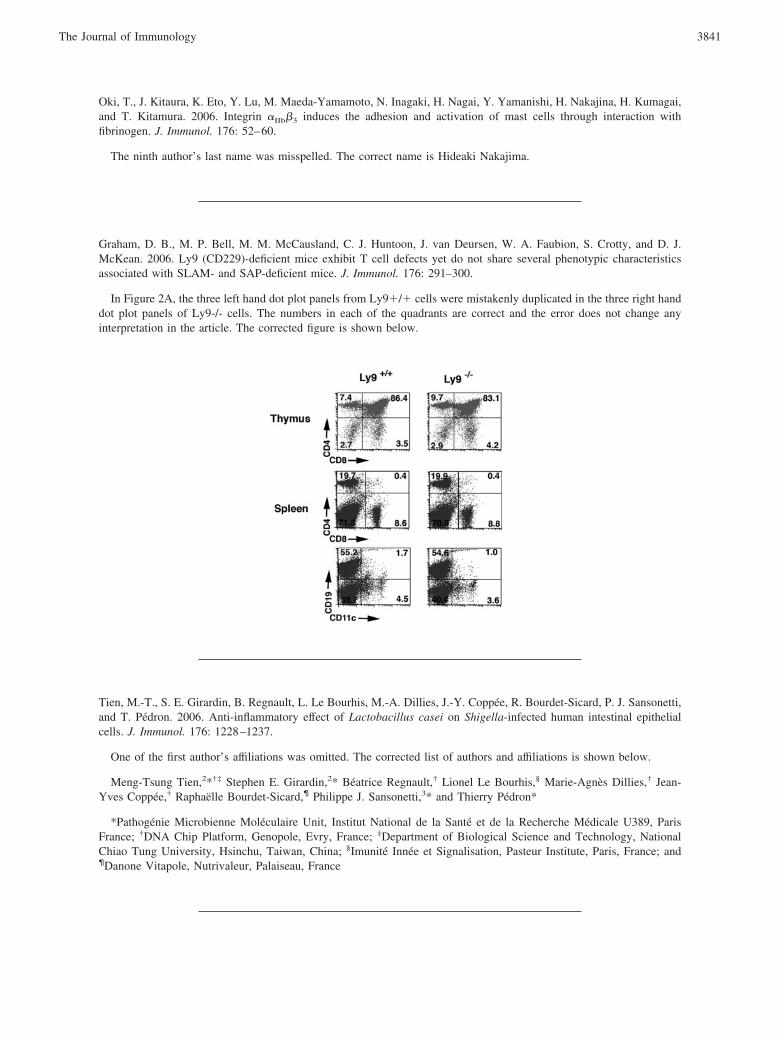
Graham, D. B., M. P. Bell, M. M. McCausland, C. J. Huntoon, J. van Deursen, W. A. Faubion, S. Crotty, and D. J.McKean. 2006. Ly9 (CD229)-deficient mice exhibit T cell defects yet do not share several phenotypic characteristicsassociated with SLAM- and SAP-deficient mice. J. Immunol. 176: 291–300.
In Figure 2A, the three left hand dot plot panels from Ly9�/� cells were mistakenly duplicated in the three right handdot plot panels of Ly9-/- cells. The numbers in each of the quadrants are correct and the error does not change anyinterpretation in the article. The corrected figure is shown below.
Tien, M.-T., S. E. Girardin, B. Regnault, L. Le Bourhis, M.-A. Dillies, J.-Y. Coppee, R. Bourdet-Sicard, P. J. Sansonetti,and T. Pedron. 2006. Anti-inflammatory effect of Lactobacillus casei on Shigella-infected human intestinal epithelialcells. J. Immunol. 176: 1228–1237.
One of the first author’s affiliations was omitted. The corrected list of authors and affiliations is shown below.
Meng-Tsung Tien,2*†‡ Stephen E. Girardin,2* Beatrice Regnault,† Lionel Le Bourhis,§ Marie-Agnes Dillies,† Jean-Yves Coppee,† Raphaelle Bourdet-Sicard,¶ Philippe J. Sansonetti,3* and Thierry Pedron*
*Pathogenie Microbienne Moleculaire Unit, Institut National de la Sante et de la Recherche Medicale U389, ParisFrance; †DNA Chip Platform, Genopole, Evry, France; ‡Department of Biological Science and Technology, NationalChiao Tung University, Hsinchu, Taiwan, China; §Imunite Innee et Signalisation, Pasteur Institute, Paris, France; and¶Danone Vitapole, Nutrivaleur, Palaiseau, France
3841The Journal of Immunology

Atherly, L. O., M. A. Brehm, R. M. Welsh, and L. J. Berg. 2006. Tec kinases Itk and Rlk are required for CD8� T cellresponses to virus infection independent of their role in CD4� T cell help. J. Immunol. 176: 1571–1581.
In Figure 1B, the WT Ca flux data line is missing from the Ca flux graph. The corrected figure is shown below.
3842 CORRECTIONS
Stone, J. D., and L. J. Stern. 2006. CD8 T cells, like CD4 T cells, are triggered by multivalent engagement of TCRs byMHC-peptide ligands but not by monovalent engagement. J. Immunol. 176: 1498–1505.
In Discussion, the last reference in the paper is incorrect. The corrected sentence and reference are shown below.
It is known that the cytoplasmic domains of several components of the TCR complex tend to homo-oligomerize at highconcentrations (41); perhaps ligand-induced clustering of the TCR drives the cytoplasmic domains of proximal receptorsto rearrange, exposing the Nck binding epitope and propelling other signaling cascade processes.
41. Sigalov A., D. Aivazian, and L. Stern. 2004. Homooligomerization of the cytoplasmic domain of the T cell receptor zetachain and of other proteins containing the immunoreceptor tyrosine-based activation motif. Biochemistry 43: 2049–61.
Serhan, C. N., K. Gotlinger, S. Hong, Y. Lu, J. Siegelman, T. Baer, R. Yang, S. P. Colgan, and N. A. Petasis. 2006.Anti-inflammatory actions of neuroprotectin D1/protectin D1 and its natural stereoisomers: assignments of dihydroxy-containing docosatrienes. J. Immunol. 176: 1848–1859.
In Discussion, in the second sentence of paragraph six, 10S-HDNA should have been 10S-HDHA. The correctedsentence is shown below.
Recently, classic steric analysis of 10S-HDHA and the formation of 10,20-diHDHA and 17-H(p)DHA were reportedlyoptimized for the plant LOs (49).
Cante-Barrett, K., E. M. Gallo, M. M. Winslow, and G. R. Crabtree. 2006. Thymocyte negative selection is mediated byprotein kinase C- and Ca2�-dependent transcriptional induction of Bim of cell death. J. Immunol. 176: 2299–2306.
The title of the article is incorrect. The corrected title is shown below. The error has been corrected in the online version,which now differs from the print version as originally published.
Thymocyte Negative Selection Is Mediated by Protein Kinase C- and Ca2�-Dependent Transcriptional Induction ofBim
3843The Journal of Immunology