antiangiogenic therapy: impact on invasion, disease ... · antiangiogenic therapy: impact on...
TRANSCRIPT
210 | APRIL 2011 | VOLUME 8 www.nature.com/nrclinonc
Division of Molecular and Cellular Biology Research, Sunnybrook Health Sciences Centre, S‑217 Research Building, 2075 Bayview Avenue, Toronto, ON M4N 3M5, Canada (J. M. L. Ebos, R. S. Kerbel).
Correspondence to: J. M. L. Ebos [email protected]
Antiangiogenic therapy: impact on invasion, disease progression, and metastasisJohn M. L. Ebos and Robert S. Kerbel
Abstract | Antiangiogenic drugs targeting the VEGF pathway have slowed metastatic disease progression in some patients, leading to progression‑free survival (PFS) and overall survival benefits compared with controls. However, the results are more modest than predicted by most preclinical testing and benefits in PFS are frequently not accompanied by overall survival improvements. Questions have emerged about the basis of drug resistance and the limitations of predictive preclinical models, and also about whether the nature of disease progression following antiangiogenic therapy is different to classic cytotoxic therapies—in particular whether therapy may lead to more invasive or metastatic behavior. In addition, because of recent clinical trial failures of antiangiogenic therapy in patients with early‑stage disease, and the fact that there are hundreds of trials underway in perioperative neoadjuvant and adjuvant settings, there is now greater awareness about the lack of appropriate preclinical testing that preceded these studies. Improved preclinical assessment of all stages of metastatic disease should be a priority for future antiangiogenic drug discovery and development.
Ebos, J. M. L. & Kerbel, R. S. Nat. Rev. Clin. Oncol. 8, 210–221 (2011); published online 1 March 2011; corrected online 8 March 2011 and 4 May 2011; doi:10.1038/nrclinonc.2011.21
IntroductionAntiangiogenic therapy is based on the theory that block-ing new blood vessel formation in tumors will stop or slow their growth. Currently, four molecular-targeted drugs are approved by the FDA for six tumor indications; all act to disrupt the VEGF pathway.1 Thus, nearly four decades after the anti angiogenesis concept was introduced by Judah Folkman,2 antiangiogenic therapy is considered a major anticancer treatment modality.3 However, with hundreds of clinical trials currently underway in multiple cancer indications and pathological stages, and dozens of other VEGF and other angiogenic-pathway-targeted agents now in experimental or clinical testing, an urgent issue is under-standing why the majority of patients stop responding -—or do not respond at all—to such drugs and how such limitations can be overcome. Numerous mechanisms of resistance to antiangiogenic therapy have been proposed4 highlighting that over two decades of positive preclinical studies have yielded only modest incremental changes in the clinic. While this is an unfortunate and common occur-rence among cancer treatments, the question remains: are the challenges facing antiangiogenic drugs unique?
In theory, targeting the host ‘tumor-supporting’ angio-genic processes has many benefits but it might also have limitations. Antiangiogenic therapies might initiate an array of stromal and microenvironmental defense mecha-nisms4 that contribute to eventual drug inefficacy and, more
provocatively, may lead to a more aggressive and invasive tumor phenotype—one with an increased ability to meta-stasize. Though perhaps surprising, this latter property is not distinct from other anticancer treatment modalities -—surgery, radiation and chemotherapy can also produce similar unwanted ‘prometastatic’ effects in certain isolated experimental settings (Box 1). However, the possibility that VEGF-pathway inhibitors, and perhaps other ‘host- targeted’ drugs as well, could augment invasive or metastatic potential (despite controlling primary tumor growth or ini-tially slowing the growth of meta stasis) could be signifi cant and has become a topic of considerable controversy. The debate has been fuelled by modest clinical benefits, high drug cost, and adverse side effects, in addition to converg-ing findings published in the past 2 years, which relate to limited drug efficacy in early-stage disease. The first finding comes from two preclinical studies showing that the bene fits from VEGF-pathway-inhibitor monotherapy can depend on disease stage and treatment circumstances and can, in certain settings, be offset by increased aggres-sive invasiveness and augmented metastatic potential.5,6 The second finding comes from two large phase III clinical trials involving bevacizumab, a monoclonal antibody to VEGF, used in combination with chemotherapy and administered as adjuvant therapy to patients with early-stage colorectal carcinoma; the treatment combination showed no benefit in the primary end point of disease-free survival (DFS) compared with the chemotherapy-alone arm.7,8 These studies have raised questions about the expectations for antiangiogenic agents in blocking different stages of tumor progression and, in particular, the benefits of these drugs in micrometastatic disease settings.
Competing interestsR. S. Kerbel declares associations with the following companies: GlaxoSmithKline, MetronomX, MolMed, Pfizer, Taiho Pharmaceutical, YM Biosciences. See the article online for full details of the relationships. J. M. L. Ebos declares no competing interests.
REVIEWS
© 2011 Macmillan Publishers Limited. All rights reserved

NATURE REVIEWS | CLINICAL ONCOLOGY VOLUME 8 | APRIL 2011 | 211
We summarize evidence that suggests anti angiogenic drugs might alter the natural history of disease progression, depending on the disease stage and tumor type, and focus on limitations that anti angiogenic drugs might have to over-come to bring about greatly improved clinical bene fits. It is possible that antiangiogenic therapy may induce a differ-ent disease progression pattern than cytotoxics and lead to worse outcomes in terms of progression, invasion, and meta-stasis. However, this result might never mat erialize outside of certain limited preclinical scenarios. It remains theoreti-cally possible that such ‘evasive resistance’ mechanisms have a role in the clinical limitations of successful antiangiogenic drugs and, perhaps most importantly, might provide a clue as to how they can be made more effective. There is no com-pelling clinical evidence that antiangio genesis treatment will make disease worse or decrease survival;9 however, neither is there a large pool of supporting preclinical evidence that such therapy will be beneficial in blocking early-stage disease, particularly in potentially curative and preventive settings where detailed analysis is rarely performed. With thousands of patients projected to be enrolled to clinical trials over the next 5 years to assess neoadjuvant or long-term adjuvant use of VEGF-pathway-targeted drugs,10 a rigorous assessment of actual and predicted outcomes for antiangiogenic therapy should be conducted using improved pre clinical models to better understand when and to what extent these new drugs are likely to work.
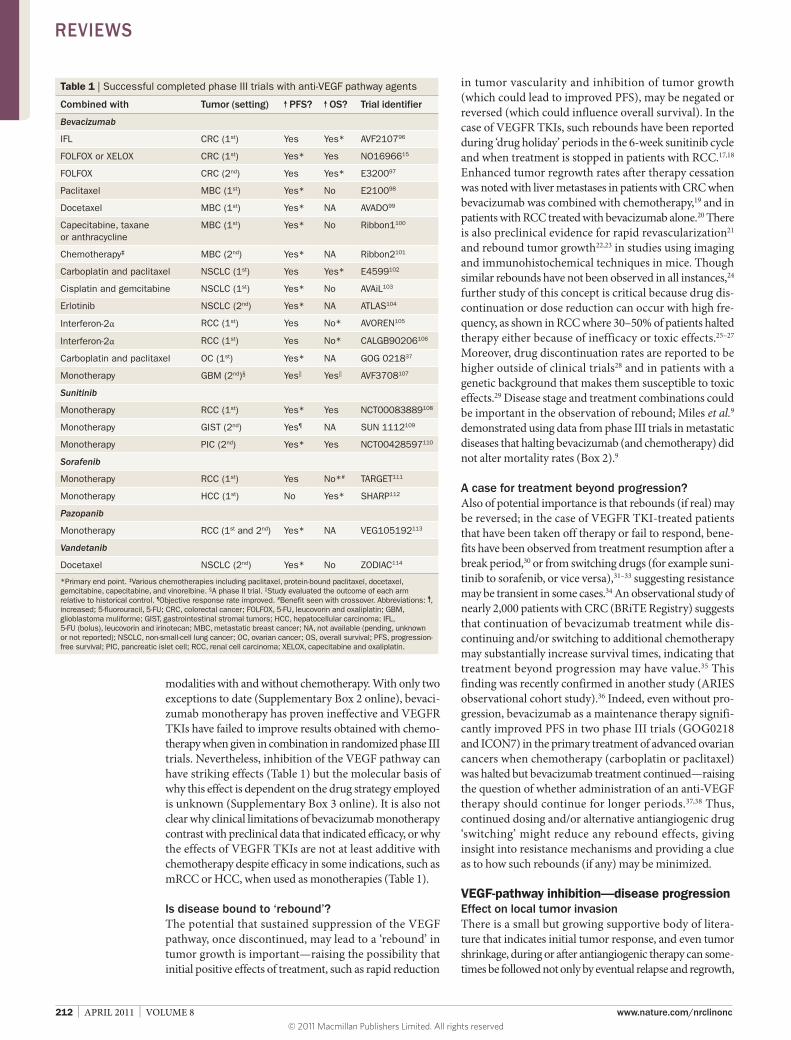
Successful therapy—but challenges remainBevacizumab was the first molecular-targeted antiangio-genic therapy approved by the FDA and is used as first-line therapy in colorectal cancer (CRC), metastatic breast cancer (MBC), non-small-cell lung cancer (NSCLC), and metastatic renal cell carcinoma (mRCC), and as second- line therapy in CRC and glioblastoma multi forme (GBM).11 With the exception of GBM, bevacizumab is only approved when combined with chemo therapy or cytokine therapy, as mono therapy failed to show robust activity in most instances of advanced-stage disease.12 A second class of approved inhibitors (sunitinib, sorafenib and pazopanib) include oral small-molecule tyrosine kinase inhibitors (TKIs) that target VEGFRs, platelet-derived growth factor (PDGF) receptors, and other kinases including KIT, Ret, BRAF and Flt-3.13 All three of these VEGFR TKIs have been approved as mono-therapies for the treatment of mRCC; sunitinib is approved to treat imatinib- refractory gastro intestional stromal tumors (GIST), and sorafenib is approved for hepato cellular carci-noma (HCC). But, these clinical successes have been accom-panied by questions that have emerged in the phase III trial setting, which represent potential challenges that must be addressed in order to overcome the limited efficacy of VEGF-pathway inhibitors (Tables 1 and 2).
PFS gains and overall survivalIn terms of objective benefits, such as disease stabiliza-tion and progression-free survival (PFS) or overall sur-vival, VEGF-pathway-targeted therapy has largely yielded only modest gains. Despite the presence of VEGF and VEGFR2, tumors either do not respond or eventually become un responsive with PFS or overall survival benefits
Key points
■ Successful clinical trials with various VEGF‑pathway inhibitors have been accompanied by numerous phase III failures
■ Trial failures in adjuvant disease, and ongoing trials in early‑stage settings, could highlight differences in antiangiogenic drug efficacy depending on disease stage
■ There is a gap between how antiangiogenics are usually tested in the clinic (late‑stage metastatic) and in preclinical mouse models (localized primary tumors)
■ There is debate whether anti‑VEGF therapy may lead to ‘rebound growth’ when halted or if it may fuel more invasive and metastatic disease phenotypes
■ Future testing of antiangiogenic therapies should be conducted in clinically relevant animal models of all disease stages
in patients receiving anti angiogenic therapy being, in most cases, measured in months.14 In some instances, trials in indications that initially yielded significant improvements in overall survival when bevaci zumab was combined with chemo therapy have sometimes not shown similar benefits when compared with more-effective chemotherapies in follow-up studies.15
Perhaps more concerning, however, is the emerging trend where patient response rate and PFS does not trans-late into significantly increased overall survival in phase III trials (Tables 1 and 2 and Supplementary Box 1 online). Currently, overall survival remains the gold standard for determining therapeutic benefit but the potential use of PFS as an ‘overall survival surrogate’ has been introduced because of a typically strong correlation between the hazard ratios for overall survival and PFS.16 However, there remains a lack of consensus on the use of PFS in this manner and results with antiangiogenic drugs suggest an example where PFS benefits are often not translated into overall survival benefits. It remains a major question as to why such robust gains in PFS seen in the majority of completed phase III trials with bevacizumab and chemo therapy, or VEGFR TKIs as monotherapy, have not frequently corresponded to robust gains in overall survival.
The paradox of chemotherapy combinationFailed trials with VEGF-pathway inhibitors have uncov-ered a disparity between the efficacy of different treatment
Box 1 | Therapy‑accelerated tumor growth and metastasis—not a new phenomenon
Nearly all anticancer treatments have been shown in some preclinical settings to enhance or facilitate metastatic disease growth and distribution (Supplementary Table 1 online). For example, antitumor effects of radiation can be offset by effects on adjacent ‘bystander’ tissues (the radiation‑induced ‘tumor bed effect’) that, in turn, allow for a more hospitable site for tumor extravasation and metastatic growth.130,131 However, preclinical studies involving therapy‑induced metastasis must be put into context. This phenomenon only occurs under certain conditions, and can be directly contrasted with positive preclinical examples of beneficial effects in cancer where treatment is sustained. Moreover, decades of clinical use of chemotherapy and radiation clearly demonstrate that antitumor effects outweigh any potential prometastatic effects. Nevertheless, no anticancer therapy has been consistently curative for patients, and prometastatic effects could counteract, or limit, the beneficial antitumor effects of any treatment strategy. Molecular host‑targeted drugs such as antiangiogenics could warrant more careful consideration ‑—particularly in micrometastatic disease settings. Chemotherapy and radiation mainly act by direct tumor cytotoxicity and are administered for defined periods (usually brief), whereas antiangiogenic agents are typically cytostatic inhibitors and meant to be administered for longer periods because of their reduced toxic effects.
REVIEWS
© 2011 Macmillan Publishers Limited. All rights reserved
212 | APRIL 2011 | VOLUME 8 www.nature.com/nrclinonc
modalities with and without chemotherapy. With only two exceptions to date (Supplementary Box 2 online), bevaci-zumab monotherapy has proven ineffective and VEGFR TKIs have failed to improve results obtained with chemo-therapy when given in combination in random ized phase III trials. Nevertheless, inhibition of the VEGF pathway can have striking effects (Table 1) but the molecular basis of why this effect is dependent on the drug strategy employed is unknown (Supplementary Box 3 online). It is also not clear why clinical limitations of bevacizumab monotherapy contrast with preclinical data that indicated efficacy, or why the effects of VEGFR TKIs are not at least additive with chemotherapy despite efficacy in some indications, such as mRCC or HCC, when used as mono therapies (Table 1).
Is disease bound to ‘rebound’?The potential that sustained suppression of the VEGF pathway, once discontinued, may lead to a ‘rebound’ in tumor growth is important—raising the possibility that initial positive effects of treatment, such as rapid reduction
in tumor vascularity and inhibition of tumor growth (which could lead to improved PFS), may be negated or reversed (which could influence overall survival). In the case of VEGFR TKIs, such rebounds have been reported during ‘drug holiday’ periods in the 6-week sunitinib cycle and when treatment is stopped in patients with RCC.17,18 Enhanced tumor regrowth rates after therapy cessation was noted with liver metastases in patients with CRC when bevacizumab was combined with chemotherapy,19 and in patients with RCC treated with bevacizumab alone.20 There is also preclinical evidence for rapid revascularization21 and rebound tumor growth22,23 in studies using imaging and immuno histochemical techniques in mice. Though similar rebounds have not been observed in all instances,24 further study of this concept is critical because drug dis-continuation or dose reduction can occur with high fre-quency, as shown in RCC where 30–50% of patients halted therapy either because of inefficacy or toxic effects.25–27 Moreover, drug discontinuation rates are reported to be higher outside of clinical trials28 and in patients with a genetic background that makes them susceptible to toxic effects.29 Disease stage and treatment combinations could be important in the observation of rebound; Miles et al.9 demonstrated using data from phase III trials in metastatic diseases that halting bevacizumab (and chemotherapy) did not alter mortality rates (Box 2).9
A case for treatment beyond progression?Also of potential importance is that rebounds (if real) may be reversed; in the case of VEGFR TKI-treated patients that have been taken off therapy or fail to respond, bene-fits have been observed from treatment resumption after a break period,30 or from switching drugs (for example suni-tinib to sorafenib, or vice versa),31–33 suggesting resistance may be transient in some cases.34 An observational study of nearly 2,000 patients with CRC (BRiTE Registry) suggests that continuation of bevacizumab treatment while dis-continuing and/or switching to additional chemo therapy may substantially increase survival times, indicating that treatment beyond progression may have value.35 This finding was recently confirmed in another study (ARIES observational cohort study).36 Indeed, even without pro-gression, bevacizumab as a maintenance therapy signifi-cantly improved PFS in two phase III trials (GOG0218 and ICON7) in the primary treatment of advanced ovarian cancers when chemotherapy (carboplatin or paclitaxel) was halted but bevacizumab treatment continued—raising the question of whether administration of an anti-VEGF therapy should continue for longer periods.37,38 Thus, continued dosing and/or alternative antiangiogenic drug ‘switching’ might reduce any rebound effects, giving insight into resistance mechanisms and providing a clue as to how such rebounds (if any) may be minimized.
VEGF-pathway inhibition—disease progressionEffect on local tumor invasionThere is a small but growing supportive body of litera-ture that indicates initial tumor response, and even tumor shrinkage, during or after antiangiogenic therapy can some-times be followed not only by eventual relapse and regrowth,
Table 1 | Successful completed phase III trials with anti‑VEGF pathway agents
Combined with Tumor (setting) PFS? OS? Trial identifier
Bevacizumab
IFL CRC (1st) Yes Yes* AVF210796
FOLFOX or XELOX CRC (1st) Yes* Yes NO1696615
FOLFOX CRC (2nd) Yes Yes* E320097
Paclitaxel MBC (1st) Yes* No E210098
Docetaxel MBC (1st) Yes* NA AVADO99
Capecitabine, taxane or anthracycline
MBC (1st) Yes* No Ribbon1100
Chemotherapy‡ MBC (2nd) Yes* NA Ribbon2101
Carboplatin and paclitaxel NSCLC (1st) Yes Yes* E4599102
Cisplatin and gemcitabine NSCLC (1st) Yes* No AVAiL103
Erlotinib NSCLC (2nd) Yes* NA ATLAS104
Interferon‑2α RCC (1st) Yes No* AVOREN105
Interferon‑2α RCC (1st) Yes No* CALGB90206106
Carboplatin and paclitaxel OC (1st) Yes* NA GOG 021837
Monotherapy GBM (2nd)§ Yes|| Yes|| AVF3708107
Sunitinib
Monotherapy RCC (1st) Yes* Yes NCT00083889108
Monotherapy GIST (2nd) Yes¶ NA SUN 1112109
Monotherapy PIC (2nd) Yes* Yes NCT00428597110
Sorafenib
Monotherapy RCC (1st) Yes No*# TARGET111
Monotherapy HCC (1st) No Yes* SHARP112
Pazopanib
Monotherapy RCC (1st and 2nd) Yes* NA VEG105192113
Vandetanib
Docetaxel NSCLC (2nd) Yes* No ZODIAC114
*Primary end point. ‡Various chemotherapies including paclitaxel, protein‑bound paclitaxel, docetaxel, gemcitabine, capecitabine, and vinorelbine. §A phase II trial. ||Study evaluated the outcome of each arm relative to historical control. ¶Objective response rate improved. #Benefit seen with crossover. Abbreviations: , increased; 5‑fluorouracil, 5‑FU; CRC, colorectal cancer; FOLFOX, 5‑FU, leucovorin and oxaliplatin; GBM, glioblastoma muliforme; GIST, gastrointestinal stromal tumors; HCC, hepatocellular carcinoma; IFL, 5‑FU (bolus), leucovorin and irinotecan; MBC, metastatic breast cancer; NA, not available (pending, unknown or not reported); NSCLC, non‑small‑cell lung cancer; OC, ovarian cancer; OS, overall survival; PFS, progression‑free survival; PIC, pancreatic islet cell; RCC, renal cell carcinoma; XELOX, capecitabine and oxaliplatin.
REVIEWS
© 2011 Macmillan Publishers Limited. All rights reserved

NATURE REVIEWS | CLINICAL ONCOLOGY VOLUME 8 | APRIL 2011 | 213
but also an enhanced invasive or infiltrative phenotype.39 Supportive evidence is largely anecdotal and limited to small studies or case reports; therefore the concept remains speculative (Box 2). GBM is the most notable example, as 30–50% of patients treated with bevacizumab develop progressive disease accompanied by a high rate of diffuse infiltrative lesions.40,41 Although GBM is already a highly infiltrative, invasive tumor, this finding has been noted in several studies42–50 and suggests an adaptive response to anti-angiogenic therapy that leads to more invasive behavior. In preclinical mouse models of GBM where VEGF or hypoxia inducible factor 1α is genetically or therapeutically blocked, initial tumor stabilization and/or shrinkage can be followed by recurrent or existing tumor regrowth, as well as increases in new microsatellite lesions in adjacent sites with infiltra-tive behavior and wide fronts of invasion (Supplementary Box 4 online).6,51–59 The caveat is that such findings are not uniformly observed60 and could manifest primarily from the initial success of therapy rather than from a direct nega-tive effect. If patients with GBM survive longer because of bevacizumab treatment, then this could create more time for tumors to become invasive. Thus, a PFS benefit might have uncovered progression patterns of a rapidly progress-ing tumor type that had not been observed as frequently and that may shorten the period between relapse and death and compromise overall survival benefits (Figure 1).
Effect on tumor dissemination and metastasisPaez-Ribes et al.6 observed increased numbers of meta-stases in distant organs after VEGF-pathway inhibition. It is critical to note that—as for previous preclinical studies -—these results were observed only after objective tumor growth inhibition in localized disease that led to prolonged overall survival.6 Therefore, despite an initial and overall benefit in survival after treatment, tumor-response mech-anisms to therapy may eventually facilitate induction of invasive and metastatic tumor outgrowths. This, in turn, might limit the overall benefits in survival times. If these findings suggest a tumor-dependent response to therapy, then it is also possible that host-dependent responses to VEGF-pathway inhibition could facilitate metastasis. Similar potent antitumor properties were observed using short-term and sustained VEGFR TKI monotherapy treat-ment in orthotopically implanted tumors, but when mice were treated before intravenous inoculation (experimen-tal metastasis) or immediately following primary tumor removal (spontaneous meta stasis) an increase in metastatic disease was observed that translated into shortened sur-vival times for mice receiving therapy.5 Thus, short-term treatment could influence early-stage micrometastatic disease initiation, independent of direct effects of drug on tumor cells, suggesting that systemic reactions to VEGF-pathway disruption could facilitate tumor dissemination. These preclinical studies demonstrate that early-stage micrometastatic growth, under certain conditions, can be elicited rather than inhibited by VEGF-pathway inhibition, and might involve both adaptive tumor-dependent and tumor- independent (host-mediated) mechanisms.5,6
The clinical relevance of these findings is unclear; however, clinical results that seem consistent with these
preclinical findings have emerged. For VEGFR TKIs, similar instances where treatment cessation and rebound regrowth has been accompanied by increases in local foci and/or distant metastasis in retrospective analyses of patients with RCC who discontinued either sunitinib
Table 2 | Unsuccessful or terminated phase III trials with anti‑VEGF pathway agents
Combined with Tumor (setting) PFS? OS? Identifier
Bevacizumab
XELOX and cetuximab CRC (1st) No*‡ NA CAIRO2115
Oxaliplatin‑ or irinotecan‑based chemotherapy and panitumumab
CRC (1st) No*‡ NA PACCE116
FOLFOX CRC (adjuvant) No§ NA NSABP‑C‑0889
Capecitabine MBC (2nd) No* No AVF2119117
Erlotinib NSCLC (2nd) Yes No* BeTa118
Capecitabine or 5‑FU and cisplatin AGC (1st) Yes No* AVAGAST119
Gemcitabine PC (1st) No No* CALGB80303120
Gemcitabine and erlotinib PC (1st) Yes No* AviTA121
Docetaxel and prednisone PR (1st) Yes No* CALGB90401122
FOLFOX or XELOX CRC (adjuvant) No§ NA AVANT24
Aflibercept
Gemcitabine PC (1st) NA No* VANILLA||
Sunitinib
Paclitaxel MBC (1st) No* NA SUN 1094||
Capecitabine MBC (2nd) No* No SUN 1099123
Docetaxel MBC (1st) No* NA SUN 1064||
FOLFIRI CRC (1st) No* NA SUN 1122||
Erlotinib NSCLC (2nd) Yes No* SUN 1087||
Monotherapy MBC (2nd) No* No SUN 1107124
Monotherapy HCC (2nd) NA No SUN 1170||
Prednisone PR (2nd) NA No* SUN 1120||
Sorafenib
Carboplatin and paclitaxel MM (2nd) No* NA PRISM||
Carboplatin and paclitaxel NSCLC (1st) No No* ESCAPE125
PTK787
FOLFOX CRC (2nd) Yes No* CONFIRM 2126
FOLFOX CRC (1st) No* No CONFIRM 1||
Semaxanib
FOLFIRI CRC (1st) NA No* NCT00021281||
Leucovorin and 5‑FU CRC (1st) NA No* NCT00004252||
Axitinib
Gemcitabine PC (1st) NA No* A4061028||
Vandetanib
Monotherapy NSCLC (2nd) No* No ZEST127
Pemetrexed NSCLC (2nd) No* No ZEAL128
Cediranib
FOLFOX CRC (1st) No* NA HORIZON III||
Monotherapy or lomustine GBM (2nd) No* No REGAL129
*Primary end point. ‡PFS worse in experimental arm; all patients received bevacizumab. §Disease‑free survival. ||No citation available, study terminated. Abbreviations: , increased; 5‑FU, 5‑fluorouracil; AGC, advanced gastric cancer; CRC, colorectal cancer; FOLFIRI, 5‑FU, leucovorin and irinotecan; FOLFOX, 5‑FU, leucovorin and oxaliplatin; GBM, glioblastoma multiforme; HCC, hepatocellular carcinoma; MBC, metastatic breast cancer; MM, metastatic melanoma; NA, not available (pending, unknown or not reported); NSCLC, non‑small‑cell lung cancer; OS, overall survival; PC, pancreatic cancer; PFS, progression‑free survival; PR, prostate cancer; XELOX, capecitabine and oxaliplatin.
REVIEWS
© 2011 Macmillan Publishers Limited. All rights reserved

214 | APRIL 2011 | VOLUME 8 www.nature.com/nrclinonc
or sorafenib,30 and in isolated case reports.61 In one study, the anatomical sites of disease progression were similar in patients who eventually failed to respond to either inter-feron or VEGF-pathway inhibitors, however, in the latter group there was an increase in metastases in previously uninvolved anatomical sites, suggesting that efficacy of therapy in sites of established metastases is superior to the prevention or inhibition of microscopic tumor growth in new ones.62
Mechanisms of evasive resistanceModes of resistance to VEGF-pathway inhibition have been discussed,4 and themes have emerged that could be related to disease progression changes in response to therapy, including tumor and host responses (Box 3 and Supplementary Box 5 online).39,63 For cytotoxic therapies, drug-resistance mechanisms involve a multitude of tumor-dependent changes, including multidrug-resistance gene or protein upregulation; clonal selection or repopulation; and resistance of cancer stem cells.4 The micro environment can also be affected by cytotoxic therapies; however, for anti-angiogenic agents—where the micro environment is the primary target—it is clearly possible that micro environment effects are of greater influence (Box 3). Disruption of the VEGF pathway could affect these functions with eventual tumor progression and disease relapse.
A change in the seed, the soil, or both?Although acquired drug resistance is an accepted reality for antiangiogenic therapy, how would resistance lead to a tumor phenotype of increased invasion or metastasis? When a locally growing primary tumor progresses to form distant metastases, several steps are involved including loss of cellular adhesion; enhanced motility and invasion capa-bilities; intravasation into the bloodstream; homing and
survival; extravasation and seeding of micrometastases; and colonization and growth in a distant site.64 Critically, as Stephen Paget theorized as the ‘seed and soil hypo thesis’,65 both the tumor (seed) and host organ environment (soil) must allow for dissemination of disease. There are many preclinical studies showing that anticancer treatments can facilitate the dissemination of tumor cells and metastases (Box 1), and there are mechanisms that could account for antiangiogenic-therapy-induced invasion or metastasis (Box 2), some driven by the host and others by the tumor, though it is likely that both have a role.
Perhaps the most important compensatory mecha-nism a tumor can acquire in response to VEGF-pathway inhibition is an elevation in tumor hypoxia, which could select for tumor populations able to grow in low oxygen environments66,67 and/or provide alternate compensa-tory pro angiogenic pathways to allow persistent neo-vascularization.68 Though the connection between antiangiogenic therapy and an increase in invasive and metastatic pheno types needs further validation, the evi-dence linking hypoxia to a more aggressive metastatic phenotype is established. Both acute and systemic oxygen deprivation facilitate tumor metastasis and studies have demonstrated that hypoxia-induced mechanisms, such as c-met upregulation (among others), can force tumors to branch and disseminate despite therapy-induced hypoxia being a key initial controller of tumor growth.69–71
A second important potential mediator of increased meta static potential after therapy could include inflamma-tory mechanisms of the host, perhaps as a result of altera-tion (or injury) to the endothelial micro environment, which assist in both the intravasive and extravasive potential of tumors.72,73 It is possible (though unproven), that such favorable conditions (or ‘premetastatic niches’) could differ significantly depending on the therapy. Chemotherapy and radiation, for example, could primar-ily act in this manner to promote metastasis (Box 1 and Supplementary Table 1 online), and it is possible that this effect could differ between VEGF-pathway inhibitors. For example, the less-specific multitargeted small- molecule TKIs could cause an increased metastatic potential, whereas antibodies or other large-molecule inhibitors, which may not evoke a systemic inflammatory response, could lack or have attenuated ‘prometastatic’ capacity. In addition, perivascular pericytes might act as a barrier to limit tumor cell intravasation and extra vasation and target-ing these cells using VEGFR TKIs (which block PDGFRs) could promote aspects of the metastatic process.74 Future investigations could illumi nate the differences between how the tumor and micro environment react to therapy, whether positively or negatively, with respect to tumor growth and metastatic dissemination.
Early-stage diseaseThere are several important ramifications for the field of antiangiogenesis therapy if one or more of the theoreti-cal mechanisms of resistance and/or preclinical findings mani fest into altered disease progression in the clinical setting. The most obvious question is how can such data be reconciled with the numerous preclinical and clinical
Box 2 | Therapy‑induced metastasis—preclinical anomaly or clinical reality?
It remains a controversial issue whether mechanisms of resistance to antiangiogenic therapy might involve increased invasive behavior with enhanced metastatic potential and there is debate about how to make the proper assessments. In terms of tumor rebound when VEGF‑targeted therapy is stopped, there is no consensus in preclinical studies. Revasularization and regrowth has been observed when treatment with VEGFR tyrosine kinase inhibitors (TKIs) is stopped,21–23 but similar rebounds were not observed in localized tumors when treated with different TKIs24 or with anti‑VEGF antibodies.132 Perhaps the critical distinction is that the latter studies did not monitor micrometastatic disease progression. Increases in invasive characteristics have been confirmed after treatment with VEGFR TKIs;79 however, acceleration of metastasis has not been observed in similar circumstances,133,134 including with antibody treatment.82 Crucially, overall survival improvement in mouse models of clinically relevant metastasis is not regularly tested or observed (Supplementary Table 2 online).
In a meta‑analysis of phase III trial data from over 4,000 patients with colorectal (NO16966 and AVF2107g), breast (AVADO), renal (AVOREN), and pancreas (AViTA) cancer treated with bevacizumab, disease progression was not accelerated when therapy was stopped.9 Unfortunately, there are caveats. First, the trials incorporate chemotherapy or immunotherapy whereas preclinical studies tested antiangiogenic drugs as monotherapy, using anti‑VEGFR2 antibodies or VEGFR TKIs. Second, the patients included have established metastatic (often refractory) disease, and there are no preclinical equivalents that mirror such clinical trials. Thus the question of whether VEGF‑pathway inhibition could negatively influence micrometastatic disease remains outstanding135,136 and further testing is required.
REVIEWS
© 2011 Macmillan Publishers Limited. All rights reserved

NATURE REVIEWS | CLINICAL ONCOLOGY VOLUME 8 | APRIL 2011 | 215
data indicating that antiangiogenic therapy inhibits, not promotes, disease progression in localized and metastatic settings? Indeed, experimental conditions (such as animal model, tumors, drugs, doses, treatment duration, or combi-nations with chemotherapy) may explain some differences in outcomes; however, antiangiogenic therapies may have different efficacies in established localized primary tumors and micrometastatic and macrometastatic disease.
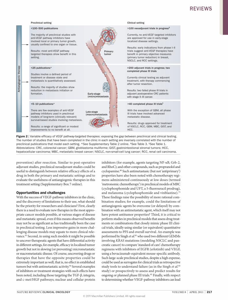
The gap between bedside and benchPerhaps foremost among the challenges in predicting disease- progression patterns and mechanisms of drug resistance to antiangiogenic therapy is a general disconnect between how VEGF-pathway inhibitors (and all anticancer therapies for that matter) are tested in experimental versus clinical settings. In preclinical evaluations, the majority of analyses have been conducted either in genetically engi-neered mouse models (GEMMS) or, more frequently, in locally grown primary ectopic (or orthotopic) tumors using human (xenograft) or mouse (syngenic) models.75 Conversely, most cancer patients receiving VEGF-pathway inhibitors have late-stage (sometimes refractory) disease involving established metastases in more than one site.71 In the preclinical setting, only a negligible fraction of studies have tested VEGF-pathway inhibitors in similar late-stage models and even fewer have compared directly antitumor efficacy in such circumstances to locally grown primary tumors (Figure 2). Also, pre clinical metastasis models are often quantitatively assessed (for example visual nodule counts, immuno histochemistry, and imaging76) and disease is measured at a defined end point—usually when a primary or localized tumor has reached an institutional ethical limit. This means studies are stopped short of overt systemic metastatic disease, and therefore the majority of preclinical studies involving VEGF-pathway inhibi-tors and metastasis have included non-survival-based analyses. These limitations have resulted in relatively few studies that are designed to investigate the impact of VEGF-pathway inhibitors on established metastasis when compared with the hundreds of publications dedicated to localized or primary disease. In addition, there are even fewer studies that include clinically relevant, survival-based evaluations of therapy in models of metastasis, and there is disparity in the tumor models employed and modes of meta stasis quantification used (Figure 2 and Supplementary Table 2 online). These preclinical studies have shown that VEGF-pathway-targeted therapy leads to the inhibition of meta stasis when quantified empirically, either after short treatment periods or when studies are terminated because of primary tumor growth. However, considering that the vast majority of patients receiving similar drugs in the clinic have established metastases, more relevant pre clinical analy ses should be conducted to determine the consequences of this on overall survival. In such rare preclinical studies, the results have been mixed, with some reporting treatment benefit77 and others noting more moderate or negligible effects78,79 (Supplementary Table 2 online). The limitations of these studies are of par-ticular relevance because metastasis is generally the cause of patient mortality,80 and antiangiogenic agents are now
being evaluated in earlier stages of disease, such as the adjuvant setting, which may involve treating early-stage occult micro metastatic disease. Moreover, as the studies by Paez-Ribes et al.6 and Ebos et al.5 show, positive effects in primary tumor models do not always translate into bene-ficial effects in blocking hematogenous micrometastatic-disease progression (the outcomes may even be worse), and comparisons of drug effects in the primary tumor and micrometastasis treatment settings can be very different. Results from similar studies have varied,79,81,82 and thus it is critical when interpreting potential conflicting data sets from preclinical studies using inhibitors of the VEGF pathway to consider variables such as disease stage, the types of drug employed (antibodies versus TKIs), and the models of metastasis that are used.
The need for optimal mouse models to study metastasis has taken on a greater urgency, particularly in the setting of micrometastatic disease. A recent Review covered this topic in detail and listed models that could be employed10 -
—an example is a model of NSCLC where sunitinib pro-longed survival but longer treatments (initiated earlier) did not translate into greater benefit.83,84 This situation empha-sizes the importance of developing models that can clearly distinguish between macroscopic and microscopic disease. In addition, use of models that employ clinically relevant end points such as PFS are promising for improving their predictions of clinical potential.85
Diagnosis or treatment initiation Relapse Death
Overall survival
a
c
b
Improvement in PFS
Improvement in overall survival
Improvement in PFS
No change in overall survival
Worse PFS
(Possible) worse overall survival
Figure 1 | Clinical results of combinations of PFS and overall survival. There are several different combinations of PFS and overall survival, including no change in either (not shown here). a | Improvement in PFS translates into improved overall survival. In completed phase III trials with anti‑VEGF‑pathway therapy (Tables 1 and 2), two additional scenarios have occurred: b | PFS benefit does not translate into improved overall survival, and c | reduced PFS (Table 2). Worse overall survival has not been shown in a phase III trial though a recent interim analysis of the AVANT trial indicated that this trend is possible.24 It is possible that response to anti‑VEGF therapy (even if leading to improved PFS) can change the natural history of disease progression to include a more aggressive phenotype—possibly explaining lack of changes in overall survival. This figure is based on conceptual ideas outlined by David Reardon. Abbreviation: PFS, progression‑free survival.
REVIEWS
© 2011 Macmillan Publishers Limited. All rights reserved

216 | APRIL 2011 | VOLUME 8 www.nature.com/nrclinonc
The perioperative settingPerhaps the area where disease progression after therapy presents the biggest challenge (and the potential to show benefit) is in the perioperative setting, when treatments are administered either before (neoadjuvant) or after (adju-vant) surgery to remove the tumor. With studies under-way in patients with CRC, RCC, NSCLC, breast and central nervous system cancers, it will be critical to determine safety parameters for wound healing and therapy toxicity to optimize guidelines,86 and to determine the efficacy of VEGF-pathway inhibition in these settings.
Adjuvant therapyCurrently, there are over 200 adjuvant clinical trials planned or underway assessing antiangio genesis drugs either alone or in combination with chemotherapy in cancer types including breast, RCC, prostate, head and neck, NSCLC, and ovarian.87 The rationale for therapeutic inter vention with VEGF-pathway inhibitors in the postoperative setting was summarized by Bagri et al.10 who highlighted the advantages of anti angiogenic blockade in preventing occult micro metastatic growth in distant sites. Most obvious is that because of the integral role of the vascu lature in the step-wise process of meta stasis, antiangio genic therapy could compromise some of these steps in primary tumors such
as preventing or delaying intravasation (for example via the destruction of the immature vasculature) and the ‘angiogenic switch’ in avascular metastases at distant sites.10 Recently, two phase III postoperative adjuvant trials (C-08 and AVANT) that assessed bevacizumab in patients with stage II–III CRC were completed. Patients in both trials received either bevaci-zumab for 1 year (in combination with chemo therapy for the first 6 months) or 6 months of chemotherapy alone. The chemo therapy regimen FOLFOX (5-fluorouracil, leuco vorin and oxaliplatin) was compared with bevacizumab in C-08, and FOLFOX or XELOX (oxaliplatin and cape citabine) was compared with bevaci zumab in AVANT. The primary end point of a benefit in DFS after 3 years was not met in either trial, although in both the C-08 and AVANT trials indica tions of DFS improvement was observed following the 6-month bevacizumab maintenance period at the 1-year interim analysis, and at subsequent interim analyses (in C-08 only)—but the extent of the benefit diminished over time in both trials.88,89 The basis of this ‘fading’ effect is unknown, and questions remain as to whether long-term bevacizumab maintenance should be tested in follow-up studies to poten-tially prolong the observed DFS benefits (as was seen in the GOG0218 and ICON7 ovarian cancer trials37). However, it is important to question if DFS bene fits translate into overall survival benefits and, if not, do they justify the associated costs and toxicity of using a drug such as bevacizumab? As well, and perhaps over shadowing such questions, the AVANT trial results demon strated that patients receiving bevacizumab with chemotherapy had numerical increases in disease relapse and death compared with chemotherapy alone.24 Though firm conclusions cannot be made based on early reporting of trial results, and it is possible that patient crossover in the control group may have had a role in these observations (these patients later received bevacizumab), it remains an open question whether bevacizumab was a detriment in this trial—a point raised by the trial organiz-ers.8 Regardless, in both trials, the fact that DFS changed rapidly after bevacizumab was halted requires further study and highlights the importance of undertaking appropriate preclinical studies to examine the mechanisms by which antiangiogenic treatments lose their activity and/or alter tumor progression and metastasis over time, especially in the adjuvant setting (Figure 2).
Neoadjuvant therapyIn neoadjuvant therapy, the theoretical advantages of antiangiogenesis treatment are twofold. First, to elicit an objective reduction in tumor size, usually to downstage an unresectable tumor or improve the impact of surgery of resectable tumors and second, to prevent micro metastatic outgrowth, increasing the potential for PFS and overall sur-vival benefits.90,91 There are over 100 ongoing neo adjuvant trials using VEGF-pathway inhibitors, either alone or in combination with chemotherapy, radiation, or other therapies (Table 3 and Supplementary Box 6 online).87
To date, there are few, if any, preclinical studies that have been conducted with antiangiogenic drugs (or any other drug types) that analyze neoadjuvant or pre surgical treat-ments,81,92 and virtually none that compare treatment effects on primary tumors to metastatic-disease progression (or
Box 3 | Possible mechanisms influencing invasion and metastasis after therapy
Tumor-dependent mechanisms ■ Increased expression of prometastatic proteins: c‑met,69,70 interleukin (IL)‑6,137
IL‑8,138 and urokinase‑type plasminogen activator receptor139
■ Suppression of antimetastatic mediators: myoglobulin140
■ Altered adhesion: upregulation or activation and secretion of exomal proteolytic enzymes, such as matrix metalloproteinases141,142
■ Bone‑marrow‑derived dendritic cell (BMDC) mobilization creates ‘premetastatic niches’143
■ Acute hypoxic stress144–146
■ Instigation of tumor epithelial–mesenchymal transition147
■ Increased vascular co‑optive behavior133
■ Activation of alternative angiogenic pathways: FGF and ephrin148
■ Induction of stromal autophagy149
■ Vascular mimicry or cancer stem cells150,151
Tumor-independent—host-mediated—mechanisms ■ Compensatory upregulation of proangiogenic or prometastatic factors contribute
to ‘rebound’21 and/or increased extravasive potential: VEGF, PlGF, G‑CSF, osteopontin, Bv8 (prokineticin), G‑CSF, angiopoieten2, PDGFA and SDF1α14,152–158
■ BMDC mobilization recruits VEGFR1‑positive bone marrow cells to distant sites to facilitate ‘premetastatic niches’;159,160 this has not been confirmed in all cases92
■ BMDC mobilization of Gr1+CD11b+ myeloid suppressor‑type cells, TIE2 expressing monocytes, and tumor‑associated macrophages to home to the tumor microenvironment and produce compensatory proangiogenic factors14,155–158
■ Pericyte dysfunction increases vessel leakiness and allows for increased extravasive and metastatic tumor potential4,74,161,162
■ Increased prothrombotic events caused by vessel damage as a result of therapy allows for increased tumor cell ‘seeding’ and growth in distant organs163
■ Altered endothelial cell adhesion molecule function may enhance VEGF‑driven angiogenesis and tumor growth164
■ Inflammatory pathway activation alters the endothelial microenvironment increasing intravasive and extravasive potential of tumor cells72
REVIEWS
© 2011 Macmillan Publishers Limited. All rights reserved
NATURE REVIEWS | CLINICAL ONCOLOGY VOLUME 8 | APRIL 2011 | 217
prevention) after resection. Similar to post- operative adjuvant studies, preclinical neoadjuvant studies could be useful to distinguish between relative efficacy effects of a drug in both the primary and metastatic settings and to evaluate the usefulness of antiangiogenic therapies in this treatment setting (Supplementary Box 7 online).
Opportunities and challengesWith the success of VEGF-pathway inhibitors in the clinic, and the discovery of limitations to their use, what should be the priority for researchers and clinicians? First, clearly there is a need to evaluate new therapies in the most appro-priate cancer models possible, at various stages of disease and metastatic spread, even if this means observed bene fits may not be as significant as has traditionally been the case in preclinical testing. Less impressive gains in more chal-lenging disease models may equate to more clinical rele-vance.93 Second, in using such models it might be possible to uncover therapeutic agents that have differential acti vity in different settings, for example, efficacy in localized tumor growth but not in slowing (or preventing) micro metastatic or macrometastatic disease. Of course, uncovering drugs or therapies that have the opposite properties could be extremely important as well, that is, no effect in established tumors but with antimetastatic acti vity.94 Several examples of inhibitors or treatment strategies with such effects have been noted, including those targeting the TGF-β, integrin, and c-met/HGF pathways; nuclear and cellular protein
inhibitors (for example, agents targeting NF-κB, Grb-2, and RhoC); and other compounds, such as propranolol and cyclopamine.80 Such anti metastasic (but not ‘anti primary’) properties have also been noted with chemotherapy regi-mens administered continuously at low doses (termed ‘metronomic chemotherapy’) in preclinical models of MBC (cyclophosphomide and UFT, a 5- fluorouracil prodrug), and melanoma (cyclo phosphomide and vin blastine).75 These findings raise the possibility of more rational com-bination studies; for example, could the limitations of antiangiogenic agents be overcome (or delayed) by com-bination with an anti metastatic agent, which itself may not have potent anti tumor properties? Third, it is critical to perform studies in preclinical models that assess drug treat-ments or combinations that closely mimic phase III clini-cal trials, ideally using similar (or equivalent) quantitative assessments to PFS and overall survival. An example was performed by Singh et al.85 who used two different GEMMs involving KRAS mutations (modeling NSCLC and pan-creatic cancer) to compare ‘standard of care’ chemo therapy regimens with inhibitors of EGFR (erlotinib) and VEGF, using a bevacizumab- equivalent mouse-specific antibody. Such large-scale preclinical studies, despite a high expense, could be used as surrogates for clinical trials as retro spective study tools to understand failure (as in the Singh et al.85 study) or prospectively to assess and predict results for ongoing or planned phase III trials.95 Finally, with respect to determining whether VEGF-pathway inhibitors can lead
<100–500 publications
The majority of preclinical studies with anti-VEGF pathway inhibitors have involved local or primary tumor growth, usually con�ned to one organ or tissue.
Results: most anti-VEGF pathway targeted therapies show bene�t in this setting.
>100 neoadjuvant trials in progress‡
Currently, no anti-VEGF targeted inhibitors are approved for use in early-stage localized disease settings.
Results: early indications from phase I–II trials suggest anti-VEGF therapies have bene�t in primary objective measures (primary tumor reduction) in breast, NSCLC, and RCC settings.
<25 publications*
Studies involve a de�ned period of treatment or disease state and metastasis is quantitatively assessed.
Results: the majority of studies show reduction in metastasis initiation or formation.
>200 adjuvant trials in progress; two completed phase III trials
Currently clinical testing as adjuvant treatment, with therapy commencing after tumor resection.
Results: two failed phase III trials in adjuvant postoperative CRC patients with stage II–III cancer.
<5–10 publications*
There are few examples of anti-VEGF pathway inhibitors used in preclinical models of long-term (clinically relevant) survival-based studies involving metastasis.
Results: a range of signi�cant or modest improvements to no bene�t at all.
>40 completed phase III trials§
With the exception of GBM, all phase III trials have involved advanced metastatic disease.
Results: drugs approved for treatment of NSCLC, RCC, GBM, MBC, GIST, and HCC.
Primarytumor
Early-stage(micro)metastasis
Late-stage(macro)metastasis
Ant
iang
ioge
nic
ther
apy
Preclinical setting Clinical setting
Figure 2 | Variable efficacy of VEGF pathway‑targeted therapies: exposing the gap between preclinical and clinical testing. The number of studies that have been completed in the clinic in each setting are inversely correlated with the number of preclinical publications that model each setting. *See Supplementary Table 2 online. ‡See Table 3. §See Table 1. Abbreviations: CRC, colorectal cancer; GBM, glioblastoma muliforme; GIST, gastrointestional stromal tumors; HCC, hepatocellular carcinoma; MBC, metastatic breast cancer; NSCLC, non‑small‑cell lung cancer; RCC, renal cell carcinoma.
REVIEWS
© 2011 Macmillan Publishers Limited. All rights reserved

218 | APRIL 2011 | VOLUME 8 www.nature.com/nrclinonc
to more invasive and metastatic phenotypes (despite initial positive effects in terms of tumor shrinkage, PFS or overall survival bene fits), it will be critical to properly assess (and compare) different modes of inhibition (antibodies versus TKIs) in different disease stages (localized verses micro-metastasis and macro metastasis) to understand disease progression on (or off) therapy. This will be essential for understanding the basis of any future rationale for extended treatments in patients, such as the initial benefits seen in certain settings where treatment extends beyond disease progression. In addition, the differences between PFS and overall survival benefits in clinical trials investigating anti-angiogenic drugs could have important implications for the future use of VEGF-pathway-targeted agents. For example, in 2010, the FDA rejected full approval of bevacizumab with chemo therapy in patients with MBC based on toxicities and the lack of overall survival benefits and diminishing PFS bene fits in the AVADO and RIBBON-1 trials compared to the earlier E2100 trial (Tables 1 and 2 and Box 3).
ConclusionsWhile efficacies of VEGF-pathway-targeted therapies in certain cancer settings represent a conceptual and
practical medical success, the lack of substantial benefits for the vast majority of patients in terms of increased long-term overall survival times remains an ongoing challenge. Understanding the basis of these treatment limitations will likely be key to devising improved strategies and to overcome the possible difficulties facing further develop-ment of antiangiogenic therapies used at all stages of tumor progression.
Table 3 | VEGF pathway‑targeted drugs currently in neoadjuvant clinical trials
Search criteria* Cancer Number of trials (drugs used)
Total Mono-therapy‡
With MTT With hormone therapy
With chemotherapy§ With radiation
Neoadjuvant and sunitinib
Breast, renal, bladder, soft‑tissue sarcoma, GIST, prostate
17 10 – 2 (exemestane, LHRH agonist)
4 (TCARB, gemcitabine, cisplatin, docetaxel)
2
Neoadjuvant and sorafenib
Breast, renal, soft‑tissue sarcoma, prostate, rectal, SCCHN
12 5 – 1 (letrozole) 6 (cisplatin, capecitabine, IE, ifosfamide, ixabepilone, LDM CTX)
5
Neoadjuvant and pazopanib
Breast, NSCLC 3 3 – – 1 (docetaxel) –
Neoadjuvant and zactima
Breast, NSCLC, esophageal 3 2 – 1 (anastrozole) 1 (docetaxel and carboplatin combination)
1
Neoadjuvant and cediranib
Breast 1 – – – 1 (docetaxel, doxorubicin and cyclophosphamide combination)
–
Neoadjuvant and semaxinib
Soft‑tissue sarcoma 1 – – – 1 (doxorubicin, ifosfamide and dacarbazine combination)
1
Neoadjuvant and bevacizumab
Breast, renal, bladder, soft‑tissue sarcoma, prostate, esophageal, cervical, colorectal, urothelial, rectal, NSCLC, glioblastoma, pancreatic, ovarian, uveal melanoma, gastric or adrenal
63 2 3 (trastuzumab, cetuximab)
4 (letrozole, AI) 65 (docetaxel, carboplatin, capecitabine, gemcitabine, irinotecan, cisplatin, 5‑FU, XELOX, FOLFOX, FOLFOXIRI, TC, M‑VAC, TAC, TCARB, DC, AC, FEC, IC, DG, OCFL‑BC, DECNC, ECX)
10
*Searched in www.clinicaltrials.gov, only active or completed studies (recruiting or non‑recruiting) included. Studies include local (primary) disease and metastasis deemed surgically operable. If perioperative therapy included separate regimens, only the VEGF‑pathway inhibitor was included in the table. In all studies overall survival is a secondary end point. All searches filtered for ‘intervention’ trials only. ‡At least one study arm testing drug alone. §Docetaxol and paclitaxol used interchangeably unless otherwise indicated. Abbreviations: 5‑FU, 5‑fluorouracil; AC, doxorubicin and cyclophosphamide; AI, undefined aromatase inhibitor; DC, docetaxel and capecitabine; DECNC, docetaxel, epirubicin, cyclophosphamide, navelbine and capecitabine; DG, docetaxol and gemcitabine; ECX, epirubicin, cisplatin and capecitabine; FEC, 5‑FU, epirubicin and cyclophosphamide; FOLFOX, 5‑FU, leucovorin and oxaliplatin; FOLFOXIRI, 5‑FU, leucovorin, oxaliplatin and irinotecan; GIST, gastrointestinal stromal tumors; IC, irinotecan and cisplatin; IE, epirubicin and ifosfamide; LDM CTX, low‑dose metronomic cyclophosphamide; LHRH, luteinizing hormone‑releasing hormone; MTT, molecular‑targeted therapy; M‑VAC, methotrexate, vinblastine, adriamycin and cisplatin; NSCLC, non‑small‑cell lung cancer; OCFL‑BC, oxaliplatin‑CPT‑11, 5‑FU, leucovorin, bevacizumab and cetuximab; SCCHN, squamous cell carcinomas of the head and neck; TC, docetaxel and cyclophosphamide; XELOX, capecitabine and oxaliplatin.
Review criteria
Data in this Review were compiled from databases searched before 1 February 2011, including PubMed and MEDLINE; conference proceedings (AACR, ASCO, and others); company website and trial information releases; and online search engines for press releases of failed or terminated trials. Information on phase III trials and drug approvals was from NIH databases (www.clinicaltrials.gov, www.cancer.gov/clinicaltrials) and the FDA website (www.fda.gov). Searches used every drug name in this article, as well as combinations of biological and disease‑stage terms such as ‘VEGF‑ or VEGFR‑inhibitors’, ‘antiangiogenic therapy’, ‘metastasis’, ‘adjuvant’, ‘neoadjuvant’, ‘perioperative’.
1. US Department of Health and Human Services FDA US Food and Drug Administration [online], www.fda.gov (2011).
2. Folkman, J. Tumor angiogenesis: therapeutic implications. N. Engl. J. Med. 285, 1182–1186 (1971).
3. Abdollahi, A. & Folkman, J. Evading tumor evasion: current concepts and perspectives of anti‑angiogenic cancer therapy. Drug Resist. Updat. 13, 16–28 (2010).
4. Bergers, G. & Hanahan, D. Modes of resistance to anti‑angiogenic therapy. Nat. Rev. Cancer 8, 592–603 (2008).
5. Ebos, J. M. et al. Accelerated metastasis after short‑ term treatment with a potent inhibitor of tumor angiogenesis. Cancer Cell 15, 232–239 (2009).
6. Pàez‑Ribes, M. et al. Antiangiogenic therapy elicits malignant progression of tumors to increased local invasion and distant
metastasis. Cancer Cell 15, 220–231 (2009).
7. Allegra, C. J. et al. Phase III trial assessing bevacizumab in stages II and III carcinoma of the colon: results of NSABP protocol C‑08. J. Clin. Oncol. 29, 11–16 (2011).
8. de Gramont, A. et al. AVANT: Results from a randomized, three‑arm multinational phase III study to investigate bevacizumab with either XELOX or FOLFOX4 versus FOLFOX4 alone as
REVIEWS
© 2011 Macmillan Publishers Limited. All rights reserved

NATURE REVIEWS | CLINICAL ONCOLOGY VOLUME 8 | APRIL 2011 | 219
adjuvant treatment for colon cancer [abstract]. J. Clin. Oncol. 29 (Suppl. 4), a362 (2011).
9. Miles, D. et al. Disease course patterns after discontinuation of bevacizumab: pooled analysis of randomized phase III trials. J. Clin. Oncol. 29, 83–88 (2011).
10. Bagri, A., Kouros‑Mehr, H., Leong, K. G. & Plowman, G. D. Use of anti‑VEGF adjuvant therapy in cancer: challenges and rationale. Trends Mol. Med. 16, 122–132 (2010).
11. Heath, V. L. & Bicknell, R. Anticancer strategies involving the vasculature. Nat. Rev. Clin. Oncol. 6, 395–404 (2009).
12. Boere, I. A., Hamberg, P. & Sleijfer, S. It takes two to tango: combinations of conventional cytotoxics with compounds targeting the vascular endothelial growth factor‑vascular endothelial growth factor receptor pathway in patients with solid malignancies. Cancer Sci. 101, 7–15 (2010).
13. Ivy, S. P., Wick, J. Y. & Kaufman, B. M. An overview of small‑molecule inhibitors of VEGFR signaling. Nat. Rev. Clin. Oncol. 6, 569–579 (2009).
14. Kerbel, R. S. Tumor angiogenesis. N. Engl. J. Med. 358, 2039–2049 (2008).
15. Saltz, L. B. et al. Bevacizumab in combination with oxaliplatin‑based chemotherapy as first‑line therapy in metastatic colorectal cancer: a randomized phase III study. J. Clin. Oncol. 26, 2013–2019 (2008).
16. Wilkerson, J. & Fojo, T. Progression‑free survival is simply a measure of a drug’s effect while administered and is not a surrogate for overall survival. Cancer J. 15, 379–385 (2009).
17. Wolter, P., Beuselinck, B., Pans, S. & Schöffski, P. Flare‑up: an often unreported phenomenon nevertheless familiar to oncologists prescribing tyrosine kinase inhibitors. Acta Oncol. 48, 621–624 (2009).
18. Desar, I. M. et al. The reverse side of the victory: flare up of symptoms after discontinuation of sunitinib or sorafenib in renal cell cancer patients. A report of three cases. Acta Oncol. 48, 927–931 (2009).
19. Cacheux, W. et al. Reversible tumor growth acceleration following bevacizumab interruption in metastatic colorectal cancer patients scheduled for surgery. Ann. Oncol. 19, 1659–1661 (2008).
20. Stein, W. D., Yang, J., Bates, S. E. & Fojo, T. Bevacizumab reduces the growth rate constants of renal carcinomas: a novel algorithm suggests early discontinuation of bevacizumab resulted in a lack of survival advantage. Oncologist 13, 1055–1062 (2008).
21. Mancuso, M. R. et al. Rapid vascular regrowth in tumors after reversal of VEGF inhibition. J. Clin. Invest. 116, 2610–2621 (2006).
22. Levashova, Z. et al. Molecular imaging of changes in the prevalence of vascular endothelial growth factor receptor in sunitinib‑treated murine mammary tumors. J. Nucl. Med. 51, 959–966 (2010).
23. Nagengast, W. B. et al. VEGF‑PET imaging is a noninvasive biomarker showing differential changes in the tumor during sunitinib treatment. Cancer Res. 71, 143–153 (2011).
24. di Tomaso, E. et al. Glioblastoma recurrence after cediranib therapy in patients: lack of “rebound” revascularization as mode of escape. Cancer Res. 71, 19–28 (2011).
25. La Vine, D. B., Coleman, T. A., Davis, C. H., Carbonell, C. E. & Davis, W. B. Frequent dose interruptions are required for patients receiving oral kinase inhibitor therapy for advanced renal cell carcinoma. Am. J. Clin. Oncol. 33, 217–220 (2010).
26. Gore, M. E. et al. Safety and efficacy of sunitinib for metastatic renal‑cell carcinoma: an
expanded‑access trial. Lancet Oncol. 10, 757–763 (2009).
27. van der Veldt, A. A. et al. Predictive factors for severe toxicity of sunitinib in unselected patients with advanced renal cell cancer. Br. J. Cancer 99, 259–265 (2008).
28. Riechelmann, R. P. et al. Sorafenib for metastatic renal cancer: the Princess Margaret experience. Am. J. Clin. Oncol. 31, 182–187 (2008).
29. Yoo, C. et al. The efficacy and safety of sunitinib in Korean patients with advanced renal cell carcinoma: high incidence of toxicity leads to frequent dose reduction. Jpn. J. Clin. Oncol. 40, 980–985 (2010).
30. Johannsen, M. et al. Can tyrosine kinase inhibitors be discontinued in patients with metastatic renal cell carcinoma and a complete response to treatment? A multicentre, retrospective analysis. Eur. Urol. 55, 1430–1438 (2009).
31. Sablin, M. P. et al. Sequential sorafenib and sunitinib for renal cell carcinoma. J. Urol. 182, 29–34 (2009).
32. Dudek, A. Z., Zolnierek, J., Dham, A., Lindgren, B. R. & Szczylik, C. Sequential therapy with sorafenib and sunitinib in renal cell carcinoma. Cancer 115, 61–67 (2009).
33. Tamaskar, I. et al. Antitumor effects of sunitinib or sorafenib in patients with metastatic renal cell carcinoma who received prior antiangiogenic therapy. J. Urol. 179, 81–86 (2008).
34. Hammers, H. J. et al. Reversible epithelial to mesenchymal transition and acquired resistance to sunitinib in patients with renal cell carcinoma: evidence from a xenograft study. Mol. Cancer Ther. 9, 1525–1535 (2010).
35. Ellis, L. M. & Haller, D. G. Bevacizumab beyond progression: does this make sense? J. Clin. Oncol. 26, 5313–5315 (2008).
36. Cohn, A. L. et al. Clinical outcomes in bevacizumab (BV)‑treated patients (pts) with metastatic colorectal cancer (mCRC): Results from ARIES observational cohort study (OCS) and confirmation of BRiTE data on BV beyond progression (BBP) [abstract]. J. Clin. Oncol. 28 (Suppl.), a3596 (2010).
37. Burger, R. A. et al. Phase III trial of bevacizumab (BEV) in the primary treatment of advanced epithelial ovarian cancer (EOC), primary peritoneal cancer (PPC), or fallopian tube cancer (FTC): A Gynecologic Oncology Group study [abstract]. J. Clin. Oncol. 28 (Suppl.), aLBA1 (2010).
38. Perren, T. et al. ICON7: A phase III Gynaecologic Cancer InterGroup (GCIG) trial of adding bevacizumab to standard chemotherapy in women with newly diagnosed epithelial ovarian, primary peritoneal or fallopian tube cancer [abstract]. Ann. Oncol. 21 (Suppl. 8), aLBA3 (2010).
39. Ebos, J. M., Lee, C. R. & Kerbel, R. S. Tumor and host‑mediated pathways of resistance and disease progression in response to antiangiogenic therapy. Clin. Cancer Res. 15, 5020–5025 (2009).
40. Norden, A. D. et al. Bevacizumab for recurrent malignant gliomas: efficacy, toxicity, and patterns of recurrence. Neurology 70, 779–787 (2008).
41. Narayana, A. et al. Antiangiogenic therapy using bevacizumab in recurrent high‑grade glioma: impact on local control and patient survival. J. Neurosurg. 110, 173–180 (2009).
42. Fischer, I. et al. High‑grade glioma before and after treatment with radiation and Avastin: initial observations. Neuro. Oncol. 10, 700–708 (2008).
43. Ellis, L. M. & Reardon, D. A. Cancer: The nuances of therapy. Nature 458, 290–292 (2009).
44. Mathews, M. S., Linskey, M. E., Hasso, A. N. & Fruehauf, J. P. The effect of bevacizumab (Avastin) on neuroimaging of brain metastases. Surg. Neurol. 70, 649–652 (2008).
45. Zuniga, R. M. et al. Efficacy, safety and patterns of response and recurrence in patients with recurrent high‑grade gliomas treated with bevacizumab plus irinotecan. J. Neurooncol. 91, 329–336 (2009).
46. Iwamoto, F. M. et al. Patterns of relapse and prognosis after bevacizumab failure in recurrent glioblastoma. Neurology 73, 1200–1206 (2009).
47. Zuniga, R. M. et al. Rebound tumour progression after the cessation of bevacizumab therapy in patients with recurrent high‑grade glioma. J. Neurooncol. 99, 237–242 (2010).
48. Verhoeff, J. J. et al. Concerns about anti‑angiogenic treatment in patients with glioblastoma multiforme. BMC Cancer 9, 444 (2009).
49. Narayana, A. et al. Bevacizumab in recurrent high‑grade pediatric gliomas. Neuro. Oncol. 12, 985–990 (2010).
50. Narayana, A. et al. Change in pattern of relapse after antiangiogenic therapy in high‑grade glioma. Int. J. Radiat. Oncol. Biol. Phys. doi:10.1016/ j.ijrobp.2010.10.038.
51. Rubenstein, J. L. et al. Anti‑VEGF antibody treatment of glioblastoma prolongs survival but results in increased vascular cooption. Neoplasia 2, 306–314 (2000).
52. Kunkel, P. et al. Inhibition of glioma angiogenesis and growth in vivo by systemic treatment with a monoclonal antibody against vascular endothelial growth factor receptor‑2. Cancer Res. 61, 6624–6628 (2001).
53. Blouw, B. et al. The hypoxic response of tumors is dependent on their microenvironment. Cancer Cell 4, 133–146 (2003).
54. Verhoeff, J. J. et al. Tumour control by whole brain irradiation of anti‑VEGF‑treated mice bearing intracerebral glioma. Eur. J. Cancer 45, 3074–3080 (2009).
55. Lucio‑Eterovic, A. K., Piao, Y. & de Groot, J. F. Mediators of glioblastoma resistance and invasion during antivascular endothelial growth factor therapy. Clin. Cancer Res. 15, 4589–4599 (2009).
56. Gomez‑Manzano, C. et al. VEGF Trap induces antiglioma effect at different stages of disease. Neuro. Oncol. 10, 940–945 (2008).
57. de Groot, J. F. et al. Tumor invasion after treatment of glioblastoma with bevacizumab: radiographic and pathologic correlation in humans and mice. Neuro. Oncol. 12, 233–242 (2010).
58. Lamszus, K. et al. Inhibition of glioblastoma angiogenesis and invasion by combined treatments directed against vascular endothelial growth factor receptor‑2, epidermal growth factor receptor, and vascular endothelial‑cadherin. Clin. Cancer Res. 11, 4934–4940 (2005).
59. Leenders, W. P. et al. Antiangiogenic therapy of cerebral melanoma metastases results in sustained tumor progression via vessel co‑option. Clin. Cancer Res. 10, 6222–6230 (2004).
60. Chamberlain, M. C. Radiographic patterns of relapse in glioblastoma. J. Neurooncol. 101, 319–323 (2011).
61. Petrelli, F., Cabiddu, M., Carpo, M., Ghilardi, M. & Barni, S. Progression of intramedullary metastasis during perioperative cessation of sunitinib. Nat. Rev. Urol. 7, 634–637 (2010).
62. Plimack, E. R., Tannir, N., Lin, E., Bekele, B. N. & Jonasch, E. Patterns of disease progression in metastatic renal cell carcinoma patients treated with antivascular agents and interferon: impact of therapy on recurrence patterns and outcome measures. Cancer 115, 1859–1866 (2009).
REVIEWS
© 2011 Macmillan Publishers Limited. All rights reserved
220 | APRIL 2011 | VOLUME 8 www.nature.com/nrclinonc
63. Crawford, Y. & Ferrara, N. Tumor and stromal pathways mediating refractoriness/resistance to anti‑angiogenic therapies. Trends Pharmacol. Sci. 30, 624–630 (2009).
64. Nguyen, D. X., Bos, P. D. & Massagué, J. Metastasis: from dissemination to organ‑specific colonization. Nat. Rev. Cancer 9, 274–284 (2009).
65. Paget, S. The distribution of secondary growths in cancer of the breast. Lancet 133, 571–573 (1889).
66. Hirota, K. & Semenza, G. L. Regulation of angiogenesis by hypoxia‑inducible factor 1. Crit. Rev. Oncol. Hematol. 59, 15–26 (2006).
67. Yu, J. L., Rak, J. W., Coomber, B. L., Hicklin, D. J. & Kerbel, R. S. Effect of p53 status on tumor response to antiangiogenic therapy. Science 295, 1526–1528 (2002).
68. Rapisarda, A. & Melillo, G. Role of the hypoxic tumor microenvironment in the resistance to anti‑angiogenic therapies. Drug Resist. Updat. 12, 74–80 (2009).
69. Pennacchietti, S. et al. Hypoxia promotes invasive growth by transcriptional activation of the met protooncogene. Cancer Cell 3, 347–361 (2003).
70. Steeg, P. S. Angiogenesis inhibitors: motivators of metastasis? Nat. Med. 9, 822–823 (2003).
71. Blagosklonny, M. V. Antiangiogenic therapy and tumor progression. Cancer Cell 5, 13–17 (2004).
72. Bidard, F. C., Pierga, J. Y., Vincent‑Salomon, A. & Poupon, M. F. A “class action” against the microenvironment: do cancer cells cooperate in metastasis? Cancer Metastasis Rev. 27, 5–10 (2008).
73. Solinas, G., Marchesi, F., Garlanda, C., Mantovani, A. & Allavena, P. Inflammation‑mediated promotion of invasion and metastasis. Cancer Metastasis Rev. 29, 243–248 (2010).
74. Xian, X. et al. Pericytes limit tumor cell metastasis. J. Clin. Invest. 116, 642–651 (2006).
75. Francia, G., Cruz‑Munoz, W., Man, S., Xu, P. & Kerbel, R. S. Mouse models of advanced spontaneous metastasis for experimental therapeutics. Nat. Rev. Cancer 11, 135–141 (2011).
76. Bos, P. D., Nguyen, D. X. & Massagué, J. Modeling metastasis in the mouse. Curr. Opin. Pharmacol. 10, 571–577 (2010).
77. Hu‑Lowe, D. D. et al. Nonclinical antiangiogenesis and antitumor activities of axitinib (AG‑013736), an oral, potent, and selective inhibitor of vascular endothelial growth factor receptor tyrosine kinases 1, 2, 3. Clin. Cancer Res. 14, 7272–7283 (2008).
78. Schomber, T. et al. Differential effects of the vascular endothelial growth factor receptor inhibitor PTK787/ZK222584 on tumor angiogenesis and tumor lymphangiogenesis. Mol. Cancer Ther. 8, 55–63 (2009).
79. Yin, J. J., Zhang, L., Munasinghe, J., Linnoila, R. I. & Kelly, K. Cediranib/AZD2171 inhibits bone and brain metastasis in a preclinical model of advanced prostate cancer. Cancer Res. 70, 8662–8673 (2010).
80. Steeg, P. S. & Theodorescu, D. Metastasis: a therapeutic target for cancer. Nat. Clin. Pract. Oncol. 5, 206–219 (2008).
81. Padera, T. P. et al. Differential response of primary tumor versus lymphatic metastasis to VEGFR‑2 and VEGFR‑3 kinase inhibitors cediranib and vandetanib. Mol. Cancer Ther. 7, 2272–2279 (2008).
82. Lee, Y. J. et al. Differential effects of VEGFR‑1 and VEGFR‑2 inhibition on tumor metastases based on host organ environment. Cancer Res. 70, 8357–8367 (2010).
83. Gandhi, L. et al. Sunitinib prolongs survival in genetically engineered mouse models of multistep lung carcinogenesis. Cancer Prev. Res. (Phila) 2, 330–337 (2009).
84. Grandis, J. R. & Argiris, A. Targeting angiogenesis from premalignancy to metastases. Cancer Prev. Res. (Phila) 2, 291–294 (2009).
85. Singh, M. et al. Assessing therapeutic responses in Kras mutant cancers using genetically engineered mouse models. Nat. Biotechnol. 28, 585–593 (2010).
86. Bose, D. et al. Vascular endothelial growth factor targeted therapy in the perioperative setting: implications for patient care. Lancet Oncol. 11, 373–382 (2010).
87. US National Institutes of Health Clinical Trials [online], www.clinicaltrials.gov (2011).
88. Allegra, C. J. et al. Initial safety report of NSABP C‑08: A randomized phase III study of modified FOLFOX6 with or without bevacizumab for the adjuvant treatment of patients with stage II or III colon cancer. J. Clin. Oncol. 27, 3385–3390 (2009).
89. Wolmark, N. et al. A phase III trial comparing mFOLFOX6 to mFOLFOX6 plus bevacizumab in stage II or III carcinoma of the colon: Results of NSABP Protocol C‑08 [abstract]. J. Clin. Oncol. 27 (Suppl.), aLBA4 (2009).
90. Kinoshita, T. et al. Preoperative induction with sorafenib pathologically downstaged advanced renal cell carcinoma: a case report. Int. J. Urol. 17, 286–288 (2010).
91. Altorki, N. et al. Phase II proof‑of‑concept study of pazopanib monotherapy in treatment‑naive patients with stage I/II resectable non‑small‑cell lung cancer. J. Clin. Oncol. 28, 3131–3137 (2010).
92. Dawson, M. R., Duda, D. G., Chae, S. S., Fukumura, D. & Jain, R. K. VEGFR1 activity modulates myeloid cell infiltration in growing lung metastases but is not required for spontaneous metastasis formation. PLoS ONE 4, e6525 (2009).
93. Steeg, P. S. et al. Preclinical drug development must consider the impact on metastasis. Clin. Cancer Res. 15, 4529–4530 (2009).
94. Mack, G. S. & Marshall, A. Lost in migration. Nat. Biotechnol. 28, 214–229 (2010).
95. Francia, G. & Kerbel, R. S. Raising the bar for cancer therapy models. Nat. Biotechnol. 28, 561–562 (2010).
96. Hurwitz, H. et al. Bevacizumab plus irinotecan, fluorouracil, and leucovorin for metastatic colorectal cancer. N. Engl. J. Med. 350, 2335–2342 (2004).
97. Giantonio, B. J. et al. Bevacizumab in combination with oxaliplatin, fluorouracil, and leucovorin (FOLFOX4) for previously treated metastatic colorectal cancer: results from the Eastern Cooperative Oncology Group Study E3200. J. Clin. Oncol. 25, 1539–1544 (2007).
98. Miller, K. et al. Paclitaxel plus bevacizumab versus paclitaxel alone for metastatic breast cancer. N. Engl. J. Med. 357, 2666–2676 (2007).
99. Miles, D. W. et al. Phase III study of bevacizumab plus docetaxel compared with placebo plus docetaxel for the first‑line treatment of human epidermal growth factor receptor 2‑negative metastatic breast cancer. J. Clin. Oncol. 28, 3239–3247 (2010).
100. Robert, N. J. et al. RIBBON‑1: Randomized, double‑blind, placebo‑controlled, phase III trial of chemotherapy with or without bevacizumab (B) for first‑line treatment of HER2‑negative locally recurrent or metastatic breast cancer (MBC) [abstract]. J. Clin. Oncol. 27 (Suppl.), a1005 (2009).
101. Brufsky, A. et al. Progression‑free survival (PFS) in patient subgroups in RIBBON‑2, a phase III trial of chemotherapy (chemo) plus or minus bevacizumab (BV) for second‑line treatment of HER2‑negative, locally recurrent or metastatic breast cancer (MBC) [abstract]. J. Clin. Oncol. 28 (Suppl.), a1021 (2010).
102. Sandler, A. et al. Paclitaxel‑carboplatin alone or with bevacizumab for non‑small‑cell lung cancer. N. Engl. J. Med. 355, 2542–2550 (2006).
103. Reck, M. et al. Overall survival with cisplatin‑gemcitabine and bevacizumab or placebo as first‑line therapy for nonsquamous non‑small‑cell lung cancer: results from a randomised phase III trial (AVAiL). Ann. Oncol. 21, 1804–1809 (2010).
104. Kabbinavar, F. F., Miller, V. A., Johnson, B. E., O’Connor, P. G. & Soh, C. Overall survival (OS) in ATLAS, a phase IIIb trial comparing bevacizumab (B) therapy with or without erlotinib (E) after completion of chemotherapy (chemo) with B for first‑line treatment of locally advanced, recurrent, or metastatic non‑small cell lung cancer (NSCLC) [abstract]. J. Clin. Oncol. 28 (Suppl.), a7526 (2010).
105. Escudier, B. et al. Phase III trial of bevacizumab plus interferon alfa‑2a in patients with metastatic renal cell carcinoma (AVOREN): final analysis of overall survival. J. Clin. Oncol. 28, 2144–2150 (2010).
106. Rini, B. I. et al. Phase III trial of bevacizumab plus interferon alfa versus interferon alfa monotherapy in patients with metastatic renal cell carcinoma: final results of CALGB 90206. J. Clin. Oncol. 28, 2137–2143 (2010).
107. Cohen, M. H., Shen, Y. L., Keegan, P. & Pazdur, R. FDA drug approval summary: bevacizumab (Avastin) as treatment of recurrent glioblastoma multiforme. Oncologist 14, 1131–1138 (2009).
108. Motzer, R. J. et al. Overall survival and updated results for sunitinib compared with interferon alfa in patients with metastatic renal cell carcinoma. J. Clin. Oncol. 27, 3584–3590 (2009).
109. Goodman, V. L. et al. Approval summary: sunitinib for the treatment of imatinib refractory or intolerant gastrointestinal stromal tumors and advanced renal cell carcinoma. Clin. Cancer Res. 13, 1367–1373 (2007).
110. Raymond, E. et al. Sunitinib malate for the treatment of pancreatic neuroendocrine tumors. N. Engl. J. Med. 364, 501–513 (2011).
111. Escudier, B. et al. Sorafenib for treatment of renal cell carcinoma: final efficacy and safety results of the phase III treatment approaches in renal cancer global evaluation trial. J. Clin. Oncol. 27, 3312–3218 (2009).
112. Llovet, J. M. et al. Sorafenib in advanced hepatocellular carcinoma. N. Engl. J. Med. 359, 378–390 (2008).
113. Sternberg, C. N. et al. Pazopanib in locally advanced or metastatic renal cell carcinoma: results of a randomized phase III trial. J. Clin. Oncol. 28, 1061–1068 (2010).
114. Herbst, R. S. et al. Vandetanib plus docetaxel versus docetaxel as second‑line treatment for patients with advanced non‑small‑cell lung cancer (ZODIAC): a double‑blind, randomised, phase 3 trial. Lancet Oncol. 11, 619–626 (2010).
115. Hecht, J. R. et al. A randomized phase IIIB trial of chemotherapy, bevacizumab, and panitumumab compared with chemotherapy and bevacizumab alone for metastatic colorectal cancer. J. Clin. Oncol. 27, 672–680 (2009).
116. Tol, J. et al. Chemotherapy, bevacizumab, and cetuximab in metastatic colorectal cancer. N. Engl. J. Med. 360, 563–572 (2009).
REVIEWS
© 2011 Macmillan Publishers Limited. All rights reserved

NATURE REVIEWS | CLINICAL ONCOLOGY VOLUME 8 | APRIL 2011 | 221
117. Miller, K. D. et al. Randomized phase III trial of capecitabine compared with bevacizumab plus capecitabine in patients with previously treated metastatic breast cancer. J. Clin. Oncol. 23, 792–799 (2005).
118. Jones, D. Avastin‑Tarceva combination fails in lung cancer. Nat. Biotechnol. 27, 108–109 (2009).
119. Kang, Y. et al. AVAGAST: A randomized double‑blind, placebo‑controlled, phase III study of first‑line capecitabine and cisplatin plus bevacizumab or placebo in patients with advanced gastric cancer (AGC) [abstract]. J. Clin. Oncol. 28 (Suppl.), aLBA4007 (2010).
120. Kindler, H. L. et al. Gemcitabine plus bevacizumab compared with gemcitabine plus placebo in patients with advanced pancreatic cancer: phase III trial of the cancer and Leukemia Group B (CALGB 80303). J. Clin. Oncol. 28, 3617–3622 (2010).
121. Van Cutsem, E. et al. Phase III trial of bevacizumab in combination with gemcitabine and erlotinib in patients with metastatic pancreatic cancer. J. Clin. Oncol. 27, 2231–2237 (2009).
122. Kelly, W. K. et al. A randomized, double‑blind, placebo‑controlled phase III trial comparing docetaxol, prednisone, and placebo with docetaxel, prednisone, and bevacizumab in men with metastatic castration‑resistant prostate cancer (mCRPC): Survival results of CALB 90401 [abstract]. J. Clin. Oncol. 28 (Suppl.), aLBA4511 (2010).
123. Crown, J. et al. Phase III trial of sunitinib (SU) in combination with capecitabine (C) versus C in previously treated advanced breast cancer (ABC) [abstract]. J. Clin. Oncol. 28 (Suppl.), aLBA1011 (2010).
124. Barrios, C. H. et al. Phase III randomized trial of sunitinib versus capecitabine in patients with previously treated HER2‑negative advanced breast cancer. Breast Cancer Res. Treat. 121, 121–131 (2010).
125. Scagliotti, G. et al. Phase III study of carboplatin and paclitaxel alone or with sorafenib in advanced non‑small‑cell lung cancer. J. Clin. Oncol. 28, 1835–1842 (2010).
126. Kohne, C. et al. Final results of CONFIRM 2: A multinational, randomized, double‑blind, phase III study in 2nd line patients (pts) with metastatic colorectal cancer (mCRC) receiving FOLFOX4 and PTK787/ZK 222584 (PTK/ZK) or placebo [abstract]. J. Clin. Oncol. 25 (Suppl.), a4033 (2007).
127. Natale, R. B. et al. Phase III trial of vandetanib compared with erlotinib in patients with previously treated advanced non‑small‑cell lung cancer. J. Clin. Oncol. doi:10.1200/JCO.2010.28.5981
128. de Boer, R. H. et. al. Vandetanib plus pemetrexed for the second‑line treatment of advanced non‑small‑cell lung cancer: a randomized, double‑blind phase III trial. J. Clin. Oncol. doi:10.1200/JCO.2010.29.5717
129. Batchelor, T. T. et al. A phase III randomized study comparing the efficacy of cediranib as monotherapy, and in combination with lomustine, with lomustine alone in recuttrent glioblastoma patients [abstract]. Ann. Oncol. 21 (Suppl. 8), aLBA7 (2010).
130. von Essen, C. F. Radiation enhancement of metastasis: a review. Clin. Exp. Metastasis 9, 77–104 (1991).
131. Kargiotis, O., Geka, A., Rao, J. S. & Kyritsis, A. P. Effects of irradiation on tumor cell survival, invasion and angiogenesis. J. Neurooncol. 100, 323–338 (2010).
132. Bagri, A. et al. Effects of anti‑VEGF treatment duration on tumor growth, tumor regrowth, and
treatment efficacy. Clin. Cancer Res. 16, 3887–3900 (2010).
133. Kienast, Y. et al. Real‑time imaging reveals the single steps of brain metastasis formation. Nat. Med. 16, 116–122 (2010).
134. Lee, S. L. et al. Hypoxia‑induced pathological angiogenesis mediates tumor cell dissemination, invasion, and metastasis in a zebrafish tumor model. Proc. Natl Acad. Sci. USA 106, 19485–19490 (2009).
135. Ellis, L. M. & Reardon, D. A. Is there really a yin and yang to VEGF‑targeted therapies? Lancet Oncol. 11, 809–811 (2010).
136. Van Cutsem, E., Lambrechts, D., Prenen, H., Jain, R. K. & Carmeliet, P. Lessons from the adjuvant bevacizumab trial on colon cancer: what next? J. Clin. Oncol. 29, 1–4 (2011).
137. Saidi, A. et al. Combined targeting of interleukin‑6 and vascular endothelial growth factor potently inhibits glioma growth and invasiveness. Int. J. Cancer 125, 1054–1064 (2009).
138. Rofstad, E. K. & Halsør, E. F. Hypoxia‑associated spontaneous pulmonary metastasis in human melanoma xenografts: involvement of microvascular hot spots induced in hypoxic foci by interleukin 8. Br. J. Cancer 86, 301–308 (2002).
139. Rofstad, E. K. et al. Hypoxia promotes lymph node metastasis in human melanoma xenografts by up‑regulating the urokinase‑type plasminogen activator receptor. Cancer Res. 62, 1847–1853 (2002).
140. Galluzzo, M., Pennacchietti, S., Rosano, S., Comoglio, P. M. & Michieli, P. Prevention of hypoxia by myoglobin expression in human tumor cells promotes differentiation and inhibits metastasis. J. Clin. Invest. 119, 865–875 (2009).
141. Cairns, R. A., Khokha, R. & Hill, R. P. Molecular mechanisms of tumor invasion and metastasis: an integrated view. Curr. Mol. Med. 3, 659–671 (2003).
142. Park, J. E. et al. Hypoxia tumor cell modulates its microenvironment to enhance angiogenic and metastastic potential by secretion of proteins and exosomes. Mol. Cell. Proteomics 9, 1085–1099 (2010).
143. Du, R. et al. HIF1alpha induces the recruitment of bone marrow‑derived vascular modulatory cells to regulate tumor angiogenesis and invasion. Cancer Cell 13, 206–220 (2008).
144. Cairns, R. A. & Hill, R. P. Acute hypoxia enhances spontaneous lymph node metastasis in an orthotopic murine model of human cervical carcinoma. Cancer Res. 64, 2054–2061 (2004).
145. Cairns, R. A., Kalliomaki, T. & Hill, R. P. Acute (cyclic) hypoxia enhances spontaneous metastasis of KHT murine tumors. Cancer Res. 61, 8903–8908 (2001).
146. Rofstad, E. K., Gaustad, J. V., Egeland, T. A., Mathiesen, B. & Galappathi, K. Tumors exposed to acute cyclic hypoxic stress show enhanced angiogenesis, perfusion and metastatic dissemination. Int. J. Cancer 127, 1535–1546 (2010).
147. Higgins, D. F. et al. Hypoxia promotes fibrogenesis in vivo via HIF‑1 stimulation of epithelial‑to‑mesenchymal transition. J. Clin. Invest. 117, 3810–3820 (2007).
148. Casanovas, O., Hicklin, D. J., Bergers, G. & Hanahan, D. Drug resistance by evasion of antiangiogenic targeting of VEGF signaling in late‑stage pancreatic islet tumors. Cancer Cell 8, 299–309 (2005).
149. Martinez‑Outschoorn, U. E. et al. The autophagic tumor stroma model of cancer or “battery‑
operated tumor growth”: A simple solution to the autophagy paradox. Cell Cycle 9, 4297–4306 (2010).
150. Ricci‑Vitiani, L. et al. Tumour vascularization via endothelial differentiation of glioblastoma stem‑like cells. Nature 468, 824–828 (2010).
151. Wang, R. et al. Glioblastoma stem‑like cells give rise to tumour endothelium. Nature 468, 829–833 (2010).
152. Crawford, Y. et al. PDGF‑C mediates the angiogenic and tumorigenic properties of fibroblasts associated with tumors refractory to anti‑VEGF treatment. Cancer Cell 15, 21–34 (2009).
153. Ebos, J. M., Lee, C. R., Christensen, J. G., Mutsaers, A. J. & Kerbel, R. S. Multiple circulating proangiogenic factors induced by sunitinib malate are tumor‑independent and correlate with antitumor efficacy. Proc. Natl Acad. Sci. USA 104, 17069–17074 (2007).
154. Batchelor, T. T. et al. AZD2171, a pan‑VEGF receptor tyrosine kinase inhibitor, normalizes tumor vasculature and alleviates edema in glioblastoma patients. Cancer Cell 11, 83–95 (2007).
155. Lewis, C. E., De Palma, M. & Naldini, L. Tie2‑expressing monocytes and tumor angiogenesis: regulation by hypoxia and angiopoietin‑2. Cancer Res. 67, 8429–8432 (2007).
156. Shojaei, F. et al. Tumor refractoriness to anti‑VEGF treatment is mediated by CD11b+Gr1+ myeloid cells. Nat. Biotechnol. 25, 911–920 (2007).
157. Joyce, J. A. & Pollard, J. W. Microenvironmental regulation of metastasis. Nat. Rev. Cancer 9, 239–252 (2009).
158. Shojaei, F. et al. G‑CSF‑initiated myeloid cell mobilization and angiogenesis mediate tumor refractoriness to anti‑VEGF therapy in mouse models. Proc. Natl Acad. Sci. USA 106, 6742–6747 (2009).
159. Kaplan, R. N. et al. VEGFR1‑positive haematopoietic bone marrow progenitors initiate the pre‑metastatic niche. Nature 438, 820–827 (2005).
160. Steeg, P. S. Cancer biology: emissaries set up new sites. Nature 438, 750–751 (2005).
161. Bergers, G., Song, S., Meyer‑Morse, N., Bergsland, E. & Hanahan, D. Benefits of targeting both pericytes and endothelial cells in the tumor vasculature with kinase inhibitors. J. Clin. Invest. 111, 1287–1295 (2003).
162. Hirschi, K. K. & D’Amore, P. A. Control of angiogenesis by the pericyte: molecular mechanisms and significance. EXS 79, 419–428 (1997).
163. Elice, F., Rodeghiero, F., Falanga, A. & Rickles, F. R. Thrombosis associated with angiogenesis inhibitors. Best. Pract. Res. Clin. Haematol. 22, 115–128 (2009).
164. Reynolds, A. R. et al. Stimulation of tumor growth and angiogenesis by low concentrations of RGD‑mimetic integrin inhibitors. Nat. Med. 15, 392–400 (2009).
AcknowledgmentsThe authors thank David Reardon for editorial comments and permission to use the concept in Figure 1, and William Cruz‑Munoz and Christina R. Lee for critically reading the manuscript.
Author contributionsJ. M. L. Ebos researched the data for the article and wrote the manuscript. Both authors had a substantial contribution to discussion and editing of content.
Supplementary information is linked to the online version of the paper at www.nature.com/nrclinonc
REVIEWS
© 2011 Macmillan Publishers Limited. All rights reserved

CORRECTION
Antiangiogenic therapy: impact on invasion, disease progression, and metastasisJohn M. L. Ebos and Robert S. Kerbel
In the version of this article initially published online, the title of Table 2 should have read ‘Unsuccessful or terminated phase III trials with anti‑VEGF pathway agents’; in the last row of Table 2 an asterisk has been added to the PFS column; the title of Box 3 should have read ‘Possible mechanisms influencing invasion and metastasis after therapy’; the sentence on page 7, column 2 beginning ‘In both trials, the rapid change in PFS after bevacizumab was halted, which requires...’ should have read ‘Regardless, in both trials, the fact that PFS changed rapidly after bevacizumab was halted requires...’; and in Table 3 ‘CAPOX’ has been deleted. The errors have been corrected for the print, HTML and PDF versions of the article.
CORRECTION
Antiangiogenic therapy: impact on invasion, disease progression, and metastasisJohn M. L. Ebos and Robert S. Kerbel
In the Review article published in the April issue of Nature Reviews Clinical Oncology there were errors in the clinical data presented in Tables 1 and 2. In addition, the primary end point of the adjuvant AVANT and NSABP‑C‑08 trials should have been disease‑free survival (DFS) instead of progression‑free survival (PFS). The errors have been corrected for the HTML and PDF versions of the article.
REVIEWS
© 2011 Macmillan Publishers Limited. All rights reserved