antibiotic resistance – impact on the irish pig industry · antibiotic resistance – its impact...
TRANSCRIPT

1
Antibiotic resistance – its impact on the Irish pig industry Presented at the Irish Pig Health Society Symposium, Mullingar, Ireland – 22nd May 2012 David G. S. Burch, Octagon Services Ltd, Old Windsor, Berkshire, UK Introduction The Irish Pig Health Society Conference Committee asked me to cover a number of topics regarding antibiotic resistance because of the growing concern surrounding this issue. These topics include: - What is antibiotic resistance? What impact does it have on me as a pig producer? What target pathogens are involved on the farm rather than food borne? What does the future look like? The latter raises many concerns, what if we won’t be allowed to use antibiotics; if we will be allowed to use them, what will be different and finally what happens if or when the ‘new product’ pipeline runs dry? I will try to run through the science behind antimicrobial resistance but much of the future aspects of antimicrobial use are potentially more political than scientific and are therefore more difficult to predict. However, there are opportunities to ensure that good ‘evidence-based’ science may be utilised by the regulators to come to reasonable and responsible conclusions. What is antibiotic/antimicrobial resistance? Firstly, to clarify the difference between antibiotic and antimicrobial, the former is a naturally produced substance, often by a fungus or bacterium, to protect themselves from neighbouring bacteria and fungi to stop them being overgrown – a part of natural selection. Antimicrobial drugs refer to synthetic chemical compounds, which have antibacterial activity, like the sulphonamides, trimethoprim and fluoroquinolones such as enrofloxacin. Antibiotics are produced commercially by fermentation, then purified and may be adapted by synthetic chemical means by adding side chains etc, to produce more active compounds e.g. penicillin is converted to amoxycillin, with a broader spectrum of activity or to methicillin, which is more resistant to penicillinases or beta-lactamase enzymes, which break down the basic penicillin ring. Antibiotic production is therefore a natural protective mechanism by organisms and antibiotic resistance is also a natural phenomenon for a bacterium to protect itself against an antibiotic, so it can survive and grow. The use of antibiotics in human and veterinary medicine has been utilised and developed to control bacteria and other pathogens over the last 60 years and has saved many lives and treated many infections. However, it is also natural for bacteria to want to survive and they have developed a variety of antibiotic resistance mechanisms to do so. ‘Simply, antibiotic use either kills the organism or it selects a resistance mechanism to survive or it is resistant to the antibiotic already and can multiply and grow.’ Each antimicrobial family and its sub-groups has its own mode of action and thereby each bacterial or mycoplasmal species develops its own way of countering the antimicrobial, as a defence or

2
resistance mechanism, so that it can survive and continue to live in the environment it inhabits e.g. the gut, the respiratory tract etc. The main antimicrobial families, their mode of action and common resistance mechanisms are summarised in Table 1.
Table 1. Antimicrobial family, mode of action and common resistance mechanism (Burch,
2011; after Giguere et al, 2006) Antimicrobial family Mode of action Resistance mechanism Beta-lactam antibiotics
Penicillins:
Penicillin G, penicillin V
Methicillin, oxacillin
Ampicillin, amoxycillin
Piperacillin
Cephalosporins:
1st and 2nd generation
Cephalexin, cephradine
3rd and 4th generation
Ceftiofur, cefquinome
Cefotaxime, ceftazidime (human
use)
Monobactams:
Aztreonam
Carbapenems:
Imipenem, meropenem
Beta-lactamase inhibitors
Clavulanic acid, sulbactam
tazobactam
Inhibit cell wall production.
Binds enzymes (PBPs) which
help form peptidoglycans.
Inhibits/binds to beta-lactamase
enzymes
-Beta-lactamase production primarily – bla genes.
-Changes cell wall protein enzymes so that they
cannot bind to PBPs. MecA gene for methicillin
resistance – S. aureus.
-TEM-1, TEM-2, SHV-1 type beta-lactamase (bla)
producing genes - plasmid transfer usually
-Cephalosporinases
-Extended-spectrum beta lactamases (ESBLs)
CTX-M beta-lactamase (bla) genes – plasmid
transfer usually (better term - expanded spectrum
cephalosporinases - ESCs)
-Carbapenemases - KPC, CMY (bla) genes – serine
based; IMP and VIM – metallo beta-lactamase (bla)
enzyme genes (Zn dependent)
-AmpC gene (blaCMY sub-group) –
cephalosporinase; Metalloenzyme genes – inhibitor
resistant genes
mecA gene - cannot bind to PBPs;
Polymixins
Colistin
Action on cell membrane –
disrupts permeability
Unclear – decreased bacterial permeability
Tetracyclines
Chlortetracycline,
oxytetracycline
Doxycycline, minocycline
rRNA – binds to 30S subunit
and interferes with amino acid
transfer
Prevents protein production
Inducible efflux in E. coli etc (tetA, tetB, tetC)
Binding site changes (tetO, tetM genes)
Rare, changes to tetracycline molecule
Aminoglycosides
Streptomycin
Neomycin, Kanamycin
Apramycin, gentamicin
Amikacin
Aminocyclitol
Spectinomycin
rRNA – binds to 30S subunit,
so misreads genetic code.
Prevents protein production.
Effect on cell membrane
permeability
Phosphorylation, adenylation and acetylation of
aminoglycoside (aph, aad, aac genes) stops them
binding.
Streptomycin – single binding site
Others – multiple binding sites, slower resistance,
primarily plasmid
Macrolides/azalides (M)
Tylosin, tylvalosin, tilmicosin
(16C)
Tulathromycin (15 & 13C)
Azithromycin (15C)
Erythromycin (13C)
rRNA – binding to 50S subunit.
Inhibits transpeptidation.
Prevents protein production
Methylation of rRNA in G+ve orgs (ermA, ermB,
ermC genes) inhibits binding. Co-resistance possible
(mlsB).
Active efflux (mef gene)
Enzymatic inactivation possible
Lincosamides (L)
Lincomycin, clindamycin
rRNA – binding to 50S subunit.
Inhibits peptidyl transferase.
Prevents protein production
Methylation of 23S subunit of rRNA, prevents
binding.
Co-resistance possible (mlsB).
Drug inactivation possible
Streptogramins (S)
Virginiamycin
rRNA – binding to 50S subunit.
Prevents protein production
A and B class
Methylation of rRNA in G+ve orgs
Class A – active efflux and drug inactivation (vgaA,
vgaC, msrA genes)
Co-resistance to S, M, L and P.
Class B – methylation of 23S subunit of rRNA (erm
genes)
Pleuromutilins (P)
Tiamulin, valnemulin
rRNA – binding to 50S subunit.
Inhibits peptidyl transferase.
Prevents protein production
Chromosomal mutations – stepwise
Methylation of rRNA in G+ve orgs
Co-resistance genes (vgaA, vgaC)
Chloramphenicols
Thiamphenicol, florfenicol
rRNA – binds irreversibly to
50S subunit. Inhibits peptidyl
transferase.
Acetylation of drug in enterobacteria (catA gene)
prevents drug binding. Plasmid transmission.
Efflux (cmlA, floR genes); mutations at target site

3
Antimicrobial family Mode of action Resistance mechanism Prevents protein production and increased permeability barriers
Sulphonamides
Sulfadiazine
Purine synthesis for DNA.
Interferes folic synthesis
Chromosomal mutations but plasmid and integron-
mediated resistance more common. Bypass blocked
pathway by resistant dihydropteroate synthetase
(sul1, sul2, sul3 genes)
Diaminopyrimidines
Trimethoprim, ormethoprim
Purine synthesis for DNA.
Interferes folic synthesis
Bypass blocked pathway by resistant dihydrofolate
reductase (dfr gene). Often transposon or integron
encoded on plasmid or chromosome
Quinolones
Nalidixic acid, oxolinic acid
Fluoroquinolones
Flumequine
Norfloxacin
Enrofloxacin, ciprofloxacin
marbofloxacin
Interrupts DNA breakage-
reunion step by binding DNA-
gyrase or topoisomerase II
(subunits GyrA & GyrB)
topoisomerase IV (ParC & Par
E subunits)
Target modification – DNA gyrase (gyrA and gyrB)
one step resistance + parC & parE – complete
resistance.
Nalidixic acid resistance - gyrA mutation only
Decreased permeability – outer membrane porins
mutations (ompF)
Efflux pumps
Resistance primarily clonal but recently found
plasmid gene (qnr) on integron. Campylobacter only
have topoisomaerase II, so one step resistance
Key: PBP - Penicillin binding protein The bacterium is constructed of an outer cell wall of variable thickness with an inner cell membrane. It has chromosomal DNA in a tightly coiled chain, which controls growth and multiplication. The DNA sends messages to the ribosome (rRNA 50S subunit and 30S subunit) via messenger RNA (mRNA) to produce polypeptides or proteins for growth. Transfer RNA (tRNA) carries the amino acids to the ribosome to form the new proteins. When the bacterium is ready to divide the DNA uncoils and divides and a new bacterial cell is formed. Some bacteria multiply rapidly, like E. coli and some grow slowly like Brachyspira spp. The rapid, prolific growers have more of a chance to develop new DNA mutants and these mutations may increase resistance to antibiotics. All the bacterial structures can be targets for antimicrobial attack. The penicillins or beta-lactam antibiotics target the cell wall, the polymixins the cell membrane, the fluoroquinolones the DNA and the tetracyclines, macrolides, pleuromutilins, aminoglycosides the rRNA. Bacteria are routinely classified as Gram positive (blue staining with Gram stain - due to a thick cell wall) these include Staphylococcus spp, Streptococcus spp, Enterococcus spp and Clostridium spp. Gram-negative (pink staining – thin cell wall) bacteria are primarily found in the gut, such as E. coli, Salmonella spp, or in the respiratory tract Actinobacillus pleuropneumoniae, Pasteurella multocida and Haemophilus parasuis. They are further divided into aerobic (need oxygen to survive) or anaerobic where they do not use oxygen and have different metabolic pathways. Enterococcus spp and Clostridium spp are examples of Gram +ve anaerobic bacteria and are found in the large intestine or colon and Brachyspira spp are examples of Gram –ve anaerobic bacteria, also found in the colon. Some bacteria can live in both environments, like E. coli. The commonly monitored bacteria for public health and regulatory resistance monitoring are the commensal bacteria, such as E. coli and Enterococcus spp, and Salmonella spp (mainly S. Typhimurium in pigs) and Campylobacter spp (mainly C. coli in pigs) for potential zoonotic infections, those infections in animals that cause disease in man. Resistance mechanisms When we look at antimicrobial resistance there are some other key factors to consider. Some bacteria are intrinsically resistant to certain antibiotics, usually due to their mode of action. For example penicillins, which act on the cell wall of a bacterium, are not effective against Mycoplasma spp, as they do not have a cell wall, only a cell membrane. Macrolides, like tylosin, cannot penetrate the cell membranes of certain Gram -ve bacteria like E. coli; aminoglycosides work poorly against

4
anaerobic bacteria, as they use an oxygen-dependent mechanism to penetrate the bacteria. Susceptible bacteria can acquire resistance by a variety of mechanisms: - 1. Prevent an antimicrobial substance reaching a target by reducing its penetration into the bacterial cell often via porin changes in the cell wall, as they are often large molecules 2. General or specific efflux pump mechanism to expel antimicrobial agents from the bacterial cell 3. Antimicrobial agent inactivated by modification or degradation either before or after penetrating the cell 4. Antimicrobial target may be modified so that it cannot act on it, or the microorganism’s activation or acquisition of an alternative pathway rendering the target dispensable (see Figure 1.) Figure 1. Common mechanisms of resistance development
Picture courtesy of A. Pridmore Acquired resistance can be achieved by a number of mechanisms, which are usually the result of selection pressure from the use of antibiotics. Mutations in the chromosomal DNA, which then alter the DNA coiling etc, are important for the fluoroquinolones. DNA changes which affects the binding sites of the ribosome are important for the macrolides, lincosamides, streptogramins and pleuromutilins and co-resistance can occur between these families, as their sites of action often are close or overlap. The acquisition of resistance genes from outside the bacterial cell is also highly important. Some bacteria pick up extraneous DNA genes from other broken down cells by transformation and insert them into the chromosome. Others receive DNA into the chromosome via transduction from viral bacteriophages but the most common route is via plasmid transfer at conjugation of two cells. The plasmid can be independent of the chromosome in the cell and made up of a variety of DNA genes or open-reading frames (ORFs), which may be significant or not. Plasmids can carry multiple-resistance genes, which are often carried in transposons or integrons, which are sections of genetic material that can insert themselves via enzymes transposases and integrases, respectively, usually into plasmids but also into the chromosome of a bacterium.

5
Plasmids are a very common route of resistance transmission between enteric bacteria, like E. coli; hence they are good indicators for monitoring resistance. What impact does resistance have on me as a pig producer? Most pig producers and their veterinarians will have come across a resistance problem at one time or another. They are using an antibiotic to control an infection and then it appears to be not working. Depending on the disease, say E. coli, this can result in death, diarrhoea, poor growth and feed conversion efficiency (FCE) in the survivors. This therefore can have major economic impact on the pig farm’s production efficiency and profitability. Respiratory problems can also be a major issue especially with infections, such as Actinobacillus pleuropneumoniae, or mixed infections involving viruses (porcine reproductive and respiratory virus (PPRSV)) mycoplasma and bacteria. Depending on when and where the infection takes place and the resulting mortality and reduced production occurs, the impact on economic performance will vary. Larger pigs are more valuable. Figure 2. Common enteric disease patterns (from Burch et al, 2008)
Figure 3. Common respiratory disease patterns (from Burch et al, 2008)
0
20
40
60
80
100
0 4 8 12 16 20 24 Clin
ica
l in
cid
en
ce
/ris
k (
%)
Weeks
E. coli B. hyodysenteriae B. pilosicoli L. intracellularis
PHE Wean
Move Move
0
10
20
30
40
50
60
0 4 8 12 16 20 24 Clin
ica
l in
cid
en
ce
/ris
k (
%)
Age (weeks)
M. hyopneumoniae P. multocida A. pleuropneumoniae
Atrophic rhinitis
Move
Move
6
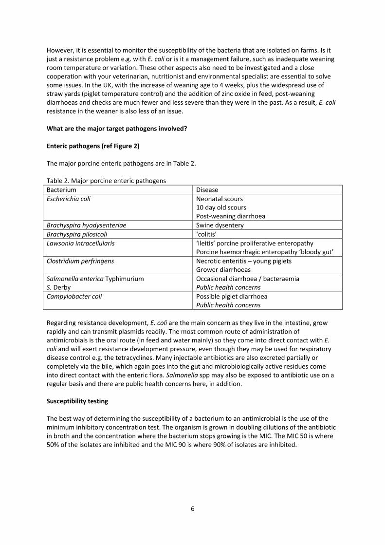
However, it is essential to monitor the susceptibility of the bacteria that are isolated on farms. Is it just a resistance problem e.g. with E. coli or is it a management failure, such as inadequate weaning room temperature or variation. These other aspects also need to be investigated and a close cooperation with your veterinarian, nutritionist and environmental specialist are essential to solve some issues. In the UK, with the increase of weaning age to 4 weeks, plus the widespread use of straw yards (piglet temperature control) and the addition of zinc oxide in feed, post-weaning diarrhoeas and checks are much fewer and less severe than they were in the past. As a result, E. coli resistance in the weaner is also less of an issue. What are the major target pathogens involved? Enteric pathogens (ref Figure 2) The major porcine enteric pathogens are in Table 2. Table 2. Major porcine enteric pathogens
Bacterium Disease
Escherichia coli Neonatal scours 10 day old scours Post-weaning diarrhoea
Brachyspira hyodysenteriae Swine dysentery
Brachyspira pilosicoli ‘colitis’
Lawsonia intracellularis ‘ileitis’ porcine proliferative enteropathy Porcine haemorrhagic enteropathy ‘bloody gut’
Clostridium perfringens Necrotic enteritis – young piglets Grower diarrhoeas
Salmonella enterica Typhimurium S. Derby
Occasional diarrhoea / bacteraemia Public health concerns
Campylobacter coli Possible piglet diarrhoea Public health concerns
Regarding resistance development, E. coli are the main concern as they live in the intestine, grow rapidly and can transmit plasmids readily. The most common route of administration of antimicrobials is the oral route (in feed and water mainly) so they come into direct contact with E. coli and will exert resistance development pressure, even though they may be used for respiratory disease control e.g. the tetracyclines. Many injectable antibiotics are also excreted partially or completely via the bile, which again goes into the gut and microbiologically active residues come into direct contact with the enteric flora. Salmonella spp may also be exposed to antibiotic use on a regular basis and there are public health concerns here, in addition. Susceptibility testing The best way of determining the susceptibility of a bacterium to an antimicrobial is the use of the minimum inhibitory concentration test. The organism is grown in doubling dilutions of the antibiotic in broth and the concentration where the bacterium stops growing is the MIC. The MIC 50 is where 50% of the isolates are inhibited and the MIC 90 is where 90% of isolates are inhibited.

7
Figure 4. MIC test – broth dilution method
Table 3. Antimicrobial susceptibility of E. coli in the EU (152 isolates) (Felmingham, 2009)
Antimicrobial MIC 50 (µg/ml) MIC 90 (µg/ml) Range (µg/ml) Resistance (%)
Amoxycillin 8.0 >128 1.0->128 43
Amoxycillin + clavulanic acid
4.0 8.0 1.0-32 0 (enteric)
Streptomycin 32 >128 4.0->128 44
Neomycin 1.0 32 0.25->128 5
Apramycin 4.0 16 1.0-32 0 (enteric)
Gentamicin 0.5 2.0 0.25->128 9
Enrofloxacin 0.03 1.0 0.008-16 20 (systemic) 7 (enteric)
Ciprofloxacin 0.015 0.5 0.008-16 20 (systemic) 7 (enteric)
Colistin 0.25 0.25 0.12-8.0 0
Trimethoprim + sulpha
0.25 >16 0.015->64 45
Tetracycline >128 >128 14->128 80
Amoxycillin and amoxycillin + clavulanic acid (beta-lactamase inhibitor) demonstrates the way beta- lactamase enzymes exert their effect and they can be blocked or inactivated by inhibitors such as clavulanic acid. There are high levels of resistance to tetracycline as it is the most widely used antibiotic in pigs.
MIC

8
Figure 5. Susceptibility patterns demonstrated by E. coli against amoxycillin and amoxycillin + clavulanic acid (Felmingham, 2009)
Figure 6. Susceptibility pattern demonstrated by E. coli against enrofloxacin (Felmingham, 2009)
With regard to enrofloxacin resistance against E. coli, there is an initial ‘wild-type’ pattern, a first stage mutant pattern, which is still susceptible if situated in the gut and a second stage, complete resistance peak at 16µg/ml. Brachyspira spp seem to develop resistance less rapidly than E. coli, fortunately. Most isolates are resistant to tylosin but many are still susceptible to the pleuromutilins, tiamulin and valnemulin.
0
10
20
30
40
50
60
70
1 2 4 8 16 32 64 128
No
of
iso
late
s
MICs (µg/ml)
Amoxycillin
Amoxycillin + clavulanate
Resistant isolates
0
10
20
30
40
50
60
70
80
90
100
0.008 0.015 0.03 0.06 0.12 0.25 0.5 1 2 4 8 16
No
of
iso
late
s
MICs (µg/ml)
Enrofloxacin
Wild type
Resistant
First stage mutants

9
Table 4. Antimicrobial susceptibility of B. hyodysenteriae in 70 UK isolates (Williamson et al, 2010) Antibiotic MIC 50 (µg/ml) MIC 90 (µg/ml) Range (µg/ml)
Tiamulin 0.125 2.0 ≤0.06->8.0
Valnemulin ≤0.03 4.0 ≤0.03->4.0
Lincomycin >32 >32 0.5->32
Tylosin >128 >128 2.0->128
Tylvalosin >32 >32 0.5->32
Table 5. Recent isolate of B. hyodysenteriae from Yorkshire outbreak 2011-12 antibiotic susceptibility (Strugnell, 2012 – personal communication) (agar plate method)
Antibiotic MIC (µg/ml) MBC (µg/ml) Tiamulin <0.031 <0.031 Valnemulin <0.031 <0.031 Lincomycin 8.0 8.0 Tylosin 128 >128 Tylvalosin 2.0 4.0
Key: MBC – minimum bactericidal concentration From Table 5, the majority of antibiotics would be considered highly susceptible, except for tylosin, and would be suitable for use in eradication programmes. A typical susceptibility pattern of 76 B. hyodysenteriae isolates was described by Karlsson et al (2002), see Figure 7. Figure 7. Typical susceptibility pattern of 76 B. hyodysenteriae isolates (Karlsson et al, 2002)
However, in recent years there has been an increase of the incidence of resistance to antibiotics by B. hyodysenteriae, following the withdrawal of a number of compounds from pig medicine, such as the growth promoters carbadox, olaquindox, salinomycin and medicines dimetridazole and ronidazole. There have been cases where multi-resistant isolates of B. hyodysenteriae have been found, which has resulted in the slaughtering out of the herd. On one side it has resulted in a major economic impact on the herd, but it has also permitted the farmer to restock with high health animals, which will not require antibiotics in the future.
0
10
20
30
40
50
60
0.0
16
0.0
31
0.0
63
0.1
25
0.2
5
0.5
1
2
4
8
16
32
64
12
8
25
6
>2
56
Iso
late
s (
%)
MICs (ug/ml)
Valnemulin Tiamulin Lincomycin Tylosin

10
Antimicrobial susceptibility of B. pilosicoli is usually better than B. hyodysenteriae. Lawsonia intracellularis, the cause of ileitis is a difficult organism to grow, as it requires cell cultures to grow the bacterium. The most comprehensive intracellular MIC work was reported by Wattanaphansak et al (2009) where he tested 10 isolates of L. intracellularis from EU and US sources. Table 6. Estimated iMICs for a number of antimicrobials of 20 results (10 isolates x 2 tests) (Wattanaphansak et al, 2009)
Antimicrobial iMIC50 (µg/ml) iMIC 90 (µg/ml) Range (µg/ml)
Tiamulin 0.125 0.125 0.125 - 0.5
Valnemulin 0.125 0.125 0.125
Tylosin 2.0 8.0 0.25 - 32
Lincomycin 64 >128 8.0 - >128
Chlortetracycline 8.0 64 0.125 - 64
The iMIC appears to be the most useful for comparison with therapeutic concentrations of antimicrobials and as such demonstrates there might be some resistance associated with lincomycin and chlortetracycline but not with the other compounds. Respiratory pathogens (ref Figure 3) One of the major respiratory pathogens with potential to develop antimicrobial resistance is A. pleuropneumoniae but generally resistance is much lower than for enteric bacteria such as E. coli. Table 7. Antimicrobial susceptibility of A. pleuropneumoniae in 129 EU isolates (Felmingham, 2009)
Antimicrobial MIC 50 (µg/ml) MIC 90 (µg/ml) Range (µg/ml) Resistance (%)
Amoxycillin 0.5 0.5 0.25-32 5
Amoxycillin + clavulanic acid
0.25 0.5 0.06-1.0 0
Cephalexin (1G) 2.0 2.0 0.12-4.0 0
Ceftiofur (3G) 0.015 0.03 0.008-0.06 0
Enrofloxacin 0.03 0.06 0.008-2.0 1
Florfenicol 0.25 0.5 0.12-0.5 0
Trimethoprim+ sulphonamide
0.06 0.25 0.008-16 5
Tetracycline 1.0 16 0.25-32 15
Tilmicosin 8.0 16 4.0-16 0
Tiamulin 8.0 16 0.25-16 0
Slightly better results are achieved against Pasteurella multocida, except for tetracyclines.

11
Table 8. Antimicrobial susceptibility of P. multocida in 135 EU isolates (Felmingham, 2009)
Antimicrobial MIC 50 (µg/ml) MIC 90 (µg/ml) Range (µg/ml) Resistance (%)
Amoxycillin 0.25 0.25 0.06-128 1
Amoxycillin + clavulanic acid
0.25 0.25 0.12-0.25 0
Cephalexin (1G) 2.0 4.0 1.0-8.0 0
Ceftiofur (3G) 0.004 0.03 0.002-0.5 0
Enrofloxacin 0.015 0.03 0.008-0.25 0
Florfenicol 0.5 0.5 0.25-1.0 0
Trimethoprim+ sulphonamide
0.06 0.5 0.008-16 3
Tetracycline 0.5 2.0 0.25-32 22
Tilmicosin 8.0 16 1.0-16 0
Streptococcus suis the cause of streptococcal meningitis still remains remarkably susceptible to the penicillins but shows poor susceptibility to tetracyclines and tilmicosin. Table 9. Antimicrobial susceptibility of S. suis in 110 EU isolates (Felmingham, 2009)
Antimicrobial MIC 50 (µg/ml) MIC 90 (µg/ml) Range (µg/ml) Resistance (%)
Amoxycillin ≤0.03 ≤0.03 0.03-0.25 0
Amoxycillin + clavulanic acid
≤0.06 ≤0.06 0.06-0.25 0
Cephalexin (1G) 0.12 0.5 0.06-4.0 0
Ceftiofur (3G) 0.12 0.5 0.06-2.0 0
Enrofloxacin 0.5 0.5 0.12-8.0 1
Florfenicol 0.5 0.5 0.25-1.0 0
Trimethoprim+ sulphonamide
0.06 1.0 0.008-16 7
Tetracycline 32 32 0.25-32 82
Tilmicosin >128 >128 4.0->128 54
Mycoplasma hyopneumoniae is also a slow growing organism and generally antibiotic resistance is low. Table 10. Antimicrobial susceptibility of M. hyopneumoniae in 21 Belgian field isolates (Vicca et al, 2004) – final MICs, 14 days after inoculation
Antimicrobial MIC 50 (µg/ml) MIC 90 (µg/ml) Range (µg/ml)
Enrofloxacin 0.06 0.5 0.03->1.0
Oxytetracycline 0.5 2.0 0.12->2.0
Doxycycline 0.5 1.0 0.12-2.0
Lincomycin ≤0.06 0.12 ≤0.06->8.0
Florfenicol 0.25 0.5 ≤0.12—1.0
Tiamulin 0.03 0.12 ≤0.015-0.12
Tylosin 0.06 0.12 ≤0.015->1.0
Tilmicosin 0.5 0.5 ≤0.25->16
Some antibiotic resistance was demonstrated against lincomycin, tylosin and tilmicosin (1 isolate and enrofloxacin (5 isolates). Acquired resistance to these antimicrobials had not been described in M. hyopneumoniae field isolates previously.

12
So apart from some organisms such as E. coli, B. hyodysenteriae and some A. pleuropneumoniae, where resistance has been determined, the antimicrobial resistance situation is not that bad and can be covered providing that we can maintain the availability of the current antimicrobials that are approved for use in pigs. Care should be taken to not overuse antimicrobials but if used responsibly and sensibly there is no major reason for dismay. Remember most of the antimicrobials used in pigs are over thirty years old and in the main are still working. Even the more modern antibiotics such as 3rd generation cephalosporins, if used carefully and not for widespread prophylaxis, will maintain their efficacy and not select for MRSA or ESBLs, as we have seen in both Ireland and the UK. What does the future look like? Firstly, it is not all doom and gloom in Europe, as some lobbying groups would make out. There are some issues in some countries with some organisms and some antimicrobials and these are being addressed. Denmark and France have voluntarily withdrawn the use of 3rd and 4th generation cephalosporins. The Netherlands is wishing to reduce its antimicrobial use in swine by 50% by 2013. They have stopped the use of feed premixes. This is fine but neither 3rd generation cephalosporins nor fluoroquinolones (enrofloxacin) are used in feed and these are the two families of antibiotics that they are particularly concerned about. The removal of feed premixes, which tend to be the older antibiotics may put more pressure on the use of the modern ones e.g. cephalosporins. Germany and Denmark have almost stopped the use of feed premixes but they have flourishing markets in oral powders (products for ‘top dressing’ feed). This to me is nonsense, as the production of medicated feed is on farm and unregulated and the homogenous mix of medication cannot be guaranteed or controlled as it is when produced by feed compounders. Feed compounders may quite like to not have to make medicated feeds, as it reduces the efficiency of their production and adds costs. Normally, they will recoup these costs though. Fortunately, the European Commission (EC) is reportedly in favour of ‘controlled’ medicated-feed production. Other aspects might involve the cessation of the use of antimicrobials for ‘prevention/prophylaxis’, which the European Parliament (EP) has passed. I do not think they have defined what they mean by prophylaxis. I suspect it is considered that low level antimicrobial inclusion to prevent disease is similar to growth promotion. The use of high levels of antimicrobial to eliminate infections such as swine dysentery in pigs coming into a finishing unit or the ‘early treatment’/ metaphylaxis of pigs carrying Streptococcus suis, I do not think should be included but one cannot be sure. Some countries, like Germany, tried to ban prophylactic use of antimicrobials and developed a major problem with B. hyodysenteriae resistance. Fortunately, the European Medicines Agency (EMA/CVMP) and EC have recently approved both low level prevention and high level prevention claims for a feed premix (Tiamutin/Denagard – Novartis) in a referral procedure (European Commission decision C(2010)5372) 27 July 2010). This suggests that there is some sound, scientific and practical sanity in key EU bodies. Dispensing of antimicrobials by veterinary surgeons is another issue – should vets make money out of supplying antimicrobial products – does this influence their judgement? Again, Denmark are leading the way with this and trying to break the association of veterinary profit from the equation. However, vets have to visit farms on a monthly basis and the costs are borne by the farmer. Their use of antibiotics is also monitored and if it is deemed too high it is investigated. Interestingly, feed premixes are primarily sourced by feed compounders and supplement manufacturers direct from companies and usually feed premixes account for over 60% of antimicrobial use (VMD, 2011), so

13
there is relatively minor profit in them for veterinarians. In Ireland, I understand you have your own competitive license remedy outlets already. Will we be allowed to use antibiotics in the Future? The answer definitely has to be YES. Even politicians would not allow animals to die on farm for the lack of antimicrobial treatment. On the grounds of animal welfare, it would not be acceptable. What will be different? What do you want to be different? If nothing, then you need to lobby hard, both pig farmers and their veterinarians, to maintain the status quo. The European Commission is reviewing antimicrobial use and resistance in both animals and man (EC, 2011) over the next 5 years. You need to lobby and work with the European bodies such as Copa-Cogeca for the farmers, via the Irish Medicines Board into the EMA, the Irish vets via Federation of Veterinarians in Europe (FVE), the Irish Animal Health industry (APHA) via IFAH Europe, which is run by an Irishman, Declan O’Brien, and consider setting up an Irish Responsible Use of Medicines Alliance. In the UK we have RUMA, which is across industry from farmers, vets, feed industry, VMD, NOAH, FSA, supermarkets, processors and medical doctors. This is now taken up by EPRUMA (the European version) which is similar to UK but has good contacts to the EC, which is critical. They promote the responsible use of medicines in general but specifically antibiotics. Table 11. Possible changes, opinions and comments
Possible change Probability Comments
Ban growth promotion
Already done (2006) ‘Precautionary principle’ – not necessarily science or factually based – politically driven.
Ban prevention use Low level use – possible Early treatment/metaphylactic use - no
Politically, associated with growth promotion. No scientific reason to ban it
Ban in-feed use Possible – but unlikely Oral powders – now established in EU
Controlled use very effective. Often uncontrolled use.
Ban certain drugs 3rd and 4th generation cephalosporins – vulnerable Fluoroquinolones – been around for over 20 years. Macrolides – on the list Colistin – now on the list?
Highly effective but attract major criticism – ESBLs & MRSA. Primarily injectable use in pigs or oral dosers for neonatal scours – would cause major welfare concerns if withdrawn. Comparably less significant than the above groups in human medicine.
Stop dispensing by veterinarians
Possible – but unlikely Would cause welfare issues and also loss of vet services in more remote areas. Competition more appropriate for POM products.

14
Possible change Probability Comments
More policing of antimicrobial use on farm
Possible – on a national basis Added bureaucracy but may add reassurance to public/politicians
What if the pipeline dries up? Personally, I think the likelihood of the development of new families of antimicrobials for pig use, which are primarily derived from human pharmaceutical research, is remote. A new macrolide injectable antibiotic, tildipirosin (Zuprevo – MSD) was introduced in the US recently (Kniffen and Wray, 2012) primarily for bacterial respiratory infections. Otherwise, there appears to be little new on the horizon. However, there are opportunities to improve the range and availability of products on the market in Ireland by utilising products that are currently approved in the rest of the EU. Many products can be imported under the ‘cascade’ and can be used in accordance with their EU/national approval. This may develop further so that there is only ‘one product, one assessment and one registration’ for a number of generic products similar to the centralised procedure registrations and referrals across the EU. Proposals are in hand to do this and early trials are being undertaken. I think there is a good opportunity for this to happen via the EMA (CMDv) and should be encouraged via your IMB representatives. Conclusions Antimicrobial resistance is not a new issue and generally can be controlled by responsible use. It is not as bad as is often painted by the organic and vegetarian lobbyists that use scare tactics to raise funds. Each country may have its own issues, which need to be addressed and resolved. If at a farm level, it cannot be resolved, i.e. in the case of multi-resistant B. hyodysenteriae this may lead to slaughtering out the herd but there are opportunities to restock with high health stock, which require minimal use of antibiotics. One cannot separate the science from politics at this stage but it is hoped that the EC will take practical steps to assess the situation and act accordingly and not be pushed by individual Member States or political lobbying groups into taking inappropriate action. It is up to the Irish swine industry to come together and lobby their national and thereby EU counterparts to assist the EC to come to the right decisions. In the meantime, the use of antimicrobials on farm should be proportionate to their need and only used in a responsible way. References Burch, D.G.S., Duran, C.O. and Aarestrup, F.M. (2008) Chapter 7. Guidelines for antimicrobial use in swine. In “Guide to Antimicrobial Use in Animals. Eds: Guardabassi, L., Jensen, L.B. and Kruse, H. Blackwell Publishing, Oxford, UK, pp 102-125. Burch, D.G.S. (2011) Antimicrobial resistance – veterinary and public health concerns. Suis, 81, pp 12-16. European Commission (DG Sanco) (2011) Communication from the Commission to the European Parliament and Council “Action plan against the rising threats from Antimicrobial Resistance”, COM (2011) 748.
15
Felmingham, D. (May 2009) Report ‘Determination of the antimicrobial susceptibility of the VetPath II (2004-2006) collection of bacterial pathogens’. Giguere, S., Prescott, J.F., Baggot, J.D., Walker, R.D. and Dowling, P.M. (2006) Antimicrobial Therapy in Veterinary Medicine (Fourth Edition). Blackwell Professional Publishing, Ames, Iowa, USA. Karlsson, M., Oxberry, S.L. and Hampson, J. (2002) Antimicrobial susceptibility testing of Australian isolates of Brachyspira hyodysenteriae using a new broth dilution method. Veterinary Microbiology, 84, 123-133. Kniffen, T.S. and Wray, M.I. (2012) Scientific and practical considerations regarding the use of a novel, injectable anti-infective in swine: Zuprevo(R) from Merck Animal Health. Proceedings of the American Association of Swine Veterinarians Meeting, Denver, Colorado, USA pp 131-134. Veterinary Medicines Directorate (VMD) (2011) Sales of antimicrobial products authorised for use as veterinary medicines in the UK in 2010. Vicca, J., Stakenborg, T., Maes, D., Butaye, P., Peeters, J., de Kruif, A. And Haesebrouck, F. (2004) In-vitro susceptibilities of Mycoplasma hyopneumoniae field isolates. Antimicrobial Agents and Chemotherapy, 48, 11, 4470-4472. Wattanaphansak, S., Singer, R.S. and Gebhart, C.J. (2009) In vitro antimicrobial activity against 10 North American and European Lawsonia intracellularis isolates. Veterinary Microbiology, 134, 305-310. Williamson, S. Rogers, J., Hunt, B. and Teale, C. (2010) Preliminary results for Brachyspira MIC assessment of isolates from England. Presentation at Pig Veterinary Society Meeting, Norwich, Norfolk, UK.