anticancer activity of some polyamine derivatives on … · anticancer activity of some polyamine...
TRANSCRIPT

Regular paper
Anticancer activity of some polyamine derivatives on human prostate and breast cancer cell linesMarta Szumilak1*, Malgorzata Galdyszynska2, Kamila Dominska2, Andrzej Stanczak1 and Agnieszka Piastowska-Ciesielska2,3*
1Department of Hospital Pharmacy, Faculty of Pharmacy, Medical University of Lodz, Łódź, Poland; 2 Department of Comparative Endocrinology, Medical University of Lodz, Łódź, Poland; 3Laboratory of Cell Cultures and Genomic Analysis, Medical University of Lodz, Łódź, Poland
The aim of this study was to expand our knowledge about anticancer activity of some polyamine derivatives with quinoline or chromane as terminal moieties. Tested compounds were evaluated in vitro towards metastatic human prostate adenocarcinoma (PC3), human carci-noma (DU145) and mammary gland adenocarcinoma (MCF7) cell lines. Cell viability was estimated on the basis of mitochondrial metabolic activity using water-soluble tetrazolium WST1 to establish effective concen-trations of the tested compounds under experimental conditions. Cytotoxic potential of polyamine derivatives was determined by the measurement of lactate dehy-drogenase activity released from damaged cells, changes in mitochondrial membrane potential, the cell cycle dis-tribution analysis and apoptosis assay. It was revealed that the tested polyamine derivatives differed markedly in their antiproliferative activity. Bischromane derivative 5a exhibited a rather cytostatic than cytotoxic effect on the tested cells, whereas quinoline derivative 3a caused changes in cell membrane integrity, inhibited cell cycle progression, as well as induced apoptosis of prostate and breast cancer cells which suggest its potential appli-cation in cancer therapy.
Key words: polyamine derivatives, prostate cancer, breast cancer, mi-tochondrial potential, cell cycle, apoptosis
Received: 24 August, 2016; revised: 21 December, 2016; accepted: 28 December, 2016; available on-line: 14 April, 2017
*e-mail: [email protected] (MS - chemistry), [email protected] (AP - in vitro biological evaluation)Abbreviations: DU145, human carcinoma; MCF7, mammary gland adenocarcinoma; PC3, metastatic human prostate adenocarcinoma
INTRODUCTION
Cancer is used as a general term describing a group of approximately 120 different diseases, which can affect various parts of the body. It can be also defined as the state characterized by uncontrolled cell proliferation and normal tissues invasion (Latosinska & Latosinska, 2013). According to the Institute for Health Metrics and Evalu-ation (IHME) report, cancer is the second leading cause of death worldwide. Increased incidence of cancer ob-served in the developed countries can be attributed in part to the changes in demographic structure i.e. greater longevity of population, as well as risk factors like smok-ing, obesity and unhealthy diet (Fitzmaurice et al., 2015).
Prostate cancer (PCa) has the highest incidence ratio and it is the second most common cause of death in men (Siegel et al., 2016). In Poland, in 2013, about 12 162 men received a prostate cancer diagnosis and an
estimated 4 281 men died of it (Dominska et al., 2012; Wojciechowska & Didkowska, 2013). When surgery is excluded, the basic method of conservative treatment in cases of advanced cancer is hormonal therapy involving the elimination of endogenous androgens and the block-ade of the androgen receptor. Hormonal therapy slows down the development of cancer but it does not lead to full recovery. After an initial period of improvement, the disease progression ensues due to the development of androgen independent cancer, followed by fully hor-mone resistant cancer. As aggressive, poorly-differentiat-ed high-grade PCa is currently incurable and potentially lethal, there is a need for a new treatment strategy which can be provided by newly designed anticancer medicines (Siegel et al., 2013; Jemal et al., 2011; Walczak & Car-ducci, 2007).
The second type of cancer cell line included in this study is derived from breast cancer, which is the most common female cancer worldwide (Wojciechowska & Didkowska, 2013; Siegel et al., 2016). In Poland, in 2013, about 17 142 women received a breast cancer diagnosis and it is estimated that 5 816 women died of the disease. Some breast cancers rapidly develop multidrug resistance to chemotherapy medicines which results in therapeutic failure (Li et al., 2015). Aforementioned data indicate that the search for novel, effective and less toxic anticancer agents is very important goal for contemporary medicine (Ma & Adjei, 2009).
Our quest for potential anticancer agents is focused on symmetrical polyamine derivatives with bicyclic ter-minal moieties designed according to bisintercalators’ structural requirements (Szulawska-Mroczek et al., 2013; Szumilak et al., 2010). Bisintercalators are able to interact reversibly with double stranded (dsDNA) by simultane-ous insertion of two chromophores usually tethered by a polyamine linker. It results in higher DNA affinity and sequence selectivity in comparison to corresponding mo-nointercalating agents (Brana et al., 2001; Lorente et al., 2004; Tse & Boger 2004). In addition, a positive cor-relation between cytotoxic potency and the strength of reversible DNA binding for bisintercalators has been ob-served (Taher & Hegazy, 2013).
Our previous studies involving synthesis and biologi-cal in vitro evaluation of polyamine derivatives with vari-ous bicyclic moieties, revealed that polyamine derivatives with quinoline 3a and chromane 5a scaffolds (Fig. 1) are the most promising entities exhibiting antiproliferative activity toward a highly aggressive melanoma cell line A375 (Szulawska-Mroczek et al., 2013; Szumilak et al., 2010). Although the chemical structure of 5a was for-merly known as a chelating agent (Trathnigg et al., 1985), we have obtained it by another route and assessed its
Vol. 64, No 2/2017307–313
https://doi.org/10.18388/abp.2016_1416
308 2017M. Szumilak and others
antiproliferative activity together with other chromone/chromane derivatives designed as potential bisintercala-tors (Szulawska-Mroczek et al., 2013).
Taking into consideration that prostate cancer in men and breast cancer in women belong to the most fre-quently registered malignant cancers (Siegel et al., 2016), we decided to evaluate the influence of 3a and 5a on well-described prostate and breast cancer cell lines that are commonly used as models of drug susceptibility: PC3, DU145 and MCF7 (Sampson et al., 2013; Ellem et al., 2014; Li et al., 2015; Ming et al., 2015). The afore-mentioned choice can be supported by studies reporting that quinoline ring system is used in many anticancer agents (Burns et al., 2002; Deady et al., 1997; Hansch & Verma, 2007; Li et al., 2016; Rescifina et al., 2014) and chromane derivatives possess promising anticancer activ-ity toward breast cancer (Rawat et al., 2016) and leukae-mia (Nawrot-Modranka et al., 2006) or prevent progres-sion to a metastatic phenotype for human prostate can-cer (Xu et al., 2010).
MATERIALS AND METHODS
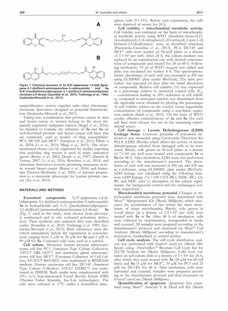
Examined compounds. N,N′-(piperazine-1,4-di-yldipropane-3,1-diyl)bis(4-aminoquinoline-3-carboxamide) 3a as hydrochloride and N,N´-[(methylimino)dipropane-3,1-diyl]bis[3-(aminomethylene)chroman-2,4-dione] 5a (Fig. 1) used in this study, were chosen from previous-ly synthesized and in vitro evaluated polyamine deriva-tives. Their synthesis and analytical data were described earlier (Szumilak et al., 2010; Trathnigg et al. 1985, Szu-lawska-Mroczek et al., 2013). Both substances were dis-solved immediately before the experiment at concentra-tions ranging from 5 μM to 50 μM for 3a and 5 μM to 90 μM for 5a. Untreated cells were used as a control.
Cell culture. Metastatic human prostate adenocarci-noma cell line PC3 (American Type Culture Collection, ATCC® CRL-1435™) and mammary gland adenocarci-noma cell line MCF7 (European Collection of Cell Cul-ture, ECACC® 86012803) were maintained in RPMI1640 medium. Human carcinoma cell line DU145 (American Type Culture Collection, ATCC® HTB81™) was main-tained in DMEM. Both media were supplemented with 10% (v/v) heat-inactivated Foetal Bovine Serum (FBS) (Thermo Fisher Scientific Inc/Life technologies). The cells were cultured at 37°C under a humidified atmo-
sphere with 5% CO2. Before each experiment, the cells were deprived of serum for 24 h.
Cell viability – mitochondrial metabolic activity. Cell viability was estimated on the basis of mitochondri-al metabolic activity using WST1 (disodium mono{4-[3-(4-iodophenyl)-2-(4-nitrophenyl)-2H-tetrazol]-3-ium-5-yl]benzene-1,3-disulfonate}) assay as described elsewhere (Piastowska-Ciesielska et al., 2013). PC3, DU145 and MCF7 cells were seeded on 96-well plates at a density of 1 × 10⁴ per well. After 24 h, the culture medium was replaced by an experimental one with desired concentra-tions of compounds and treated for 24 or 48 h. Follow-ing incubation, 10 μl of WST1 reagent was added and plate was incubated for further 4 h. The spectrophoto-metric absorbance of each well was measured at 450 nm using ELX808IU plate reader (BioTeck). The same pro-cedure was repeated 14 days after the initial dissolution of compounds. Relative cell viability (%) was expressed as a percentage relative to untreated control cells. IC50 – concentration leading to 50% reduction in cell viabili-ty, compared to untreated control, was determined from the sigmoidal curve obtained by plotting the percentages of cell viability relative to the control versus logarithmic concentration of compounds using a non-linear regres-sion analysis (Saleh et al., 2015). On the basis of WST1 results, effective concentrations of 3a and 5a (for each cell line) were chosen for use in the remaining experi-mental assays.
Cell damage – Lactate Dehydrogenase (LDH) Leakage Assay. Cytotoxic potential of polyamine de-rivatives was measured using Cytotoxicity Detection Kit PLUS (LDH) (Roche) which allows the activity of lactate dehydrogenase released from damaged cells to be mea-sured. Briefly, cells grown in 96-well plates at a density of 7 × 10³ per well were treated with compounds 3a or 5a for 48 h. After incubation, LDH assay was performed according to the manufacturer’s protocol. The absor-bance of each well was measured at 490 nm and 690 nm as a reference, using ELX808IU plate reader (BioTeck). LDH leakage was calculated using the following func-tion: LDH leakage (%) = 100 × (CS–BC)/(NDC–BC). CS, BC and NDC refer to absorption of the culture super-natant, the background control and the undamaged con-trol, respectively.
Mitochondrial membrane potential. Changes in mi-tochondrial membrane potential were determined with Muse™ Mitopotential Kit (Merck Millipore) which mea-sures the accumulation of dye within the inner mem-brane of intact mitochondria. Briefly, cells grown in 6-well plates (at a density of 2.5 × 10⁵ per well) were treated with 3a or 5a. After 48 h of incubation, cells were collected by trypsinization, resuspended in media and counted. All samples were prepared according to the manufacturer’s protocol and measured on Muse™ Cell Analyzer (Merck Millipore) according to manufacturer’s instruction, standardized to control probes.
Cell cycle analysis. The cell cycle distribution anal-ysis was performed with Guava® easyCyte (Merck Mil-lipore) using FlowCellect™ Bivariate Cell Cycle Kit for G2/M Analysis kit (Merck Millipore). Cells were cul-tured on cell-culture dish at a density of 1 × 10⁶ for 24 h, after which they were treated with 3a (20 µM for all cell lines) and 5a (5 µM for MCF7, 10 µM for PC3 and 25 µM for DU145) for 48 h. After incubation, cells were harvested and counted. Samples were prepared accord-ing to the manufacturer protocol and then measured on Guava® easyCyte (Merck Millipore).
Quantification of apoptosis. Apoptosis was exam-ined using Muse™ Annexin V & Dead cell Kit (Merck
Figure 1. Chemical structure of 3a N,N’-(piperazine-1,4-diyl-dipro-pane-3,1-diyl)bis(4-aminoquinoline-3-carboxamide) and 5a N,N’-[(methylimino)dipropane-3,1-diyl]bis[3-(aminomethylene)chroman-2,4-dione] (Szumilak et al., 2010; Trathnigg et al., 1985; Szulawska-Mroczek et al., 2013).

Vol. 64 309Polyamine derivatives as potential anticancer compounds
Millipore). Cells were cultured on 6-well plates at a den-sity of 2.5 × 10⁵ for 24 h, after which they were treated with compound 3a or 5a for 48 h. After treatment, cells were collected and incubated with Annexin V and 7-ami-noactinomycin D (7AAD), a dead cell marker, for 20 min at room temperature in the dark. All samples were measured using Muse™ Cell Analyzer (Merck Millipore).
Statistical analysis. Results were expressed as means of results from a minimum of three independent exper-iments with similar patterns. IC50 was estimated from the sigmoidal curve obtained by plotting the percentages of cell viability relative to the control versus logarithmic concentration of compounds using non-linear regression analysis. Statistical analysis was performed using one-way ANOVA. All calculations were performed using Graph-Pad Prism 6 software (GraphPad Software, San Diego, California, USA). A p-value below 0.05 was considered statistically significant. All experiments were performed as three independent repetitions.
RESULTS
Compounds’ stability in cell culture media
The influence on cancer cell viability of 3a and 5a im-mediately after initial dissolution (T0) and after 14 days
(T14) was compared to determine the compounds’ sta-bility. A decrease in cell viability after treatment with 5a was denoted after both incubation times (24 and 48 h) for T0 and T14 as well. However, 5a at 50 μM, de-creased the cell viability by 50% in PC3 cells at T0 (24 h incubation), but only by 31% at T14. Similar results were observed after 48 h of incubation (data not shown). Fur-thermore, the ability of compound 3a to inhibit cancer cell viability fell 14 days after dissolution. For example, after 24 h of incubation with compound 3a at 25 μM, a 45% decrease in cell viability was observed at T0, but only 21% at T14. No significant difference between the results of 24 and 48 h of incubation for each time point was noticed. Therefore, both compounds were dissolved immediately before each experiment and a 48 hr incuba-tion period was used.
Influence of compounds on cancer cells – deter-mination of IC50. Water-soluble tetrazolium (WST1) was used to assess the influence of compounds 3a and 5a on cell viability of prostate and breast cancer cells (measured via mitochondrial metabolic activity). The as-say is based on WST1, a highly sensitive tetrazolium that produces soluble formazan via the NADPH oxidase re-duction in mitochondria. The amount of formazan dye yielded, directly correlates to the number of metabolical-ly-active live cells in the culture (Xiong et al., 2015). As before, in the first step, a concentration response course
Figure 2. Polyamine derivative 3a containing quinoline moiety (A–C) and polyamine derivative 5a containing chromane moiety (A’–C’), inhibit the growth of human cancer cells. The sigmoidal curve was obtained by plotting the percentages of cell viability relative to the control against the logarithmic concentra-tion of compounds using non-linear regression analysis. The phase-contrast micrographs (lower panel) represent cells treated with 3a and 5a (15 µM, 20 µM and 25 µM) and untreated cells (control) which were cultured on serum-free medium. Photomicrographs were taken at a magnification of ×400 (Olympus CKX41 with digital camera Olympus DP20) from representative experiments.
310 2017M. Szumilak and others
analysis was performed to determine the compounds’ concentration required to inhibit the growth of cancer cells by 50% (IC50) after 48 h of incubation (Szumilak et al., 2010). Compounds 3a and 5a were tested in a wide range of concentrations from 5 μM to 90 μM. Treat-ment of prostate and breast cancer cells with examined compounds resulted in concentration-dependent inhibi-tion of cell mitochondrial activity which corresponded to cell viability (Fig. 2). IC50 values for compound 3a were found to be 23.70 μM for PC3, 26.64 μM for DU145 and 18.54 μM for MCF7. Chromane derivative 5a ex-hibited a lower inhibitory activity than the quinoline one (3a) which is illustrated by following the IC50 values: 36.19 μM, 49.20 μM and 21.39 μM for PC3, DU145 and MCF7, respectively.
Effect of compounds on cancer cell lactate dehydrogenase leakage and mitochondrial membrane potential
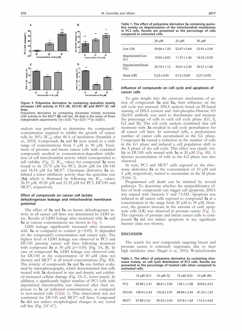
The effect of 3a and 5a on lactate dehydrogenase ac-tivity in all cancer cell lines was determined by LDH as-say. Results of LDH leakage after treatment with 3a and 5a at various concentrations are shown in Fig. 3.
LDH leakage significantly increased after treatment with 3a as compared to control (p < 0.05). It depended on the compound’s concentration and cancer type. The highest level of LDH leakage was observed in PC3 and DU145 prostate cancer cell lines following treatment with compound 3a at 30 µM (p < 0.05) (Fig. 3A, B). In case of compound 5a, LDH leakage was observed only for DU145 at the concentration of 50 µM (data not shown) and MCF7 at all tested concentrations (Fig. 3D). The toxicity of compounds 3a and 5a was further evalu-ated by microphotographs, which demonstrated that cells treated with 3a decreased in size and density and exhibit-ed increased cellular damage (Fig. 2A–C, lower panel). In addition, a significantly higher number of PC3 cells with depolarized mitochondria was observed after their ex-posure to 3a (at indicated concentrations), as compared to non-treated cells (Table 1). This observation was not confirmed for DU145 and MCF7 cell lines. Compound 5a did not induce morphological changes in any tested cell line (Fig. 2A’–C’).
Influence of compounds on cell cycle and apoptosis of cancer cells
To gain insight into the cytotoxic mechanism of ac-tion of compounds 3a and 5a, their influence on the cell cycle was assessed. DNA analysis based on PI-based staining of DNA content and Anti-phospho-Histone H3 (Ser10) antibody was used to discriminate and measure the percentage of cells in each cell cycle phase (G1, S, G2 and M). The cell cycle analysis confirmed that cell treatment with 3a resulted in cell cycle perturbation for all cancer cell lines. In untreated cells, a predominant number of cancer cells accumulated in the G1 phase. Compound 3a caused a reduction in the number of cells in the G1 phase and induced a cell population shift to the S phase of the cell cycle. This effect was clearly visi-ble in DU145 cells treated with 3a at 20 µM (Fig. 4B). A discreet accumulation of cells in the G2 phase was also observed.
In turn, PC3 and MCF7 cells exposed to the chro-mane derivative 5a at the concentration of 10 µM and 5 µM, respectively, started to accumulate in the M phase (Table 2).
Programmed cell death can be initiated by several pathways. To determine whether the antiproliferative ef-fect of both compounds can trigger cell apoptosis, DNA was stained with Annexin V and 7AAD. Apoptosis was induced in all cancer cells exposed to compound 3a at a concentration in the range from 20 µM to 30 µM. How-ever, the greatest increase in the amount of early apop-totic cells (LR) was observed in prostate cancer (Fig. 5). The exposure of prostate and breast cancer cells to com-pound 5a did not induce apoptosis in any significant manner (data not shown).
DISCUSSION
The search for new compounds targeting breast and prostate cancer is extremely important, due to their high incidence rates (Siegel et al., 2016; Wojciechowska
Figure 3. Polyamine derivative 3a containing quinoline moiety increases LDH activity in PC3 (A), DU145 (B) and MCF7 (C) cell lines. Polyamine derivative 5a containing chromane moiety increases LDH activity in the MCF7 (D) cell line. All data is the mean of three independent experiments (*p < 0.05; **p < 0.01; ***p < 0.001).
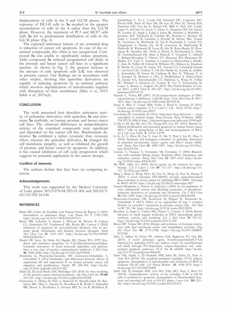
Table 1. The effect of polyamine derivative 3a containing quino-line moiety on depolarization of the mitochondrial membranes in PC3 cells. Results are presented as the percentage of cells compared to untreated cells.
20 µM 25 µM 30 µM
Live (LR) 59.66 ± 1.59 52.87 ± 3.64 52.91 ± 2.04
Depolarized/Live (LL) 10.93 ± 0.65 11.37 ± 1.46 16.52 ± 0.50
Depolarized/Dead (UL) 29.19 ± 1.12 35.61 ± 2.39 30.31 ± 1.66
Dead (UR) 0.22 ± 0.05 0.15 ± 0.04 0.27 ± 0.05
Table 2. The effect of polyamine derivative 5a containing chro-mane moiety, on cell cycle distribution of PC3 cells. Results are presented as the percentage of treated cells when compared to untreated cells.
10 µM (G1) 10 µM (S) 10 µM (G2) 10 µM (M)
PC3 95.98 ± 2.41 88.61 ± 3.95 138.1 ± 1.08 204.8 ± 2.61
DU145 100.9 ± 2.43 102.8 ± 2.91 84.89 ± 3.44 47.23 ± 1.67
MCF7 97.89 ± 3.0 95.55 ± 3.45 107.8 ± 1.64 114.2 ± 4.62

Vol. 64 311Polyamine derivatives as potential anticancer compounds
& Didkowska, 2013), as well as rapid development of multidrug resistance to chemotherapies, which block ef-fective, life-saving therapies (Li et al., 2015; Walczak & Carducci, 2007).
As it was previously demonstrated, polyamine deriv-atives structurally related to known bisintercalators con-taining the quinoline 3a and chromane 5a moieties as terminal scaffolds, exhibit antiproliferative activity on a human melanoma cell line A375 (Szumilak et al., 2010), but the influence of these compounds on breast and prostate cancer has not been studied yet.
The results of current screening revealed significant differences in the anticancer activity of the examined compounds depending on a cancer cell line. Compound 5a was found to exhibit antiproliferative activity at a lower concentration in MCF7 (21 μM) in comparison to PC3 and DU145 cell lines (36 μM and 49 μM, respec-tively) (Fig. 2). However, it had no significant influence on the lactate dehydrogenase leakage or mitochondrial membrane depolarization which may suggest its limited anticancer properties. Our present findings also con-firmed that the compound 3a exhibited anticancer ac-tivity against prostate and breast cancer cells. Inhibition of mitochondrial activity was directly connected with the increase of the compound concentration. Estimated IC50 values for PC3, DU145 and MCF7 cancer cells ranged
from 18 µM to 27 µM (Fig. 2). Furthermore, in contrast to the chromane derivative 5a, the quinoline one (3a) caused changes in cell membrane integrity, as evidenced by increased lactate dehydrogenase leakage which cor-related with mitochondrial membrane depolarization in the PC3 line (Table 1, Fig. 3).
To expand our knowledge about possible cytotoxic mechanism of action of the tested compounds, their ef-fect on the cell cycle progression in breast and prostate cancer cells was examined. Compound 5a applied at the concentration approaching IC50 was found to have no statistically significant influence on changes in the pros-tate cancer cell cycle. In the examined prostate cell line, compound 5a was not able to decrease the number of cells in the G1, G2 or M phases, but an accumulation of cells in the S phase of the cell cycle was observed. How-ever, when applied at the concentration needed for cell viability reduction to 60% (10 µM), compound 5a was responsible for changes in the number of PC3 cells in the G1, S, G2/M phases, and the increase of cell num-ber in the G2 and M phases of the cell cycle (Table 2).
Compound 3a strongly affected changes in the cell cy-cle progression. Most of the cells from untreated PC3, DU145 and MCF7 lines accumulated in the G1 phase. In contrast, compound 3a induced a noticeable reduc-tion in the number of cells in the G1 phase and the
Figure 4. The effect of compound 3a on cell cycle distribution. DNA content measurement by Guava® easyCyte after 48 h of treatment. (A–C) data from three independent experiments; (A’–C’) representative histograms: higher panel – untreated (C), lower panel – treated cells (3a at 20 µM).
Figure 5. Cell apoptosis analysis of human prostate and breast cancer cell lines after 48 h treatment with compound 3a at dif-ferent concentrations. (A–C) data from three independent experiments: A: PC3 cell line, B: DU145 cell line and C: MCF7 cell line. The data is given as means of three independent experiments (*p < 0.05; **p < 0.01; ***p < 0.001). Representative histograms: A’: 1, untreated (C); 2: 20 µM, 3: 25 µM, 4: 30 µM; B’: 1, untreated (C); 2: 23 µM, 3: 25 µM, 4: 27 µM; C’, 1: untreated (C), 2: 20 µM, 3: 23 µM, 4: 25 µM.
312 2017M. Szumilak and others
displacement of cells in the S and G2/M phases. The exposure of DU145 cells to 3a resulted in the greatest accumulation of cells in the S rather than the G2/M phase. However, the treatment of PC3 and MCF7 cells with 3a led to predominant distribution of cells in the G2/M phase (Fig. 4).
The expected anticancer feature of any potential drug is induction of cancer cell apoptosis. In case of the ex-amined compounds, this effect is not unequivocal. Com-pound 5a was unable to significantly induce apoptosis, while compound 3a induced programmed cell death in the prostate and breast cancer cell lines in a significant manner. As shown in Fig. 5, the greatest increase in the amount of early apoptotic cells (LR) was observed in prostate cancer. Our findings are in accordance with other studies, showing that quinoline derivatives are capable of inducing apoptosis by an intrinsic pathway which involves depolarization of mitochondria together with disruption of their membranes (Sahu et al., 2013; Saleh et al., 2015a,b).
CONCLUSION
The work presented here describes anticancer activ-ity of polyamine derivatives with quinoline 3a and chro-mane 5a scaffolds, on human prostate and breast cancer cell lines. The observed differences in antiproliferative activity of the examined compounds were significant and depended on the cancer cell line. Bischromane de-rivative 5a exhibited a rather cytostatic than cytotoxic effect, whereas the quinoline derivative 3a changed the cell membrane integrity, as well as inhibited the growth of prostate and breast cancer by apoptosis. In addition, it has caused inhibition of cell cycle progression which suggests its potential application in the cancer therapy.
Conflicts of interests
The authors declare that they have no competing in-terests.
Acknowledgements
This work was supported by the Medical University of Lodz grants 503/0-078-04/503-01-004 and 502-03/3-011-03/502-34-036
REFERENCES
Brana MF, Cacho M, Gradillas A,de Pascual-Teresa B, Ramos A (2001) Intercalators as anticancer drugs. Curr Pharm Des 7: 1745–1780. http://dx.doi.org/10.2174/1381612013397113
Burns MR, LaTurner S, Ziemer J, McVean M, Devens B, Carlson CL, Graminski GF, Vanderwerf SM, Weeks RS, Carreon J (2002) Induction of apoptosis by aryl-substituted diamines: role of aro-matic group substituents and distance between nitrogens. Bioorg Med Chem Lett 12: 1263–1267. http://dx.doi.org/10.1016/S0960-894X(02)00156-7
Deady LW, Kaye AJ, Finlay GJ, Baguley BC, Denny WA (1997) Syn-thesis and antitumor properties of N-[2-(dimethylamino)ethyl]car-boxamide derivatives of fused tetracyclic quinolines and quinoxa-lines: a new class of putative topoisomerase inhibitors. J Med Chem. 40: 2040–2046. http://dx.doi.org/10.1021/jm970044r
Dominska K, Piastowska-Ciesielska AW, Lachowicz-Ochedalska A, Ochedalski T (2012) Similarities and differences between effects of angiotensin III and angiotensin II on human prostate cancer cell migration and proliferation. Peptides 37: 200–206. http://dx.doi.org/10.1016/j.peptides.2012.07.022
Ellem SJ, De-Juan-Pardo EM, Risbridger GP (2014) In vitro modeling of the prostate cancer microenvironment. Adv Drug Deliv Rev 79–80: 214–221. http://dx.doi.org/10.1016/j.addr.2014.04.008
Fitzmaurice C, Dicker D, Pain A, Hamavid H, Moradi-Lakeh M, Mac-Intyre MF, Allen C, Hansen G, Woodbrook R, Wolfe C, Hamadeh RR, Moore A, Werdecker A, Gessner BD, Te Ao B, McMahon B,
Karimkhani C, Yu C, Cooke GS, Schwebel DC, Carpenter DO, Pereira DM, Nash D, Kazi DS, De Leo D, Plass D, Ukwaja KN, Thurston GD, Yun Jin K, Simard EP, Mills E, Park EK, Catalá-López F, deVeber G, Gotay C, Khan G, Hosgood HD 3rd, Santos IS, Leasher JL, Singh J, Leigh J, Jonas JB, Sanabria J, Beardsley J, Jacobsen KH, Takahashi K, Franklin RC, Ronfani L, Montico M, Naldi L, Tonelli M, Geleijnse J, Petzold M, Shrime MG, Younis M, Yonemoto N, Breitborde N, Yip P, Pourmalek F, Lotufo PA, Esteghamati A, Hankey GJ, Ali R, Lunevicius R, Malekzadeh R, Dellavalle R, Weintraub R, Lucas R, Hay R, Rojas-Rueda D, West-erman R, Sepanlou SG, Nolte S, Patten S, Weichenthal S, Abera SF, Fereshtehnejad SM, Shiue I, Driscoll T, Vasankari T, Alsharif U, Rahimi-Movaghar V, Vlassov VV, Marcenes WS, Mekonnen W, Melaku YA, Yano Y, Artaman A, Campos I, MacLachlan J, Mueller U, Kim D, Trillini M, Eshrati B, Williams HC, Shibuya K, Dandona R, Murthy K, Cowie B, Amare AT, Antonio CA, Castañeda-Orjuela C, van Gool CH, Violante F, Oh IH, Deribe K, Soreide K, Knibbs L, Kereselidze M, Green M, Cardenas R, Roy N, Tillmann T, Li Y, Krueger H, Monasta L, Dey S, Sheikhbahaei S, Hafezi-Nejad N, Kumar GA, Sreeramareddy CT, Dandona L, Wang H, Vollset SE, Mokdad A, Salomon JA, Lozano R, Vos T, Forouzanfar M, Lopez A, Murray C, Naghavi M (2015) The Global Burden of Can-cer 2013. JAMA Oncol 1: 505–527. http://dx.doi.org/doi:10.1001/jamaoncol.2015.0735
Hansch C, Verma RP (2007) 20-(S)-camptothecin analogues as DNA topoisomerase I inhibitors: a QSAR study. Chem Med Chem 2: 1807–1813. http://dx.doi.org/10.1002/cmdc.200700138
Jemal A, Bray F, Center MM, Ferlay J, Ward E, Forman D (2011) Global cancer statistics. CA-A Cancer J Clin 61(2): 69-90. http://dx.doi.org/10.3322/caac.20107
Latosinska JN, Latosinska M (2013) Anticancer drug discovery — from serendipity to rational design. Drug Discovery. Hany El-Shemy. ISBN 978-953-51-0906-8 http://cdn.intechopen.com/pdfs-wm/41943.pdf
Li HJ, Li XJ, Bai ML, Suo YE, Zhang GH, Cao XY (2015) Matrine in-hibited proliferation and increased apoptosis in human breast cancer MCF-7 cells via upregulation of Bax and downregulation of Bcl-2. Int J Clin Exp Pathol 8: 14793–14799
Li K, Li Y, Zhou D, Fan Y, Guo H, Ma T, Wen J, Liu D, Zhao L (2016) Synthesis and biological evaluation of quinoline derivatives as potential anti-prostate cancer agents and Pim-1 kinase inhibi-tors. Bioorg Med Chem 24: 1889–1897. http://dx.doi.org/10.1016/j.bmc.2016.03.016.
Lorente A, Vázquez Y, Fernández MJ, Ferrández A (2004) Bisacrid-ines with aromatic linking chains. Synthesis, DNA interaction, and antitumor activity. Bioorg Med Chem 12: 4307–4312 http://dx.doi.org/10.1016/j.bmc.2004.06.021
Ma WW, Adjei AA (2009) Novel agents on the horizon for cancer therapy. CA-A Cancer J Clin 59: 111–137. http://dx.doi.org/111-137. 10.3322/caac.20003
Ming J, Ruan S, Wang M,Ye D, Fan N, Meng Q, Tian B, Huang T (2015) A novel chemical, STF-083010, reverses tamoxifen-related drug resistance in breast cancer by inhibiting IRE1/XBP1. Oncotarget 6: 40692–40703. http://dx.doi.org/10.18632/oncotarget.5827
Nawrot-Modranka J, Nawrot E, Graczyk J (2006) In vivo antitumor, in vitro antibacterial activity and alkylating properties of phosphoro-hydrazine derivatives of coumarin and chromone. Eur J Med Chem 41:1301–1309. http://dx.doi.org/10.1016/j.ejmech.2006.06.004
Piastowska-Ciesielska AW, Kozlowski M, Wagner W, Dominska K, Ochedalski T (2013) Effect of an angiotensin II type 1 receptor blocker on caveolin-1 expression in prostate cancer cells. Arch Med Sci 9: 739–744. http://dx.doi.org/10.5114/aoms.2012.30955
Rescifina A, Zagni C, Varrica MG, Pistara V, Corsaro A (2014) Recent advances in small organic molecules as DNA intercalating agents: synthesis, activity, and modeling. Eur J Med Chem 74: 95–115. http://dx.doi.org/10.1016/j.ejmech.2013.11.029
Rawat RP, Verma SM (2016) Design and synthesis of chroman deriva-tives with dual anti-breast cancer and antiepileptic activities. Drug Des Devel Ther 10: 2779–2788. https://doi.org/10.2147/DDDT.S111266
Sahu U, Sidhar H, Ghate PS, Advirao GM, Raghavan SC, Giri RK (2013) A novel anticancer agent, 8-methoxypyrimido[4’,5’:4,5]thieno(2,3-b) quinoline-4(3H)-one induces neuro 2a neuroblastoma cell death through P53-dependent, caspase-dependent and -inde-pendent apoptotic pathways. PLoS One 8: e66430. http://dx.doi.org/10.1371/journal.pone.0066430
Saleh AM, Aljada A, El-Abadelah MM, Sabri SS, Zahra JA, Nasr A, Aziz MA (2015a) The pyridone-annelated isoindigo (5’-Cl) induces apoptosis, dysregulation of mitochondria and formation of ROS in leukemic HL-60 cells. Cell Physiol Biochem 35: 1958–1974. http://dx.doi.org/10.1159/000374004
Saleh AM, El-Abadelah MM, Aziz MA, Taha MO, Nasr A, Rizvi SA (2015b) Antiproliferative activity of the isoindigo 5’-Br in HL-60 cells is mediated by apoptosis, dysregulation of mitochondrial func-tions and arresting cell cycle at G0/G1 phase. Cancer Lett 361: 251–261. http://dx.doi.org/10.1016/j.canlet.2015.03.013

Vol. 64 313Polyamine derivatives as potential anticancer compounds
Sampson N, Neuwirt H, Puhr M, Klocker H, Eder IE (2013) In vitro model systems to study androgen receptor signaling in prostate can-cer. Endocr Relat Cancer 20: R49–R64. http://dx.doi.org/10.1530/ERC-12-0401
Siegel R, Naishadham D, Jemal A (2013) Cancer statistics, 2013. CA-A Cancer J Clin 63: 11–30. http://dx.doi.org/10.3322/caac.21166
Siegel RL, Miller KD, Jemal A (2016) Cancer statistics, 2016. CA-A Cancer J Clin 66: 7–30. http://dx.doi.org/10.3322/caac.21332
Szulawska-Mroczek A, Szumilak M, Szczesio M, Olczak A, Nazarski RB, Lewgowd W, Czyz M, Stanczak A (2013) Synthesis and bio-logical evaluation of new bischromone derivatives with antiprolif-erative activity. Arch Pharm 346: 34–43. http://dx.doi.org/10.1002/ardp.201200220
Szumilak M, Szulawska-Mroczek A, Koprowska K, Stasiak M, Lew-gowd W, Stanczak A, Czyz M (2010) Synthesis and in vitro biologi-cal evaluation of new polyamine conjugates as potential anticancer drugs. Eur J Med Chem 45: 5744–5751. http://dx.doi.org/10.1016/j.ejmech.2010.09.032
Taher AT, Hegazy GH (2013) Synthesis of novel bis-anthraquinone derivatives and their biological evaluation as antitumor agents. Arch Pharm Res 36: 573–578. http://dx.doi.org/10.1007/s12272-013-0074-x
Trathnigg B, Golob K, Junek H, Popitsch A (1985) Chelating enami-noketones, II. Syntheses of symmetric ligands. Monatsh Chem 116: 323–339
Tse WC, Boger DL (2004) Sequence-selective DNA recognition: natu-ral products and nature’s lessons. Chem Biol 11: 1607–1617. http://dx.doi.org/10.1016/j.chembiol.2003.08.012
Walczak JR, Carducci MA (2007) Prostate cancer: a practical approach to current management of recurrent disease. Mayo Clin Proc 82: 243–249. http://dx.doi.org/10.4065/82.2.243
Wojciechowska U, Didkowska J (2013) Illness and deaths from ma-lignant tumors in Poland. National Cancer Registry, Cancer Cen-tre - Institute for them. Maria Sklodowska-Curie. ISSN 0867–8251 http://onkologia.org.pl/wp-content/uploads/BIUL 2013.pdf
Xiong P, Wang R, Zhang X, DeLa TE, Leon F, Zhang Q, Zheng S, Wang G, Chen QH (2015) Design, synthesis, and evaluation of gen-istein analogues as anti-cancer agents. Anticancer Agents Med Chem 15: 1197–1203. http://dx.doi.org/10.2174/1871520615666150520142437
Xu L, Farmer R, Huang X, Pavese J, Voll E, Irene O, Biddle M, Nibbs A, Valsecchi M, Scheidt K, Bergan R (2010) Discovery of a novel drug KBU2046 that inhibits conversion of human prostate cancer to a metastatic phenotype. Cancer Prev Res 3 (12 Suppl): B58 http://dx.doi.org /10.1158/1940-6207.PREV-10-B58