anticoagulant-induced breast hematoma
TRANSCRIPT
Anticoagulant-induced breast hematomaEbubekir Gündeş, M.D.,1 Kamuran Cumhur Değer, M.D.,1 Erdal Taşcı, M.D.,2
Aziz Serkan Senger, M.D.,1 Mustafa Duman, M.D.1
1DepartmentofGastroenterologySurgery,KartalKoşuyoluYüksekİhtisasTrainingandResearchHospital,İstanbul-Turkey2DepartmentofThoracicSurgery,KartalKoşuyoluYüksekİhtisasTrainingandResearchHospital,İstanbul-Turkey
ABSTRACT
Warfarin is the most commonly used oral anticoagulant and is widely prescribed to prevent thromboembolic events. Warfarin-depen-dent spontaneous breast hematoma is a very rare complication. Presently described is rare case of warfarin-induced breast hematoma.
Keywords: Anticoagulant; hematoma; warfarin.
Warfarin inhibits the vitamin K-dependent clotting factors in the liver; the most important complication of warfarin use is excessive bleeding.[1] Warfarin-induced spontaneous breast hematoma is a very rare complication.
A 58-year-old female patient was hospitalized in cardiology clinic of the hospital with diagnosis of heart failure. Consulta-tion was requested because of left breast swelling, redness, and pain. There was no history of trauma. Mitral and aortic valve replacement had been performed 17 years earlier. For the last year, she had been in follow-up due to right heart failure. She was taking warfarin 5 mg/day on a regular basis.
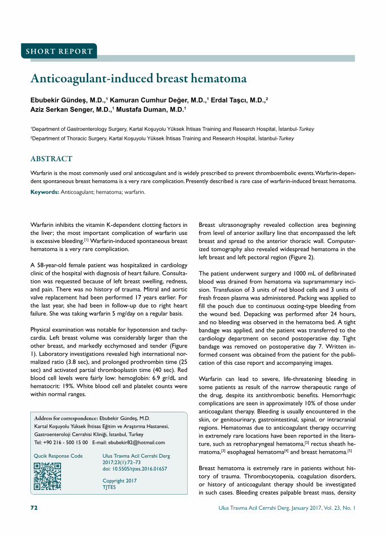
Physical examination was notable for hypotension and tachy-cardia. Left breast volume was considerably larger than the other breast, and markedly ecchymosed and tender (Figure 1). Laboratory investigations revealed high international nor-malized ratio (3.8 sec), and prolonged prothrombin time (25 sec) and activated partial thromboplastin time (40 sec). Red blood cell levels were fairly low: hemoglobin: 6.9 gr/dL and hematocrit: 19%. White blood cell and platelet counts were within normal ranges.
Breast ultrasonography revealed collection area beginning from level of anterior axillary line that encompassed the left breast and spread to the anterior thoracic wall. Computer-ized tomography also revealed widespread hematoma in the left breast and left pectoral region (Figure 2).
The patient underwent surgery and 1000 mL of defibrinated blood was drained from hematoma via supramammary inci-sion. Transfusion of 3 units of red blood cells and 3 units of fresh frozen plasma was administered. Packing was applied to fill the pouch due to continuous oozing-type bleeding from the wound bed. Depacking was performed after 24 hours, and no bleeding was observed in the hematoma bed. A tight bandage was applied, and the patient was transferred to the cardiology department on second postoperative day. Tight bandage was removed on postoperative day 7. Written in-formed consent was obtained from the patient for the publi-cation of this case report and accompanying images.
Warfarin can lead to severe, life-threatening bleeding in some patients as result of the narrow therapeutic range of the drug, despite its antithrombotic benefits. Hemorrhagic complications are seen in approximately 10% of those under anticoagulant therapy. Bleeding is usually encountered in the skin, or genitourinary, gastrointestinal, spinal, or intracranial regions. Hematomas due to anticoagulant therapy occurring in extremely rare locations have been reported in the litera-ture, such as retropharyngeal hematoma,[2] rectus sheath he-matoma,[3] esophageal hematoma[4] and breast hematoma.[5]
Breast hematoma is extremely rare in patients without his-tory of trauma. Thrombocytopenia, coagulation disorders, or history of anticoagulant therapy should be investigated in such cases. Bleeding creates palpable breast mass, density
S H O R T R E P O R T
Address for correspondence: Ebubekir Gündeş, M.D.
Kartal Koşuyolu Yüksek İhtisas Eğitim ve Araştırma Hastanesi,
Gastroenteroloji Cerrahisi Kliniği, İstanbul, Turkey
Tel: +90 216 - 500 15 00 E-mail: [email protected]
Qucik Response Code Ulus Travma Acil Cerrahi Derg2017;23(1):72–73doi: 10.5505/tjtes.2016.01657
Copyright 2017TJTES
Ulus Travma Acil Cerrahi Derg, January 2017, Vol. 23, No. 172

Ulus Travma Acil Cerrahi Derg, January 2017, Vol. 23, No. 1 73
Gündeş et al. Anticoagulant-induced breast hematoma
increase, and color change. Bleeding can also spread to the chest wall. Red blood cell transfusion and surgery may be needed in such cases.[5]
In conclusion, medical treatment may be adequate for self-limited small breast hematoma; however, in cases with risk of necrosis in the skin of the breast or if hematoma is extensive
Figure 1. Markedlyenlarged,ecchymosed,andtenderbreastduetohematoma.
enough to require blood transfusion, we believe that surgery must be considered without delay.
Conflict of interest: None declared.
REFERENCES
1. Landefeld CS, Beyth RJ. Anticoagulant-related bleeding: clinical epide-miology, prediction, and prevention. Am J Med 1993;95:315–28.
2. Toker I, Duman Atilla O, Yesilaras M, Ursavas B. Retropharyngeal He-matoma due to Oral Warfarin Usage. Turk J Emerg Med 2016;14:182–4.
3. Yunokizaki H, Tamura K, Li ZL, Abe T. Large spontaneous rectus sheath hematoma associated with severe anemia. Internal medicine 2015;54:349.
4. Guzman R, Ding L, Watson TJ, Hobbs SK, Litle VR. Spontaneous esophageal hematoma in a patient with atrial fibrillation. Ann Thorac Surg 2013;95(3):1089–91.
5. Özdemir B, Bayram AS, Bolca N, Kumbay E. Warfarin-Induced Chest Wall and Breast Hematoma in an Elderly Female Patient with Atrial Fi-brillation: Original Image. Turkiye Klinikleri J Med Sci 2008;6:28.
Figure 2. Computerizedtomographyshowingwidespreadhema-tomainleftbreastandpectoralregion.
KISA RAPOR - ÖZET
Antikoagülan tedaviye bağlı meme hematomuDr. Ebubekir Gündeş,1 Dr. Kamuran Cumhur Değer,1 Dr. Erdal Taşcı,2 Dr. Aziz Serkan Senger,1 Dr. Mustafa Duman1
1KartalKoşuyoluYüksekİhtisasEğitimveAraştırmaHastanesi,GastroenterolojiCerrahisiKliniği,İstanbul2KartalKoşuyoluYüksekİhtisasEğitimveAraştırmaHastanesi,GöğüsCerrahisiKliniği,İstanbul
Tromboembolik olayların önlenmesinde yaygın olarak kullanılan varfarin en sık kullanılan oral antikoagülandır. Varfarine bağlı spontan meme hema-tomu oldukça nadiren görülen bir komplikasyondur. Varfarine bağlı memede hematom gelişmesi nadir bir durumdur.Anahtar sözcükler: Antikoagülan; hematom; varfarin.
Ulus Travma Acil Cerrahi Derg 2017;23(1):72–73 doi: 10.5505/tjtes.2016.01657

A rare cause of ileus: late jejunal stricture followingblunt abdominal traumaUlaş Aday, M.D.,1 Ebubekir Gündeş, M.D.,1 Kamuran Cumhur Değer, M.D.,1
Hüseyin Çiyiltepe, M.D.,1 Şükran Kayıpmaz, M.D.,2 Mustafa Duman, M.D.1
1DepartmentofGastrointestinalSurgery,KartalKoşuyoluYüksekİhtisasTrainingandResearchHospital,İstanbul-Turkey2DepartmentofPatology,KartalDr.LütfiKırdarTrainingandResearchHospital,İstanbul-Turkey
ABSTRACT
Small intestinal stricture forming in the late phase following nonpenetrating abdominal trauma is rare cause of ileus. It has often been suggested that it is result of localized feeding deficiency on the intestinal wall related to minor trauma in the mesentery. Laparoscopy has been increasingly used for diagnosis and treatment. Diagnosis should be supported by pathological analyses in case of intestinal stenosis related to blunt abdominal traumas.
Keywords: Intestinal stenosis; laparoscopy; trauma.
INTRODUCTION
Small intestinal obstruction forming in the late phase follow-ing blunt abdominal trauma is quite rare; studies have report-ed rate as 1%.[1,2] It is often caused by fibrotic scar formation and blockage of passage entry during healing process of isch-emic areas, which develop as result of localized damage to the intestinal wall or the mesentery.[1–4] A 37-year-old male patient, who had occupational accident 2 years prior, was sur-gically treated at our clinic after presenting with occasional abdominal pain and swelling that had been going on for a year. Presently described is rarely seen case of patient diagnosed with post-traumatic jejunal stricture.
CASE REPORT
A 37-year-old male patient presented at the clinic with com-plaints of occasional abdominal pain and distension that had been going on for a year. The patient’s history revealed that
he had previously been treated at another clinic for left iliac wing fracture following a fall from a tractor and that he had been discharged without surgical procedure. His physical ex-amination revealed distended abdomen, increase in intestinal sounds and tympanism in the left upper quadrant, and mild sensitivity. He had linear scar tissue 10 cm in length on the left iliac wing as result of the traumatic skin laceration. The patient had no known chronic disease or earlier history of surgery. Standing abdominal computed tomography indicated dilated loops of small bowel segments. The patient’s hemoglobin level was 9.6 g/dL (normal range: 11.1–17.1 g/dL), hematocrit volume was 29.9% (normal range: 33–54%), albumin level was 3.2 g/dL (normal range: 3.5–5.2 g/dL), and C-reactive protein level was 4.74 mg /dL (normal range 0–0.34 mg/dL). His other laboratory parameters were within normal limits. Oral and intravenous contrasted abdominopelvic tomography revealed dilated jejunal loops and obstructed area with partial passage at the end point of dilatation. There was also thickening and irregularity in the mesentery of the same segment (Figure 1). Nasogastric tube was inserted and medical observation was initiated; however, upon seeing no development in his clinical condition, laparoscopic exploration was performed. Fibrotic thickening was seen in the mesentery of the jejunal segment about 80 cm from the ligament of Treitz, and circular fibrotic area of 1 cm diameter was observed on the intestinal wall. It was also seen that the proximal segment was quite dilated. Laparoscopic segmental resection and side–to-side jejuno-jejunal anastomosis were performed. Macroscopic evaluation of the resected portion revealed circular cicatricial area caus-ing narrowness, fibrotic thickening in the mesentery, and dila-
C A S E R E P O R T
Address for correspondence: Ulaş Aday, M.D.
Kartal Koşuyolu Yüksek İhtisas Eğitim ve Araştırma Hastanesi,
34000 İstanbul, Turkey
Tel: +90 216 - 459 44 40 E-mail: [email protected]
Qucik Response Code Ulus Travma Acil Cerrahi Derg2017;23(1):74–76doi: 10.5505/tjtes.2016.06981
Copyright 2017TJTES
Ulus Travma Acil Cerrahi Derg, January 2017, Vol. 23, No. 174

Ulus Travma Acil Cerrahi Derg, January 2017, Vol. 23, No. 1 75
Aday et al. A rare cause of ileus
tation and edema in the proximal loop. It was also observed that distal crossing diameter was quite narrowed (Figure 2). The patient was discharged on post-operative day 8 without any problems. Pathological analysis demonstrated focal ulcer-ated area and active chronic nonspecific inflammation of the site (Figure 3).
DISCUSSION
Ileus related to post-traumatic intestinal stenosis is rare; studies have reported that rate is about 1%.[1,2] Jejunal stricture related to blunt abdominal trauma often form as result of minor trauma to the mesentery or the intestinal wall. There may be no symptoms that can be seen in the clinical condition of the patient in the early phase.[1,3] Small laceration, hematoma in the mesentery, or contusion and mural hematoma on the intestinal wall give way to local-ized ischemia. Inadequate mucosal feeding causes bacterial translocation, and ulcer formation and inflammation during tissue healing result in fibrosis and scarring. Authors agree that feeding deficiency related to damaged mesentery is pri-mary reason for stricture formation.[4–6] In the present case, macroscopic evaluation showed fibrotic thickening in the mesentery of the strictured segment. It was suggested that the damage to the mesentery was the principal reason for the patient’s condition as pathological analysis revealed focal ulcerated area and active chronic nonspecific inflammation in the area.
Although post-traumatic symptoms are frequently seen 5 weeks after the trauma, there are also reports present-
ing cases that remained asymptomatic for a long time.[2–4] In our case, the patient’s complaints started a year after the traumatic incident. Symptoms related to partial small bowel obstruction, such as intermittent abdominal pain, distension, nausea, and vomiting are seen.[4,6] Rate of proximal jejunal stricture reported in the literature varies; however, Konobu et al. provided rate of 17.9% in their study.[4,7]
Diagnostic criteria for the condition are as follows: a) previous history of blunt abdominal trauma, b) absence of described pathology before trauma, c) start of symptoms after trauma, d) radiological detection of intestinal stenosis, e) malignity or signs of specific inflammatory diseases seen in pathological evaluation of the resected portion.[1,2,7] Pathological confirma-tion is significant for cases in which no differential diagnosis
Figure 1. Abdominaltomographysectionwithoralandintravenouscontrast.
Figure 2. Imageoftheresectedportion.
Figure 3. Low-powerviewoftheulcerillustratesthedepthofthelesionandintensemixed-typeinflammation(hematoxylinandeo-sin,x40).

Aday et al. A rare cause of ileus
can be reached through radiological evaluation, and for dif-ferentiating cases of Crohn’s disease, intestinal tuberculosis, radiation enteritis, or cancer.[1,2,7,8]
There has been increase in laparoscopic surgery as result of technological developments and accumulated experience with laparoscopic procedures. Small intestinal obstruction related to adhesion occurs far less often in intra-abdominal laparo-scopic procedure in comparison with open surgery. Studies with large scope have demonstrated that laparoscopic treat-ment has low morbidity and mortality rates for adhesions, which are the most common reason for small bowel obstruc-tion.[8,9] Adhesions requiring re-operation formed within 30 years in 29% of cases of laparoscopic adhesiolysis.[9,10] In our case, trauma-related stenosis was not initially thought of in the preoperative period. Macroscopic results cast doubt on the diagnostic laparoscopy and the patient was diagnosed based on pathological evaluation. Less invasive laparoscopy to complete the surgical procedure decreased risk of adhesions forming later.
Intestinal stenosis in the late phase related to blunt abdominal trauma is a rare cause of ileus in surgical practice. Resection of the segment causing stricture not only achieves treatment, but also enables histopathological confirmation. Increased surgical experience with laparoscopic procedures has facili-tated safe resection of pathological segment and thereby de-creased rate of post-operative adhesion formation.
Conflict of interest: None declared.
REFERENCES
1. Kang GH, Jeon TJ, Seo DD, Oh TH, Kim SH, Cho HS, et al. Ileal ste-nosis occurred 3 months after blunt abdominal trauma. Korean J Gastro-enterol 2011;57:370–3.
2. Kaban G, Somani RA, Carter J. Delayed presentation of small bowel in-jury after blunt abdominal trauma: case report. J Trauma 2004;56:1144–5.
3. De Backer AI, De Schepper AM, Vaneerdeweg W, Pelckmans P. Intes-tinal stenosis from mesenteric injury after blunt abdominal trauma. Eur Radiol 1999;9:1429–31.
4. Jones VS, Soundappan SV, Cohen RC, Pitkin J, La Hei ER, Martin HC, et al. Posttraumatic small bowel obstruction in children. J Pediatr Surg 2007;42:1386–8.
5. Loberant N, Szvalb S, Herskovits M, Cohen I, Salamon V. Posttraumatic intestinal stenosis: radiographic and sonographic appearance. Eur Radiol 1997;7:524–6.
6. Maharaj D, Perry A, Ramdass M, Naraynsingh V. Late small bowel ob-struction after blunt abdominal trauma. Postgrad Med J 2003;79:57–8.
7. Konobu T, Murao Y, Miyamoto S, Nakamura T, Imanishi M, Ueda S, et al. Posttraumatic intestinal stenosis presenting as a perforation: report of a case. Surg Today 1999;29:564–7.
8. O’Connor DB, Winter DC. The role of laparoscopy in the management of acute small-bowel obstruction: a review of over 2,000 cases. Surg En-dosc 2012;26:12–7.
9. Ghosheh B, Salameh JR. Laparoscopic approach to acute small bowel ob-struction: review of 1061 cases. Surg Endosc 2007;21:1945–9.
10. Gutt CN, Oniu T, Schemmer P, Mehrabi A, Büchler MW. Fewer adhe-sions induced by laparoscopic surgery? Surg Endosc 2004;18:898–906.
OLGU SUNUMU - ÖZET
Nadir görülen ileus nedeni: Künt karın travması sonrası geç dönem gelişen jejunal striktürDr. Ulaş Aday,1 Dr. Ebubekir Gündeş,1 Dr. Kamuran Cumhur Değer,1 Dr. Hüseyin Çiyiltepe,1
Dr. Şükran Kayıpmaz,2 Dr. Mustafa Duman1
1KartalKoşuyoluYüksekİhtisasEğitimveAraştırmaHastanesi,GastroenterolojiCerrahisiKliniği,İstanbul2KartalDr.LütfiKırdarEğitimveAraştırmaHastanesi,PatolojiBölümü,İstanbul
Penetran olmayan karın travmalarına bağlı geç dönemde oluşan ince bağırsak striktürü nadir ileus nedenidir. Sıklıkla mezenterde oluşan minör trav-maya bağlı bağırsak duvarındaki lokalize beslenme bozukluğunun sonucu olduğu düşünülmektedir. Laparoskopi tanı ve tedavi uygulamalarında artan oranlarda uygulanmaktadır. Künt karın travmasına bağlı intestinal stenozlarda tanının patolojik değerlendirme ile desteklenmesi gerekir.Anahtar sözcükler: İntestinal stenoz; laparoskopi; travma.
Ulus Travma Acil Cerrahi Derg 2017;23(1):74–76 doi: 10.5505/tjtes.2016.06981
Ulus Travma Acil Cerrahi Derg, January 2017, Vol. 23, No. 176