antigenic variation
TRANSCRIPT
1
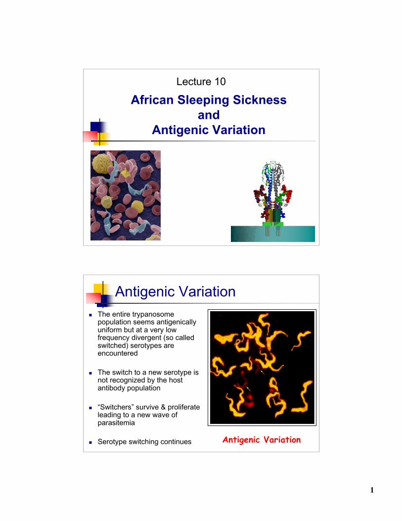
African Sleeping Sicknessand
Antigenic Variation
Lecture 10
Antigenic Variation The entire trypanosome
population seems antigenicallyuniform but at a very lowfrequency divergent (so calledswitched) serotypes areencountered
The switch to a new serotype isnot recognized by the hostantibody population
“Switchers” survive & proliferateleading to a new wave ofparasitemia
Serotype switching continues Antigenic Variation
2
Antigenic Variation
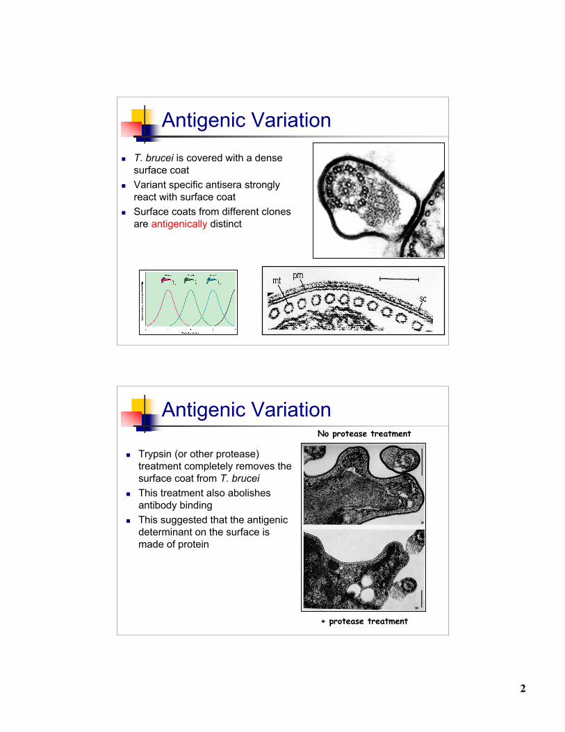
T. brucei is covered with a densesurface coat
Variant specific antisera stronglyreact with surface coat
Surface coats from different clonesare antigenically distinct
Antigenic Variation
Trypsin (or other protease)treatment completely removes thesurface coat from T. brucei
This treatment also abolishesantibody binding
This suggested that the antigenicdeterminant on the surface ismade of protein
No protease treatment
+ protease treatment
3
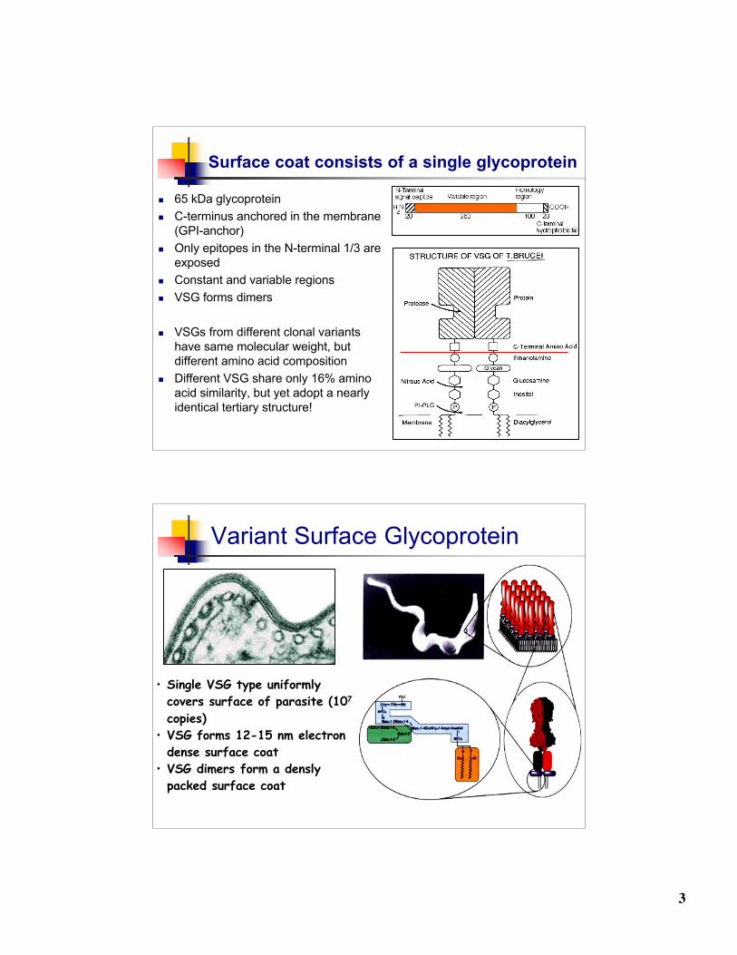
Surface coat consists of a single glycoprotein
65 kDa glycoprotein C-terminus anchored in the membrane
(GPI-anchor) Only epitopes in the N-terminal 1/3 are
exposed Constant and variable regions VSG forms dimers
VSGs from different clonal variantshave same molecular weight, butdifferent amino acid composition
Different VSG share only 16% aminoacid similarity, but yet adopt a nearlyidentical tertiary structure!
Variant Surface Glycoprotein
• Single VSG type uniformlycovers surface of parasite (107
copies)• VSG forms 12-15 nm electron
dense surface coat• VSG dimers form a densly
packed surface coat

4
Variant Surface Glycoprotein
Different VSG share only 16%similarity, but yet adopt anearly identical tertiarystructure!
Variableregion
Constantregion
T. brucei life cycle
non-dividingfuel=glucose
VSG coatmito=“low”
Dividing formfuel=glucose
VSG coatmito=“off”
non-dividingfuel=?
mVSG coatmito=?
Dividing formfuel=amino acids
Procyclin coatmito=“on”
5
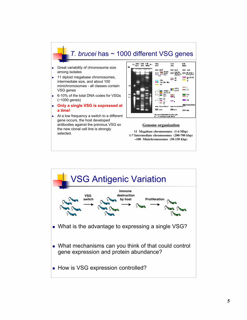
T. brucei has ~ 1000 different VSG genes
Great variability of chromosome sizeamong isolates
11 diploid megabase chromosomes,intermediate size, and about 100minichromosomes - all classes containVSG genes
6-10% of the total DNA codes for VSGs(~1000 genes)
Only a single VSG is expressed ata time!
At a low frequency a switch to a differentgene occurs, the host developedantibodies against the previous VSG sothe new clonal cell line is stronglyselected.
Genome organization11 Megabase chromosomes (1-6 Mbp)
1-7 Intermediate chromosomes (200-700 kbp)~100 Minichromosomes (50-150 kbp)
VSG Antigenic Variation
What is the advantage to expressing a single VSG?
What mechanisms can you think of that could controlgene expression and protein abundance?
How is VSG expression controlled?
VSGswitch
Immune destruction
by host Proliferation
6
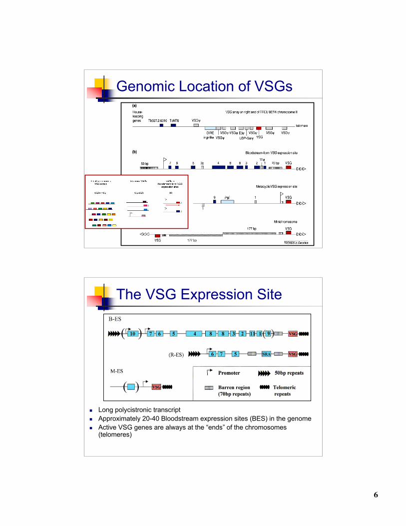
Genomic Location of VSGs
The VSG Expression Site
Long polycistronic transcript Approximately 20-40 Bloodstream expression sites (BES) in the genome Active VSG genes are always at the “ends” of the chromosomes
(telomeres)
7
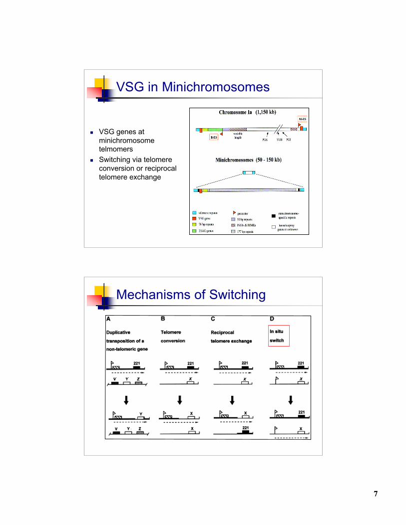
VSG in Minichromosomes
VSG genes atminichromosometelmomers
Switching via telomereconversion or reciprocaltelomere exchange
Mechanisms of Switching
8
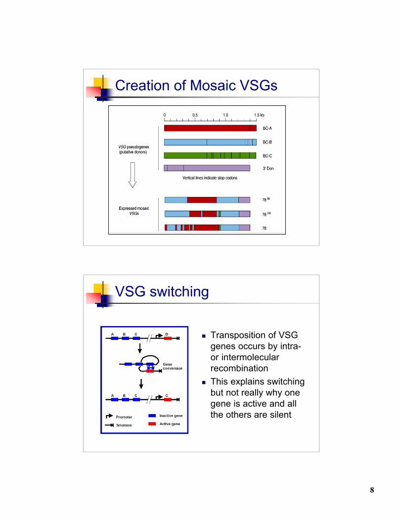
Creation of Mosaic VSGs
VSG switching
Transposition of VSGgenes occurs by intra-or intermolecularrecombination
This explains switchingbut not really why onegene is active and allthe others are silent
9
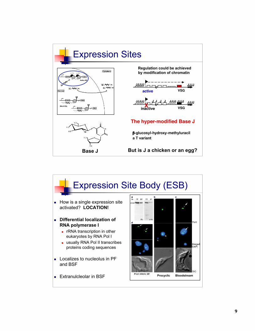
Expression Sites
VSG
VSG
active
inactiveJJJJJJJJ JJ JJJJJJ JJ
JJJJJJJJJ
JJJX
The hyper-modified Base J
But is J a chicken or an egg?
Regulation could be achieved by modification of chromatin
β-glucosyl-hydroxy-methyluracil a T variant
Base J
Expression Site Body (ESB) How is a single expression site
activated? LOCATION!
Differential localization ofRNA polymerase I rRNA transcription in other
eukaryotes by RNA Pol I usually RNA Pol II transcribes
proteins coding sequences
Localizes to nucleolus in PFand BSF
Extranulcleolar in BSF BloodstreamProcyclic
10
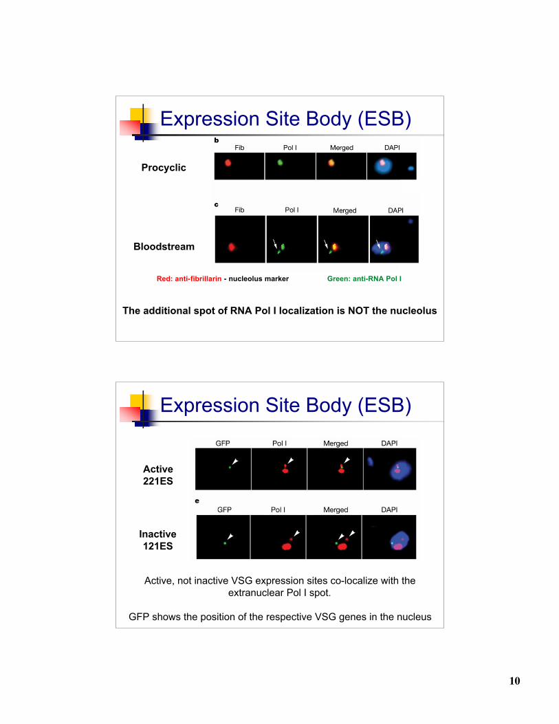
Expression Site Body (ESB)
Procyclic
Bloodstream
Red: anti-fibrillarin - nucleolus marker Green: anti-RNA Pol I
The additional spot of RNA Pol I localization is NOT the nucleolus
Expression Site Body (ESB)
Active, not inactive VSG expression sites co-localize with theextranuclear Pol I spot.
GFP shows the position of the respective VSG genes in the nucleus
Active221ES
Inactive121ES
11
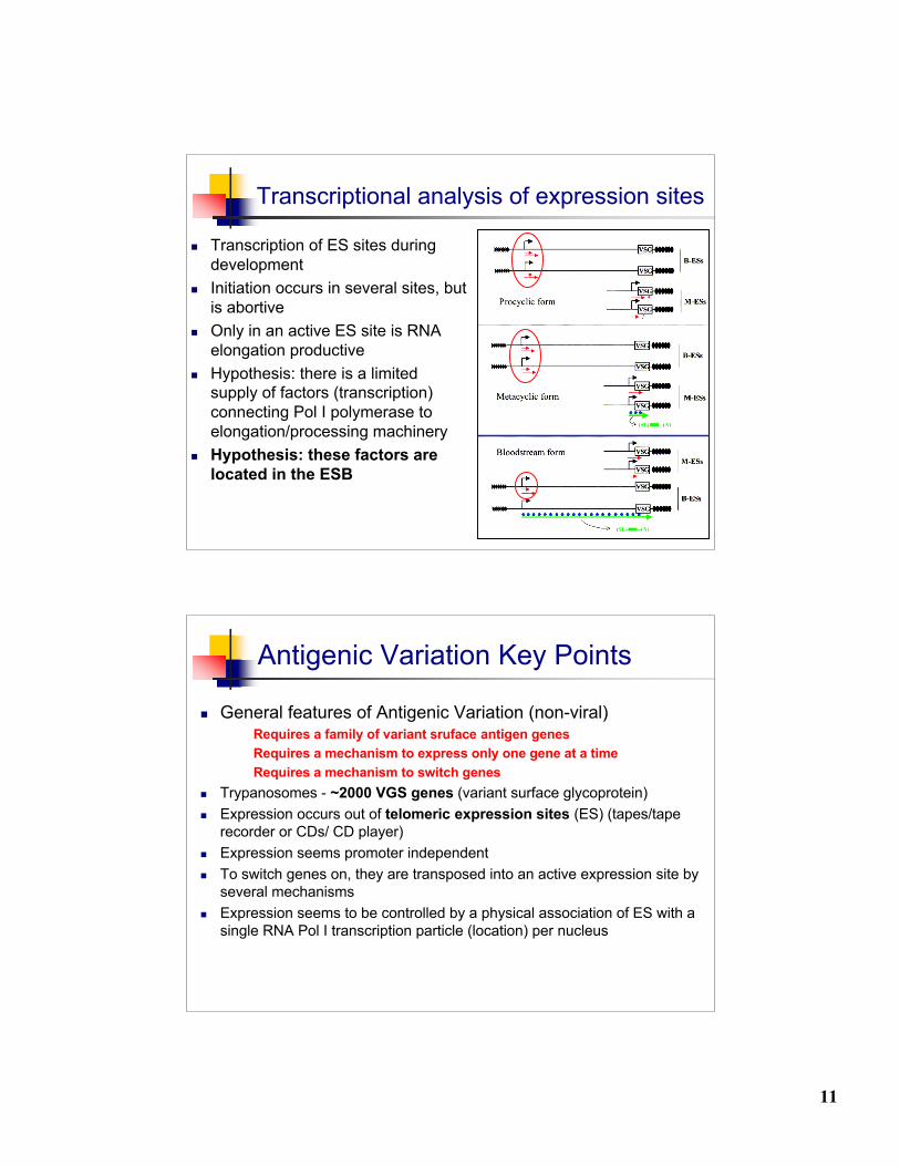
Transcriptional analysis of expression sites
Transcription of ES sites duringdevelopment
Initiation occurs in several sites, butis abortive
Only in an active ES site is RNAelongation productive
Hypothesis: there is a limitedsupply of factors (transcription)connecting Pol I polymerase toelongation/processing machinery
Hypothesis: these factors arelocated in the ESB
Antigenic Variation Key Points
General features of Antigenic Variation (non-viral)Requires a family of variant sruface antigen genesRequires a mechanism to express only one gene at a timeRequires a mechanism to switch genes
Trypanosomes - ~2000 VGS genes (variant surface glycoprotein) Expression occurs out of telomeric expression sites (ES) (tapes/tape
recorder or CDs/ CD player) Expression seems promoter independent To switch genes on, they are transposed into an active expression site by
several mechanisms Expression seems to be controlled by a physical association of ES with a
single RNA Pol I transcription particle (location) per nucleus
12
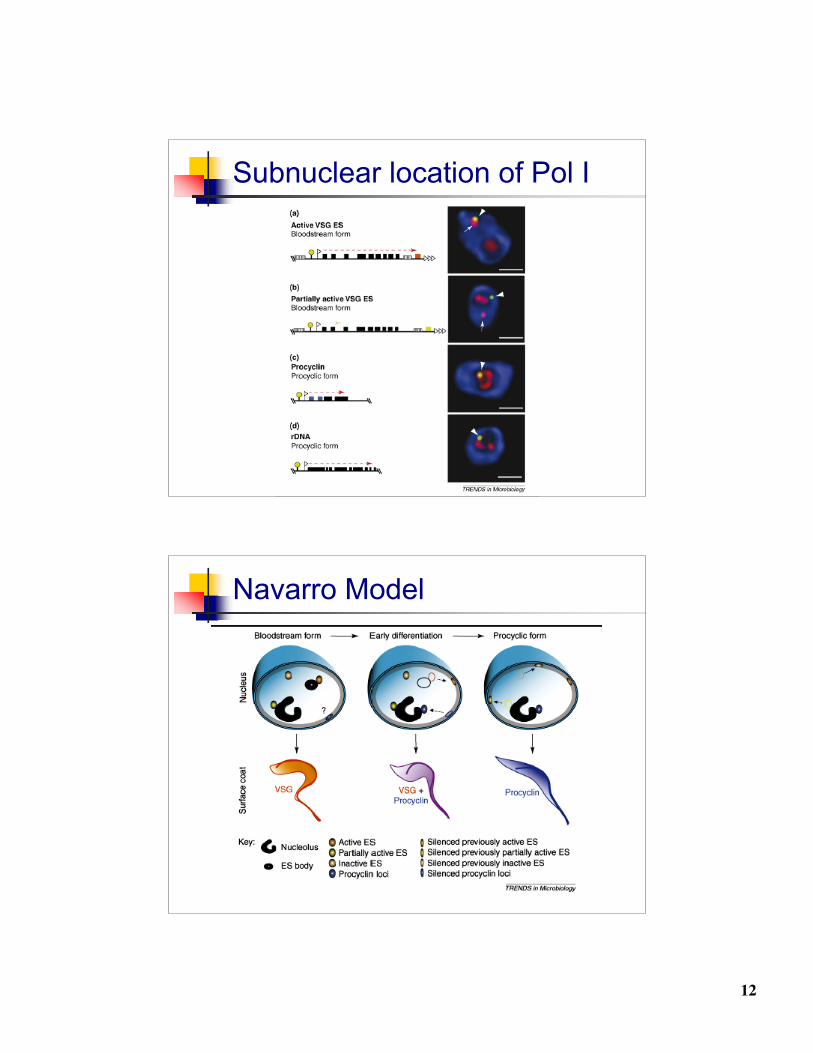
Subnuclear location of Pol I
Navarro Model
13
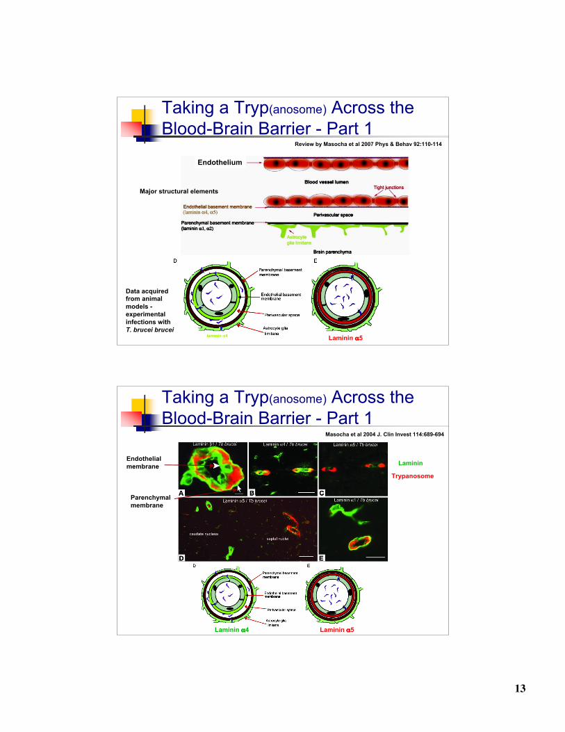
Taking a Tryp(anosome) Across theBlood-Brain Barrier - Part 1
Data acquiredfrom animalmodels - experimental infections with T. brucei brucei
Laminin α5
Endothelium
Major structural elements
Review by Masocha et al 2007 Phys & Behav 92:110-114
Taking a Tryp(anosome) Across theBlood-Brain Barrier - Part 1
Laminin α4 Laminin α5
Trypanosome
Masocha et al 2004 J. Clin Invest 114:689-694
Endothelialmembrane
Parenchymalmembrane
Laminin
14
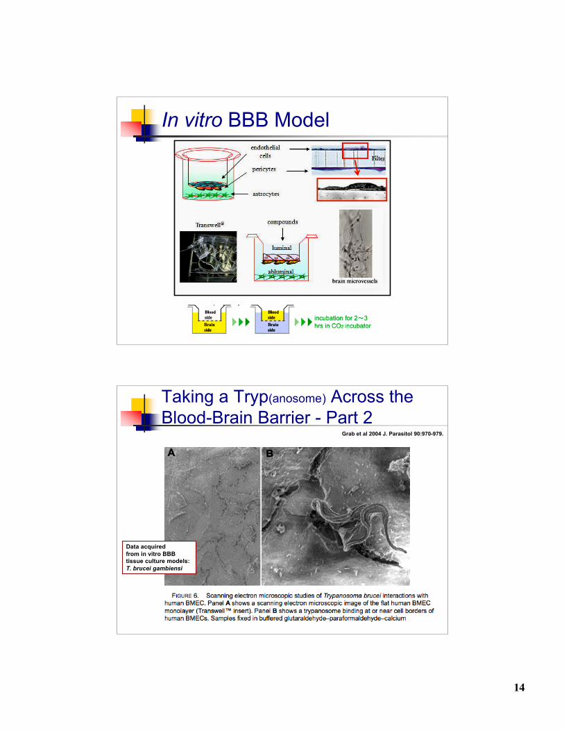
In vitro BBB Model
Taking a Tryp(anosome) Across theBlood-Brain Barrier - Part 2
Laminin α4 Laminin α5
Grab et al 2004 J. Parasitol 90:970-979.
Data acquiredfrom in vitro BBBtissue culture models:T. brucei gambiensi

15
Taking a Tryp(anosome) Across theBlood-Brain Barrier - Part 2
Grab et al 2004 J. Parasitol 90:970-979.