antigens three cryptosporidium species - iai.asm.orgiai.asm.org/content/60/4/1509.full.pdf ·...
TRANSCRIPT

INFEcrION AND IMMUNITY, Apr. 1992, p. 1509-15130019-9567/92/041509-05$02.00/0Copyright X 1992, American Society for Microbiology
Analysis of Oocyst Wall and Sporozoite Antigens from ThreeCryptosporidium Species
JAIME M. S. NINA,' VINCENT McDONALD,l* DAVID A. DYSON,2 JANET CATCHPOLE,3 SHIGEHIKO UNI,4MOTOHERO ISEKI,4 PETER L. CHIODINI,' AND KEITH P. W. J. McADAM'
Department of Clinical Sciences, London School of Hygiene and Tropical Medicine, Keppel Street, London WClE 7HT,and Hospital for Tropical Diseases, St. Pancras Way, London NW1 OPE, 1 Veterinary Investigation Centre, MinistryofAgriculture, Fisheries and Food, Calthwaite, Penrith, Cumbria CAll 9RR,2 and M.A.F.F. Central Veterinary
Laboratory, New Haw, Weybridge, Surrey, KT15 3NB,3 United Kingdom, and Department ofMedical Zoology, Osaka City University Medical School, Abeno-ku, Osaka 545, Japan4
Received 9 September 1991/Accepted 14 January 1992
A comparison was made of the antigenic composition of oocyst walls and sporozoites from Cryptosporidiumbaileyi from turkeys, C. muris from rodents, and C. parvum from ruminants, employing immunoblotting andimmunofluorescence. In immunoblotting, oocyst antigens were subjected to sodium dodecyl sulfate-polyacryl-amide gel electrophoresis and Western blotting (immunoblotting) and detected with rabbit polyclonal anti-C.muris or -C. parvum antibodies or murine monoclonal antibodies developed against C. parvum. Immunofluo-rescence was used to investigate the reactivity of these monoclonal antibodies with air-dried excystationmixtures of sporozoites and oocysts of the different species. The results from both types of experiment indicatedthat the three Cryptosporidium species could be differentiated immunologically. In comparison, few antigenicdifferences were found between a number of isolates of C. parnum in immunoblotting. There was also evidenceto suggest that C. parvum and C. bailkyi were more closely related antigenically to one another than to C. muris.
The coccidian genus Cryptosporidium is the causativeagent of cryptosporidiosis, a disease found worldwide in avariety of vertebrates. Since the first report of the genus byTyzzer (20), a large number of species have been recorded inthe literature, each usually being distinguished by descrip-tive features including parasite morphology, site of infection,and host species of origin (6). Because little information isavailable on the biology of the parasite, including strainvariation, it is not clear whether information on thesefeatures alone is sufficient to determine species of Crypto-sporidium. Certainly, the inclusion of host species of originas a factor in identifying "new" species has caused someconfusion. It has often been assumed that Cryptosporidium,like many other coccidia, has strict host specificity, butrecent evidence suggests that the genus has a relatively widehost range. C. parvum and C. muris, for example, are eachable to infect diverse mammalian species, and the develop-ment of C. baileyi may occur in separate avian host species(6, 7, 21). The recent finding of disseminated C. baileyiinfection in an AIDS patient (5) also suggests that immuno-suppression of the host weakens any barrier to infection withcryptosporidia of other vertebrate classes.The employment of techniques in biochemistry, immunol-
ogy, and molecular genetics is likely to be helpful in theidentification of Cryptosporidium species and strains andshould be valuable in epidemiology and taxonomy.
In studies by earlier workers, Cryptosporidium specieswere differentiated by polypeptide composition of the oocystwall (12, 19) and electrophoretic mobility of chromosomes(15). Antigen profiles of oocysts in Western blots (immuno-blots) have been reported (9, 15), and significant differenceswere observed between the profiles of C. parvum and C.baileyi by using a polyclonal anti-C. parvum serum (2). Someantigenic diversity has also been observed between separate
* Corresponding author.
isolates of C. parvum (17, 18), and a sporozoite antigen ofthis species was found not to be conserved among differentisolates (3).
In the present report, results are presented of a newcomparative investigation of the antigenic composition ofseparate Cryptosporidium species. Oocyst antigens fromthree species, C. baileyi, C. munis, and C. parvum, wereexamined by Western blotting and immunofluorescence.Polyclonal antisera against two of the species or monoclonalantibodies (MAbs) developed against C. parvum were usedfor antigen detection. The results demonstrated that therewas greater antigenic diversity between the three species ofparasite than there was between isolates of a single species(C. parvum). There was also evidence that C. parvum and C.baileyi are more closely related antigenically to one anotherthan to C. muris.
MATERIALS AND METHODS
Parasites. The MD isolate of C. parvum, originally isolatedfrom a deer, was passaged in calves. This isolate was used torepresent C. parvum in experiments to compare antigens ofdifferent Cryptosporidium species. Other isolates of thisspecies were provided by clinicians or field veterinary work-ers. The RN 66 strain of C. muris (7) was passaged inC57BL/6 mice or BALB/c mice with severe combined im-munodeficiency, and an isolate of C. baileyi was obtainedfrom turkeys. Purified oocysts of each species were obtainedby sucrose or salt flotation (7, 14). Excystation of sporozo-ites was obtained by incubation of oocysts at 37°C in RPMI1640 medium with 0.4% bovine bile salts for 60 to 120 min.Bile salts were found not to be required for C. munis.
Preparation of antibodies against oocyst antigens. Poly-clonal antibodies against C. parvum or C. muris antigenswere produced in New Zealand White rabbits by threeintramuscular injections, at intervals of 14 days, of 5 x 10⁶homogenized oocysts (13) of a human isolate of C. parvum
1509
Vol. 60, No. 4
on June 7, 2018 by guesthttp://iai.asm
.org/D
ownloaded from
1510 NINA ET AL.
or the RN 66 strain of C. muris incorporated in Freund'scomplete adjuvant. The serum was collected 10 days afterthe final injection of antigen.Murine MAbs against the same human isolate of C.
parvum were prepared from fusions between BALB/c spleencells, following immunization of animals with disruptedoocysts, and SP2/0 myeloma cells, as described by McDon-ald et al. (13). The specificity of the MAbs for parasites wasdetermined by the indirect fluorescent-antibody test (IFAT),using an air-dried and acetone-fixed excystation mixture ofoocysts and sporozoites as the antigen (13).
Electrophoresis and Western blotting. Sodium dodecylsulfate-polyacrylamide gel electrophoresis (SDS-PAGE) wasperformed as described previously (13), based on the methodof Laemmli (8), using 5% stacking gels and 7.5% separatinggels. Oocyst antigens were prepared by freeze-thawing,using liquid nitrogen, of fresh oocysts in a standard electro-phoresis sample buffer containing 1.5 mM dithiothreitol.Particulate matter was removed from the freeze-thawedpreparation by centrifugation at 13,000 rpm for 5 min, andthe soluble material, which formed the electrophoretic sam-ples, was heated at 100°C for 90 s. Each sample contained asimilar amount of protein, estimated by the method ofLowry et al. (11); thus, for C. baileyi, C. muris, and C.parvum, respectively, samples contained the equivalent of1.0 x 106, 4 x 10⁵, and 2.5 x 10⁶ oocysts. Electrophoresiswas done in a Biotech Easy apparatus (15 by 15 cm) with aconstant current of 20 mA.
Antigens were transferred electrophoretically from gels toImmobilon-P membranes (Millipore) by using a semi-dryWestern blotting system (Biotech). Transfer was obtainedwith a constant current of 100 mA for 90 min.
After incubation of the blots with anti-Cryptospondiumantibodies, blocking was performed with 4% bovine serumalbumin in Tris-buffered saline (pH 10.3) (13). The blots werethen treated with a second antibody, goat anti-rabbit immu-noglobulin G or anti-mouse immunoglobulin, conjugatedwith alkaline phosphatase (Sigma), and antigens were visu-alized following the addition of 5-bromo-4-chloro-3-indolylphosphate and nitroblue tetrazolium (Sigma).
RESULTS
Antigenic differences between species were observed inIFAT. The anti-C. parvum MAbs used in this study wereknown to recognize the oocyst wall (181B5) or part(s) of thesporozoite (all remaining antibodies). The reactivities of theMAbs with sporozoites and oocysts from C. baileyi, C.muris, and C. parvum are presented in Table 1. As inprevious experiments, all the antibodies reacted stronglywith C. parvum. The oocyst wall-reactive antibody, 181B5,also bound to the same component of C. baileyi and C.muris, although with less intensity. Only two of the sporo-zoite-reactive MAbs, 182C1 and 182C2b, were observed tobind to sporozoites of C. muns, and the fluorescence wasweak. Sporozoites of C. baileyi reacted strongly with MAbs181C3 and 182C1, moderately with 182C2b, and weakly with182A6. The remaining five sporozoite-reactive antibodiesfailed to bind to C. baileyi.Immunoblots with polyclonal antisera demonstrated anti-
genic differences between Cryptosporidium spp. Oocysts iso-lated from a number of unrelated human cases ofcryptosporidiosis in southeast England had the typical shapeand size (almost spherical and approximately 5 ,um indiameter) of those associated with C. parvum. Freeze-thawed oocyst material was subjected to SDS-PAGE and
TABLE 1. Interaction in IFAT of murine MAbs developedagainst C. parvum with sporozoites or oocysts of
C. parvum, C. baileyi, and C. muris
MAb Staining patterna C. parvumb C. baileyib C. muris"
181B5 Oocyst wall + + + +
172A4 Whole ++ - -181A4 Whole + +181C3 Interior + + + +182A6 Surface ++ +182B2 Surface ++182B4 Surface + +182B7 Whole + +182C1 Interior and surface ++ ++ +182C2b Surface ++ + +
a The part(s) of the sporozoite of the homologous isolate of C. parvumrecognized by each MAb is given; the exceptional antibody is 181B5, whichreacted only with the oocyst wall.
b A semiquantitative assessment was made of fluorescence: + +, strong; +,moderate; +, weak; -, none.
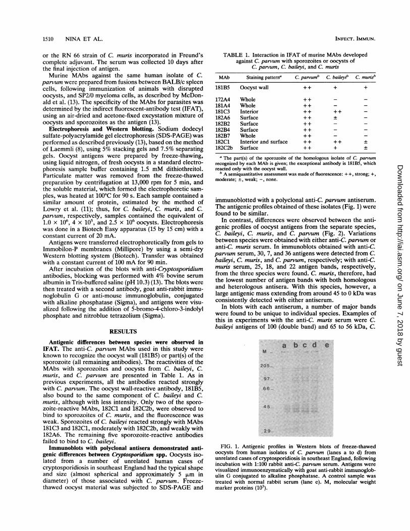
immunoblotted with a polyclonal anti-C parvum antiserum.The antigenic profiles obtained of these isolates (Fig. 1) werefound to be similar.
In contrast, differences were observed between the anti-genic profiles of oocyst antigens from the separate species,C. baileyi, C. muris, and C. parvum (Fig. 2). Variationsbetween species were obtained with either anti-C. parvum oranti-C. muris serum. In immunoblots obtained with anti-C.parvum serum, 30, 7, and 36 antigens were detected from C.baileyi, C. muris, and C. parvum, respectively; with anti-C.muris serum, 25, 18, and 22 antigen bands, respectively,from the three species were found. C. muris, therefore, hadthe lowest number of antigen bands with both homologousand heterologous antisera. With this species, however, alarge antigenic mass extending from around 45 to 0 kDa wasconsistently detected with either antiserum.
In blots with each antiserum, a number of major bandswere found to be unique to individual species. Examples ofthis in experiments with the anti-C. munis serum were C.baileyi antigens of 100 (double band) and 65 to 56 kDa, C.
a b c d e
205-
97-
66 -
45
2 9
FIG. 1. Antigenic profiles in Western blots of freeze-thawedoocysts from human isolates of C. parvum (lanes a to d) fromunrelated cases of cryptosporidiosis in southeast England, followingincubation with 1:100 rabbit anti-C. parvum serum. Antigens werevisualized immunoenzymatically with goat anti-rabbit immunoglob-ulin G conjugated to alkaline phosphatase. A control sample wastreated with normal rabbit serum (lane e). M, molecular weightmarker proteins (103).
INFEcr. IMMUN.
on June 7, 2018 by guesthttp://iai.asm
.org/D
ownloaded from

ANTIGENS FROM THREE CRYPTOSPORIDIUM SPECIES 1511
c b cm cp cb cm cp cb cm c p
M
205-
66 -
45_ alli'
29- " |
Mcb cp cm cp cb cb cp cm
205
97-
66-
46-
29-
antlop normsl
FIG. 2. Antigenic profiles in Western blots of freeze-thawedoocysts of C. baileyi (cb), C. muris (cm), and C. parvum (cp) afterincubation with 1:100 serum from rabbits which either were immu-nized with oocyst antigens of C. muris (anti-cm) or C. parvum(anti-cp) or remained unimmunized. Antigens were visualized enzy-matically as described in the legend to Fig. 1. M, molecular weightmarker proteins (103).
muris antigens of 110 and 51 to 54 kDa (more than one band),and C. parvum antigen bands of 58 and 48 kDa. With theanti-C. parvum serum, bands of 56 and 51 kDa and of 48, 42,and 38 kDa were seen only with C. baileyi and C. parvum,respectively. Some antigen bands of each species weredetected with only one antiserum or were more prominent inblots with one antiserum than with the other. At least twobands of C. parvum (58 and 48 kDa) were observed moreprominently with the heterologous antiserum than with thehomologous one.Immunoblots with MAbs showed antigenic differences be-
tween species. The MAbs used in IFAT were also used inimmunoblots of oocyst antigens from the three species ofparasite. Similar antigenic profiles were usually obtainedwith different isolates of C. parvum, and examples of this arepresented in Fig. 3. The oocyst wall-specific MAb, 181B5,
Ma b c d
16.
97-
S.-
45-
t h I
*116..47-
iI
FIG. 3. Detection of parasite antigens in Western blots fromdifferent freeze-thawed oocyst isolates of C. parvum from animalsand humans, following treatment with MAb 181B5 (lanes a to d) or182A6 (lanes f to h) in ascites diluted 1:50. Antigens were visualizedimmunoenzymatically with goat anti-mouse immunoglobulin conju-gated to alkaline phosphatase. Samples treated with 181B5: lane a,cervine isolate, passaged in calves; lane b, calf isolate; lane c, sheepisolate 1; lane d, sheep isolate 2; lane e, same isolate as in lane a buttreated with 1:50 normal mouse ascites. Samples treated with182A6: lane f, immunocompetent child; lane g, AIDS patient 1; laneh, AIDS patient 2; lane i, control (same as lane e). M, molecularweight marker proteins (103).
18185 182A8 controlFIG. 4. Comparison of antigens of freeze-thawed oocysts of
different Cryptosporidium species detected by MAbs 181B5 and182A6 in ascites diluted 1:50. The parasite species were C. baileyi(cb), C. muris (cm), and C. parvum (cp). Controls were treated with1:50 normal mouse ascites. Antigens were visualized as described inthe legend to Fig. 3. The positions of the faint bands of C. baileyirecognized by 182A6 are marked with arrowheads. M, molecularweight marker proteins (103).
reacted strongly with a 44-kDa band from each of fouranimal isolates and, except in one case, similarly reactedwith a 41-kDa band (Fig. 3, lanes a to d). Many isolates of C.parvum have now been examined in immunoblots with181B5, and the 44- and 41-kDa bands were detected in mostinstances. Treatment of blots from three human isolates ofC. parvum with the sporozoite surface-reactive MAb,182A6, resulted in the detection of a 47-kDa antigen (Fig. 3,lanes f to h).The oocyst wall-reactive MAb 181B5 also recognized
antigens of C. baileyi and C. muns in blots, but the antigenicprofiles from the three species were different (Fig. 4). The44- and 41-kDa bands of C. parvum previously recognized bythis antibody were again evident in the cervine isolate of thisspecies. In contrast, the most prominent antigenic bands ofC. muris observed were of 110 and 72 kDa (which probablycorresponded with two of the seven bands recognized by thepolyclonal anti-C. parvum serum). Furthermore, with C.baileyi, a number of bands were observed between >200 and35 kDa; this included a 41-kDa band which was fainter thanthat of the same size found in C. parvum. In the sameexperiment, a comparison was made between the antigens ofC. baileyi and C. parvum recognized by MAb 182A6 (Fig. 4).A single prominent 48-kDa band of C. parvum was observed,whereas with C. baileyi, two weak bands (positions markedwith arrowheads in Fig. 4) were just detectable at positionscorresponding to 178 and 140 kDa. In a similar experiment toexamine the reactivity of MAb 182C2b (data not presented),a 48-kDa band of C. parvum was again recognized, as werea number of less intense bands of larger molecular masses
(> 150 kDa), but with C. baileyi, a single band of 42 kDa wasidentified.The sporozoite-reactive MAb 181C3 recognized a double
antigen band from C. parvum at 168 kDa (Fig. 5). With C.baileyi, this MAb detected a number of bands between 207and 40 kDa, the most prominent band being the smallest. Asimilar result was obtained with 182C1 (data not shown).MAbs 182C1 and 182C2b, which reacted weakly with C.
anti-cm
VOL. 60, 1992
i
I-W.*JW40 gmw.
on June 7, 2018 by guesthttp://iai.asm
.org/D
ownloaded from

1512 NINA ET AL.
M cb cp cb CP205-
116l97-
_6
.. .4.
45-
29-
181C3 CONTROL
FIG. 5. Antigens of freeze-thawed oocysts of C. baileyi (cb) andC. parvum (cp) recognized by MAb 181C3. Blots were treated with1:50 ascites containing the MAb or normal ascites. Antigens werevisualized as described in the legend to Fig. 3. M, molecular weightmarker proteins (103).
muris in IFAT, did not detect any antigens of this species inblots. In addition, MAbs which did not provide a positiveresult in IFAT with C. baileyi or C. muris also failed toidentify antigens of these species in blots.
DISCUSSION
The results of this study clearly demonstrated that oocystsfrom three Cryptosponidium species, C. baileyi (from tur-keys), C. muns (from mice), and C. parvum (from deer andcattle), could be readily distinguished immunologically.
In IFAT, each MAb from a panel of 10 anti-C. parvumMAbs reacted with C. parvum, but only 50 and 33% of theseantibodies, respectively, recognized C. baileyi and C. muris,and the binding to these parasites was usually weaker than toC. parvum.
Antigenic differences were also found between the threespecies by immunoblotting with either polyclonal antisera orMAbs. Using immunoblotting with an anti-C. parvum se-rum, Current (2) had previously shown variation betweenthe antigenic profiles of C. parvum and C. baileyi oocysts. Inthe present study, unique antigenic profiles were obtainedwith each of the three species in blots treated with eitherrabbit anti-C. parvum or anti-C. munis polyclonal serum. Inaddition, the two polyclonal antisera produced differentprofiles for each species. The MAbs used in the study eachdetected one or more antigens of C. parvum in blots (unpub-lished data), but usually only the MAbs which reactedstrongly or moderately with C. baileyi or C. muris in IFAT(i.e., a minority of the antibodies) were able to detectantigens of these two species in blots (182A6 was an excep-tion). Furthermore, when MAbs reacted with more than onespecies in blots, the antigen(s) recognized by each MAbvaried in size from species to species. For example, 181B5,an oocyst wall-reactive antibody and the only MAb torecognize all three species in blots, bound strongly in blotswith 44- and 41-kDa antigens of C. parvum, but antigens ofthese sizes were not found in C. muns and only a weakantigen of 41 kDa was found in C. baileyi (Fig. 4). A similarMAb, described by other workers (1), reacted with theoocyst wall of both C. parvum and C. muris; it recognized a40-kDa antigen of C. parvum which may correspond with the41-kDa band detected by 181B5 in the present study.
In previous studies, some variation of antigenic composi-tion was observed between isolates of C. parvum (17, 18). Inour own studies, for example, the molecular mass of theantigen recognized by MAb 182A6 was usually 47 kDa inhuman isolates from the United Kingdom or Portugal, butthe equivalent antigen of bovine, ovine, or cervine isolateswas consistently slightly larger (18a). However, the differ-ences between strains of C. parvum were less markedcompared with those found in the present study betweenseparate Cryptosporidium species.Our data suggested that C. parvum and C. baileyi were
more closely related antigenically to each other than eitherspecies was to C. muris. In blots treated with anti-C. parvumserum, similar numbers of antigen bands were obtained withC. baileyi and C. parvum, and visually there appeared to bea degree of homology between the antigenic profiles of thesespecies. In contrast, the profile of C. muris obtained withthis antiserum contained only about 25% of the numbers ofbands found with the other species, and in addition, a largediffuse antigenic mass found in the lower-molecular-weightrange of immunoblots of C. mu-is was not detected with theother species. Similar conclusions about the antigenic rela-tionships between the three species of parasite could bemade from results of experiments involving sporozoite-reactive MAbs developed against C. parvum, since a largernumber of antibodies recognized C. baileyi than C. muris inIFAT and immunoblotting.The antigenic relationships between the three species may
reflect biological functions of the parasites associated withthe normal sites of development in the host. C. parvum andC. baileyi may develop in a number of mucosal sites. In theimmunocompetent host, C. parvum is normally found in theintestine (21) and C. baileyi affects the respiratory system,intestine, and bursa of Fabricius (4, 10); in the immunocom-promised host, C. parvum has been observed throughout thealimentary tract, including the stomach and respiratorysystem (21), and C. baileyi infection of an AIDS patientaffected the intestine, the gall bladder, and urinary bladder(5). In contrast, C. muris has been reported to develop onlyin the stomachs of immunocompetent animals (7, 20), and inour experience, even in immunodeficient (severe combinedimmunodeficiency) mice, no developing stages of C. muriswere observed in extragastric locations (unpublished obser-vations).
In conclusion, the results of this study demonstrated thatCryptospondium species could be readily differentiated an-tigenically with MAbs or polyclonal antibodies, while anti-genic differences between isolates of one species, C. par-vum, were less marked.
ACKNOWLEDGMENTSThis research was supported by grants from the Wellcome Trust
and the European Economic Community.
REFERENCES1. Anusz, K. Z., P. H. Mason, M. W. Riggs, and L. E. Perryman.
1990. Detection of Cryptosporidium parvum oocysts in bovinefeces by monoclonal antibody capture enzyme-linked immuno-sorbent assay. J. Clin. Microbiol. 28:2770-2774.
2. Current, W. L. 1989. Cryptospondium spp., p. 281-341. InP. W. Walzer and R. M. Genta (ed.), Parasitic infections in theimmunocompromised host. Marcel Dekker, Inc., New York.
3. Current, W. L., and P. W. Bick. 1989. Immunobiology ofCryptosporidium spp. Pathol. Immunopathol. Res. 8:141-160.
4. Current, W. L., S. J. Upton, and T. B. Haynes. 1986. Thelife-cycle of Cryptosporidium baileyi n. sp. (Apicomplexa,Cryptosporidiidae) infecting chickens. J. Protozool. 33:289-296.
INFECT. IMMUN.
on June 7, 2018 by guesthttp://iai.asm
.org/D
ownloaded from

ANTIGENS FROM THREE CRYPTOSPORIDIUM SPECIES 1513
5. Ditrich, O., L. Palkovic, J. Sterba, J. Prokopic, J. Loudova, andM. Giboda. 1991. The first finding of Cryptosporidium baileyi inman. Parasitol. Res. 77:44 47.
6. Fayer, R., and B. L. P. Ungar. 1986. Cryptosporidium spp. andcryptosporidiosis. Microbiol. Rev. 50:458-483.
7. Iseki, M., T. Maekawa, K. Moriya, S. Uni, and S. Takada. 1989.Infectivity of Cryptosporidium muris (strain RN 66) in variouslaboratory animals. Parasitol. Res. 75:218-222.
8. Laemmli, U. K. 1970. Cleavage of structural proteins during theassembly of the head of bacteriophage T4. Nature (London)227:680-685.
9. Lazo, A., 0. 0. Barriga, D. R. Redman, and S. Bech-Nielsen.1986. Identification by transfer blot of antigens reactive with theenzyme-linked immunosorbent assay (ELISA) in rabbits immu-nized and a calf infected with Cryptosporidium sp. Vet. Parasi-tol. 21:151-163.
10. Lindsay, D. S., C. A. Sundermann, and B. L. Blagburn. 1988.Cultivation of Cryptosporidium baileyi: studies with cell cul-tures, avian embryos, and pathogenicity of chicken embryo-passaged oocysts. J. Parasitol. 74:288-293.
11. Lowry, 0. H., N. J. Rosebrough, A. L. Farr, and R. J. Randall.1951. Protein measurement with the Folin phenol reagent. J.Biol. Chem. 193:265-275.
12. Lumb, R., J. A. Lanser, and P. J. O'Donoghue. 1988. Electro-phoretic and immunoblot analysis of Cryptosporidium. Immu-nol. Cell Biol. 66:369-376.
13. McDonald, V., R. M. A. Deer, J. M. S. Nina, S. Wright, P. L.Chiodini, and K. P. W. J. McAdam. 1991. Characteristics andspecificity of hybridoma antibodies against oocyst antigens ofCryptosporidium parvum from man. Parasite Immunol. 13:251-259.
14. McDonald, V., R. Stables, D. C. Warhurst, M. R. Barer, D. A.Blewett, H. D. Chapman, G. M. Connolly, P. L. Chiodini, andK. P. W. J. McAdam. 1990. In vitro cultivation of Cryptospo-ridium parvum and screening for anticryptosporidial drugs.Antimicrob. Agents Chemother. 34:1498-1500.
15. Mead,J. R., M. J. Arrowood, W. L. Current, and C. R. Sterling.1988. Field inversion gel electrophoretic separation of Crypto-sporidium spp. chromosome-sized DNA. J. Parasitol. 74:366-369.
16. Mead, J. R., M. J. Arrowood, and C. R. Sterling. 1988. Antigensof Cryptosporidium sporozoites recognized by immune sera ofinfected animals and humans. J. Parasitol. 74:135-143.
17. Nichols, G., D. Samuel, and J. McLauchlin. 1989. Characteriza-tion of oocyst surface antigens of Cryptosporidium spp., p. 121.In K. W. Angus and D. A. Blewett (ed.), Cryptosporidiosis.Proceedings of the First International Workshop. Animal Dis-ease Research Association, Edinburgh.
18. Nina, J. M. S., V. McDonald, R. Deer, D. A. Dyson, P. L.Chiodini, and K. McAdam. 1991. Antigenic differences betweenoocyst isolates of Cryptospondium parvum. Trans. R. Soc.Trop. Med. Hyg. 85:315.
18a.Nina, J. M. S., et al. Parasite Immunol., in press.19. Tilley, M., S. J. Upton, B. L. Blagburn, and B. C. Anderson.
1990. Identification of outer oocyst wall proteins of threeCryptospondium (Apicomplexa: Cryptosporidiidae) species by"1I surface labeling. Infect. Immun. 58:252-253.
20. Tyzzer, E. E. 1907. A protozoan found in the peptic glands of thecommon mouse. Proc. Soc. Exp. Biol. Med. 5:12-13.
21. Tzipori, S. 1988. Cryptosporidiosis in perspective. Adv. Parasi-tol. 27:63-129.
VOL. 60, 1992
on June 7, 2018 by guesthttp://iai.asm
.org/D
ownloaded from