antimicrobial and cytotoxic activities of the crude extracts of dietes bicolor leaves, flowers and...
TRANSCRIPT
South African Journal of Botany 95 (2014) 97–101
Contents lists available at ScienceDirect
South African Journal of Botany
j ourna l homepage: www.e lsev ie r .com/ locate /sa jb
Short communication
Antimicrobial and cytotoxic activities of the crude extracts of Dietesbicolor leaves, flowers and rhizomes
I.M. Ayoub a, M. El-Shazly a, M.-C. Lu b,c, A.N.B. Singab a,⁎a Department of Pharmacognosy, Faculty of Pharmacy, Ain Shams University, African Union Organization Street, Cairo 11566, Egyptb Graduate Institute of Marine Biotechnology, National Dong Hwa University, Pingtung 944, Taiwanc National Museum of Marine Biology & Aquarium, Pingtung 944, Taiwan
Abbreviations:MIC,minimuminhibitoryconcentrationtion;MTT, 3-(4,5-dimethylthiazol-2-yl)-2,5-diphenyltetra⁎ Corresponding author. Tel.: +20 2 2405 1120; fax: +
E-mail address: [email protected] (A.N.B. Sing
http://dx.doi.org/10.1016/j.sajb.2014.08.0120254-6299/© 2014 Published by Elsevier B.V. on behalf of
a b s t r a c t
a r t i c l e i n f oArticle history:Received 11 July 2014Received in revised form 25 August 2014Accepted 27 August 2014Available online xxxx
Edited by I Vermaak
Keywords:Dietes bicolorYellow wild irisIridaceaeAntimicrobial activityCytotoxicity
The crude extracts of Dietes bicolor leaves, flowers and rhizomes were subjected to comparative antimicrobialscreening against two Gram-positive, two Gram-negative bacteria and four fungal strains using the agar well diffu-sion method. The minimum inhibitory concentrations (MIC) of the tested extracts were also determined. Further-more, the cytotoxic activity was evaluated. D. bicolor extracts generally demonstrated notable broad spectrumantimicrobial activities (MIC values≤ 500 μg/mL) against all tested pathogens.D. bicolor leaf extract showed potentbroad spectrum antimicrobial activity with MIC values ranging between 0.24 and 31.25 μg/mL against all testedpathogens.Moreover, theflowers extract exhibitedpromising antimicrobial activities, displayingMIC values rangingbetween1.95 and 125 μg/mL against the tested bacteria and fungi. However, the rhizomes extract showedmoderateantimicrobial activity with MIC values ranging between 31.25 and 500 μg/mL. Despite the potent antimicrobial ac-tivity of D. bicolor extracts, they were ineffective as cytotoxic agents against nine tested cancer cell lines, displaying50% inhibitory concentration (IC50) values above 100 μg/mL. The reported potent antimicrobial activity along withthe lack of measurable cytotoxic activity indicated that the antimicrobial activity of D. bicolor crude extracts is me-diated through a mechanism other than cytotoxicity. These results suggest that D. bicolor can act as a potentialsource for natural antibacterial and antifungal agents with a good safety profile at a preliminary level.
© 2014 Published by Elsevier B.V. on behalf of SAAB.
1. Introduction
Infectious diseases are still a leading cause of morbidity and mortal-ityworldwide, despite the great progress inmedical technology and sci-entific knowledge (Moellering et al., 2007). Historically, naturalproducts and their derivatives have been an invaluable source of thera-peutic agents. In vitro and in vivo antimicrobial assays have effectivelyserved as reliablemethods to detect several classes of secondarymetab-oliteswith potent antimicrobial activity such as penicillins, tetracyclinesand glycopeptides (Koehn and Carter, 2005; Silver and Bostian, 1990).However, resistance to antibiotics is intensifying, thus searching fornew antimicrobial agents is an urgent unmet goal (Tekwu et al.,2012). Besides synthetic small molecules, natural products are still con-sidered as the major sources of innovative new therapeutic entitiestargeting a plethora of ailments including infectious diseases (Clardyand Walsh, 2004). The screening for new antimicrobials often involvesplant secondarymetabolites exhibiting pharmacological activity against
; IC50, 50% inhibitory concentra-zoliumbromide.20 2 2405 1107.ab).
SAAB.
pathogens. Among the modern antifungals used nowadays, about 40%are of natural origin (Freiesleben and Jäger, 2014).
According to the WHO, 70–95% of the population in developingcountries depend mainly on herbal medicines for their health care(WHO, 2011). Medicinal plants are freely available, less expensive,and their use is based on expertise and extensive knowledge amonglocal communities (Mabona and Van Vuuren, 2013; Shai et al., 2008).Medicinal plants have been commonly used in traditional medicinefor the treatment of infectious diseases. Antimicrobials from plantsources may exert their activity through mechanisms different fromthose of currently used synthetic drugs, thus they can significantlyhelp in the treatment of resistant microbial strains. Examples of micro-organisms that have gained resistance to antimicrobials include Pseudo-monas aeruginosa, Escherichia coli, Salmonella typhi, Salmonellaenteritidis, Shigella dysenteriae, Streptococcus faecalis, Staphylococcus au-reus and Candida albicans (Barbour et al., 2004).
Dietes is amember of family Iridaceae, subfamily Iridoideae (Goldblatt,1981). It is closely related to both genera Iris andMoraea. The genusDietescomprises of six species; five are native to South Africa and one (Dietesrobinsoniana) is restricted to Lord Howe Island between Australia andNew Zealand. The five African species include Dietes bicolor, Dietesiridioides, Dietes flavida, Dietes grandiflora and Dietes butcheriana(Goldblatt, 1981). D. bicolor (Steud.) Sweet ex Klatt, commonly known
98 I.M. Ayoub et al. / South African Journal of Botany 95 (2014) 97–101
as the Yellow wild iris, Peacock Flower or Butterfly Iris, is a rhizomatousperennial plant with sword-like leaves and Iris-like flowers that arewhite to creamy yellow in color and have three dark spots eachsurrounded by an orange outline (Pooley, 1999; Rudall, 1983). Thegenus name “Dietes” is derived from the Greek word “dis”which meanstwice and from the Latin “etum” which means a plant association for itsclose relationship to both Iris and Moraea. The species name “bicolor”means two-colored (Cunliff and Teicher, 2005).
Family Iridaceae is widely used in traditional medicine to treat flu,cold, toothache, malaria, and bruise (Lin et al., 2002). Extracts or activeprinciples obtained from members of this family have shown a widerange of biological activities including antibacterial, antiprotozoal, anti-viral, antioxidant, antinociceptive, anti-inflammatory cytotoxic, and im-munomodulatory activities (Lucena et al., 2007; Rahman et al., 2003).The major secondary metabolites isolated from members of Iridaceaeinclude isoflavonoids, flavonoids, triterpenoids, naphthoquinones, an-thraquinones, naphthalene derivatives, xanthones and simple phenolics(Kukula-Koch et al., 2013; Mahabusarakam et al., 2010).
Many plants are used in traditionalmedicine in the formof crude ex-tracts or infusions to treat various infections without scientific evidence(Noumi and Yomi, 2001). Scientific evidence formedicinal claims repre-sents themost important limiting factor for indigenous herbal productsdevelopment (VanWyk, 2011). Infusions of the inner part ofD. iridioidesrhizomes are used by traditional healers in South Africa for the treat-ment of diarrhea and dysentery (Pujol, 1990). Despite the traditionaluse of Dietes for the treatment of diarrhea and dysentery, there are noscientific reports studying this effect. We could not even find any datain literature regarding the phytochemical or pharmacological propertiesof this genus. The scarcity of scientific information on the therapeuticactivity ofD. bicolorhinders its full utilization in evidence based comple-mentary medicine and as a source of anti-infective agents. Aiming totackle this problem, we evaluated the in vitro antimicrobial activity ofD. bicolor crude extracts obtained from various parts including leaves,flowers and rhizomes. Moreover, the cytotoxic activity of these extractson nine different cell lines was also evaluated.
2. Materials and methods
2.1. Plant material
D. bicolor leaves, flowers and rhizomes were collected in April, 2011from a botanical garden on Cairo–Alexandria road and were kindlyidentified and authenticated by Mrs Therease Labib, Consultant ofPlant Taxonomy at the Ministry of Agriculture and Director of OrmanBotanical Garden, Giza, Egypt. Voucher specimens were deposited inthe herbarium of the Department of Pharmacognosy, Faculty of Phar-macy, Ain Shams University (voucher specimen number IA-10411).
2.2. Preparation of D. bicolor extracts
Plant material (leaves, flowers and rhizomes) were air-dried andground into coarse powder. Fifty grams from each powderwere extract-ed with 80% methanol (Kuete et al., 2012) for 3 days at room tempera-ture. Extracts were filtered, evaporated under vacuum at 45 °C, thenlyophilized and kept in the refrigerator for further biological studies.
2.3. Chemicals and reagents
Solvents used in the study were of analytical grade. All microorgan-isms and culturemedia for the antibacterial and antifungal screening as-says were supplied by the Regional Center for Mycology andBiotechnology (RCMB), Al-Azhar University, Nasr City, Cairo, Egypt. Clo-trimazole, the antifungal standard drug, was obtained from BayerHealth Care Pharmaceuticals, Germany. Gentamicin, the antibacterialstandard drug, was obtained from Memphis Co. for Pharmaceuticaland Chemical Industries, Cairo, Egypt. For the assessment of cytotoxic
activity, cell culture reagents were obtained from Lonza (Basel,Switzerland). Cancer cell lines including HeLa (cervical cancer cellline), DLD-1 (colorectal adenocarcinoma cell line), HCT 116 (colorectalcarcinoma), T47D (human ductal breast epithelial tumor cell line),MCF-7 (breast cancer cell line), MDA-231 (breast cancer cell line),K562 (human erythromyeloblastoid leukemia cell line) and Molt4(human acute lymphoblastic leukemia cell line) were obtained fromthe Graduate Institute of Natural Products, College of Pharmacy,Kaohsiung Medical University, Kaohsiung 807, Taiwan. Mucosal mastcell-derived rat basophilic leukemia (RBL-2H3) cell line was purchasedfrom the American Type Culture Collection line (Bioresource Collectionand Research Centre, Hsin-Chu, Taiwan). Dulbecco's modified Eagle'sminimal essential medium (DMEM) powder and 3-(4,5-dimethyl-thiazol-2-yl)-2,5-diphenyltetrazolium bromide (MTT) were obtainedfrom Sigma-Aldrich (St. Louis, MO, USA).
2.4. Antimicrobial activity
2.4.1. Microorganisms used in the antimicrobial assaysThe antimicrobial activity of the tested extracts was assessed against
standard Gram-positive bacteria including Staphylococcus aureus(RCMB 000108) and Bacillus subtilis (RCMB 000102) and Gram-negative bacteria including Pseudomonas aeruginosa (RCMB 000104)and Escherichia coli (RCMB 000107). The tested fungi included Aspergil-lus fumigatus (RCMB 002007), Geotrichum candidum (RCMB 052009),Candida albicans (RCMB 005008) and Syncephalastrum racemosum(RCMB 005005). All microorganism cultures were obtained from theRegional Center forMycology and Biotechnology (RCMB), Al-AzharUni-versity, Nasr City, Cairo, Egypt.
2.4.2. Antibacterial activityThe antibacterial activity was evaluated in vitro using the agar well
diffusionmethod. Centrifuged pellets of bacteria from a 24 h old culturecontaining approximately 104–106 CFU (colony forming unit) per mLwere spread on the surface of the nutrient agar (NaCl 0.5%, tryptone1%, yeast extract 0.5%, agar 1%, 1000 mL of distilled water, pH 7.0)which was autoclaved at 121 °C for at least 20 min then cooled downto 45 °C, poured into Petri dishes and allowed to settle. After solidifica-tion, wells of 6 mm diameter were cut in themediumwith the help of asterile metallic borer. Extracts were prepared at a concentration of10 mg/mL in dimethyl sulfoxide (DMSO). The tested samples (100 μL)were loaded into the wells. DMSO was loaded as the negative control.All plates were observed for a zone of inhibition at 37 °C for 24 h. Theactivity was determined by measuring the diameter of the inhibitionzone (in mm). Each inhibition zone was measured three times by cali-per to get an average value. The test was carried out three times foreach bacterium culture (Rahman et al., 2001). Gentamicin was used asthe positive control at a concentration of 30 μg/mL.
2.4.3. Antifungal activityThe tested samples were separately screened for their antifungal ac-
tivity in vitro against A. fumigatus, G. candidum, C. albicans andS. racemosum using agar well diffusion method on Sabouraud dextroseagar plates (Rathore et al., 2000). The fungi culturewas purified by a sin-gle spore isolation technique (Choi et al., 1999). Sabouraud dextrose agarplates were prepared using a homogeneous mixture of glucose–pep-tone–agar (40:10:15) sterilized by autoclaving at 121 °C for 20 min.The sterilized solution (25 mL) was poured in each sterilized Petri dishin the laminar flow and left for 20 min to form the solidified Sabourauddextrose agar plate. These plates were inverted and kept at 30 °C in theincubator to remove moisture and to check for any contamination.
2.4.4. Antifungal assayFungal strainwas grown in 5mLSabouraud dextrose broth (glucose:
peptone; 40:10) for 3 to 4 days to achieve 105 CFU/mL cells. The fungalculture (0.1 mL) was spread out uniformly on the Sabouraud dextrose

99I.M. Ayoub et al. / South African Journal of Botany 95 (2014) 97–101
agar plates by a sterilized triangular folded glass rod. Plates were left for5–10 min so that the culture was properly adsorbed on the surface ofSabouraud dextrose agar plates. Small wells (4 mm × 2 mm) were cutinto the plates with the help of a well cutter and the bottom of thewells was sealed with 0.8% soft agar to prevent the flow of the testedsample at the bottom of the well. The tested samples were preparedin DMSO at a concentration of 10 mg/mL. Tested samples (100 μL)were loaded into the wells. DMSO was loaded as the negative control.The plates were incubated at 30 °C for 3 to 4 days and then examinedfor the formation of the inhibition zone. Each inhibition zone was mea-sured three times by a caliper to get an average value. The test was per-formed three times for each fungus. Clotrimazole (30 μg/mL) was usedas the positive control (Rathore et al., 2000).
2.4.5. Determination of the minimum inhibitory concentration (MIC)The agar plate method was used to determine the MIC of the tested
extracts. Nutrient agar (for bacteria) and Sabouraud dextrose agar (forfungi) were heated in the autoclave for 25 min at 121 °C and allowedto cool to 45 °C. Two-fold serial dilutions of each sample were addedto the medium immediately before it was poured into the Petri dishes.DMSO was used as the negative control. The culture of each organismin the nutrient broth (beef extract 5 g/L and peptone 10 g/L, pH =7.0) for bacteria and Sabouraud dextrose broth for fungi was dilutedwith sterile distilled water to 105–106 CFU/mL. A loop of each suspen-sion was inoculated in the appropriate medium with the sample orthe control added. After inoculation, the plates were incubated at 37 °Cfor 24 h for bacteria, and at 30 °C for three to four days for fungi. TheMIC was considered to be the lowest concentration that completelyinhibited the visible growth of amicroorganism compared with the con-trol (Rahman et al., 2001). Each test was performed in duplicate. The an-tibiotics gentamicin and clotrimazole were used as the positive controls.
2.5. Cytotoxicity studies
2.5.1. Cell cultureThe cytotoxicity of D. bicolor aqueous methanolic extracts was ex-
amined on nine cancer cell lines including HeLa, DLD-1, HCT 116,T47D, MCF-7, MDA-231, K562, Molt4 and RBL-2H3. Cells were main-tained in DMEM supplemented with 10% heat-inactivated fetal bovineserum(FBS), 1mMsodiumpyruvate, 2mM L-glutamine, 100U/mLpen-icillin, and 100 μg/mL streptomycin. Cells were cultured in Cellstar
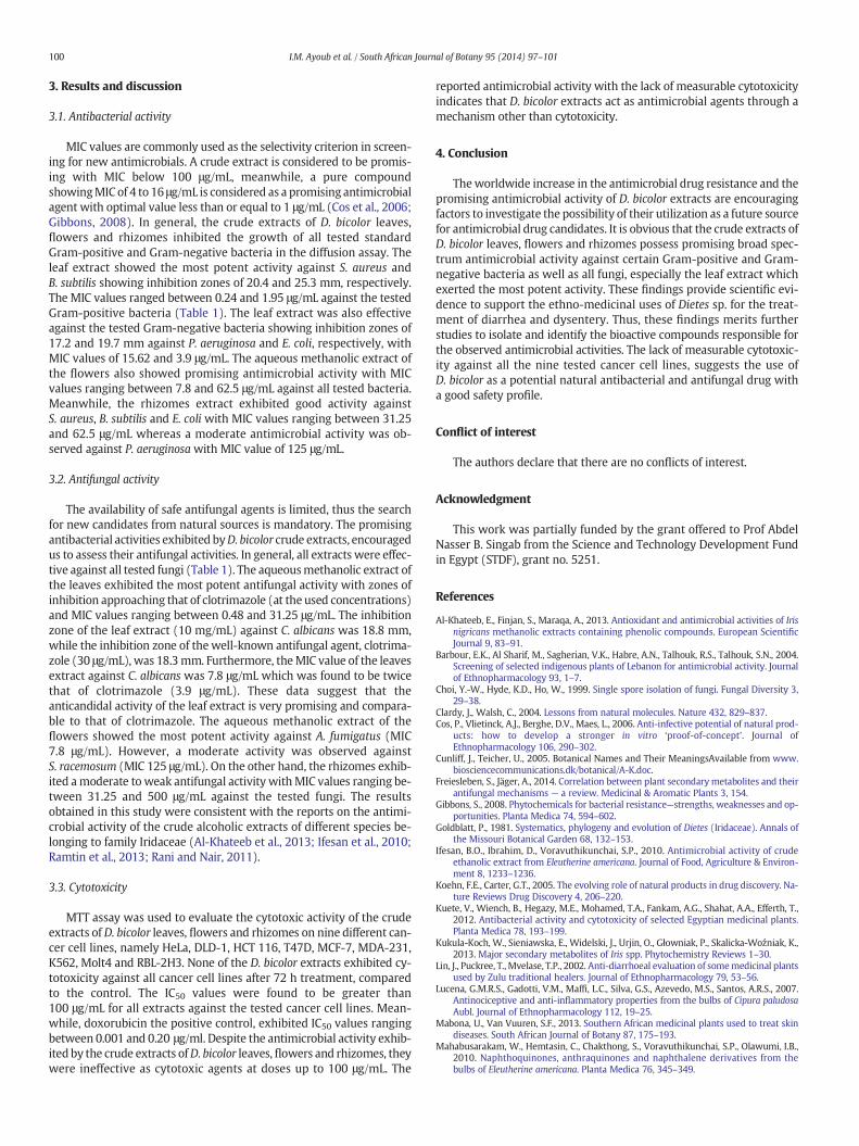
Table 1Mean inhibition zones and minimum inhibitory concentrations (MICs) of various extracts agai
Microorganism DBLa DBRb
Inhibition zone(mm)d
MIC(μg/mL)
Inhibition z(mm)
Gram positive bacteriaBacillus subtilis (RCMBf 000102) 25.3 ± 0.7 0.24 16.2 ± 0.Staphylococcus aureus (RCMB000108)
20.4 ± 0.9 1.95 14.1 ± 0.
Gram negative bacteriaEscherichia coli (RCMB 000107) 19.7 ± 0.6 3.9 15.7 ± 0.Pseudomonas aeruginosa (RCMB000104)
17.2 ± 0.3 15.62 12.2 ± 0.
FungiAspergillus fumigatus (RCMB 002007) 22.1 ± 0.5 0.48 16.1 ± 0.Candida albicans (RCMB 005008) 18.8 ± 0.1 7.8 10.2 ± 0.Geotrichum candidum (RCMB 052009) 19.2 ± 0.4 1.95 12.4 ± 0.Syncephalastrum racemosum (RCMB005005)
16.3 ± 0.1 31.25 9.7 ± 0.3
a D. bicolor leaf extract.b D. bicolor rhizomes extract.c D. bicolor flowers extract.d Data are measured in triplicate (n = 3) and presented as means ± S.D., well diameter: 6.e Gentamicin.f Regional Centre of Mycology and Biotechnology, Al-Azhar University.g Clotrimazole.
(10 cm cell culture dishes) in a humidified chamber with 5% (v/v) CO2
in air at 37 °C. The cells were maintained asmonolayer culture by serialsubculturing. All experiments were carried out using cells in the loga-rithmic growth phase.
2.5.2. Cytotoxicity assayThe sensitivity of human cancer cells to the tested D. bicolor aqueous
methanolic extracts was determined using the MTT assay (Marks et al.,1992; Van de Loosdrecht et al., 1991). The assay depends on the factthat MTT, a yellow colored compound, is converted by viable cells intoformazan, a purple derivative. Freshly trypsinized cell suspensions wereseeded at densities of 7000 cells per well (for HeLa, DLD-1, HCT 116,T47D, MCF-7, MDA-231), 2 × 10⁴ cells per well (for K562, RBL-2H3)and 4 × 10⁴ cells per well (for Molt4) in a 96-well culture plate and incu-bated overnight. The tested extracts were dissolved in DMSO to formstock solutions of 1 mg/mL. Cells were treated with various concentra-tions of the tested samples (1, 10 and 100 μg/mL). The appropriate dilu-tions of each extract were prepared in the culture medium and addedto the cells (the final concentration of DMSOwas less than 0.2% in the cul-ture well) and were incubated for 72 h at 37 °C under 5% (v/v) CO2. Themedium was removed from each well. A stock MTT solution (5 mg/mL)was diluted 1:10 in culture medium and was added to the wells (100 μLper well). Cells were then incubated for 4 h. The medium was removedand the formed formazan crystals were dissolved in DMSO and the absor-bance was recorded at 550 nm. The assay was performed in triplicate foreach sample concentration. The % cell viability of three independent ex-periments was calculated by the following formula:
% cell viability ¼ O:D of treated cells−O:D of culture mediumO:D of untreated cells−O:D of culture medium
� 100
where O.D = optical density.Cytotoxicity was expressed as IC50 which is the concentration that re-
duces the absorbance value of the treated cells by 50% compared to thecontrol (untreated cell). Doxorubicin was used as the positive control.
2.6. Data analysis
Data were measured in triplicates (n = 3) and expressed asmeans ± S.D. The 50% inhibitory concentration (IC50) was calculatedby calcuSyn software (Biosoft, Ferguson, MO, USA).
nst different microorganisms.
DBFc Standard
one MIC(μg/mL)
Inhibition zone(mm)
MIC(μg/mL)
Inhibition zone(mm)
MIC(μg/mL)
Gent.e
6 31.25 20.3 ± 0.7 1.95 29.1 ± 0.04 0.032 62.5 18.8 ± 0.5 7.8 25.1 ± 0.08 0.24
Gent.3 31.25 17.4 ± 0.5 15.62 25.6 ± 0.04 0.123 125 14.3 ± 0.1 62.5 24.3 ± 0.08 0.24
Clot.g
2 31.25 18.2 ± 0.9 7.8 26.1 ± 0.01 0.128 250 14.4 ± 0.1 62.5 18.3 ± 0.01 3.96 125 15.4 ± 0.5 62.5 23.1 ± 0.03 0.48
500 13.1 ± 0.3 125 20.5 ± 0.02 0.97
0 mm (100 μL was tested).
100 I.M. Ayoub et al. / South African Journal of Botany 95 (2014) 97–101
3. Results and discussion
3.1. Antibacterial activity
MIC values are commonly used as the selectivity criterion in screen-ing for new antimicrobials. A crude extract is considered to be promis-ing with MIC below 100 μg/mL, meanwhile, a pure compoundshowingMIC of 4 to 16 μg/mL is considered as a promising antimicrobialagent with optimal value less than or equal to 1 μg/mL (Cos et al., 2006;Gibbons, 2008). In general, the crude extracts of D. bicolor leaves,flowers and rhizomes inhibited the growth of all tested standardGram-positive and Gram-negative bacteria in the diffusion assay. Theleaf extract showed the most potent activity against S. aureus andB. subtilis showing inhibition zones of 20.4 and 25.3 mm, respectively.The MIC values ranged between 0.24 and 1.95 μg/mL against the testedGram-positive bacteria (Table 1). The leaf extract was also effectiveagainst the tested Gram-negative bacteria showing inhibition zones of17.2 and 19.7 mm against P. aeruginosa and E. coli, respectively, withMIC values of 15.62 and 3.9 μg/mL. The aqueous methanolic extract ofthe flowers also showed promising antimicrobial activity with MICvalues ranging between 7.8 and 62.5 μg/mL against all tested bacteria.Meanwhile, the rhizomes extract exhibited good activity againstS. aureus, B. subtilis and E. coli with MIC values ranging between 31.25and 62.5 μg/mL whereas a moderate antimicrobial activity was ob-served against P. aeruginosawith MIC value of 125 μg/mL.
3.2. Antifungal activity
The availability of safe antifungal agents is limited, thus the searchfor new candidates from natural sources is mandatory. The promisingantibacterial activities exhibited byD. bicolor crude extracts, encouragedus to assess their antifungal activities. In general, all extracts were effec-tive against all tested fungi (Table 1). The aqueousmethanolic extract ofthe leaves exhibited the most potent antifungal activity with zones ofinhibition approaching that of clotrimazole (at the used concentrations)and MIC values ranging between 0.48 and 31.25 μg/mL. The inhibitionzone of the leaf extract (10 mg/mL) against C. albicans was 18.8 mm,while the inhibition zone of the well-known antifungal agent, clotrima-zole (30 μg/mL), was 18.3mm. Furthermore, theMIC value of the leavesextract against C. albicans was 7.8 μg/mL which was found to be twicethat of clotrimazole (3.9 μg/mL). These data suggest that theanticandidal activity of the leaf extract is very promising and compara-ble to that of clotrimazole. The aqueous methanolic extract of theflowers showed the most potent activity against A. fumigatus (MIC7.8 μg/mL). However, a moderate activity was observed againstS. racemosum (MIC 125 μg/mL). On the other hand, the rhizomes exhib-ited amoderate to weak antifungal activity withMIC values ranging be-tween 31.25 and 500 μg/mL against the tested fungi. The resultsobtained in this study were consistent with the reports on the antimi-crobial activity of the crude alcoholic extracts of different species be-longing to family Iridaceae (Al-Khateeb et al., 2013; Ifesan et al., 2010;Ramtin et al., 2013; Rani and Nair, 2011).
3.3. Cytotoxicity
MTT assay was used to evaluate the cytotoxic activity of the crudeextracts of D. bicolor leaves, flowers and rhizomes on nine different can-cer cell lines, namely HeLa, DLD-1, HCT 116, T47D, MCF-7, MDA-231,K562, Molt4 and RBL-2H3. None of the D. bicolor extracts exhibited cy-totoxicity against all cancer cell lines after 72 h treatment, comparedto the control. The IC50 values were found to be greater than100 μg/mL for all extracts against the tested cancer cell lines. Mean-while, doxorubicin the positive control, exhibited IC50 values rangingbetween 0.001 and 0.20 μg/ml. Despite the antimicrobial activity exhib-ited by the crude extracts ofD. bicolor leaves,flowers and rhizomes, theywere ineffective as cytotoxic agents at doses up to 100 μg/mL. The
reported antimicrobial activity with the lack of measurable cytotoxicityindicates that D. bicolor extracts act as antimicrobial agents through amechanism other than cytotoxicity.
4. Conclusion
Theworldwide increase in the antimicrobial drug resistance and thepromising antimicrobial activity of D. bicolor extracts are encouragingfactors to investigate the possibility of their utilization as a future sourcefor antimicrobial drug candidates. It is obvious that the crude extracts ofD. bicolor leaves, flowers and rhizomes possess promising broad spec-trum antimicrobial activity against certain Gram-positive and Gram-negative bacteria as well as all fungi, especially the leaf extract whichexerted the most potent activity. These findings provide scientific evi-dence to support the ethno-medicinal uses of Dietes sp. for the treat-ment of diarrhea and dysentery. Thus, these findings merits furtherstudies to isolate and identify the bioactive compounds responsible forthe observed antimicrobial activities. The lack of measurable cytotoxic-ity against all the nine tested cancer cell lines, suggests the use ofD. bicolor as a potential natural antibacterial and antifungal drug witha good safety profile.
Conflict of interest
The authors declare that there are no conflicts of interest.
Acknowledgment
This work was partially funded by the grant offered to Prof AbdelNasser B. Singab from the Science and Technology Development Fundin Egypt (STDF), grant no. 5251.
References
Al-Khateeb, E., Finjan, S., Maraqa, A., 2013. Antioxidant and antimicrobial activities of Irisnigricans methanolic extracts containing phenolic compounds. European ScientificJournal 9, 83–91.
Barbour, E.K., Al Sharif, M., Sagherian, V.K., Habre, A.N., Talhouk, R.S., Talhouk, S.N., 2004.Screening of selected indigenous plants of Lebanon for antimicrobial activity. Journalof Ethnopharmacology 93, 1–7.
Choi, Y.-W., Hyde, K.D., Ho, W., 1999. Single spore isolation of fungi. Fungal Diversity 3,29–38.
Clardy, J., Walsh, C., 2004. Lessons from natural molecules. Nature 432, 829–837.Cos, P., Vlietinck, A.J., Berghe, D.V., Maes, L., 2006. Anti-infective potential of natural prod-
ucts: how to develop a stronger in vitro ‘proof-of-concept’. Journal ofEthnopharmacology 106, 290–302.
Cunliff, J., Teicher, U., 2005. Botanical Names and Their MeaningsAvailable from www.biosciencecommunications.dk/botanical/A-K.doc.
Freiesleben, S., Jäger, A., 2014. Correlation between plant secondary metabolites and theirantifungal mechanisms — a review. Medicinal & Aromatic Plants 3, 154.
Gibbons, S., 2008. Phytochemicals for bacterial resistance—strengths, weaknesses and op-portunities. Planta Medica 74, 594–602.
Goldblatt, P., 1981. Systematics, phylogeny and evolution of Dietes (Iridaceae). Annals ofthe Missouri Botanical Garden 68, 132–153.
Ifesan, B.O., Ibrahim, D., Voravuthikunchai, S.P., 2010. Antimicrobial activity of crudeethanolic extract from Eleutherine americana. Journal of Food, Agriculture & Environ-ment 8, 1233–1236.
Koehn, F.E., Carter, G.T., 2005. The evolving role of natural products in drug discovery. Na-ture Reviews Drug Discovery 4, 206–220.
Kuete, V., Wiench, B., Hegazy, M.E., Mohamed, T.A., Fankam, A.G., Shahat, A.A., Efferth, T.,2012. Antibacterial activity and cytotoxicity of selected Egyptian medicinal plants.Planta Medica 78, 193–199.
Kukula-Koch, W., Sieniawska, E., Widelski, J., Urjin, O., Głowniak, P., Skalicka-Woźniak, K.,2013. Major secondary metabolites of Iris spp. Phytochemistry Reviews 1–30.
Lin, J., Puckree, T., Mvelase, T.P., 2002. Anti-diarrhoeal evaluation of somemedicinal plantsused by Zulu traditional healers. Journal of Ethnopharmacology 79, 53–56.
Lucena, G.M.R.S., Gadotti, V.M., Maffi, L.C., Silva, G.S., Azevedo, M.S., Santos, A.R.S., 2007.Antinociceptive and anti-inflammatory properties from the bulbs of Cipura paludosaAubl. Journal of Ethnopharmacology 112, 19–25.
Mabona, U., Van Vuuren, S.F., 2013. Southern African medicinal plants used to treat skindiseases. South African Journal of Botany 87, 175–193.
Mahabusarakam, W., Hemtasin, C., Chakthong, S., Voravuthikunchai, S.P., Olawumi, I.B.,2010. Naphthoquinones, anthraquinones and naphthalene derivatives from thebulbs of Eleutherine americana. Planta Medica 76, 345–349.
101I.M. Ayoub et al. / South African Journal of Botany 95 (2014) 97–101
Marks, D.C., Belov, L., Davey, M.W., Davey, R.A., Kidman, A.D., 1992. The MTT cell viabilityassay for cytotoxicity testing in multidrug-resistant human leukemic cells. LeukemiaResearch 16, 1165–1173.
Moellering Jr., R.C., Graybill, J.R., McGowan Jr., J.E., Corey, L., 2007. Antimicrobial resistanceprevention initiative—an update: proceedings of an expert panel on resistance. TheAmerican Journal of Medicine 120, S4–S25.
Noumi, E., Yomi, A., 2001. Medicinal plants used for intestinal diseases in Mbalmayo Re-gion, Central Province, Cameroon. Fitoterapia 72, 246–254.
Pooley, E., 1999. A Field Guide to Wild Flowers KwaZulu-Natal and the Eastern Region.Natal Flora Publications Trust, Durban and Scottsville.
Pujol, J., 1990. NaturAfrica: the herbalist handbook: African Flora, Medicinal Plants. J. PujolNatural Healers Foundation.
Rahman, A.-u, Choudhary, M.I., Thomsen,W.J., 2001. Bioassay Techniques for Drug Devel-opment. Harwood Academic Publishers, The Netherlands.
Rahman, A.-u, Nasim, S., Baig, I., Jalil, S., Orhan, I., Sener, B., Choudhary, M.I., 2003. Anti-inflammatory isoflavonoids from the rhizomes of Iris germanica. Journal ofEthnopharmacology 86, 177–180.
Ramtin, M., Massiha, A., Khoshkholgh-Pahlaviani, M.R.M., Issazadeh, K., Assmar, M.,Zarrabi, S., 2013. In vitro antimicrobial activity of Iris Pseudacorus and Urtica Dioica.Zahedan Journal of Research in Medical Sciences 35–39.
Rani, V.S., Nair, B.R., 2011. Antimicrobial effects of crude extracts of Eleutherine bulbosa.Journal of Medicinal and Aromatic Plant Sciences 33, 46–52.
Rathore, H., Mittal, S., Kumar, S., 2000. Synthesis, characterization and antifungal activitiesof 3d-transition metal complexes of 1-acetylpiperazinyldithiocarbamate, M (acpdtc)2. Pesticide Research Journal 12, 103–107.
Rudall, P., 1983. Leaf anatomy and relationships of Dietes (Iridaceae). Nordic Journal ofBotany 3, 471–477.
Shai, L.J., McGaw, L.J., Masoko, P., Eloff, J.N., 2008. Antifungal and antibacterial activity ofseven traditionally used South African plant species active against Candida albicans.South African Journal of Botany 74, 677–684.
Silver, L., Bostian, K., 1990. Screening of natural products for antimicrobial agents.European Journal of Clinical Microbiology & Infectious Diseases 9, 455–461.
Tekwu, E.M., Pieme, A.C., Beng, V.P., 2012. Investigations of antimicrobial activity of someCameroonian medicinal plant extracts against bacteria and yeast with gastrointesti-nal relevance. Journal of Ethnopharmacology 142, 265–273.
Van de Loosdrecht, A.A., Nennie, E., Ossenkoppele, G.J., Beelen, R.H., Langenhuijsen, M.M.,1991. Cell mediated cytotoxicity against U 937 cells by human monocytes and mac-rophages in a modified colorimetric MTT assay: a methodological study. Journal ofImmunological Methods 141, 15–22.
VanWyk, B.E., 2011. The potential of South African plants in the development of newme-dicinal products. South African Journal of Botany 77, 812–829.
WHO, 2011. The World Medicines Situation, Traditional Medicines: Global Situation, Is-sues and Challenges. World Health Organization, Geneva.