antimicrobial treatment of bacterial cns infections in the - swab
TRANSCRIPT
SWAB Guidelines Bacterial CNS Infections
SWAB Guidelines on Antibacterial Therapy of Patients with Bacterial Central Nervous System Infections.
Part 1. Community-acquired bacterial meningitis
Part 2. Nosocomial bacterial meningitis
Part 3. Bacterial intracerebral abscess
Part 4. Tuberculous meningitis
Dutch Working Party on Antibiotic Policy (SWAB), 2012
Preparatory Committee: Dr. M.C. Brouwer, Drs. S.G.B. Heckenberg, Dr. G.T.J. van Well
(Nederlandse Vereniging voor Kindergeneeskunde), Dr. A. Brouwer (Vereniging voor
Infectieziekten), Dr. E.J. Delwel (Nederlandse Vereniging voor Neurochirurgie), Dr. L. Spanjaard
(Nederlandse Vereniging voor Medisch Microbiologie), Prof. dr. D. van de Beek (Nederlandse
Vereniging voor Neurologie), Prof. dr. J.M. Prins (SWAB).
© 2012 SWAB
Secretariaat SWAB
p/a Universitair Medisch Centrum St Radboud
Medische Microbiologie Huispost 574, route 574
Postbus 9101
6500 HB Nijmegen
www.swab.nl
SWAB Guidelines Bacterial CNS Infections 2
Table of contents
Chapter 1 Introduction
General introduction
Scope of the guideline
Definitions
Diagnosis of bacterial meningitis
Key questions
Data sources
Part 1 Community acquired bacterial meningitis
Chapter 2 Epidemiology and empirical antibiotic treatment of community-acquired bacterial
meningitis
Chapter 3 Specific antimicrobial treatment for community acquired bacterial meningitis
Part 2 Nosocomial and post-traumatic bacterial meningitis
Chapter 4 Epidemiology and empirical antibiotic treatment of nosocomial and posttraumatic bacterial
meningitis
Chapter 5 Withdrawal of antibiotic therapy in suspected postoperative meningitis
Chapter 6 Duration of treatment before CSF catheter replacement
Chapter 7 Intraventricular antibiotic treatment
Part 3
Chapter 8 Epidemiology, empirical antibiotic treatment and treatment duration of bacterial
intracerebral abscess
Part 4
Chapter 9 Epidemiology, empirical antibiotic treatment and treatment duration of bacterial
tuberculous meningitis (in preparation)
Supplementary Tables
References
SWAB Guidelines Bacterial CNS Infections 3
Chapter 1.
Introduction
General introduction
The Dutch Working Party on Antibiotic Policy (SWAB; Stichting Werkgroep Antibiotica Beleid), established by
the Dutch Society for Infectious Diseases (VIZ), the Dutch Society of Medical Microbiology (NVMM) and the
Dutch Society for Hospital Pharmacists (NVZA), develops evidence-based guidelines for the use of antibiotics in
hospitalized patients in order to optimize the quality of prescribing, thus, contributing to the containment of
antimicrobial drug costs and resistance. By means of the development of national guidelines, SWAB offers local
antibiotic and formulary committees a guideline for the development of their own, local antibiotic policy.
These are the first SWAB guidelines on bacterial central nervous system infections. It is developed according to
the Evidence Based Guideline Development method (EBRO; www.cbo.nl). The AGREE criteria
(www.agreecollaboration.org) provided a structured framework both for the development and the assessment of
the draft guideline.
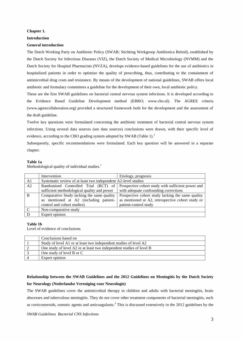
Twelve key questions were formulated concerning the antibiotic treatment of bacterial central nervous system
infections. Using several data sources (see data sources) conclusions were drawn, with their specific level of
evidence, according to the CBO grading system adopted by SWAB (Table 1).1
Subsequently, specific recommendations were formulated. Each key question will be answered in a separate
chapter.
Table 1a
Methodological quality of individual studies.1
Intervention Etiology, prognosis
A1 Systematic review of at least two independent A2-level studies
A2 Randomised Controlled Trial (RCT) of
sufficient methodological quality and power
Prospective cohort study with sufficient power and
with adequate confounding corrections
B Comparative Study lacking the same quality
as mentioned at A2 (including patient-
control and cohort studies)
Prospective cohort study lacking the same quality
as mentioned at A2, retrospective cohort study or
patient-control study
C Non-comparative study
D Expert opinion
Table 1b
Level of evidence of conclusions
Conclusions based on
1 Study of level A1 or at least two independent studies of level A2
2 One study of level A2 or at least two independent studies of level B
3 One study of level B or C
4 Expert opinion
Relationship between the SWAB Guidelines and the 2012 Guidelines on Meningitis by the Dutch Society
for Neurology (Nederlandse Vereniging voor Neurologie)
The SWAB guidelines cover the antimicrobial therapy in children and adults with bacterial meningitis, brain
abscesses and tuberculous meningitis. They do not cover other treatment components of bacterial meningitis, such
as corticosteroids, osmotic agents and anticoagulants.2 This is discussed extensively in the 2012 guidelines by the
SWAB Guidelines Bacterial CNS Infections 4
Dutch Society for Neurology (Nederlandse Vereniging voor Neurologie).3 The Nederlandse Vereniging voor
Neurologie guidelines adopted the SWAB guidelines on meningitis to be the treatment part of their meningitis
guidelines.
Scope of the SWAB guidelines
Core issues on cryptococcal meningitis are extensively discussed in the 2008 SWAB guidelines on fungal
infections. Diagnostics for bacterial meningitis are briefly discussed in the introduction, but not systematically
reviewed in these guidelines. Encephalitis falls outside the scope of these guidelines.
For this guideline we made a distinction based on the setting in which bacterial meningitis was acquired:
community-acquired versus nosocomial. Further, we provide recommendations for empirical antimicrobial
therapy for clinical subgroups of bacterial meningitis patients. The choice of initial antimicrobial therapy for these
subgroups is based on the bacteria most commonly causing the disease, taking into account the patient’s age and
clinical setting, and patterns of antimicrobial susceptibility. After the results of culture and susceptibility testing
have become available, antimicrobial therapy can be modified for optimal treatment.
SWAB Guidelines Bacterial CNS Infections 5
Definitions
Other relevant definitions
Community-acquired bacterial meningitis: bacterial meningitis acquired in the community.
Nosocomial bacterial meningitis: bacterial meningitis resulting from invasive procedures (e.g., craniotomy,
placement of internal or external ventricular catheters, lumbar puncture, intrathecal infusions of medications, or
spinal anesthesia), complicated head trauma, or in rare cases, metastatic infection in patients with hospital-
acquired bacteremia.4
Empirical antibiotic therapy: therapy that is started before the pathogen and its susceptibility patterns are known.
The choice of antibacterial therapy is largely based on expected etiology and antimicrobial resistance.
Diagnosis of bacterial meningitis
To diagnose bacterial meningitis, CSF examination is mandatory. CSF culture remains the gold standard for the
diagnosis of bacterial meningitis; aerobic culturing techniques are obligatory for community-acquired bacterial
meningitis. Anaerobic culture may be important for post-neurosurgical meningitis or for the investigation of CSF
shunt meningitis. In a retrospective series of 875 community-acquired bacterial meningitis patients for whom the
diagnosis was defined by a CSF white blood cell count of over 1,000 cells per mm3 and/or more than 80%
polymorphonuclear cells, the CSF culture was positive for 85% of cases in the absence of prior antibiotic
treatment.5
Tuberculous meningitis
Tuberculous meningitis is defined as an infection of the meninges caused by Mycobacterium tuberculosis,
proven by CSF culture or CSF PCR, or suspected by clinical characteristics and/or CSF markers of
inflammation (CSF leukocyte count, CSF protein and/or CSF glucose level) combined with evidence or
suspicion of other foci of tuberculosis (e.g. lungs).
Bacterial meningitis
Bacterial meningitis is defined as a bacterial infection of the meninges, proven by cerebrospinal fluid (CSF)
culture, CSF polymerase chain reaction (PCR), CSF gram stain or antigen testing, or suspected by clinical
characteristics and/or CSF markers of inflammation (CSF leukocyte count, CSF protein and/or CSF glucose
level).4
Brain abscess
Brain abscess is defined as focal intraparenchymal bacterial infection in the cerebrum, cerebellum or
midbrain identified by cranial computed tomography, magnetic resonance imaging or during neurosurgery.
SWAB Guidelines Bacterial CNS Infections 6
CSF culture is obligatory to obtain the in vitro susceptibility of the causative microorganism and to rationalize
treatment. CSF Gram staining, latex agglutination testing and PCR are additional diagnostic tools that might aid in
etiological diagnoses, especially for patients with negative CSF cultures (i.e., after antibiotic pretreatment).6
Characteristic CSF findings for bacterial meningitis consist of polymorphonuclear pleocytosis,
hypoglycorrhachia, and raised CSF protein levels. A prediction model based on 422 patients with bacterial or viral
meningitis showed that individual predictors of community-acquired bacterial meningitis were a glucose
concentration of less than 1.9 mmol per liter (0.34 g/liter), a ratio of CSF glucose to blood glucose of less than
0.23, a protein concentration of more than 2.2 g per liter, or a white cell count of more than 2,000 cells per mm3.7
Diagnosis of brain abscess
The most important investigation to diagnose brain abscesses is cranial imaging, either cranial tomography (CT)
or magnetic resonance imaging (MRI).8,9
These will usually show round contrast-enhancing lesions similar to
intracranial malignancies. Using diffusion weight MRI a distinction can be made between tumor and cerebral
abscess: typical brain abscesses are hyperintense on T2 and FLAIR images, show a cavity with ring enhancement
on T1 with gadolinium and limited diffusion in the cavity. The microbiological diagnosis in brain abscesses can
be difficult. In only a minority of cases the causative pathogen(s) is identified in blood cultures or CSF culture.
Therefore, if no primary focus of infection can be identified, a stereotactic aspiration of the abscess should be
considered.8,9
Both aerobe and anaerobe cultures of the aspirate should be performed.
Diagnosis of tuberculous meningitis
Tuberculous (TB) meningitis is difficult to diagnose, as presenting clinical features are usually non-specific.11,12
CSF usually shows a predominant lymphocytic pleiocytosis (>50%), with a low CSF to blood glucose ratio (<0.5)
and a high protein concentration (>1.0 g/L).12
Due to usually low numbers of mycobacteria in the CSF, the yield
of Ziehl-Neelsen staining is only adequate (80%) if a large volume of CSF is examined (>5 ml), and in some
cases repeated investigations need to be performed.11
PCR for detection of Mycobacterium tuberculosis has a
lower sensitivity than Ziehl-Neelsen staining, but is especially useful in patients who already started anti-
tuberculosis medication.11
CSF Ziehl-Neelsen staining, TB culture and PCR may all remain negative. In some
cases repeated CSF investigations will aid in establishing the diagnosis. An important clue to the diagnosis can be
pulmonary tuberculosis, which should always be looked for by chest X-ray in patients suspected of TB
meningitis. A prediction model was devised in a Vietnamese population, which showed that age below 36, blood
leukocyte count below 15.000/ml, duration of illness >6 days, CSF leukocyte count below 750/ml and CSF
neutrophil percentage <90% were all indicators of TB meningitis in that setting.12
Key questions
1a. What is the epidemiology and optimal empirical antibiotic therapy of community-acquired bacterial
meningitis in the Netherlands in neonates (0-28 days)
1b. What is the epidemiology and optimal empirical antibiotic therapy of community-acquired bacterial
meningitis in the Netherlands in children (29 days – 16 years)

SWAB Guidelines Bacterial CNS Infections 7
1c. What is the epidemiology and optimal empirical antibiotic therapy of community-acquired bacterial
meningitis in the Netherlands in adults (> 16 years)?
2. What is the optimal antibiotic therapy and treatment duration for community-acquired bacterial
meningitis?
a. caused by Streptococcus pneumoniae.
b. caused by Neisseria meningitidis
c. caused by Listeria monocytogenes
d. caused by Haemophilus influenzae
e. caused by Streptococcus agalactiae
f. caused by Escherichia coli
g. with negative CSF and blood cultures
3a. What is the epidemiology and empirical treatment of bacterial meningitis related to external CSF drainage?
3b. What is the epidemiology and empirical treatment of bacterial meningitis related to internal CSF drainage?
3c. What is the epidemiology and empirical treatment of bacterial meningitis related to postoperative bacterial
meningitis?
3d. What is the epidemiology and empirical treatment of bacterial meningitis related to posttraumatic bacterial
meningitis?
4. When should antibiotic treatment in patients with suspected postoperative bacterial meningitis be
withdrawn?
5. How long should infected CSF catheters be treated before removal/replacement?
6. What are the indications and regimens for intraventricular antimicrobial treatment?
7. What is the epidemiology of bacterial intracerebral abscesses in Europe/US?
8. What is the optimal empirical antibiotic therapy in intracerebral abscesses?
9. What is the optimal dose of antibiotic therapy for intracerebral abscesses?
10. What is the optimal duration of therapy for intracerebral abscesses?
11. What is the epidemiology and empirical treatment for tuberculous meningitis (in preparation)?
SWAB Guidelines Bacterial CNS Infections 8
12. What is the optimal duration of therapy for tuberculous meningitis (in preparation)?
Data sources
Community-acquired bacterial meningitis
The Netherlands Reference Laboratory for Bacterial Meningitis
The most important source of information for bacterial meningitis in the Netherlands is the Netherlands Reference
Laboratory for Bacterial Meningitis (NRLBM; http://www.amc.uva.nl/index.cfm?pid=5627). The NRLBM
started collecting isolates of Neisseria meningitidis in 1959 and of other bacteria causing meningitis in 1975. The
Reference Laboratory isolates are available for studies on the epidemiology of bacterial meningitis and on the
pathogenicity and antibiotic susceptibility of isolates.
The objectives of the Reference Laboratory are to perform surveillance of bacterial meningitis, to describe the
epidemiology of bacterial meningitis in the Netherlands, to provide keys for the development of potential vaccine
components and to provide data about antibiotic susceptibility of isolates. The NRLBM receives isolates from
>85% of all patients with bacterial meningitis in the Netherlands. It is not possible to grade the data from the
NRLBM with a specific level of evidence. However, the committee considers these surveillance data to be the
most reliable, as the NRLBM covers >85% of the Dutch patients with bacterial meningitis and assigned it a level
A2.
Dutch cohorts of adults with community-acquired bacterial meningitis
The Dutch Bacterial Meningitis Study (DBMS) and MeninGene Study are 2 prospective cohort studies including
adult patients with community-acquired meningitis in the Netherlands, who had bacterial meningitis confirmed by
cerebrospinal fluid culture.10,11
In both studies patients were identified by the NRLBM, which provided the names
of the hospitals where patients with bacterial meningitis had been admitted 2-6 days previously. The treating
physician was contacted, and informed consent was obtained from all participating patients or their legally
authorized representatives. The Dutch Bacterial Meningitis Study lasted from 1998 to 2002 and included 696
patients. MeninGene started in 2006 and continues until today, and has thus far included over 1200 patients.
Detailed information on patients with bacterial meningitis is collected through Case-record forms (CRF). CRFs
are used to collect data on patients’ history, symptoms and signs on admission, laboratory findings at admission,
clinical course, outcome and neurologic findings at discharge, and treatment. We used data from these studies for
all adults with bacterial meningitis.

SWAB Guidelines Bacterial CNS Infections 9
Literature
The NRLBM data does not distinguish between community-acquired and nosocomial bacterial meningitis.
Therefore, we substantiated the NRLBM data with literature studies where appropriate using the following search
strategy:
((((("Meningitis, Bacterial"[Mesh]) OR (bacterial meningitis*[TIAB]) OR (pneumococcal meningitis*[TIAB])
OR (meningococcal meningitis*[TIAB]) OR (staphylococcal meningitis*[TIAB]) OR (nosocomial
meningitis*[TIAB]) OR (hospital acquired meningitis*[TIAB]) OR (e coli meningitis*[TIAB]) OR (escherichia
coli meningitis*[TIAB]) OR (neonatal meningitis*[TIAB])) AND (("Anti-Bacterial Agents"[Mesh] OR "Anti-
Bacterial Agents "[Pharmacological Action]) OR (antibiotic*[TIAB]) OR (antimicrobial*[TIAB])) AND
((Humans[Mesh]) AND (English[lang]))) NOT (tuberc*[TI] OR anthra*[TI])) NOT (case reports[PT] OR
editorial[PT]))
Nosocomial bacterial meningitis
Literature
Pubmed was searched using search strategies
Search 1): Pubmed search: bacterial meningitis related to internal/external CSF drainage
(("Ventriculoperitoneal Shunt"[Mesh] OR ventriculoperitoneal shunt*[TIAB]) OR (ventricular drain*[TIAB]) OR
(lumbar drain*[TIAB]) OR (ventricular atrial drain*) OR (ventriculoatrial drain*[TIAB]) OR (ventriculopleural
drain*[TIAB])) AND (("cross infection"[MeSH Terms] OR cross infection*[TIAB]) OR ("infection"[MeSH
Terms] OR "infection"[TIAB])))
Search 2): Nosocomial bacterial meningitis (Selected from meningitis search)
((((("Meningitis, Bacterial"[Mesh]) OR (bacterial meningitis*[TIAB]) OR (pneumococcal meningitis*[TIAB])
OR (meningococcal meningitis*[TIAB]) OR (staphylococcal meningitis*[TIAB]) OR (nosocomial
meningitis*[TIAB]) OR (hospital acquired meningitis*[TIAB]) OR (e coli meningitis*[TIAB]) OR (escherichia
coli meningitis*[TIAB]) OR (neonatal meningitis*[TIAB])) AND (("Anti-Bacterial Agents"[Mesh] OR "Anti-
Bacterial Agents "[Pharmacological Action]) OR (antibiotic*[TIAB]) OR (antimicrobial*[TIAB])) AND
((Humans[Mesh]) AND (English[lang]))) NOT (tuberc*[TI] OR anthra*[TI])) NOT (case reports[PT] OR
editorial[PT]))
Brain abscess
Literature
Pubmed was searched using search strategie:
((((("Brain Abscess"[Mesh:noexp]) OR (Brain Abscess*[TIAB]) OR (cerebral Abscess*[TIAB]) OR (cerebellar
Abscess*[TIAB])) OR (brain[TIAB] AND abscess*[TIAB])) AND (("Anti-Bacterial Agents"[Mesh] OR "Anti-
Bacterial Agents "[Pharmacological Action]) OR (antibiotic*[TIAB]) OR (antibacterial agent*[TIAB]) OR
(antimicrobial agent*[TIAB])) AND ((Humans[Mesh]) AND (English[lang]))) AND ((Humans[Mesh]) AND
(English[lang]))) NOT ((((("Brain Abscess"[Mesh:noexp]) OR (Brain Abscess*[TIAB]) OR (cerebral

SWAB Guidelines Bacterial CNS Infections 10
Abscess*[TIAB]) OR (cerebellar Abscess*[TIAB])) OR (brain[TIAB] AND abscess*[TIAB])) AND (("Anti-
Bacterial Agents"[Mesh] OR "Anti-Bacterial Agents "[Pharmacological Action]) OR (antibiotic*[TIAB]) OR
(antibacterial agent*[TIAB]) OR (antimicrobial agent*[TIAB])) AND ((Humans[Mesh]) AND (English[lang])))
AND (Humans[Mesh] AND Case Reports[ptyp] AND English[lang]))
Limits used were: English, not case reports, humans. This resulted in 517 hits (March 8th
2010)
To avoid differences due to geographical location, patients in Europe/North America/Western Asia (i.e., Israel
and Turkey) were included. Case series of >10 patients were included. Microbiological data were extracted from
each study according to a pre-specified protocol.
Parts of the recommendations in these guidelines have been published previously.4,6,12
Tuberculous meningitis (in preparation)
Literature
SWAB Guidelines Bacterial CNS Infections 11
Chapter 2
Epidemiology and empirical antibiotic treatment of community-acquired bacterial meningitis
In patients with bacterial meningitis, other treatment next to antibiotics may be indicated. Adjunctive
dexamethasone therapy needs to be administered simultaneous with the antibiotics in most children and adults
with bacterial meningitis, and consultation with an ENT specialist may be necessary to assess whether surgical
treatment of otitis or sinusitis is indicated. Although the SWAB guidelines only cover antibiotic therapy and refers
to the 2012 guidelines by the Dutch Society for Neurology (Nederlandse Vereniging voor Neurologie) for
adjunctive treatments, the committee likes to stress the importance of these therapies.
Key question 1a. What is the epidemiology and optimal empirical antibiotic therapy of bacterial meningitis in
the Netherlands in neonates (0-28 days)?
Common causative microorganisms of neonatal meningitis during the first week of life are Streptococcus
agalactiae (Group B streptococci), Escherichia coli, and Listeria monocytogenes (Supplementary table 1).6,13-15
Late-onset neonatal meningitis is formally classified as meningitis after the first week until the 28th
day of
life.13,16,17
Late-onset neonatal meningitis may be caused by a wide variety of species, including staphylococci,
and Gram-negative bacilli.6 Children born with a hydrocephalus or those that develop a hydrocephalus after an
intraventricular bleeding (on the neonatal intensive care unit) are often treated with repetitive CSF drainage from
an Ommaya reservoir, or a temporary or permanent CSF shunt and are at increased risk of nosocomial
meningitis.4,18
The causative microorganisms in these children are different from those in “spontaneous”
meningitis and are similar to those seen in nosocomial meningitis (Chapter 4).
Empirical therapy for neonatal meningitis should consist of amoxicillin combined with cefotaxime (Table 2). The
use of gentamicin to cover neonatal meningitis due to Gram-negative bacteria has been debated. The
recommendation for the addition of gentamicin has been based on data from in vitro studies, which showed
synergistic activity with amoxicillin in antimicrobial killing of S. agalactiae.6,19
However, as CSF concentrations
are usually only minimally above the MIC, third generation cephalosporins are considered superior to
gentamicin.17
In neonates with suspected sepsis gentamicin is combined with amoxicillin, but physicians should
be aware this regimen is sub-optimal for meningitis treatment. In neonates with suspected sepsis or meningitis
CSF examination to establish whether concurrent meningitis is present is vital to determine the optimal empirical
treatment. We recommend amoxicillin and cefotaxime in children with a high suspicion of bacterial meningitis.
The dose is depends on gestational age and birth weight, and is given in the online SWAB-ID (www.swab.nl).
Ceftriaxone is contraindicated in children < 4 weeks because of a high risk of precipitation of calcium-ceftriaxone
complexes in the gallbladder.
SWAB Guidelines Bacterial CNS Infections 12
1b. What is the epidemiology and optimal empirical antibiotic therapy of bacterial meningitis in the
Netherlands for children (29 days – 16 years)?
The most common causative bacteria of community-acquired bacterial meningitis in children aged 1 month and
older are S. pneumoniae and N. meningitidis, causing 80% of cases in the Netherlands (Supplementary table 2).
The remainder of cases is caused by group B streptococci, E. coli, H. influenzae, other Gram-negative bacilli, L.
monocytogenes, and group A streptococci. The epidemiology has shifted substantially in the past 10 years and is
still changing due to the introduction of the meningococcus group C vaccine (2002), and the 7- (2006) and 10-
valent (2011) pneumococcal conjugate vaccines.6
Empirical coverage with a third generation cephalosporin (cefotaxime or ceftriaxone) is recommended based on a
broad spectrum of activity and excellent penetration into the CSF under inflammatory conditions. Third
generation cephalosporins are preferred in this age group above amoxicillin as resistance of H. influenzae to
amoxicillin due to beta-lactamase production occurs in 11% of meningitis cases.20
A clinical trial comparing
cefotaxime with meropenem showed similar efficacy; therefore meropenem may be considered as alternative
empirical treatment in children >3 months of age in specific cases (e.g. cephalosporin allergy).21
However, the
committee recommends keeping the use of meropenem restricted to the use as last resort antibiotic in bacterial
meningitis patients.
1c. What is the epidemiology and optimal empirical antibiotic therapy of bacterial meningitis in the
Netherlands for adults (> 16 years)
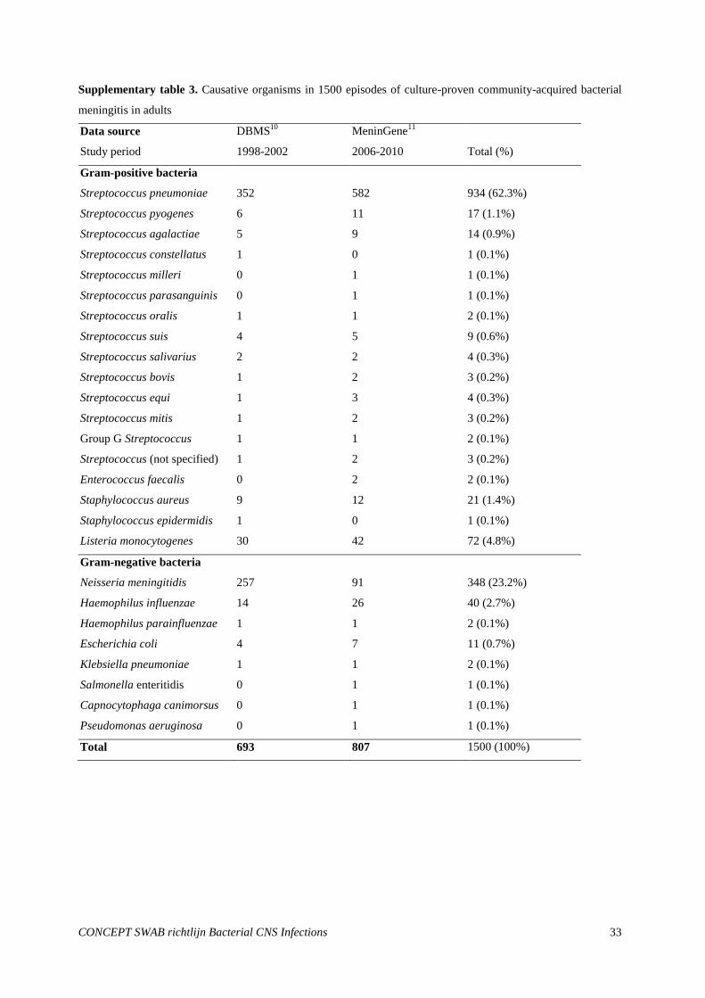
All causative organisms of bacterial meningitis in adults from two nation-wide prospective cohort studies in the
Netherlands, including 1500 patients, are presented in Supplementary table 3. S. pneumoniae is the most common
causative microorganism of adult bacterial meningitis, and was identified in 70% of culture positive cases
included between 2006 and 2010.22
N. meningitidis was the second most common causative microorganism and is
predominantly found in young adults.23
The proportion of meningococcal meningitis decreased from 37% of
bacterial meningitis cases between 1998-2002 to 16% in 2006-2010, probably due to vaccination against
serogroup C and to normal variation in meningococcal disease incidence. Patients over 50 years of age and those
with an immunocompromised state are at increased risk for Listeria monocytogenes, which is found in 5% of
cases.24
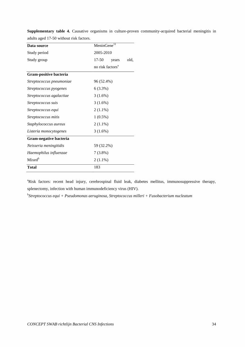
However, L. monocytogenes meningitis also occurs in previously healthy adults without risk factors
(Supplementary table 4). Therefore age and immunocompromised status cannot be used to rule out L.
monocytogenes meningitis. Meningitis due to mostly unencapsulated H. influenzae and group A streptococci
occurs in 3-4% of cases. Other causative organisms occur sporadically.
There have been no randomized controlled trials or comparative studies to evaluate the optimal empirical
antibiotic treatment in adults with bacterial meningitis. Therefore all recommendations are based on
epidemiological data (level 4 evidence – expert opinion).
The committee recommends empirical therapy for bacterial meningitis in adults to consist of amoxicillin and a
third generation cephalosporin (ceftriaxone or cefotaxime). Use of these antibiotics will ensure coverage of the
SWAB Guidelines Bacterial CNS Infections 13
four most common causative microorganisms (pneumococci, N. meningitidis, L. monocytogenes and H.
influenzae) and most sporadic causes. This combination therapy is preferred over monotherapy with third
generation cephalosporins, as this does not cover L. monocytogenes, while monotherapy with amoxicillin does not
cover β-lactamase producing H. influenzae strains and E.coli.
In the majority of patients the CSF or blood cultures will show the causative microorganism within 48 hours, with
the exception of L. monocytogenes which is a notably slow grower.6 If a microorganism is cultured, therapy can
be adjusted accordingly (see Ch 3). If cultures are negative after 48 hours, the third generation cephalosporins can
be discontinued, and monotherapy with amoxicillin will suffice because infection with β-lactamase producing H.
influenzae is virtually excluded.
Conclusions
Level 1 Early neonatal meningitis is most commonly caused by S. agalactiae, E. coli, and L.
monocytogenes. Late neonatal meningitis is most commonly caused by staphylococci,
and Gram-negative bacilli
A2 Garges (2006)13
, Holt (2001)15
, NRLBM (2011)20
B Hristeva (1993)14
Level 2 Bacterial meningitis in children is caused by S. pneumoniae (44%), N. meningitidis
(39%) and H. influenzae (11%)
A2 NRLBM20
Level 2 Bacterial meningitis in adults is caused by S. pneumoniae (62%), N. meningitidis (23%)
L. monocytogens (5%) and H. influenzae (3%)
A2 NRLBM20
Level 3 Cefotaxime and meropenem are equally effective as empirical therapy for bacterial
meningitis in children. However, meropenem should only be given in specific cases and
kept as last resort.
B Odio (1999)21
* No comparative studies have been performed to evaluate empirical antibiotic therapy
regimens for community-acquired bacterial meningitis in neonates and adults.
Recommendations
1. Empirical antibiotic therapy in neonates with bacterial meningitis should consist of amoxicillin and
cefotaxime. If a microorganism is cultured, therapy can be adjusted accordingly (see Ch 3).
2. Empirical antibiotic therapy in children aged 1 month-16 years with bacterial meningitis should consist of a
third generation cephalosporin (cefotaxime or ceftriaxone). Meropenem may be used as an alternative in
specific cases (e.g. known allergic reactions). If a microorganism is cultured, therapy can be adjusted
accordingly (see Ch 3).

SWAB Guidelines Bacterial CNS Infections 14
3. Empirical antibiotic therapy in adults with bacterial meningitis should consist of the combination of
amoxicillin and a third generation cephalosporin (ceftriaxone or cefotaxime).
4. If a microorganism is cultured in adults with bacterial meningitis, therapy can be adjusted accordingly (see
Ch 3). If cultures are negative after 48 hours, the third generation cephalosporin can be discontinued,
and monotherapy amoxicillin will suffice.
Table 2. Empirical antimicrobial therapy for community-acquired bacterial meningitis, stratified for age.
Age / risk group Treatment Alternative
Neonates* amoxicillin 100-200 mg/kg/day (6h) and
cefotaxime 50-150 mg/kg/day (8h)
amoxicillin 100-200 mg/kg/day (6h)
and gentamicin 4 mg/kg/day (24-
48h)
Children ceftriaxone 100 mg/kg/day (max 4g)
(24h) or cefotaxime 150 mg/kg/day
(max 12g) (6h)
meropenem 120 mg/kg/day (max 6
g) (8h)
Adults amoxicillin 6x2 g/dag plus
ceftriaxone 2x2 g/day or cefotaxime 6x2
g/day
Dosages derived from: SWAB-ID: Nationale antibioticaboekje van de SWAB,
http://customid.duhs.duke.edu/NL/Main/Start.asp. Between brackets interval between dosages.
* Dose and dosage interval dependent on gestational age and weight, see SWAB-ID for exact recommendations.
SWAB Guidelines Bacterial CNS Infections 15
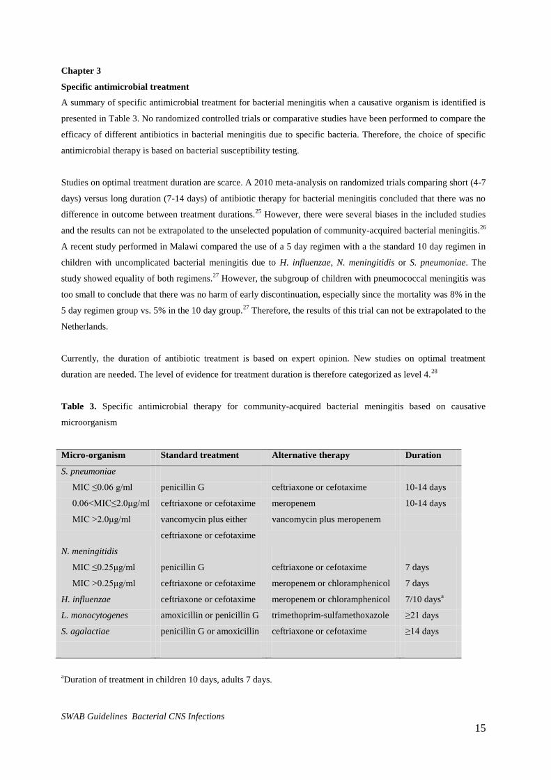
Chapter 3
Specific antimicrobial treatment
A summary of specific antimicrobial treatment for bacterial meningitis when a causative organism is identified is
presented in Table 3. No randomized controlled trials or comparative studies have been performed to compare the
efficacy of different antibiotics in bacterial meningitis due to specific bacteria. Therefore, the choice of specific
antimicrobial therapy is based on bacterial susceptibility testing.
Studies on optimal treatment duration are scarce. A 2010 meta-analysis on randomized trials comparing short (4-7
days) versus long duration (7-14 days) of antibiotic therapy for bacterial meningitis concluded that there was no
difference in outcome between treatment durations.25
However, there were several biases in the included studies
and the results can not be extrapolated to the unselected population of community-acquired bacterial meningitis.26
A recent study performed in Malawi compared the use of a 5 day regimen with a the standard 10 day regimen in
children with uncomplicated bacterial meningitis due to H. influenzae, N. meningitidis or S. pneumoniae. The
study showed equality of both regimens.27
However, the subgroup of children with pneumococcal meningitis was
too small to conclude that there was no harm of early discontinuation, especially since the mortality was 8% in the
5 day regimen group vs. 5% in the 10 day group.27
Therefore, the results of this trial can not be extrapolated to the
Netherlands.
Currently, the duration of antibiotic treatment is based on expert opinion. New studies on optimal treatment
duration are needed. The level of evidence for treatment duration is therefore categorized as level 4.28
Table 3. Specific antimicrobial therapy for community-acquired bacterial meningitis based on causative
microorganism
Micro-organism Standard treatment Alternative therapy Duration
S. pneumoniae
MIC ≤0.06 g/ml penicillin G ceftriaxone or cefotaxime 10-14 days
0.06<MIC≤2.0μg/ml ceftriaxone or cefotaxime meropenem 10-14 days
MIC >2.0μg/ml vancomycin plus either
ceftriaxone or cefotaxime
vancomycin plus meropenem
N. meningitidis
MIC ≤0.25μg/ml penicillin G ceftriaxone or cefotaxime 7 days
MIC >0.25μg/ml ceftriaxone or cefotaxime meropenem or chloramphenicol 7 days
H. influenzae ceftriaxone or cefotaxime meropenem or chloramphenicol 7/10 daysa
L. monocytogenes amoxicillin or penicillin G trimethoprim-sulfamethoxazole ≥21 days
S. agalactiae penicillin G or amoxicillin ceftriaxone or cefotaxime ≥14 days
aDuration of treatment in children 10 days, adults 7 days.
SWAB Guidelines Bacterial CNS Infections 16
Key question 2a. What is the optimal antibiotic therapy and duration for community-acquired bacterial
meningitis caused by Streptococcus pneumoniae?
Resistance of S. pneumoniae to penicillin and third generation cephalosporins (cefotaxime or ceftriaxone) has
been reported to cause treatment failure.6 The rate of pneumococcal resistance rates in the Netherlands is still very
low, with only 0.5-1% strains showing intermediate resistance to penicillin (0.06<MIC≤2.0).10,11
So far, one strain
resistant to third generation cephalosporins has been described (MIC>2.0).29
Therefore, in the overwhelming
majority of patients with pneumococcal meningitis monotherapy with amoxicillin or penicillin suffices. When
susceptibility testing shows intermediate resistance to penicillin, a third generation cephalosporin should be used.
Patients at risk for infection with a resistant strain, such as inhabitants of countries with high pneumococcal
resistance rates (e.g. South-European countries, or the US) or recent travelers from these countries should be
treated with combination therapy consisting of vancomycin and a third generation cephalosporin until
susceptibility testing is performed.6 When the isolate shows resistance to penicillin (MIC>2.0), combination
therapy of vancomycin and a third generation cephalosporin should be used. The duration of therapy for S.
pneumoniae meningitis is 10 days, unless persistent or recurrent fever or other complications occur that warrant
prolonged treatment.28
Key question 2b. What is the optimal antibiotic therapy and duration for community-acquired bacterial
meningitis caused by Neisseria meningitidis?
Currently (2012), there is no consensus in Europe about the penicillin breakpoints of N.meningitidis. According to
EUCAST the meningococcus has intermediate resistance (reduced susceptibility) when the penicillin MIC is
>0.06 en ≤0.25 mg/l. Meningococcal strains with reduced susceptibility to penicillin have been described, but the
clinical significance remains unclear. In the Netherlands, 17 of 392 (4%) meningococcal strains isolated from
CSF between 2005-2009 showed intermediate resistance to penicillin. In 2010 the number increased to 10 out of
54 (19%). Penicillin-resistant strains are very rare. The majority of patients with N. meningitidis strains with
intermediate resistance to penicillin reported in the literature have responded well to penicillin treatment.6
Treatment failures have been described in a few case reports, and none of these occurred in the Netherlands.
Therefore, treatment with amoxicillin or penicillin for 7 days remains first choice.28
Infection with a penicillin-
resistant strain (MIC >0.25) should be treated with a third generation cephalosporin.6
All meningococcal meningitis cases have to be reported to the Public Health Service (GGD/GG&GD). Household
contacts and others who have been in close contact with meningococcal meningitis patients in the week prior to
disease onset should receive prophylaxis to minimize the risk of secondary cases. Health care workers are only at
risk after intimate contact such as mouth-to mouth resuscitation or endotracheal intubation. Rifampicin and
ciprofloxacin are both effective as prophylaxis for treating patient contacts.3 More information on this subject can
be found in the bacterial meningitis guidelines of the Dutch Neurological Society.3

SWAB Guidelines Bacterial CNS Infections 17
Key question 2c. What is the optimal antibiotic therapy and duration for community-acquired bacterial
meningitis caused by Listeria monocytogenes?
Amoxicillin and penicillin are highly effective against L. monocytogenes, and one of these should be used in
patients with proven or suspected L. monocytogenes meningitis. Third generation cephalosporins are ineffective
against this pathogen and therefore empirical monotherapy with these agents in patients at higher risk for L.
monocytogenes meningitis, e.g. elderly (>50 years) and immunocompromised patients, should be avoided.24
As it
is difficult to define patients that have an increased risk of L. monocytogenes, the empirical treatment for all adults
includes amoxicillin to cover L. monocytogenes. Patients allergic to penicillin can be treated with trimethoprim-
sulfamethoxazole.6
The addition of aminoglycosides is debated. Synergistic activity of gentamicin with other antibiotics has been
shown in in vitro experiments. However, a cohort of 118 Spanish adults with L. monocytogenes disease showed
increased mortality and renal failure in the aminoglycoside-treated patients.30
Therefore, aminoglycosides are not
advised in adults with L. monocytogenes meningitis. Minimal duration of treatment of L. monocytogenes
meningitis is 21 days for children and adults.28,31,32
Key question 2d. What is the optimal antibiotic therapy and duration for community-acquired bacterial
meningitis caused by Haemophilus influenzae?
The rate of beta-lactamase producing H. influenzae strains in the Netherlands has fluctuated in the last 25 years
from 0.9% to 12.5%, with an average around 5% (data NRLBM). Therefore, third generation cephalosporins have
become standard for H. influenzae meningitis, until susceptibility testing is performed. If the strain is susceptible
to amoxicillin, amoxicillin should be used. The standard duration of therapy for adults is 7 days and for children
10 days.28,32
Key question 2e. What is the optimal antibiotic therapy and duration for community-acquired bacterial
meningitis caused by Streptococcus agalactiae?
S. agalactiae is invariably susceptible to penicillin, amoxicillin, and cephalosporins. Treatment of S. agalactiae
meningitis consists of penicillin or amoxicillin, and alternatively third generation cephalosporins can be used. The
minimal advised treatment duration is 14 days, but courses up to 21 days should be considered in patients with a
complicated disease course. In neonates with S. agalactiae meningitis gentamicin is added for 3 days.
Key question 2f. What is the optimal antibiotic therapy and duration for community-acquired bacterial
meningitis caused by Escherichia coli?
E. coli strains are mostly susceptible to third generation cephalosporins and 30-40% is susceptible to amoxicillin.
Strains producing extended spectrum beta-lactamases (ESBL) are increasing in frequency but have not been
described in meningitis patients in the Netherlands so far. Treatment of E. coli meningitis consists of third
generation cephalosporins combined with gentamicin during the first three days of treatment in neonates. In

SWAB Guidelines Bacterial CNS Infections 18
children beyond the neonatal age and adults third generation cephalosporins are advised, or amoxicillin if the
strain is sensitive to amoxicillin. When ESBL producing E. coli are suspected or proven, meropenem is the
treatment of choice. The minimal advised treatment duration is 21 days.
Key question 2g. What is the optimal antibiotic therapy and duration for community-acquired bacterial
meningitis caused by unidentified pathogens (culture-negative cases)?
In 10 to 20% of bacterial meningitis cases, defined by elevated CSF markers of inflammation, CSF and blood
cultures remain negative.6 No studies have been performed to determine the treatment regimen for these patients
and the optimal duration of therapy is therefore also unclear. The given recommendations are based on the
duration of therapy advised for the most common causative microorganisms. For children empirical therapy with
a third generation cephalosporin should be continued for 14 days in culture-negative cases. In adults the empirical
therapy can be changed, from amoxicillin combined with a third generation cephalosporin to monotherapy with
amoxicillin for 14 days if after 48 hours cultures remain negative. Monotherapy amoxicillin is the antimicrobial
therapy of choice in adults because infection with β-lactamase producing H. influenzae is virtually excluded if
cultures remain negative after 48 hours. L. monocytogenes should still be covered after 48 hours as it is a slow
growing microorganism. As L. monocytogenes is extremely rare in children beyond the neonatal age,
monotherapy with a third generation cephalosporin suffices for children.
Conclusions
Level 1 No randomized controlled trials or adequate comparative studies have been
performed to determine the optimal antibiotic treatment duration of bacterial
meningitis caused by specific pathogens or in culture negative cases.
A1 Karageorgeopoulous (2007)25
, vd Beek (2010)26
Recommendations
1. Bacterial meningitis caused by penicillin susceptible (MIC≤0.06) S. pneumoniae should be treated with
penicillin 200.000 IU/kg/day (4h – maximum 12 million units) in children and penicillin 12 million IU /day
(4h or continuously) in adults, for a minimum of 10 days. Meningitis due to S. pneumoniae with
intermediate resistance (0.06<MIC≤2.0) should be treated with ceftriaxone 100 mg/kg/day (24h – max 4g)
in children and ceftriaxone 4 g/day (12h) or cefotaxime 8-12 g/day (4-6h) in adults, for a minimum of 10
days. Meningitis due to penicillin-resistant S. pneumoniae (MIC >2.0) should be treated with vancomycin
60 mg/kg/day (12h – max 2g) plus ceftriaxone 100 mg/kg/day (24h – max 4g) in children and vancomycin
2 g/day (12h) plus ceftriaxone 4 g/day (12h) or cefotaxime 8-12 g/day (4-6h) in adults.
2. Bacterial meningitis caused by penicillin susceptible N. meningitidis should be treated with penicillin
200.000 IU/kg/day (4h – maximum 12 million units) in children and penicillin 12 million IU /day (4h or
continuously) in adults, for 7 days. Penicillin resistant strains (MIC >0.25) should be treated with
ceftriaxone 100 mg/kg/day (24h – max 4g) in children and ceftriaxone 4 g/day (12h) or cefotaxime 8-12
g/day (4-6h) in adults, for 7 days.

SWAB Guidelines Bacterial CNS Infections 19
3. Bacterial meningitis caused by L. monocytogenes should be treated with amoxicillin 200 mg/kg/day (6h –
max 12g ) in children and amoxicillin 12 g/day (4h) in adults, for at least 21 days.
4. Bacterial meningitis caused by H. influenzae, ß-lactamase positive or ß-lactamase unknown, should be
treated with ceftriaxone 100 mg/kg/day (24h – max 4 g) in children and ceftriaxone 4 gr/day (12h) or
cefotaxime 8-12 gr/day (4-6h) in adults, for 7 days. If the strain is susceptible to amoxicillin, it should be
treated with amoxicillin 200 mg/kg/day (6h – max 12g) in children and amoxicillin 12g/day (4h) in adults,
for 7 days.
5. Bacterial meningitis caused by Streptococcus agalactiae should be treated with penicillin 200.00 IU/kg/day
(4h – maximum 12 million units) and penicillin 12 million units/day (4h or continuously) in adults, for at
least 14 days.
6. Bacterial meningitis caused by Escherichia coli should be treated with cefotaxime plus gentamicin for 3
days in neonates (dose depends on gestational age and birth weight – SWAB website), ceftriaxone 100
mg/kg/day (24h – max 4g) for children and ceftriaxone 4g (12h) or cefotaxime 8-12g/day (4-6h) in adults,
for 21 days. ESBL positive strains should be treated with meropenem 120mg/kg/day (8h - maximum 6 g) in
children and 6g (8h) for adults.
7. Bacterial meningitis with negative CSF and blood cultures after 48 hours should be treated with ceftriaxone
100 mg/kg/day (24h – max 4g) in children and amoxicillin 12g/day (4h) in adults, for 14 days.

SWAB Guidelines Bacterial CNS Infections 20
Part 2.
Nosocomial and posttraumatic bacterial meningitis
Chapter 4
Epidemiology and empirical antibiotic treatment of nosocomial and posttraumatic bacterial meningitis
Key question 3a. What is the epidemiology and empirical treatment of bacterial meningitis related to external
CSF drainage?
External ventricular and lumbar drains are used for monitoring intracranial pressure and temporary diversion of
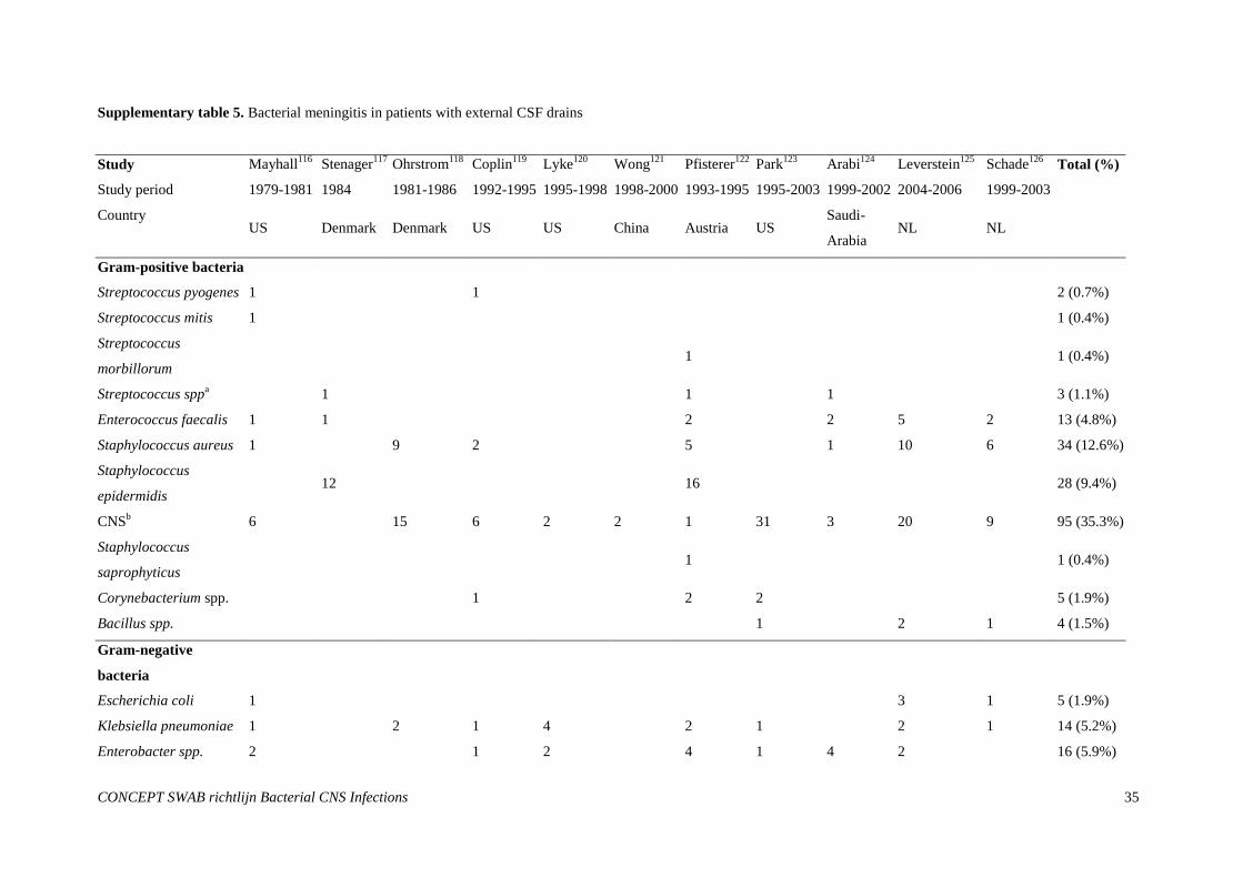
CSF, or as part of treatment of infected internal catheters.4 The risk of infection of external ventricular catheters is
reported to be approximately 8%, while infection of external lumbar catheters has been estimated to occur in
5%.34
Most common pathogens in infections of external CSF catheters are staphylococci, which are cultured in
approximately 60% of cases. Common staphylococcal species are coagulase-negative staphylococci, mostly S.
epidermidis, and S. aureus (Supplementary table 5).35-44
Other common pathogens are Klebsiella pneumoniae,
Enterobacter species, Enterococcus faecalis, Acinetobacter species and Pseudomonas aeruginosa. Multiple
uncommon pathogens can be found in external CSF catheter infection and therefore empirical antibiotic coverage
should be broad. A combination of vancomycin plus either ceftazidime or meropenem should be used (Table 4).
4
Key question 3b. What is the epidemiology and empirical treatment of bacterial meningitis related to internal
CSF drainage?
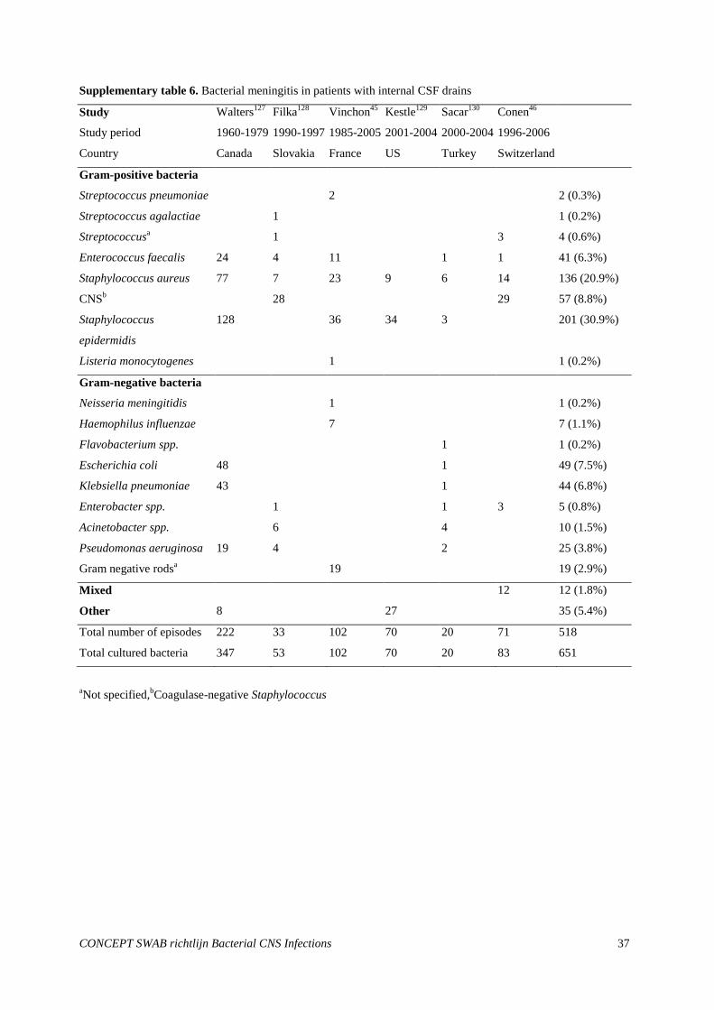
The reported incidence of meningitis associated with internal ventricular or lumbar catheters ranges from 4 to
17%.4,45,46
Approximately 60% of cases are caused by Staphylococcus species, and other frequent pathogens are
Escherichia coli, K. pneumoniae, E. faecalis and Acinetobacter species (Supplementary table 6).45-50
Mixed
infections were described in 17% of cases in a recent Swiss study including 71 patients.46
Because of the wide
range of pathogens and the occurrence of mixed infections the antibiotic coverage should be broad and should
consist of vancomycin plus either ceftazidime or meropenem (Table 4).
4
Key question 3c. What is the epidemiology and empirical treatment of bacterial meningitis related to
postoperative bacterial meningitis?
Bacterial meningitis is a serious complication of neurosurgical procedures and occurs in 0.8-1.5% of patients who
undergo craniotomy.4,51,52
Development of bacterial meningitis has been associated with concomitant infection of
the site of incision and duration of operation > 4 hours.4 One third of meningitis cases develop within the first
week after the operation, one third in the second week and one third after the second week.51
Most cases are
caused by Staphylococcus species (coagulase negative staphylococci, mostly S. epidermidis and S. aureus;
Supplementary table 752-57
). E. coli, K. pneumoniae, Enterobacter spp, Serratia spp, Acinetobacter spp, and P.
aeruginosa were all found in approximately 5% of cases. Frequent causes of community-acquired bacterial
meningitis such as Streptococcus pneumoniae and Haemophilus influenzae occur infrequently in postoperative
bacterial meningitis. Because the significance of coagulase negative staphylococci is not evident in the patients
without internal or external drains, and vancomycin is inferior to β-lactam antibiotics in S.aureus infections,
SWAB Guidelines Bacterial CNS Infections 21
empirical therapy should consist of flucloxacillin combined with ceftazidime, or meropenem monotherapy (Table
4).
Key question 3d. What is the epidemiology and empirical treatment of posttraumatic bacterial meningitis?
The incidence of meningitis after moderate and severe head trauma is estimated at 1.4%.4,58
In patients with open
cranial fractures the rate of meningitis is higher at 2-11%. Data on causative microorganisms in posttraumatic
bacterial meningitis are scarce (Supplementary table 8).58
Posttraumatic meningitis following skull base fractures
are caused by microorganisms that colonize the nasopharynx (S. pneumoniae, H. influenzae and Group A
streptococci),4 while bacterial meningitis following an impression fracture of the skull will be likely caused by
skin flora (staphylococci). Also in these patients the significance of coagulase negative staphylococci is not
evident in the absence of internal or external drains, and vancomycin is inferior to β-lactam antibiotics in S.aureus
infections. Therefore, empirical therapy in patients with skull base fractures consists of a third generation
cephalosporin, and patients with bacterial meningitis following open skull fractures should likewise be treated
with a third generation cephalosporin (ceftriaxone or cefotaxime)(Table 4).
Conclusions
Level 2 Bacterial meningitis related to external and internal ventricular drains, and postoperative
bacterial meningitis is caused by Staphylococcus spp. in 50-60% of cases
B Vinchon (2006)45
, Conen (2008)46
, Walters (1984)47
, Filka (1999)48
, Kestle (2006)49
,
Sacar (2006)50
, McClelland (2007)52
, Aucoin (1986)53
, Kourbeti (2007)54
, Federico
(2001)55
, Wang (2005)56
, Zarrouk (2007)57
Level 4 Bacterial meningitis following severe head trauma due to skull base fractures is mostly
caused by S. pneumoniae, H. influenzae and group A streptococci and bacterial
meningitis due to open skull fractures is mostly caused by skin flora (staphylococci).
* No studies have been performed on empirical antibiotic therapy for nosocomial bacterial
meningitis
Recommendations
1. Nosocomial meningitis associated with external or internal ventricular drains should be empirically treated
with vancomycin plus either ceftazidime or meropenem (Table 4).
2. Postoperative bacterial meningitis should be treated with flucloxacillin combined with ceftazidime, or with
meropenem monotherapy.
3. Posttraumatic bacterial meningitis due to a skull base fracture should be treated with a third generation
cephalosporin.
SWAB Guidelines Bacterial CNS Infections 22
4. Posttraumatic bacterial meningitis due to open skull fractures should be empirically treated with a third
generation cephalosporin (ceftriaxone or cefotaxime).
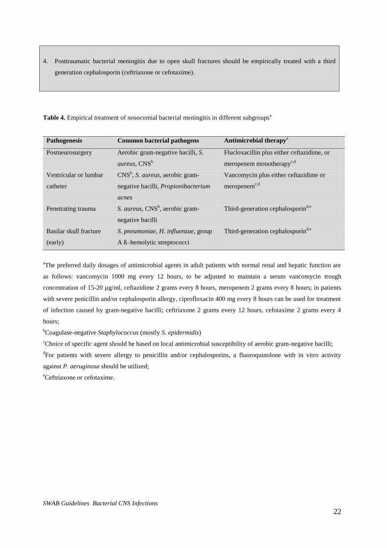
Table 4. Empirical treatment of nosocomial bacterial meningitis in different subgroupsa
Pathogenesis Common bacterial pathogens Antimicrobial therapya
Postneurosurgery Aerobic gram-negative bacilli, S.
aureus, CNSb
Flucloxacillin plus either ceftazidime, or
meropenem monotherapyc,d
Ventricular or lumbar
catheter
CNSb, S. aureus, aerobic gram-
negative bacilli, Propionibacterium
acnes
Vancomycin plus either ceftazidime or
meropenemc,d
Penetrating trauma S. aureus, CNSb, aerobic gram-
negative bacilli
Third-generation cephalosporind,e
Basilar skull fracture
(early)
S. pneumoniae, H. influenzae, group
A ß–hemolytic streptococci
Third-generation cephalosporind,e
aThe preferred daily dosages of antimicrobial agents in adult patients with normal renal and hepatic function are
as follows: vancomycin 1000 mg every 12 hours, to be adjusted to maintain a serum vancomycin trough
concentration of 15-20 µg/ml, ceftazidime 2 grams every 8 hours, meropenem 2 grams every 8 hours; in patients
with severe penicillin and/or cephalosporin allergy, ciprofloxacin 400 mg every 8 hours can be used for treatment
of infection caused by gram-negative bacilli; ceftriaxone 2 grams every 12 hours, cefotaxime 2 grams every 4
hours;
bCoagulase-negative Staphylococcus (mostly S. epidermidis)
cChoice of specific agent should be based on local antimicrobial susceptibility of aerobic gram-negative bacilli;
dFor patients with severe allergy to penicillin and/or cephalosporins, a fluoroquinolone with in vitro activity
against P. aeruginosa should be utilized;
eCeftriaxone or cefotaxime.
SWAB Guidelines Bacterial CNS Infections 23
Chapter 5
Withdrawal of antibiotic therapy in suspected postoperative meningitis.
Key question 4. When should antibiotic treatment in patients with suspected postoperative bacterial meningitis
be withdrawn?
A clinical suspicion of nosocomial bacterial meningitis should prompt a diagnostic workup and initiation of
empirical antimicrobial therapy.4 The most consistent clinical features are fever and a decreased level of
consciousness, although they are non-specific and difficult to recognize in patients who are sedated or have just
undergone neurosurgical treatment.4 The British Society for Antimicrobial Chemotherapy recommends empirical
treatment for all patients with signs of postoperative meningitis and withdrawal of treatment after 72 hours if the
results of CSF culture remain negative.59
This regimen was evaluated in a prospective study and showed to be
associated with few complications in patients with negative CSF Gram stain and culture.57
However, the effect of
preventive use of antibiotics in ICU patients on this regimen is unknown. If a high suspicion of nosocomial
bacterial meningitis remains even though CSF cultures are negative, prolongation of antibiotic treatment is
advised. When the causative microorganism is identified the antibiotic regimen can be adapted according to the
results of CSF culture and susceptibility testing.
Conclusions
Level 3 Empirical antibiotic therapy can be safely withdrawn after 72 hours in patients with
suspected postoperative meningitis if CSF cultures remain negative.
B Zarrouk57
Recommendations
1. In patients with suspected postoperative bacterial meningitis, empirical antibiotic treatment may be stopped
if cultures remain negative after three days.
SWAB Guidelines Bacterial CNS Infections 24
Chapter 6.
Duration of treatment before CSF catheter replacement
Key question 5. How long should infected CSF catheters be treated before removal/replacement?
If patients with internal ventricular catheters develop bacterial meningitis, the catheter should be removed to
increase the chances of resolving the infection.4 Observational studies showed that removal of the catheter
hardware, followed by immediate replacement and intravenous antimicrobial therapy, cures approximately 65%
of patients with catheter-related infections.60
The internal catheter should be replaced with an external ventricular
catheter to divert CSF and monitor CSF infection parameters, and this is associated with more rapid resolution of
ventriculitis.4 Conservative management (i.e., leaving the internal catheter in place and administering intravenous
or intraventricular antimicrobial therapy) has generally been associated with a low success rate (approximately
35%).60
Timing of reimplantation of the internal ventricular catheter depends on the causative microorganism and
results of repeated CSF cultures. As a general guideline shunt infections with coagulase-negative staphylococci
or Propionibacterium acnes are treated for 7 days before a new catheter is placed. However, if CSF cultures
remain positive the catheter should only be placed after cultures have been negative for at least 10 consecutive
days. For shunt infections by S. aureus and Gram-negative bacilli, 10 days of antimicrobial therapy after repeated
negative cultures are recommended before placement of a new internal catheter.4 When cultures remain negative
10 days of empirical treatment are recommended before placement of a new internal catheter.
Conclusions
Level 3 Removal of infected internal ventricular drains and replacement by external ventrical
drains combined with intravenous antibiotic treatment is associated with more rapid
resolution of ventriculitis.
B Schreffler (2002)60
* No randomized controlled trials or comparative studies have been performed to
determine the timing of reimplantation of internal CSF catheters in patients with
infected ventricular drains.
Recommendations
1. Reimplantation of CSF catheters in shunt infections caused by coagulase-negative staphylococci or
Propionibacterium acnes can be performed after 7 days of antimicrobial therapy.
If CSF cultures remain positive the catheter should only be placed after cultures have been negative for 10
consecutive days.
2. Reimplantation of CSF catheters in shunt infections caused by Staphylococcus aureus, Gram-negative
bacilli or in patients with negative CSF cultures can be performed 10 days after CSF cultures are negative.
SWAB Guidelines Bacterial CNS Infections 25
Chapter 7.
Intraventricular antibiotic treatment
Key question 6. What are the indications and regimens for intraventricular antimicrobial treatment?
Direct infusion of antimicrobial agents into the ventricles through external ventricular catheters is sometimes
necessary to treat infections after neurosurgical procedures or infections associated with CSF catheters that are
difficult to eradicate with parenteral antibiotics alone.4,28,61,62
The indications for intraventricular antibiotics are
however, not well defined. The antibiotic is administered through the catheter, which should subsequently be
closed for one hour. Dosages have been determined empirically and are generally adjusted according to the
measured CSF concentration of the antibiotic.4 Determination of CSF antibiotic concentration is performed in a
sample taken just before the next dose. The trough concentration should be at least 10-20 times the minimal
inhibitory concentration to achieve a constant sterilization of CSF.4 Antibiotics most used for intraventricular
administration are vancomycin and gentamicin (Table 5, adapted from 4).
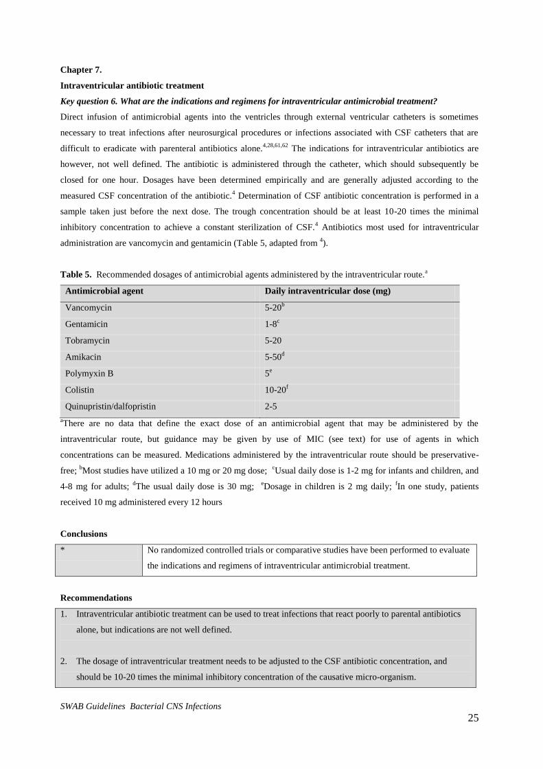
Table 5. Recommended dosages of antimicrobial agents administered by the intraventricular route.a
Antimicrobial agent Daily intraventricular dose (mg)
Vancomycin 5-20b
Gentamicin 1-8c
Tobramycin 5-20
Amikacin 5-50d
Polymyxin B 5e
Colistin 10-20f
Quinupristin/dalfopristin 2-5
aThere are no data that define the exact dose of an antimicrobial agent that may be administered by the
intraventricular route, but guidance may be given by use of MIC (see text) for use of agents in which
concentrations can be measured. Medications administered by the intraventricular route should be preservative-
free; bMost studies have utilized a 10 mg or 20 mg dose;
cUsual daily dose is 1-2 mg for infants and children, and
4-8 mg for adults; dThe usual daily dose is 30 mg;
eDosage in children is 2 mg daily;
fIn one study, patients
received 10 mg administered every 12 hours
Conclusions
* No randomized controlled trials or comparative studies have been performed to evaluate
the indications and regimens of intraventricular antimicrobial treatment.
Recommendations
1. Intraventricular antibiotic treatment can be used to treat infections that react poorly to parental antibiotics
alone, but indications are not well defined.
2. The dosage of intraventricular treatment needs to be adjusted to the CSF antibiotic concentration, and
should be 10-20 times the minimal inhibitory concentration of the causative micro-organism.
SWAB Guidelines Bacterial CNS Infections 26
Chapter 8.
The epidemiology of bacterial intracerebral abscess in Europe/US.
Key question 7: What is the epidemiology of bacterial intracerebral abscess in Europe/US?
We identified 51 studies reporting bacterial causes of brain abscesses that met the inclusion criteria.63-112,113
In
these studies 3,583 patients were included (Supplementary table 9). In 2536 patients (71%) data on bacterial
cultures were provided, and in 435 patients no culture was performed (17%). Cultures were negative in 30% and
in studies reporting the number of cultured bacteria, 26% of the cultures were polymicrobial. This may be an
underestimation as local microbiology data suggest that the rate of polymicrobial intracranial abscesses may be as
high as 50%.
Of the 2,653 bacteria cultured in the included studies, Streptococcus species were the most prevalent and were
identified in 36% of the positive cultures (Supplementary table 10). Specification of streptococcal species was not
available for the majority of strains. Streptococci from the milleri group were the most frequently identified
species, occurring in 9% of all patients and 72% of specified streptococcal species. S. pneumoniae, the most
frequent pathogen of bacterial meningitis, was identified in only 3% of all positive cultures in brain abscess
patients.6 The second most common microorganisms identified in brain abscesses were Staphylococcus species
(frequency 17%). Staphylococcus aureus was identified in 11% of patients and Staphylococcus epidermidis in
2%. The species were not specified of the remaining strains. Gram negative enteric bacteria were found in 12% of
cases and most of these were Proteus spp. (7%). Other relatively frequent pathogens were Bacteroides spp. (7%),
Haemophilus spp (3%) and Peptostreptococcus spp (3%). In these included studies 17% of bacteria were
classified as ‘other’ or were not specified.
Anaerobe bacteria are present in virtually all polymicrobial abscesses and are frequently found in cerebral
abscesses with a dentogenic focus. The frequency of anaerobes given in Supplementary table 10 may be an
underestimation due to suboptimal culture techniques and transportation.
16S ribosomal DNA sequencing has expanded the yield of microbiological investigation in patients with
intracerebral abscesses.114
The number of causative pathogens identified by sequencing of brain abscess aspirate
was over threefold higher compared to standard culture techniques (22 strains identified by culture vs. 72 by
sequencing).114
This technique is currently not commonly used, but will probably become part of routine
diagnostics in the coming years.
Key question 8: What is the optimal (empirical) antibiotic therapy in intracerebral abscess?
No clinical trials have been performed to determine the optimal regimen in bacterial intracerebral abscesses,
therefore the recommendations are all based on expert opinion.8,9
Empirical treatment consists of a third
generation cephalosporin (ceftriaxone or cefotaxime) combined with metronidazole in appropriate doses to
penetrate the blood brain barrier. Alternatively penicillin may be used instead of third generation cephalosporins.
After identification of the causative microorganism the antimicrobial therapy can be adapted according to the
antimicrobial resistance pattern. In culture-negative cases the empirical therapy should be continued until clinical
resolution (see question 10).

SWAB Guidelines Bacterial CNS Infections 27
The primary source of infection can give a clue to which bacteria cause the cerebral abscesses. For instance,
Enterobacteriaceae are the most frequently found in cerebral abscesses due to otogenic infections. However,
since virtually all common pathogens have been reported for the various sources of infection, the choice of
empirical antimicrobial therapy cannot be guided by the source of infection (e.g. dental abscess, endocarditis).
Key question 9. What is the optimal dose of antimicrobial therapy?
Only a limited number of antimicrobials have been studied for their penetration into brain abscesses, mostly in
small studies.53
Antimicrobials have to be able to first penetrate the blood brain barrier and then to achieve
sufficient levels inside the abscess to resolve the infection. Penicillin concentrations were only found consistently
in brain abscess pus if administered in a high dose of 24 million units per day.115
Commonly advised doses of
penicillin are however 12 million units per day in adults.8,9
Other antimicrobials that have shown adequate or
good penetration into the abscess cavity are metronidazole, vancomycin and third generation cephalosporins.53
The dosage and dose intervals of antimicrobials commonly used for brain abscesses are presented in table 6. For
vancomycin and aminoglycosides, the dose needs to be adjusted according to the peak and trough serum
concentrations.
Table 6. Dosages of antibiotics for use in adults with bacterial brain abscesses.a
Antibiotic Total daily dose Dose interval (hour)
Amikacinb 15 mg/kg 24
Amoxicillin 12 gr 4
Azitromycin 1200-1500 mg 24
Cefotaxime 8-12 gr 4-6
Ceftazidime 6 gr 8
Ceftriaxone 4 gr 12
Chloramphenicol 4-6 gr or 50 mg/kg/day 6
Ciprofloxacin 800-1200 mg 8-12
Clindamycin 2400-4800 mg 6
Gentamicinb 5 mg/kg 24
Meropenem 6 gr 8
Metronidazol 1500 mg 8
Penicillin 12x106 units 4 (or continuous)
Rifampicin 600 24
Tobramycin b
5 mg/kg 24
Trimethoprim-sulfamethoxazole
(TMP/SMX)
10-20 mg/kg iv (based on TMP component),
max. 960/4800 mg TMP/SMX iv
8
8
Vancomycinb 30-45 mg/kg, max. 2000 mg 12
Adapted from8.
aPatients with normal hepatic and renal function, intravenous administration.
bAdjust dosage
based on peak and trough serum concentrations.
SWAB Guidelines Bacterial CNS Infections 28
Key question 10. What is the optimal duration of therapy?
No studies have been performed to evaluate the optimal duration of therapy in bacterial brain abscesses. The
common strategy is to treat the abscess at least 6 weeks and to use clinical, laboratory and radiological follow-up
data to decide on further treatment beyond this period.8,9
Studies on antimicrobial treatment duration for cerebral
abscesses are needed.
Conclusions
* No clinical trials have been performed to determine the optimal antibiotic treatment in
bacterial intracerebral abscesses,
Recommendations
1. Empirical treatment for bacterial cerebral abscesses consists of a third generation cephalosporin
(ceftriaxone or cefotaxime) combined with metronidazole in appropriate doses to penetrate the blood brain
barrier (Table 6). Alternatively, penicillin may be used instead of a third generation cephalosporin.
2. If a microorganism is cultured, therapy can be adjusted accordingly. In culture-negative cases the empirical
therapy should be continued until clinical resolution.
3. The common strategy for antibiotic treatment of cerebral abscesses is to treat the abscess at least 6 weeks
and to use clinical, laboratory and radiological follow-up data to decide on further treatment beyond this
period.
SWAB Guidelines Bacterial CNS Infections 29
Chapter 9
Epidemiology, empirical antibiotic treatment and treatment duration of bacterial tuberculous meningitis
(in preparation)
Key question 11. What is the epidemiology and empirical treatment for tuberculous meningitis (in
preparation)?
Key question 12. What is the optimal duration of therapy for tuberculous meningitis (in preparation)?

SWAB Guidelines Bacterial CNS Infections
Supplementary table 1. Causative organisms of bacterial meningitis in neonates
Data source Garges (2006)13
Holt (2001)15
Hristeva (1993)14
NRLBM Total
Country US UK UK NL
Age 0-150 days (NICU) 2-28 days 0-28 days 0-28 days
Study period 1997-2004 1996-1997 1984-1991 2005-2010
Gram-positive
Streptococcus agalactiae 37 69 7 71 178 (54%)
Streptococcus mitis 0 0 1 0 1 (0.3%)
Listeria monocytogenes 1 7 1 1 10 (3.0%)
Streptococcus pneumoniae 2 8 0 3 13 (3.9%)
Gram-positive cocci (not specified) 12 0 0 0 12 (3.6%)
Enterococcus spp. 6 0 0 0 6 (1.8%)
Staphylococcus aureus 4 0 0 1 5 (1.5%)
Other Gram-positive bacteria (not specified) 0 17 0 2 19 (5.7%)
Gram-negative
Haemophilus influenzae 2 1 0 0 3 (0.9%)
Escherichia coli 12 26 0 25 57 (15.7%)
Enterobacter spp. 4 0 0 0 4 (1.2%)
Serratia spp. 2 0 1 0 3 (0.9%)
Acinetobacter spp. 3 0 0 0 3 (0.9%)
Pseudomonas spp. 3 0 1 0 4 (1.2%)
Proteus spp. 1 0 0 0 1 (0.3%)
Citrobacter spp. 1 0 0 0 1 (0.3%)
Salmonella spp. 1 0 0 1 2 (0.6%)
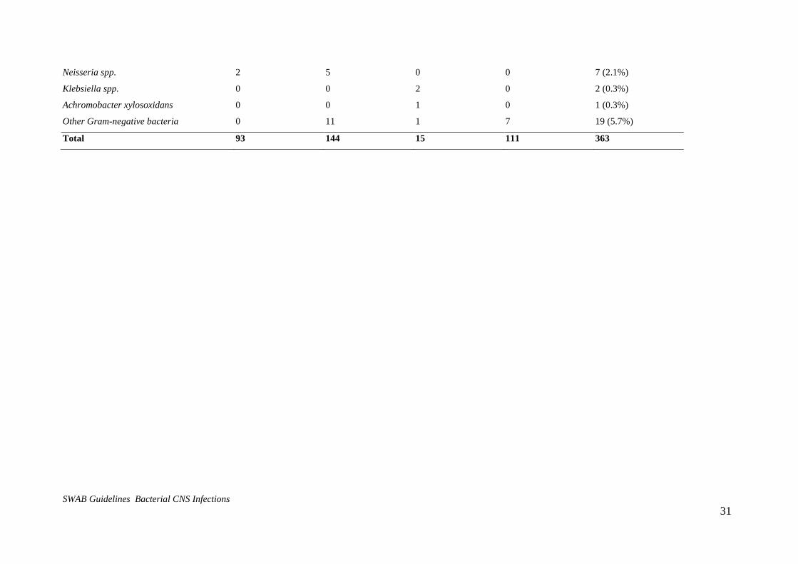
SWAB Guidelines Bacterial CNS Infections 31
Neisseria spp. 2 5 0 0 7 (2.1%)
Klebsiella spp. 0 0 2 0 2 (0.3%)
Achromobacter xylosoxidans 0 0 1 0 1 (0.3%)
Other Gram-negative bacteria 0 11 1 7 19 (5.7%)
Total 93 144 15 111 363

CONCEPT SWAB richtlijn Bacterial CNS Infections
32
Supplementary table 2. Causative organisms of bacterial meningitis in children aged 29 days-16 years
Data source NRLBM
Study period 2005-2010
Gram-positive bacteria
Streptococcus pneumoniae 305 (43.6%)
Streptococcus agalactiae 9 (1.3%)
Streptococcus pyogenes 14 (2.0%)
Streptococcus sanguinis 1 (0.1%)
“α-haemolytic” streptococcus 1 (0.1%)
Staphylococcus aureus 4 (0.6%)
Staphylococcus lugdunensis 1 (0.1%)
Staphylococcus capitis 1 (0.1%)
Staphylococcus epidermidis 2 (0.3%)
Coagulase-negative staphylococcus 1 (0.1%)
Listeria monocytogenes 3 (0.4%)
Gram-negative bacteria
Neisseria meningitidis 271 (38.8%)
Haemophilus influenzae 76 (10.9%)
Escherichia coli 10 (1.4%)
Total 699 (100.0%)
CONCEPT SWAB richtlijn Bacterial CNS Infections
33
Supplementary table 3. Causative organisms in 1500 episodes of culture-proven community-acquired bacterial
meningitis in adults
Data source DBMS10
MeninGene11
Study period 1998-2002 2006-2010 Total (%)
Gram-positive bacteria
Streptococcus pneumoniae 352 582 934 (62.3%)
Streptococcus pyogenes 6 11 17 (1.1%)
Streptococcus agalactiae 5 9 14 (0.9%)
Streptococcus constellatus 1 0 1 (0.1%)
Streptococcus milleri 0 1 1 (0.1%)
Streptococcus parasanguinis 0 1 1 (0.1%)
Streptococcus oralis 1 1 2 (0.1%)
Streptococcus suis 4 5 9 (0.6%)
Streptococcus salivarius 2 2 4 (0.3%)
Streptococcus bovis 1 2 3 (0.2%)
Streptococcus equi 1 3 4 (0.3%)
Streptococcus mitis 1 2 3 (0.2%)
Group G Streptococcus 1 1 2 (0.1%)
Streptococcus (not specified) 1 2 3 (0.2%)
Enterococcus faecalis 0 2 2 (0.1%)
Staphylococcus aureus 9 12 21 (1.4%)
Staphylococcus epidermidis 1 0 1 (0.1%)
Listeria monocytogenes 30 42 72 (4.8%)
Gram-negative bacteria
Neisseria meningitidis 257 91 348 (23.2%)
Haemophilus influenzae 14 26 40 (2.7%)
Haemophilus parainfluenzae 1 1 2 (0.1%)
Escherichia coli 4 7 11 (0.7%)
Klebsiella pneumoniae 1 1 2 (0.1%)
Salmonella enteritidis 0 1 1 (0.1%)
Capnocytophaga canimorsus 0 1 1 (0.1%)
Pseudomonas aeruginosa 0 1 1 (0.1%)
Total 693 807 1500 (100%)
CONCEPT SWAB richtlijn Bacterial CNS Infections
34
Supplementary table 4. Causative organisms in culture-proven community-acquired bacterial meningitis in
adults aged 17-50 without risk factors.
Data source MeninGene11
Study period 2005-2010
Study group 17-50 years old,
no risk factorsa
Gram-positive bacteria
Streptococcus pneumoniae 96 (52.4%)
Streptococcus pyogenes 6 (3.3%)
Streptococcus agalactiae 3 (1.6%)
Streptococcus suis 3 (1.6%)
Streptococcus equi 2 (1.1%)
Streptococcus mitis 1 (0.5%)
Staphylococcus aureus 2 (1.1%)
Listeria monocytogenes 3 (1.6%)
Gram-negative bacteria
Neisseria meningitidis 59 (32.2%)
Haemophilus influenzae 7 (3.8%)
Mixedb 2 (1.1%)
Total 183
aRisk factors: recent head injury, cerebrospinal fluid leak, diabetes mellitus, immunosuppressive therapy,
splenectomy, infection with human immunodeficiency virus (HIV).
bStreptococcus equi + Pseudomonas aeruginosa, Streptococcus milleri + Fusobacterium nucleatum
CONCEPT SWAB richtlijn Bacterial CNS Infections
35
Supplementary table 5. Bacterial meningitis in patients with external CSF drains
Study Mayhall116
Stenager117
Ohrstrom118
Coplin119
Lyke120
Wong121
Pfisterer122
Park123
Arabi124
Leverstein125
Schade126
Total (%)
Study period 1979-1981 1984 1981-1986 1992-1995 1995-1998 1998-2000 1993-1995 1995-2003 1999-2002 2004-2006 1999-2003
Country US Denmark Denmark US US China Austria US
Saudi-
Arabia NL NL
Gram-positive bacteria
Streptococcus pyogenes 1 1 2 (0.7%)
Streptococcus mitis 1 1 (0.4%)
Streptococcus
morbillorum 1 1 (0.4%)
Streptococcus sppa 1 1 1 3 (1.1%)
Enterococcus faecalis 1 1 2 2 5 2 13 (4.8%)
Staphylococcus aureus 1 9 2 5 1 10 6 34 (12.6%)
Staphylococcus
epidermidis 12 16 28 (9.4%)
CNSb 6 15 6 2 2 1 31 3 20 9 95 (35.3%)
Staphylococcus
saprophyticus 1 1 (0.4%)
Corynebacterium spp. 1 2 2 5 (1.9%)
Bacillus spp. 1 2 1 4 (1.5%)
Gram-negative
bacteria
Escherichia coli 1 3 1 5 (1.9%)
Klebsiella pneumoniae 1 2 1 4 2 1 2 1 14 (5.2%)
Enterobacter spp. 2 1 2 4 1 4 2 16 (5.9%)

CONCEPT SWAB richtlijn Bacterial CNS Infections
36
Serratia spp. 1 1 1 3 (1.1%)
Proteus spp. 1 1 1 3 (1.1%)
Acinetobacter spp. 2 1 1 1 2 1 1 6 1 16 (5.9%)
Pseudomonas
aeruginosa 1 2 2 2 1 8 (3.0%)
Flavobacterium spp. 1 1 (0.4%)
Mixed 1 4 1 6 (2.2%)
Other 1 1 6 2 10 (3.7%)
Total 19 16 27 13 11 6 44 51 19 46 22 269
aNot specified,
bCoagulase-negative Staphylococcus
CONCEPT SWAB richtlijn Bacterial CNS Infections
37
Supplementary table 6. Bacterial meningitis in patients with internal CSF drains
Study Walters127
Filka128
Vinchon45
Kestle129
Sacar130
Conen46
Study period 1960-1979 1990-1997 1985-2005 2001-2004 2000-2004 1996-2006
Country Canada Slovakia France US Turkey Switzerland
Gram-positive bacteria
Streptococcus pneumoniae 2 2 (0.3%)
Streptococcus agalactiae 1 1 (0.2%)
Streptococcusa 1 3 4 (0.6%)
Enterococcus faecalis 24 4 11 1 1 41 (6.3%)
Staphylococcus aureus 77 7 23 9 6 14 136 (20.9%)
CNSb 28 29 57 (8.8%)
Staphylococcus
epidermidis
128 36 34 3 201 (30.9%)
Listeria monocytogenes 1 1 (0.2%)
Gram-negative bacteria
Neisseria meningitidis 1 1 (0.2%)
Haemophilus influenzae 7 7 (1.1%)
Flavobacterium spp. 1 1 (0.2%)
Escherichia coli 48 1 49 (7.5%)
Klebsiella pneumoniae 43 1 44 (6.8%)
Enterobacter spp. 1 1 3 5 (0.8%)
Acinetobacter spp. 6 4 10 (1.5%)
Pseudomonas aeruginosa 19 4 2 25 (3.8%)
Gram negative rodsa 19 19 (2.9%)
Mixed 12 12 (1.8%)
Other 8 27 35 (5.4%)
Total number of episodes 222 33 102 70 20 71 518
Total cultured bacteria 347 53 102 70 20 83 651
aNot specified,
bCoagulase-negative Staphylococcus
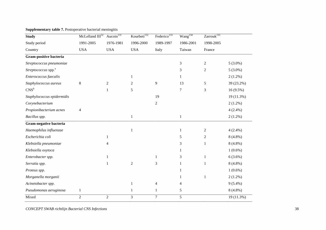
CONCEPT SWAB richtlijn Bacterial CNS Infections
38
Supplementary table 7. Postoperative bacterial meningitis
Study McLelland III52
Aucoin131
Kourbeti132
Federico133
Wang134
Zarrouk135
Study period 1991-2005 1976-1981 1996-2000 1989-1997 1986-2001 1998-2005
Country USA USA USA Italy Taiwan France
Gram-positive bacteria
Streptococcus pneumoniae 3 2 5 (3.0%)
Streptococcus spp.a 3 2 5 (3.0%)
Enterococcus faecalis 1 1 2 (1.2%)
Staphylococcus aureus 8 2 2 9 13 5 39 (23.2%)
CNSb 1 5 7 3 16 (9.5%)
Staphylococcus epidermidis 19 19 (11.3%)
Corynebacterium 2 2 (1.2%)
Propionibacterium acnes 4 4 (2.4%)
Bacillus spp. 1 1 2 (1.2%)
Gram-negative bacteria
Haemophilus influenzae 1 1 2 4 (2.4%)
Escherichia coli 1 5 2 8 (4.8%)
Klebsiella pneumoniae 4 3 1 8 (4.8%)
Klebsiella oxytoca 1 1 (0.6%)
Enterobacter spp. 1 1 3 1 6 (3.6%)
Serratia spp. 1 2 3 1 1 8 (4.8%)
Proteus spp. 1 1 (0.6%)
Morganella morganii 1 1 2 (1.2%)
Acinetobacter spp. 1 4 4 9 (5.4%)
Pseudomonas aeruginosa 1 1 1 5 8 (4.8%)
Mixed 2 2 3 7 5 19 (11.3%)
CONCEPT SWAB richtlijn Bacterial CNS Infections
39
Total 14 12 12 52 62 20 168
aNot specified,
bCoagulase-negative Staphylococcus
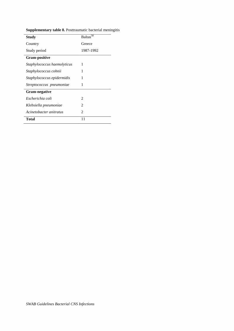
SWAB Guidelines Bacterial CNS Infections
Supplementary table 8. Posttraumatic bacterial meningitis
Study Baltas58
Country Greece
Study period 1987-1992
Gram-positive
Staphylococcus haemolyticus 1
Staphylococcus cohnii 1
Staphylococcus epidermidis 1
Streptococcus pneumoniae 1
Gram-negative
Escherichia coli 2
Klebsiella pneumoniae 2
Acinetobacter anitratus 2
Total 11
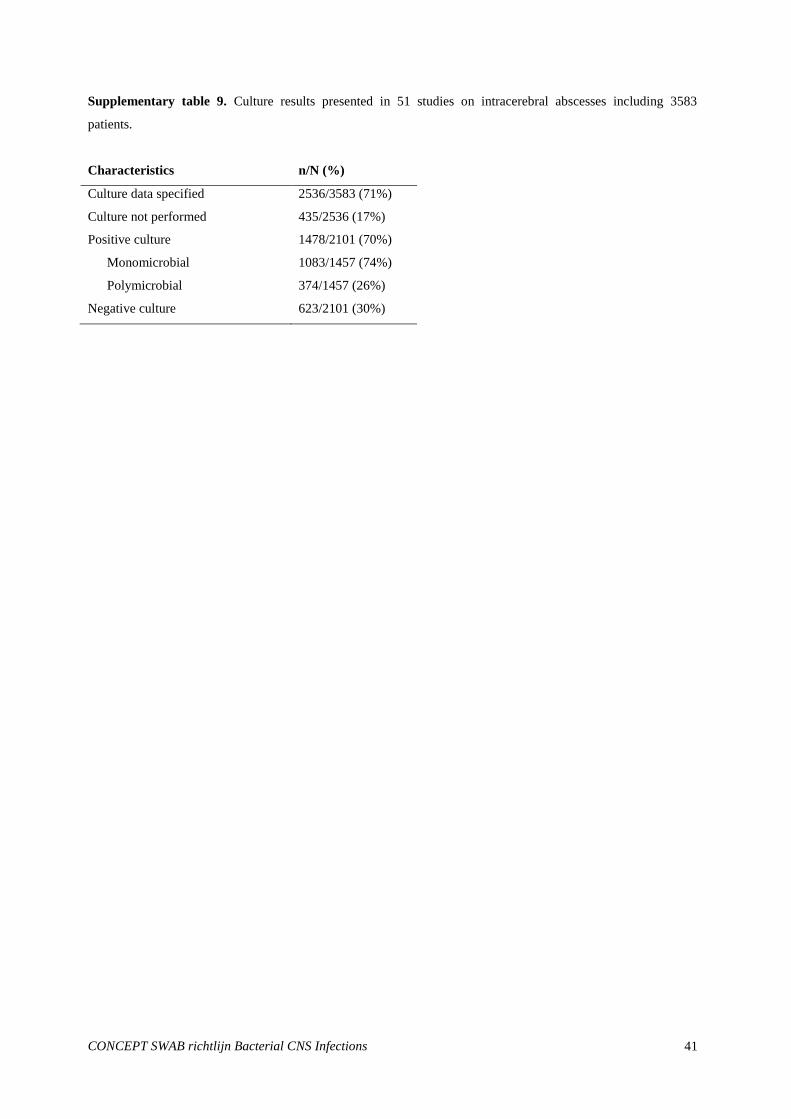
CONCEPT SWAB richtlijn Bacterial CNS Infections
41
Supplementary table 9. Culture results presented in 51 studies on intracerebral abscesses including 3583
patients.
Characteristics n/N (%)
Culture data specified 2536/3583 (71%)
Culture not performed 435/2536 (17%)
Positive culture 1478/2101 (70%)
Monomicrobial 1083/1457 (74%)
Polymicrobial 374/1457 (26%)
Negative culture 623/2101 (30%)

CONCEPT SWAB richtlijn Bacterial CNS Infections
42
Supplementary table 10. Overview of 2653 causative bacteria cultured from patients with intracerebral abscesses
from 51 studies including >10 patients.
Bacteria No (%)
Staphylococcus spp 454 (17.1%)
Staphylococcus aureus 292 (11.0%)
Staphylococcus epidermidis 62 (2.3%)
Staphylococci other/not specified 100 (3.8%)
Streptococcus spp 953 (35.9%)
Aerobic streptococci 39 (1.5%)
Haemolytic streptococci 34 (1.3%)
Anhaemolytic streptococci 27 (1.0%)
Streptococcus milleri/ viridans streptococci 219 (8.3%)
Microaerophilic streptococci 55 (2.1%)
Streptococcus pneumoniae 72 (2.7%)
Streptococcus other/not specified 397 (15.0%)
Gram negative enteric rods 313 (11.8%)
Escherichia coli 42 (1.6%)
Klebsiella spp 30 (1.1%)
Proteus spp 183 (6.9%)
Other Gram-negative enteric rods 58 (2.2%)
Pseudomonas spp 47 (1.8%)
Haemophilus spp
Actinomycetales
85 (3.2%)
80 (3.0%)
Nocardia 14 (0.5%)
Actinomyces 25 (0.9%)
Corynebacterium 29 (1.1%)
Other Actinomycetales 20 (0.8%)
Peptostreptoccus spp 88 (3.3%)
Anaerobic streptococci
Bacteroides spp
71 (2.7%)
181 (6.8%)
Other 199 (7.5%)
Not specified 253 (9.5%)
CONCEPT SWAB richtlijn Bacterial CNS Infections
43
REFERENCES
1. CBO. Kwaliteitsinstituut voor de Gezondheidszorg CBO, Handleiding voor werkgroepleden.
http://www.cbo.nl/Downloads/222/EBRO_handl_totaal.pdf
2. van de Beek D, Weisfelt M, de Gans J, Tunkel AR, Wijdicks EF. Drug Insight: adjunctive therapies in
adults with bacterial meningitis. Nat Clin Pract Neurol 2006; 2: 504-16.
3. van de Beek D, Brouwer MC, de Gans J et al. Richtlijn bacteriele meningitis. Utrecht: Nederlandse
Vereniging voor Neurologie; 2011.
4. van de Beek D, Drake JM, Tunkel AR. Nosocomial bacterial meningitis. N Engl J Med 2010; 362: 146-
54.
5. Bohr V, Rasmussen N, Hansen B et al. 875 cases of bacterial meningitis: diagnostic procedures and the
impact of preadmission antibiotic therapy. Part III of a three-part series. J Infect 1983; 7: 193-202.
6. Brouwer MC, Tunkel AR, van de Beek D. Epidemiology, diagnosis, and antimicrobial treatment of acute
bacterial meningitis. Clin Microbiol Rev 2010; 23: 467-92.
7. Spanos A, Harrell FE, Jr., Durack DT. Differential diagnosis of acute meningitis. An analysis of the
predictive value of initial observations. JAMA 1989; 262: 2700-7.
8. Tunkel AR. Brain abscess. In: Mandell GL, Bennett JE, Dolin R, editors. Principles and practive of
infectious diseases. 7 ed. Philadelphia: Churchill livingstone; 2010. p. 1265-78.
9. Kastenbauer S, Pfister HW, Wispelwey B, Scheld WM. Brain abscess. In: Scheld WM, Whitley RJ,
Marra CM, editors. Infections of the central nervous system. 3 ed. Philadelphia: Lippincott Williams &
Wilkins; 2004. p. 479-507.
10. van de Beek D, de Gans J, Spanjaard L, Weisfelt M, Reitsma JB, Vermeulen M. Clinical features and
prognostic factors in adults with bacterial meningitis. N Engl J Med 2004; 351: 1849-59.
11. Brouwer MC, Heckenberg SG, de GJ, Spanjaard L, Reitsma JB, van de Beek D. Nationwide
implementation of adjunctive dexamethasone therapy for pneumococcal meningitis. Neurology 2010.
12. van de Beek D, de Gans J, Tunkel AR, Wijdicks EF. Community-acquired bacterial meningitis in adults.
N Engl J Med 2006; 354: 44-53.
13. Garges HP, Moody MA, Cotten CM et al. Neonatal meningitis: what is the correlation among
cerebrospinal fluid cultures, blood cultures, and cerebrospinal fluid parameters? Pediatrics 2006; 117:
1094-100.
14. Hristeva L, Booy R, Bowler I, Wilkinson AR. Prospective surveillance of neonatal meningitis. Arch Dis
Child 1993; 69: 14-8.
15. Holt DE, Halket S, de LJ, Harvey D. Neonatal meningitis in England and Wales: 10 years on. Arch Dis
Child Fetal Neonatal Ed 2001; 84: F85-F89.
16. Galiza EP, Heath PT. Improving the outcome of neonatal meningitis. Curr Opin Infect Dis 2009; 22:
229-34.
17. Heath PT, Nik Yusoff NK, Baker CJ. Neonatal meningitis. Arch Dis Child Fetal Neonatal Ed 2003; 88:
F173-F178.
18. Siegal T, Pfeffer MR, Steiner I. Antibiotic therapy for infected Ommaya reservoir systems. Neurosurgery
1988; 22: 97-100.
CONCEPT SWAB richtlijn Bacterial CNS Infections
44
19. Cooper MD, Keeney RE, Lyons SF, Cheatle EL. Synergistic effects of ampicillin-aminoglycoside
combinations on group B streptococci. Antimicrob Agents Chemother 1979; 15: 484-6.
20. Netherlands Reference Laboratory for Bacterial Meningitis (AMC/RIVM). Bacterial meningitis in the
Netherlands Annual report 2010. Amsterdam: University of Amsterdam; 2011.
21. Odio CM, Puig JR, Feris JM et al. Prospective, randomized, investigator-blinded study of the efficacy
and safety of meropenem vs. cefotaxime therapy in bacterial meningitis in children. Meropenem
Meningitis Study Group. Pediatr Infect Dis J 1999; 18: 581-90.
22. Brouwer MC, Heckenberg SG, de Gans J, Spanjaard L, Reitsma JB, van de Beek D. Nationwide
implementation of adjunctive dexamethasone therapy for pneumococcal meningitis. Neurology 2010; 75:
1533-9.
23. Heckenberg SG, de Gans J, Brouwer MC et al. Clinical features, outcome, and meningococcal genotype
in 258 adults with meningococcal meningitis: a prospective cohort study. Medicine (Baltimore) 2008; 87:
185-92.
24. Brouwer MC, van de Beek D, Heckenberg SG, Spanjaard L, de Gans J. Community-acquired Listeria
monocytogenes meningitis in adults. Clin Infect Dis 2006; 43: 1233-8.
25. Karageorgopoulos DE, Valkimadi PE, Kapaskelis A, Rafailidis PI, Falagas ME. Short versus long
duration of antibiotic therapy for bacterial meningitis: a meta-analysis of randomised controlled trials in
children. Arch Dis Child 2009; 94: 607-14.
26. van de Beek D, Brouwer MC. No difference between short-course and long-course antibiotics for
bacterial meningitis in children, but available evidence limited. Evid Based Med 2010; 15: 6-7.
27. Molyneux E, Nizami SQ, Saha S et al. 5 versus 10 days of treatment with ceftriaxone for bacterial
meningitis in children: a double-blind randomised equivalence study. Lancet 2011; 377: 1837-45.
28. Tunkel AR, Hartman BJ, Kaplan SL et al. Practice guidelines for the management of bacterial
meningitis. Clin Infect Dis 2004; 39: 1267-84.
29. van der Meer H, van ZA, Spanjaard L, Van FM. [An infant with meningitis caused by resistant
pneumococcus: infection despite vaccination]. Ned Tijdschr Geneeskd 2012; 156: A3806.
30. Mitja O, Pigrau C, Ruiz I et al. Predictors of mortality and impact of aminoglycosides on outcome in
listeriosis in a retrospective cohort study. J Antimicrob Chemother 2009; 64: 416-23.
31. Lorber B. Listeriosis. Clin Infect Dis 1997; 24: 1-9.
32. Red Book: 2009 report of the committee on infectious diseases. 28 ed. Elk Grove Village, IL, USA:
American Academy of Pediatrics; 2009.
33. Falagas ME, Bliziotis IA, Tam VH. Intraventricular or intrathecal use of polymyxins in patients with
Gram-negative meningitis: a systematic review of the available evidence. Int J Antimicrob Agents 2007;
29: 9-25.
34. Lozier AP, Sciacca RR, Romagnoli MF, Connolly ES, Jr. Ventriculostomy-related infections: a critical
review of the literature. Neurosurgery 2008; 62 Suppl 2: 688-700.
35. Mayhall CG, Archer NH, Lamb VA et al. Ventriculostomy-related infections. A prospective
epidemiologic study. N Engl J Med 1984; 310: 553-9.
36. Stenager E, Gerner-Smidt P, Kock-Jensen C. Ventriculostomy-related infections--an epidemiological
study. Acta Neurochir (Wien ) 1986; 83: 20-3.
37. Coplin WM, Avellino AM, Kim DK, Winn HR, Grady MS. Bacterial meningitis associated with lumbar
drains: a retrospective cohort study. J Neurol Neurosurg Psychiatry 1999; 67: 468-73.
CONCEPT SWAB richtlijn Bacterial CNS Infections
45
38. Lyke KE, Obasanjo OO, Williams MA, O'Brien M, Chotani R, Perl TM. Ventriculitis complicating use
of intraventricular catheters in adult neurosurgical patients. Clin Infect Dis 2001; 33: 2028-33.
39. Wong GK, Poon WS, Wai S, Yu LM, Lyon D, Lam JM. Failure of regular external ventricular drain
exchange to reduce cerebrospinal fluid infection: result of a randomised controlled trial. J Neurol
Neurosurg Psychiatry 2002; 73: 759-61.
40. Pfisterer W, Muhlbauer M, Czech T, Reinprecht A. Early diagnosis of external ventricular drainage
infection: results of a prospective study. J Neurol Neurosurg Psychiatry 2003; 74: 929-32.
41. Park P, Garton HJ, Kocan MJ, Thompson BG. Risk of infection with prolonged ventricular
catheterization. Neurosurgery 2004; 55: 594-9.
42. Arabi Y, Memish ZA, Balkhy HH et al. Ventriculostomy-associated infections: incidence and risk
factors. Am J Infect Control 2005; 33: 137-43.
43. Schade RP, Schinkel J, Visser LG, Van Dijk JM, Voormolen JH, Kuijper EJ. Bacterial meningitis caused
by the use of ventricular or lumbar cerebrospinal fluid catheters. J Neurosurg 2005; 102: 229-34.
44. Ohrstrom JK, Skou JK, Ejlertsen T, Kosteljanetz M. Infected ventriculostomy: bacteriology and
treatment. Acta Neurochir (Wien ) 1989; 100: 67-9.
45. Vinchon M, Dhellemmes P. Cerebrospinal fluid shunt infection: risk factors and long-term follow-up.
Childs Nerv Syst 2006; 22: 692-7.
46. Conen A, Walti LN, Merlo A, Fluckiger U, Battegay M, Trampuz A. Characteristics and treatment
outcome of cerebrospinal fluid shunt-associated infections in adults: a retrospective analysis over an 11-
year period. Clin Infect Dis 2008; 47: 73-82.
47. Walters BC, Hoffman HJ, Hendrick EB, Humphreys RP. Cerebrospinal fluid shunt infection. Influences
on initial management and subsequent outcome. J Neurosurg 1984; 60: 1014-21.
48. Filka J, Huttova M, Tuharsky J, Sagat T, Kralinsky K, Krcmery V, Jr. Nosocomial meningitis in children
after ventriculoperitoneal shunt insertion. Acta Paediatr 1999; 88: 576-8.
49. Kestle JR, Garton HJ, Whitehead WE et al. Management of shunt infections: a multicenter pilot study. J
Neurosurg 2006; 105: 177-81.
50. Sacar S, Turgut H, Toprak S et al. A retrospective study of central nervous system shunt infections
diagnosed in a university hospital during a 4-year period. BMC Infect Dis 2006; 6:43.: 43.
51. Korinek AM, Baugnon T, Golmard JL, van Effenterre R, Coriat P, Puybasset L. Risk factors for adult
nosocomial meningitis after craniotomy: role of antibiotic prophylaxis. Neurosurgery 2008; 62 Suppl 2:
532-9.
52. McClelland S, III, Hall WA. Postoperative central nervous system infection: incidence and associated
factors in 2111 neurosurgical procedures. Clin Infect Dis 2007; 45: 55-9.
53. Aucoin PJ, Kotilainen HR, Gantz NM, Davidson R, Kellogg P, Stone B. Intracranial pressure monitors.
Epidemiologic study of risk factors and infections. Am J Med 1986; 80: 369-76.
54. Kourbeti IS, Jacobs AV, Koslow M, Karabetsos D, Holzman RS. Risk factors associated with
postcraniotomy meningitis. Neurosurgery 2007; 60: 317-25.
55. Federico G, Tumbarello M, Spanu T et al. Risk factors and prognostic indicators of bacterial meningitis
in a cohort of 3580 postneurosurgical patients. Scand J Infect Dis 2001; 33: 533-7.
56. Wang KW, Chang WN, Huang CR et al. Post-neurosurgical nosocomial bacterial meningitis in adults:
microbiology, clinical features, and outcomes. J Clin Neurosci 2005; 12: 647-50.
CONCEPT SWAB richtlijn Bacterial CNS Infections
46
57. Zarrouk V, Vassor I, Bert F et al. Evaluation of the management of postoperative aseptic meningitis. Clin
Infect Dis 2007; 44: 1555-9.
58. Baltas I, Tsoulfa S, Sakellariou P, Vogas V, Fylaktakis M, Kondodimou A. Posttraumatic meningitis:
bacteriology, hydrocephalus, and outcome. Neurosurgery 1994; 35: 422-6.
59. Infection in neurosurgery working party of the british society for antimicrobial chemotherapy. The
management of neurosurgical patients with postoperative bacterial or aseptic meningitis or external
ventricular drain-associated ventriculitis. Infection in Neurosurgery Working Party of the British Society
for Antimicrobial Chemotherapy. Br J Neurosurg 2000; 14: 7-12.
60. Schreffler RT, Schreffler AJ, Wittler RR. Treatment of cerebrospinal fluid shunt infections: a decision
analysis. Pediatr Infect Dis J 2002; 21: 632-6.
61. Kim BN, Peleg AY, Lodise TP et al. Management of meningitis due to antibiotic-resistant Acinetobacter
species. Lancet Infect Dis 2009; 9: 245-55.
62. Pfausler B, Spiss H, Beer R et al. Treatment of staphylococcal ventriculitis associated with external
cerebrospinal fluid drains: a prospective randomized trial of intravenous compared with intraventricular
vancomycin therapy. J Neurosurg 2003; 98: 1040-4.
63. Beller AJ, Sahar A, Praiss I. Brain abscess. Review of 89 cases over a period of 30 years. J Neurol
Neurosurg Psychiatry 1973; 36: 757-68.
64. Berlit P, Fedel C, Tornow K, Schmiedek P. [Bacterial brain abscess--experiences with 67 patients].
Fortschr Neurol Psychiatr 1996; 64: 297-306.
65. Carpenter J, Stapleton S, Holliman R. Retrospective analysis of 49 cases of brain abscess and review of
the literature. Eur J Clin Microbiol Infect Dis 2007; 26: 1-11.
66. Hakan T, Ceran N, Erdem I, Berkman MZ, Goktas P. Bacterial brain abscesses: an evaluation of 96
cases. J Infect 2006; 52: 359-66.
67. Tattevin P, Bruneel F, Clair B et al. Bacterial brain abscesses: a retrospective study of 94 patients
admitted to an intensive care unit (1980 to 1999). Am J Med 2003; 115: 143-6.
68. Smith SJ, Ughratdar I, MacArthur DC. Never go to sleep on undrained pus: a retrospective review of
surgery for intraparenchymal cerebral abscess. Br J Neurosurg 2009; 23: 412-7.
69. Al Masalma M, Armougom F, Scheld WM et al. The expansion of the microbiological spectrum of brain
abscesses with use of multiple 16S ribosomal DNA sequencing. Clin Infect Dis 2009; 48: 1169-78.
70. de Lastours V, Kalamarides M, Leflon V et al. Optimization of bacterial diagnosis yield after needle
aspiration in immunocompetent adults with brain abscesses. Neurosurgery 2008; 63: 362-7.
71. Cavusoglu H, Kaya RA, Turkmenoglu ON, Colak I, Aydin Y. Brain abscess: analysis of results in a
series of 51 patients with a combined surgical and medical approach during an 11-year period.
Neurosurg Focus 2008; 24: E9.
72. Goodkin HP, Harper MB, Pomeroy SL. Intracerebral abscess in children: historical trends at Children's
Hospital Boston. Pediatrics 2004; 113: 1765-70.
73. Le Moal G, Landron C, Grollier G et al. Characteristics of brain abscess with isolation of anaerobic
bacteria. Scand J Infect Dis 2003; 35: 318-21.
74. Sennaroglu L, Sozeri B. Otogenic brain abscess: review of 41 cases. Otolaryngol Head Neck Surg 2000;
123: 751-5.
75. Barlas O, Sencer A, Erkan K, Eraksoy H, Sencer S, Bayindir C. Stereotactic surgery in the management
of brain abscess. Surg Neurol 1999; 52: 404-10.
CONCEPT SWAB richtlijn Bacterial CNS Infections
47
76. Mamelak AN, Mampalam TJ, Obana WG, Rosenblum ML. Improved management of multiple brain
abscesses: a combined surgical and medical approach. Neurosurgery 1995; 36: 76-85.
77. O'Donoghue MA, Green HT, Shaw MD. Cerebral abscess on Merseyside 1980-1988. J Infect 1992; 25:
163-72.
78. Tekkok IH, Erbengi A. Management of brain abscess in children: review of 130 cases over a period of 21
years. Childs Nerv Syst 1992; 8: 411-6.
79. Renier D, Flandin C, Hirsch E, Hirsch JF. Brain abscesses in neonates. A study of 30 cases. J Neurosurg
1988; 69: 877-82.
80. Chun CH, Johnson JD, Hofstetter M, Raff MJ. Brain abscess. A study of 45 consecutive cases. Medicine
(Baltimore) 1986; 65: 415-31.
81. Brook I. Bacteriology of intracranial abscess in children. J Neurosurg 1981; 54: 484-8.
82. Shahzad K, Hamid MH, Khan MA, Malik N, Maqbool S. Brain abscess in children. J Coll Physicians
Surg Pak 2005; 15: 609-11.
83. de Louvois, ., Gortavai P, Hurley R. Bacteriology of abscesses of the central nervous system: a
multicentre prospective study. Br Med J 1977; 2: 981-4.
84. Jansson AK, Enblad P, Sjolin J. Efficacy and safety of cefotaxime in combination with metronidazole for
empirical treatment of brain abscess in clinical practice: a retrospective study of 66 consecutive cases.
Eur J Clin Microbiol Infect Dis 2004; 23: 7-14.
85. Bartt R. Listeria and atypical presentations of Listeria in the central nervous system. Semin Neurol 2000;
20: 361-73.
86. Roche M, Humphreys H, Smyth E et al. A twelve-year review of central nervous system bacterial
abscesses; presentation and aetiology. Clin Microbiol Infect 2003; 9: 803-9.
87. Mampalam TJ, Rosenblum ML. Trends in the management of bacterial brain abscesses: a review of 102
cases over 17 years. Neurosurgery 1988; 23: 451-8.
88. Bagdatoglu H, Ildan F, Cetinalp E et al. The clinical presentation of intracranial abscesses. A study of
seventy-eight cases. J Neurosurg Sci 1992; 36: 139-43.
89. Miller ES, Dias PS, Uttley D. CT scanning in the management of intracranial abscess: a review of 100
cases. Br J Neurosurg 1988; 2: 439-46.
90. Tonon E, Scotton PG, Gallucci M, Vaglia A. Brain abscess: clinical aspects of 100 patients. Int J Infect
Dis 2006; 10: 103-9.
91. Gomez J, Garcia-Vazquez E, Martinez PM et al. [Brain abscess. The experience of 30 years]. Med Clin
(Barc ) 2008; 130: 736-9.
92. Lunardi P, Acqui M, Ferrante L, Mastronardi L, Fortuna A. Non-traumatic brain abscess. Neurosurg Rev
1993; 16: 189-96.
93. Nicolosi A, Hauser WA, Musicco M, Kurland LT. Incidence and prognosis of brain abscess in a defined
population: Olmsted County, Minnesota, 1935-1981. Neuroepidemiology 1991; 10: 122-31.
94. Richards J, Sisson PR, Hickman JE, Ingham HR, Selkon JB. Microbiology, chemotherapy and mortality
of brain abscess in Newcastle-upon-Tyne between 1979 and 1988. Scand J Infect Dis 1990; 22: 511-8.
95. Svanteson B, Nordstrom CH, Rausing A. Non-traumatic brain abscess. Epidemiology, clinical symptoms
and therapeutic results. Acta Neurochir (Wien ) 1988; 94: 57-65.
CONCEPT SWAB richtlijn Bacterial CNS Infections
48
96. Danziger A, Price H, Schechter MM. An analysis of 113 intracranial infections. Neuroradiology 1980;
19: 31-4.
97. Hilmani S, Riyahi S, Ibahioin K, Naja A, El KA, El AA. [Brain abscess (80 cases)]. Neurochirurgie
2009; 55: 40-4.
98. Morgan H, Wood MW, Murphey F. Experience with 88 consecutive cases of brain abscess. J Neurosurg
1973; 38: 698-704.
99. Carey ME, Chou SN, French LA. Long-term neurological residua in patients surviving brain abscess
with surgery. J Neurosurg 1971; 34: 652-6.
100. Rish BL, Caveness WF, Dillon JD, Kistler JP, Mohr JP, Weiss GH. Analysis of brain abscess after
penetrating craniocerebral injuries in Vietnam. Neurosurgery 1981; 9: 535-41.
101. Gruszkiewicz J, Doron Y, Peyser E, Borovich B, Schachter J, Front D. Brain abscess and its surgical
management. Surg Neurol 1982; 18: 7-17.
102. Bradley PJ, Manning KP, Shaw MD. Brain abscess secondary to otitis media. J Laryngol Otol 1984; 98:
1185-91.
103. Ersahin Y, Mutluer S, Guzelbag E. Brain abscess in infants and children. Childs Nerv Syst 1994; 10: 185-
9.
104. Schliamser SE, Backman K, Norrby SR. Intracranial abscesses in adults: an analysis of 54 consecutive
cases. Scand J Infect Dis 1988; 20: 1-9.
105. Mathisen GE, Meyer RD, George WL, Citron DM, Finegold SM. Brain abscess and cerebritis. Rev Infect
Dis 1984; 6 Suppl 1: S101-S106.
106. Shaw MD, Russell JA. Cerebellar abscess. A review of 47 cases. J Neurol Neurosurg Psychiatry 1975;
38: 429-35.
107. Nielsen H. Cerebral abscess in children. Neuropediatrics 1983; 14: 76-80.
108. Hirsch JF, Roux FX, Sainte-Rose C, Renier D, Pierre-Kahn A. Brain abscess in childhood. A study of 34
cases treated by puncture and antibiotics. Childs Brain 1983; 10: 251-65.
109. Moss SD, McLone DG, Arditi M, Yogev R. Pediatric cerebral abscess. Pediatr Neurosci 1988; 14: 291-
6.
110. Fischer EG, McLennan JE, Suzuki Y. Cerebral abscess in children. Am J Dis Child 1981; 135: 746-9.
111. Dohrmann PJ, Elrick WL. Observations on brain abscess. Review of 28 cases. Med J Aust 1982; 2: 81-3.
112. Sharma R, Mohandas K, Cooke RP. Intracranial abscesses: changes in epidemiology and management
over five decades in Merseyside. Infection 2009; 37: 39-43.
113. van Alphen HA, Dreissen JJ. Brain abscess and subdural empyema. Factors influencing mortality and
results of various surgical techniques. J Neurol Neurosurg Psychiatry 1976; 39: 481-90.
114. Al MM, Armougom F, Scheld WM et al. The expansion of the microbiological spectrum of brain
abscesses with use of multiple 16S ribosomal DNA sequencing. Clin Infect Dis 2009; 48: 1169-78.
115. de LJ, Gortvai P, Hurley R. Antibiotic treatment of abscesses of the central nervous system. Br Med J
1977; 2: 985-7.
116. Mayhall CG, Archer NH, Lamb VA et al. Ventriculostomy-related infections. A prospective
epidemiologic study. N Engl J Med 1984; 310: 553-9.
CONCEPT SWAB richtlijn Bacterial CNS Infections
49
117. Stenager E, Gerner-Smidt P, Kock-Jensen C. Ventriculostomy-related infections--an epidemiological
study. Acta Neurochir (Wien ) 1986; 83: 20-3.
118. Ohrstrom JK, Skou JK, Ejlertsen T, Kosteljanetz M. Infected ventriculostomy: bacteriology and
treatment. Acta Neurochir (Wien ) 1989; 100: 67-9.
119. Coplin WM, Avellino AM, Kim DK, Winn HR, Grady MS. Bacterial meningitis associated with lumbar
drains: a retrospective cohort study. J Neurol Neurosurg Psychiatry 1999; 67: 468-73.
120. Lyke KE, Obasanjo OO, Williams MA, O'Brien M, Chotani R, Perl TM. Ventriculitis complicating use
of intraventricular catheters in adult neurosurgical patients. Clin Infect Dis 2001; 33: 2028-33.
121. Wong GK, Poon WS, Wai S, Yu LM, Lyon D, Lam JM. Failure of regular external ventricular drain
exchange to reduce cerebrospinal fluid infection: result of a randomised controlled trial. J Neurol
Neurosurg Psychiatry 2002; 73: 759-61.
122. Pfisterer W, Muhlbauer M, Czech T, Reinprecht A. Early diagnosis of external ventricular drainage
infection: results of a prospective study. J Neurol Neurosurg Psychiatry 2003; 74: 929-32.
123. Park P, Garton HJ, Kocan MJ, Thompson BG. Risk of infection with prolonged ventricular
catheterization. Neurosurgery 2004; 55: 594-9.
124. Arabi Y, Memish ZA, Balkhy HH et al. Ventriculostomy-associated infections: incidence and risk
factors. Am J Infect Control 2005; 33: 137-43.
125. Leverstein-van Hall MA, Hopmans TE, van der Sprenkel JW et al. A bundle approach to reduce the
incidence of external ventricular and lumbar drain-related infections. J Neurosurg 2010; 112: 345-53.
126. Schade RP, Schinkel J, Visser LG, Van Dijk JM, Voormolen JH, Kuijper EJ. Bacterial meningitis caused
by the use of ventricular or lumbar cerebrospinal fluid catheters. J Neurosurg 2005; 102: 229-34.
127. Walters BC, Hoffman HJ, Hendrick EB, Humphreys RP. Cerebrospinal fluid shunt infection. Influences
on initial management and subsequent outcome. J Neurosurg 1984; 60: 1014-21.
128. Filka J, Huttova M, Tuharsky J, Sagat T, Kralinsky K, Krcmery V, Jr. Nosocomial meningitis in children
after ventriculoperitoneal shunt insertion. Acta Paediatr 1999; 88: 576-8.
129. Kestle JR, Garton HJ, Whitehead WE et al. Management of shunt infections: a multicenter pilot study. J
Neurosurg 2006; 105: 177-81.
130. Sacar S, Turgut H, Toprak S et al. A retrospective study of central nervous system shunt infections
diagnosed in a university hospital during a 4-year period. BMC Infect Dis 2006; 6:43.: 43.
131. Aucoin PJ, Kotilainen HR, Gantz NM, Davidson R, Kellogg P, Stone B. Intracranial pressure monitors.
Epidemiologic study of risk factors and infections. Am J Med 1986; 80: 369-76.
132. Kourbeti IS, Jacobs AV, Koslow M, Karabetsos D, Holzman RS. Risk factors associated with
postcraniotomy meningitis. Neurosurgery 2007; 60: 317-25.
133. Federico G, Tumbarello M, Spanu T et al. Risk factors and prognostic indicators of bacterial meningitis
in a cohort of 3580 postneurosurgical patients. Scand J Infect Dis 2001; 33: 533-7.
134. Wang KW, Chang WN, Huang CR et al. Post-neurosurgical nosocomial bacterial meningitis in adults:
microbiology, clinical features, and outcomes. J Clin Neurosci 2005; 12: 647-50.
135. Zarrouk V, Vassor I, Bert F et al. Evaluation of the management of postoperative aseptic meningitis. Clin
Infect Dis 2007; 44: 1555-9.